Embed Size (px)
Citation preview

Exsavi
J. La Heb Pacc U.S
WA
Veterinary Microbiology 166 (2013) 91–101
A R
Artic
Rece
Rece
Acce
Keyw
Vira
Atla
Path
Imm
*
Wild
Oxfo
037
http
perimental infection studies demonstrating Atlanticlmon as a host and reservoir of viral hemorrhagic septicemiarus type IVa with insights into pathology and host immunity
ovy a,b,*, P. Piesik b, P.K. Hershberger c, K.A. Garver b
rring Conservation & Research Society, 1-5765 Turner Road, Suite 403, Nanaimo, BC V9T 6M4, Canada
ific Biological Station, 3190 Hammond Bay Road, Nanaimo, BC V9T 6N7, Canada
. Geological Survey, Western Fisheries Research Center, Marrowstone Marine Field Station, 616 Marrowstone Point Road, Nordland,
98358, USA
1. Introduction
Viral hemorrhagic septicemia virus (VHSV) is arhabdovirus in the genus Novirhabdovirus. The virus occursas four genotypes (I–IV) that show some degree of host andgeographic specificity. In North America VHSV genotype IVis divided into three subgroups with type IVa occurring inthe marine environment of the Northeastern Pacific Oceanand Japan (Bernard et al., 1992; Traxler et al., 1999), type
T I C L E I N F O
le history:
ived 18 March 2013
ived in revised form 3 May 2013
pted 22 May 2013
ords:
l hemorrhagic septicemia
ntic salmon
ology
une gene
A B S T R A C T
In British Columbia, Canada (BC), aquaculture of finfish in ocean netpens has the potential
for pathogen transmission between wild and farmed species due to the sharing of an
aquatic environment. Viral hemorrhagic septicemia virus (VHSV) is enzootic in BC and
causes serious disease in wild Pacific herring, Clupea pallasii, which often enter and remain
in Atlantic salmon, Salmo salar, netpens. Isolation of VHSV from farmed Atlantic salmon
has been previously documented, but the effects on the health of farmed salmon and the
wild fish sharing the environment are unknown. To determine their susceptibility, Atlantic
salmon were exposed to a pool of 9 isolates of VHSV obtained from farmed Atlantic salmon
in BC by IP-injection or by waterborne exposure and cohabitation with diseased Pacific
herring. Disease intensity was quantified by recording mortality, clinical signs,
histopathological changes, cellular sites of viral replication, expression of interferon-
related genes, and viral tissue titers. Disease ensued in Atlantic salmon after both VHSV
exposure methods. Fish demonstrated gross disease signs including darkening of the
dorsal skin, bilateral exophthalmia, light cutaneous hemorrhage, and lethargy. The virus
replicated within endothelial cells causing endothelial cell necrosis and extensive
hemorrhage in anterior kidney. Infected fish demonstrated a type I interferon response as
seen by up-regulation of genes for IFNa, Mx, and ISG15. In a separate trial infected salmon
transmitted the virus to sympatric Pacific herring. The results demonstrate that farmed
Atlantic salmon can develop clinical VHS and virus can persist in the tissues for at least 10
weeks. Avoiding VHS epizootics in Atlantic salmon farms would limit the potential of VHS
in farmed Atlantic salmon, the possibility for further host adaptation in this species, and
virus spillback to sympatric wild fishes.
Published by Elsevier B.V.
Corresponding author. Present address: N.J. Division of Fish &
life, Office of Fish & Wildlife Health & Forensics, 605 Pequest Road,
rd, NJ 07863, USA. Tel.: +1 908 637 4173; fax: +1 908 637 6735.
E-mail address: [email protected] (J. Lovy).
Contents lists available at SciVerse ScienceDirect
Veterinary Microbiology
jo u rn al ho m epag e: ww w.els evier .c o m/lo cat e/vetmic
8-1135/$ – see front matter . Published by Elsevier B.V.
://dx.doi.org/10.1016/j.vetmic.2013.05.019

J. Lovy et al. / Veterinary Microbiology 166 (2013) 91–10192
IVb in the freshwater Laurentian Great Lakes region(Elsayed et al., 2006), and type IVc in the marineenvironment of the Atlantic Ocean (Gagne et al., 2007;Pierce and Stepien, 2012). VHSV type IVa causes disease inmarine species including Pacific herring Clupea pallasi,Pacific hake Merluccius productus, walleye pollock Theragra
chalcogramma, and Pacific sardines Sardinops sagax
(Meyers et al., 1999; Traxler et al., 1999) and the virushas been isolated from a number of other marine species(Hedrick et al., 2003), including Pacific salmon (Brunsonet al., 1989; Bernard et al., 1992; Amos et al., 1998). Thebroad host range for VHSV suggests that it can adapt to avariety of hosts, although its pathogenicity varies acrosshost species. Viral hemorrhagic septicemia is considered aserious disease of wild Pacific herring, causing large scalefish kills (Meyers et al., 1999; Traxler et al., 1999) and likelycontributing to population level declines (Marty et al.,2010). Pacific herring are highly susceptible to the diseaseresulting in mortalities approaching 100% when exposedto low concentrations of the virus (Hershberger et al.,2011). In 1995, genotype IVa was first isolated from farmedAtlantic salmon Salmo salar, a non-native species farmed inmarine netpens in coastal British Columbia BC (Traxleret al., 1995). Since its initial isolation, it has been regularlyisolated from farmed Atlantic salmon throughout theregion (Garver et al., 2013). With the open nature of net-pen aquaculture throughout British Columbia, farmedAtlantic salmon can be exposed to pathogens occurringnaturally in the BC marine environment. In particular,VHSV is one pathogen whereby transmission from wild tofarmed fish has been identified (Garver et al., 2013).Nonetheless despite the near annual occurrence of thispathogen in net-pen farmed Atlantic salmon, little to noinformation is available on the susceptibility of Atlanticsalmon to VHSV type IVa and what the potential risks maybe for this virus to salmon farming.
For a fish to be susceptible to VHSV the virus mustsuccessfully enter the host, replicate, and exit in order tospread to new hosts. Susceptibility of the host can bemeasured by mortality or the development of clinicaldisease signs after exposure to the virus. Gross diseasesigns caused by VHSV type IVa include subcutaneoushemorrhage, lethargy, and darkening of the skin;histological changes most commonly include focalhepatic necrosis, necrosis and hemorrhage in hemato-poietic tissues, and necrosis in the intestinal submucosa(Kocan et al., 1997; Marty et al., 1998; Lovy et al., 2012).In addition to causing host pathological changes,rhabdoviruses also stimulate a robust type I interferon(IFN) response (Rogel-Gaillard et al., 1993; Congleton &Sun, 1996; Tafalla et al., 2008; Hansen et al., 2012). Theintent of the current study was to determine the effectsof VHSV type IVa in Atlantic salmon by experimentalexposure to viral isolates from farmed Atlantic salmon(Garver et al., 2013). The major objectives were todetermine (1) if Atlantic salmon were experimentallysusceptible to VHSV type IVa, (2) if salmon can be a hostfor the virus and transmit it back to herring, and (3) toinvestigate the pathogenesis of VHSV in Atlantic salmonby histopathology and expression of interferon-related
2. Materials and methods
2.1. Fish
Mowi strain Atlantic salmon parr (mean weight � 50 g)were obtained from a freshwater hatchery with noprevious history of VHSV. The fish were vaccinated againstYersinia ruckeri, Vibrio spp., and Aeromonas salmonicida.Parr were kept in flow-through dechlorinated andcharcoal-filtered freshwater at 6–7 8C and fed 2 mm pellets(EWOS) at 2% body weight every two days. To monitorsmoltification, ten fish were randomly sampled at twotime points to determine gill ATPase; average gill ATPaseone month prior to experimental infection was 6.49 mmolADP/h/mg protein. Specific Pathogen-Free (SPF) herringwere reared at the USGS-Marrowstone Marine FieldStation in Nordland, Washington, USA. Herring weretransported to the Pacific Biological Station and main-tained in flow-through, UV-irradiated and sand-filtered(65 mm) seawater (temperature range 8–9 8C) taken at adepth of 22 m from Departure Bay, Nanaimo. Herring werefed to satiation every two days with 2 mm pellets (Bio-Olympic Fry, Bio-Oregon).
2.2. Virus inoculum
A cocktail of nine VHSV type IVa isolates (Table 1),originating from net-pen farmed Atlantic salmon in BritishColumbia isolated between 1998 and 2010 (Garver et al.,2013), was used as the experimental inoculum. In order toamplify the virus to obtain necessary concentrations, allisolates were passed through Epithelioma papillosum
cyprini (EPC) cells no more than two times and frozen at�80 8C in Minimal Essential Media (MEM)-10 supplemen-ted with 10% fetal bovine serum and 1% GlutaMAX (Gibco).Isolates were then thawed, using EPC cells and pooled toform a VHSV cocktail containing equal concentrations ofeach virus isolate. Fish were exposed to the VHSV-IVacocktail either through intra-peritoneal (IP) injection orwaterborne immersion as described in Sections 2.3 and 2.4,respectively.
2.3. Experiment 1: intra-peritoneal infection with VHSV
To determine whether transfer of Atlantic salmon fromfreshwater into seawater affects their susceptibility toVHSV, mortality was compared between VHSV-exposedgroups that were either seawater-acclimated or seawater-shocked. The seawater-shocked group consisted of 240
Table 1
Origin and date of VHSV isolates used for experimental infection.
Isolate name Date Location
BC 98-250 February 1998 Samsun narrows
BC 99-292 April 1999 Campbell river
BC 02-03 March 2002 Arrow pass
BC 04-040 March 2004 Clayoquot sound
BC 05-011 January 2005 Clayoquot sound
BC 07-15-7 January 2007 Arrow pass
BC 07-286-10 January 2007 Arrow pass
BC 10-21-3 March 2010 Barkley sound
BC 10-42-13 April 2010 Barkley sound
genes.
freswewitin
VHunitranwitwaaccsalmaccVHPacin
inocontanlieuthranaEpi
Win
an
founegwaassfrozviscformana
2.4.
VHS
VHwit(lendistonefromtanstoaddandaernegtancon(PEinfeModeasalmclinfishoveof m
J. Lovy et al. / Veterinary Microbiology 166 (2013) 91–101 93
hwater held Atlantic salmon (length � 17.8 cm,ight � 64.1 g; n = 60) that were lightly anesthetizedh 50 mg/l of tricaine methanesulfonate (TMS) buffered50 mg/l sodium bicarbonate and IP-injected with theSV cocktail in a 100 ml dose of 4 � 105 plaque formingts (pfu) fish�1. The fish were then immediatelysferred into 4 replicate 400 L research tanks supplied
h flow-through, sand-filtered, and UV-irradiated sea-ter (temp 9 8C) at a flow rate of 7 l/h. The seawater-limated group consisted of 120 separate Atlantic
on that were transferred to two 400 L tanks andlimated to seawater for one week prior to analogousSV exposure. A positive virus control consisted of 40 SPFific herring (a species known to be highly susceptible)a separate tank that were IP-injected with the sameculum. Negative controls consisted of two tanks eachtaining 60 seawater-shocked Atlantic salmon, and onek of 40 Pacific herring that were injected with PBS in
of virus. Mortality was monitored over the followingee weeks. Mortalities were frozen at �80 8C untillyzed for VHSV concentration using plaque assay on
thelioma papulosum cyprini (EPC) cells (Batts andton, 1989; Winton et al., 2010).
Subsamples of live fish (n = 5/tank) were euthanized inoverdose of TMS at weekly intervals from two of ther replicates of seawater-shocked salmon and one of theative control salmon tanks. A portion of the mid-kidney
s collected for enumeration of virus titer by plaqueay using EPC cells, a 30–50 mg piece of head kidney wasen in liquid nitrogen for quantitative RT-PCR, anderal organs were fixed in 10% neutral-bufferedalin for histological and immunohistochemical
lyses.
Experiment 2: immersion and cohabitation infection with
V
To determine the susceptibility of Atlantic salmon toSV through waterborne immersion and cohabitationh diseased herring, one-hundred herringgth � 13.9 cm, weight � 31.5 g, n = 60) were evenlyributed in five tanks (400 L each) and acclimated for
day. About 200 Atlantic salmon smolts were transferred a freshwater tank and evenly distributed into the same
ks (40–42 salmon + 20 herring/tank). The water flow waspped and the level was dropped to 200 L; VHSV wased to four replicates to yield a dose of 6.8 � 105 pfu ml�1
fish were maintained in the bath with supplementalation for 2 h. PBS was added to the remaining tank as aative control. Water samples were collected from theks weekly for the first 4 weeks (W) to determine thecentration of shed virus. Thirteen days post-exposure) an additional 26 herring were added to each of thection tanks to sustain an ongoing infection with VHSV.
rtality of both herring and salmon was recorded and thed fish were frozen at �80 8C for future viral assay. Live
on were subsampled weekly for 10 W PE; fish withical signs were selected and if no signs were present then were randomly sampled. Fish were euthanized with anrdose of buffered TMS and aseptically dissected. A piece
id-kidney was taken for viral titer, head kidney for
RT-PCR, and the visceral organs were fixed in 10% NBF forhistology and immunohistochemistry. During 4 W PE onlyone fish that exhibited clinical signs was sampled forhistopathological and immunohistochemical analysis. Todetermine the duration that salmon remained infected, 20total live fish (5 from each of the 4 replicates) were randomlysampled from 5 to 10 W PE and euthanized with an overdoseof buffered TMS. Head kidney, spleen, and brain wereaseptically dissected and pooled for viral plaque assay/titerusing EPC cells.
2.5. Experiment 3: transmission of VHSV from Atlantic
salmon to herring
A cohabitation experiment was performed to determinewhether Atlantic salmon that were previously exposed toVHSV were capable of transmitting the disease to sympatricPacific herring. This experiment was initiated as anextension of the previous immersion experiment, whereall herring were lost to mortality after 4 W PE. Two days afterthe last herring mortality the salmon were temporarilytransferred from the 4 replicate tanks and one of the VHSVIP-injected salmon tanks to separate holding tanks. Theexperimental tanks were then drained, flushed, anddisinfected with 250 ppm Ovadine with a contact time of15 min. The tanks were rinsed, refilled with saltwater at aflow rate of 7 l/h, and salmon were transferred back into thedisinfected experimental tanks. One-hundred SPF herring(sentinels) were evenly distributed into each of the 5 tanksand monitored for mortality for a period of 2 weeks. Deadherring were frozen at �80 8C and 5 of these herring wererandomly selected from each tank and processed for viralassay. Fish were aseptically dissected and pools of kidney,spleen, and brain were processed for viral assay by passageon EPC cells to determine if fish were positive for VHSV.
2.6. Histology and immunohistochemistry
All tissues (liver, heart, spleen, kidney, pyloric caeca,intestine, and pectoral fin) for histological and immunohis-tochemical analyses were immediately fixed in 10% NBF for24–48 h. Following standard histological processing, threemicron sections were cut and serial sections were mountedon silane-coated glass slides; one slide was stained withHematoxylin and Eosin (H&E) for routine histology and theother slides were stained for VHSV immunohistochemistry.Immunohistochemical staining for VHSV was done onAtlantic salmon as previously reported (Lovy et al., 2012).Briefly, sections were deparaffinized in xylene, blocked ofendogenous peroxidases in 3% H2O2 in methanol, rehy-drated, and incubated in Tris-EDTA buffer (pH 9.0) contain-ing Tween 20 for 30 min at 100 8C for antigen retrieval.Primary antibody was a commercially available anti-VHSVmonoclonal antibody (Aquatic Diagnostics Ltd., Scotland)and negative controls including incubation in PBS. Second-ary antibody staining was done using a goat anti-mouse IgGwhole molecule antibody with a biotin conjugate (Sigma,MO, USA), the enzyme conjugate used was streptavidin-horseradish peroxidase (Invitrogen, CA, USA), and colordevelopment was done by incubating sections in 3.30-diaminobenzidine (DAB) (Sigma, MO, USA).
J. Lovy et al. / Veterinary Microbiology 166 (2013) 91–10194
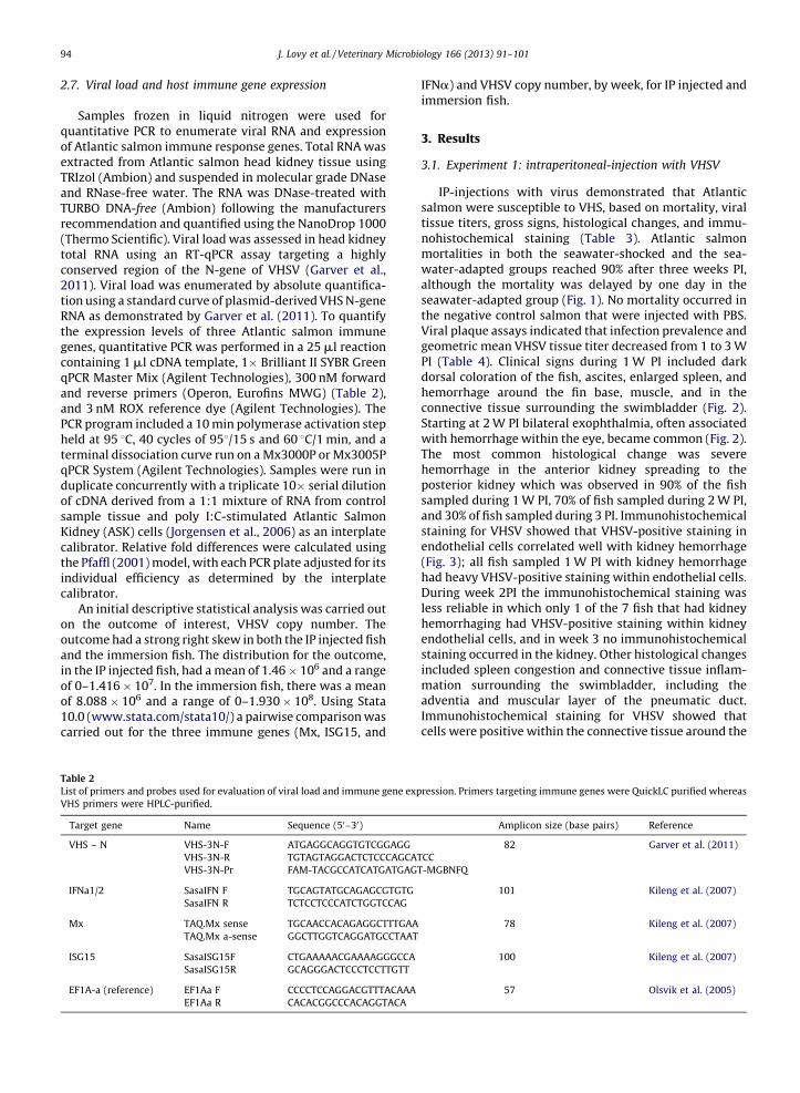
2.7. Viral load and host immune gene expression
Samples frozen in liquid nitrogen were used forquantitative PCR to enumerate viral RNA and expressionof Atlantic salmon immune response genes. Total RNA wasextracted from Atlantic salmon head kidney tissue usingTRIzol (Ambion) and suspended in molecular grade DNaseand RNase-free water. The RNA was DNase-treated withTURBO DNA-free (Ambion) following the manufacturersrecommendation and quantified using the NanoDrop 1000(Thermo Scientific). Viral load was assessed in head kidneytotal RNA using an RT-qPCR assay targeting a highlyconserved region of the N-gene of VHSV (Garver et al.,2011). Viral load was enumerated by absolute quantifica-tion using a standard curve of plasmid-derived VHS N-geneRNA as demonstrated by Garver et al. (2011). To quantifythe expression levels of three Atlantic salmon immunegenes, quantitative PCR was performed in a 25 ml reactioncontaining 1 ml cDNA template, 1� Brilliant II SYBR GreenqPCR Master Mix (Agilent Technologies), 300 nM forwardand reverse primers (Operon, Eurofins MWG) (Table 2),and 3 nM ROX reference dye (Agilent Technologies). ThePCR program included a 10 min polymerase activation stepheld at 95 8C, 40 cycles of 958/15 s and 60 8C/1 min, and aterminal dissociation curve run on a Mx3000P or Mx3005PqPCR System (Agilent Technologies). Samples were run induplicate concurrently with a triplicate 10� serial dilutionof cDNA derived from a 1:1 mixture of RNA from controlsample tissue and poly I:C-stimulated Atlantic SalmonKidney (ASK) cells (Jorgensen et al., 2006) as an interplatecalibrator. Relative fold differences were calculated usingthe Pfaffl (2001) model, with each PCR plate adjusted for itsindividual efficiency as determined by the interplatecalibrator.
An initial descriptive statistical analysis was carried outon the outcome of interest, VHSV copy number. Theoutcome had a strong right skew in both the IP injected fishand the immersion fish. The distribution for the outcome,in the IP injected fish, had a mean of 1.46 � 106 and a rangeof 0–1.416 � 107. In the immersion fish, there was a meanof 8.088 � 106 and a range of 0–1.930 � 108. Using Stata10.0 (www.stata.com/stata10/) a pairwise comparison wascarried out for the three immune genes (Mx, ISG15, and
IFNa) and VHSV copy number, by week, for IP injected andimmersion fish.
3. Results
3.1. Experiment 1: intraperitoneal-injection with VHSV
IP-injections with virus demonstrated that Atlanticsalmon were susceptible to VHS, based on mortality, viraltissue titers, gross signs, histological changes, and immu-nohistochemical staining (Table 3). Atlantic salmonmortalities in both the seawater-shocked and the sea-water-adapted groups reached 90% after three weeks PI,although the mortality was delayed by one day in theseawater-adapted group (Fig. 1). No mortality occurred inthe negative control salmon that were injected with PBS.Viral plaque assays indicated that infection prevalence andgeometric mean VHSV tissue titer decreased from 1 to 3 WPI (Table 4). Clinical signs during 1 W PI included darkdorsal coloration of the fish, ascites, enlarged spleen, andhemorrhage around the fin base, muscle, and in theconnective tissue surrounding the swimbladder (Fig. 2).Starting at 2 W PI bilateral exophthalmia, often associatedwith hemorrhage within the eye, became common (Fig. 2).The most common histological change was severehemorrhage in the anterior kidney spreading to theposterior kidney which was observed in 90% of the fishsampled during 1 W PI, 70% of fish sampled during 2 W PI,and 30% of fish sampled during 3 PI. Immunohistochemicalstaining for VHSV showed that VHSV-positive staining inendothelial cells correlated well with kidney hemorrhage(Fig. 3); all fish sampled 1 W PI with kidney hemorrhagehad heavy VHSV-positive staining within endothelial cells.During week 2PI the immunohistochemical staining wasless reliable in which only 1 of the 7 fish that had kidneyhemorrhaging had VHSV-positive staining within kidneyendothelial cells, and in week 3 no immunohistochemicalstaining occurred in the kidney. Other histological changesincluded spleen congestion and connective tissue inflam-mation surrounding the swimbladder, including theadventia and muscular layer of the pneumatic duct.Immunohistochemical staining for VHSV showed thatcells were positive within the connective tissue around the
Table 2
List of primers and probes used for evaluation of viral load and immune gene expression. Primers targeting immune genes were QuickLC purified whereas
VHS primers were HPLC-purified.
Target gene Name Sequence (50–30) Amplicon size (base pairs) Reference
VHS – N VHS-3N-F ATGAGGCAGGTGTCGGAGG 82 Garver et al. (2011)
VHS-3N-R TGTAGTAGGACTCTCCCAGCATCC
VHS-3N-Pr FAM-TACGCCATCATGATGAGT-MGBNFQ
IFNa1/2 SasaIFN F TGCAGTATGCAGAGCGTGTG 101 Kileng et al. (2007)
SasaIFN R TCTCCTCCCATCTGGTCCAG
Mx TAQ.Mx sense TGCAACCACAGAGGCTTTGAA 78 Kileng et al. (2007)
TAQ.Mx a-sense GGCTTGGTCAGGATGCCTAAT
ISG15 SasaISG15F CTGAAAAACGAAAAGGGCCA 100 Kileng et al. (2007)
SasaISG15R GCAGGGACTCCCTCCTTGTT
EF1A-a (reference) EF1Aa F CCCCTCCAGGACGTTTACAAA 57 Olsvik et al. (2005)
EF1Aa R CACACGGCCCACAGGTACA

Tab
Num
wee
In
IP
IP
IP
IP
IM
IM
IM
IMa
b
enlac
d
e
f
g
h
Fig.
adap
shoc
Tab
Num
sam
W
1
2
3
4
5
6
7
8
9
10a
*
J. Lovy et al. / Veterinary Microbiology 166 (2013) 91–101 95
swim bladder and pneumatic duct (Fig. 4). During weeks1–3 immunohistochemical staining was observed withinthe connective tissue near the swimbladder and thiscorrelated well in all weeks with inflammation within theconnective tissue. Mortalities in the IP-injected herringreached 95% after 10 days PI demonstrating the highvirulence of the virus to herring.
3.2. Experiment 2: immersion and cohabitation infection with
VHSV
Waterborne-exposure to VHSV and cohabitation withdiseased herring led to clinical VHS in Atlantic salmon(Table 3). In the VHSV-immersion tanks all herring diedwithin 2 W of their introduction and these fish had clinicalsigns consistent with VHS, including dark coloration, skin
le 3
ber of Atlantic salmon positive for virology by plaque assay and qPCR, gross clinical observations, histopathology, and immunohistochemistry for 1–4
ks following infection with VHSV by either intraperitoneal injection or immersion.
fection/time (sample size) Virus titera qPCR+ Gross findingsb Histopathc Immu-
nod
N L M H A B C D E F G H I J
e/1 W (n = 10) 0 0 1 9 10 8 8 0 3 3 9 4 5 9 4
/2 W (n = 10) 2 1 5 2 8 2 0 3 1 2 7 4 5 1 4
/3 W (n = 10) 3 6 1 0 6 3 0 2 1 3 3 1 2 0 2
/4 W (n = 5) 2 1 2 0 NAf 0 0 0 0 0 1 1 1 0 1g/1 W (n = 10) 8 0 1 1 4 1 2 0 0 0 1 0 0 1 0
/2 W (n = 10) 4 2 4 0 6 6 3 6 0 0 4 0 1 0 0
/3 W (n = 10) 7 2 1 0 5 2 0 2 0 1 3 0 2 0 0
/4 W (n = 12) 9 0 2 1 NA 0 0 1 1 2 *h * * * *
Virus titer by (N) negative, (L) low titer <104 pfu/g, (M) medium titer 104–106 pfu/g, (H) high titer �107 pfu/g.
Gross findings include number of fish that had (A) skin or fin hemorrhage, (B) dark dorsal coloration, (C) bilateral exophthalmia, (D) internally had an
rged spleen, (E) presence of ascites.
Histopathology includes fish with (F) severe kidney hemorrhage, (G) swim bladder connective tissue inflammation, (H) congestion in the spleen.
VHSV-positive staining in (I) kidney endothelial cells, (J) connective tissue near swim bladder.
IP, infection by intraperitoneal injection.
NA, not applicable.
IM, infection by immersion.
*, samples not taken.
1. Cumulative percent mortality of Atlantic salmon infected with either VHSV or PBS by intraperitoneal injection. Combined replicates of the saltwater-
ted group (IP-VHS SW-adapted) (n = 120), saltwater-shocked (IP-VHS SW-shocked) (n = 120), compared to a single control replicate of saltwater-
le 4
ber of VHSV-positive Atlantic salmon and viral titer in weekly
ples after exposure by either IP-injection or immersion.
eeks PE IP-injected salmon Immersion-exposed
salmon
VHSV+/total Viral titera VHSV+/total Viral titer
10/10 9.21 � 106 2/10 2.65 � 106
8/10 1.10 � 106 6/10 1.60 � 105
7/10 2.81 � 105 3/10 2.74 � 104
6/10 3.02 � 104 3/12 3.16 � 106
* * 5/18 4.37 � 106
* * 4/20 9.12 � 105
* * 2/20 9.93 � 106
* * 2/20 1.33 � 107
* * 2/20 9.86 � 104
* * 3/12 3.59 � 106
Viral titer is expressed as the geometric mean (pfu/g�1).
Samples not taken.
ked fish injected with PBS (IP-control SW-shocked) (n = 60).

J. Lovy et al. / Veterinary Microbiology 166 (2013) 91–10196
and fin hemorrhage, and lethargic swimming. All herringmortalities from these tanks tested positive for VHSV.Water samples from the 400 L tanks taken 1 W followingthe immersion challenge demonstrated that 3 of the 4
replicate tanks had an average of 1 � 103 pfu/ml, demon-strating that virus was being amplified and shed, mainly byherring in the tanks. Atlantic salmon mortality reachedapproximately 8% of the population, although weekly
Fig. 2. Gross clinical signs of VHSV in Atlantic salmon, similar signs appear in fish infected by IP-injection and immersion; shown in (a) through (d) are fish
infected by immersion and (e) is a fish infected by IP-injection. Common signs included (a) skin hemorrhage frequently near the pectoral fin, (b) dark dorsal
coloration and bilateral exophthalmia, (c) hemorrhage occurring within exophthalmic eyes, (d) hemorrhage in the brain cavity, and (e) hemorrhage on the
swim bladder.
Fig. 3. Histology and immunohistochemistry of Atlantic salmon infected with VHSV by immersion. (a) Anterior kidney with a blood vessel (*) containing
necrotic endothelial cells (arrows) with hemorrhage within the tissue (bar = 20 mm). (b) Serial section from (a) showing the same region with endothelial
cells staining positive for VHSV (arrows) (bar = 20 mm). (c) A fish sampled during week 4 PE with extensive cellular necrosis and destruction of muscle tissue
in the muscularis (bar = 100 mm). (d) Higher magnification of boxed in area of (c) showing fragmentation of the muscle and necrotic cells (bar = 20 mm). (e)
Serial section of similar region from (d) with positive VHSV immunohistochemical staining (bar = 20 mm).

samlowtrea10
straandgretestso ttrau152ass26.perPE,1 �in fiVHdesdorand2 Wfindfollat
musho
Fig.
with
pred
(bar
Fig.
high
line
J. Lovy et al. / Veterinary Microbiology 166 (2013) 91–101 97
pling bias toward clinically ill fish likely resulted iner overall reported mortality. From the 4 replicatetment tanks 13 total Atlantic salmon died during the
week duration of the trial. Viral plaque assay demon-ted that 12/13 dead salmon tested positive for VHSV
titers were all higher than 4 � 105 pfu/g and 7/12 wereater than 1 � 107 pfu/g. The one dead salmon thated negative for VHSV died 2 days into the experimenthe death was likely related to transport and handlingma. A total of 165 salmon, including 13 mortalities and
sampled fish, were tested for VHSV by viral plaqueay throughout the 10 W duration of the experiment and7% of the fish tested positive (Table 4). The virussisted in salmon until the final sampling period at 10 W
where 3 out of 12 fish had titers higher than 106 pfu/g (Table 4). Gross clinical signs were absentsh sampled from week 5 to 10 PE. Clinically affected
SV-immersion salmon had similar symptoms ascribed for the IP-injected fish, including darkenedsal color and hemorrhage on the skin evident 1 W PE,
bilateral exophthalmia that became first evident in the PE sample (Fig. 2). The most common histologicaling was hemorrhage in the anterior kidney (Fig. 3)
owed by spleen congestion. One fish that was sampled4 W PE had necrosis and inflammation within thescularis layer of the intestine. Immunohistochemistrywed VHSV positive cells within the muscularis lesions
(Fig. 3). No Atlantic salmon were lost in the control mock-immersion tank, although all herring died within 1 W oftheir introduction into this tank. All control herring testednegative for VHSV and their death was likely caused bytheir incompatibility with salmon in the research tanks,although no aggressive behavior was observed by theresearchers.
3.3. Experiment 3: transmission of VHSV from Atlantic
salmon to herring
Transmission of VHSV from Atlantic salmon to sympatricPacific herring was indicated by 100% mortality andrecovery of VHSV with titers exceeding 1 � 106 from herringsentinels in 3 out of 4 immersion replicates. Prior to theaddition of herring into the tanks, no virus could be isolatedfrom the tank water sample. In the fourth replicate none ofthe sampled sentinel herring cultured positive for VHSV.Sentinel herring from the one IP-injected salmon tank alsoall tested positive for VHSV with titers exceeding 1 � 106.
3.4. Viral load and Atlantic salmon immune gene expression
Exposure of Atlantic salmon to VHSV by both immersionand injection exposure resulted in up-regulation of genesinvolved in the innate immune system (Figs. 5 and 6). In IP-injected fish IFNa was expressed the highest during 1 W PI
4. Histology and immunohistochemistry of Atlantic salmon infected with VHSV by IP-injection. (a) Pneumatic duct with inflammation and necrosis
in the muscular layer and the adventia (bar = 100 mm). (b) Serial section of similar region in (a) with positive VHSV staining in the muscular layer and
ominantly in the adventia of the pneumatic duct (bar = 100 mm). (c) Connective tissue around the swim bladder with extensive cellular necrosis
= 40 mm). (d) Serial section of similar region as (c) with positive staining for VHSV (bar = 40 mm).
5. Expression of immune genes and VHSV following IP-injection of Atlantic salmon with VHSV; samples in each week were ordered from lowest to
est based on viral load; individual fish represented by number on x axis are grouped by week of sampling (1–3 weeks). Point connections with dashed
s were done to better show trends in gene expression, these lines do not denote continuous data.

J. Lovy et al. / Veterinary Microbiology 166 (2013) 91–10198
with up to 300 fold higher expression compared to controlfish. ISG15 was expressed the highest during 1 and 2 W PIwith variable expression from 0 to 840-fold higherexpression than controls (Fig. 6). During 2–3 W PI IFNaexpression was strongly correlated (p = 0.00) with viral load(Table 5). In the immersion fish, VHSV load was stronglycorrelated with expression of IFNa in all 3 weeks followingexposure to VHSV (Table 5). In all 3 weeks for both IP-injected and immersion fish there was a moderate to strongcorrelation between the expression of ISG15 and IFNa(Table 5).
4. Discussion
The virulence of VHSV to Atlantic salmon is variabledepending on infection route with IP-injection resulting inheavier mortality. This study is the first to demonstratethat Atlantic salmon are experimentally susceptible toVHSV genotype IVa through waterborne immersion. Theviral-exposure method utilizing an immersion and coha-bitation model in fish freshly transferred to seawater wasintended to duplicate more closely what could occur infield situations with farmed Atlantic salmon. In industry,farmed Atlantic salmon smolts are transferred fromfreshwater hatcheries into seawater netpens located alongcoastal BC, where VHSV genotype IVa is present. In this
study the immersion route caused clinical signs consistentwith VHS disease, although the severity of the disease wasmuch lower than in IP-injected fish. The doses used forimmersion in the present study resulted in about 27% offish becoming infected with the virus. This is in contrast toSPF herring, in which VHSV levels that are much lowerthan used here (e.g. below the normal detection thresholdof the viral plaque assay) can initiate VHS outbreaks andaffect most of the population (Hershberger et al., 2011).Previous studies looked at the susceptibility of Atlanticsalmon to European isolates of VHSV and found thatfollowing exposure by immersion, no clinical disease signsoccurred and virus was rarely isolated from fish (DeKinkelin and Castric, 1982; King et al., 2001). Thedifferences in susceptibility between the European studiesand the study herein may be explained by the genotypesand isolates used; previous studies used European isolatesfrom other host species, whereas the current study usedisolates of genotype IVa from farmed Atlantic salmon.Additionally in the current study the salmon were infectedwith VHSV during seawater transfer, which could be anadded stressor making fish more vulnerable to infection.Considering the differences in the severity of disease ingroups of salmon exposed to VHSV by IP-injectioncompared to immersion, it is possible that, in additionto differences in dose, that host and virus characteristics
Fig. 6. Expression of immune genes and VHSV following immersion of Atlantic salmon with VHSV; samples in each week were ordered from lowest to
highest based on viral load; individual fish represented by number on x axis are grouped by week of sampling (1–3 weeks). Point connections with dashed
lines were done to better show trends in gene expression, these lines do not denote continuous data.
Table 5
Correlation coefficients and p values for the expression of VHSV and immune genes.
W–Ga IP-Injected salmon Immersion salmon
VHSV Mx IFNa VHSV Mx IFNa
1-Mx 0.06, p = 0.86 0.056, p = 0.93
1-IFNa 0.05, p = 0.90 0.74, p = 0.01 0.95, p = 0.014 0.09, p = 0.88
1-ISG 0.31, p = 0.39 0.75, p = 0.01 0.81, p = 0.01 0.54, p = 0.34 0.68, p = 0.21 0.74, p = 0.15
2-Mx 0.12, p = 0.74 0.27, p = 0.44
2-IFNa 0.96, p = 0.00b 0.21, p = 0.57 0.98, p = 0.00 0.39, p = 0.26
2-ISG 0.62, p = 0.06 0.69, p = 0.03 0.62, p = 0.05 0.72, p = 0.02 0.81, p = 0.00 0.80, p = 0.01
3-Mx 0.06, p = 0.87 0.49, p = 0.18
3-IFNa 0.63, p = 0.05 0.42, p = 0.23 0.99, p = 0.00 0.57, p = 0.11
3-ISG 0.26, p = 0.48 0.67, p = 0.03 0.88, p = 0.00 0.77, p = 0.02 0.88, p = 0.00 0.82, p = 0.01a Weeks post-infection (W) – immune gene (G).b Shading represents a statistically significant correlation between the groups; dark shading is a strong correlation and light shading is a mild to moderate
correlation.

preandothbilifindviruthaperfactdev
thetheprethoImmbotconto t(Al-201detsen1 WregkidstrarespatherexpinfegenregagaRokrepdouet aexpvirucorimmhavexainteredresAtlaherfact
perfor
infeheret aIVbinfeAtlaviru
J. Lovy et al. / Veterinary Microbiology 166 (2013) 91–101 99
vent efficient viral entry to the host through the skin gills. Predisposing factors, such as the presence ofer diseases and host condition/immunological varia-ty, may explain these differences in susceptibility. Theings of this study suggest that these isolates are mildlylent to Atlantic salmon, yet field observations indicate
t VHS epizootics in penned Atlantic salmon occuriodically. Future research is needed to identify viralors and host stressors that may predispose salmon toeloping clinical VHS.VHS in Atlantic salmon is similar to other species with
most common gross signs being lethargy, darkening of skin, and bilateral exophthalmia. Skin hemorrhage wassent, although lesions were not nearly as severe asse observed in Pacific herring suffering from VHS.
unohistochemistry showed that viral replication inh infection routes occurred within endothelial cells andnective tissue cells in Atlantic salmon, which is similarhe cellular tropism of other susceptible fish to VHSVHussinee et al., 2011; Brudeseth et al., 2005; Lovy et al.,2). Immunohistochemistry was a useful method inermining VHSV cellular tropism, although it was not asitive detection method as it was only reliable during
PI in the IP-injected fish. The findings of heavily up-ulated type I-interferon system genes in the anteriorney of both IP-injected and immersion fish demon-ted that Atlantic salmon were immunologically
ponding to VHSV through the interferon-induciblehway, similar to the response occurring in Pacificring (Hansen et al., 2012). The genes for Mx and ISG15ression were the most heavily up-regulated in VHSV-cted salmon and generally the expression of thesees correlated with each other. Both of these interferon-ulated proteins are known for their anti-viral responseinst single stranded RNA viruses (Nygaard et al., 2000;enes et al., 2007). Mx has also been found to inhibit thelication of infectious pancreatic necrosis virus (IPNV), able-stranded RNA virus, in Atlantic salmon (Larsenl., 2004). There was a high degree of variation in theression in these genes and Mx never correlated withs expression, while ISG15 seemed to positively
relate with virus expression in fish infected byersion in 2–3 W PE. Viruses in susceptible species
e found ways to avoid the interferon response, formple Atlantic salmon infected with ISAV have a strongrferon response, although the response is unable to
uce replication of the virus (Kileng et al., 2007). Furtherearch into comparing immune responses betweenntic salmon and more susceptible species, such as
ring, may aid in determining if immunity is a majoror in susceptibility.
An important conclusion from this study was that VHSVsisted in asymptomatic Atlantic salmon at high titersthe duration of the experiment. Persistent VHSVctions have been reported in symptom-free Pacificring for up to 224 days following infection (Hershbergerl., 2010). In muskellunge it was found that VHSV type
is shed from infected fish for 15 weeks following initialction (Kim and Faisal, 2012). The extent to whichntic salmon may remain infective reservoirs of thes will need to be addressed with future research,
although it is clearly more than 10 weeks. Disease causedby VHSV type IVa is also known to cause chronicneurological disease in other susceptible hosts (Lovyet al., 2012) and it is possible that the virus may followthis pattern in long-term infected salmon, although thiswas not the focus of the current study. The transmission ofVHSV from Atlantic salmon to Pacific herring demon-strated that Atlantic salmon shed virus following infectionand that this shed virus is in high enough quantities toinfect a highly susceptible species like Pacific herring. Inthe current study Atlantic salmon infected Pacific herringsentinels with VHS in three out of four replicate tanks.Variability of VHS infection in Atlantic salmon and thelower shedding rates of Atlantic salmon compared toherring, supported by the fact that virus could not bedetected in water samples from tanks that containedsalmon only, were likely factors for herring to remainuninfected in the fourth replicate.
Unlike many viruses, VHSV has a broad host rangewhich has undoubtedly led to its global spread andsignificant impact to wild and farmed fish. During a herringVHS outbreak a single fish can release 5 � 108 pfu/fish/day(Hershberger et al., 2011) and herring biomass in salmonnetpens can measure in the tons in some salmon farms(unpublished observation, K. Garver). With high densitiesof farmed salmon and susceptible wild fish being exposedto VHSV at these levels, an environment conducive forvirus adaptation may be created in this situation. Instancesin which VHSV has developed higher virulence towardcertain hosts have been documented; Schonherz et al.(2012) found that cross-species transmission from turbot,Scophthalmus maximus, to rainbow trout, Oncorhynchus
mykiss, occurred with only a trout-adapted isolate that isinfective to both turbot and trout, whereas a marine turbotisolate did not infect rainbow trout. Nonetheless, fieldobservation since the initial detection of VHSV-IVa infarmed Atlantic salmon have not revealed an evolutiontoward increased virulence rather VHSV isolations in net-pen aquaculture have been associated with low mortal-ities. Additionally molecular epidemiology studies demon-strate that virus isolations from salmon do not form anAtlantic salmon adapted lineage (Garver et al., 2013)suggesting that ecological factors in addition to thoseassociated with aquaculture are involved in the evolutionof VHSV-IVa.
Understanding of the mechanisms which drive theevolution of VHSV-IVa is crucial to evaluating the riskposed to aquaculture through transmission from wildmarine species. A study conducted by Snow and Cunning-ham (2000) found that serial passage of VHSV through anovel host species leads to increased virulence, althoughthe genomic changes associated with the virulenceincreased remain unknown. Evidence suggests that VHSVisolates can quickly evolve to become more virulent withonly a few nucleotide substitutions (Cho et al., 2012). Adifference between 20% and 80% cumulative mortality wasobserved among groups of olive flounder, Paralichthys
olivaceus, exposed to two isolates of VHSV genotype IVathat were found to be identical for all genes except for thenucleoprotein and glycoprotein genes that had 99.7% and99.8% amino acid sequence identity (Cho et al., 2012).

J. Lovy et al. / Veterinary Microbiology 166 (2013) 91–101100
Genetic analysis of VHSV type IVb isolates from fish in theGreat Lakes basin of the USA and Canada suggests thatidentical isolates can occur in a number of species; a studyby Thompson et al. (2011) showed that 90% of VHSV typeIVb isolates from 31 different species belonged to twomajor genetic types, based on a partial sequence of theglycoprotein gene. Future research examining geneticdifferences among the isolates comprising the cocktailused in the study herein may provide insights to themolecular basis of VHS virulence in Atlantic salmon andPacific herring. Overall, the broad species range of VHSVand experimental evidence of evolution toward highervirulence in novel species warrants further research toquantify the adaptation potential of VHSV IVa to highervirulence in Atlantic salmon.
5. Conclusions
The study herein shows that Atlantic salmon, althoughnot a natural host for VHSV type IVa, were susceptible tothe virus and developed clinical signs consistent with VHS.The virus was able to complete its cycle including entry tothe host, replication, and exiting for transmission. VHSVcan cause mild mortality in Atlantic salmon and salmoncan be a reservoir of virus subsequently transmitting VHSdisease to cohabitating naı̈ve Pacific herring. Althoughresults herein suggest that salmon do not shed highconcentrations of virus that a highly susceptible species,such as Pacific herring, would.
Conflict of interest statement
The authors do not have any financial or personalrelationships that would pose a conflict of interest in thisstudy.
Acknowledgements
The authors are grateful for the assistance of NicoleLewis in statistical analysis of the data, William Bennett forhistological support, John Richard and Laura Hawley forassistance in the laboratory, and Holly Hicklin and RobertKennedy for helping to maintain fish in the aquaticsfacility. Funding was supported by the Fisheries andOceans Canada Aquaculture Collaborative Research andDevelopment Program (Project no. P11-02-006) and theHerring Conservation and Research Society. Pacific herringfrom this study were provided by Project # 10100132-I,funded by the Exxon Valdez Oil Spill Trustee Council. Theuse of trade, firm, or corporation names in this publicationis for the information and convenience of the reader. Suchuse does not constitute an official endorsement or approvalby the Canadian government, U.S. Department of Interioror the U.S. Geological Survey of any product or service tothe exclusion of others that may be suitable.
References
Al-Hussinee, L., Lord, S., Stevenson, R.M.W., Casey, R.N., Groocock, G.H., Britt,K.L., Kohler, K.H., Wooster, G.A., Getchell, R.G., Bowser, P.R., Lumsden,J.S., 2011. Immunohistochemistry and pathology of multiple GreatLakes fish from mortality events associated with viral hemorrhagicsepticemia virus type IVb. Dis. Aquat. Organ. 93, 117–127.
Amos, K., Thomas, J., Hopper, K., 1998. A case history of adaptive manage-ment strategies for viral hemorrhagic septicemia virus (VHSV) inWashington State. J. Aquat. Anim. Health 10, 152–159.
Batts, W., Winton, J., 1989. Enhanced detection of infectious hematopoie-tic necrosis virus and other fish viruses by pretreatment of cellmonolayers with polyethylene glycol. J. Aquat. Anim. Health 1,284–290.
Bernard, J., Bremont, M., Winton, J., 1992. Nucleocapsid gene sequence ofa North American isolate of viral haemorrhagic septicaemia virus, afish rhabdovirus. J. Gen. Virol. 73, 1011–1014.
Brudeseth, B.E., Raynard, R.S., King, J.A., Evenson, O., 2005. Sequentialpathology after experimental infection with marine viral hemorrha-gic septicemia virus isolates of low and high virulence in turbot(Scophthalmus maximus L.). Vet. Pathol. 42, 9–18.
Brunson, R., True, K., Yancey, J., 1989. VHS virus isolated at MakahNational Fish Hatcher. Am. Fish. Soc. Fish Health Sect. Newsl. 17,3–4.
Cho, M.Y., Lee, U.H., Moon, C.H., Bang, J.D., Jee, B.Y., Cha, S.J., Kim, J.W.,Park, M.A., Do, J.W., Park, J.W., 2012. Genetically similar VHSV isolatesare differentially virulent in olive flounder Paralichthys olivaceus. Dis.Aquat. Organ 101, 105–114.
Congleton, J., Sun, B., 1996. Interferon-like activity produced by anteriorkidney leucocytes of rainbow trout stimulated in vitro by infectioushematopoietic necrosis virus or Poly I:C. Dis. Aquat. Organ. 25, 185–195.
De Kinkelin, P., Castric, J., 1982. An experimental study of the suscept-ibility of Atlantic salmon fry, Salmo salar L., to viral haemorrhagicsepticaemia. J. Fish Dis. 5, 57–65.
Elsayed, E., Faisal, M., Thomas, M., Whelan, G., Batts, W., Winton, J., 2006.Isolation of viral hemorrhagic septicemia virus from muskellunge,Esox masquinongy (Mitchell), in Lake St. Clair, Michigan, USA reveals anew sub-lineage of the North American genotype. J. Fish Dis. 29, 611–619.
Gagne, N., Mackinnon, A.M., Boston, L., Souter, B., Cook-Versloot, M.,Griffiths, S., Olivier, G., 2007. Isolation of viral haemorrhagic septi-caemia virus from mummichog, stickleback, striped bass and browntrout in eastern Canada. J. Fish Dis. 30, 213–223.
Garver, K.A., Hawley, L.M., McClure, C.A., Schroeder, T., Aldous, S., Doig, F.,Snow, M., Edes, S., Baynes, C., Richard, J., 2011. Development andvalidation of a reverse transcription quantitative PCR for universaldetection of viral hemorrhagic septicemia virus. Dis. Aquat. Organ. 95,97–112.
Garver, K.A., Traxler, G.S., Hawley, L.M., Richard, J., Ross, J., Lovy, J., 2013.Molecular epidemiology of viral hemorrhagic septicemia (VHSV) inBritish Columbia, Canada reveals transmission from wild to farmedfish. Dis. Aquat. Organ. 104, 93–104.
Hansen, J.D., Woodson, J.C., Hershberger, P.K., Grady, C., Gregg, J.L., Purcell,M.K., 2012. Induction of anti-viral genes during acute infection withviral hemorrhagic septicemia virus (VHSV) genogroup IVa in Pacificherring (Clupea pallasii). Fish Shellfish Immunol. 32, 259–267.
Hedrick, R.P., Batts, W.N., Yun, S., Traxler, G.S., Kaufman, J., Winton, J.R.,2003. Host and geographic range extensions of the North Americanstrain of viral hemorrhagic septicemia virus. Dis. Aquat. Organ. 55,211–220.
Hershberger, P.K., Gregg, J.L., Grady, C.A., Hart, L.M., Roon, S.R., Winton,J.R., 2011. Factors controlling the early stages of viral haemorrhagicsepticaemia epizootics: low exposure levels, virus amplification andfish-to-fish transmission. J. Fish Dis. 34, 893–899.
Hershberger, P.K., Gregg, J.L., Grady, C.A., Taylor, L., Winton, J.R., 2010.Chronic and persistent viral hemorrhagic septicemia virus infectionsin Pacific herring. Dis. Aquat. Organ. 93, 43–49.
Jorgensen, S.M., Syvertsen, B.L., Lukacs, M., Grimholt, U., Gjoen, T., 2006.Expression of MHC class I pathway genes in response to infectioussalmon anaemia virus in Atlantic salmon (Salmo salar L.) cells. FishShellfish Immunol. 5, 548–560.
Kileng, O., Brundtland, M.I., Robertsen, B., 2007. Infectious salmon anemiavirus is a powerful inducer of key genes of the type I interferon systemof Atlantic salmon, but is not inhibited by interferon. Fish ShellfishImmunol. 23, 378–389.
Kim, R.K., Faisal, M., 2012. Shedding of viral hemorrhagic septicemia virus(genotype IVb) by experimentally infected muskellunge (Esox mas-quinongy). J. Microbiol. 50, 278–284.
King, J.A., Snow, M., Skall, H.F., Raynard, R.S., 2001. Experimental suscept-ibility of Atlantic salmon Salmo salar and turbot Scophthalmus max-imus to European freshwater and marine isolates of viralhaemorrhagic septicaemia virus. Dis. Aquat. Organ 47, 25–31.
Kocan, R., Bradley, M., Elder, N., Meyers, T., Batts, W., Winton, J., 1997.North American strain of viral hemorrhagic septicemia virus is highlypathogenic for laboratory-reared Pacific herring. J. Aquat. Anim.Health 9, 279–290.

Lars
Lovy
Mar
Mar
Mey
Nyg
Olsv
Pfaf
Pier
Rog
J. Lovy et al. / Veterinary Microbiology 166 (2013) 91–101 101
en, R., Rokenes, T.P., Robertsen, B., 2004. Inhibition of infectiouspancreatic necrosis virus replication by Atlantic salmon Mx1 protein.J. Virol. 78, 7938–7944., J., Lewis, N.L., Hershberger, P.K., Bennett, W., Meyers, T.R., Garver,
K.A., 2012. Viral tropism and pathology associated with viral hemor-rhagic septicemia in larval and juvenile Pacific herring. Vet. Microbiol.161, 66–76.ty, G.D., Freiberg, E.F., Meyers, T.R., Wilcock, J., Farver, T.B., Hinton, D.E.,1998. Viral hemorrhagic septicemia virus, Ichthyophonus hoferi, andother causes of morbidity in Pacific herring Clupea pallasi spawning inPrince William Sound, Alaska, USA. Dis. Aquat. Organ. 32, 15–40.ty, G.D., Hulson, P.J.F., Miller, S.E., Quinn, T.J.I.I., Moffitt, S.D., Merizon,R.A., 2010. Failure of population recovery in relation to disease inPacific herring. Dis. Aquat. Organ. 90, 1–14.ers, T.R., Short, S., Lipson, K., 1999. Isolation of the North Americanstrain of viral hemorrhagic septicemia virus (VHSV) associated withepizootic mortality in two new host species of Alaskan marine fish.Dis. Aquat. Organ. 38, 81–86.aard, R., Husgard, S., Sommer, A.I., Leong, J.A., Robertsen, B., 2000.Induction of Mx protein by interferon and double-stranded RNA insalmonid cells. Fish Shellfish Immunol. 10, 435–450.ik, P.A., Lie, K.K., Jordal, A.E., Nilsen, T.O., Hordvik, I., 2005. Evaluationof potential reference genes in real-time RT-PCR studies of Atlanticsalmon. BMC Mol. Biol. 6, 21.fl, M.W., 2001. A new mathematical model for relative quantificationin real-time RT-PCR. Nucleic Acids Res. 29, 2002–2007.ce, L.R., Stepien, C.A., 2012. Evolution and biogeography of an emer-ging quasispecies: diversity patterns of the fish viral hemorrhagicsepticemia virus (VHSv). Mol. Phylogenet. Evol. 63, 327–341.el-Gaillard, C., Chilmonczyk, S., De Kinkelin, P., 1993. In vitro inductionof interferon-like activity from rainbow trout leucocytes stimulatedby Egtved virus. Fish Shellfish Immunol. 3, 383–394.
Rokenes, T.P., Larsen, R., Robertsen, B., 2007. Atlantic salmon ISG15:expression and conjugation to cellular proteins in response to inter-feron, double stranded RNA and virus infections. Mol. Immunol. 44,950–959.
Schonherz, A.A., Lorenzen, N., Einer-Jensen, K., 2012. Inter-species trans-mission of viral haemorrhagic septicaemia virus (VHSV) from turbot(Scophthalmus maximus) to rainbow trout (Oncorhynchus mykiss). J.Gen. Virol. (December) (Epub ahead of print).
Snow, M., Cunningham, C.O., 2000. Virulence and nucleotide sequenceanalysis of marine viral haemorrhagic septicaemia virus following invivo passage in rainbow trout Oncorhynchus mykiss. Dis. Aquat. Organ42, 17–26.
Tafalla, C., Sanchez, E., Lorenzen, N., DeWitte-Orr, S.J., Bols, N.C., 2008.Effects of viral hemorrhagic septicemia virus (VHSV) on the rainbowtrout (Oncorhynchus mykiss) monocyte cell line RTS-11. Mol. Immu-nol. 45, 1439–1448.
Thompson, T.M., Batts, W.N., Faisal, M., Bowser, P., Casey, J.W., Phillips, K.,Garver, K.A., Winton, J., Kurath, G., 2011. Emergence of viral hemor-rhagic septicemia virus in the North American Great Lakes region isassociated with low viral genetic diversity. Dis. Aquat. Organ. 96,29–43.
Traxler, G.S., Kieser, D., Evelyn, T.P.T., 1995. Isolation of North Americanstrain of VHS virus from farmed Atlantic salmon. Aquacult. Update 72,1–2.
Traxler, G.S., Kieser, D., Richard, J., 1999. Mass mortality of pilchardand herring associated with viral hemorrhagic septicemia virus inBritish Columbia, Canada. Am. Fish. Soc. Fish Health Sect. Newsl.27, 3–4.
Winton, J., Batts, W., de Kinkelin, P., LeBerre, M., Bremont, M., Fijan, N.,2010. Current lineages of the Epithelioma papulosum cyprinid (EPC)cell line are contaminated with fathead minnow, Pimephales promelas,cells. J. Fish Dis. 33, 701–704.