Embed Size (px)
Citation preview
Neural circuits are defined by the structure of axons and dendrites and the synapses that connect them. Axons route a neuron’s output to diverse target regions, which can span most of the brain. Individual dendrites integrate inputs from several sources over hundreds of micrometers. In the adult brain, circuit changes medi-ated by structural plasticity, accompanied by synapse formation and elimination, are thought to underlie aspects of long-term memory formation1. Given that in most areas of the brain, including the cerebral cortex, neurons are sparsely connected, structural plasticity could provide a substantial boost in the memory stor-age capacity, compared with plasticity due to changes in synaptic strength alone2. Structural rearrangements over long distances allows more variability and there-fore a larger number of potential circuits to be gener-ated, implying a larger memory capacity per synapse. Structural plasticity might also be involved in recovery from brain injury3–5.
Neuronal processes are studded with a high density of synapses. A synapse is typically defined by the pres-ence of a presynaptic active zone with synaptic vesicles, a well-defined synaptic cleft and a postsynaptic density (PSD)6,7. Most excitatory cortical synapses occur at con-tacts between axonal en passant boutons and dendritic spines. En passant boutons are small axonal varicosities that typically contain one active zone and one cluster of
synaptic vesicles8. A subpopulation of axons, for example those of cortical layer 6 pyramidal cells, harbours a high density of terminaux boutons that often form syn-apses with dendritic shafts9. Spines are tiny protrusions that emanate from the dendritic shaft10. Typically one bouton and one spine correspond to one synapse6,8. A small number of boutons8 and spines11–14 lack synapses and a few boutons participate in more than one synapse (multiple synapse boutons, MSBs)8,14–17. Electron micro-scopy (EM) reconstructions have revealed that spines are structurally extremely diverse: their volumes can range from 0.001 to 1 μm3,18,19; their shapes include thin, filopodia-like protrusions (‘thin spines’), short spines without a well-defined spine neck (‘stubby spines’) and spines with a large bulbous head (‘mushroom spines’)18,20. Different cortical cells, and perhaps even individual cells within a class, can have dramatically different spine densities21–23.
Changes in synaptic connectivity through the de novo growth and retraction of dendritic spines and axonal boutons might contribute to functional changes in the brain. Compared with synaptic strength changes alone, such structural spine plasticity would hugely increase the memory storage capacity of the brain, because a large number of synaptic connectivity pat-terns are attainable by spine or bouton growth, even without large-scale remodelling of dendritic and axonal
*Department of Basic Neurosciences, Medical Faculty, University of Geneva, Switzerland. ‡Janelia Farm Research Campus, HHMI, Ashburn, Virginia 20147, USA.e-mails: [email protected]; [email protected]:10.1038/nrn2699Corrected online 21 August 2009
Experience-dependent structural synaptic plasticity in the mammalian brainAnthony Holtmaat* and Karel Svoboda‡
Abstract | Synaptic plasticity in adult neural circuits may involve the strengthening or weakening of existing synapses as well as structural plasticity, including synapse formation and elimination. Indeed, long-term in vivo imaging studies are beginning to reveal the structural dynamics of neocortical neurons in the normal and injured adult brain. Although the overall cell-specific morphology of axons and dendrites, as well as of a subpopulation of small synaptic structures, are remarkably stable, there is increasing evidence that experience- dependent plasticity of specific circuits in the somatosensory and visual cortex involves cell type-specific structural plasticity: some boutons and dendritic spines appear and disappear, accompanied by synapse formation and elimination, respectively. This Review focuses on recent evidence for such structural forms of synaptic plasticity in the mammalian cortex and outlines open questions.
R E V I E W S
NATurE rEvIEwS | NeuroscieNce voluME 10 | SEPTEMBEr 2009 | 647
© 2009 Macmillan Publishers Limited. All rights reserved
Golgi methodA method that is used to label a sparse subset of neurons in fixed tissue using potassium dichromate and silver nitrate; neurons are stained by microcrystallization of silver chromate. The labelling seems stochastic, but the mechanisms underlying sparse labelling remain unknown.
arbors2,24–26. Consistent with this notion, in cortical neuronal cultures that are still undergoing develop-mental circuitry changes, spines appear and disappear over tens of minutes27–29, a process that can be trig-gered by synaptic activity29–34. Furthermore, de novo spine growth has been linked to synapse formation in cultured preparations32,35–37. Axonal boutons also grow, retract and remodel in an activity-dependent manner in vitro, but much less is known about this process38,39.
Are these forms of structural plasticity ongoing in the adult brain? To what extent does structural plas-ticity contribute to experience-dependent rewiring in vivo? Answers to these questions will shed light on the storage capacity of neural circuits and also on the cellular and molecular mechanisms underlying learning and memory. Here we review recent work on structural plasticity in the adult mammalian brain, focusing on longitudinal imaging experiments in the mouse neocortex (see Supplementary informa-tion S1 (table) for a summary of results) that used two-photon excitation laser scanning microscopy40–42 of neurons expressing fluorescent proteins39,43,44 (see Supplementary information S2 (box))
The dynamics of axonal and dendritic arborsMost studies of large-scale structural plasticity so far have relied on static measurements and comparisons between neurons in different animals. They suggest that neurons can undergo large-scale structural changes after relatively drastic long-term manipulations, lead-ing to the conclusion that synapses are eliminated or formed with changes in connectivity. More recent stud-ies have used long-term time-lapse imaging to probe structural dynamics in more detail and have yielded a different picture.
Axonal dynamics. Evidence for axonal growth in the adult brain has come mostly from experiments involving focal brain injury or lesions of the sensory periphery. For example, in squirrel monkeys ischaemic injury to the primary motor cortex induces axonal sprouting of premotor cortical projections near the infarct site4. Inducing epilepsy in rats leads to axonal re organizations in the hippocampus45. limb amputation in macaques causes growth of intracortical axons across several millimeters in the somatosensory cortex46, a phenom-enon that may underlie functional reorganization of cortical maps47 and phantom limb syndrome48. Similar rewiring is observed in the cat visual cortex after focal retinal lesions49 and in the rat barrel cortex after par-tial vibrissectomy50. Although these experiments reveal that cortical axons maintain the capacity to grow and branch in the adult neocortex, their relevance to expe-rience-dependent plasticity is unclear. However, in the hippocampus there are indications that axonal sprout-ing occurs during spatial learning51,52 or in response to environmental enrichment53.
long-term imaging experiments have shown that in naive adult mice a subset of axonal branches in the cortex can undergo structural rearrangements over lengths of tens of micrometers over several days54. As
these retractions and elongations are associated with turnover of numerous boutons, they are likely to be associated with synapse formation and elimination54. Similarly, in the cerebellum a subset of olivocerebel-lar climbing fibre branches are dynamic, whereas other branches of the same neurons are stable55. overall, these changes are modest, amounting to a few percent of axonal length within the terminal arbor (Supplementary information S1 (table)). The large-scale organization of axonal arbors in the mouse54,56 and macaque57 neocortex are therefore relatively sta-ble. Similar stability has been observed in the terminal arborizations of the parasympathetic submandibular ganglion58; this stability stands in dramatic contrast to the rapid and large-scale rearrangements that occur in the developing cortex59,60. long-term imaging studies of axons in response to behavioural training have not been reported so far.
Dendritic plasticity. Classic studies of dendritic plas-ticity, mostly in rats, have relied on the Golgi method. Curiously, it is still not known which neurons are labelled by this method and how complete the labelling is. Such experiments have revealed that environmental enrichment61, extensive training62, stress levels63 and drugs of abuse64,65 all might have profound influences on the complexity of dendritic arbors in some corti-cal areas, but not in others. large-scale reorganization of dendritic arbors has also been observed in the rat sensorimotor cortex after damage to the contralateral homotopic cortex66, and in the somatosensory cortex after vibrissal deafferentation67 and forepaw denerva-tion68. Training-induced changes in dendrites seem to be more subtle69. As these static measurements are only sensitive to robust changes in morphometric parame-ters, the studies might underestimate the actual changes in arbors in response to manipulations.
In contrast to the Golgi method, long-term imaging experiments in mice have shown that the large-scale organization of dendritic arbors is relatively stable (Supplementary information S1 (table)). The dendritic arborization of cortical layer 5 (REF. 13) and layer 2/3 pyramidal neurons22,70,71, and of mitral/tufted cells in the olfactory bulb72, remains unchanged over several months, even in response to learning72 and after enrich-ment22. Small length changes (a few micrometers) can be detected at the branch tips of pyramidal neurons (A. H. and K. S., unpublished observations). A subset of inhibitory dendritic branches show a larger degree of structural plasticity (∼10 μm over 4 weeks), but the fractional change of the entire dendritic arbor is very small70,73, in the order of 1–5 % of the overall dendritic length. Apart from the growth of dendritic trees of adult-born neurons74,75, complete retractions or de novo growth of dendritic branches are extremely rare under any of the conditions studied in wild-type mice.
Thus, results from static and longitudinal experi-ments seem to be at odds. The discrepancies might be due to differences in the imaged cell types, brain region, animal species or the experimental manipu-lations. The capriciousness of the Golgi method is
R E V I E W S
648 | SEPTEMBEr 2009 | voluME 10 www.nature.com/reviews/neuro
© 2009 Macmillan Publishers Limited. All rights reserved
Nature Reviews | Neuroscience
a
c
b
d
g
e
f
Layer 2/3 pyramidal cell dendrites–PSD-95-paGFP/mCherry
Layer 2/3 pyramidal cell dendrite–PSD-95-GFP/DsRed Express
Layer 2/3 pyramidal cell axon–Synaptophysin-GFP/DsRed Express
Layer 6 pyramidal cell axon–m-GFP Layer 5 pyramidal cell dendrite–GFP
Thalamocortical axon–GFP Intracortical axon–GFP
82 86 90
62 66 112
240 264 280 310
91 107 135
pre-pa pa, 0 min +90 min +24 h
86 94 102
2 µm 2 µm
2 µm
5 µm
5 µm
5 µm
2 µm
70 106 188
another complication: neurons with different average dendritic complexities could be selectively labelled under particular experimental conditions, leading to the reporting of spurious structural plasticity. It is therefore of considerable interest to probe the effects of enrichment, training, stress, drugs and lesions on dendrite structure using genetic labelling methods76 in combination with long-term imaging.
The dynamics of synaptic structuresEven in the absence of large-scale remodelling of dendritic and axonal arbors, changes in synaptic con-nectivity through the de novo growth and elimination of boutons and dendritic spines may contribute to functional rewiring. Numerous studies have exam-ined cortical tissue by EM for synaptic changes after behavioural enrichment, learning or long-term poten-tiation (lTP) induction1. These studies have provided evidence for synaptogenesis in the adult brain after
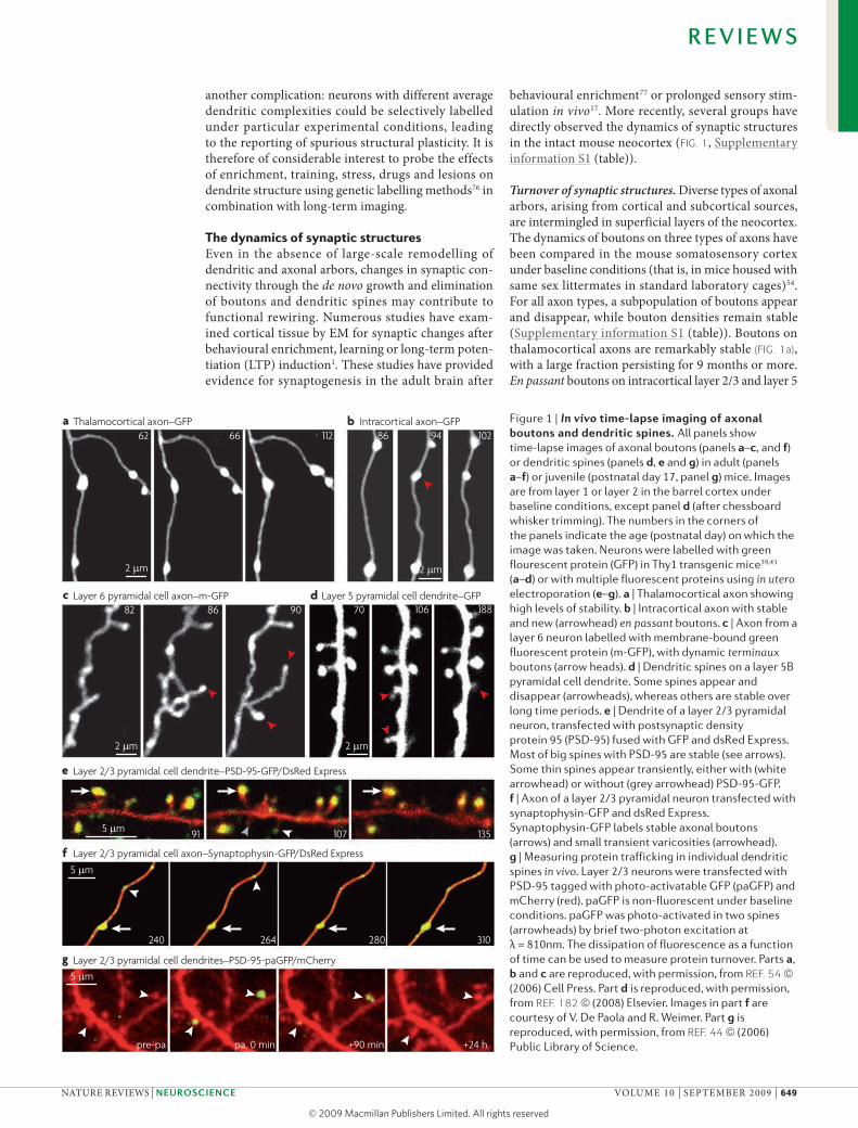
behavioural enrichment77 or prolonged sensory stim-ulation in vivo17. More recently, several groups have directly observed the dynamics of synaptic structures in the intact mouse neocortex (FIG. 1, Supplementary information S1 (table)).
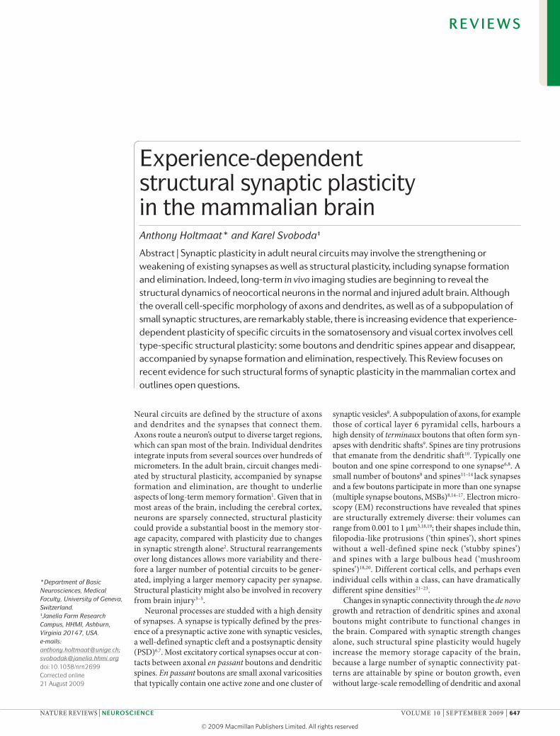
Turnover of synaptic structures. Diverse types of axonal arbors, arising from cortical and subcortical sources, are intermingled in superficial layers of the neocortex. The dynamics of boutons on three types of axons have been compared in the mouse somatosensory cortex under baseline conditions (that is, in mice housed with same sex littermates in standard laboratory cages)54. For all axon types, a subpopulation of boutons appear and disappear, while bouton densities remain stable (Supplementary information S1 (table)). Boutons on thalamocortical axons are remarkably stable (FIG. 1a), with a large fraction persisting for 9 months or more. En passant boutons on intracortical layer 2/3 and layer 5
Figure 1 | In vivo time-lapse imaging of axonal boutons and dendritic spines. All panels show time-lapse images of axonal boutons (panels a–c, and f) or dendritic spines (panels d, e and g) in adult (panels a–f) or juvenile (postnatal day 17, panel g) mice. Images are from layer 1 or layer 2 in the barrel cortex under baseline conditions, except panel d (after chessboard whisker trimming). The numbers in the corners of the panels indicate the age (postnatal day) on which the image was taken. Neurons were labelled with green flourescent protein (GFP) in Thy1 transgenic mice39,43 (a–d) or with multiple fluorescent proteins using in utero electroporation (e–g). a | Thalamocortical axon showing high levels of stability. b | Intracortical axon with stable and new (arrowhead) en passant boutons. c | Axon from a layer 6 neuron labelled with membrane-bound green fluorescent protein (m-GFP), with dynamic terminaux boutons (arrow heads). d | Dendritic spines on a layer 5B pyramidal cell dendrite. Some spines appear and disappear (arrowheads), whereas others are stable over long time periods. e | Dendrite of a layer 2/3 pyramidal neuron, transfected with postsynaptic density protein 95 (PSD-95) fused with GFP and dsRed Express. Most of big spines with PSD-95 are stable (see arrows). Some thin spines appear transiently, either with (white arrowhead) or without (grey arrowhead) PSD-95-GFP. f | Axon of a layer 2/3 pyramidal neuron transfected with synaptophysin-GFP and dsRed Express. Synaptophysin-GFP labels stable axonal boutons (arrows) and small transient varicosities (arrowhead). g | Measuring protein trafficking in individual dendritic spines in vivo. Layer 2/3 neurons were transfected with PSD-95 tagged with photo-activatable GFP (paGFP) and mCherry (red). paGFP is non-fluorescent under baseline conditions. paGFP was photo-activated in two spines (arrowheads) by brief two-photon excitation at λ = 810nm. The dissipation of fluorescence as a function of time can be used to measure protein turnover. Parts a, b and c are reproduced, with permission, from REF. 54 (2006) Cell Press. Part d is reproduced, with permission, from REF. 182 (2008) Elsevier. Images in part f are courtesy of V. De Paola and R. Weimer. Part g is reproduced, with permission, from REF. 44 (2006) Public Library of Science.
R E V I E W S
NATurE rEvIEwS | NeuroscieNce voluME 10 | SEPTEMBEr 2009 | 649
© 2009 Macmillan Publishers Limited. All rights reserved
Critical periodThe developmental age when an animal displays a heightened sensitivity to certain environmental stimuli, such as sensory experiences, which impact (often irreversibly) the development of neural circuits.
pyramidal cell axons are more plastic, with a turnover of 20% over one month of imaging (FIG. 1b). These num-bers are in quantitative agreement with studies of the same axon types in the macaque visual cortex57 and with a study in the mouse visual cortex, although in the latter the identity of the imaged axons was not ana-lysed56. The turnover of en passant boutons is lower than dendritic spines, suggesting that spine growth and that of en passant boutons are not tightly coupled54,56. Axons from layer 6 pyramidal cells are rich in small terminaux boutons. Terminaux boutons appear and disappear at higher rates, with more than 50% turnover in a month (FIG. 1c). Thus the extent of bouton plasticity depends on the cell type. All reported real-time meas-urements in vivo so far have come from mice that were studied under baseline conditions. Experiments using plasticity paradigms have not yet been reported.
Several groups have imaged dendritic spines in the developing22,78,79 and adult neocortex in vivo5,13,22,56,80–86 (Supplementary information S1 (table)). During the second week of life, spines appear and disappear at a rapid rate78. Spine densities in the mouse neocortex were found to increase during the second and third week of life, followed by a period of net spine pruning22,79. This corroborates former findings in the developing primate and rodent cortex that synapse densities increase rap-idly during a relatively brief postnatal period, followed by a protracted period of net synapse elimination87,88
(but see REFS 89,90). As the brain matures, spine turno-ver decreases; this trend continues at least up to the sixth month of a rodent’s life, long after the closure of all known critical periods13,22,56,79,80.
In adult mice, a subpopulation of spines continues to turn over (FIG. 1d). Some spines appear and disap-pear over days, whereas others persist for months, perhaps even for the life of the animals13,22,56,79,80. The
reported fractions of persistent, appearing and dis-appearing spines differ considerably between studies (see BOX 1, Supplementary information S1 (table) and Supplementary information S3 (box)) The consen-sus is that spines are less persistent in young adults (65–85% over 1 month) than in mature adults (75–95% over 1 month), and that the majority of spines persist over long periods, at least under baseline laboratory conditions.
Spine turnover has been measured in various cell types (layer 5B and layer 2/3 pyramidal cells) and cor-tical areas (somatosensory, motor, auditory, visual and frontal)13,22,56,79,80,83. like bouton turnover, the rate of spine turnover is cell type-dependent and might be brain region-specific. For example, apical dendrites of layer 2/3 pyramidal cells show a lower fractional spine turnover rate compared with layer 5 pyramidal cells22. Some studies have reported lower spine turnover22 or spine motility56 (see below) in the visual cortex com-pared with the somatosensory cortex, but other studies have failed to detect a difference56,79.
Time-lapse studies have revealed that spines have widely differing lifetimes. under baseline conditions, spines that appear are likely to disappear again within a few days13,22,83; we have called these structures ‘transient’ spines. Conversely, spines that persist for over a week are likely to persist for months22,79 (FIG. 1d); we have called these structures ‘persistent’ spines. Similarly, in other studies thin, filopodia-like protrusions were dynamic and had short lifetimes, whereas the majority of the big mushroom-type spines were stable56,79,80. Thus, on aver-age, there is a relationship between spine stability and spine size; transient spines are typically small, whereas persistent spines are large22. However, the correlation between structure and stability is not absolute. Small spines can persist, and large spines can disappear. In some studies spines were separated for analysis into structural categories: spines (protrusions with a well-defined head) and filopodia (thin protrusions), with the implication that only spines correspond to synapses56,79,80. However, expression of cytoplasmic green fluorescent protein (GFP) does not allow segregation of spines into struc-tural classes by imaging, let alone to assess for the pres-ence of synapses. In addition, detailed imaging and EM studies indicate that all dendritic protrusions do not fall neatly into a limited number of recognizable structural subtypes, but rather form a structural continuum14,22,91,92. Furthermore, even the thinnest protrusions often make synapses13,14,19,36,93 or express synaptic proteins (FIG. 1e). It might therefore be preferable to class all dendritic pro-trusions as spines and to analyse their dynamics as a function of objective structural parameters22,91.
Spine motility. In addition to spine growth and retrac-tion, several studies have measured rapid (seconds to minutes) spine head motility (also called twitching) as an indicator of developmental and experience-dependent synaptic plasticity56,81,94–96. This work has been inspired by the finding that the actin cytoskeleton of dendritic spines is constantly rearranging97, and the supposition that spine twitching modulates synaptic
Box 1 | Quantitative differences between imaging studies
Even after taking differences in animal age, cell type and cortical area into account, it seems that the extent of spine structural plasticity found in several recent imaging studies is still quantitatively inconsistent13,22,56,79,80,83,86,164,165. At least three factors could contribute to the discrepancies166. First, differences in surgical preparations could matter167, but the contributions are likely to be small (Supplementary information S3 (box))165. Second, spine quantification is performed manually. Although individual researchers and research groups have established consistent and rational analysis criteria, these criteria differ between groups. Some of these differences in criteria are necessitated by variations in image quality in different brain regions and in different types of transgenic mice (Supplementary information S2 (box)). Nevertheless, these differences can cause up to a twofold variation in reported spine turnover166. The scoring of changes in structural plasticity, for example after sensory deprivation, is likely to be more robust as identical criteria are used before and after the manipulation, provided the analyser is blind to the experimental manipulation. Third, differences in sampling are likely to matter as well. Cell type-dependent differences in spine turnover are difficult to detect in densely labelled brains as only short dendritic segments of individual neurons can be traced (Supplementary information S2 (box)); spines must be pooled over multiple neurons and therefore turnover measurements might be biased to the lower rates that are characteristic of cells with the highest spine densities22. Furthermore, turnover rates based on the average of a modest number of single cells in sparsely labelled mice might not adequately represent spine turnover of the entire neuronal population22,23. A combination of these three factors probably explains the discrepancies in reported spine turnover rates.
R E V I E W S
650 | SEPTEMBEr 2009 | voluME 10 www.nature.com/reviews/neuro
© 2009 Macmillan Publishers Limited. All rights reserved
Nature Reviews | Neuroscience
LTP
AMPAR NMDAR
Spine volume
Spin
e br
ight
ness
(inte
grat
ed f
lure
scen
ce)
Spine volume
PSD
siz
e, A
MPA
R co
nten
t
LTP
LTD
Spinehead
Spinehead
LTD
PSDSynaptic vesicles
PSD size
Num
ber o
f doc
ked
syna
ptic
ves
icle
s
Optical point spread functionThe point spread function (PSF) describes the response of an imaging system to a point object. In microscopy the PSF is a measure of the resolution.
Optophysiological recordingOptical microscopybased imaging of cellular function, such as calcium imaging.
function98,99. Spine motility decreases with develop-mental age in vitro94,100 and in vivo, and is regulated by sensory experience56,78,81. Changes in spine motility might be triggered by synaptic activity and precede more prominent morphological changes, such as spine retraction or stabilization56,78.
Synapse stability. An emerging consensus is that a subpopulation of dendritic spines and axonal boutons is remarkably stable, with lifetimes on the order of the lifespan of the mouse. Even more remarkable is that the relative sizes of individual dendritic spines and bou-tons can be maintained for months (FIG. 1), suggesting that synaptic weights could also be stable for months
(BOX 2). By contrast, in vitro studies indicate that synaptic protein complexes are highly labile, with protein life-times of a day or two, orders of magnitude shorter than synapse lifetimes101. In addition, synaptic molecules (such as ras, PSD-95, Shank3, bassoon and synapto-physin) continuously redistribute between synapses on the same neurite over minutes to hours44,102,103104. How can stable synapses exist in the context of high pro-tein turnover either through redistribution or through unstable protein components? Answers to this ques-tion might come from experiments that track the fates of synaptic proteins in vivo. For example, PSD-95 is an abundant multi-domain postsynaptic scaffolding protein that clusters glutamate receptors and organ-izes the associated signalling complexes105. PSD-95 is thought to determine the size and strength of synapses. using two-photon photo-activation of PSD-95 tagged with photo-activatable GFP (paGFP), the trafficking of PSD-95 molecules in and out of single PSDs was measured in vivo44. Synaptic PSD-95 in single PSDs in vivo turned over remarkably quickly (with a half-life of approximately one hour) and exchanged with PSD-95 in neighbouring spines by diffusion (FIG. 1g). large PSDs in large spines captured more diffusing PSD-95 and also retained PSD-95 longer than small PSDs. Changes in the sizes of individual PSDs over days were associated with concomitant changes in PSD-95 retention times. In other words, the kinetic interactions between PSD-95 molecules and individual PSDs are tuned to regulate and maintain synapse size.
Spine growth and synapse formationHigh-resolution optical microscopy alone typically cannot detect synapse formation and elimination. Contact of dendrite and axon is a poor predictor of synapses, as sev-eral non-synaptic contacts occur per actual synapse24,26. Furthermore, as a volume corresponding to the optical point spread function often contains multiple synapses, optical overlap of fluorescent presynaptic and postsynap-tic molecules does not provide proof of a synapse. Instead, detection of synapses requires retrospective analysis using EM13,106, array tomography107, direct imaging of synaptic proteins in vivo (FIG. 1e,f), or perhaps optophysiological recordings with single synapse sensitivity28,108–110.
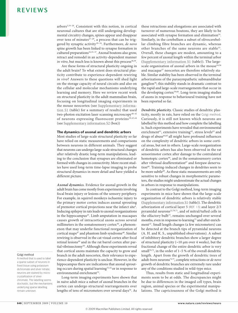
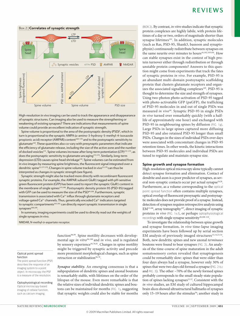
To investigate the relationship between spine growth and synapse formation, in vivo time-lapse imaging experiments have been followed up by serial section EM analysis of previously imaged structures13,14,23,54. Both, new dendritic spines and new axonal terminaux boutons were found to bear synapses (FIG. 2). An analy-sis of the time-course of spine maturation in the adult somatosensory cortex revealed that synaptogenesis could be remarkably slow: spines that were older than four days always had a synapse, however only 30% of spines that were two days old formed a synapse (FIG. 2 b,c and FIG. 3). The other ~70% of the newly formed spines probably corresponds to the small steady-state popula-tion of spines lacking synapses11,12. Consistent with the in vivo studies, an EM study of cultured hippocampal brain slices showed ultrastructural hallmarks of synapses only 15–19 hours after the stimulus36; another study in
Box 2 | Correlates of synaptic strength
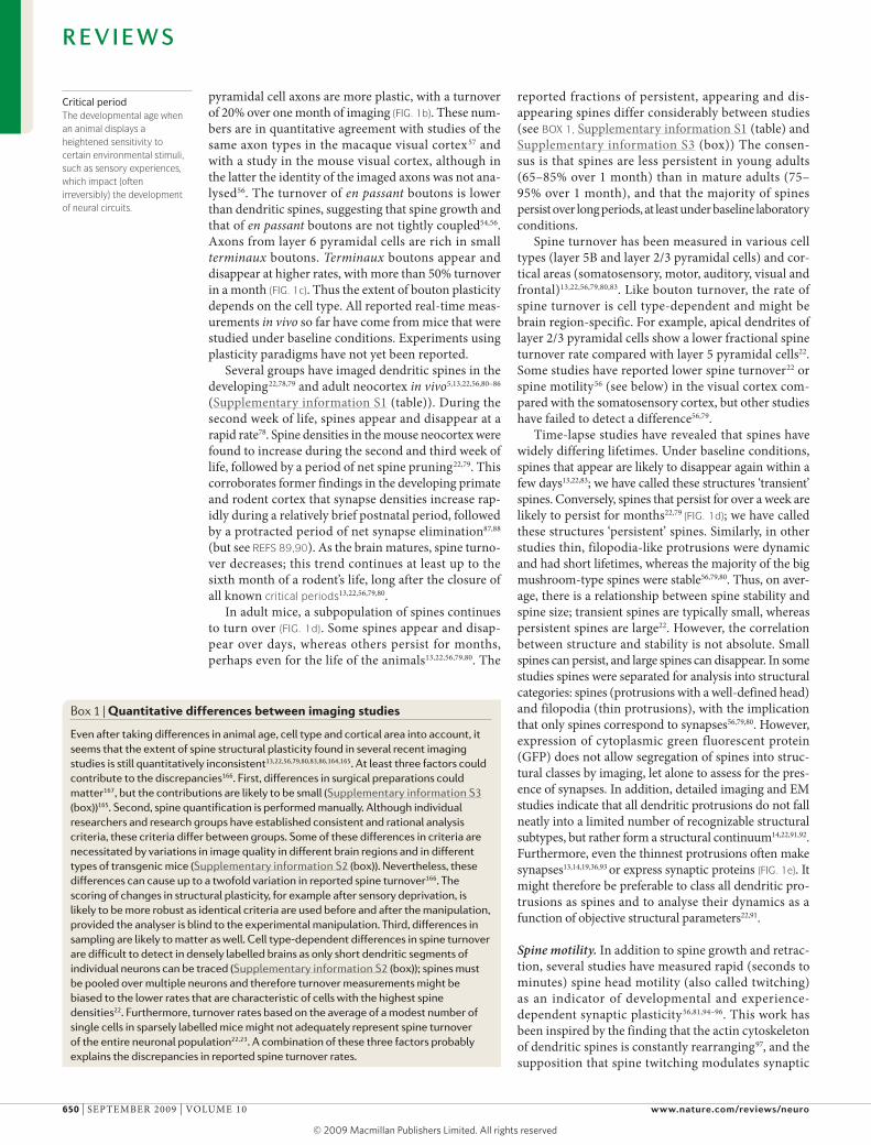
High-resolution in vivo imaging can be used to track the appearance and disappearance of synaptic structures. Can imaging also be used to measure the strengthening or weakening of existing synapses? There are indications that measurements of spine volume could provide an excellent indication of synaptic strength.
Spine volume is proportional to the area of the postsynaptic density (PSD)6, which in turn is proportional to the synaptic AMPA (α-amino-3-hydroxy-5-methyl-4-isoxazole propionic acid) receptor (AMPAR) content168–170 and to the postsynaptic sensitivity to glutamate110. These quantities also co-vary with presynaptic parameters that indicate the efficiency of glutamate release, including the size of the active zone and the number of docked vesicles171. Spine volumes increase after long-term potentiation (LTP)172–175, as does the postsynaptic sensitivity to glutamate uncaging172,174. Similarly, long-term depression (LTD) causes spine head shrinkage176. Spine volumes can be estimated from in vivo images by measuring spine brightness, the fluorescent signal integrated over a dendritic spine22,177,178. Changes in spine volume tracked in vivo23,179 can thus be interpreted as changes in synaptic strength (see figure).
Synaptic strength might also be tracked more directly with recombinant fluorescent synaptic proteins. For example, the AMPAR subunit GluR1 tagged with pH-sensitive green fluorescent protein (GFP) has been used to report the synaptic GluR1 content in the membrane of single spines173,180. Postsynaptic density protein 95 (PSD-95) tagged with GFP can be used to measure the size of the PSD44,104. Furthermore, synaptic transmission is associated with Ca2+ influx through glutamate receptors and voltage-gated Ca2+ channels. Thus, genetically encoded Ca2+ indicators targeted to synaptic compartments163,181 can directly report synaptic transmission in single synapses108,109.
In summary, imaging experiments could be used to directly read out the weights of single synapses in vivo.
NMDAR, N-methyl-d-aspartate receptor.
R E V I E W S
NATurE rEvIEwS | NeuroscieNce voluME 10 | SEPTEMBEr 2009 | 651
© 2009 Macmillan Publishers Limited. All rights reserved
Nature Reviews | Neuroscience
b
c
a
New spine without synapse
New spine with synapse
New terminaux bouton with synapse
162
166
111
116
60
83
1
1
22
1
21
1
2
1
2
1
2
1
1
2
2 µm
2 µm
2 µm
1 µm
0.5 µm
1 µm
0.5 µm
2
brain slices (usually derived from early postnatal brain) showed a much shorter delay (< 1 hour) between spon-taneous spine growth and synapse formation111. It is not clear if all transient spines participate in synapses at some point during their life cycle. It could even be that a subpopulation of transient spines serves completely distinct, non-synaptic functions, such as chemosensa-tion. These studies highlight the importance of combin-ing time-lapse imaging with ultrastructural analysis to elucidate the role of structural plasticity.
New spines preferentially form synapses with large axonal boutons that already bear a synapse, resulting in so-called MSBs14,36,112 (FIG. 3). In the somatosensory cor-tex most new spines (~65%) make synapses on MSBs, whereas ~35% make synapses with single synapse boutons, which are presumably new14. These numbers are consistent with the finding that in the cortex the turnover of en passant boutons is twofold lower com-pared with dendritic spines22,54,56. remarkably, quan-titative analysis of cultured hippocampal slices36 and the dentate gyrus in vivo112 reveal similar results. These observations suggest that new spines on MSBs initi-ate a competition that leads to the pruning of one of the synapses at a later stage112 (FIG. 3). Increases in the fraction of MSBs have also been observed after learn-ing113 and hormonal stimulation114. This suggests that the formation of new synapses on MSBs is a common mechanism for adaptive plasticity in the adult brain and might help to ensure local homeostasis in neural networks.
Is spine growth deterministic or random? Does the presynaptic target site release factors that promote directed spine growth or do spines make synapses with targets they happen to encounter? These questions remain largely unanswered. It has been shown that presynaptic activity29–32 and glutamate33,115 can trigger spine growth. New protrusions seem to preferentially grow towards axonal boutons with active synapses, often ignoring other potential presynaptic elements in the direct vicinity of the dendrite14,36,112. These observa-tions suggest that glutamate may act as a trophic fac-tor to guide new spines to active boutons. However, it is likely that numerous other cell-autonomous and secreted molecules also contribute to guide new spine growth116.
Experience-dependent spine growthFunctional circuits in the adult neocortex adjust to novel experience. It has long been hypothesized that spine growth and retraction could be a substrate or mecha-nism for experience-dependent plasticity in the devel-oping and adult brain. In the developing cortex, spine pruning79 or stabilization117,118 are driven by normal sen-sory experience. In the adult brain, spine and synapse densities can change upon manipulation of sensory experience119. Increased spine and synapse densities have been reported after rearing or training in enriched environments119–122,62, and also after long-term sensory stimulation (for example, REF. 17) and deprivation (for example, REF. 79).
long-term imaging experiments have provided further support for a role of spine growth and retrac-tion in experience-dependent plasticity (FIG. 4). The mouse barrel cortex is a powerful model system to study experience-dependent plasticity123. Trimming a subset of mystacial whiskers causes experience-depend-ent changes in receptive fields (FIG. 4b). For example, responses to deflection of the spared whiskers poten-tiate. This so-called response potentiation has several key properties124: First, it can be detected after a day or so, but continues to progress over several weeks.
Figure 2 | retrospective electron microscopy analysis of previously imaged neurons in vivo. a | New axonal terminaux boutons (numbered 1 and 2) that were imaged in vivo (left) were reconstructed using serial section electron microscopy (EM) (centre, right). Both new boutons (green) bear synapses, one with a dendritic spine (bouton 1) and one with a dendritic shaft (bouton 2). In the reconstruction, the axonal boutons are green, synapses are red and mitochondria are blue. b | A new (age < 4 days) thin spine (numbered 1, bottom left panel; green, centre panel) that was reconstructed using serial section EM (centre, right). The tip of the protrusion makes contact with an axonal bouton (pink), but there are no clear signs of a synapse. c | Thin new (age 1–5 days) spine (numbered 2, bottom left panel) that was reconstructed together with an older persistent spine (numbered 1, bottom left panel) using serial section EM (centre, right). The new spine (green) weaved its way through a dense neuropil to form a clear synapse with an axonal bouton (red), 2 μm away from the parent dendrite. In the reconstruction synapses are coulored red. Age (postnatal day) of mice is shown in top corner of the panels. Part a is reproduced, with permission, from REF. 54 (2006) Cell Press. Parts b and c are reproduced, with permission, from REF. 14 (2006) Macmillan Publishers Ltd. All rights reserved.
R E V I E W S
652 | SEPTEMBEr 2009 | voluME 10 www.nature.com/reviews/neuro
© 2009 Macmillan Publishers Limited. All rights reserved
Nature Reviews | Neuroscience
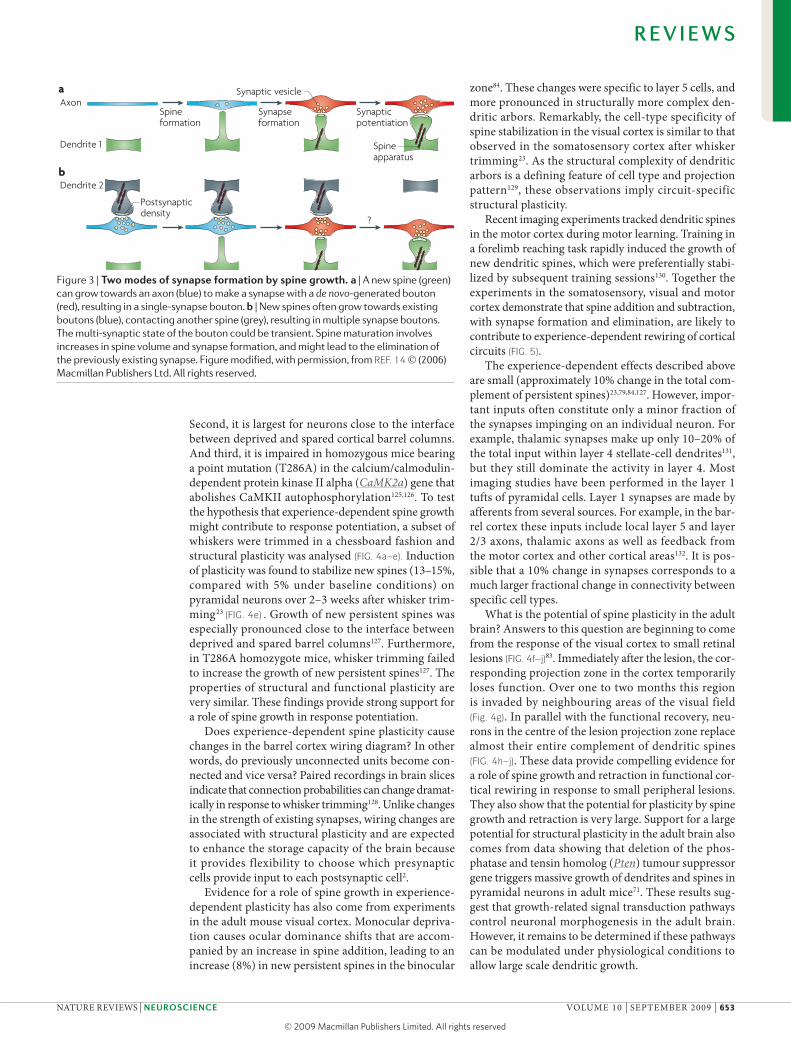
?
Spine formation
Synapseformation
Synapticpotentiation
Axon
Dendrite 1
Dendrite 2
Spineapparatus
a
b
Synaptic vesicle
Postsynaptic density
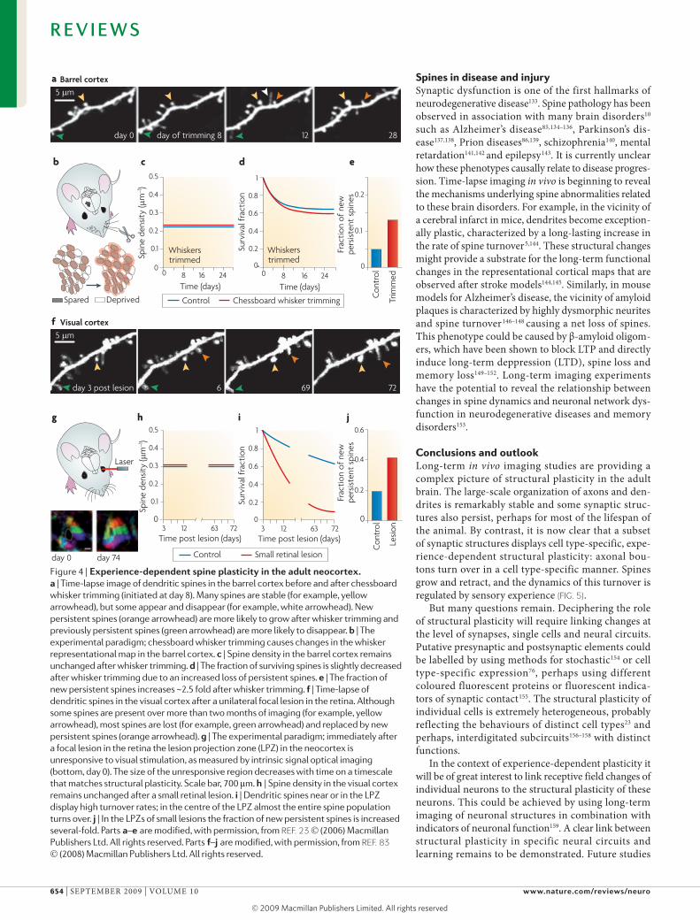
Second, it is largest for neurons close to the interface between deprived and spared cortical barrel columns. And third, it is impaired in homozygous mice bearing a point mutation (T286A) in the calcium/calmodulin-dependent protein kinase II alpha (CaMK2a) gene that abolishes CaMKII autophosphorylation125,126. To test the hypothesis that experience-dependent spine growth might contribute to response potentiation, a subset of whiskers were trimmed in a chessboard fashion and structural plasticity was analysed (FIG. 4a–e). Induction of plasticity was found to stabilize new spines (13–15%, compared with 5% under baseline conditions) on pyramidal neurons over 2–3 weeks after whisker trim-ming23 (FIG. 4e) . Growth of new persistent spines was especially pronounced close to the interface between deprived and spared barrel columns127. Furthermore, in T286A homozygote mice, whisker trimming failed to increase the growth of new persistent spines127. The properties of structural and functional plasticity are very similar. These findings provide strong support for a role of spine growth in response potentiation.
Does experience-dependent spine plasticity cause changes in the barrel cortex wiring diagram? In other words, do previously unconnected units become con-nected and vice versa? Paired recordings in brain slices indicate that connection probabilities can change dramat-ically in response to whisker trimming128. unlike changes in the strength of existing synapses, wiring changes are associated with structural plasticity and are expected to enhance the storage capacity of the brain because it provides flexibility to choose which presynaptic cells provide input to each postsynaptic cell2.
Evidence for a role of spine growth in experience-dependent plasticity has also come from experiments in the adult mouse visual cortex. Monocular depriva-tion causes ocular dominance shifts that are accom-panied by an increase in spine addition, leading to an increase (8%) in new persistent spines in the binocular
zone84. These changes were specific to layer 5 cells, and more pronounced in structurally more complex den-dritic arbors. remarkably, the cell-type specificity of spine stabilization in the visual cortex is similar to that observed in the somatosensory cortex after whisker trimming23. As the structural complexity of dendritic arbors is a defining feature of cell type and projection pattern129, these observations imply circuit-specific structural plasticity.
recent imaging experiments tracked dendritic spines in the motor cortex during motor learning. Training in a forelimb reaching task rapidly induced the growth of new dendritic spines, which were preferentially stabi-lized by subsequent training sessions130. Together the experiments in the somatosensory, visual and motor cortex demonstrate that spine addition and subtraction, with synapse formation and elimination, are likely to contribute to experience-dependent rewiring of cortical circuits (FIG. 5).
The experience-dependent effects described above are small (approximately 10% change in the total com-plement of persistent spines)23,79,84,127. However, impor-tant inputs often constitute only a minor fraction of the synapses impinging on an individual neuron. For example, thalamic synapses make up only 10–20% of the total input within layer 4 stellate-cell dendrites131, but they still dominate the activity in layer 4. Most imaging studies have been performed in the layer 1 tufts of pyramidal cells. layer 1 synapses are made by afferents from several sources. For example, in the bar-rel cortex these inputs include local layer 5 and layer 2/3 axons, thalamic axons as well as feedback from the motor cortex and other cortical areas132. It is pos-sible that a 10% change in synapses corresponds to a much larger fractional change in connectivity between specific cell types.
what is the potential of spine plasticity in the adult brain? Answers to this question are beginning to come from the response of the visual cortex to small retinal lesions (FIG. 4f–j)83. Immediately after the lesion, the cor-responding projection zone in the cortex temporarily loses function. over one to two months this region is invaded by neighbouring areas of the visual field (Fig. 4g). In parallel with the functional recovery, neu-rons in the centre of the lesion projection zone replace almost their entire complement of dendritic spines (FIG. 4h–j). These data provide compelling evidence for a role of spine growth and retraction in functional cor-tical rewiring in response to small peripheral lesions. They also show that the potential for plasticity by spine growth and retraction is very large. Support for a large potential for structural plasticity in the adult brain also comes from data showing that deletion of the phos-phatase and tensin homolog (Pten) tumour suppressor gene triggers massive growth of dendrites and spines in pyramidal neurons in adult mice71. These results sug-gest that growth-related signal transduction pathways control neuronal morphogenesis in the adult brain. However, it remains to be determined if these pathways can be modulated under physiological conditions to allow large scale dendritic growth.
Figure 3 | Two modes of synapse formation by spine growth. a | A new spine (green) can grow towards an axon (blue) to make a synapse with a de novo-generated bouton (red), resulting in a single-synapse bouton. b | New spines often grow towards existing boutons (blue), contacting another spine (grey), resulting in multiple synapse boutons. The multi-synaptic state of the bouton could be transient. Spine maturation involves increases in spine volume and synapse formation, and might lead to the elimination of the previously existing synapse. Figure modified, with permission, from REF. 14 (2006) Macmillan Publishers Ltd. All rights reserved.
R E V I E W S
NATurE rEvIEwS | NeuroscieNce voluME 10 | SEPTEMBEr 2009 | 653
© 2009 Macmillan Publishers Limited. All rights reserved
5 µm
day 3 post lesion 6 69 72
5 µm
day 0 day of trimming 8 12 28
Nature Reviews | Neuroscience
g h i j
f
b
a
c d e
Laser
Spared Deprived
day 0 day 74
Control Chessboard whisker trimming
3 12 63
0.2
0.4
0.6
0.8
1
Time post lesion (days)
Surv
ival
fra
ctio
n
720
3 12 63Time post lesion (days)
72
0.2
0.4
0.6
0.8
1
0
Whiskerstrimmed
Whiskerstrimmed
Control Small retinal lesion
Visual cortex
Barrel cortex
0
0.1
0.2
0.3
0.4
0.5
Spin
e de
nsity
(µm
–1)
0.6
0
0.2
0.4
Con
trol
Frac
tion
of n
ewpe
rsist
ent
spin
esFr
actio
n of
new
pers
isten
t sp
ines
Lesi
on
0
0.1
0.2
0.3
0.4
0.5
Spin
e de
nsity
(µm
–1)
Time (days)
0 8 16 240
0.1
0.2
Con
trol
Trim
med
Time (days)
0 8 16 24
Surv
ival
frac
tion
Spines in disease and injurySynaptic dysfunction is one of the first hallmarks of neurodegenerative disease133. Spine pathology has been observed in association with many brain disorders10 such as Alzheimer’s disease85,134–136, Parkinson’s dis-ease137,138, Prion diseases86,139, schizophrenia140, mental retardation141,142 and epilepsy143. It is currently unclear how these phenotypes causally relate to disease progres-sion. Time-lapse imaging in vivo is beginning to reveal the mechanisms underlying spine abnormalities related to these brain disorders. For example, in the vicinity of a cerebral infarct in mice, dendrites become exception-ally plastic, characterized by a long-lasting increase in the rate of spine turnover5,144. These structural changes might provide a substrate for the long-term functional changes in the representational cortical maps that are observed after stroke models144,145. Similarly, in mouse models for Alzheimer’s disease, the vicinity of amyloid plaques is characterized by highly dysmorphic neurites and spine turnover146–148 causing a net loss of spines. This phenotype could be caused by β-amyloid oligom-ers, which have been shown to block lTP and directly induce long-term deppression (lTD), spine loss and memory loss149–152. long-term imaging experiments have the potential to reveal the relationship between changes in spine dynamics and neuronal network dys-function in neurodegenerative diseases and memory disorders153.
Conclusions and outlooklong-term in vivo imaging studies are providing a complex picture of structural plasticity in the adult brain. The large-scale organization of axons and den-drites is remarkably stable and some synaptic struc-tures also persist, perhaps for most of the lifespan of the animal. By contrast, it is now clear that a subset of synaptic structures displays cell type-specific, expe-rience-dependent structural plasticity: axonal bou-tons turn over in a cell type-specific manner. Spines grow and retract, and the dynamics of this turnover is regulated by sensory experience (FIG. 5).
But many questions remain. Deciphering the role of structural plasticity will require linking changes at the level of synapses, single cells and neural circuits. Putative presynaptic and postsynaptic elements could be labelled by using methods for stochastic154 or cell type-specific expression76, perhaps using different coloured fluorescent proteins or fluorescent indica-tors of synaptic contact155. The structural plasticity of individual cells is extremely heterogeneous, probably reflecting the behaviours of distinct cell types23 and perhaps, interdigitated subcircuits156–158 with distinct functions.
In the context of experience-dependent plasticity it will be of great interest to link receptive field changes of individual neurons to the structural plasticity of these neurons. This could be achieved by using long-term imaging of neuronal structures in combination with indicators of neuronal function159. A clear link between structural plasticity in specific neural circuits and learning remains to be demonstrated. Future studies
Figure 4 | experience-dependent spine plasticity in the adult neocortex. a | Time-lapse image of dendritic spines in the barrel cortex before and after chessboard whisker trimming (initiated at day 8). Many spines are stable (for example, yellow arrowhead), but some appear and disappear (for example, white arrowhead). New persistent spines (orange arrowhead) are more likely to grow after whisker trimming and previously persistent spines (green arrowhead) are more likely to disappear. b | The experimental paradigm; chessboard whisker trimming causes changes in the whisker representational map in the barrel cortex. c | Spine density in the barrel cortex remains unchanged after whisker trimming. d | The fraction of surviving spines is slightly decreased after whisker trimming due to an increased loss of persistent spines. e | The fraction of new persistent spines increases ~2.5 fold after whisker trimming. f | Time-lapse of dendritic spines in the visual cortex after a unilateral focal lesion in the retina. Although some spines are present over more than two months of imaging (for example, yellow arrowhead), most spines are lost (for example, green arrowhead) and replaced by new persistent spines (orange arrowhead). g | The experimental paradigm; immediately after a focal lesion in the retina the lesion projection zone (LPZ) in the neocortex is unresponsive to visual stimulation, as measured by intrinsic signal optical imaging (bottom, day 0). The size of the unresponsive region decreases with time on a timescale that matches structural plasticity. Scale bar, 700 μm. h | Spine density in the visual cortex remains unchanged after a small retinal lesion. i | Dendritic spines near or in the LPZ display high turnover rates; in the centre of the LPZ almost the entire spine population turns over. j | In the LPZs of small lesions the fraction of new persistent spines is increased several-fold. Parts a –e are modified, with permission, from REF. 23 (2006) Macmillan Publishers Ltd. All rights reserved. Parts f–j are modified, with permission, from REF. 83 (2008) Macmillan Publishers Ltd. All rights reserved.
R E V I E W S
654 | SEPTEMBEr 2009 | voluME 10 www.nature.com/reviews/neuro
© 2009 Macmillan Publishers Limited. All rights reserved
Nature Reviews | Neuroscience
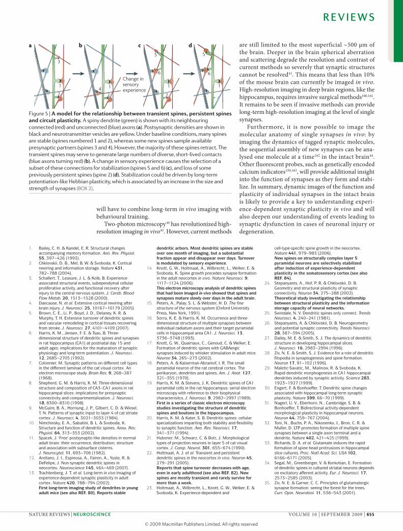
1
2
5
6
5
1
6
1
2
3
1
24
a b c d
Change insensory experience
will have to combine long-term in vivo imaging with behavioural training.
Two-photon microscopy40 has revolutionized high-resolution imaging in vivo42. However, current methods
are still limited to the most superficial ~500 μm of the brain. Deeper in the brain spherical aberration and scattering degrade the resolution and contrast of current methods so severely that synaptic structures cannot be resolved41. This means that less than 10% of the mouse brain can currently be imaged in vivo. High-resolution imaging in deep brain regions, like the hippocampus, requires invasive surgical methods160,161. It remains to be seen if invasive methods can provide long-term high-resolution imaging at the level of single synapses.
Furthermore, it is now possible to image the molecular anatomy of single synapses in vivo: by imaging the dynamics of tagged synaptic molecules, the sequential assembly of new synapses can be ana-lysed one molecule at a time162 in the intact brain44. other fluorescent probes, such as genetically encoded calcium indicators159,163, will provide additional insight into the function of synapses as they form and stabi-lize. In summary, dynamic images of the function and plasticity of individual synapses in the intact brain is likely to provide a key to understanding experi-ence-dependent synaptic plasticity in vivo and will also deepen our understanding of events leading to synaptic dysfunction in cases of neuronal injury or degeneration.
Figure 5 | A model for the relationship between transient spines, persistent spines and circuit plasticity. A spiny dendrite (green) is shown with its neighbouring connected (red) and unconnected (blue) axons (a). Postsynaptic densities are shown in black and neurotransmitter vesicles are yellow. Under baseline conditions, many spines are stable (spines numbered 1 and 2), whereas some new spines sample available presynaptic partners (spines 3 and 4). However, the majority of these spines retract. The transient spines may serve to generate large numbers of diverse, short-lived contacts (blue axons turning red) (b). A change in sensory experience causes the selection of a subset of these connections for stabilization (spines 5 and 6) (c), and loss of some previously persistent spines (spine 2) (d). Stabilization could be driven by long-term potentiation-like Hebbian plasticity, which is associated by an increase in the size and strength of synapses (BOX 2).
1. Bailey, C. H. & Kandel, E. R. Structural changes accompanying memory formation. Ann. Rev. Physiol. 55, 397–426 (1993).
2. Chklovskii, D. B., Mel, B. W. & Svoboda, K. Cortical rewiring and information storage. Nature 431, 782–788 (2004).
3. Schallert, T., Leasure, J. L. & Kolb, B. Experience-associated structural events, subependymal cellular proliferative activity, and functional recovery after injury to the central nervous system. J. Cereb. Blood Flow Metab. 20, 1513–1528 (2000).
4. Dancause, N. et al. Extensive cortical rewiring after brain injury. J. Neurosci. 25, 10167–10179 (2005).
5. Brown, C. E., Li, P., Boyd, J. D., Delaney, K. R. & Murphy, T. H. Extensive turnover of dendritic spines and vascular remodeling in cortical tissues recovering from stroke. J. Neurosci. 27, 4101–4109 (2007).
6. Harris, K. M., Jensen, F. E. & Tsao, B. Three-dimensional structure of dendritic spines and synapses in rat hippocampus (CA1) at postnatal day 15 and adult ages: implications for the maturation of synaptic physiology and long-term potentiation. J. Neurosci. 12, 2685–2705 (1992).
7. Colonnier, M. Synaptic patterns on different cell types in the different laminae of the cat visual cortex. An electron microscope study. Brain Res. 9, 268–287 (1968).
8. Shepherd, G. M. & Harris, K. M. Three-dimensional structure and composition of CA3--CA1 axons in rat hippocampal slices: implications for presynaptic connectivity and compartmentalization. J. Neurosci. 18, 8300–8310 (1998).
9. McGuire, B. A., Hornung, J. P., Gilbert, C. D. & Wiesel, T. N. Patterns of synaptic input to layer 4 of cat striate cortex. J. Neurosci. 4, 3021–3033 (1984).
10. Nimchinsky, E. A., Sabatini, B. L. & Svoboda, K. Structure and function of dendritic spines. Annu. Rev. Physiol. 64, 313–353 (2002).
11. Spacek, J. ‘Free’ postsynaptic-like densities in normal adult brain: their occurrence, distribution, structure and association with subsurface cisterns. J. Neurocytol. 11, 693–706 (1982).
12. Arellano, J. I., Espinosa, A., Fairen, A., Yuste, R. & DeFelipe, J. Non-synaptic dendritic spines in neocortex. Neuroscience 145, 464–469 (2007).
13. Trachtenberg, J. T. et al. Long-term in vivo imaging of experience-dependent synaptic plasticity in adult cortex. Nature 420, 788–794 (2002).First long-term imaging study of dendrites in young adult mice (see also REF. 80). Reports stable
dendritic arbors. Most dendritic spines are stable over one month of imaging, but a substantial fraction appear and disappear over days. Turnover is modulated by sensory experience.
14. Knott, G. W., Holtmaat, A., Wilbrecht, L., Welker, E. & Svoboda, K. Spine growth precedes synapse formation in the adult neocortex in vivo. Nature Neurosci. 9, 1117–1124 (2006).This electron microscopy analysis of dendritic spines that had been imaged in vivo showed that spines and synapses mature slowly over days in the adult brain.
15. Peters, A., Palay, S. L. & Webster, H. D. The fine structure of the nervous system (Oxford University Press, New York, 1991).
16. Sorra, K. E. & Harris, K. M. Occurrence and three-dimensional structure of multiple synapses between individual radiatum axons and their target pyramidal cells in hippocampal area CA1. J. Neurosci. 13, 3736–3748 (1993).
17. Knott, G. W., Quairiaux, C., Genoud, C. & Welker, E. Formation of dendritic spines with GABAergic synapses induced by whisker stimulation in adult mice. Neuron 34, 265–273 (2002).
18. Peters, A. & Kaiserman-Abramof, I. R. The small pyramidal neuron of the rat cerebral cortex. The perikaryon, dendrites and spines. Am. J. Anat. 127, 321–355 (1970).
19. Harris, K. M. & Stevens, J. K. Dendritic spines of CA1 pyramidal cells in the rat hippocampus: serial electron microscopy with reference to their biophysical characterisitcs. J. Neurosci. 9, 2982–2997 (1989).First in a series of classic electron microscopy studies investigating the structure of dendritic spines and boutons in the hippocampus.
20. Harris, K. M. & Kater, S. B. Dendritic spines: cellular specializations imparting both stability and flexibility to synaptic function. Ann. Rev. Neurosci. 17, 341–371 (1994).
21. Hubener, M., Schwarz, C. & Bolz, J. Morphological types of projection neurons in layer 5 of cat visual cortex. J. Comp. Neurol. 301, 655–674 (1990).
22. Holtmaat, A. J. et al. Transient and persistent dendritic spines in the neocortex in vivo. Neuron 45, 279–291 (2005).Reports that spine turnover decreases with age, even in early adulthood (see also REF. 82). New spines are mostly transient and rarely survive for more than a week.
23. Holtmaat, A., Wilbrecht, L., Knott, G. W., Welker, E. & Svoboda, K. Experience-dependent and
cell-type-specific spine growth in the neocortex. Nature 441, 979–983 (2006).New spines on structurally complex layer 5 pyramidal neurons are selectively stabilized after induction of experience-dependent plasticity in the somatosensory cortex (see also REF. 84).
24. Stepanyants, A., Hof, P. R. & Chklovskii, D. B. Geometry and structural plasticity of synaptic connectivity. Neuron 34, 275–288 (2002).Theoretical study investigating the relationship between structural plasticity and the information storage capacity of neural networks.
25. Swindale, N. V. Dendritic spines only connect. Trends Neurosci. 4, 240–241 (1981).
26. Stepanyants, A. & Chklovskii, D. B. Neurogeometry and potential synaptic connectivity. Trends Neurosci. 28, 387–394 (2005).
27. Dailey, M. E. & Smith, S. J. The dynamics of dendritic structure in developing hippocampal slices. J. Neurosci. 16, 2983–2994 (1996).
28. Ziv, N. E. & Smith, S. J. Evidence for a role of dendritic filopodia in synaptogenesis and spine formation. Neuron 17, 91–102 (1996).
29. Maletic-Savatic, M., Malinow, R. & Svoboda, K. Rapid dendritic morphogenesis in CA1 hippocampal dendrites induced by synaptic activity. Science 283, 1923–1927 (1999).
30. Engert, F. & Bonhoeffer, T. Dendritic spine changes associated with hippocampal long-term synaptic plasticity. Nature 399, 66–70 (1999).
31. Nagerl, U. V., Eberhorn, N., Cambridge, S. B. & Bonhoeffer, T. Bidirectional activity-dependent morphological plasticity in hippocampal neurons. Neuron 44, 759–767 (2004).
32. Toni, N., Buchs, P. A., Nikonenko, I., Bron, C. R. & Muller, D. LTP promotes formation of multiple spine synapses between a single axon terminal and a dendrite. Nature 402, 421–425 (1999).
33. Richards, D. A. et al. Glutamate induces the rapid formation of spine head protrusions in hippocampal slice cultures. Proc. Natl Acad. Sci. USA 102, 6166–6171 (2005).
34. Segal, M., Greenberger, V. & Korkotian, E. Formation of dendritic spines in cultured striatal neurons depends on excitatory afferent activity. Eur. J. Neurosci. 17, 2573–2585 (2003).
35. Ziv, N. E. & Garner, C. C. Principles of glutamatergic synapse formation: seeing the forest for the trees. Curr. Opin. Neurobiol. 11, 536–543 (2001).
R E V I E W S
NATurE rEvIEwS | NeuroscieNce voluME 10 | SEPTEMBEr 2009 | 655
© 2009 Macmillan Publishers Limited. All rights reserved
36. Nagerl, U. V., Kostinger, G., Anderson, J. C., Martin, K. A. & Bonhoeffer, T. Protracted synaptogenesis after activity-dependent spinogenesis in hippocampal neurons. J. Neurosci. 27, 8149–8156 (2007).
37. De Roo, M., Klauser, P., Mendez, P., Poglia, L. & Muller, D. Activity-dependent PSD formation and stabilization of newly formed spines in hippocampal slice cultures. Cereb. Cortex 18, 151–161 (2008).
38. Tashiro, A., Dunaevsky, A., Blazeski, R., Mason, C. A. & Yuste, R. Bidirectional regulation of hippocampal mossy fiber filopodial motility by kainate receptors: a two-step model of synaptogenesis. Neuron 38, 773–784 (2003).
39. De Paola, V., Arber, S. & Caroni, P. AMPA receptors regulate dynamic equilibrium of presynaptic terminals in mature hippocampal networks. Nature Neurosci. 6, 491–500 (2003).
40. Denk, W., Strickler, J. H. & Webb, W. W. Two-photon laser scanning microscopy. Science 248, 73–76 (1990).
41. Helmchen, F. & Denk, W. Deep tissue two-photon microscopy. Nature Methods 2, 932–940 (2005).Pithy review highlighting the opportunites and limitations of two-photon microscopy applied to intact tissue.
42. Svoboda, K. & Yasuda, R. Principles of two-photon excitation microscopy and its applications to neuroscience. Neuron 50, 823–839 (2006).
43. Feng, G. et al. Imaging neuronal subsets in transgenic mice expressing multiple spectral variants of GFP. Neuron 28, 41–51 (2000).Describes Thy1 transgenic mice expressing fluorescent proteins in a sparse subset of projection neurons. These mice have made routine in vivo imaging possible.
44. Gray, N. W., Weimer, R. M., Bureau, I. & Svoboda, K. Rapid redistribution of synaptic PSD-95 in the neocortex in vivo. PLoS Biol. 4, e370 (2006).Measurement of turnover of PSD-95 at individual synapses in vivo. PSD-95 half-life is on the order of hours, several orders of magnitude lower than synaptic lifetimes.
45. Sutula, T., He, X. X., Cavazos, J. & Scott, G. Synaptic reorganization in the hippocampus induced by abnormal functional activity. Science 239, 1147–1150 (1988).
46. Florence, S. L., Taub, H. B. & Kaas, J. H. Large-scale sprouting of cortical connections after peripheral injury in adult macaque monkeys. Science 282, 1117–1121 (1998).
47. Buonomano, D. V. & Merzenich, M. M. Cortical plasticity: from synapses to maps. Annu. Rev. Neurosci. 21, 149–186 (1998).
48. Flor, H., Nikolajsen, L. & Staehelin Jensen, T. Phantom limb pain: a case of maladaptive CNS plasticity? Nature Rev. Neurosci. 7, 873–881 (2006).
49. Darian-Smith, C. & Gilbert, C. D. Axonal sprouting accompanies functional reorganization in adult cat striate cortex. Nature 368, 737–740 (1994).
50. Kossut, M. & Juliano, S. L. Anatomical correlates of representational map reorganization induced by partial vibrissectomy in the barrel cortex of adult mice. Neuroscience 92, 807–817 (1999).
51. Ramirez-Amaya, V., Balderas, I., Sandoval, J., Escobar, M. L. & Bermudez-Rattoni, F. Spatial long-term memory is related to mossy fiber synaptogenesis. J. Neurosci. 21, 7340–7348 (2001).
52. Holahan, M. R., Rekart, J. L., Sandoval, J. & Routtenberg, A. Spatial learning induces presynaptic structural remodeling in the hippocampal mossy fiber system of two rat strains. Hippocampus 16, 560–570 (2006).
53. Galimberti, I. et al. Long-term rearrangements of hippocampal mossy fiber terminal connectivity in the adult regulated by experience. Neuron 50, 749–763 (2006).
54. De Paola, V. et al. Cell type-specific structural plasticity of axonal branches and boutons in the adult neocortex. Neuron 49, 861–875 (2006).This paper compares the structural plasticity in different types of axon. Turnover of boutons is high in some types of intracortical axons and extremely low in thalamocortical axons.
55. Nishiyama, H., Fukaya, M., Watanabe, M. & Linden, D. J. Axonal motility and its modulation by activity are branch-type specific in the intact adult cerebellum. Neuron 56, 472–487 (2007).In vivo imaging study of climbing fibres in the cerebellum. Reports branch-type-specific dynamics of different branches of the same axon. Indicates that structural plasticity can be regulated locally.
56. Majewska, A. K., Newton, J. R. & Sur, M. Remodeling of synaptic structure in sensory cortical areas in vivo. J. Neurosci. 26, 3021–3029 (2006).Comparison of structural plasticity in different cortical areas. Reports that axon terminals are more stable than dendritic spines.
57. Stettler, D. D., Yamahachi, H., Li, W., Denk, W. & Gilbert, C. D. Axons and synaptic boutons are highly dynamic in adult visual cortex. Neuron 49, 877–887 (2006).Tour de force study investigating turnover of axonal boutons in macaques.
58. Gan, W. B., Kwon, E., Feng, G., Sanes, J. R. & Lichtman, J. W. Synaptic dynamism measured over minutes to months: age-dependent decline in an autonomic ganglion. Nature Neurosci. 6, 956–960 (2003).
59. Portera-Cailliau, C., Weimer, R. M., Paola, V. D., Caroni, P. & Svoboda, K. Diverse modes of axon elaboration in the developing neocortex. PLoS Biol. 3, e272 (2005).
60. Antonini, A. & Stryker, M. P. Rapid remodeling of axonal arbors in the visual cortex. Science 260, 1819–1821 (1993).
61. Volkmar, F. R. & Greenough, W. T. Differential rearing effects on rat visual cortical plasticity. Science 176, 1445–1447 (1972).
62. Kolb, B., Cioe, J. & Comeau, W. Contrasting effects of motor and visual spatial learning tasks on dendritic arborization and spine density in rats. Neurobiol. Learn. Mem. 90, 295–300 (2008).
63. Magarinos, A. M., McEwen, B. S., Flugge, G. & Fuchs, E. Chronic psychosocial stress causes apical dendritic atrophy of hippocampal CA3 pyramidal neurons in subordinate tree shrews. J. Neurosci. 16, 3534–3540 (1996).
64. Robinson, T. E. & Kolb, B. Alterations in the morphology of dendrites and dendritic spines in the nucleus accumbens and prefrontal cortex following repeated treatment with amphetamine or cocaine. Eur. J. Neurosci. 11, 1598–1604 (1999).
65. Robinson, T. E., Gorny, G., Mitton, E. & Kolb, B. Cocaine self-administration alters the morphology of dendrites and dendritic spines in the nucleus accumbens and neocortex. Synapse 39, 257–266 (2001).
66. Jones, T. A. & Schallert, T. Use-dependent growth of pyramidal neurons after neocortical damage. J. Neurosci. 14, 2140–2152 (1994).
67. Tailby, C., Wright, L. L., Metha, A. B. & Calford, M. B. Activity-dependent maintenance and growth of dendrites in adult cortex. Proc. Natl Acad. Sci. USA 102, 4631–4636 (2005).
68. Hickmott, P. W. & Steen, P. A. Large-scale changes in dendritic structure during reorganization of adult somatosensory cortex. Nature Neurosci. 8, 140–142 (2005).
69. Chang, F. L. & Greenough, W. T. Lateralized effects of monocular training on dendritic branching in adult split-brain rats. Brain Res. 232, 283–292 (1982).
70. Lee, W. C. et al. Dynamic remodeling of dendritic arbors in GABAergic interneurons of adult visual cortex. PLoS Biol. 4, e29 (2006).
71. Chow, D. K. et al. Laminar and compartmental regulation of dendritic growth in mature cortex. Nature Neurosci. 12, 116–118 (2009).
72. Mizrahi, A. & Katz, L. C. Dendritic stability in the adult olfactory bulb. Nature Neurosci. 6, 1201–1207 (2003).First long-term imaging study of neuronal structure in the olfactory bulb. Reports that the large-scale structure of mitral cell dendrites is stable over months.
73. Lee, W. C. et al. A dynamic zone defines interneuron remodeling in the adult neocortex. Proc. Natl Acad. Sci. USA 105, 19968–19973 (2008).
74. Zhao, C., Teng, E. M., Summers, R. G., Jr., Ming, G. L. & Gage, F. H. Distinct morphological stages of dentate granule neuron maturation in the adult mouse hippocampus. J. Neurosci. 26, 3–11 (2006).
75. Mizrahi, A. Dendritic development and plasticity of adult-born neurons in the mouse olfactory bulb. Nature Neurosci. 10, 444–452 (2007).
76. Luo, L., Callaway, E. M. & Svoboda, K. Genetic dissection of neural circuits. Neuron 57, 634–660 (2008).
77. Turner, A. M. & Greenough, W. T. Differential rearing effects on rat visual cortex synapses. I. Synaptic and neuronal density and synapses per neuron. Brain Res. 329, 195–203 (1985).
78. Lendvai, B., Stern, E., Chen, B. & Svoboda, K. Experience-dependent plasticity of dendritic spines in
the developing rat barrel cortex in vivo. Nature 404, 876–881 (2000).First time-lapse imaging study of dendritic spines in vivo.
79. Zuo, Y., Yang, G., Kwon, E. & Gan, W. B. Long-term sensory deprivation prevents dendritic spine loss in primary somatosensory cortex. Nature 436, 261–265 (2005).
80. Grutzendler, J., Kasthuri, N. & Gan, W. B. Long-term dendritic spine stability in the adult cortex. Nature 420, 812–816 (2002).First long-term imaging study of dendritic spines in adult mice (see also REF. 13). Reports largely stable dendritic spines over up to three months of imaging.
81. Majewska, A. & Sur, M. Motility of dendritic spines in visual cortex in vivo: Changes during the critical period and effects of visual deprivation. Proc. Natl Acad. Sci. USA 100, 16024–16029 (2003).
82. Zuo, Y., Lin, A., Chang, P. & Gan, W. B. Development of long-term dendritic spine stability in diverse regions of cerebral cortex. Neuron 46, 181–189 (2005).Reports that spines gradually stabilize during development and adolescence (see also REF. 22). In adults most spines persist for more than a year and a half.
83. Keck, T. et al. Massive restructuring of neuronal circuits during functional reorganization of adult visual cortex. Nature Neurosci. 11, 1162–1167 (2008).This study describes a large-scale turnover of dendritic spines in the visual cortex after induction of a retinal scotoma, indicating the huge potential for structural plasticity in the adult brain.
84. Hofer, S. B., Mrsic-Flogel, T. D., Bonhoeffer, T. & Hubener, M. Experience leaves a lasting structural trace in cortical circuits. Nature 457, 313–317 (2009).New spines grow and are stabilized after induction of experience-dependent plasticity in the visual cortex, specifically on structurally complex layer 5 pyramidal neurons (see also REF. 23).
85. Spires, T. L. et al. Dendritic spine abnormalities in amyloid precursor protein transgenic mice demonstrated by gene transfer and intravital multiphoton microscopy. J. Neurosci. 25, 7278–7287 (2005).
86. Fuhrmann, M., Mitteregger, G., Kretzschmar, H. & Herms, J. Dendritic pathology in prion disease starts at the synaptic spine. J. Neurosci. 27, 6224–6233 (2007).
87. Rakic, P., Bourgeois, J. P., Eckenhoff, M. F., Zecevic, N. & Goldman-Rakic, P. S. Concurrent overproduction of synapses in diverse regions of the primate cerebral cortex. Science 232, 232–235 (1986).
88. De Felipe, J., Marco, P., Fairen, A. & Jones, E. G. Inhibitory synaptogenesis in mouse somatosensory cortex. Cereb. Cortex 7, 619–634 (1997).
89. Blue, M. E. & Parnavelas, J. G. The formation and maturation of synapses in the visual cortex of the rat. II. Quantitative analysis. J. Neurocytol. 12, 697–712 (1983).
90. Micheva, K. D. & Beaulieu, C. Quantitative aspects of synaptogenesis in the rat barrel field cortex with special reference to GABA circuitry. J. Comp. Neurol. 373, 340–354 (1996).
91. Portera-Cailliau, C., Pan, D. T. & Yuste, R. Activity-regulated dynamic behavior of early dendritic protrusions: evidence for different types of dendritic filopodia. J. Neurosci. 23, 7129–7142 (2003).
92. Arellano, J. I., Benavides-Piccione, R., Defelipe, J. & Yuste, R. Ultrastructure of dendritic spines: correlation between synaptic and spine morphologies. Front. Neurosci. 1, 131–143 (2007).
93. Fiala, J. C., Feinberg, M., Popov, V. & Harris, K. M. Synaptogenesis via dendritic filopodia in developing hippocampal area CA1. J. Neurosci. 18, 8900–8911 (1998).
94. Dunaevsky, A., Tashiro, A., Majewska, A., Mason, C. & Yuste, R. Developmental regulation of spine motility in the mammalian central nervous system. Proc. Natl Acad. Sci. USA 96, 13438–13443 (1999).
95. Konur, S., Rabinowitz, D., Fenstermaker, V. L. & Yuste, R. Systematic regulation of spine sizes and densities in pyramidal neurons. J. Neurobiol. 56, 95–112 (2003).
96. Oray, S., Majewska, A. & Sur, M. Dendritic spine dynamics are regulated by monocular deprivation and extracellular matrix degradation. Neuron 44, 1021–1030 (2004).
97. Fischer, M., Kaech, S., Knutti, D. & Matus, A. Rapid actin-based plasticity in dendritic spines. Neuron 20, 847–854 (1998).
R E V I E W S
656 | SEPTEMBEr 2009 | voluME 10 www.nature.com/reviews/neuro
© 2009 Macmillan Publishers Limited. All rights reserved
98. Matus, A. Actin-based plasticity in dendritic spines. Science 290, 754–758 (2000).
99. Crick, F. Do dendritic spines twitch? Trends Neurosci. 5, 44–46 (1982).
100. Korkotian, E. & Segal, M. Regulation of dendritic spine motility in cultured hippocampal neurons. J. Neurosci. 21, 6115–6124 (2001).
101. Ehlers, M. D. Activity level controls postsynaptic composition and signaling via the ubiquitin-proteasome system. Nature Neurosci. 6, 231–242 (2003).
102. Tsuriel, S. et al. Exchange and redistribution dynamics of the cytoskeleton of the active zone molecule bassoon. J. Neurosci. 29, 351–358 (2009).
103. Tsuriel, S. et al. Local sharing as a predominant determinant of synaptic matrix molecular dynamics. PLoS Biol. 4, e271 (2006).
104. Steiner, P. et al. Destabilization of the postsynaptic density by PSD-95 serine 73 phosphorylation inhibits spine growth and synaptic plasticity. Neuron 60, 788–802 (2008).
105. Kim, E. & Sheng, M. PDZ domain proteins of synapses. Nature Rev. Neurosci. 5, 771–781 (2004).
106. Zito, K., Parnas, D., Fetter, R. D., Isacoff, E. Y. & Goodman, C. S. Watching a synapse grow: noninvasive confocal imaging of synaptic growth in Drosophila. Neuron 22, 719–729 (1999).
107. Micheva, K. D. & Smith, S. J. Array tomography: a new tool for imaging the molecular architecture and ultrastructure of neural circuits. Neuron 55, 25–36 (2007).
108. Oertner, T. G., Sabatini, B. S., Nimchinsky, E. A. & Svoboda, K. Facilitation at single synapses probed with optical quantal analysis. Nature Neurosci. 5, 657–664 (2002).
109. Yuste, R. & Denk, W. Dendritic spines as basic functional units of neuronal integration. Nature 375, 682–684 (1995).
110. Matsuzaki, M. et al. Dendritic spine geometry is critical for AMPA receptor expression in hippocampal CA1 pyramidal neurons. Nature Neurosci. 4, 1086–1092 (2001).
111. Zito, K., Scheuss, V., Knott, G., Hill, T. & Svoboda, K. Rapid functional maturation of nascent dendritic spines. Neuron 61, 247–258 (2009).
112. Toni, N. et al. Synapse formation on neurons born in the adult hippocampus. Nature Neurosci. 10, 727–734 (2007).
113. Geinisman, Y., Berry, R. W., Disterhoft, J. F., Power, J. M. & Van der Zee, E. A. Associative learning elicits the formation of multiple-synapse boutons. J. Neurosci. 21, 5568–5573 (2001).
114. Yankova, M., Hart, S. A. & Woolley, C. S. Estrogen increases synaptic connectivity between single presynaptic inputs and multiple postsynaptic CA1 pyramidal cells: A serial electron-microscopic study. Proc. Natl Acad. Sci. USA 98, 3525–3530 (2001).
115. Smith, S. J. & Jahr, C. E. in The Nerve Growth Cone (eds. Letourneau, P. C., Kater, S. B. & Macagno, E. R.) 19–26 (Raven Press, New York, 1992).
116. Tada, T. & Sheng, M. Molecular mechanisms of dendritic spine morphogenesis. Curr. Opin. Neurobiol. 16, 95–101 (2006).
117. Mataga, N., Mizuguchi, Y. & Hensch, T. K. Experience-dependent pruning of dendritic spines in visual cortex by tissue plasminogen activator. Neuron 44, 1031–1041 (2004).
118. Ruiz-Marcos, A. & Valverde, F. The temporal evolution of the distribution of dendritic spines in the visual cortex of normal and dark raised mice. Exp. Brain Res. 8, 284–294 (1969).
119. Moser, M. B. Making more synapses: a way to store information? Cell. Mol. Life Sci. 55, 593–600 (1999).
120. Moser, M. B., Trommwald, M. & Andersen, P. An increase in dendritic spine density on hippocampal CA1 cells following spatial-learning in adult rats suggests the formation of new synapses. Proc. Natl. Acad. Sci. USA 91, 12673–12675 (1994).
121. Greenough, W. T., Hwang, H. M. & Gorman, C. Evidence for active synapse formation or altered postsynaptic metabolism in visual cortex of rats reared in complex environments. Proc. Natl. Acad. Sci. USA 82, 4549–4552 (1985).
122. Beaulieu, C. & Colonnier, M. Effect of the richness of the environment on the cat visual cortex. J. Comp. Neurol. 266, 478–494 (1987).
123. Feldman, D. E. & Brecht, M. Map plasticity in somatosensory cortex. Science 310, 810–815 (2005).
124. Fox, K. Anatomical pathways and molecular mechanisms for plasticity in the barrel cortex. Neuroscience 111, 799–814 (2002).
125. Glazewski, S., Giese, K. P., Silva, A. & Fox, K. The role of alpha-CaMKII autophosphorylation in neocortical experience- dependent plasticity. Nature Neurosci. 3, 911–918 (2000).
126. Lisman, J., Schulman, H. & Cline, H. The molecular basis of CaMKII function in synaptic and behavioural memory. Nature Rev. Neurosci. 3, 175–190 (2002).
127. Wilbrecht, L. E., Holtmaat, A. & Svoboda, K. Lack of experience-dependent spine growth in alphaCamKII-T286A phosphorylation mutant mice. Abstract No. 147.23 (Society for Neuroscience Meeting, San Diego, 2007).
128. Cheetham, C. E., Hammond, M. S., Edwards, C. E. & Finnerty, G. T. Sensory experience alters cortical connectivity and synaptic function site specifically. J. Neurosci. 27, 3456–3465 (2007).
129. Hattox, A. M. & Nelson, S. B. Layer V neurons in mouse cortex projecting to different targets have distinct physiological properties. J. Neurophysiol. 98, 3330–3340 (2007).
130. Xu, T. et al. Rapid formation and selective stabilization of synapses for enduring motor memories. Nature (in the press).
131. Benshalom, G. & White, E. L. Quantification of thalamocortical synapses with spiny stellate neurons in layer IV of mouse somatosensory cortex. J. Comp. Neurol. 253, 303–314 (1986).
132. Petreanu, L., Mao, T., Sternson, S. M. & Svoboda, K. The subcellular organization of neocortical excitatory connections. Nature 457, 1142–1145 (2009).
133. Selkoe, D. J. Alzheimer’s disease is a synaptic failure. Science 298, 789–791 (2002).
134. Uylings, H. B. & de Brabander, J. M. Neuronal changes in normal human aging and Alzheimer’s disease. Brain Cogn. 49, 268–276 (2002).
135. Lanz, T. A., Carter, D. B. & Merchant, K. M. Dendritic spine loss in the hippocampus of young PDAPP and Tg2576 mice and its prevention by the ApoE2 genotype. Neurobiol. Dis. 13, 246–253 (2003).
136. Knafo, S. et al. Widespread changes in dendritic spines in a model of Alzheimer’s disease. Cereb. Cortex 28 Jul 2008 (doi:10.1093/cercor/bhn111).
137. Patt, S., Gertz, H. J., Gerhard, L. & Cervos-Navarro, J. Pathological changes in dendrites of substantia nigra neurons in Parkinson’s disease: a Golgi study. Histol. Histopathol. 6, 373–380 (1991).
138. Day, M. et al. Selective elimination of glutamatergic synapses on striatopallidal neurons in Parkinson disease models. Nature Neurosci. 9, 251–259 (2006).
139. Hogan, R. N., Baringer, J. R. & Prusiner, S. B. Scrapie infection diminishes spines and increases varicosities of dendrites in hamsters: a quantitative Golgi analysis. J. Neuropathol. Exp. Neurol. 46, 461–473 (1987).
140. Garey, L. J. et al. Reduced dendritic spine density on cerebral cortical pyramidal neurons in schizophrenia. J. Neurol. Neurosurg. Psychiatry 65, 446–453 (1998).
141. Nimchinsky, E. A., Oberlander, A. M. & Svoboda, K. Abnormal development of dendritic spines in FMR1 knock-out mice. J. Neurosci. 21, 5139–5146 (2001).
142. Comery, T. A. et al. Abnormal dendritic spines in fragile X knockout mice: maturation and pruning deficits. Proc. Natl Acad. Sci. USA 94, 5401–5404 (1997).
143. Jiang, M., Lee, C. L., Smith, K. L. & Swann, J. W. Spine loss and other persistent alterations of hippocampal pyramidal cell dendrites in a model of early-onset epilepsy. J. Neurosci. 18, 8356–8368 (1998).
144. Brown, C. E., Aminoltejari, K., Erb, H., Winship, I. R. & Murphy, T. H. In vivo voltage-sensitive dye imaging in adult mice reveals that somatosensory maps lost to stroke are replaced over weeks by new structural and functional circuits with prolonged modes of activation within both the peri-infarct zone and distant sites. J. Neurosci. 29, 1719–1734 (2009).Reports a correlation between functional re-mapping and neuronal structural plasticity in somatosensory cortical areas near a local infarct.
145. Winship, I. R. & Murphy, T. H. In vivo calcium imaging reveals functional rewiring of single somatosensory neurons after stroke. J. Neurosci. 28, 6592–6606 (2008).
146. Meyer-Luehmann, M. et al. Rapid appearance and local toxicity of amyloid-β plaques in a mouse model of Alzheimer’s disease. Nature 451, 720–724 (2008).
147. Tsai, J., Grutzendler, J., Duff, K. & Gan, W. B. Fibrillar amyloid deposition leads to local synaptic
abnormalities and breakage of neuronal branches. Nature Neurosci. 7, 1181–1183 (2004).First long-term imaging study of neuronal structure in a mouse model of Alzheimer’s disease. Reports progressive neurite abnormalities and increased spine turnover in the vicinity of fibrillar amyloid-β deposits.
148. Spires-Jones, T. L. et al. Impaired spine stability underlies plaque-related spine loss in an Alzheimer’s disease mouse model. Am. J. Pathol. 171, 1304–1311 (2007).
149. Shankar, G. M. et al. Amyloid-β protein dimers isolated directly from Alzheimer’s brains impair synaptic plasticity and memory. Nature Med. 14, 837–842 (2008).
150. Shankar, G. M. et al. Natural oligomers of the Alzheimer amyloid-β protein induce reversible synapse loss by modulating an NMDA-type glutamate receptor-dependent signaling pathway. J. Neurosci. 27, 2866–2875 (2007).
151. Hsieh, H. et al. AMPAR removal underlies Aβ-induced synaptic depression and dendritic spine loss. Neuron 52, 831–843 (2006).
152. Shrestha, B. R. et al. Amyloid-β peptide adversely affects spine number and motility in hippocampal neurons. Mol. Cell Neurosci. 33, 274–282 (2006).
153. Busche, M. A. et al. Clusters of hyperactive neurons near amyloid plaques in a mouse model of Alzheimer’s disease. Science 321, 1686–1689 (2008).
154. Livet, J. et al. Transgenic strategies for combinatorial expression of fluorescent proteins in the nervous system. Nature 450, 56–62 (2007).
155. Feinberg, E. H. et al. GFP Reconstitution Across Synaptic Partners (GRASP) defines cell contacts and synapses in living nervous systems. Neuron 57, 353–363 (2008).
156. Yoshimura, Y., Dantzker, J. L. & Callaway, E. M. Excitatory cortical neurons form fine-scale functional networks. Nature 433, 868–873 (2005).
157. Kampa, B. M., Letzkus, J. J. & Stuart, G. J. Cortical feed-forward networks for binding different streams of sensory information. Nature Neurosci. 9, 1472–1473 (2006).
158. Song, S., Sjostrom, P. J., Reigl, M., Nelson, S. & Chklovskii, D. B. Highly nonrandom features of synaptic connectivity in local cortical circuits. PLoS Biol. 3, 1–13 (2005).
159. Mank, M. et al. A genetically encoded calcium indicator for chronic in vivo two-photon imaging. Nature Methods 5, 805–811 (2008).First long-term imaging study of neurons expressing genetically encoded calcium indicators.
160. Mizrahi, A., Crowley, J. C., Shtoyerman, E. & Katz, L. C. High-resolution in vivo imaging of hippocampal dendrites and spines. J. Neurosci. 24, 3147–3151 (2004).
161. Flusberg, B. A. et al. Fiber-optic fluorescence imaging. Nature Methods 2, 941–950 (2005).
162. Friedman, H. V., Bresler, T., Garner, C. C. & Ziv, N. E. Assembly of new individual excitatory synapses: time course and temporal order of synaptic molecule recruitment. Neuron 27, 57–69 (2000).
163. Mao, T., O’Connor, D. H., Scheuss, V., Nakai, J. & Svoboda, K. Characterization and subcellular targeting of GCaMP-type genetically-encoded calcium indicators. PLoS ONE 3, e1796 (2008).
164. Meyer, M. P., Niell, C. M. & Smith, S. J. Brain imaging: how stable are synaptic connections? Curr. Biol. 13, R180–182 (2003).
165. Alvarez, V. A. & Sabatini, B. L. Anatomical and physiological plasticity of dendritic spines. Annu. Rev. Neurosci. 30, 79–97 (2007).
166. Holtmaat, A. et al. Long-term, high-resolution imaging in the mouse neocortex through a chronic cranial window. Nat. Protoc. 4, 1128–1144 (2009).
167. Xu, H. T., Pan, F., Yang, G. & Gan, W. B. Choice of cranial window type for in vivo imaging affects dendritic spine turnover in the cortex. Nature Neurosci. 10, 549–551 (2007).
168. Nusser, Z. et al. Cell type and pathway dependence of synaptic AMPA receptor number and variability in the hippocampus. Neuron 21, 545–559 (1998).
169. Takumi, Y., Ramirez-Leon, V., Laake, P., Rinvik, E. & Ottersen, O. P. Different modes of expression of AMPA and NMDA receptors in hippocampal synapses. Nature Neurosci. 2, 618–624 (1999).
170. Kharazia, V. N. & Weinberg, R. J. Immunogold localization of AMPA and NMDA receptors in somatic sensory cortex of albino rat. J. Comp. Neurol. 412, 292–302 (1999).
R E V I E W S
NATurE rEvIEwS | NeuroscieNce voluME 10 | SEPTEMBEr 2009 | 657
© 2009 Macmillan Publishers Limited. All rights reserved

171. Schikorski, T. & Stevens, C. F. Quantitative ultrastructural analysis of hippocampal excitatory synapses. J. Neurosci. 17, 5858–5867 (1997).
172. Matsuzaki, M., Honkura, N., Ellis-Davies, G. C. & Kasai, H. Structural basis of long-term potentiation in single dendritic spines. Nature 429, 761–766 (2004).
173. Kopec, C. D., Li, B., Wei, W., Boehm, J. & Malinow, R. Glutamate receptor exocytosis and spine enlargement during chemically induced long-term potentiation. J. Neurosci. 26, 2000–2009 (2006).
174. Harvey, C. D. & Svoboda, K. Locally dynamic synaptic learning rules in pyramidal neuron dendrites. Nature 450, 1195–1200 (2007).
175. Yang, Y., Wang, X. B., Frerking, M. & Zhou, Q. Spine expansion and stabilization associated with long-term potentiation. J. Neurosci. 28, 5740–5751 (2008).
176. Zhou, Q., Homma, K. J. & Poo, M. M. Shrinkage of dendritic spines associated with long-term depression of hippocampal synapses. Neuron 44, 749–757 (2004).
177. Svoboda, K., Tank, D. W. & Denk, W. Direct measurement of coupling between dendritic spines and shafts. Science 272, 716–719 (1996).
178. Svoboda, K. Do spines and dendrites distribute dye evenly? Trends Neurosci. 27, 445–446 (2004).
179. Mrsic-Flogel, T. D. et al. Homeostatic regulation of eye-specific responses in visual cortex during ocular dominance plasticity. Neuron 54, 961–972 (2007).
180. Matsuo, N., Reijmers, L. & Mayford, M. Spine-type-specific recruitment of newly synthesized AMPA receptors with learning. Science 319, 1104–1107 (2008).
181. Guerrero, G. et al. Heterogeneity in synaptic transmission along a Drosophila larval motor axon. Nature Neurosci. 8, 1188–1196 (2005).
182. Holtmaat, A., De Paola, V., Wilbrechta, L. & Knott, G. W. Imaging of experience-dependent structural plasticity in the mouse neocortex in vivo. Behav. Brain Res. 192, 20–25 (2008).
AcknowledgementsWe thank V. De Paola, S. Kuhlman, R. Weimer for sharing unpublished figures and V. De Paola, K. Fox, M. Hübener, T. Keck, G. Knott, S. Kuhlman, C. Portera-Cailliau and K. Zito for their input and comments on the manuscript.
DATABASESEntrez Gene: http://www.ncbi.nlm.nih.gov/entrez/query.fcgi?db=geneCaMK2a | Pten
FURTHER INFORMATIONKarel Svoboda’s homepage: http://research.janelia.org/Svoboda
SUPPLEMENTARY INFORMATIONSee online article: S1 (table) | S2 (box) | S3 (box)
All liNks Are AcTive iN The oNliNe pdf
R E V I E W S
658 | SEPTEMBEr 2009 | voluME 10 www.nature.com/reviews/neuro
© 2009 Macmillan Publishers Limited. All rights reserved

ERRATUM
Experience-dependent structural synaptic plasticity in the mammalian brainAnthony Holtmaat and Karel SvobodaNature Reviews Neuroscience 10, 647–658 (2009)
On page 654 of the above article, the scale bar in parts a and f of figure 4 should represent 5 μm rather than 5 mm. This has been corrected in the online version.
© 2009 Macmillan Publishers Limited. All rights reserved