Embed Size (px)
Citation preview

Exotic Handbook by Jonathan Clarke 2000 1
EXOTIC Exon Trapping Insert Consortium
The EXOTIC project is funded through the Fifth Framework programme of the EU for PlantBiotechnology. The EXOTIC project initiates a large-scale programme aimed at determining theexpression patterns of approximately 5000 genes from Arabidopsis. These expression patterns reveal oneof the important facets of gene function that can be linked to others, such as phenotypes from loss-of-function and mis-expression mutants, and predictions based on protein sequence and structure, to reveal anholistic and predictive view of the cellular roles of gene products. This approach may reveal aspects of thefunctions of essential genes that are not amenable to genetic analysis.
The EXOTIC Handbook was conceived as an aid to participants within the consortium and to researchersand Plant Biotechnologist. The Handbook gives an overview of Gene-Trapping in Arabidopsis, using theDs gene-traps designed by Rob Martienssen and colleagues at the Cold Spring Harbor Laboratory, and aseries of detailed protocols. The Handbook will shortly be available via the web and will be regularlyupdated and revised to give up-to-date protocols and details of new constructs currently in development.
I hope you find the Handbook a useful aid in your research and will be glad to receive your feedback onchanges and improvements. mailto:[email protected]
Jonathan Clarke Ph.D. John Innes Centre, March 2000.
Notes for Second Edition:To complement the first Gene Trap Workshop, held at the JIC from January 29-31, 2001 the Handbookhas been revised and updated. I have included more images of the phenotypes observed during theselection of F2 transposant, and extended the range of protocols to cover the preparation of DNA,diagnostic PCRs to eliminate escapes from the F2 selection, and all our greenhouse protocols.Thanks to all those who helped in revising the protocols, especially Sally Langham, Beth McCullagh,Lesley Phillips, Bee Bowles and Paul Langham at ATIS.
Jonathan Clarke Ph.D., John Innes Centre, June 2001
Exotic Handbook by Jonathan Clarke 2000 2
Table of Contents
1. Objectives and Expected Achievements 3
2. Enhancer and Gene Trap Transposon Mutagenesis in ArabidopsisGene trap transposons 5Mutagenesis 5Screening transposant lines 6
Lethal insertions 6Screening for reporter gene expression 6
Molecular analysis of transposants 7Insertions into T-DNA 7Amplification of flanking DNA 7Amplification of cDNA from gene trap fusions 7
Genetic analysis of transposants 8Mapping 8Reversion 8Reinsertion 9
3. FiguresFigure 1. Schematic Transposon Constructs 10Figure 2A & 2B. Mobilization Scheme for Unlinked Transposition 11Figure 3. NAM/KAN Double Selection 12Figure 4. Gene-trap Reporter Gene Expression 13Figure 5. Re-mobilization Scheme for Linked Transposition 14
4. ProtocolsProtocol 1A. Growth of Arabidopsis 16Protocol 1B. MutagenesisProtocol 2. Selecting transposants from F2 families 18Protocol 3. Screening F3 seedlings for reporter gene expression 19Protocol 4A. Amplification of flanking DNA – Inverse PCR 21Protocol 4B. Amplification of flanking DNA – TAIL PCR 24Protocol 5. Amplification of gene trap cDNA by 5’ RACE PCR 29Protocol 6: Remobilization of transposed elements 31
5. Participants’ Addresses 32
Exotic Handbook by Jonathan Clarke 2000 3
Objectives and Expected Achievements
The goal of this project is to initiate a large -scale project aimed at determining the expression patternsof approximately 5,000 genes from Arabidopsis thaliana (Arabidopsis). The propensity of transposonsto jump to linked sites from a launching pad will be exploited on a large scale for the first time togenerate a new spectrum of gene disruptions. The Ds transposable element has been modified to detectplant gene expression patterns by incorporating a splice acceptor in three reading frames with a GUSreporter gene. The GUS reporter sensitively detects very low levels of gene expression, and is usedwidely to define the cellular specificity and inducibility of plant gene expression patterns.Defining the expression patterns of genes reveals one of the important facets of gene function. Linkedto others data sources, such as phenotypes from loss-of-function and mis-expression mutants, andpredictions based on protein sequence and structure, expression patterns reveal an holistic andpredictive view of the cellular roles of gene products. Strategies involving the definition of geneexpression patterns have revealed the functions of both individuals and family members moreefficiently, and lead to phenotypes in the heterozygous state that reveal the function of genes necessaryfor cell viability. This approach may reveal aspects of the functions of essential genes that are notamenable to genetic analysis. Furthermore, patterns of gene expression directed by promoter sequencesreveal the information content of non-coding regulatory regions of the genome. This proposal willform an important component of world-wide activities aimed at determining the functions of plantgenes using these complementary approaches, as it offers a new strategy for obtaining genome-widecoverage of a large number of insertions.A systematic approach to revealing phenotypes and describing gene expression patterns will be carriedout. First, an established system for generating Ds insertions at new positions, both linked and unlinkedto the launching pad will be used to initiate large-scale mutagenesis. This activity will be carried out byall partners to generate a large population of insertions at new locations throughout the genome.Second, an improved method for generating translational fusions will be tested and utilized in an“unlinked” strategy. GUS expression patterns in transposant populations will be screened in particulartissues and in several growth conditions, according to the interests and expertise of the participants.Any phenotypes associated with homozygous insertions will be assessed in specific screens adapted tothe screens, will be described using a controlled vocabulary. The data will be pooled and a specificdatabase made that contains images of expression patterns, sequence of the disrupted gene and positionof insertion in the gene, and possible mutant phenotypes and a description of the expression patternsand phenotypes. This database will be an intrinsic part of the new Arabidopsis database and will alsobe linked to, and be an annotated component of, the major Arabidopsis sequence databases.
Exotic Handbook by Jonathan Clarke 2000 4
Summary of Objectives
• Generation of a new population of approximately 50,000 transposon insertions in the Arabidopsisgenome, with a significant proportion of insertions in sequenced regions.
• Approximately 10-20% of the insertions will generate translational fusions with the GUS reportergene, and many of these will be enzymatically active.
• Expression patterns will be assessed in several tissue types and in response to several treatmentsand environmental stimuli.
• Further studies of possible subcellular localisation of the GUS gene fusion are possible.• The transposon insertion sites in lines expressing GUS will be sequenced to relate expression and
potential phenotype to a disrupted gene.• A catalogue of expression patterns and any associated phenotypes caused by loss of gene function
will be made and placed in a searchable database, and linked to genome sequence and anyquantitative microarray data generated.
• The information content of non-coding regulatory regions of the genome is revealed for manygenes.
• A catalog of promoter expression patterns will be generated.
Exotic Handbook by Jonathan Clarke 2000 5
Gene Trap Transposon Mutagenesis in Arabidopsis
(adapted from “Enhancer and Gene trap transposon mutagenesis in Arabidopsis”, R. Martienssen and PSpringer, 1998 in “Insertional Mutagenesis: a practical approach” Oxford University Press (GCoupland, ed.)
Gene traps are reporter gene constructs that can respond to cis-acting transcriptional signals whenintegrated into chromosomal DNA. Insertional mutagenesis using these “traps” involves generating alarge number of individuals that have the reporter gene integrated at different sites throughout thegenome. Their progeny are collected and examined for expression of the reporter gene and mutantphenotypes caused by insertion. In lines in which the reporter gene is inserted within or near achromosomal gene, reporter gene expression mimics that the chromosomal gene. In the last few yearsgene traps have been extensively exploited in Drosophila and in mouse developmental genetics, andseveral modifications have been made to the basic systems. In plants, gene traps have taken a numberof different forms, depending on the reporter gene construct and the vector used for insertionalmutagenesis. T-DNA and transposons have both been used to introduce promoter traps intoArabidopsis. In this summary, we will describe the gene trap transposon system that we, and ourcolleagues have utilizing in Arabidopsis.
Gene trap transposons
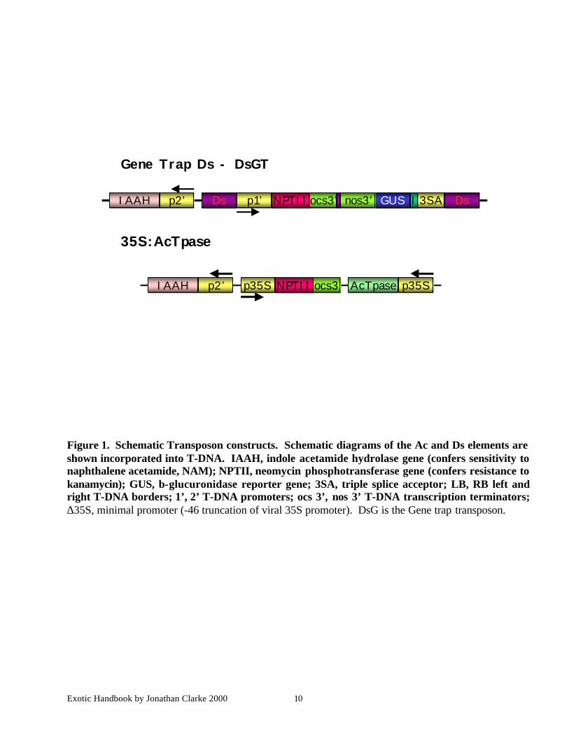
Gene trap reporter genes (promoter traps and exon traps) have no promoter, so that reporter geneexpression can occur only when the reporter gene inserts within a transcribed chromosomal gene,creating a transcriptional fusion. Our system uses gene trap reporters, and is based on the Maize Acand Ds transposable elements (Figure 1). The Ds elements carry the β-glucuronidase (GUS) gene as areporter and the Neomycin phosphotransferase (NPTII) gene (conferring resistance to kanamycin) as aselectable marker. The Ds element used in the gene trap has a multiple splice acceptor fused to theGUS gene. Random insertions of the Ds element throughout the genome allow us to detectchromosomal gene expression through the activation of the GUS gene.
Mutagenesis
Ds elements are transactivated by crossing to transgenic plants that provide a source of transposase,namely an immobilized Ac element (Figure 1). The Indole acetamide hydrolase (IAAH) gene has beenincorporated in the T-DNAs carrying both the Ds elements and the Ac element. Selection against theIAAH gene using the herbicide analog napthalene acetamide (NAM), and for the Ds element usingkanamycin allows the recovery of transposition events that have lost (by recombination) the donorlocus, and thereby enriches for unlinked transposition events. Since we also select against the Acelement, the insertion is immediately stabilized.
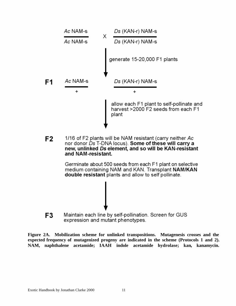
Mutagenesis is initiated by crossing plants homozygous for one of the Ds elements to plants containingthe Ac transposase gene (Protocol 1; Figure 2A & B). The resulting F1 seed are planted, and the plantsare allowed to self-pollinate. The F2 seed are harvested from each individual F1 plant, and plated onmedia containing kanamycin (selects for the Ds element) and NAM (selects against both T-DNAs)(Protocol 2). The double resistant F2 seedlings (called transposants) contain a transposed Ds element(Figure 3). The NamR KanR seedlings are transplanted to soil, and allowed to self-fertilize. F3 seed
Exotic Handbook by Jonathan Clarke 2000 6
are collected, and grown, and the resulting plants are stained for GUS activity and examined for mutantphenotypes.
For detailed methods see protocols1, 2 & 3.
Screening transposant lines
Transposant lines can be screened for mutant phenotypes caused by insertion, and for patterns ofreporter gene expression. In some cases, it may be beneficial to stain transposants first, and then usethe staining pattern to guide phenotypic examination. Although only a fraction of insertions into geneswill result in reporter gene expression, the remaining lines without reporter gene provide a source ofgene disruptions. Individual F2 plants may be either homozygous or heterozygous for the transposedDs element. Any phenotype associated with the insertion will be present in every F3 plant (in the caseof a F2 homozygote) or segregate 3:1 normal to mutant (if the F2 plant was heterozygous and themutation is recessive). Segregation ratios substantially more than 3:1 may indicate that the mutationarose after transposition and is not associated with the insertion.
Lethal insertions
Lethal insertions can be scored most conveniently in F2 transposants, by opening developing siliquesand scoring unfertilized ovules and colorless embryos. Lethal situations arise at a frequency of about4%. Lethals should be carefully tested for heritability and association with the Ds insertion by platingF3 seed on kanamycin (the resistance gene within the Ds element). If the insertion is responsible forthe lethal phenotype, all resistant F3 plants (which are heterozygous for the insertion) should give riseto plants with semi-sterile siliques or defective embryos. Lethality in the gametophyte or in theembryo should result in poor transmission of the kanamycin resistant trait, with ratios of between 1:1and 2:1 resistant to sensitive F3 seed.
Screening for reporter gene expression
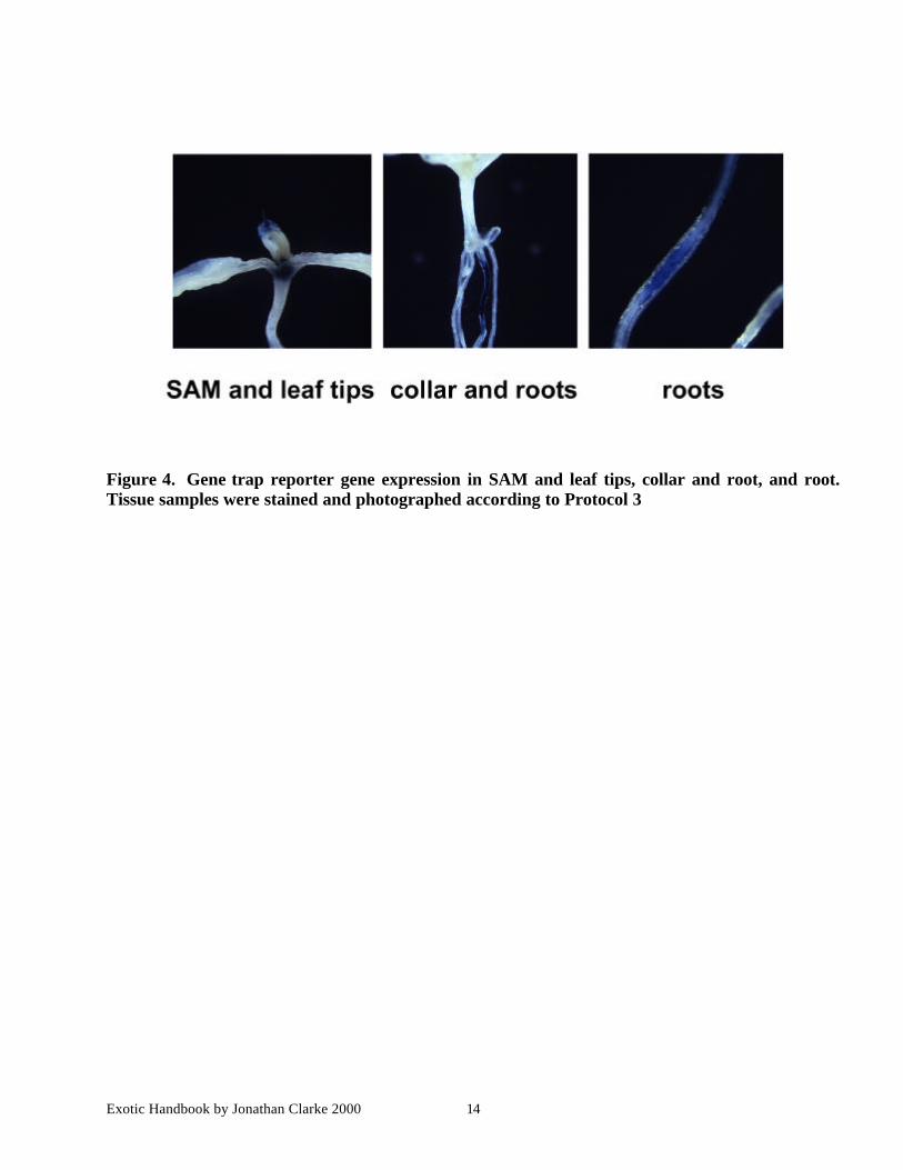
Screening for reporter gene expression patterns should be done wherever possible in heterozygousplants or in segregating families that include phenotypically normal individuals (Protocol 3). This isbecause homozygosity for the insertion can affect report gene expression patterns in many cases. It iswise to examine several individual plants, so staining in the F3 is recommended. Potassiumferricyanide is included in the staining reaction in order to catalyze dimerization of the indigomonomer, which is the product of the glucuronidase reaction. The colorless monomer is soluble, whilethe dimer is not, resulting in a blue precipitate at the site of enzyme activity. In the absence of at least1.5mM ferricyanide, diffusion of the indigo monomer results in artefactual staining patterns.Unfortunately, ferricyanide ions also inhibit the GUS enzyme at these concentrations. This inhibitioncan be partially overcome by using long incubation times for weaker staining patterns. Clearing of thetissue following staining is most simply and gently accomplished using 70% ethanol, which isadequate for all tissues. However, other more drastic clearing methods can be employed, but need tobe carefully controlled with respect to re-dissolving the stain and damaging the tissue. Cleared tissueis mounted in glycerol and viewed with Nomarski optics. An example of a line in which the reportergene is expressed in roots is shown in Figure 4.
Exotic Handbook by Jonathan Clarke 2000 7
Molecular analysis of transposants
Each transposant line that has a staining pattern or phenotype of interest can be analyzed usingmolecular methods to determine the location of the transposed element in the Arabidopsis genome.Flanking genomic DNA or cDNA corresponding to the gene nearest the insertion site can be rapidlyobtained by PCR amplification. The resulting products can be sequenced directly, or used ashybridization probes for further analysis.
Insertions into T-DNA
Although most insertions are randomly distributed around the genome, about 5 percent of transposantshave Ds insertions into the IAAH gene on the T-DNA. These insertions disrupt the IAAH generesulting in resistance to the negative marker NAM. IAAH insertions typically have strong ubiquitousreporter gene expression in seedling tissues and can often be discarded on the basis of this expressionpattern or on the basis of further molecular analysis (see below). However, if this is not possible,flanking DNA can be sequenced as described below.
Amplification of flanking genomic DNA
Chromosomal sequences flanking gene trap insertions can be amplified using I (inverse) PCR(Protocol 4A) and TAIL (thermal asymmetric Inter-laced) PCR (Protocol 4B) using standard protocolsand primers from the Ds element. These PCR products can be sequenced directly after purification onspin columns or gels by cycle sequencing using dye terminators. Each of these methods depends onthe fortuitous location of a primer sequence or restriction site close to the insertion, and consequentlyis only successful about 50% of the time for a given primer/enzyme combination. It is wise, therefore,to use several prime combinations or several approaches when attempting to amplify a given insertionsite.
Amplification of cDNA from gene trap fusions
Gene trap insertions result in transcriptional fusions between the reporter gene and the chromosomalgene into which it is inserted. Consequently, flanking sequences can be amplified by 5’RACE PCRusing RNA isolated from the gene trap line (Protocol 5). This is useful when the transposon is insertedinto a large intron, or when multiple introns make chromosomal sequence hard to interpret. RACEPCR products can also be sequenced directly, except that alternate splicing will result in mixedsequence reads in many cases. In these cases, sub-cloning will be required.
In either case, it is wise to confirm the assignment of a given sequence to a give line by hybridizationof the products to genomic Southern blots, or by PCR using specific primers derived from thesequence.
Exotic Handbook by Jonathan Clarke 2000 8
Genetic analysis of transposants
In many cases, insertion of a transposon will be associated with a mutant phenotype. Spontaneousmutants arise at a surprisingly high frequency in transposon lines, and it is important to determinewhether the transposon is responsible for any observed phenotype.
• First, the insertion should be mapped genetically, relative to the mutant phenotype and to othergenetic and molecular markers.
• Second, the transposon at the locus in question should be re-mobilized. The ability to remobilizetransposons so that they leave one locus and re-insert elsewhere allows the construction of theallelic series.
The most important application of the second approach is in the analysis of revertants. The restorationof a wild-type phenotype when the transposon excises provides strong evidence that the transposonwas responsible for the mutation. A second application is in the disruption of nearby genes. Theseprocedures are described below.
Mapping
Mapping of transposed elements can be most readily accomplished by amplifying flanking DNA (seeprotocol 4A & B) and aligning with the genomic sequence of Arabidopsis by BLAST searches. If noalignment is identified the resulting PCR product may be used as a probes to hybridize to anchoredlibraries, or to Southern blots of DNA from mapping populations such as recombinant inbred lines.
Phenotypically, the DsG transposons each carry a kanamycin-resistance gene. This means that eachinsertion can be mapped with respect to any associated mutant genotype, as well as to previouslymapped phenotypic and molecular markers. Plants heterozygous for the insertion are outcrossed towild-type plants, and F1 progeny are sowed to self-pollinate. F2 families are sown once to screen forany mutant phenotype, and again to screen for kanamycin resistance. Mutations that are caused by theinsertion will only be found in F2 families with kanamycin resistant progeny. If some kanamycinresistant families have no mutant progeny, this might indicate poor penetrance of the mutation, or thepresence of a second insertion elsewhere in the genome. If the insertion causes a lethal mutation, theratio of kanamycin resistant to kanamycin sensitive seedlings should be less than 3:1 on self-pollination of a heterozygous F1 plant. The ratio will be 2:1 for an embryo lethal, and 1:1 for agametophyte lethal.
By using a wild-type parent from a different ecotype (Columbia), F2 seed can be used to map theinsertion. This is accomplished by preparing DNA from pooled F2 seedlings from kanamycin resistantand kanamycin sensitive families, and screening the DNA samples with PCR-based polymorphicmarkers.
Reversion
Insertion of a Ds transposon results in the duplication of 8 bp of target sequence immediately flankingthe insertion site. When Ds excises, the target duplication is partially removed, resulting in small
Exotic Handbook by Jonathan Clarke 2000 9
insertions and deletions at the original locus. If the Ds is inserted into the coding region of the gene,only those events that restore the reading frame and result in a functional protein will revert the mutantphenotype back to wild-type. In contrast, almost all reversions from non-essential sequences such asintrons will result in reversion of the mutant phenotype.
Reversion is accomplished by crossing mutant plants to transgenic plants that carry the transposasegene (Protocol 6). The resulting F1 plants are then planted and allowed to self-pollinate. The F2progeny will now include mutants that carry the transposase gene. The transposon responsible for themutant phenotype will excise in these plants resulting in somatic sectors of tissue that have lost thetransposon. If these plants are mosaics for the mutant phenotype, this is good evidence that thephenotype can be reverted by transposase. More importantly, a proportion of the F3 progeny of theseplants should be wild-type, in contrast to the progeny of mutants that do not carry the transposase gene,which should be true-breeding mutant. Revertant alleles can be amplified from wild-type progenyusing primers that flank the insertion site. These products can then be sequence to determine thenature of the reversion event. In the special case of lethal mutations, reversion can be observed in theF1 plants themselves. Most of the siliques on these plants should be semi-sterile, or carry dead seed,depending on whether the mutation is lethal at the gametophytic or the embryonic stage. However,reversion early in development will result in normal, fully fertile siliques provided reversion occursearly enough to detect revertant branches.
Reinsertion
There are many circumstances when reinsertion of a transposon by short-range transposition isadvantageous. For example, gene trap insertions may not disrupt gene function, as insertions withinintrons may be spliced from the RNA transcript without phenotypic effect. In these cases, it can beuseful to obtain a secondary insertion into the nearest exon by inducing a short-range transposition. Ahigh proportion of transpositions of Ds are to closely linked sites when these transpositions are notcounter-selected (see introduction). A protocol for re-mobilization is given below.
In brief, the transposon is re-mobilized by crossing to transposase, and is then stabilized by selectingagainst the transposase gene in the next generation (Figure 4). The parental transposon is not selectedagainst by this procedure, and so a large proportion (more than half) of the resulting plants will stillhave the transposon inserted at the original location. Those plants that have new insertions thereforeneed to be identified molecularly, phenotypically, or by staining for reporter gene expressions. In ourexperience, a collection of 2,000 plants selected in this manner will carry between 500 and 1,000 newtranspositions. About 20% of these will be within 100kb. This should be sufficient to saturate thenearby region with new insertions (C. Yordan and R. Martienssen, unpubl. Observations).
Exotic Handbook by Jonathan Clarke 2000 10
35S:AcTpase
Gene Trap Ds - DsGT
AcTpaseIAAH p35S ocs3’NPTIIp2’ p35S
IAAH Ds 3SAGUS Ip2’ p1’ ocs3’NPTII nos3’ Ds
Figure 1. Schematic Transposon constructs. Schematic diagrams of the Ac and Ds elements areshown incorporated into T-DNA. IAAH, indole acetamide hydrolase gene (confers sensitivity tonaphthalene acetamide, NAM); NPTII, neomycin phosphotransferase gene (confers resistance tokanamycin); GUS, b-glucuronidase reporter gene; 3SA, triple splice acceptor; LB, RB left andright T-DNA borders; 1’, 2’ T-DNA promoters; ocs 3’, nos 3’ T-DNA transcription terminators;∆35S, minimal promoter (-46 truncation of viral 35S promoter). DsG is the Gene trap transposon.
Exotic Handbook by Jonathan Clarke 2000 11
Figure 2A. Mobilization scheme for unlinked transpositions. Mutagenesis crosses and theexpected frequency of mutagenized progeny are indicated in the scheme (Protocols 1 and 2).NAM, naphthalene acetamide; IAAH indole acetamide hydrolase; kan, kanamycin.
Exotic Handbook by Jonathan Clarke 2000 12
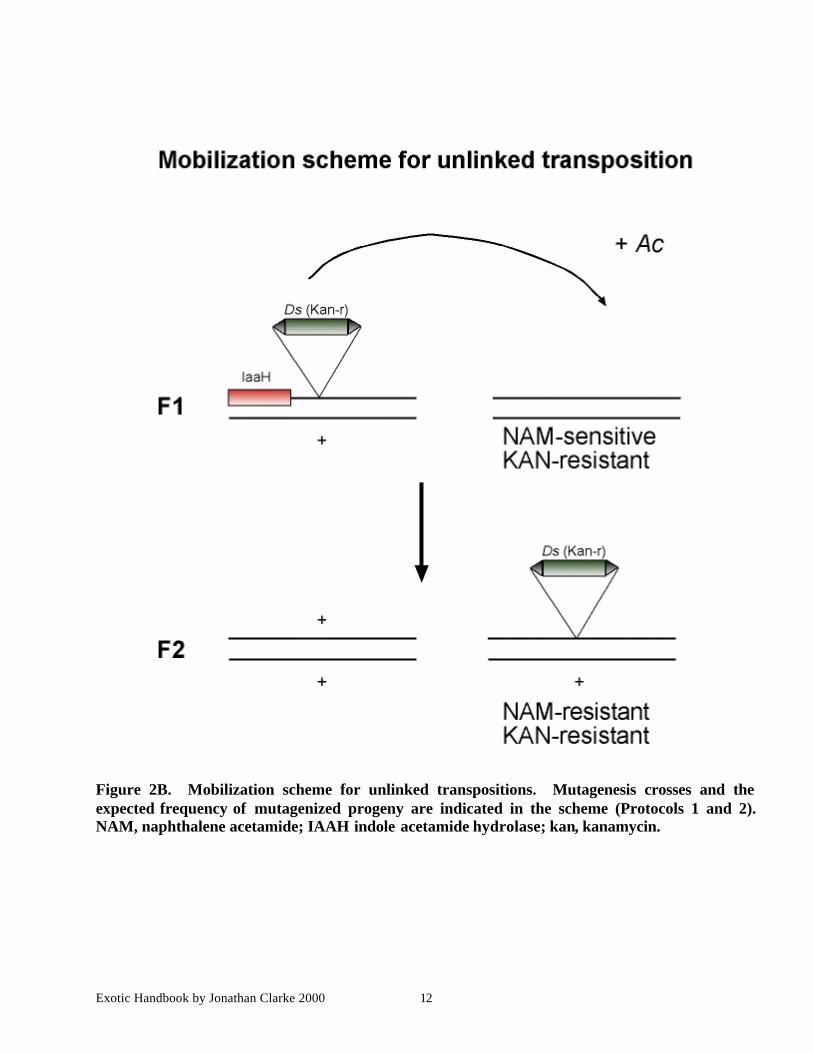
Figure 2B. Mobilization scheme for unlinked transpositions. Mutagenesis crosses and theexpected frequency of mutagenized progeny are indicated in the scheme (Protocols 1 and 2).NAM, naphthalene acetamide; IAAH indole acetamide hydrolase; kan, kanamycin.
Exotic Handbook by Jonathan Clarke 2000 13
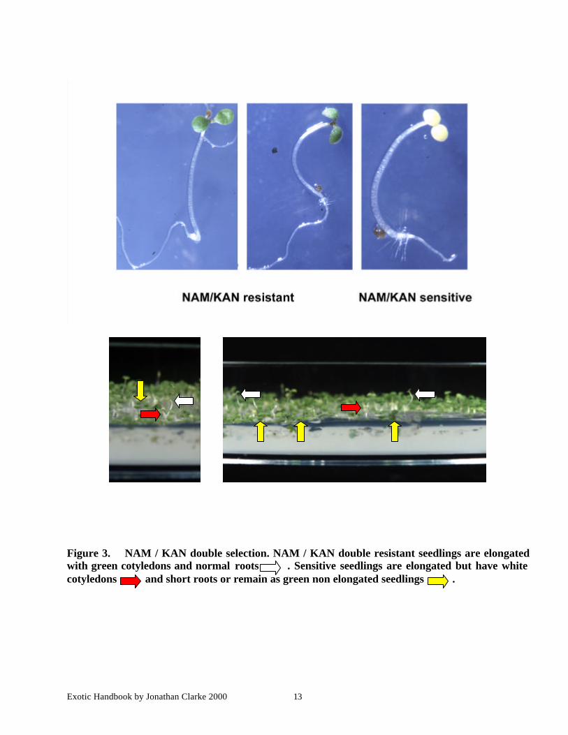
Figure 3. NAM / KAN double selection. NAM / KAN double resistant seedlings are elongatedwith green cotyledons and normal roots . Sensitive seedlings are elongated but have whitecotyledons and short roots or remain as green non elongated seedlings .
Exotic Handbook by Jonathan Clarke 2000 14
Figure 4. Gene trap reporter gene expression in SAM and leaf tips, collar and root, and root.Tissue samples were stained and photographed according to Protocol 3
Exotic Handbook by Jonathan Clarke 2000 15
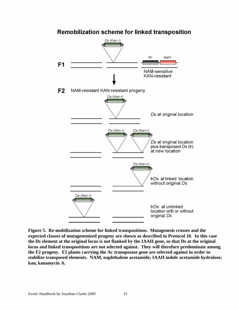
Figure 5. Re-mobilization scheme for linked transpositions. Mutagenesis crosses and theexpected classes of mutagenenized progeny are shown as described in Protocol 10. In this casethe Ds element at the original locus is not flanked by the IAAH gene, so that Ds at the originallocus and linked transpositions are not selected against. They will therefore predominate amongthe F2 progeny. F2 plants carrying the Ac transposase gene are selected against in order tostabilize transposed elements. NAM, naphthalene acetamide; IAAH indole acetamide hydrolase;kan, kanamycin A.
Exotic Handbook by Jonathan Clarke 2000 16
Protocol 1A. Growth of Arabidopsis
Equipment and Reagents
• Greenhouse or plant growth cabinets• Arabidopsis soil mix JI#A: 150 l Intercept® (Levingtons) compost with 54.4 l fine grit (small
volume) or 600 l Intercept® compost with 217.6 l fine grit (large volume). Transfer the compost to60 cell trays from PlantPac (horticultural suppliers) and firm in gently and top up. Immediatelybefore use water trays then drench with Nemasys® (nematode) (BioCentre).
• 0.15% Agar: 150 mg Micro Agar (Duchfuca) in 1 l dH20, autoclave sterilized.• 15ml or 50 ml Falcon tubes• Multi-dispenser (100µl aliquots from 6ml)
High through-put sowing, plant growth and harvesting
1. Weigh Arabidopsis seed to determine required seed number (100/2.5 mg) and place in a Falcontube of the required volume.
2. Calculate volume of agar solution require to give 1 seed/100µl eg. 60 seed requires 6 ml.3. Add the required volume of 0.15% agar solution to the falcon tube containing the seed and mix
by inversion.4. Chill the seed/agar mix at 4°C for 4 days to break dormancy.5. Cut the tip from a multi-dispenser tip to prevent blockage when dispensing the seed/ agar mix.
Re-shake the tube to mix the seed evenly. Draw up the seed/agar mix into the multi-dispenser.Set the dispense volume to 100 µl and dispense 100 µl aliquots into each cell of the seed tray.
6. Cover the trays with horticulture fleece; recheck for watering each day. Start to lift the fleecefor short periods to allow air circulation at day 3.
7. At day 7 thin/prick out seedlings covering with the fleece for 2 days during weaning.8. At day 10 tube the plants and start bottom watering.9. Plants will generally have completed flowering after 3 months. Start to reduce the watering
after 2.5 months to stimulate seed set. Stop watering when approximately 50% of the sliquesare brown. Fully dry the plants before harvest.
Seed processing/threshing
10. Ensure the seed have fully dried before threshing.11. Run your hand over the seed bag against a bench to open the sliques (brushing).12. Shake the seed into one corner of the bag.13. Place two tea strainers, one inside the other over the mouth of a new seed bag. Cut off the
corner of the seed bag containing the seed and empty the seed through the tea strainer into thenew seed bag (threshing). This will remove sufficient chaff to provide seed suitable forsterilization prior to in-vitro work.
Exotic Handbook by Jonathan Clarke 2000 17
Protocol 1B. Mutagenesis
Equipment and Reagents
• Fine pointed forceps• Dissecting microscope with fiber optic light source• Greenhouse or plant growth cabinets and soil
Generation of F1 and F2 seed
1. Sow parental seed homozygous for the transposase gene in individual 2” pots and grow underoptimal conditions: 16 hour days are recommended. Plant homozygous Ds seed one weeklater.
2. Emasculate the five buds from each Ds parent (before pollen is shed) and remove the remainingbuds. Up to 60 plants may be prepared in a day. Leave each flower overnight beforepollination with homozygous transposase pollen. Cover the fertilized buds with a small squareof cling film to prevent loss of seed upon silique shattering. Remove any unfertilized buds, andmark the branch. Over the next 2 weeks remove new flowers and branches.
3. Collect the seed approximately 2 weeks later, just before the siliques open.4. Plant the F1 seed individually with a damp spatula on moistened compost in the greenhouse.
On bolting, provide each plant with a small plastic or wire stake, and a 12” polythene collectiontube to avoid seed contamination.
5. Harvest 5-10,000 F2 seed from each F1 plant. It is convenient to work in batches of 2-4,000 F1plants at a time. Avoid seed contamination.
Starter lines
Rob Martienssen’s lab have provided four DsG starter lines and four Ac starter lines:DsG1 (3734-1) Ac1 (4345-1)DsG6 (4224-1) Ac2 (4562-9)DsG7 (1536-9) Ac4 (3680-2)DsG8 (4225-1) Ac5 (4347-4)
For optimal results these should be crossed in the following combinations:1. DsG1 (3734-1) X Ac4 (3680-2)2. DsG6 (4224-1) X Ac4 (3680-2)3. DsG6 (4224-1) X Ac1 (4345-1)4. DsG7 (1536-9) X Ac2 (4562-9)5. DsG8 (4225-1) X Ac4 (3680-2)6. DsG8 (4225-1) X Ac5 (4347-4)
Exotic Handbook by Jonathan Clarke 2000 18
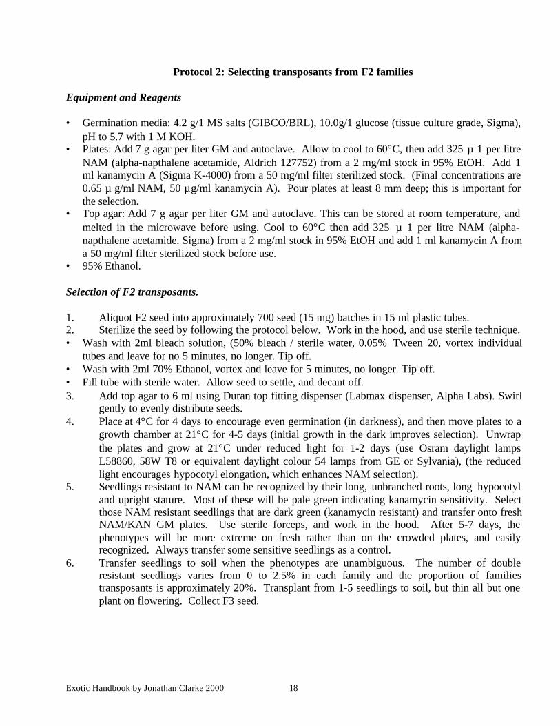
Protocol 2: Selecting transposants from F2 families
Equipment and Reagents
• Germination media: 4.2 g/1 MS salts (GIBCO/BRL), 10.0g/1 glucose (tissue culture grade, Sigma),pH to 5.7 with 1 M KOH.
• Plates: Add 7 g agar per liter GM and autoclave. Allow to cool to 60°C, then add 325 µ 1 per litreNAM (alpha-napthalene acetamide, Aldrich 127752) from a 2 mg/ml stock in 95% EtOH. Add 1ml kanamycin A (Sigma K-4000) from a 50 mg/ml filter sterilized stock. (Final concentrations are0.65 µ g/ml NAM, 50 µg/ml kanamycin A). Pour plates at least 8 mm deep; this is important forthe selection.
• Top agar: Add 7 g agar per liter GM and autoclave. This can be stored at room temperature, andmelted in the microwave before using. Cool to 60°C then add 325 µ 1 per litre NAM (alpha-napthalene acetamide, Sigma) from a 2 mg/ml stock in 95% EtOH and add 1 ml kanamycin A froma 50 mg/ml filter sterilized stock before use.
• 95% Ethanol.
Selection of F2 transposants.
1. Aliquot F2 seed into approximately 700 seed (15 mg) batches in 15 ml plastic tubes.2. Sterilize the seed by following the protocol below. Work in the hood, and use sterile technique.• Wash with 2ml bleach solution, (50% bleach / sterile water, 0.05% Tween 20, vortex individual
tubes and leave for no 5 minutes, no longer. Tip off.• Wash with 2ml 70% Ethanol, vortex and leave for 5 minutes, no longer. Tip off.• Fill tube with sterile water. Allow seed to settle, and decant off.3. Add top agar to 6 ml using Duran top fitting dispenser (Labmax dispenser, Alpha Labs). Swirl
gently to evenly distribute seeds.4. Place at 4°C for 4 days to encourage even germination (in darkness), and then move plates to a
growth chamber at 21°C for 4-5 days (initial growth in the dark improves selection). Unwrapthe plates and grow at 21°C under reduced light for 1-2 days (use Osram daylight lampsL58860, 58W T8 or equivalent daylight colour 54 lamps from GE or Sylvania), (the reducedlight encourages hypocotyl elongation, which enhances NAM selection).
5. Seedlings resistant to NAM can be recognized by their long, unbranched roots, long hypocotyland upright stature. Most of these will be pale green indicating kanamycin sensitivity. Selectthose NAM resistant seedlings that are dark green (kanamycin resistant) and transfer onto freshNAM/KAN GM plates. Use sterile forceps, and work in the hood. After 5-7 days, thephenotypes will be more extreme on fresh rather than on the crowded plates, and easilyrecognized. Always transfer some sensitive seedlings as a control.
6. Transfer seedlings to soil when the phenotypes are unambiguous. The number of doubleresistant seedlings varies from 0 to 2.5% in each family and the proportion of familiestransposants is approximately 20%. Transplant from 1-5 seedlings to soil, but thin all but oneplant on flowering. Collect F3 seed.
Exotic Handbook by Jonathan Clarke 2000 19
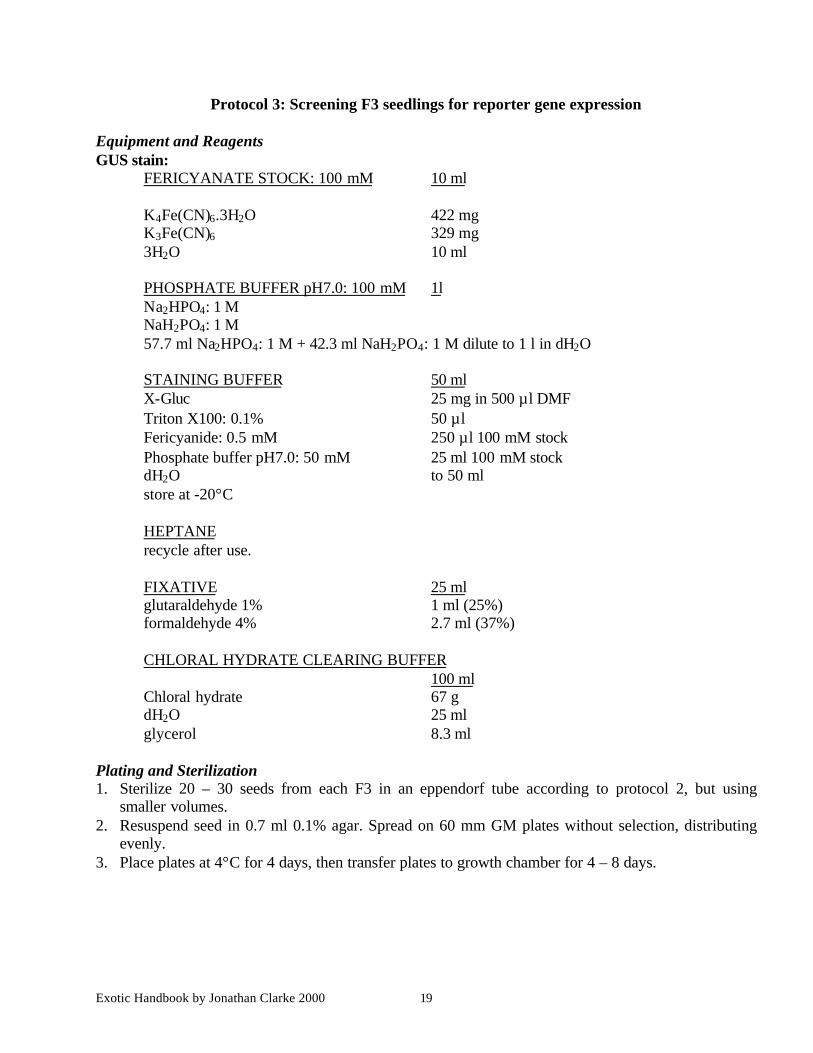
Protocol 3: Screening F3 seedlings for reporter gene expression
Equipment and ReagentsGUS stain:
FERICYANATE STOCK: 100 mM 10 ml
K4Fe(CN)6.3H2O 422 mgK3Fe(CN)6 329 mg3H2O 10 ml
PHOSPHATE BUFFER pH7.0: 100 mM 1lNa2HPO4: 1 MNaH2PO4: 1 M57.7 ml Na2HPO4: 1 M + 42.3 ml NaH2PO4: 1 M dilute to 1 l in dH2O
STAINING BUFFER 50 mlX-Gluc 25 mg in 500 µl DMFTriton X100: 0.1% 50 µlFericyanide: 0.5 mM 250 µl 100 mM stockPhosphate buffer pH7.0: 50 mM 25 ml 100 mM stockdH2O to 50 mlstore at -20°C
HEPTANErecycle after use.
FIXATIVE 25 mlglutaraldehyde 1% 1 ml (25%)formaldehyde 4% 2.7 ml (37%)
CHLORAL HYDRATE CLEARING BUFFER100 ml
Chloral hydrate 67 gdH2O 25 mlglycerol 8.3 ml
Plating and Sterilization1. Sterilize 20 – 30 seeds from each F3 in an eppendorf tube according to protocol 2, but using
smaller volumes.2. Resuspend seed in 0.7 ml 0.1% agar. Spread on 60 mm GM plates without selection, distributing
evenly.3. Place plates at 4°C for 4 days, then transfer plates to growth chamber for 4 – 8 days.

Exotic Handbook by Jonathan Clarke 2000 20
Staining procedure
1. Dissect out tissues for staining. Place tissue in a multi-compartment petri dish. Add 1 – 2 ml 100%heptane for 10 min. Remove heptane (& recycle); air dry for 5 min.
2. Add 1 ml of X-Gluc solution. Place in a vacuum dessicator and draw vacuum for 10 min, thenincubate at 37°C. Run a time course for staining at 30 min intervals for 5 h and o/n. Remove thestaining solution.
3. Post staining fixation: add 1 – 2 ml of 1% glutaraldehyde / 4% formaldehyde. Place in a vacuumdessicator and draw vacuum for 10 min then fix o/n at 4°C. Tissue may now be stored forembedding or cleared for whole mounts.
4. Clear in 95% ethanol for 1 h then 70% ethanol for 2 h for routine screening. For more detailedanalysis clear with chloral hydrate solution for 1 h. Replace the clearing solution with fresh chloralhydrate solution, mount on a slide in the clearing solution. Apply a cover slip and view usingNomarski optics.
Exotic Handbook by Jonathan Clarke 2000 21
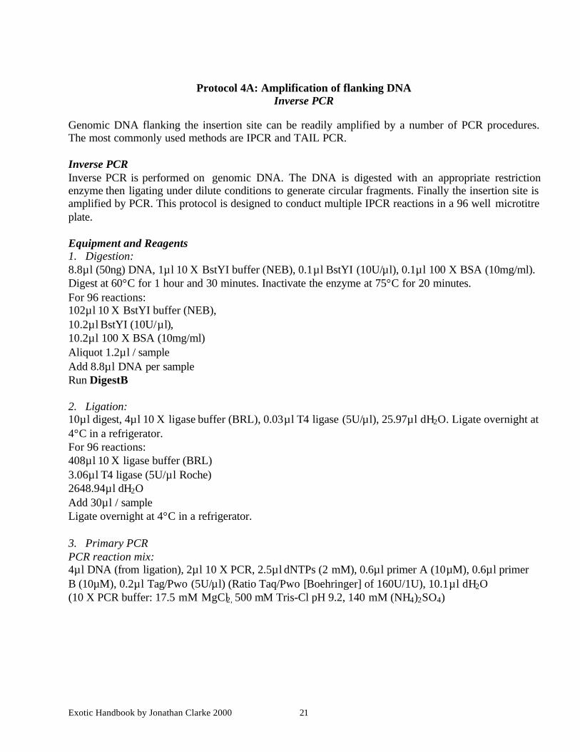
Protocol 4A: Amplification of flanking DNAInverse PCR
Genomic DNA flanking the insertion site can be readily amplified by a number of PCR procedures.The most commonly used methods are IPCR and TAIL PCR.
Inverse PCRInverse PCR is performed on genomic DNA. The DNA is digested with an appropriate restrictionenzyme then ligating under dilute conditions to generate circular fragments. Finally the insertion site isamplified by PCR. This protocol is designed to conduct multiple IPCR reactions in a 96 well microtitreplate.
Equipment and Reagents1. Digestion:8.8µl (50ng) DNA, 1µl 10 X BstYI buffer (NEB), 0.1µl BstYI (10U/µl), 0.1µl 100 X BSA (10mg/ml).Digest at 60°C for 1 hour and 30 minutes. Inactivate the enzyme at 75°C for 20 minutes.For 96 reactions:102µl 10 X BstYI buffer (NEB),10.2µl BstYI (10U/µl),10.2µl 100 X BSA (10mg/ml)Aliquot 1.2µl / sampleAdd 8.8µl DNA per sampleRun DigestB
2. Ligation:10µl digest, 4µl 10 X ligase buffer (BRL), 0.03µl T4 ligase (5U/µl), 25.97µl dH2O. Ligate overnight at4°C in a refrigerator.For 96 reactions:408µl 10 X ligase buffer (BRL)3.06µl T4 ligase (5U/µl Roche)2648.94µl dH2OAdd 30µl / sampleLigate overnight at 4°C in a refrigerator.
3. Primary PCRPCR reaction mix:4µl DNA (from ligation), 2µl 10 X PCR, 2.5µl dNTPs (2 mM), 0.6µl primer A (10µM), 0.6µl primerB (10µM), 0.2µl Tag/Pwo (5U/µl) (Ratio Taq/Pwo [Boehringer] of 160U/1U), 10.1µl dH2O(10 X PCR buffer: 17.5 mM MgCl2, 500 mM Tris-Cl pH 9.2, 140 mM (NH4)2SO4)
Exotic Handbook by Jonathan Clarke 2000 22
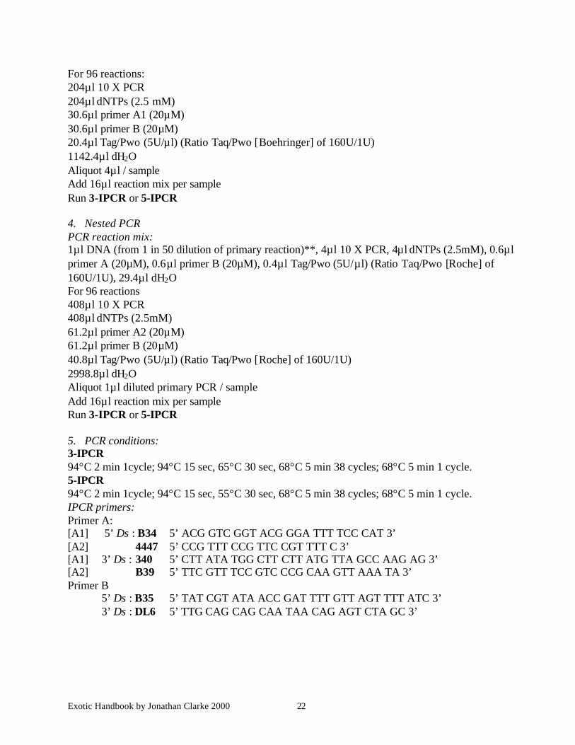
For 96 reactions:204µl 10 X PCR204µl dNTPs (2.5 mM)30.6µl primer A1 (20µM)30.6µl primer B (20µM)20.4µl Tag/Pwo (5U/µl) (Ratio Taq/Pwo [Boehringer] of 160U/1U)1142.4µl dH2OAliquot 4µl / sampleAdd 16µl reaction mix per sampleRun 3-IPCR or 5-IPCR
4. Nested PCRPCR reaction mix:1µl DNA (from 1 in 50 dilution of primary reaction)**, 4µl 10 X PCR, 4µl dNTPs (2.5mM), 0.6µlprimer A (20µM), 0.6µl primer B (20µM), 0.4µl Tag/Pwo (5U/µl) (Ratio Taq/Pwo [Roche] of160U/1U), 29.4µl dH2OFor 96 reactions408µl 10 X PCR408µl dNTPs (2.5mM)61.2µl primer A2 (20µM)61.2µl primer B (20µM)40.8µl Tag/Pwo (5U/µl) (Ratio Taq/Pwo [Roche] of 160U/1U)2998.8µl dH2OAliquot 1µl diluted primary PCR / sampleAdd 16µl reaction mix per sampleRun 3-IPCR or 5-IPCR
5. PCR conditions:3-IPCR94°C 2 min 1cycle; 94°C 15 sec, 65°C 30 sec, 68°C 5 min 38 cycles; 68°C 5 min 1 cycle.5-IPCR94°C 2 min 1cycle; 94°C 15 sec, 55°C 30 sec, 68°C 5 min 38 cycles; 68°C 5 min 1 cycle.IPCR primers:Primer A:[A1] 5’ Ds : B34 5’ ACG GTC GGT ACG GGA TTT TCC CAT 3’[A2] 4447 5’ CCG TTT CCG TTC CGT TTT C 3’[A1] 3’ Ds : 340 5’ CTT ATA TGG CTT CTT ATG TTA GCC AAG AG 3’[A2] B39 5’ TTC GTT TCC GTC CCG CAA GTT AAA TA 3’Primer B
5’ Ds : B35 5’ TAT CGT ATA ACC GAT TTT GTT AGT TTT ATC 3’3’ Ds : DL6 5’ TTG CAG CAG CAA TAA CAG AGT CTA GC 3’

Exotic Handbook by Jonathan Clarke 2000 23
6. 1.2% Gel:Run 8µl of 3° amplification products on a 1.2% gel to check for fragments. If thereaction is successful there will be 50-100ng of product in each lane. Multiple productsshould be anchored by the Ds primer and should not confuse sequencing reactions.
7. Product Purification:Purify the remaining 22µl of successful 3° products by running over a Qiaquick PCRpurification kit to remove primers and nucleotides.
Add 100µl of PB buffer (5x vol) and mixLoad onto columns and spinWash 2 x with 900µl PE and spinPlace columns onto collection tubes and elute with 60µl EBStand for 1 minute then spin
8. Sequencing:Sequencing directly using 5-10µl (8µl) of the 3° product as template using dyeterminator chemistry and primer B39 or 4447.
NOTES** Always spin plates after mixing dilutions
Exotic Handbook by Jonathan Clarke 2000 24
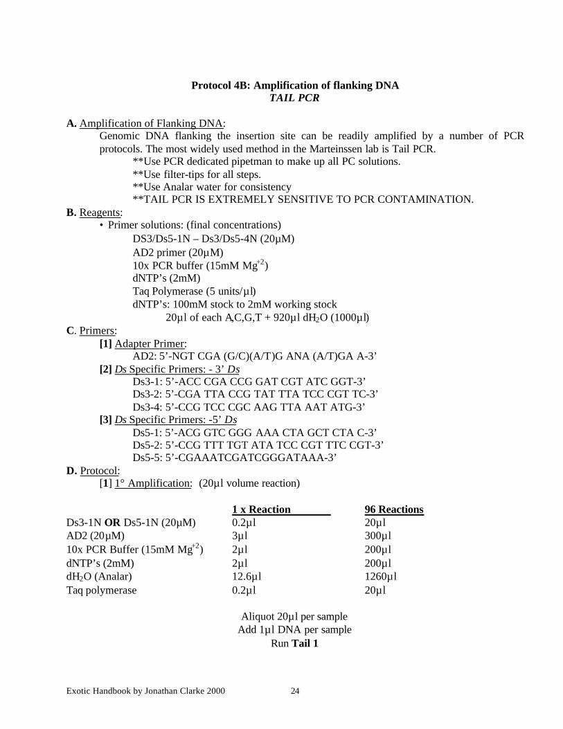
Protocol 4B: Amplification of flanking DNATAIL PCR
A. Amplification of Flanking DNA:Genomic DNA flanking the insertion site can be readily amplified by a number of PCRprotocols. The most widely used method in the Marteinssen lab is Tail PCR.
**Use PCR dedicated pipetman to make up all PC solutions.**Use filter-tips for all steps.**Use Analar water for consistency**TAIL PCR IS EXTREMELY SENSITIVE TO PCR CONTAMINATION.
B. Reagents:• Primer solutions: (final concentrations)
DS3/Ds5-1N – Ds3/Ds5-4N (20µM)AD2 primer (20µM)10x PCR buffer (15mM Mg+2)dNTP’s (2mM)Taq Polymerase (5 units/µl)dNTP’s: 100mM stock to 2mM working stock
20µl of each A,C,G,T + 920µl dH2O (1000µl)C. Primers:
[1] Adapter Primer:AD2: 5’-NGT CGA (G/C)(A/T)G ANA (A/T)GA A-3’
[2] Ds Specific Primers: - 3’ DsDs3-1: 5’-ACC CGA CCG GAT CGT ATC GGT-3’Ds3-2: 5’-CGA TTA CCG TAT TTA TCC CGT TC-3’Ds3-4: 5’-CCG TCC CGC AAG TTA AAT ATG-3’
[3] Ds Specific Primers: -5’ DsDs5-1: 5’-ACG GTC GGG AAA CTA GCT CTA C-3’Ds5-2: 5’-CCG TTT TGT ATA TCC CGT TTC CGT-3’Ds5-5: 5’-CGAAATCGATCGGGATAAA-3’
D. Protocol:[1] 1° Amplification: (20µl volume reaction)
1 x Reaction 96 ReactionsDs3-1N OR Ds5-1N (20µM) 0.2µl 20µlAD2 (20µM) 3µl 300µl10x PCR Buffer (15mM Mg+2) 2µl 200µldNTP’s (2mM) 2µl 200µldH2O (Analar) 12.6µl 1260µlTaq polymerase 0.2µl 20µl
Aliquot 20µl per sampleAdd 1µl DNA per sample
Run Tail 1
Exotic Handbook by Jonathan Clarke 2000 25
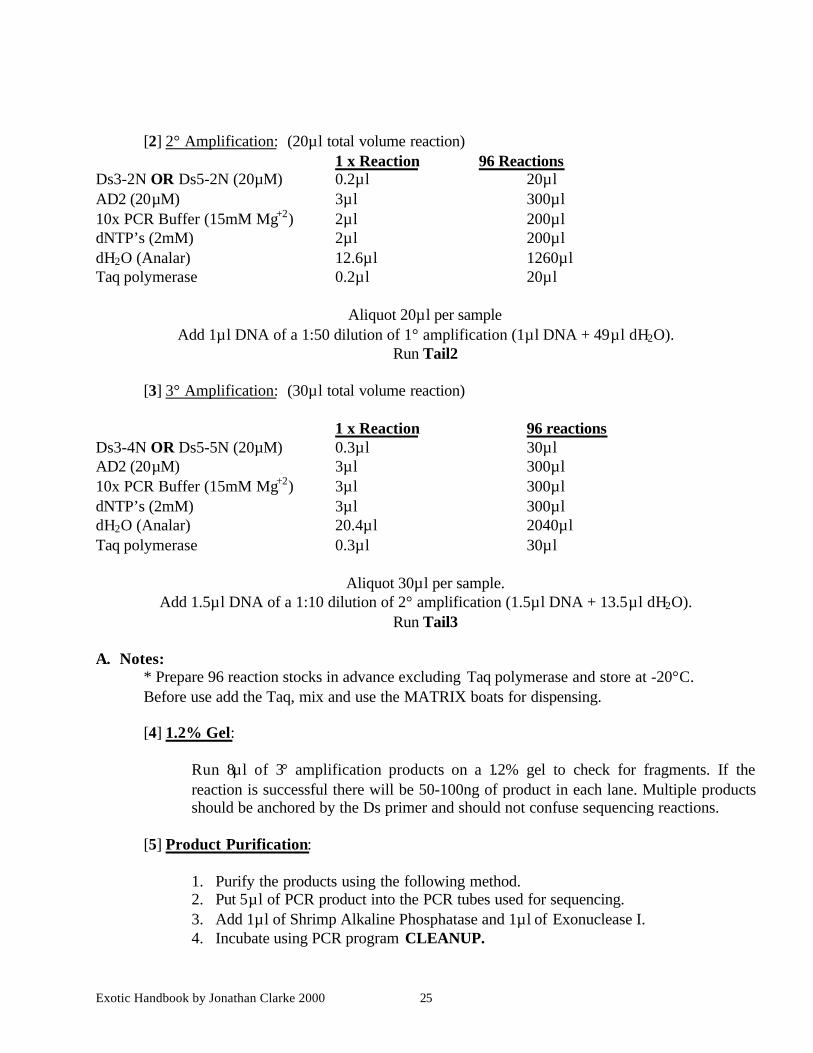
[2] 2° Amplification: (20µl total volume reaction)1 x Reaction 96 Reactions
Ds3-2N OR Ds5-2N (20µM) 0.2µl 20µlAD2 (20µM) 3µl 300µl10x PCR Buffer (15mM Mg+2) 2µl 200µldNTP’s (2mM) 2µl 200µldH2O (Analar) 12.6µl 1260µlTaq polymerase 0.2µl 20µl
Aliquot 20µl per sampleAdd 1µl DNA of a 1:50 dilution of 1° amplification (1µl DNA + 49µl dH2O).
Run Tail2
[3] 3° Amplification: (30µl total volume reaction)
1 x Reaction 96 reactionsDs3-4N OR Ds5-5N (20µM) 0.3µl 30µlAD2 (20µM) 3µl 300µl10x PCR Buffer (15mM Mg+2) 3µl 300µldNTP’s (2mM) 3µl 300µldH2O (Analar) 20.4µl 2040µlTaq polymerase 0.3µl 30µl
Aliquot 30µl per sample.Add 1.5µl DNA of a 1:10 dilution of 2° amplification (1.5µl DNA + 13.5µl dH2O).
Run Tail3
A. Notes:* Prepare 96 reaction stocks in advance excluding Taq polymerase and store at -20°C.Before use add the Taq, mix and use the MATRIX boats for dispensing.
[4] 1.2% Gel:
Run 8µl of 3° amplification products on a 1.2% gel to check for fragments. If thereaction is successful there will be 50-100ng of product in each lane. Multiple productsshould be anchored by the Ds primer and should not confuse sequencing reactions.
[5] Product Purification:
1. Purify the products using the following method.2. Put 5µl of PCR product into the PCR tubes used for sequencing.3. Add 1µl of Shrimp Alkaline Phosphatase and 1µl of Exonuclease I.4. Incubate using PCR program CLEANUP.
Exotic Handbook by Jonathan Clarke 2000 26
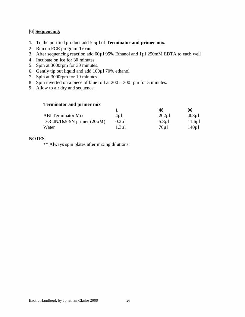
[6] Sequencing:
1. To the purified product add 5.5µl of Terminator and primer mix.2. Run on PCR program Term.3. After sequencing reaction add 60µl 95% Ethanol and 1µl 250mM EDTA to each well4. Incubate on ice for 30 minutes.5. Spin at 3000rpm for 30 minutes.6. Gently tip out liquid and add 100µl 70% ethanol7. Spin at 3000rpm for 10 minutes8. Spin inverted on a piece of blue roll at 200 – 300 rpm for 5 minutes.9. Allow to air dry and sequence.
Terminator and primer mix1 48 96
ABI Terminator Mix 4µl 202µl 403µlDs3-4N/Ds5-5N primer (20µM) 0.2µl 5.8µl 11.6µlWater 1.3µl 70µl 140µl
NOTES** Always spin plates after mixing dilutions

Exotic Handbook by Jonathan Clarke 2000 27
E. PCR Programmes
Tail1 Run time 4 hr 16 minutes
94°C for 1 minute94°C for 10 seconds62°C for 1 minute72°C for 2:30 minutes
Goto step 2, four times94°C for 10 seconds25°C for 3 minutes
ramp 0.2°C/s to 72°C72°C for 2:30 minutes94°C for 10 seconds68°C for 1 minute72°C for 2:30 minutes94°C for 10 seconds68°C for 1 minute72°C for 2:30 minutes94°C for 10 seconds44°C for 1 minute72°C for 2:30 minutes
Goto step 10, fourteen times72°C for 5 minutes4°C foreverEnd
Tail2 Run time 3 hr
94°C for 10 seconds64°C for 1 minute72°C for 2:30 minutes94°C for 10 seconds64°C for 1 minutes72°C for 2:30 minutes94°C for 10 seconds44°C for 1 minute72°C for 2:30 minutes
Goto step 1, eleven times72°C for 5 minutes4°C foreverEnd

Exotic Handbook by Jonathan Clarke 2000 28
Tail3 Run time 1 hr 52 minutes
94°C for 15 seconds44°C for 1 minute72°C for 2:30 minutes
Goto step 1, nineteen times72°C for 5 minutes4°C foreverEnd
CLEANUP
37°C for 30 mins80°C for 10 mins4°C ForeverEnd
Term
1.0°C/s to 96°C96°C for 2 minutes96°C for 10 seconds1.0°C/s to 50°C50°C for 5 seconds1.0°C/s to 60°C60°C for 4 minutesgoto step 3 twenty four times4°C foreverEnd
Exotic Handbook by Jonathan Clarke 2000 29
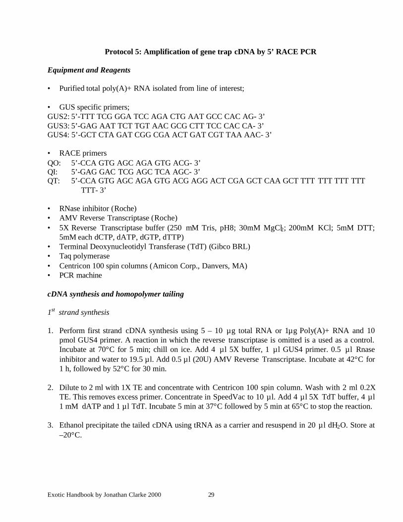
Protocol 5: Amplification of gene trap cDNA by 5’ RACE PCR
Equipment and Reagents
• Purified total poly(A)+ RNA isolated from line of interest;
• GUS specific primers;GUS2: 5’-TTT TCG GGA TCC AGA CTG AAT GCC CAC AG- 3’GUS3: 5’-GAG AAT TCT TGT AAC GCG CTT TCC CAC CA- 3’GUS4: 5’-GCT CTA GAT CGG CGA ACT GAT CGT TAA AAC- 3’
• RACE primersQO: 5’-CCA GTG AGC AGA GTG ACG- 3’QI: 5’-GAG GAC TCG AGC TCA AGC- 3’QT: 5’-CCA GTG AGC AGA GTG ACG AGG ACT CGA GCT CAA GCT TTT TTT TTT TTT
TTT- 3’
• RNase inhibitor (Roche)• AMV Reverse Transcriptase (Roche)• 5X Reverse Transcriptase buffer (250 mM Tris, pH8; 30mM MgCl2; 200mM KCl; 5mM DTT;
5mM each dCTP, dATP, dGTP, dTTP)• Terminal Deoxynucleotidyl Transferase (TdT) (Gibco BRL)• Taq polymerase• Centricon 100 spin columns (Amicon Corp., Danvers, MA)• PCR machine
cDNA synthesis and homopolymer tailing
1st strand synthesis
1. Perform first strand cDNA synthesis using 5 – 10 µg total RNA or 1µg Poly(A)+ RNA and 10pmol GUS4 primer. A reaction in which the reverse transcriptase is omitted is a used as a control.Incubate at 70°C for 5 min; chill on ice. Add 4 µl 5X buffer, 1 µl GUS4 primer. 0.5 µl Rnaseinhibitor and water to 19.5 µl. Add 0.5 µl (20U) AMV Reverse Transcriptase. Incubate at 42°C for1 h, followed by 52°C for 30 min.
2. Dilute to 2 ml with 1X TE and concentrate with Centricon 100 spin column. Wash with 2 ml 0.2XTE. This removes excess primer. Concentrate in SpeedVac to 10 µl. Add 4 µl 5X TdT buffer, 4 µl1 mM dATP and 1 µl TdT. Incubate 5 min at 37°C followed by 5 min at 65°C to stop the reaction.
3. Ethanol precipitate the tailed cDNA using tRNA as a carrier and resuspend in 20 µl dH2O. Store at–20°C.
Exotic Handbook by Jonathan Clarke 2000 30
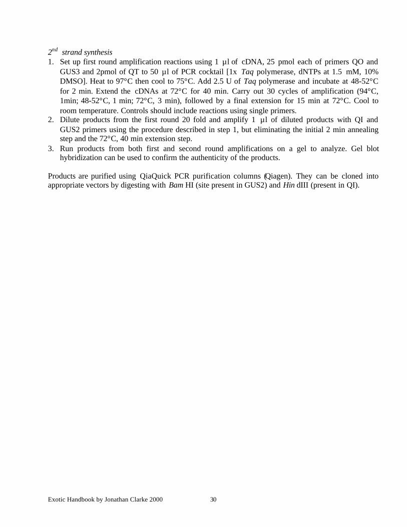
2nd strand synthesis1. Set up first round amplification reactions using 1 µl of cDNA, 25 pmol each of primers QO and
GUS3 and 2pmol of QT to 50 µl of PCR cocktail [1x Taq polymerase, dNTPs at 1.5 mM, 10%DMSO]. Heat to 97°C then cool to 75°C. Add 2.5 U of Taq polymerase and incubate at 48-52°Cfor 2 min. Extend the cDNAs at 72°C for 40 min. Carry out 30 cycles of amplification (94°C,1min; 48-52°C, 1 min; 72°C, 3 min), followed by a final extension for 15 min at 72°C. Cool toroom temperature. Controls should include reactions using single primers.
2. Dilute products from the first round 20 fold and amplify 1 µl of diluted products with QI andGUS2 primers using the procedure described in step 1, but eliminating the initial 2 min annealingstep and the 72°C, 40 min extension step.
3. Run products from both first and second round amplifications on a gel to analyze. Gel blothybridization can be used to confirm the authenticity of the products.
Products are purified using QiaQuick PCR purification columns (Qiagen). They can be cloned intoappropriate vectors by digesting with Bam HI (site present in GUS2) and Hin dIII (present in QI).
Exotic Handbook by Jonathan Clarke 2000 31
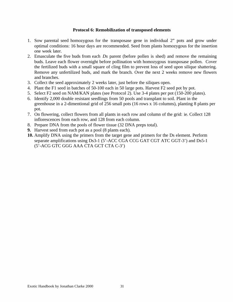
Protocol 6: Remobilization of transposed elements
1. Sow parental seed homozygous for the transposase gene in individual 2” pots and grow underoptimal conditions: 16 hour days are recommended. Seed from plants homozygous for the insertionone week later.
2. Emasculate the five buds from each Ds parent (before pollen is shed) and remove the remainingbuds. Leave each flower overnight before pollination with homozygous transposase pollen. Coverthe fertilized buds with a small square of cling film to prevent loss of seed upon silique shattering.Remove any unfertilized buds, and mark the branch. Over the next 2 weeks remove new flowersand branches.
3. Collect the seed approximately 2 weeks later, just before the siliques open.4. Plant the F1 seed in batches of 50-100 each in 50 large pots. Harvest F2 seed pot by pot.5. Select F2 seed on NAM/KAN plates (see Protocol 2). Use 3-4 plates per pot (150-200 plates).6. Identify 2,000 double resistant seedlings from 50 pools and transplant to soil. Plant in the
greenhouse in a 2-dimentional grid of 256 small pots (16 rows x 16 columns), planting 8 plants perpot.
7. On flowering, collect flowers from all plants in each row and column of the grid: ie. Collect 128inflorescences from each row, and 128 from each column.
8. Prepare DNA from the pools of flower tissue (32 DNA preps total).9. Harvest seed from each pot as a pool (8 plants each).10. Amplify DNA using the primers from the target gene and primers for the Ds element. Perform
separate amplifications using Ds3-1 (5’-ACC CGA CCG GAT CGT ATC GGT-3’) and Ds5-1(5’-ACG GTC GGG AAA CTA GCT CTA C-3’)
Exotic Handbook by Jonathan Clarke 2000 32
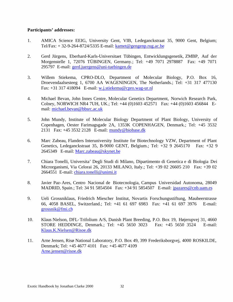
Participants’ addresses:
1. AMICA Science EEIG, University Gent, VIB, Ledeganckstraat 35, 9000 Gent, Belgium;Tel/Fax: + 32-9-264-8724/5335 E-mail: [email protected]
2. Gerd Jürgens, Eberhard-Karls-Universitaet Tübingen, Entwicklungsgenetik, ZMBP, Auf derMorgenstelle 1, 72076 TÜBINGEN, Germany.; Tel: +49 7071 2978887 Fax: +49 7071295797 E-mail: [email protected]
3. Willem Stiekema, CPRO-DLO, Department of Molecular Biology, P.O. Box 16,Droevendaalsesteeg 1, 6700 AA WAGENINGEN, The Netherlands.; Tel: +31 317 477130Fax: +31 317 418094 E-mail: [email protected]
4. Michael Bevan, John Innes Centre, Molecular Genetics Department, Norwich Research Park,Colney, NORWICH NR4 7UH, UK.; Tel: +44 (0)1603 452571 Fax: +44 (0)1603 456844 E-mail: [email protected]
5. John Mundy, Institute of Molecular Biology Department of Plant Biology, University ofCopenhagen, Oester Farimagsgade 2A, 1353K COPENHAGEN, Denmark.; Tel: +45 35322131 Fax: +45 3532 2128 E-mail: [email protected]
6. Marc Zabeau, Flanders Interuniversity Institute for Biotechnology VZW, Department of PlantGenetics, Ledeganckstraat 35, B-9000 GENT, Belgium.; Tel: +32 9 2645170 Fax: +32 92645349 E-mail: [email protected]
7. Chiara Tonelli, Universita’ Degli Studi di Milano, Dipartimento di Genetica e di Biologia DeiMicrorganismi, Via Celorai 26, 20133 MILANO, Italy.; Tel: +39 02 26605 210 Fax: +39 022664551 E-mail: [email protected]
8. Javier Paz-Ares, Centro Nacional de Biotecnologia, Campus Universidad Autonoma, 28049MADRID, Spain.; Tel: 34 91 5854504 Fax: +34 91 5854507 E-mail: [email protected]
9. Ueli Grossniklaus, Friedrich Miescher Institut, Novartis Forschungsstiftung, Maubeerstrasse66, 4058 BASEL, Switzerland.; Tel: +41 61 697 6983 Fax: +41 61 697 3976 E-mail:[email protected]
10. Klaus Nielson, DFL-Trifolium A/S, Danish Plant Breeding, P.O. Box 19, Højerupvej 31, 4660STORE HEDDINGE, Denmark.; Tel: +45 5650 3023 Fax: +45 5650 3524 E-mail:[email protected]
11. Arne Jensen, Risø National Laboratory, P.O. Box 49, 399 Frederiksborgvej, 4000 ROSKILDE,Denmark; Tel: +45 4677 4101 Fax: +45 4677 [email protected]