Embed Size (px)
Citation preview

Proc. Nati. Acad. Sci. USAVol. 89, pp. 9779-9783, October 1992Genetics
Establishment of a highly sensitive and specific exon-trapping system
(chromosome 11q13/RNA splicing)
MASAAKI HAMAGUCHI*t, HIROMI SAKAMOTO*, HIROYUKI TSURUTA*, HIROKI SASAKI*, TETSUICHIRO MUTOt,TAKASHI SUGIMURA*, AND MASAAKI TERADA**Genetics Division, National Cancer Center Research Institute, 1-1, Tsukiji 5-chome, Chuo-ku, Tokyo 104, Japan; and tDepartment of Surgery, University ofTokyo, 3-1, Hongo 7-chome, Bunkyo-ku, Tokyo 113, Japan
Contributed by Takashi Sugimura, July 13, 1992
ABSTRACT We have established a highly sensitive andspecific exon-trapping system (SETS) with a specific plasmidvector in which an exon in a given DNA segment is identifiedby its ability to remain as a mature mRNA after splicing. TheSETS provides us with the isolation of possible exons rapidlyand easily from DNA fragments in chromosomal regions ofmore than 300 kilobase pairs. Genomic DNA fragments werepartially digested and subsequently cloned into plasmidpMHC2, an exon-trapping vector we have constructed. Theseconstructs were transfected into COS-7 cells, and consequentRNA transcripts were spliced in the cells. The resulting maturemRNA was harvested and amplified by using reverse tran-scription-PCR. Possible exons can be recognized by the sizes ofPCR products and cloned into a plasmid vector. The SETSprovides a direct means of cloning exons from genomic DNA ofmore than 300 kilobase pairs within a short period of time.Using this system, we have screened 300-kilobase-pair genomicDNA segments derived from human chromosome 11q13. Hu-man chromosome 11q13 may contain genes responsible forhuman cancers, because DNA amplification is observed inseveral malignant tumors. We have successfully identified exon2 of the HSTI gene and additional transcribed sequences.
Detailed genetic and physical maps for human chromosomeshave been constructed to determine the chromosomal loca-tions of the genes responsible for many human genetic disor-ders and cancers. The minimal region in which a gene ofinterest can be found spans several hundred kilobase pairs.Identification and recovery of transcribed sequences from theregion of interest are necessary to isolate candidate genes.This strategy has been successful in isolating a number ofcandidate genes, including Wilms tumor (1-3), neurofibroma-tosis type 1 (4, 5), and familial adenomatous polyposis (6, 7).However, available methods for isolation of transcribed
sequences from given DNA fragments are inefficient. Thosemethods most frequently used are interspecies cross-hybridization to search for evolutionarily conserved se-quences (3, 8). Other methods include search for openreading frames (9), enhancers (10), promoters (11, 12), orhypomethylated CpG islands (13). But none of these strate-gies involves direct cloning of transcribed sequences.
Alternative strategies to isolate transcribed sequences in-volve direct cloning ofhuman transcripts from human-rodentsomatic cell hybrids (14, 15). These strategies, however, alsohave limitations. Possible transcripts derived from humanDNA fragments might not be expressed sufficiently enough tobe detected in hybrid cells. It is also not easy to establish thosehybrid cell lines that contain only the target human DNAfragments.
Two exon-trapping systems based on RNA splicing havebeen reported and could be important tools for direct cloningof transcribed sequences. However, both of the exon-trapping systems reported previously have disadvantages asdescribed below. A strategy using the retroviral shuttlevector system described by Duyk et al. (16) takes much timeto complete a round of screening, and the procedure iscomplicated. The other system, described by Buckler et al.(17), worked efficiently; however, its usefulness for identi-fication of exons in a given DNA fragment was still limitedbecause of the following points. First, it was demonstratedthat no more than one phage or cosmid can be screened bytheir system in a single transfection, and no mention wasmade about the maximum length of the target DNA frag-ments. The sensitivity of their system would not allow us toapply it to screening for possible exons from a large DNAsegment such as is carried in yeast artificial chromosomes(YACs). Second, the combination of vector splice sites andcryptic splice sites of inserts might cause unusual splicing andresult in production of false-positive exons. It was reportedthat three false positives out of four possible exons weregenerated by Buckler's system. To improve the sensitivityand specificity, we checked genomic DNA sequences ofintrons and searched for a sequence suitable for a trappingcassette. Check points were the following: first, a pyrimidinetract should be long enough to prevent exon skipping (18).Second, consensus sequences at branch and splice sitesshould be best fitted to those of small nuclear RNAs involvedin splicing. Third, the length of the intron should be longerthan 500 base pairs (bp), because a short intron in the trappingcassette makes it difficult to distinguish spliced fragmentsfrom unspliced fragments and because most exons are shorterthan 500 bp (19). We found suitable sequences for thetrapping cassette and have constructed an exon-trappingvector. Here we demonstrate establishment ofa sensitive andspecific exon-trapping system (SETS) both sensitive andspecific enough to carry out large-scale screening of genomicDNA segments for the presence of exons, using this vector.We tested the SETS by isolation of transcribed sequences inoverlapping cosmid clones corresponding to human chromo-some 11q13 containing INT2 and HSTI (20). We have suc-cessfully isolated three novel transcribed sequences from theregion by this strategy, demonstrating the usefulness of thissystem, which is a powerful tool for simple screening ofmegabase genomic DNA segments for the presence of pos-sible exons.
MATERIALS AND METHODSConstruction of the Exon-Trapping Vector. Construction
was initiated by PCR amplification of genomic DNA frag-
Abbreviations: SETS, sensitive and specific exon-trapping system;RT, reverse transcription; YAC, yeast artificial chromosome.
9779
The publication costs of this article were defrayed in part by page chargepayment. This article must therefore be hereby marked "advertisement"in accordance with 18 U.S.C. §1734 solely to indicate this fact.

Proc. Nati. Acad. Sci. USA 89 (1992)
ments for the trapping cassette. The primers (5'-CGT-GAAAQCTCGAGATGTTCCGA-3' and 5'-CAAGCTTC-TGACGCACATTTATTGCAAGCAA-3') were designed sothat PCR products contain part ofexon 10, intron 10, and partofexon 11 of the p53 gene, with synthetic HindIII recognitionsequence indicated by underlining. GenomicDNA ofan adultmale was amplified by using these primers. PCR productswere digested with HindIII and partially filled in with dATPand dGTP to produce a fragment of 1 kilobase (kb) with a 5'protruding AG sequence. The parent vector pEUK-C2 (Clon-tech) was cleaved at a unique Xba I restriction site, followedby partial fill-in with dCTP and dTTP to produce a 5'protruding sequence of CT. The PCR products were ligatedwith the vector to construct the pMHC2 vector (see Fig. 1).
Cell Lines. COS-7 cells (Riken Cell Bank, Tsukuba) weremaintained in Dulbecco's modified Eagle's medium contain-ing 10%6 (vol/vol) heat-inactivated fetal bovine serum. TE-1cells and TE-10 cells derived from esophageal cancers weregrown in RPMI 1640 medium containing 7% heat-inactivatedcalf serum (21).Genomic DNA Fragments. Plasmid pLBS6.2 carries 6.2
kilobase pairs (kbp) of genomic sequences of the HSTJ gene(22). Cosmid 35N contains about 40 kbp of uncharacterizedgenomic DNA segments. Cosmid clones LYI-61, LYH-1,LYH-11, LP-3 [1], LP-9 [2], LP-15 [3], LP-2 [4], and LP-4 [5]contain 30- to 50-kbp genomic DNA segments derived fromhuman chromosome 11q13 (22).DNA Transfection and Transient Expression. Two micro-
grams ofgenomic DNA fragments was partially digested with0.2 unit of restriction endonuclease Sau3AI for 2 hr andfractionated through 1% agarose gels. DNA fragments from0.6 kbp to 5 kbp were isolated from agarose gels and clonedinto a pMHC2 vector that had been cleaved at a unique BglII recognition site and had been dephosphorylated with calfintestinal alkaline phosphatase (23). Plasmid DNA was pre-pared by using equilibrium centrifugation in cesium chloride/ethidium bromide gradients. COS-7 cells were transformedwith the plasmids by electroporation with a Gene Pulser(Bio-Rad) according to the supplier's protocol. Briefly, 5million semiconfluent cells were suspended in 0.7 ml ofphosphate-buffered saline and placed in a 0.4-cm cuvette, and10 ,ug of a mixture of plasmid DNAs was added. The cellswere electroporated at 1.2 kV and 25 ,uF. Sixty hours aftertransformation, poly(A)+ RNA was isolated by using Fast-Track (Invitrogen, San Diego).
Amplification of Poly(A)+ RNA by Reverse Transcription(RT)-PCR. First-strand cDNA was synthesized frompoly(A)+ RNA by RT from oligo(dT) primers (24) and puri-fied with Bio-Spin 30 columns (Bio-Rad). Primary PCRamplification was performed between the forward primer 7rA(5'-TGAGGCCTTGGAACTCA-3') and the reverse primerirR (5'-GGAGAATGTCAGTCTGA-3') for 25 cycles. Thirtycycles of secondary PCR amplification were carried outbetween the forward primer irAB (5'-AGGGATCCCAGGC-TGGGAAGGA-3') and the reverse primer irRB (5'-ACGGAICCTTTTTGGACTTCAGGT-3') using 1/100th ofthe primary PCR products as a template. Both 1rAB and irRBprimers contain a synthetic BamHI recognition sequenceindicated by underlining. Positions of the primers are indi-cated in Fig. 1.
Cloning of Trapped Fragments. The amplified DNA frag-ments were electrophoresed through polyacrylamide or 4%agarose gels, recovered from the gel (25), digested withBamHI, and cloned into the plasmid vector pBluescript IISK+.DNA Sequencing. Trapped DNA fragments were se-
quenced on the Applied Biosystems automated sequencerwith Taq DNA polymerase in a cycle sequencing protocol(Applied Biosystems) with dideoxynucleotide fluorescentterminators.
Blot Analysis. Poly(A)+ RNA was isolated from TE-1 cellsor TE-10 cells using FastTrack (Invitrogen), and 2 j.g ofpoly(A)+ RNA was electrophoretically separated in 1% aga-rose/formaldehyde gels and transferred to Hybond N nylonmembranes (Amersham) as recommended by the supplier.Genomic DNA digested with restriction endonucleases waselectrophoresed through 1% agarose gels and transferred toHybond N+ membranes (Amersham). To make probes, plas-mid DNA with trapped fragments was linearized with BssHIIand transcribed in the presence of 32P-labeled CTP withphage T3 or T7 RNA polymerases (23). Hybridizations werecarried out by standard procedures (23).
Ribonuclease (RNase) Protection Assay. One microgram ofpBluescript II SK+ was digested with BssHII, and labeledRNA probes were synthesized from the linearized plasmidsas described above. The RNA probes were hybridized to 1 ,ugof poly(A)+ RNA from TE-1 cells at 450C for 16 hr. Unhy-bridized single-stranded RNA was digested with RNase Aand RNase T1 (Ambion, Austin, TX). RNase digestionproducts were analyzed on denaturing polyacrylamide gelsfollowed by autoradiography.PCR Amplification of a cDNA Mixture from TE-1 Cells. A
cDNA mixture derived from a AZAPII library of TE-1 cellscontaining about 1 million recombinant bacteriophages wasamplified by PCR using primers 38A (5'-CATAAGTGC-CGATGGAGTAGGA-3'), 38R (5'-CTCTCCAATTTAGGA-TTCTCA), 40A (5'-TAGCTGACATTCAAAGCCTGGG-3'), and 40R (5'-CTGTGAGGACAGGAGCTGACAGA-3').These primers were part of trapped fragments and were usedfor amplification of trapped fragments; 38A and 38R were forMB38 fragments. Primers 40A and 40R were for MB40fragments (see Table 1).PCR Amplification. The PCR reaction mixture contained
10mM Tris HCl (pH 8.3), 50mM KCl, 1.5 mM MgCI2, gelatinat 0.1 mg/ml, dNTPs at 250 nM each, 2 units of Taqpolymerase (TaKaRa), and 0.5 ,ug of primers. PCR amplifi-cation of the p53 fragment was carried out in a total volumeof 100 ,ul for 30 cycles of 94°C for 1 min, 55°C for 30 sec, and72°C for 2 min. Primary RT-PCR was carried out with 20cycles followed by 30 cycles ofsecondary amplification. PCRamplification of the cDNA mixture was performed with 35cycles of 94°C for 1 min, 60°C for 15 sec, and 72°C for 1 min.
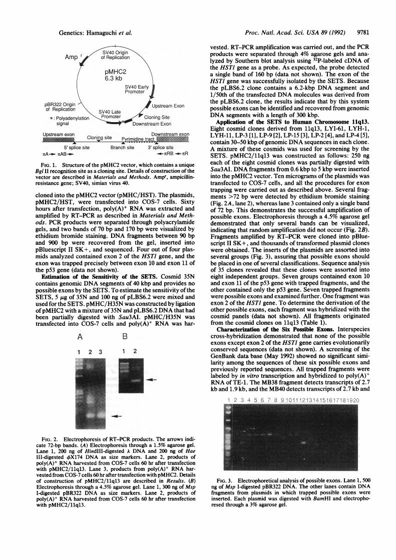
RESULTSExperimental Strategy. The constructed pMHC2 vector
contains the entire intron 10 of the p53 gene, including thelong pyrimidine tract and the consensus sequences of the 5'splice site [5'-CAG/GTGAGT-3'], the 3' splice site (5'-AG-3'), and the branch site (5'-TACTCAC-3') (Fig. 1) (26, 34, 35).The pMHC2 vector has a unique Bgl II site as a cloning sitebetween the 5' splice site and the branch site (Fig. 1). Whenan entire exon and the flanking intron derived from the testgenomic DNA segments are inserted into the pMHC2 vectorin the sense orientation, the transcripts from these plasmidsare expected to be processed in the COS-7 cells after trans-fection, and exons can be identified by RT-PCR. Otherwise,RT-PCR products consist only of the exons of the p53 gene,and a single band of 72 bp is detected by ethidium bromidestaining (Fig. 2). Since amplified fragments longer than 72 bpin length contain possible exons and fragments longer than 1kbp may contain unspliced transcripts, fragments between 90bp and 900 bp were subcloned into pBluescript II SK+,sequenced, and analyzed for expression.Trapping of the Known Exon with the SETS. As an initial
examination of the system, derivatives of pMHC2 wereconstructed. pLBS6.2 plasmid, which carries a 6.2-kb DNAfragment including genomic sequences of the HSTJ genefrom exon 1 to exon 3, was partially digested with Sau3AI,and fragments between 0.6 kbp and 5 kbp in length were
9780 Genetics: Harnaguchi et al.
Proc. Natl. Acad. Sci. USA 89 (1992) 9781
Amp r
pBR322 Origin 1Upstream Exonof Replication SV40 Late U
*: Polyadenylation :Promoter Cloning Sitesignal Downstream Exon
Upstream exon Downstream exon_ ~~~~cloning site_ Pyrimidine tract _
5' splice site Branch site 3' splice sitenA-o nAB_ -_a-RB -_7-R
FIG. 1. Structure of the pMHC2 vector, which contains a uniqueBgl II recognition site as a cloning site. Details of construction of thevector are described in Materials and Methods. Ampr, ampicillin-resistance gene; SV40, simian virus 40.
cloned into the pMHC2 vector (pMHC/HST). The plasmids,pMHC2/HST, were transfected into COS-7 cells. Sixtyhours after transfection, poly(A)+ RNA was extracted andamplified by RT-PCR as described in Materials and Meth-ods. PCR products were separated through polyacrylamidegels, and two bands of 70 bp and 170 bp were visualized byethidium bromide staining. DNA fragments between 90 bpand 900 bp were recovered from the gel, inserted intopBluescript II SK+, and sequenced. Four out of four plas-mids analyzed contained exon 2 of the HSTJ gene, and theexon was trapped precisely between exon 10 and exon 11 ofthe p53 gene (data not shown).
Estimation of the Sensitivity of the SETS. Cosmid 35Ncontains genomic DNA segments of 40 kbp and provides nopossible exons by the SETS. To estimate the sensitivity oftheSETS, 5 ,g of 35N and 100 ng of pLBS6.2 were mixed andused for the SETS. pMHC/H35N was constructed by ligationofpMHC2 with a mixture of35N and pLBS6.2 DNA that hadbeen partially digested with Sau3AI. pMHC/H35N wastransfected into COS-7 cells and poly(A)+ RNA was har-
A B
1 2 3 1 2
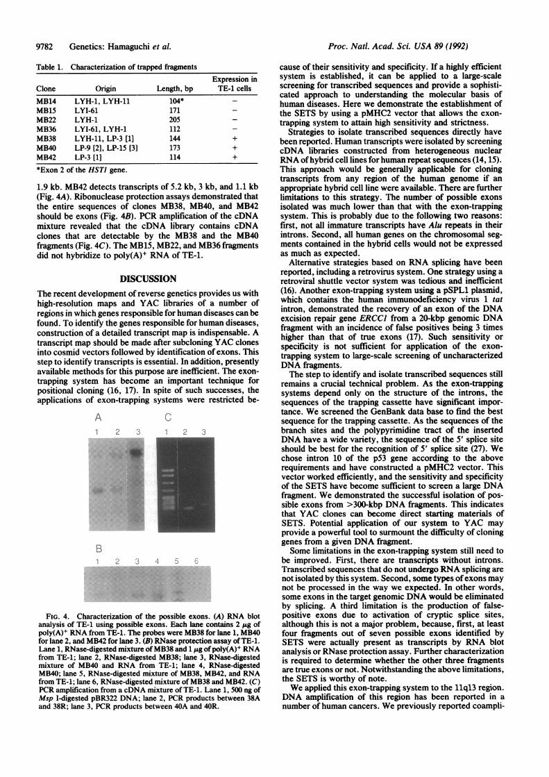
vested. RT-PCR amplification was carried out, and the PCRproducts were separated through 4% agarose gels and ana-lyzed by Southern blot analysis using 32P-labeled cDNA ofthe HSTI gene as a probe. As expected, the probe detecteda single band of 160 bp (data not shown). The exon of theHSTJ gene was successfully isolated by the SETS. Becausethe pLBS6.2 clone contains a 6.2-kbp DNA segment and1/50th of the transfected DNA molecules was derived fromthe pLBS6.2 clone, the results indicate that by this systempossible exons can be identified and recovered from genomicDNA segments with a length of 300 kbp.
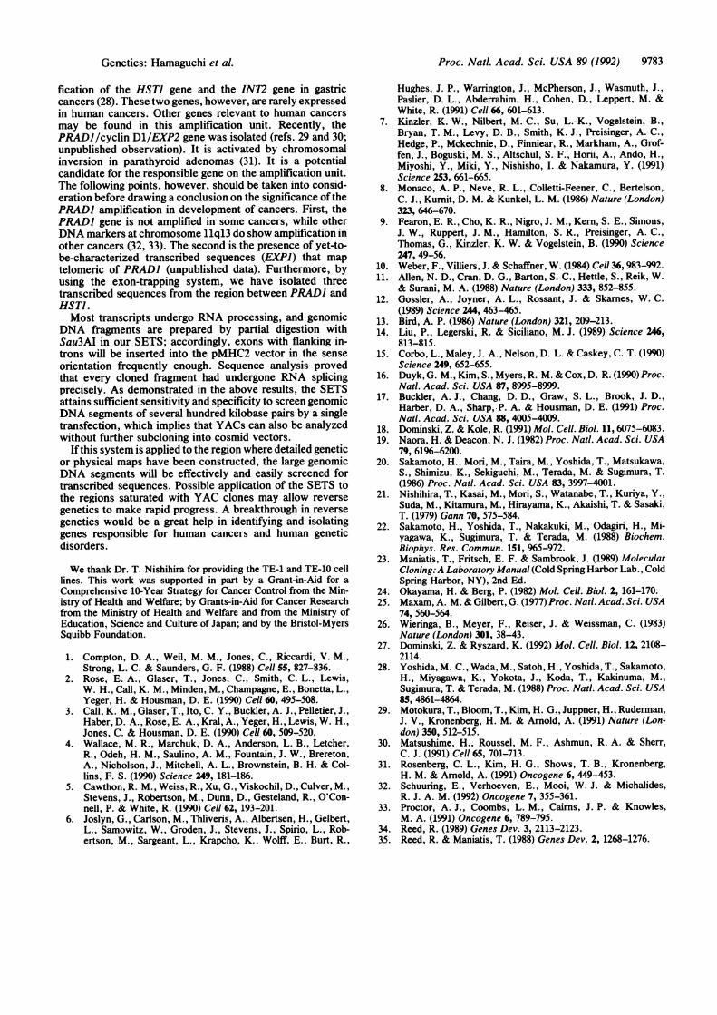
Application of the SETS to Human Chromosome 11q13.Eight cosmid clones derived from 11q13, LYI-61, LYH-1,LYH-11, LP-3 [1], LP-9 [2], LP-15 [3], LP-2 [4], and LP-4 [5],contain 30-50 kbp ofgenomic DNA sequences in each clone.A mixture of these cosmids was used for screening by theSETS. pMHC2/llql3 was constructed as follows: 250 ngeach of the eight cosmid clones was partially digested withSau3AI. DNA fragments from 0.6 kbp to 5 kbp were insertedinto the pMHC2 vector. Ten micrograms of the plasmids wastransfected to COS-7 cells, and all the procedures for exontrapping were carried out as described above. Several frag-ments >72 bp were detected by ethidium bromide staining(Fig. 2A, lane 2), whereas lane 3 contained only a single bandof 72 bp. This demonstrates the successful amplification ofpossible exons. Electrophoresis through a 4.5% agarose geldemonstrated that only several bands can be visualized,indicating that random amplification did not occur (Fig. 2B).Fragments amplified by RT-PCR were cloned into pBlue-script II SK+, and thousands of transformed plasmid cloneswere obtained. The inserts of the plasmids are assorted intoseveral groups (Fig. 3), assuring that possible exons shouldbe placed in one of several classifications. Sequence analysisof 35 clones revealed that these clones were assorted intoeight independent groups. Seven groups contained exon 10and exon 11 of the p53 gene with trapped fragments, and theother contained only the p53 gene. Seven trapped fragmentswere possible exons and examined further. One fragment wasexon 2 of the HSTJ gene. To determine the derivation of theother possible exons, each fragment was hybridized with thecosmid panels (data not shown). All fragments originatedfrom the cosmid clones on 11q13 (Table 1).
Characterization of the Six Possible Exons. Interspeciescross-hybridization demonstrated that none of the possibleexons except exon 2 of the HSTJ gene carries evolutionarilyconserved sequences (data not shown). A screening of theGenBank data base (May 1992) showed no significant simi-larity among the sequences of these six possible exons andpreviously reported sequences. All trapped fragments werelabeled by in vitro transcription and hybridized to poly(A)+RNA of TE-1. The MB38 fragment detects transcripts of 2.7kb and 1.9 kb, and the MB40 detects transcripts of 2.7 kb and
1 2 3 4 5 6 7 8 9 1011121314151617181
FIG. 2. Electrophoresis of RT-PCR products. The arrows indi-cate 72-bp bands. (A) Electrophoresis through a 1.5% agarose gel.Lane 1, 200 ng of HindIII-digested A DNA and 200 ng of HaeIII-digested 4X174 DNA as size markers. Lane 2, products ofpoly(A)+ RNA harvested from COS-7 cells 60 hr after transfectionwith pMHC2/llql3. Lane 3, products from poly(A)+ RNA har-vested from COS-7 cells 60 hr after transfection with pMHC2. Detailsof construction of pMHC2/llql3 are described in Results. (B)Electrophoresis through a 4.5% agarose gel. Lane 1, 300 ng of MspI-digested pBR322 DNA as size markers. Lane 2, products ofpoly(A)+ RNA harvested from COS-7 cells 60 hr after transfectionwith pMHC2/llql3.
FIG. 3. Electrophoretical analysis of possible exons. Lane 1, 500ng of Msp I-digested pBR322 DNA. The other lanes contain DNAfragments from plasmids in which trapped possible exons wereinserted. Each plasmid was digested with BamHI and electropho-resed through a 3% agarose gel.
Genetics: Harnaguchi et al.
Proc. Nati. Acad. Sci. USA 89 (1992)
Table 1. Characterization of trapped fragmentsExpression in
Clone Origin Length, bp TE-1 cells
MB14 LYH-1, LYH-11 104* -MB15 LYI-61 171 -MB22 LYH-1 205 -MB36 LYI-61, LYH-1 112 -MB38 LYH-11, LP-3 [1] 144 +MB40 LP-9 [2], LP-15 [3] 173 +MB42 LP-3 [1] 114 +
*Exon 2 of the HSTI gene.
1.9 kb. MB42 detects transcripts of 5.2 kb, 3 kb, and 1.1 kb(Fig. 4A). Ribonuclease protection assays demonstrated thatthe entire sequences of clones MB38, MB40, and MB42should be exons (Fig. 4B). PCR amplification of the cDNAmixture revealed that the cDNA library contains cDNAclones that are detectable by the MB38 and the MB40fragments (Fig. 4C). The MB15, MB22, and MB36 fragmentsdid not hybridize to poly(A)+ RNA of TE-1.
DISCUSSIONThe recent development of reverse genetics provides us withhigh-resolution maps and YAC libraries of a number ofregions in which genes responsible for human diseases can befound. To identify the genes responsible for human diseases,construction of a detailed transcript map is indispensable. Atranscript map should be made after subcloning YAC clonesinto cosmid vectors followed by identification of exons. Thisstep to identify transcripts is essential. In addition, presentlyavailable methods for this purpose are inefficient. The exon-trapping system has become an important technique forpositional cloning (16, 17). In spite of such successes, theapplications of exon-trapping systems were restricted be-
A1 2 z
*: .-.A..*: .: ::: .. Si
............'$<.l
*:5'i* .. .' .. 'a.I* .'$,. '.', ''aS'...'.gSo id it
.';^'.S.,,'W.
B1 2 3
c1 2
4
FIG. 4. Characterization of the possible exons. (A) RNA blotanalysis of TE-1 using possible exons. Each lane contains 2 ,ug ofpoly(A)+ RNA from TE-1. The probes were MB38 for lane 1, MB40for lane 2, and MB42 for lane 3. (B) RNase protection assay of TE-1.Lane 1, RNase-digested mixture ofMB38 and 1 pg of poly(A)+ RNAfrom TE-1; lane 2, RNase-digested MB38; lane 3, RNase-digestedmixture of MB40 and RNA from TE-1; lane 4, RNase-digestedMB40; lane 5, RNase-digested mixture of MB38, MB42, and RNAfrom TE-1; lane 6, RNase-digested mixture of MB38 and MB42. (C)PCR amplification from a cDNA mixture of TE-1. Lane 1, 500 ng ofMsp I-digested pBR322 DNA; lane 2, PCR products between 38Aand 38R; lane 3, PCR products between 40A and 40R.
cause of their sensitivity and specificity. If a highly efficientsystem is established, it can be applied to a large-scalescreening for transcribed sequences and provide a sophisti-cated approach to understanding the molecular basis ofhuman diseases. Here we demonstrate the establishment ofthe SETS by using a pMHC2 vector that allows the exon-trapping system to attain high sensitivity and strictness.
Strategies to isolate transcribed sequences directly havebeen reported. Human transcripts were isolated by screeningcDNA libraries constructed from heterogeneous nuclearRNA ofhybrid cell lines for human repeat sequences (14, 15).This approach would be generally applicable for cloningtranscripts from any region of the human genome if anappropriate hybrid cell line were available. There are furtherlimitations to this strategy. The number of possible exonsisolated was much lower than that with the exon-trappingsystem. This is probably due to the following two reasons:first, not all immature transcripts have Alu repeats in theirintrons. Second, all human genes on the chromosomal seg-ments contained in the hybrid cells would not be expressedas much as expected.
Alternative strategies based on RNA splicing have beenreported, including a retrovirus system. One strategy using aretroviral shuttle vector system was tedious and inefficient(16). Another exon-trapping system using a pSPL1 plasmid,which contains the human immunodeficiency virus 1 tatintron, demonstrated the recovery of an exon of the DNAexcision repair gene ERCCI from a 20-kbp genomic DNAfragment with an incidence of false positives being 3 timeshigher than that of true exons (17). Such sensitivity orspecificity is not sufficient for application of the exon-trapping system to large-scale screening of uncharacterizedDNA fragments.The step to identify and isolate transcribed sequences still
remains a crucial technical problem. As the exon-trappingsystems depend only on the structure of the introns, thesequences of the trapping cassette have significant impor-tance. We screened the GenBank data base to find the bestsequence for the trapping cassette. As the sequences of thebranch sites and the polypyrimidine tract of the insertedDNA have a wide variety, the sequence of the 5' splice siteshould be best for the recognition of 5' splice site (27). Wechose intron 10 of the p53 gene according to the aboverequirements and have constructed a pMHC2 vector. Thisvector worked efficiently, and the sensitivity and specificityof the SETS have become sufficient to screen a large DNAfragment. We demonstrated the successful isolation of pos-sible exons from >300-kbp DNA fragments. This indicatesthat YAC clones can become direct starting materials ofSETS. Potential application of our system to YAC mayprovide a powerful tool to surmount the difficulty of cloninggenes from a given DNA fragment.Some limitations in the exon-trapping system still need to
be improved. First, there are transcripts without introns.Transcribed sequences that do not undergo RNA splicing arenot isolated by this system. Second, some types ofexons maynot be processed in the way we expected. In other words,some exons in the target genomic DNA would be eliminatedby splicing. A third limitation is the production of false-positive exons due to activation of cryptic splice sites,although this is not a major problem, because, first, at leastfour fragments out of seven possible exons identified bySETS were actually present as transcripts by RNA blotanalysis or RNase protection assay. Further characterizationis required to determine whether the other three fragmentsare true exons or not. Notwithstanding the above limitations,the SETS is worthy of note.We applied this exon-trapping system to the 11q13 region.
DNA amplification of this region has been reported in anumber of human cancers. We previously reported coampli-
9782 Genetics: Harnaguchi et al.
Proc. Natl. Acad. Sci. USA 89 (1992) 9783
fication of the HSTJ gene and the INT2 gene in gastriccancers (28). These two genes, however, are rarely expressedin human cancers. Other genes relevant to human cancersmay be found in this amplification unit. Recently, thePRADI/cyclin D1/EXP2 gene was isolated (refs. 29 and 30;unpublished observation). It is activated by chromosomalinversion in parathyroid adenomas (31). It is a potentialcandidate for the responsible gene on the amplification unit.The following points, however, should be taken into consid-eration before drawing a conclusion on the significance of thePRADJ amplification in development of cancers. First, thePRAD) gene is not amplified in some cancers, while otherDNA markers at chromosome 11q13 do show amplification inother cancers (32, 33). The second is the presence of yet-to-be-characterized transcribed sequences (EXPI) that maptelomeric of PRADI (unpublished data). Furthermore, byusing the exon-trapping system, we have isolated threetranscribed sequences from the region between PRADI andHSTJ.Most transcripts undergo RNA processing, and genomic
DNA fragments are prepared by partial digestion withSau3AI in our SETS; accordingly, exons with flanking in-trons will be inserted into the pMHC2 vector in the senseorientation frequently enough. Sequence analysis provedthat every cloned fragment had undergone RNA splicingprecisely. As demonstrated in the above results, the SETSattains sufficient sensitivity and specificity to screen genomicDNA segments of several hundred kilobase pairs by a singletransfection, which implies that YACs can also be analyzedwithout further subcloning into cosmid vectors.
If this system is applied to the region where detailed geneticor physical maps have been constructed, the large genomicDNA segments will be effectively and easily screened fortranscribed sequences. Possible application of the SETS tothe regions saturated with YAC clones may allow reverse
genetics to make rapid progress. A breakthrough in reversegenetics would be a great help in identifying and isolatinggenes responsible for human cancers and human geneticdisorders.
We thank Dr. T. Nishihira for providing the TE-1 and TE-10 celllines. This work was supported in part by a Grant-in-Aid for a
Comprehensive 10-Year Strategy for Cancer Control from the Min-istry of Health and Welfare; by Grants-in-Aid for Cancer Researchfrom the Ministry of Health and Welfare and from the Ministry ofEducation, Science and Culture of Japan; and by the Bristol-MyersSquibb Foundation.
1. Compton, D. A., Weil, M. M., Jones, C., Riccardi, V. M.,Strong, L. C. & Saunders, G. F. (1988) Cell 55, 827-836.
2. Rose, E. A., Glaser, T., Jones, C., Smith, C. L., Lewis,W. H., Call, K. M., Minden, M., Champagne, E., Bonetta, L.,Yeger, H. & Housman, D. E. (1990) Cell 60, 495-508.
3. Call, K. M., Glaser, T., Ito, C. Y., Buckler, A. J., Pelletier, J.,Haber, D. A., Rose, E. A., Kral, A., Yeger, H., Lewis, W. H.,Jones, C. & Housman, D. E. (1990) Cell 60, 509-520.
4. Wallace, M. R., Marchuk, D. A., Anderson, L. B., Letcher,R., Odeh, H. M., Saulino, A. M., Fountain, J. W., Brereton,A., Nicholson, J., Mitchell, A. L., Brownstein, B. H. & Col-lins, F. S. (1990) Science 249, 181-186.
5. Cawthon, R. M., Weiss, R., Xu, G., Viskochil, D., Culver, M.,Stevens, J., Robertson, M., Dunn, D., Gesteland, R., O'Con-nell, P. & White, R. (1990) Cell 62, 193-201.
6. Joslyn, G., Carlson, M., Thliveris, A., Albertsen, H., Gelbert,L., Samowitz, W., Groden, J., Stevens, J., Spirio, L., Rob-ertson, M., Sargeant, L., Krapcho, K., Wolff, E., Burt, R.,
Hughes, J. P., Warrington, J., McPherson, J., Wasmuth, J.,Paslier, D. L., Abderrahim, H., Cohen, D., Leppert, M. &White, R. (1991) Cell 66, 601-613.
7. Kinzler, K. W., Nilbert, M. C., Su, L.-K., Vogelstein, B.,Bryan, T. M., Levy, D. B., Smith, K. J., Preisinger, A. C.,Hedge, P., Mckechnie, D., Finniear, R., Markham, A., Grof-fen, J., Boguski, M. S., Altschul, S. F., Horii, A., Ando, H.,Miyoshi, Y., Miki, Y., Nishisho, I. & Nakamura, Y. (1991)Science 253, 661-665.
8. Monaco, A. P., Neve, R. L., Colletti-Feener, C., Bertelson,C. J., Kurnit, D. M. & Kunkel, L. M. (1986) Nature (London)323, 646-670.
9. Fearon, E. R., Cho, K. R., Nigro, J. M., Kern, S. E., Simons,J. W., Ruppert, J. M., Hamilton, S. R., Preisinger, A. C.,Thomas, G., Kinzler, K. W. & Vogelstein, B. (1990) Science247, 49-56.
10. Weber, F., Villiers, J. & Schaffner, W. (1984) Cell 36, 983-992.11. Allen, N. D., Cran, D. G., Barton, S. C., Hettle, S., Reik, W.
& Surani, M. A. (1988) Nature (London) 333, 852-855.12. Gossler, A., Joyner, A. L., Rossant, J. & Skarnes, W. C.
(1989) Science 244, 463-465.13. Bird, A. P. (1986) Nature (London) 321, 209-213.14. Liu, P., Legerski, R. & Siciliano, M. J. (1989) Science 246,
813-815.15. Corbo, L., Maley, J. A., Nelson, D. L. & Caskey, C. T. (1990)
Science 249, 652-655.16. Duyk, G. M., Kim, S., Myers, R. M.&Cox, D. R. (1990) Proc.
Natl. Acad. Sci. USA 87, 8995-8999.17. Buckler, A. J., Chang, D. D., Graw, S. L., Brook, J. D.,
Harber, D. A., Sharp,.P. A. & Housman, D. E. (1991) Proc.Natl. Acad. Sci. USA 88, 4005-4009.
18. Dominski, Z. & Kole, R. (1991) Mol. Cell. Biol. 11, 6075-6083.19. Naora, H. & Deacon, N. J. (1982) Proc. Natl. Acad. Sci. USA
79, 6196-6200.20. Sakamoto, H., Mori, M., Taira, M., Yoshida, T., Matsukawa,
S., Shimizu, K., Sekiguchi, M., Terada, M. & Sugimura, T.(1986) Proc. Natl. Acad. Sci. USA 83, 3997-4001.
21. Nishihira, T., Kasai, M., Mori, S., Watanabe, T., Kuriya, Y.,Suda, M., Kitamura, M., Hirayama, K., Akaishi, T. & Sasaki,T. (1979) Gann 70, 575-584.
22. Sakamoto, H., Yoshida, T., Nakakuki, M., Odagiri, H., Mi-yagawa, K., Sugimura, T. & Terada, M. (1988) Biochem.Biophys. Res. Commun. 151, 965-972.
23. Maniatis, T., Fritsch, E. F. & Sambrook, J. (1989) MolecularCloning:A Laboratory Manual (Cold Spring Harbor Lab., ColdSpring Harbor, NY), 2nd Ed.
24. Okayama, H. & Berg, P. (1982) Mol. Cell. Biol. 2, 161-170.25. Maxam, A. M. & Gilbert, G. (1977) Proc. Natl. Acad. Sci. USA
74, 560-564.26. Wieringa, B., Meyer, F., Reiser, J. & Weissman, C. (1983)
Nature (London) 301, 38-43.27. Dominski, Z. & Ryszard, K. (1992) Mol. Cell. Biol. 12, 2108-
2114.28. Yoshida, M. C., Wada, M., Satoh, H., Yoshida, T., Sakamoto,
H., Miyagawa, K., Yokota, J., Koda, T., Kakinuma, M.,Sugimura, T. & Terada, M. (1988) Proc. Natl. Acad. Sci. USA85, 4861-4864.
29. Motokura, T., Bloom, T., Kim, H. G., Juppner, H., Ruderman,J. V., Kronenberg, H. M. & Arnold, A. (1991) Nature (Lon-don) 350, 512-515.
30. Matsushime, H., Roussel, M. F., Ashmun, R. A. & Sherr,C. J. (1991) Cell 65, 701-713.
31. Rosenberg, C. L., Kim, H. G., Shows, T. B., Kronenberg,H. M. & Arnold, A. (1991) Oncogene 6, 449-453.
32. Schuuring, E., Verhoeven, E., Mooi, W. J. & Michalides,R. J. A. M. (1992) Oncogene 7, 355-361.
33. Proctor, A. J., Coombs, L. M., Cairns, J. P. & Knowles,M. A. (1991) Oncogene 6, 789-795.
34. Reed, R. (1989) Genes Dev. 3, 2113-2123.35. Reed, R. & Maniatis, T. (1988) Genes Dev. 2, 1268-1276.
Genetics: Harnaguchi et al.


![CRISPR/Cas9-mediated genome editing induces exon skipping ... · HeLa cells can cause skipping of exon 3, exon 4, or exons 3, 4, and 5 [18]. We also detected infrequent exon skipping](https://img.pdfslide.us/doc/110x75/60db8f117fb86d112c69c947/crisprcas9-mediated-genome-editing-induces-exon-skipping-hela-cells-can-cause.jpg)