Embed Size (px)
Citation preview

Evolutionary Tendencies in Flowers of Marantaceae with special
reference to the Style Movement Mechanism
Dissertation zur Erlangung des Grades Doktor der Naturwissenschaften
Im Fachbereich Biologie der Johannes-Gutenberg-Universität Mainz
Elke Pischtschan
Geboren in Dresden
Mainz 2007
Contents Page
Summary of the thesis 1
1. General introduction 3
2. The setting up of tension in the styles of Maranta noctiflora Koernicke
Abstract 5
2.1 Introduction 5
2.1.1 The flowers in Marantaceae 5
2.1.2 Historical outline of interpretations 8
2.2 Materials and methods 11
2.2.1 Materials 11
2.2.2 Methods 11
2.3 Results 13
2.4 Discussion 20
3. A hydraulic model of the ´explosive´ style movement in Marantaceae – evidence
from functional anatomic studies
Abstract 24
3.1 Introduction 24
3.2 Materials and methods 26
3.2.1 Materials 26
3.2.2 Methods 27
3.2.2.1 Cellulose detection 27
3.2.2.2 Histology 27
3.2.2.3 Scanning electron microscopy 28
3.2.2.4 Fluorescence 30
3.2.2.5 Transmission electron microscopy 30
3.3 Results 31
3.3.1 Cellulose detection 31
3.3.2 Histology and SEM 31
3.3.2.1 The suitability of methods of fixation 31
3.3.2.2 Histology and SEM - first impressions 32
3.3.2.3 SEM - texture and surfaces 37
3.3.3 The genuinity of the porosity 39
3.3.3.1 Fluorescence 39
3.3.3.2 TEM 41
3.3.4 The time of emergence of the porosity 42
3.3.5 Other members of the order 43
3.3.5.1 Comparison of different Marantaceae 43
3.3.5.2 Comparison of different Zingiberales 44
3.3.6 Summary of the results 45
3.4 Discussion 45
4. Diversity and systematic value of the cucullate staminode in Marantaceae
Abstract 51
4.1 Introduction 51
4.2 Materials and methods 55
4.2.1 Materials 55
4.2.2 Methods 57
4.3 Results 58
4.4 Discussion 70
4.4.1 Discussion of the ´bauplan´ of cucullate staminodes 70
4.4.2 Discussion of the types 71
4.4.3 Discussion of evolutionary pathways 77
5. General conclusions 81
References 83
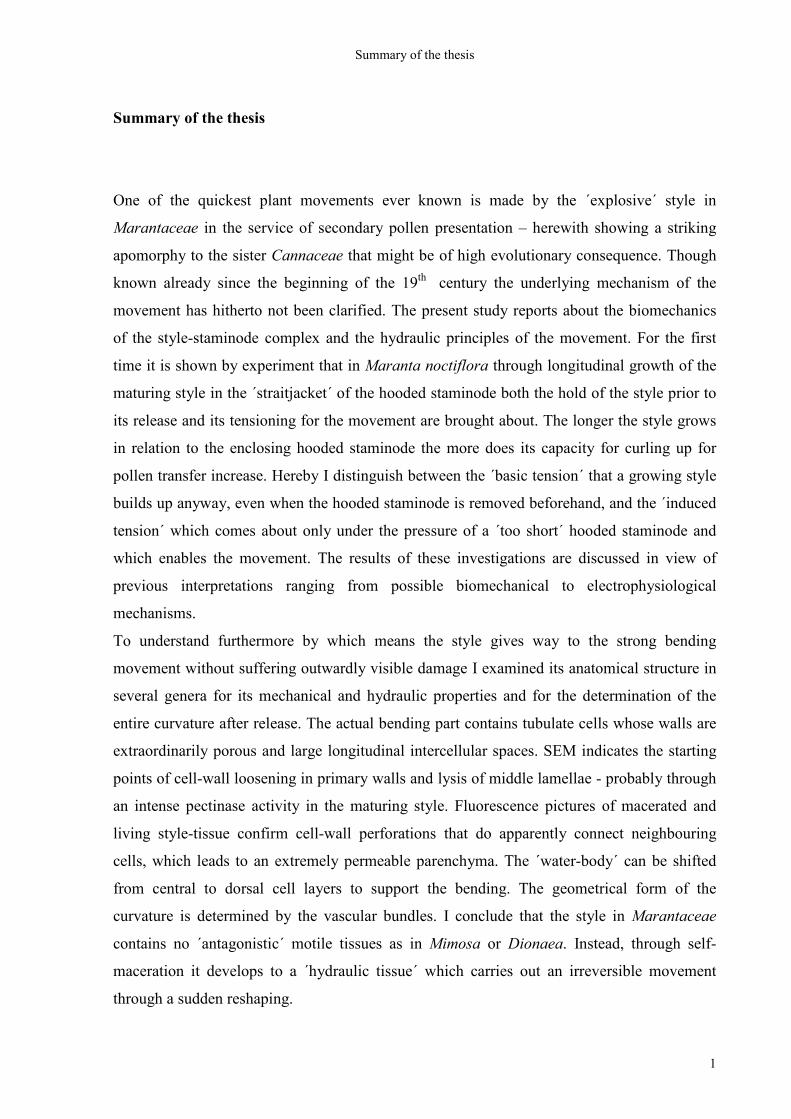
Summary of the thesis
1
Summary of the thesis
One of the quickest plant movements ever known is made by the ´explosive´ style in
Marantaceae in the service of secondary pollen presentation – herewith showing a striking
apomorphy to the sister Cannaceae that might be of high evolutionary consequence. Though
known already since the beginning of the 19th century the underlying mechanism of the
movement has hitherto not been clarified. The present study reports about the biomechanics
of the style-staminode complex and the hydraulic principles of the movement. For the first
time it is shown by experiment that in Maranta noctiflora through longitudinal growth of the
maturing style in the ´straitjacket´ of the hooded staminode both the hold of the style prior to
its release and its tensioning for the movement are brought about. The longer the style grows
in relation to the enclosing hooded staminode the more does its capacity for curling up for
pollen transfer increase. Hereby I distinguish between the ´basic tension´ that a growing style
builds up anyway, even when the hooded staminode is removed beforehand, and the ´induced
tension´ which comes about only under the pressure of a ´too short´ hooded staminode and
which enables the movement. The results of these investigations are discussed in view of
previous interpretations ranging from possible biomechanical to electrophysiological
mechanisms.
To understand furthermore by which means the style gives way to the strong bending
movement without suffering outwardly visible damage I examined its anatomical structure in
several genera for its mechanical and hydraulic properties and for the determination of the
entire curvature after release. The actual bending part contains tubulate cells whose walls are
extraordinarily porous and large longitudinal intercellular spaces. SEM indicates the starting
points of cell-wall loosening in primary walls and lysis of middle lamellae - probably through
an intense pectinase activity in the maturing style. Fluorescence pictures of macerated and
living style-tissue confirm cell-wall perforations that do apparently connect neighbouring
cells, which leads to an extremely permeable parenchyma. The ´water-body´ can be shifted
from central to dorsal cell layers to support the bending. The geometrical form of the
curvature is determined by the vascular bundles. I conclude that the style in Marantaceae
contains no ´antagonistic´ motile tissues as in Mimosa or Dionaea. Instead, through self-
maceration it develops to a ´hydraulic tissue´ which carries out an irreversible movement
through a sudden reshaping.
Summary of the thesis
2
To ascertain the evolutionary consequence of this apomorphic pollination mechanism the
diversity and systematic value of hooded staminodes are examined. For this hooded
staminodes of 24 genera are sorted according to a minimalistic selection of shape characters
and eight morphological types are abstracted from the resulting groups. These types are
mapped onto an already available maximally parsimonious tree comprising five major clades.
An amazing correspondence is found between the morphological types and the clades; several
sister-relationships are confirmed and in cases of uncertain position possible evolutionary
pathways, such as convergence, dispersal or re-migration, are discussed, as well as the great
evolutionary tendencies for the entire family in which – at least as regards the shape of
hooded staminodes – there is obviously a tendency from complicated to strongly simplified
forms. It suggests itself that such simplifying derivations may very likely have taken place as
adaptations to pollinating animals about which at present too little is known. The value of
morphological characters in relation to modern phylogenetic analysis is discussed and
conditions for the selection of morphological characters valuable for a systematic grouping
are proposed: they are to be characters of shape that are typical of a certain group. They shall
be rather rare than frequent, rather discrete than continuous, and rather obligatory than
facultative. From such characters as few as possible should be taken to carry out a grouping,
contrary to previous methods of grouping that included numerous characters.
Altogether, in view of the evolutionary success of Marantaceae compared with Cannaceae
the movement mechanism of the style-staminode complex can safely be considered a key
innovation within the order Zingiberales.
General introduction
3
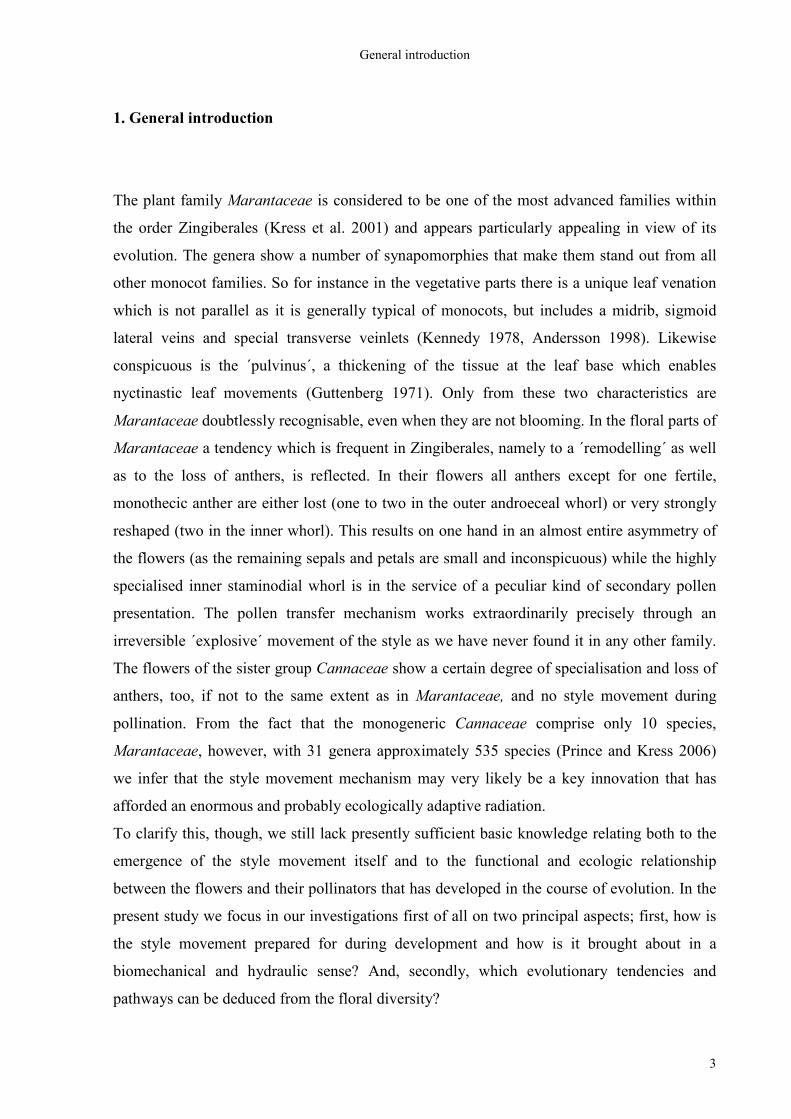
1. General introduction
The plant family Marantaceae is considered to be one of the most advanced families within
the order Zingiberales (Kress et al. 2001) and appears particularly appealing in view of its
evolution. The genera show a number of synapomorphies that make them stand out from all
other monocot families. So for instance in the vegetative parts there is a unique leaf venation
which is not parallel as it is generally typical of monocots, but includes a midrib, sigmoid
lateral veins and special transverse veinlets (Kennedy 1978, Andersson 1998). Likewise
conspicuous is the ´pulvinus´, a thickening of the tissue at the leaf base which enables
nyctinastic leaf movements (Guttenberg 1971). Only from these two characteristics are
Marantaceae doubtlessly recognisable, even when they are not blooming. In the floral parts of
Marantaceae a tendency which is frequent in Zingiberales, namely to a ´remodelling´ as well
as to the loss of anthers, is reflected. In their flowers all anthers except for one fertile,
monothecic anther are either lost (one to two in the outer androeceal whorl) or very strongly
reshaped (two in the inner whorl). This results on one hand in an almost entire asymmetry of
the flowers (as the remaining sepals and petals are small and inconspicuous) while the highly
specialised inner staminodial whorl is in the service of a peculiar kind of secondary pollen
presentation. The pollen transfer mechanism works extraordinarily precisely through an
irreversible ´explosive´ movement of the style as we have never found it in any other family.
The flowers of the sister group Cannaceae show a certain degree of specialisation and loss of
anthers, too, if not to the same extent as in Marantaceae, and no style movement during
pollination. From the fact that the monogeneric Cannaceae comprise only 10 species,
Marantaceae, however, with 31 genera approximately 535 species (Prince and Kress 2006)
we infer that the style movement mechanism may very likely be a key innovation that has
afforded an enormous and probably ecologically adaptive radiation.
To clarify this, though, we still lack presently sufficient basic knowledge relating both to the
emergence of the style movement itself and to the functional and ecologic relationship
between the flowers and their pollinators that has developed in the course of evolution. In the
present study we focus in our investigations first of all on two principal aspects; first, how is
the style movement prepared for during development and how is it brought about in a
biomechanical and hydraulic sense? And, secondly, which evolutionary tendencies and
pathways can be deduced from the floral diversity?
General introduction
4
For the biomechanical question we examine at first in which way and in which time of bud
development the biomechanical potential for the style movement is built up and in a following
examination we look into the question how the tissue of the style is ´designed´ to enable such
an enormous movement. For the examination of the evolutionary pathways a single floral
organ, namely the cucullate staminode of the inner androeceal whorl, is brought in because it
has obviously an important share in the style movement and, moreover, it displays a
conspicuous diversity and appears to be of high diagnostic value. The ´bauplan´ of the
cucullate staminode is determined and its diversity is compared with an already available
phylogenetic analysis (Prince and Kress 2006) and conceivable evolutionary pathways are
deduced and discussed.
The setting-up of tension in the style of Maranta noctiflora
5
2. The setting-up of tension in the style of Maranta noctiflora
Abstract
Marantaceae stand out from other plant families through their unique ´style movement´ that
is combined with a highly derived form of secondary pollen presentation. Though known for
a long time the mechanism underlying the movement is not yet understood. In the present
paper we report about an investigation into the biomechanical principles of the movement.
For the first time we confirm by experiment that in the case of Maranta noctiflora
longitudinal growth of the maturing style within the ´straitjacket´ of the hooded staminode
involves both the arresting of the style before tripping and the building up of the potential for
the movement. The longer the style grows in relation to the enclosing hooded staminode the
more does its capacity for curling up increase. Hereby we distinguish between the ´basic
tension´ that a growing style builds up anyway, even when the hooded staminode is removed
beforehand, and the ´induced tension´ which comes about only under the pressure of a ´too
short´ hooded staminode and which enables the movement. The results of our investigations
are discussed in view of previous interpretations ranging from biomechanical to
electrophysiological mechanisms.
2.1 Introduction
Marantaceae (31/535) are characterised by an explosive pollination mechanism which
includes a unique ´style movement´ and a highly derived form of secondary pollen
presentation (Kunze 1984, Claßen-Bockhoff 1991, Yeo 1993, Kennedy 2000, Locatelli et al.
2004). Though known for a long time the mechanism underlying the movement is not yet
understood. In the present paper we report about an experimental investigation into the
biomechanical principles of the movement.
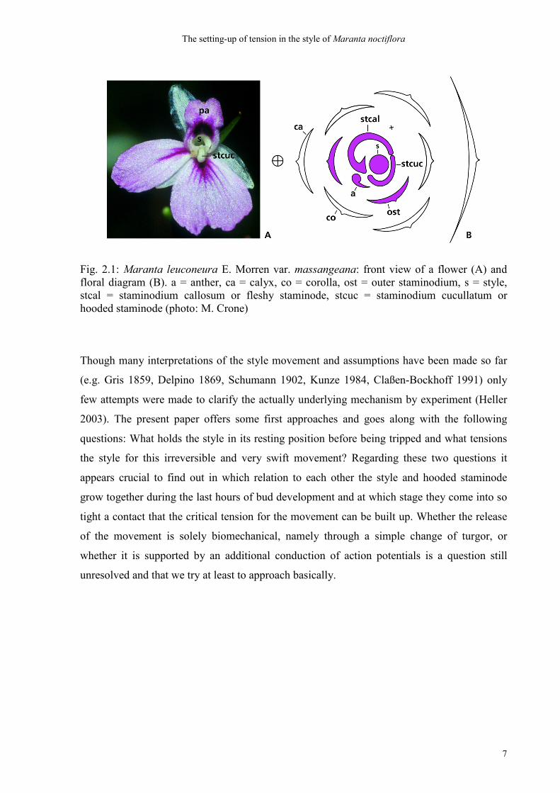
2.1.1 The Flowers in Marantaceae
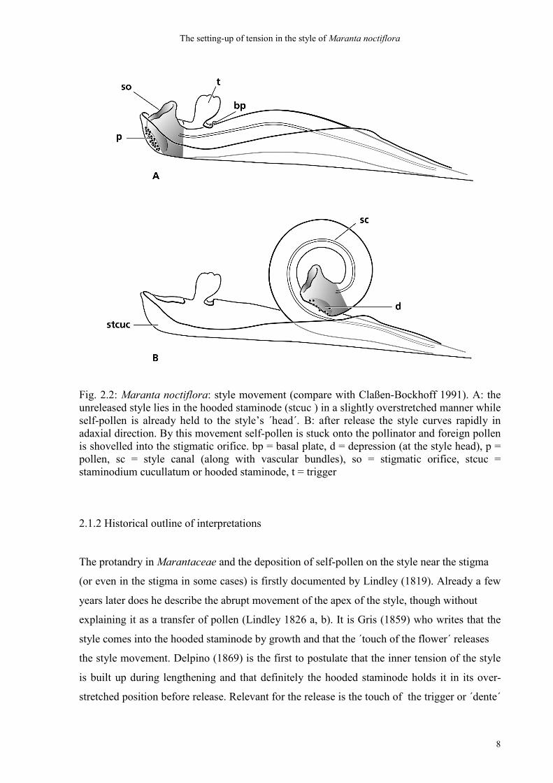
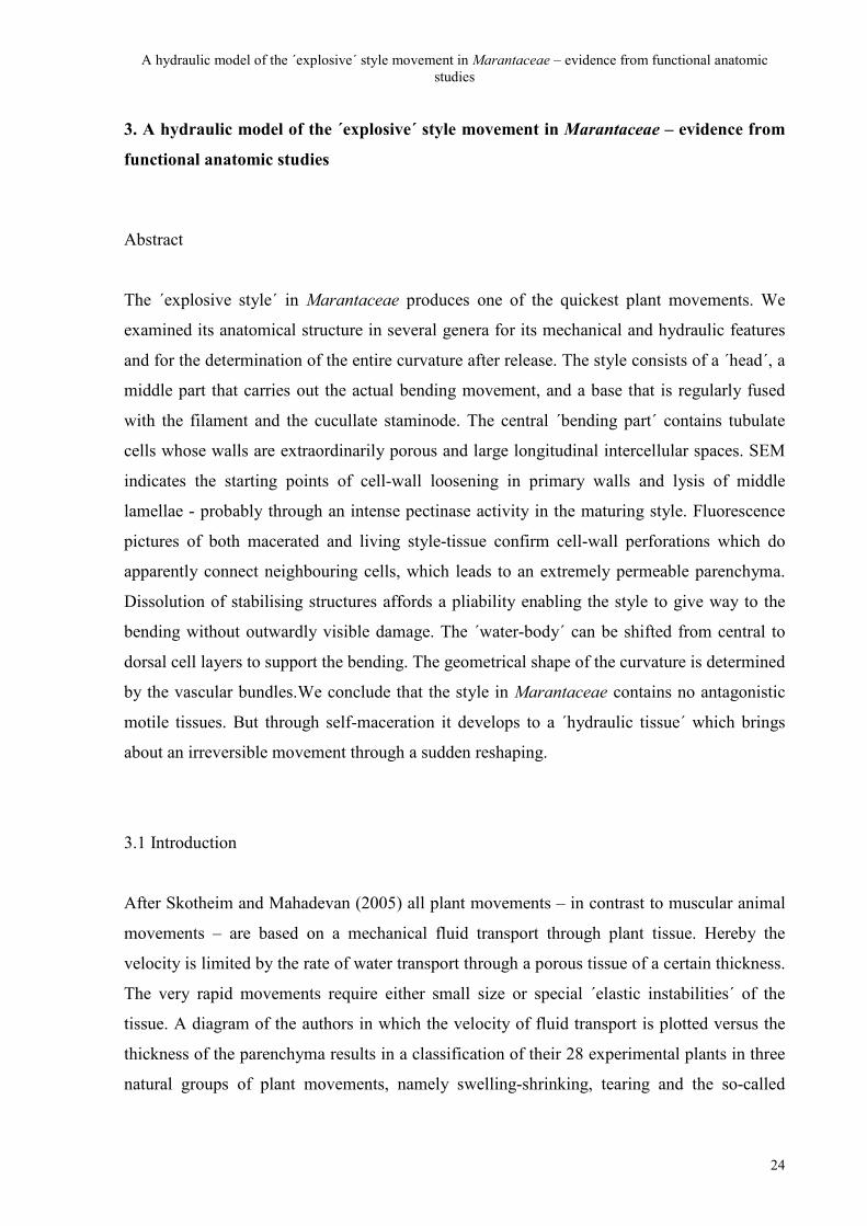
Flowers of Marantaceae are asymmetric (Fig. 2.1 A, B) and mostly arranged in pairs, being
mirror images of each other (Kennedy 1978). The perianth is usually inconspicuous and the
The setting-up of tension in the style of Maranta noctiflora
6
showy parts of the flowers are staminodes. One or two outer staminodes are petaloid (Fig. 2.1
B: ost) while the inner androeceal whorl is differentiated into functionally modified
structures: the fleshy staminode (staminodium callosum; Fig. 2.1 B: stcal), which forms the
upper part of the androeceal tube and which bears a petaloid appendage in some species (Fig.
2.1 A: pa), the hooded staminode (staminodium cucullatum; Fig. 2.1: stcuc), which encloses
the style, and the single stamen with its monothecic anther (Fig. 2.1 B: a) and petaloid
appendage. Usually pollen is forced out of the anther before anthesis by the growing style
(Kennedy 2000), hereby giving an example of extreme protandry (Heller 2003). It gets firmly
attached to a small dorsal depression at the head of the style (Kennedy 1978, ´stamp´ of
Andersson 1981; Fig. 2.2 B: d) by means of a pollen coat substance (´mucus´ of Gris 1859).
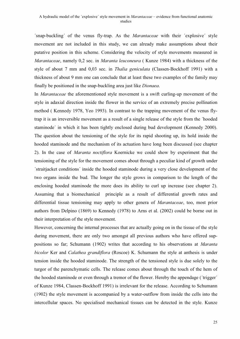
At anthesis the style lies in the hooded staminode in a more or less ´overstretched´ way (Fig.
2.2 A). When an insect enters the flower it touches the so-called ´trigger´ (Kunze 1984,
Claßen-Bockhoff 1991) or ´appendage´ (Andersson 1998) (Fig. 2.2 A: t) and the style curls up
in adaxial (ventral) direction immediately (Fig. 2.2 B). In the course of this single movement
the foreign pollen is scraped from the insect´s body and scooped into the stigmatic orifice
(Locatelli et al. 2004). At the same time the self-pollen at the style head gets attached to the
pollinator´s body by an adhesive secretion (Claßen-Bockhoff 1991, Yeo 1993). The pollen
transfer is thus completed in a single and irreversible action and each flower has but one
chance of being cross-pollinated (Kennedy 1978). The movement of the style goes very
swiftly; slow motion pictures revealed that in Maranta leuconeura E. Morren the entire
movement of the style was completed in 0,2 sec. (Kunze 1984). Also in a high frequency film
it was shown that in Thalia geniculata L. the mere exchange of pollen was performed in a
short space of even 0,03 sec. within the course of the complete style movement (Claßen-
Bockhoff 1991).
The setting-up of tension in the style of Maranta noctiflora
7
Fig. 2.1: Maranta leuconeura E. Morren var. massangeana: front view of a flower (A) and floral diagram (B). a = anther, ca = calyx, co = corolla, ost = outer staminodium, s = style, stcal = staminodium callosum or fleshy staminode, stcuc = staminodium cucullatum or hooded staminode (photo: M. Crone)
Though many interpretations of the style movement and assumptions have been made so far
(e.g. Gris 1859, Delpino 1869, Schumann 1902, Kunze 1984, Claßen-Bockhoff 1991) only
few attempts were made to clarify the actually underlying mechanism by experiment (Heller
2003). The present paper offers some first approaches and goes along with the following
questions: What holds the style in its resting position before being tripped and what tensions
the style for this irreversible and very swift movement? Regarding these two questions it
appears crucial to find out in which relation to each other the style and hooded staminode
grow together during the last hours of bud development and at which stage they come into so
tight a contact that the critical tension for the movement can be built up. Whether the release
of the movement is solely biomechanical, namely through a simple change of turgor, or
whether it is supported by an additional conduction of action potentials is a question still
unresolved and that we try at least to approach basically.
The setting-up of tension in the style of Maranta noctiflora
8
Fig. 2.2: Maranta noctiflora: style movement (compare with Claßen-Bockhoff 1991). A: the unreleased style lies in the hooded staminode (stcuc ) in a slightly overstretched manner while self-pollen is already held to the style’s ´head´. B: after release the style curves rapidly in adaxial direction. By this movement self-pollen is stuck onto the pollinator and foreign pollen is shovelled into the stigmatic orifice. bp = basal plate, d = depression (at the style head), p = pollen, sc = style canal (along with vascular bundles), so = stigmatic orifice, stcuc = staminodium cucullatum or hooded staminode, t = trigger
2.1.2 Historical outline of interpretations
The protandry in Marantaceae and the deposition of self-pollen on the style near the stigma
(or even in the stigma in some cases) is firstly documented by Lindley (1819). Already a few
years later does he describe the abrupt movement of the apex of the style, though without
explaining it as a transfer of pollen (Lindley 1826 a, b). It is Gris (1859) who writes that the
style comes into the hooded staminode by growth and that the ´touch of the flower´ releases
the style movement. Delpino (1869) is the first to postulate that the inner tension of the style
is built up during lengthening and that definitely the hooded staminode holds it in its over-
stretched position before release. Relevant for the release is the touch of the trigger or ´dente´
The setting-up of tension in the style of Maranta noctiflora
9
by a pollinator. Hildebrand (1870) follows him in this opinion and adds that the pollen
transfer is carried out during the released movement, hereby preventing autogamy. He
interprets the bending of the style as a consequence of differential tensioning of cell-layers in
both ´sides of the style´. Eichler (1884) makes even more differentiated observations: The
style is enclosed in the hooded staminode and held in its resting position by the upper cap (i.e.
the ´hood´) and is, as a consequence of stronger longitudinal growth at the dorsal side, arch-
like tensioned. Touching the hook-like appendage loosens the hood and releases the
movement. Eichler (1884) confirms Hildebrand´s (1870) opinion that autogamy is hindered
by this method and cross pollination supported. The appendage serves the release of the
movement, but also strong vibrations can release the style. Self-release is still doubted, but
not absolutely excluded by Eichler (1884). Schumann (1902) bears Eichler out in all his
observations relating to the inner tension and the hold of the style, but doubts plainly the
importance of the appendage for the release mechanism because simply the touch of the edge
of the hooded staminode or even just the tremor of the entire flower releases the style. In any
case, an insect is believed to bring about the relief from the tautness. He also agrees that auto-
gamy is prevented or at least impeded by this method of pollination. Furthermore, he says that
no mechanical tissues can be found inside the style and that the strength of the style (as well
as the fleshy staminode) is due only to the turgor of the parenchymatic tissues in these organs.
According to Schumann (1902) the movement is made possible through a shift of water from
inside the cells into the intercellular spaces. Costerus (1918) sees the style held in position by
the hooded staminode and the adnate anther and the release brought about through either
´exciting the style´ or ´pressing the tooth to the other side of the hood´. Loesener (1930)
comes to the conclusion that the style is in tension, being held in its position by the hooded
staminode. Hereby the displacement of the ´little ear´ (i.e. the trigger, the authors) brings
about the movement. Herein he is supported by Kennedy (1978). Petersen (1981) observes
that an ´object´ entering the flower and touching the appendage loosens the hood and hereby
releases the style. Andersson (1998) comes to the conclusion that the style is in tension due to
´differential lengthening´, being held in its backwards curved position by the ´hood-shaped´
staminodium cucullatum. Hereby the displacement of the ´appendage´ actually triggers off the
pollination mechanism. Andersson´s notion of a differential lengthening is supported by
Claßen-Bockhoff and Pischtschan (2000) who illustrate that the parenchymatic cells of the
ventral and dorsal sides of the style alter their length and volume with the style movement.
Kennedy (2000) speaks of a ´static equilibrium´ in which the tensioned style is held by the
The setting-up of tension in the style of Maranta noctiflora
10
hooded staminode and she supports the observation that the trigger or appendage releases the
style movement. In contrast to suppositions of Hildebrand (1870) and Eichler (1884) she
confirms autogamy in twelve genera. Arns et al. (2002) follow most previous authors in the
sense that the style is in tension, being held in its position by the hooded staminode before
visited by a pollinator. The touch of the ´apendice´ is the cause of the movement. Hereby the
authors make more differentiated observations relating to the position of the appendage and
the form of the curvature of the released style in different genera. Altogether, through
observations of many authors up to now, a rather uniform picture comes about including an
extremely close biomechanic connection between the style and the hooded staminode with the
style being held in its position against its inner tension by the hooded staminode. Hereby
either vibrations, an ´excitement´ or a displacement of the trigger bring about the release. Entirely in contrast to all prior interpretations are the results of Kunze´s (1984) investigations
of Maranta leuconeura and Calathea undulata Lind. et Aust., indicating that the hooded
staminode cannot in any case be strong enough to hold the tensioned style, an opinion in
which he is followed by Yeo (1993). Kunze (1984) postulates that the style only on being
excited at the side leaning against the basal plate of the trigger builds up the tension which is
then rapidly converted into the movement by an immediate change of turgor. This change of
turgor is accompanied by a shift of water into the intercellular spaces, an opinion already
given by Schumann (1902). The idea of a thigmonastic movement (see Costerus 1918) is
strongly confirmed by observations of Claßen-Bockhoff (1991) at Thalia geniculata L.. She
says that the inner tension of the style is built up during a phase of enormous lengthening
inside the bud. The mature style is directly excitable in the area at which rests the basal plate
of the trigger. She also confirms self-release in Thalia geniculata. The high speed of the style
movement (Claßen-Bockhoff 1991) which is faster than mere diffusion may point to an
electrophysiological reaction to trigger off the movement. An extraordinary interpretation of
the style movement is given by Howell et al. (1993), namely that in the case of Calathea
zebrina (Sims) Lindley the style can reset. This is a completely erroneous statement founded
on a description of the pollination process given by Petersen (1981: 531) where it is said
“.....in the course of its movement, the style curves back, blocking the way to the nectar.....”.
The expression ´curves back´ is here obviously misinterpreted in the sense of a reversibility of
the style. In fact, it means exactly the regular curling-up movement in adaxial (or ventral)
direction. Thorough investigations on our part have proven that there is no reset of the style in
Calathea zebrina.
The setting-up of tension in the style of Maranta noctiflora
11
2.2 Materials and methods
2.2.1 Materials
Living plants of the neotropical species Maranta depressa E. Morren and Maranta noctiflora
Koernicke were chosen for the experiments. Both are cultivated in the greenhouses of the
Botanical Garden of the University of Mainz, Germany. Flowers open at 8°° and 20°°,
respectively, and wilt after few hours. Buds develop almost exactly simultaneously what
makes them very suitable for our experiments
2.2.2 Methods
The main growth phase of the buds was determined in Maranta depressa. A total of 23
equally old buds from different individuals were labelled at 14°°, i. e. eighteen hours before
anthesis. Every two hours (14°° to 2°°) and every hour (2°° to 6°°) their length was measured
from the base of the sepals (above the ovary) to the tip of the bud. The examinations were
carried out at 32° Celsius.
The ´growth dynamics´ in Maranta depressa revealed that the strongest longitudinal growth
took place at night during the last hours before anthesis. To have a more convenient test plant
for the experiments we turned to Maranta noctiflora which regularly flowers at night and has
provided us with a good number of simultaneously developing buds during the afternoon.
A total of 42 flowers were labelled at 13°°, i. e 7 hours before anthesis. In regular intervals of
90 minutes 6-8 fresh buds were picked from the plant to measure the length of the bud as in
M. depressa. Then the styles were released by hand manipulation at the ´trigger´ and their
length was measured by means of a thread going along with the entire dorsal line from the
distal edge of the ovary to the rim of the stigmatic orifice as well as the length of the hooded
staminodes in the corresponding places. The style´s increasing capacity for curling up at each
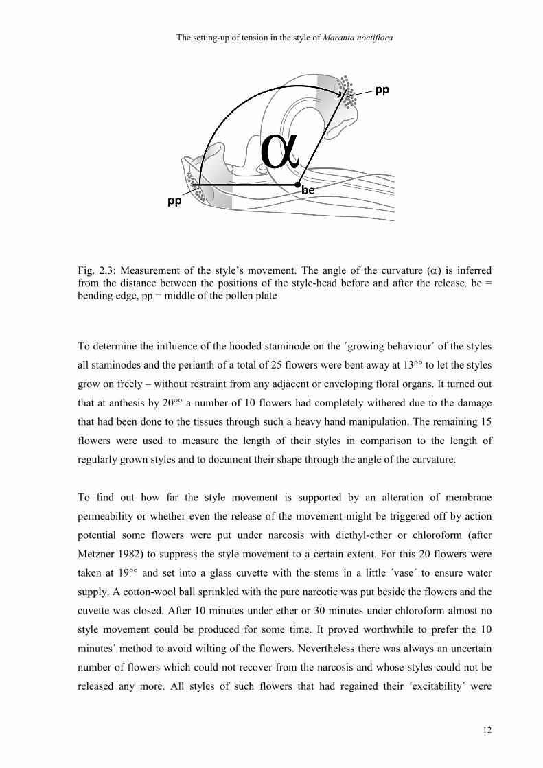
time was determined by measuring the angle of curvature (see Fig. 2.3: α ).
The setting-up of tension in the style of Maranta noctiflora
12
Fig. 2.3: Measurement of the style’s movement. The angle of the curvature (α) is inferred from the distance between the positions of the style-head before and after the release. be = bending edge, pp = middle of the pollen plate
To determine the influence of the hooded staminode on the ´growing behaviour´ of the styles
all staminodes and the perianth of a total of 25 flowers were bent away at 13°° to let the styles
grow on freely – without restraint from any adjacent or enveloping floral organs. It turned out
that at anthesis by 20°° a number of 10 flowers had completely withered due to the damage
that had been done to the tissues through such a heavy hand manipulation. The remaining 15
flowers were used to measure the length of their styles in comparison to the length of
regularly grown styles and to document their shape through the angle of the curvature.
To find out how far the style movement is supported by an alteration of membrane
permeability or whether even the release of the movement might be triggered off by action
potential some flowers were put under narcosis with diethyl-ether or chloroform (after
Metzner 1982) to suppress the style movement to a certain extent. For this 20 flowers were
taken at 19°° and set into a glass cuvette with the stems in a little ´vase´ to ensure water
supply. A cotton-wool ball sprinkled with the pure narcotic was put beside the flowers and the
cuvette was closed. After 10 minutes under ether or 30 minutes under chloroform almost no
style movement could be produced for some time. It proved worthwhile to prefer the 10
minutes´ method to avoid wilting of the flowers. Nevertheless there was always an uncertain
number of flowers which could not recover from the narcosis and whose styles could not be
released any more. All styles of such flowers that had regained their ´excitability´ were
The setting-up of tension in the style of Maranta noctiflora
13
afterwards released by hand manipulation. Their length and the length of the hooded
staminodes and the angle of the curvature were measured in the usual way.
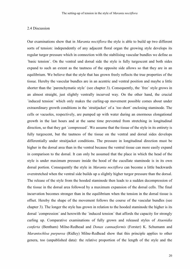
2.3 Results
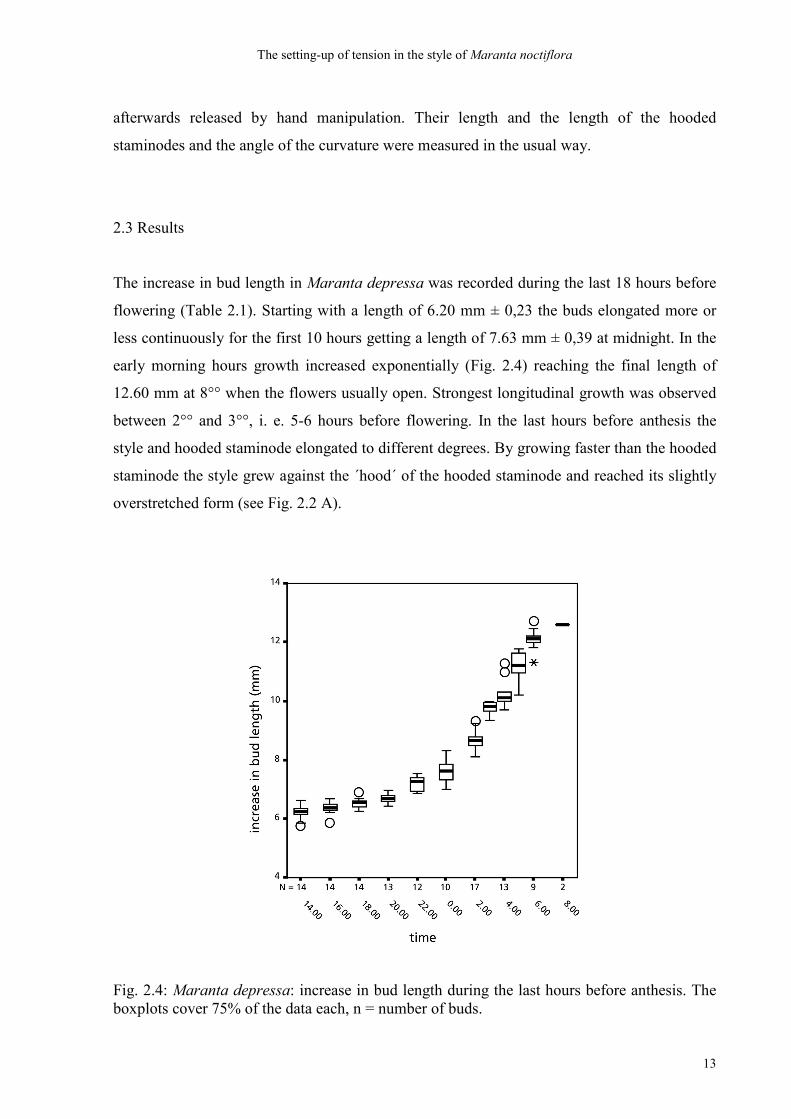
The increase in bud length in Maranta depressa was recorded during the last 18 hours before
flowering (Table 2.1). Starting with a length of 6.20 mm ± 0,23 the buds elongated more or
less continuously for the first 10 hours getting a length of 7.63 mm ± 0,39 at midnight. In the
early morning hours growth increased exponentially (Fig. 2.4) reaching the final length of
12.60 mm at 8°° when the flowers usually open. Strongest longitudinal growth was observed
between 2°° and 3°°, i. e. 5-6 hours before flowering. In the last hours before anthesis the
style and hooded staminode elongated to different degrees. By growing faster than the hooded
staminode the style grew against the ´hood´ of the hooded staminode and reached its slightly
overstretched form (see Fig. 2.2 A).
Fig. 2.4: Maranta depressa: increase in bud length during the last hours before anthesis. The boxplots cover 75% of the data each, n = number of buds.
The setting-up of tension in the style of Maranta noctiflora
14
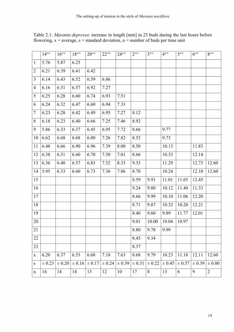
Table 2.1: Maranta depressa: increase in length [mm] in 23 buds during the last hours before flowering, x = average, s = standard deviation, n = number of buds per time unit
14°° 16°° 18°° 20°° 22°° 24°° 2°° 3°° 4°° 5°° 6°° 8°°
1 5.76 5.87 6.25
2 6.21 6.39 6.41 6.42
3 6.14 6.43 6.52 6.59 6.86
4 6.16 6.51 6.57 6.92 7.27
5 6.25 6.28 6.60 6.74 6.93 7.51
6 6.24 6.32 6.47 6.60 6.94 7.33
7 6.23 6.28 6.42 6.49 6.95 7.27 8.12
8 6.18 6.23 6.40 6.66 7.25 7.46 8.92
9 5.86 6.33 6.37 6.45 6.95 7.72 8.66 9.77
10 6.62 6.68 6.68 6.80 7.26 7.82 8.52 9.73
11 6.48 6.66 6.90 6.96 7.39 8.00 8.50 10.13 11.83
12 6.38 6.51 6.60 6.70 7.50 7.01 8.66 10.33 12.14
13 6.36 6.40 6.57 6.83 7.52 8.33 9.33 11.29 12.73 12.60
14 5.95 6.33 6.60 6.73 7.36 7.86 8.70 10.24 12.10 12.60
15 8.59 9.91 11.01 11.65 12.45
16 9.24 9.80 10.12 11.40 11.33
17 8.66 9.99 10.10 11.06 12.20
18 8.71 9.87 10.32 10.20 12.21
19 8.40 9.60 9.89 11.77 12.01
20 9.01 10.00 10.04 10.97
21 8.80 9.78 9.99
22 8.45 9.34
23 8.37
x 6.20 6.37 6.53 6.68 7.18 7.63 8.68 9.79 10.23 11.18 12.11 12.60
s ± 0.23 ± 0.20 ± 0.16 ± 0.17 ± 0.24 ± 0.39 ± 0.31 ± 0.22 ± 0.45 ± 0.57 ± 0.39 ± 0.00
n 14 14 14 13 12 10 17 8 13 6 9 2
The setting-up of tension in the style of Maranta noctiflora
15
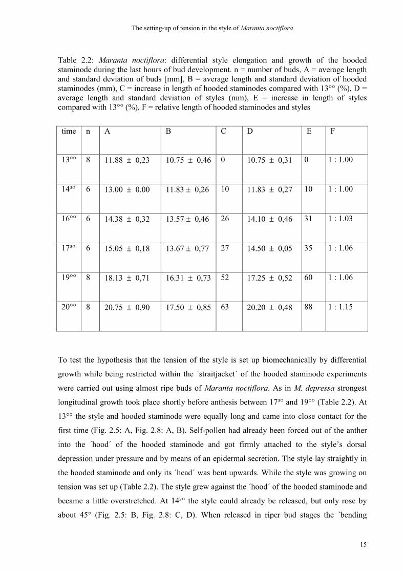
Table 2.2: Maranta noctiflora: differential style elongation and growth of the hooded staminode during the last hours of bud development. n = number of buds, A = average length and standard deviation of buds [mm], B = average length and standard deviation of hooded staminodes (mm), C = increase in length of hooded staminodes compared with 13°° (%), D = average length and standard deviation of styles (mm), E = increase in length of styles compared with 13°° (%), F = relative length of hooded staminodes and styles
time n
A B C D E F
13°° 8 11.88 ± 0,23 10.75 ± 0,46 0 10.75 ± 0,31 0 1 : 1.00
14³° 6 13.00 ± 0.00 11.83 ± 0,26 10 11.83 ± 0,27 10 1 : 1.00
16°° 6 14.38 ± 0,32 13.57 ± 0,46 26 14.10 ± 0,46 31 1 : 1.03
17³° 6 15.05 ± 0,18 13.67 ± 0,77 27 14.50 ± 0,05 35 1 : 1.06
19°° 8 18.13 ± 0,71 16.31 ± 0,73 52 17.25 ± 0,52 60 1 : 1.06
20°° 8 20.75 ± 0,90 17.50 ± 0,85 63 20.20 ± 0,48 88 1 : 1.15
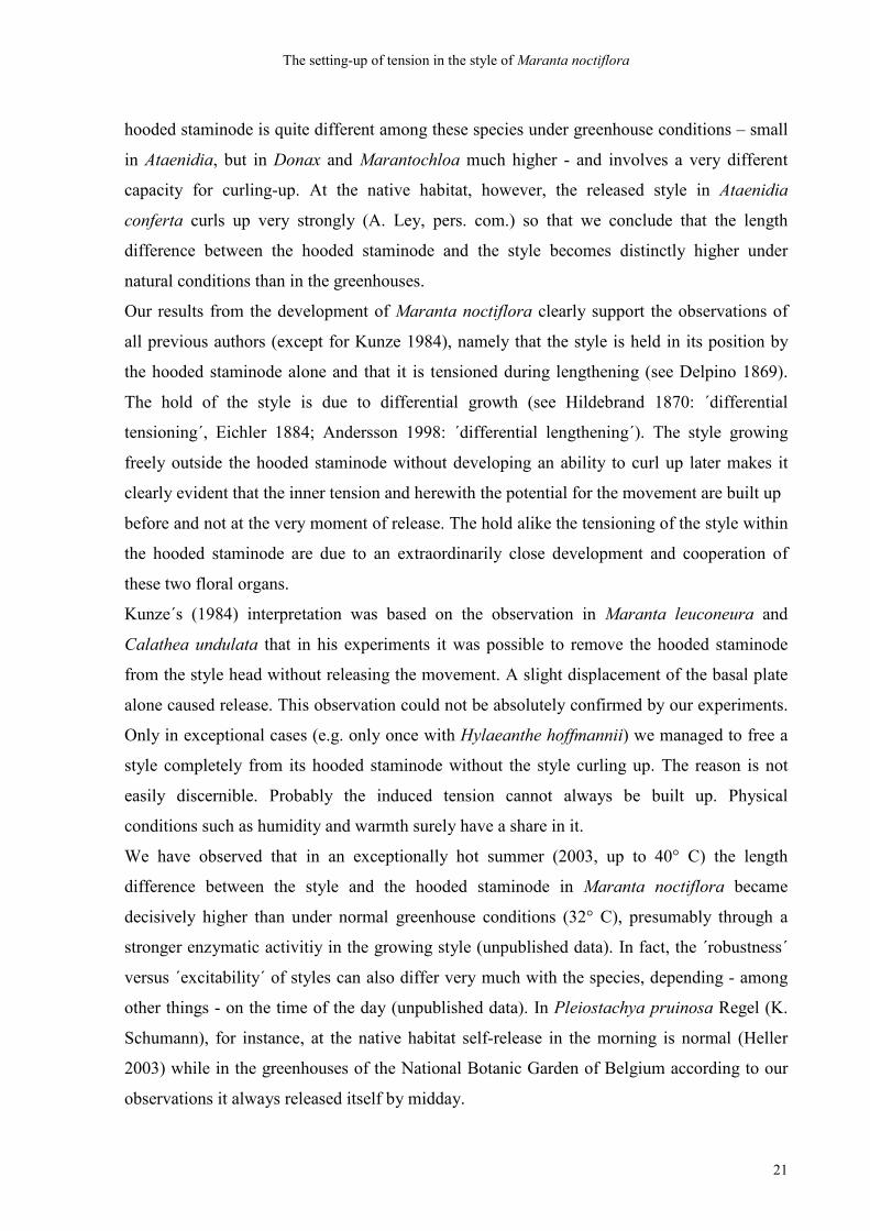
To test the hypothesis that the tension of the style is set up biomechanically by differential
growth while being restricted within the ´straitjacket´ of the hooded staminode experiments
were carried out using almost ripe buds of Maranta noctiflora. As in M. depressa strongest
longitudinal growth took place shortly before anthesis between 17³° and 19°° (Table 2.2). At
13°° the style and hooded staminode were equally long and came into close contact for the
first time (Fig. 2.5: A, Fig. 2.8: A, B). Self-pollen had already been forced out of the anther
into the ´hood´ of the hooded staminode and got firmly attached to the style’s dorsal
depression under pressure and by means of an epidermal secretion. The style lay straightly in
the hooded staminode and only its ´head´ was bent upwards. While the style was growing on
tension was set up (Table 2.2). The style grew against the ´hood´ of the hooded staminode and
became a little overstretched. At 14³° the style could already be released, but only rose by
about 45° (Fig. 2.5: B, Fig. 2.8: C, D). When released in riper bud stages the ´bending
The setting-up of tension in the style of Maranta noctiflora
16
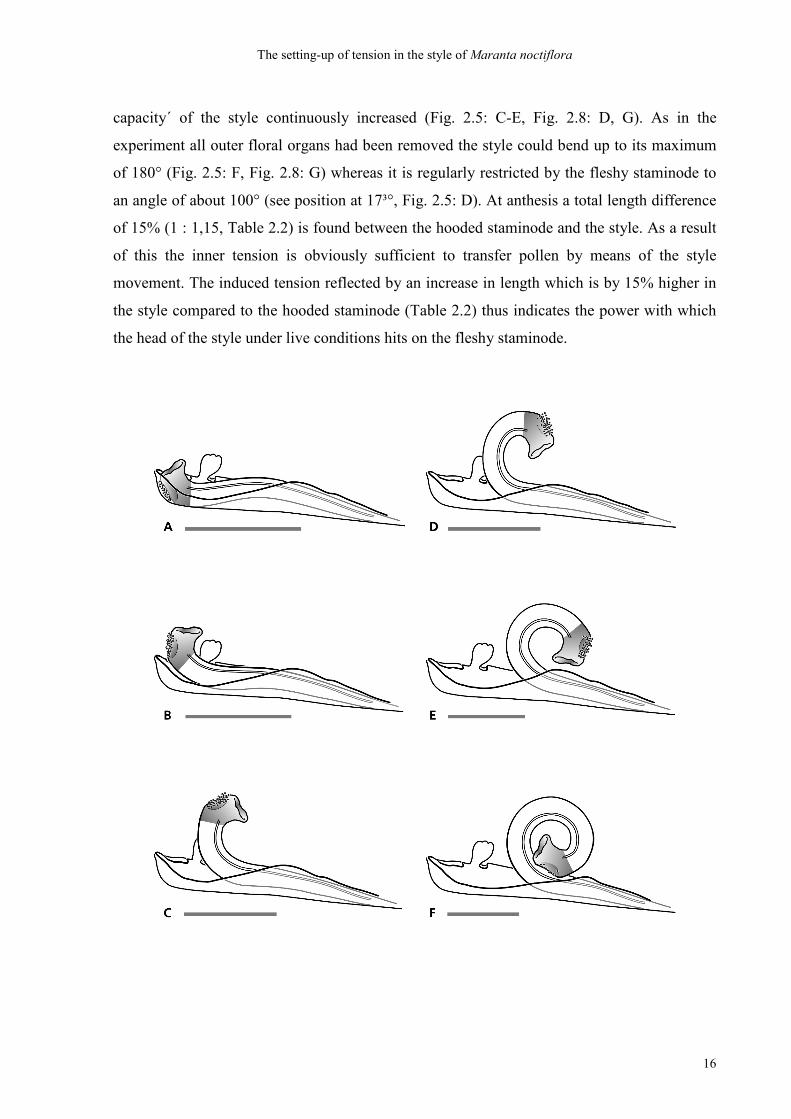
capacity´ of the style continuously increased (Fig. 2.5: C-E, Fig. 2.8: D, G). As in the
experiment all outer floral organs had been removed the style could bend up to its maximum
of 180° (Fig. 2.5: F, Fig. 2.8: G) whereas it is regularly restricted by the fleshy staminode to
an angle of about 100° (see position at 17³°, Fig. 2.5: D). At anthesis a total length difference
of 15% (1 : 1,15, Table 2.2) is found between the hooded staminode and the style. As a result
of this the inner tension is obviously sufficient to transfer pollen by means of the style
movement. The induced tension reflected by an increase in length which is by 15% higher in
the style compared to the hooded staminode (Table 2.2) thus indicates the power with which
the head of the style under live conditions hits on the fleshy staminode.
The setting-up of tension in the style of Maranta noctiflora
17
Fig. 2.5: Maranta noctiflora: increase in the capacity for curling up during the last hours before anthesis (see Table 2.2). Every 1.5 hours flowers were taken from the plant. Styles were released by hand manipulation and their lengths were compared to the ones of the hooded staminodes A 13°°, B 14³°, C 16°°, D 17³°, E 19°°, F 20°° (anthesis). Bar = 5 mm; note that the decreasing length of the bar indicates the growth of the styles and hooded staminodes.
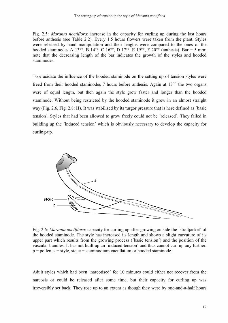
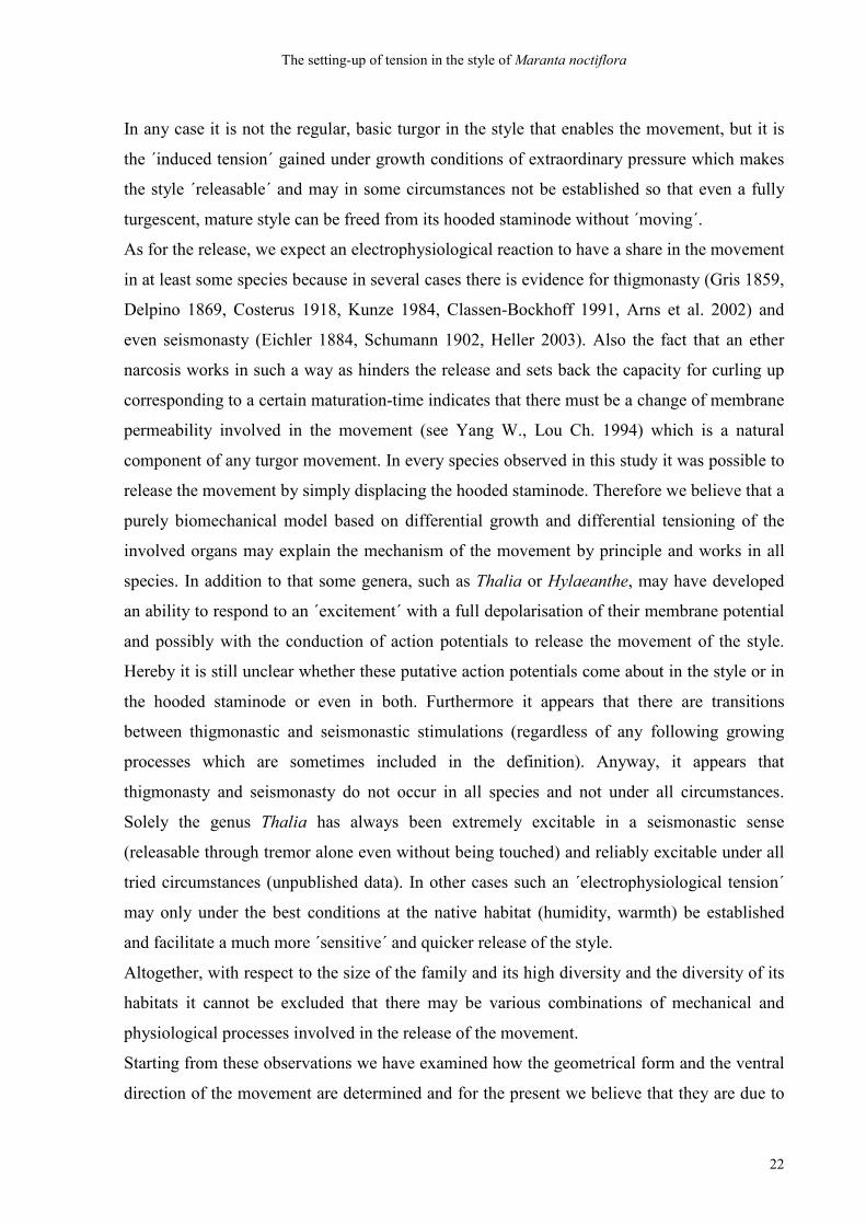
To elucidate the influence of the hooded staminode on the setting up of tension styles were
freed from their hooded staminodes 7 hours before anthesis. Again at 13°° the two organs
were of equal length, but then again the style grew faster and longer than the hooded
staminode. Without being restricted by the hooded staminode it grew in an almost straight
way (Fig. 2.6, Fig. 2.8: H). It was stabilised by its turgor pressure that is here defined as ´basic
tension´. Styles that had been allowed to grow freely could not be ´released´. They failed in
building up the ´induced tension´ which is obviously necessary to develop the capacity for
curling-up.
Fig. 2.6: Maranta noctiflora: capacity for curling up after growing outside the ´straitjacket´ of the hooded staminode. The style has increased its length and shows a slight curvature of its upper part which results from the growing process (´basic tension´) and the position of the vascular bundles. It has not built up an ´induced tension´ and thus cannot curl up any further. p = pollen, s = style, stcuc = staminodium cucullatum or hooded staminode.
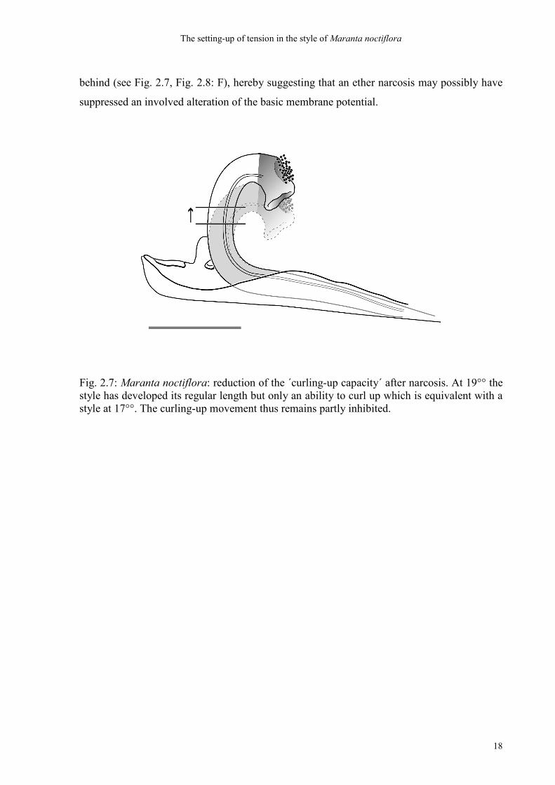
Adult styles which had been ´narcotised´ for 10 minutes could either not recover from the
narcosis or could be released after some time, but their capacity for curling up was
irreversibly set back. They rose up to an extent as though they were by one-and-a-half hours
The setting-up of tension in the style of Maranta noctiflora
18
behind (see Fig. 2.7, Fig. 2.8: F), hereby suggesting that an ether narcosis may possibly have
suppressed an involved alteration of the basic membrane potential.
Fig. 2.7: Maranta noctiflora: reduction of the ´curling-up capacity´ after narcosis. At 19°° the style has developed its regular length but only an ability to curl up which is equivalent with a style at 17°°. The curling-up movement thus remains partly inhibited.
The setting-up of tension in the style of Maranta noctiflora
19
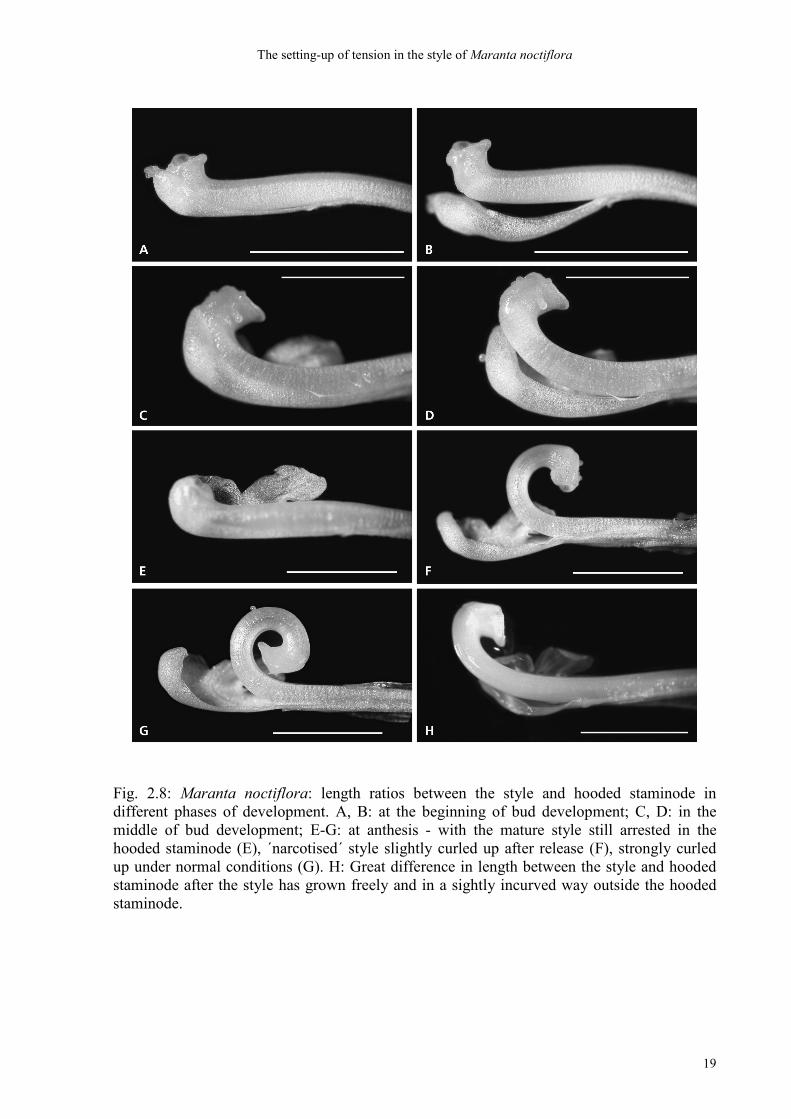
Fig. 2.8: Maranta noctiflora: length ratios between the style and hooded staminode in different phases of development. A, B: at the beginning of bud development; C, D: in the middle of bud development; E-G: at anthesis - with the mature style still arrested in the hooded staminode (E), ´narcotised´ style slightly curled up after release (F), strongly curled up under normal conditions (G). H: Great difference in length between the style and hooded staminode after the style has grown freely and in a sightly incurved way outside the hooded staminode.
The setting-up of tension in the style of Maranta noctiflora
20
2.4 Discussion
Our examinations show that in Maranta noctiflora the style is able to build up two different
sorts of tension: independently of any adjacent floral organ the growing style develops its
regular turgor pressure which in connection with the stabilising vascular bundles we define as
´basic tension´. On the ventral und dorsal side the style is fully turgescent and both sides
expand to such an extent as the tautness of the opposite side allows so that they are in an
equilibrium. We believe that the style that has grown freely reflects the true properties of the
tissue. Hereby the vascular bundles are in an acentric and ventral position and maybe a little
shorter than the ´parenchymatic style´ (see chapter 3). Consequently, the ´free´ style grows in
an almost straight, just slightly ventrally incurved way. On the other hand, the crucial
´induced tension´ which only makes the curling-up movement possible comes about under
extraordinary growth conditions in the ´straitjacket´ of a ´too short´ enclosing staminode. The
cells or vacuoles, respectively, are pumped up with water during an enormous elongational
growth in the last hours and at the same time prevented from stretching in longitudinal
direction, so that they get ´compressed´. We assume that the tissue of the style in its entirety is
fully turgescent, but the tautness of the tissue on the ventral and dorsal sides develops
differentially under straitjacket conditions. The pressure in longitudinal direction must be
higher in the dorsal area than in the ventral because the ventral tissue can more easily expand
in comparison to the dorsal. It can only be assumed that the place in which the head of the
style is under maximum pressure inside the hood of the cucullate staminode is in its own
dorsal portion. Consequently the style in Maranta noctiflora can become a little backwards
overstretched when the ventral side builds up a slightly higher turgor pressure than the dorsal.
The release of the style from the hooded staminode then leads to a sudden decompression of
the tissue in the dorsal area followed by a maximum expansion of the dorsal cells. The final
incurvation becomes stronger than in the equilibrium when the tension in the dorsal tissue is
offset. Hereby the shape of the movement follows the course of the vascular bundles (see
chapter 3). The longer the style has grown in relation to the hooded staminode the higher is its
dorsal ´compression´ and herewith the ´induced tension´ that affords the capacity for strongly
curling up. Comparative examinations of fully grown and released styles of Ataenidia
conferta (Bentham) Milne-Redhead and Donax cannaeformis (Forster) K. Schumann and
Marantochloa purpurea (Ridley) Milne-Redhead show that this principle applies to other
genera, too (unpublished data): the relative proportion of the length of the style and the
The setting-up of tension in the style of Maranta noctiflora
21
hooded staminode is quite different among these species under greenhouse conditions – small
in Ataenidia, but in Donax and Marantochloa much higher - and involves a very different
capacity for curling-up. At the native habitat, however, the released style in Ataenidia
conferta curls up very strongly (A. Ley, pers. com.) so that we conclude that the length
difference between the hooded staminode and the style becomes distinctly higher under
natural conditions than in the greenhouses.
Our results from the development of Maranta noctiflora clearly support the observations of
all previous authors (except for Kunze 1984), namely that the style is held in its position by
the hooded staminode alone and that it is tensioned during lengthening (see Delpino 1869).
The hold of the style is due to differential growth (see Hildebrand 1870: ´differential
tensioning´, Eichler 1884; Andersson 1998: ´differential lengthening´). The style growing
freely outside the hooded staminode without developing an ability to curl up later makes it
clearly evident that the inner tension and herewith the potential for the movement are built up
before and not at the very moment of release. The hold alike the tensioning of the style within
the hooded staminode are due to an extraordinarily close development and cooperation of
these two floral organs.
Kunze´s (1984) interpretation was based on the observation in Maranta leuconeura and
Calathea undulata that in his experiments it was possible to remove the hooded staminode
from the style head without releasing the movement. A slight displacement of the basal plate
alone caused release. This observation could not be absolutely confirmed by our experiments.
Only in exceptional cases (e.g. only once with Hylaeanthe hoffmannii) we managed to free a
style completely from its hooded staminode without the style curling up. The reason is not
easily discernible. Probably the induced tension cannot always be built up. Physical
conditions such as humidity and warmth surely have a share in it.
We have observed that in an exceptionally hot summer (2003, up to 40° C) the length
difference between the style and the hooded staminode in Maranta noctiflora became
decisively higher than under normal greenhouse conditions (32° C), presumably through a
stronger enzymatic activitiy in the growing style (unpublished data). In fact, the ´robustness´
versus ´excitability´ of styles can also differ very much with the species, depending - among
other things - on the time of the day (unpublished data). In Pleiostachya pruinosa Regel (K.
Schumann), for instance, at the native habitat self-release in the morning is normal (Heller
2003) while in the greenhouses of the National Botanic Garden of Belgium according to our
observations it always released itself by midday.
The setting-up of tension in the style of Maranta noctiflora
22
In any case it is not the regular, basic turgor in the style that enables the movement, but it is
the ´induced tension´ gained under growth conditions of extraordinary pressure which makes
the style ´releasable´ and may in some circumstances not be established so that even a fully
turgescent, mature style can be freed from its hooded staminode without ´moving´.
As for the release, we expect an electrophysiological reaction to have a share in the movement
in at least some species because in several cases there is evidence for thigmonasty (Gris 1859,
Delpino 1869, Costerus 1918, Kunze 1984, Classen-Bockhoff 1991, Arns et al. 2002) and
even seismonasty (Eichler 1884, Schumann 1902, Heller 2003). Also the fact that an ether
narcosis works in such a way as hinders the release and sets back the capacity for curling up
corresponding to a certain maturation-time indicates that there must be a change of membrane
permeability involved in the movement (see Yang W., Lou Ch. 1994) which is a natural
component of any turgor movement. In every species observed in this study it was possible to
release the movement by simply displacing the hooded staminode. Therefore we believe that a
purely biomechanical model based on differential growth and differential tensioning of the
involved organs may explain the mechanism of the movement by principle and works in all
species. In addition to that some genera, such as Thalia or Hylaeanthe, may have developed
an ability to respond to an ´excitement´ with a full depolarisation of their membrane potential
and possibly with the conduction of action potentials to release the movement of the style.
Hereby it is still unclear whether these putative action potentials come about in the style or in
the hooded staminode or even in both. Furthermore it appears that there are transitions
between thigmonastic and seismonastic stimulations (regardless of any following growing
processes which are sometimes included in the definition). Anyway, it appears that
thigmonasty and seismonasty do not occur in all species and not under all circumstances.
Solely the genus Thalia has always been extremely excitable in a seismonastic sense
(releasable through tremor alone even without being touched) and reliably excitable under all
tried circumstances (unpublished data). In other cases such an ´electrophysiological tension´
may only under the best conditions at the native habitat (humidity, warmth) be established
and facilitate a much more ´sensitive´ and quicker release of the style.
Altogether, with respect to the size of the family and its high diversity and the diversity of its
habitats it cannot be excluded that there may be various combinations of mechanical and
physiological processes involved in the release of the movement.
Starting from these observations we have examined how the geometrical form and the ventral
direction of the movement are determined and for the present we believe that they are due to
The setting-up of tension in the style of Maranta noctiflora
23
the acentric position and the relative shortness of the vascular bundles (see above).
Furthermore, we know now, entirely in contrast to Schumann´s opinion (1902), that there are
certain characteristics to the inner tissues of the style which allow the style to undergo such an
enormous deformation without visible ruptures and which we call a ´hydraulic´ dorsal tissue.
Here also the question about a shift of water from inside the cells into the intercellular spaces
(Schumann 1902, Kunze 1984) has been examined by means of histological and anatomical
research methods (see chapter 3).
A hydraulic model of the ´explosive´ style movement in Marantaceae – evidence from functional anatomic studies
24
3. A hydraulic model of the ´explosive´ style movement in Marantaceae – evidence from
functional anatomic studies
Abstract
The ´explosive style´ in Marantaceae produces one of the quickest plant movements. We
examined its anatomical structure in several genera for its mechanical and hydraulic features
and for the determination of the entire curvature after release. The style consists of a ´head´, a
middle part that carries out the actual bending movement, and a base that is regularly fused
with the filament and the cucullate staminode. The central ´bending part´ contains tubulate
cells whose walls are extraordinarily porous and large longitudinal intercellular spaces. SEM
indicates the starting points of cell-wall loosening in primary walls and lysis of middle
lamellae - probably through an intense pectinase activity in the maturing style. Fluorescence
pictures of both macerated and living style-tissue confirm cell-wall perforations which do
apparently connect neighbouring cells, which leads to an extremely permeable parenchyma.
Dissolution of stabilising structures affords a pliability enabling the style to give way to the
bending without outwardly visible damage. The ´water-body´ can be shifted from central to
dorsal cell layers to support the bending. The geometrical shape of the curvature is determined
by the vascular bundles.We conclude that the style in Marantaceae contains no antagonistic
motile tissues. But through self-maceration it develops to a ´hydraulic tissue´ which brings
about an irreversible movement through a sudden reshaping.
3.1 Introduction
After Skotheim and Mahadevan (2005) all plant movements – in contrast to muscular animal
movements – are based on a mechanical fluid transport through plant tissue. Hereby the
velocity is limited by the rate of water transport through a porous tissue of a certain thickness.
The very rapid movements require either small size or special ´elastic instabilities´ of the
tissue. A diagram of the authors in which the velocity of fluid transport is plotted versus the
thickness of the parenchyma results in a classification of their 28 experimental plants in three
natural groups of plant movements, namely swelling-shrinking, tearing and the so-called
A hydraulic model of the ´explosive´ style movement in Marantaceae – evidence from functional anatomic studies
25
´snap-buckling´ of the venus fly-trap. As the Marantaceae with their ´explosive´ style
movement are not included in this study, we can already make assumptions about their
putative position in this scheme. Considering the velocity of style movements measured in
Marantaceae, namely 0,2 sec. in Maranta leuconeura ( Kunze 1984) with a thickness of the
style of about 7 mm and 0,03 sec. in Thalia geniculata (Classen-Bockhoff 1991) with a
thickness of about 9 mm one can conclude that at least these two examples of the family may
finally be positioned in the snap-buckling area just like Dionaea.
In Marantaceae the aforementioned style movement is a swift curling-up movement of the
style in adaxial direction inside the flower in the service of an extremely precise pollination
method ( Kennedy 1978, Yeo 1993). In contrast to the trapping movement of the venus fly-
trap it is an irreversible movement as a result of a single release of the style from the ´hooded
staminode´ in which it has been tightly enclosed during bud development (Kennedy 2000).
The question about the tensioning of the style for its rapid shooting up, its hold inside the
hooded staminode and the mechanism of its actuation have long been discussed (see chapter
2). In the case of Maranta noctiflora Koernicke we could show by experiment that the
tensioning of the style for the movement comes about through a peculiar kind of growth under
´straitjacket conditions´ inside the hooded staminode during a very close development of the
two organs inside the bud. The longer the style grows in comparison to the length of the
enclosing hooded staminode the more does its ability to curl up increase (see chapter 2).
Assuming that a biomechanical principle as a result of differential growth rates and
differential tissue tensioning may apply to other genera of Marantaceae, too, most prior
authors from Delpino (1869) to Kennedy (1978) to Arns et al. (2002) could be borne out in
their interpretation of the style movement.
However, concerning the internal processes that are actually going on in the tissue of the style
during movement, there are only two amongst all previous authors who have offered sup-
positions so far; Schumann (1902) writes that according to his observations at Maranta
bicolor Ker and Calathea grandiflora (Roscoe) K. Schumann the style at anthesis is under
tension inside the hooded staminode. The strength of the tensioned style is due solely to the
turgor of the parenchymatic cells. The release comes about through the touch of the hem of
the hooded staminode or even through a tremor of the flower. Hereby the appendage (´trigger´
of Kunze 1984, Classen-Bockhoff 1991) is irrelevant for the release. According to Schumann
(1902) the style movement is accompanied by a water-outflow from inside the cells into the
intercellular spaces. No specialised mechanical tissues can be detected in the style. Kunze
A hydraulic model of the ´explosive´ style movement in Marantaceae – evidence from functional anatomic studies
26
(1984) on the other hand says that according to his investigations of Maranta leuconeura E.
Morren and Calathea undulata Linden et André the tautness of the style is built up not before
release but only at the very moment of an ´excitement´ of the style through a slight
displacement of the basal plate. Hereby the change of turgor comes about through a water-
outflow from the cells into the intercellular spaces at the ´upper side´ (i.e. the ventral side; the
authors) of the style and enables its shooting up. Only in this point, concerning a shift of
water through the intercellular spaces, is Kunze (1984) in accordance with Schumann (1902).
Up to now no experimental investigation has been made to form an idea of the hydraulic
principles of the movement and to make out the special properties of the style tissue that make
such a strong curvature possible without outwardly visible ruptures. The only discernible
difference detected so far between unreleased and released styles is that the length and
volume of dorsal cells in Maranta leuconeura increase after the movement (Claßen-Bockhoff
and Pischtschan 2000).
In this study we start from the assumption that there must be structures inside the style that
determine the shape alike the adaxial direction of the movement. Furthermore, entirely in
contrast to Schumann´s opinion (1902) that there exist no mechanical tissues, we expect to
find at least certain characteristics to the inner tissues of the style because the demands on this
tissue are extreme: to undergo such an enormous bending without visible damage the tissue
needs an extraordinary pliabilty. The fact that the length and volume of dorsal cells in the
style increase after release in Maranta leuconeura indicates that there is indeed a ´water body´
to be shifted - presumably from central to periferal (mainly dorsal) cell layers which requires
an enhanced hydraulic permeability of the parenchyma.
We present investigations that we made to show by which means the style in several genera of
Marantaceae meets these special mechanical and hydraulic requirements.
3.2 Materials and methods
3.2.1 Materials
The following species are chosen for the experiments: Calathea veitchiana Hook, f., Donax
cannaeformis (G. Forster) K. Schumann, Halopegia azurea K. Schumann, Maranta depressa
E. Morren, Maranta leuconeura E. Morren, Maranta noctiflora Koernicke, Marantochloa
A hydraulic model of the ´explosive´ style movement in Marantaceae – evidence from functional anatomic studies
27
purpurea (Ridley) Milne-Redhead, Thalia geniculata Linné. Furthermore, the following
members of the order Zingiberales are selected for investigations: Canna indica Linné, Costus
pictus G. Don, Zingiber spectabile Griffith. They are all cultivated in the greenhouses of the
Botanical Garden of the University of Mainz, Germany.
3.2.2 Methods
3.2.2.1 Cellulose detection
To find out in advance which materials contribute to the solidity of the style or whether the
parenchyma contains only primary or already secondary cell walls the Lugol test is performed
on Maranta leuconeura. Tissue samples of fresh styles are put in a watch-glass with Lugol
solution (2g KJ, 1g J, 100ml aqua destillata) for ten minutes. The pieces are then transferred
to a slide with a drop of the same solution and covered with a cover slip. When some thinned
sulfuric acid (H2SO4 : H2O = 2:1) is drawn through the slide preparation cellulose turns blue
whereas lignin, suberin and cutin turn yellow.
3.2.2.2 Histology
Styles of Maranta leuconeura, Halopegia azurea and Thalia geniculata and Marantochloa
purpurea are selected for sectioning. Unreleased styles are obtained by applying a 10´ ether
narcosis to the flowers immediately before fixation (see chapter 2). In the case of Thalia
geniculata, as these styles are extremely easily released, a 60´ narcosis with chloroform is
carried out. The released and unreleased styles are first fixed in a solution of 1 %
glutaraldehyde, 4 % formaldehyde and 10 % ethanol in a potassium-sodium-phosphate-buffer
for at least a week. After two days´ evacuation in an exsiccator, the tissue is gradually
dehydrated in an alcohol sequence starting with 15 % and ascending in unusually small steps
of 5 % (2 h each) in order to avoid artifacts in this very fragile tissue up to 96 % ethanol in
which they stay over night. In the preinfiltration medium (ethanol 96 % and Historesin
infiltration-medium Leica 1:1) the material remains for at least four days to prevent pieces of
style tissue from falling out of the blocks during sectioning. In the pure infiltration medium
(Historesin, Leica) the tissue rests for another two days. The samples are then embedded in
A hydraulic model of the ´explosive´ style movement in Marantaceae – evidence from functional anatomic studies
28
the embedding medium (Historesin infiltration-medium and Historesin hardener, Leica 15:1).
In silicone-rubber moulds the objects are accurately positioned for sectioning and then dried
for four days at 60° Celsius in the oven. Should they still be too soft and wet for sectioning
after that, they may stay in an exsiccator for another day or two. Finally, they are cut in
longitudinal and cross sections and tangential sections of 8 µm (Maranta leuconeura once 5
µm and Thalia geniculata longitudinally 6 µm) by means of a Leitz microtome (d-blade) and
stretched on wet slides (in 15 % ethanol) at 80° Celsius on a slide warmer for eight hours. The
samples are thereafter stained with toluidin blue 0.05 % for 5-10 minutes, then washed in
distilled water, fixed in 0.1 % HCL for 5 minutes, washed again and fixed once more in an
aqueous solution of ammonium-molybdate 5 % for 5 minutes, washed, dried and eventually
enclosed in a mounting medium (Eukitt, Leica) and covered with a cover slip.
Buds of Maranta leuconeura two days before anthesis are fixed in ethanol 70 % for a week
and after two days´ evacuation dehydrated in an alcohol sequence starting with 75 % up to 96
% and treated by the same method as above. The thickness of the longitudinal sections in this
case is 10 µm.
Styles of Marantochloa purpurea are fixed in AFP (alcohol 70 %, formaldehyde, propionic
acid in a volume ratio of 90:5:5) for a week and then, after another week in 70 % ethanol,
evacuated and dehydrated as above, starting with ethanol 75 % up to 96 %. The sections of
these specimen are 8 µm thick.
Furthermore, unreleased styles of Maranta leuconeura are fixed in pure methanol for a week
and put into a preinfiltration medium (methanol and Historesin infiltration-medium Leica 1:1)
for another week. In the pure infiltration medium (Historesin, Leica) the tissue rests for
another week before being embedded and dried in the 60° C oven for five days. They are then
cut in longitudinal sections of 6 µm and stained with toluidin-blue as described above.
3.2.2.3 Scanning electron microscopy
Experiments on a trial basis have shown that the tissue of the styles in Marantaceae is very
soft and soaked with water and consequently very difficult to fix. Therefore, several different
methods of chemical fixation are applied to produce the tissue at its best.
A hydraulic model of the ´explosive´ style movement in Marantaceae – evidence from functional anatomic studies
29
Fixation with glutaraldehyde
Released and unreleased styles of Halopegia azurea, Maranta leuconeura, Maranta depressa
and Thalia geniculata and entire buds of Maranta leuconeura (two days before anthesis) are
first fixed in a solution of 1 % glutaraldehyde, 4 % formaldehyde and 10 % ethanol in a
potassium-sodium-phosphate-buffer for at least a week. After evacuation in an exsiccator the
tissue is gradually dehydrated in an alcohol sequence starting with 15 % and ascending in
small steps of 5 % (2 h each) in order to avoid tissue artifacts up to 96% ethanol in which they
stay over night. Then, after 3 x 15 minutes in aceton they are all critical point dried at 37°
Celsius with CO2 (critical point dryer CPD 030, BAL-TEC). The dried styles of Maranta
leuconeura and Thalia geniculata are then cut in longitudinal halves and crosswise in several
thin pieces. The released styles of Halopegia azurea are once crosswise broken in two pieces
exactly on the vertex of the incurvation. All these pieces are afterwards sputtered with gold
(sputter coater SCD 005, BAL-TEC) to prepare them for SEM.
Fixation with 70 % ethanol
The experimental plants are here Maranta noctiflora, Donax cannaeformis, Canna indica,
Costus pictus and Zingiber spectabile.
Unreleased and released styles of Maranta noctiflora and Donax cannaeformis and mature
styles of all other species are fixed in 70 % ethanol for a week. The samples are then, after
evacuation, dehydrated in an alcohol sequence starting with 75 % and ascending in small
steps (as above) up to 96 % ethanol for one night. After 3 x 15 minutes in aceton, they are
critical point dried and cut in two longitudinal pieces each and sputtered with gold for SEM as
above.
Fixation with AFP
Released and unreleased styles and buds of Calathea veitchiana (one day before anthesis) are
fixed in AFP (70 % ethanol, formaldehyde, propionic acid in a volume ratio of 90:5:5) for a
week. The samples are then transferred to 70 % ethanol for at least 24 hours followed by the
same treatment (evacuation, dehydration) as the alcohol material above.
Fixation with methanol
Released and unreleased styles of Maranta leuconeura and Maranta noctiflora and entire
buds of Maranta noctiflora (one day before anthesis) are fixed in pure methanol for at least 24
A hydraulic model of the ´explosive´ style movement in Marantaceae – evidence from functional anatomic studies
30
hours and then transferred to aceton for 3 x 15 minutes. Hereafter they are immediately
critical point dried, cut in longitudinal direction in halves and sputtered with gold to prepare
them for SEM.
3.2.2.4 Fluorescence
The experimental samples for fluorescence detection are freshly picked flowers of Maranta
leuconeura and Thalia geniculata. The styles are first macerated in 10 % and 1 % aqueous
solutions of pectinase (Merck) and the solutions are left on a slide warmer for 1, 2 and 3 hours
at 35° Celsius (pectinase should be warmed in a water bath to make it more easily soluble).
Others are macerated by cooking them for 12 minutes in an aqueous solution of 0.5 % KOH.
All styles are then torn apart, the cell layers ruffled to thin the tissue out. The remaining cell
clusters are then stained with 0.5 % acridine-orange and immediately examined in a
fluorescence microscope (Leitz Diaplan). To ensure that artifacts caused by possibly too
aggressive chemical treatments can be entirely excluded we tear living styles of Maranta
leuconeura in small pieces and layers, too, and stain them with 0.5 % acridine-orange as
before and examine them by the fluorescence method.
3.2.2.5 Transmission electron microscopy
For TEM six open flowers and two buds (one day before anthesis) of Maranta leuconeura are
selected. Half of the open flowers are put under narcosis before. The fixation medium is a
solution of 3 % glutaraldehyde in a 0.1 molar cacodylate buffer. After prefixation for 0.5
hours the styles of open flowers and entire buds are dissected and then fixed in the same
solution for another hour. Then follow three washing steps in cacodylate buffer at a pH-value
of 7.3 to 7.4 and afterwards another fixation (extractor hood) for 1.5 h in a solution of 2 %
osmium-tetroxide (OsO4) in 0.1 molar cacodylate buffer, followed by triple washing in
distilled water. Thereafter comes the ascending alcohol sequence up to the absolute alcohol
for dehydration of the tissue (2 x in 30 % - 2 x in 50 % - 2 x in 70 % - 2 x in 96 % - 3 x in
99.5 % - each for 15 minutes). The absolute alcohol is then exchanged for propylene-oxide (=
1,2-epoxy propane, an intermediate solvent before araldite) in which the objects remain for 2
x 15 minutes. In a mixture of 50 % propylene-oxide and 50 % araldite the styles rest over
night openly under the extractor hood for evaporation. Next day the objects are put in pure
A hydraulic model of the ´explosive´ style movement in Marantaceae – evidence from functional anatomic studies
31
araldite for another 24 hours under the extractor hood at room temperature. Semi-thin sections
of 1 µm are made and stained on slides ´after Richardson´ (methylene blue/ azure-b) in order
to find perforated patches in the cell walls of the ´bending portion´ of the styles which are
suitable for TEM examination. Hence longitudinal sections of an unreleased and a released
style are selected. The buds do not yet show any special structures in the putative bending
portion and are no further prepared. The longitudinal sections of styles are correctly orientated
and embedded in silicone-rubber moulds for polymerisation of the epoxy resin in the oven at
60-65° C for 48 hours. The blocks are eventually cut in sections of 50-70 ηm by means of an
ultra-microtome (Reichert ultracut S). The sections are transferred to copper grids and stained
with a solution of 2 % uranyl-acetate in 50 % ethanol for 10 minutes and in an aqueous
solution of 1 % lead-citrate for 5 minutes. Finally they are viewed in a transmission-electron-
microscope (Zeiss EM 900).
3.3 Results
3.3.1 Cellulose detection
The Lugol test for cellulose on living style tissue of Maranta leuconeura reveals through the
blue colouring that cellulose is more strongly concentrated in the dorsal region of the style
than in the ventral portion. No yellow colour does indicate the presence of suberin or cutin or
lignin. We therefore assume that especially in the dorsal area of the parenchyma of the mature
style more cell-wall material is incorporated - maybe even already some very thin secondary
cell walls - and take this as a basis for other genera, too.
3.3.2 Histology and SEM
3.3.2.1 The suitability of methods of fixation
The different methods of fixation preparing for histological sectioning or for scanning
electron microscopy (SEM) seem to be of varying reliability. Fixation with AFP for
histological sections applied to styles of Marantochloa purpurea has caused that the tissue
looks somewhat ´crumbly´, so as if deposits of damaged cellular components or proteins had
A hydraulic model of the ´explosive´ style movement in Marantaceae – evidence from functional anatomic studies
32
formed and spread over the picture. These ´dirty tissues´ are therefore left out for the
evaluation. The same goes for the fixation of styles of Halopegia azurea and Maranta
depressa in glutaraldehyde. On the other hand we have obtained clear enough histological
sections of Maranta leuconeura and Thalia geniculata in glutaraldehyde. After methanol
fixation the histological cutting and staining can be a little difficult due to the resulting
brittleness of the blocks as we have found in Maranta leuconeura. Nevertheless the sections
can still be interpreted and the preparation for SEM is not disturbed. After preparation for
SEM we have obtained very clean pictures of the dorsal tissue of styles in buds of Calathea
veitchiana in AFP as well as clear enough SEM pictures of Maranta leuconeura, Thalia
geniculata and Halopegia azurea in glutaraldehyde. Fixations with ethanol and methanol
have almost always resulted in very clean histological and SEM pictures. Especially the
water-free methanol fixation generally has the advantage that the tissue is fixed in a
seemingly fully turgescent condition which makes some interpretations safer. Solutions that
still contain a certain percentage of water, such as 70 % ethanol, always lead to an inevitable
shrinking of the tissue which one must take into consideration (H. Edelmann, pers. com.). The
pectinase maceration of styles of Maranta leuconeura and Thalia geniculata and in the case
of Thalia geniculata even the maceration with KOH for some unclear reasons have lead to
similarly ´dirty´ tissues as in the case of Marantochloa purpurea in AFP and Halopegia
azurea and Maranta depressa in glutaraldehyde (see above) and must therefore be left out for
the evaluation, too.
3.3.2.2 Histology and SEM – first impressions
Longitudinal histological sections of mature styles in glutaraldehyde result in a schematic
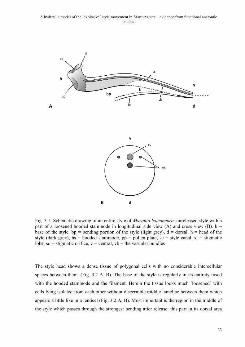
division of the style tissue in three parts (Fig. 3.1 A): the head of the style, the bending
portion in the middle and the base of the style. The three vascular bundles are in a ventral
position accompanying the style canal so that the dorsal region of the style is much thicker
and contains many more cell layers than the ventral (Fig. 3.1 A, B). The head of the style,
which is slightly bent upwards, includes the stigmatic orifice with three stigmatic lobes and
on its dorsal side a depression on which the own pollen is placed during secondary
presentation, the pollen plate.
A hydraulic model of the ´explosive´ style movement in Marantaceae – evidence from functional anatomic studies
33
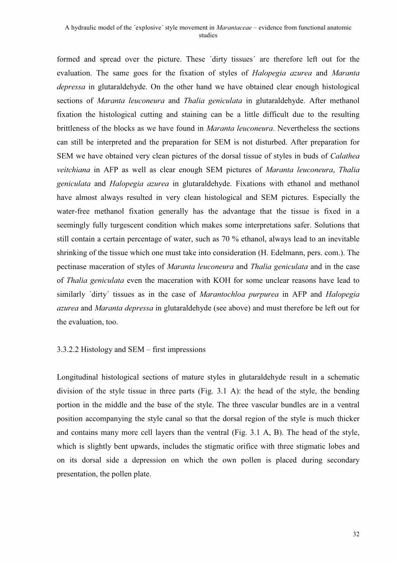
Fig. 3.1: Schematic drawing of an entire style of Maranta leuconeura: unreleased style with a part of a loosened hooded staminode in longitudinal side view (A) and cross view (B). b = base of the style, bp = bending portion of the style (light grey), d = dorsal, h = head of the style (dark grey), hs = hooded staminode, pp = pollen plate, sc = style canal, sl = stigmatic lobe, so = stigmatic orifice, v = ventral, vb = the vascular bundles
The style head shows a dense tissue of polygonal cells with no considerable intercellular
spaces between them. (Fig. 3.2 A, B). The base of the style is regularly in its entirety fused
with the hooded staminode and the filament. Herein the tissue looks much ´loosened´ with
cells lying isolated from each other without discernible middle lamellae between them which
appears a little like in a lenticel (Fig. 3.2 A, B). Most important is the region in the middle of
the style which passes through the strongest bending after release: this part in its dorsal area
A hydraulic model of the ´explosive´ style movement in Marantaceae – evidence from functional anatomic studies
34
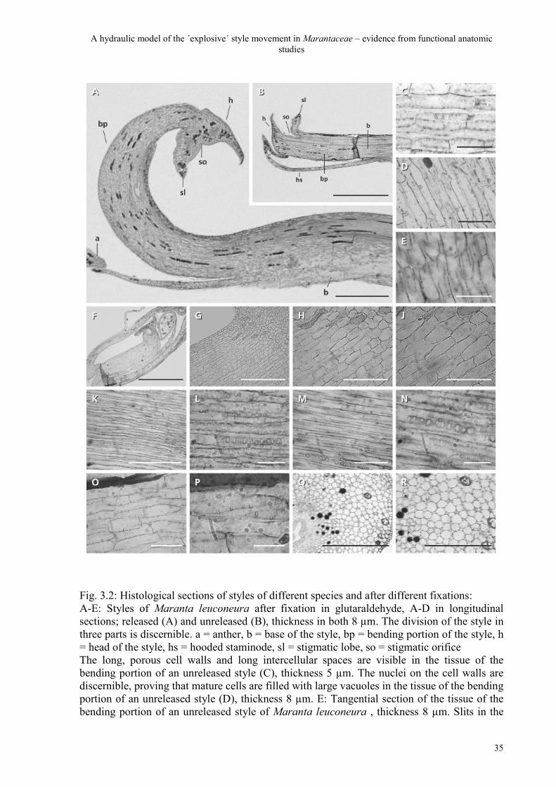
consists of longitudinal cell-layers lying parallel just like hoses or tubes. The tubulate cells are
separated from each other by even longer intercellular spaces where no middle lamellae can
be detected and the cell walls are conspicuously porous (Fig. 3.2 C). The contents of these
mature cells consists only of very large vacuoles and a thin cytoplasmatic ´wall covering´
including a nucleus on the wall (Fig. 3.2 D) – in sharp contrast to the short and highly plasma-
filled cells in styles of buds of Maranta leuconeura fixed in ethanol two days before anthesis
(Fig. 3.2 F-J). Tangential histological sections from the same dorsal region in styles of
Maranta leuconeura - fixed in glutaraldehyde - show a similar pattern (Fig. 3.2 E) which
proves that this porosity reaches clearly into the dorsal region and all lateral directions inside
the tissue.
Many of these longitudinal cell walls in mature styles are conspicuously porous – a great
number of very large holes lying close together catch the eye, here in a dorsal tissue of a style
of Maranta leuconeura (Fig. 3.2 C) and Thalia geniculata (Fig. 3.2 K-N), both fixed in
glutaraldehyde. A comparison of histological sections of glutaraldehyde-fixed tissue samples
of unreleased (Fig. 3.2 K, L) and released (Fig. 3.2 M, N) styles of Thalia geniculata also
confirms that these holes are present already in the dorsal parenchyma of unreleased styles.
The histological experiment with Maranta leuconeura after fixation in methanol shows that
the sections are not very strongly or easily stained but the holes are clearly discernible here,
too (Fig. 3.2 O, P). It is obvious from these histological sections that neither the different
fixations nor the released or unreleased condition of the styles make any difference for the
interpretation of this porosity in the dorsal cell walls. The cross section of the bending portion
of a style of Maranta leuconeura, that was before fixed in glutaraldehyde, confirms that the
longitudinal cells are highly turgescent with wide intercellular spaces between them (Fig. 3.2
Q, R).
A hydraulic model of the ´explosive´ style movement in Marantaceae – evidence from functional anatomic studies
35
Fig. 3.2: Histological sections of styles of different species and after different fixations: A-E: Styles of Maranta leuconeura after fixation in glutaraldehyde, A-D in longitudinal sections; released (A) and unreleased (B), thickness in both 8 µm. The division of the style in three parts is discernible. a = anther, b = base of the style, bp = bending portion of the style, h = head of the style, hs = hooded staminode, sl = stigmatic lobe, so = stigmatic orifice The long, porous cell walls and long intercellular spaces are visible in the tissue of the bending portion of an unreleased style (C), thickness 5 µm. The nuclei on the cell walls are discernible, proving that mature cells are filled with large vacuoles in the tissue of the bending portion of an unreleased style (D), thickness 8 µm. E: Tangential section of the tissue of the bending portion of an unreleased style of Maranta leuconeura , thickness 8 µm. Slits in the
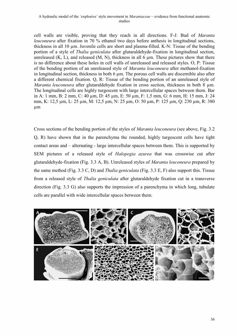
A hydraulic model of the ´explosive´ style movement in Marantaceae – evidence from functional anatomic studies
36
cell walls are visible, proving that they reach in all directions. F-J: Bud of Maranta leuconeura after fixation in 70 % ethanol two days before anthesis in longitudinal sections, thickness in all 10 µm. Juvenile cells are short and plasma-filled. K-N: Tissue of the bending portion of a style of Thalia geniculata after glutaraldehyde-fixation in longitudinal section, unreleased (K, L), and released (M, N), thickness in all 6 µm. These pictures show that there is no difference about these holes in cell walls of unreleased and released styles. O, P: Tissue of the bending portion of an unreleased style of Maranta leuconeura after methanol-fixation in longitudinal section, thickness in both 6 µm. The porous cell walls are discernible also after a different chemical fixation. Q, R: Tissue of the bending portion of an unreleased style of Maranta leuconeura after glutaraldehyde fixation in cross section, thickness in both 8 µm. The longitudinal cells are highly turgescent with large intercellular spaces between them. Bar in A: 1 mm, B: 2 mm, C: 40 µm, D: 45 µm, E: 50 µm, F: 1,5 mm, G: 6 mm, H: 15 mm, J: 24 mm, K: 12,5 µm, L: 25 µm, M: 12,5 µm, N: 25 µm, O: 50 µm, P: 125 µm, Q: 230 µm, R: 380 µm
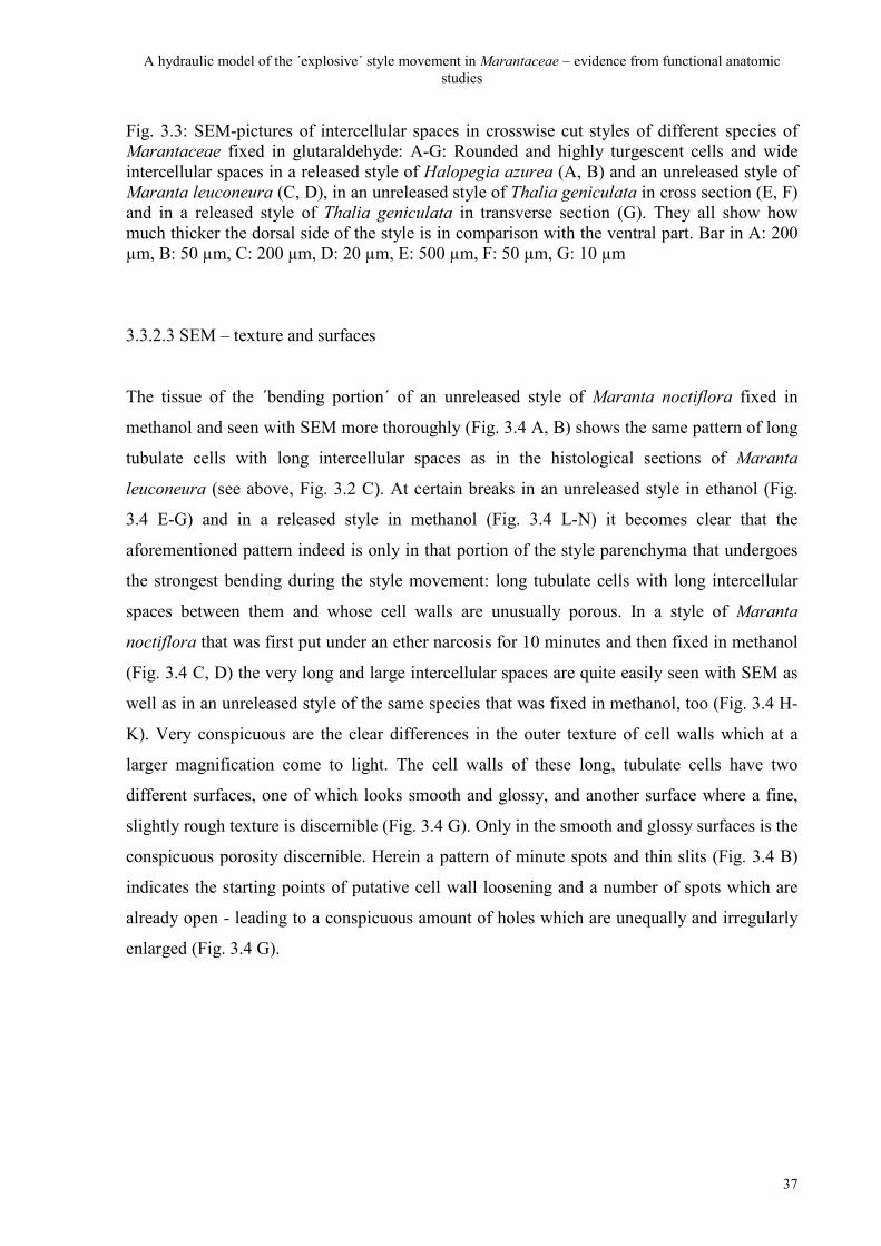
Cross sections of the bending portion of the styles of Maranta leuconeura (see above, Fig. 3.2
Q, R) have shown that in the parenchyma the rounded, highly turgescent cells have tight
contact areas and – alternating - large intercellular spaces between them. This is supported by
SEM pictures of a released style of Halopegia azurea that was crosswise cut after
glutaraldehyde-fixation (Fig. 3.3 A, B). Unreleased styles of Maranta leuconeura prepared by
the same method (Fig. 3.3 C, D) and Thalia geniculata (Fig. 3.3 E, F) also support this. Tissue
from a released style of Thalia geniculata after glutaraldehyde fixation cut in a transverse
direction (Fig. 3.3 G) also supports the impression of a parenchyma in which long, tubulate
cells are parallel with wide intercellular spaces between them.
A hydraulic model of the ´explosive´ style movement in Marantaceae – evidence from functional anatomic studies
37
Fig. 3.3: SEM-pictures of intercellular spaces in crosswise cut styles of different species of Marantaceae fixed in glutaraldehyde: A-G: Rounded and highly turgescent cells and wide intercellular spaces in a released style of Halopegia azurea (A, B) and an unreleased style of Maranta leuconeura (C, D), in an unreleased style of Thalia geniculata in cross section (E, F) and in a released style of Thalia geniculata in transverse section (G). They all show how much thicker the dorsal side of the style is in comparison with the ventral part. Bar in A: 200 µm, B: 50 µm, C: 200 µm, D: 20 µm, E: 500 µm, F: 50 µm, G: 10 µm
3.3.2.3 SEM – texture and surfaces
The tissue of the ´bending portion´ of an unreleased style of Maranta noctiflora fixed in
methanol and seen with SEM more thoroughly (Fig. 3.4 A, B) shows the same pattern of long
tubulate cells with long intercellular spaces as in the histological sections of Maranta
leuconeura (see above, Fig. 3.2 C). At certain breaks in an unreleased style in ethanol (Fig.
3.4 E-G) and in a released style in methanol (Fig. 3.4 L-N) it becomes clear that the
aforementioned pattern indeed is only in that portion of the style parenchyma that undergoes
the strongest bending during the style movement: long tubulate cells with long intercellular
spaces between them and whose cell walls are unusually porous. In a style of Maranta
noctiflora that was first put under an ether narcosis for 10 minutes and then fixed in methanol
(Fig. 3.4 C, D) the very long and large intercellular spaces are quite easily seen with SEM as
well as in an unreleased style of the same species that was fixed in methanol, too (Fig. 3.4 H-
K). Very conspicuous are the clear differences in the outer texture of cell walls which at a
larger magnification come to light. The cell walls of these long, tubulate cells have two
different surfaces, one of which looks smooth and glossy, and another surface where a fine,
slightly rough texture is discernible (Fig. 3.4 G). Only in the smooth and glossy surfaces is the
conspicuous porosity discernible. Herein a pattern of minute spots and thin slits (Fig. 3.4 B)
indicates the starting points of putative cell wall loosening and a number of spots which are
already open - leading to a conspicuous amount of holes which are unequally and irregularly
enlarged (Fig. 3.4 G).
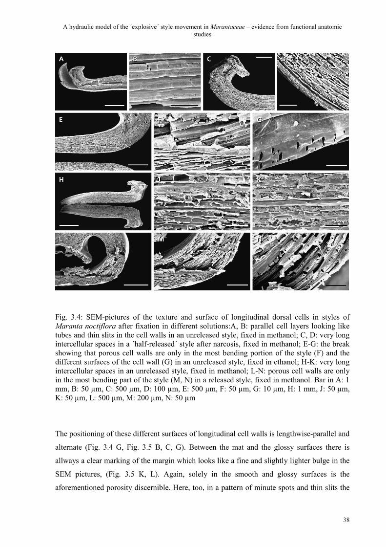
A hydraulic model of the ´explosive´ style movement in Marantaceae – evidence from functional anatomic studies
38
Fig. 3.4: SEM-pictures of the texture and surface of longitudinal dorsal cells in styles of Maranta noctiflora after fixation in different solutions:A, B: parallel cell layers looking like tubes and thin slits in the cell walls in an unreleased style, fixed in methanol; C, D: very long intercellular spaces in a ´half-released´ style after narcosis, fixed in methanol; E-G: the break showing that porous cell walls are only in the most bending portion of the style (F) and the different surfaces of the cell wall (G) in an unreleased style, fixed in ethanol; H-K: very long intercellular spaces in an unreleased style, fixed in methanol; L-N: porous cell walls are only in the most bending part of the style (M, N) in a released style, fixed in methanol. Bar in A: 1 mm, B: 50 µm, C: 500 µm, D: 100 µm, E: 500 µm, F: 50 µm, G: 10 µm, H: 1 mm, J: 50 µm, K: 50 µm, L: 500 µm, M: 200 µm, N: 50 µm
The positioning of these different surfaces of longitudinal cell walls is lengthwise-parallel and
alternate (Fig. 3.4 G, Fig. 3.5 B, C, G). Between the mat and the glossy surfaces there is
allways a clear marking of the margin which looks like a fine and slightly lighter bulge in the
SEM pictures, (Fig. 3.5 K, L). Again, solely in the smooth and glossy surfaces is the
aforementioned porosity discernible. Here, too, in a pattern of minute spots and thin slits the
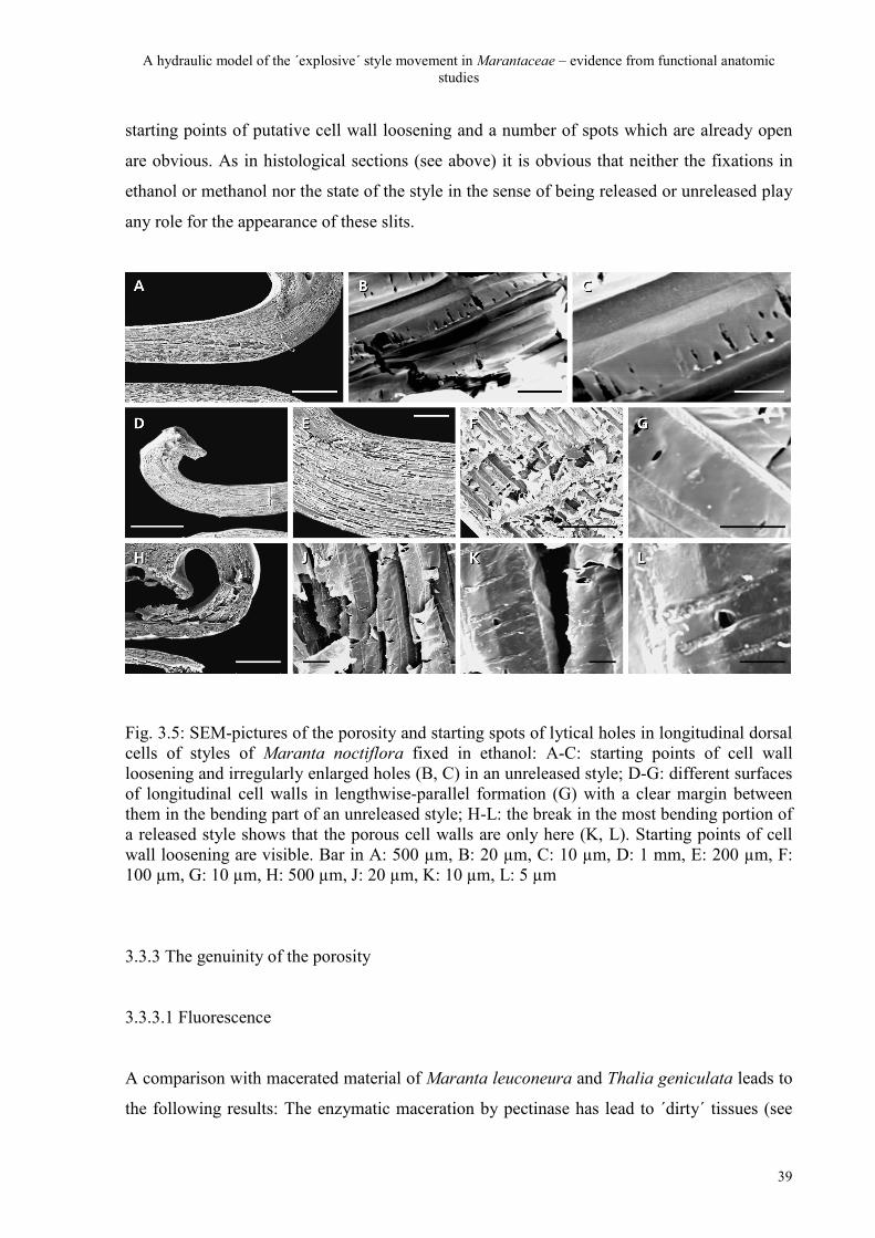
A hydraulic model of the ´explosive´ style movement in Marantaceae – evidence from functional anatomic studies
39
starting points of putative cell wall loosening and a number of spots which are already open
are obvious. As in histological sections (see above) it is obvious that neither the fixations in
ethanol or methanol nor the state of the style in the sense of being released or unreleased play
any role for the appearance of these slits.
Fig. 3.5: SEM-pictures of the porosity and starting spots of lytical holes in longitudinal dorsal cells of styles of Maranta noctiflora fixed in ethanol: A-C: starting points of cell wall loosening and irregularly enlarged holes (B, C) in an unreleased style; D-G: different surfaces of longitudinal cell walls in lengthwise-parallel formation (G) with a clear margin between them in the bending part of an unreleased style; H-L: the break in the most bending portion of a released style shows that the porous cell walls are only here (K, L). Starting points of cell wall loosening are visible. Bar in A: 500 µm, B: 20 µm, C: 10 µm, D: 1 mm, E: 200 µm, F: 100 µm, G: 10 µm, H: 500 µm, J: 20 µm, K: 10 µm, L: 5 µm
3.3.3 The genuinity of the porosity
3.3.3.1 Fluorescence
A comparison with macerated material of Maranta leuconeura and Thalia geniculata leads to
the following results: The enzymatic maceration by pectinase has lead to ´dirty´ tissues (see
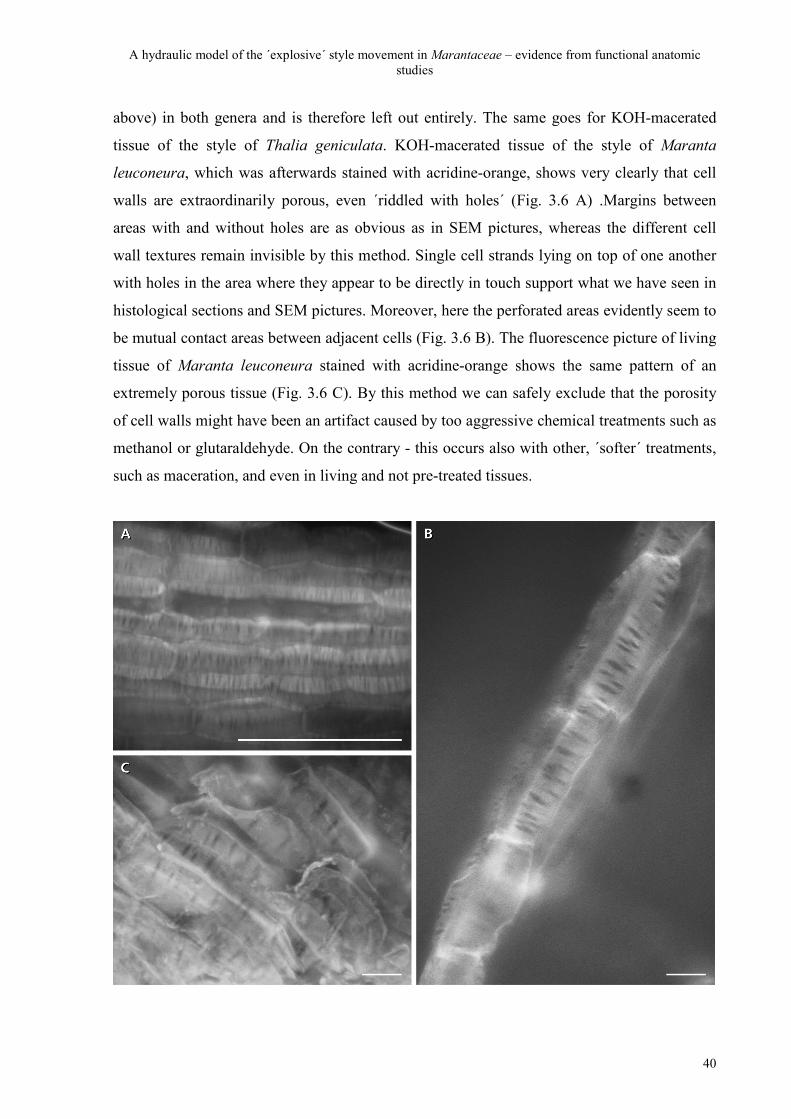
A hydraulic model of the ´explosive´ style movement in Marantaceae – evidence from functional anatomic studies
40
above) in both genera and is therefore left out entirely. The same goes for KOH-macerated
tissue of the style of Thalia geniculata. KOH-macerated tissue of the style of Maranta
leuconeura, which was afterwards stained with acridine-orange, shows very clearly that cell
walls are extraordinarily porous, even ´riddled with holes´ (Fig. 3.6 A) .Margins between
areas with and without holes are as obvious as in SEM pictures, whereas the different cell
wall textures remain invisible by this method. Single cell strands lying on top of one another
with holes in the area where they appear to be directly in touch support what we have seen in
histological sections and SEM pictures. Moreover, here the perforated areas evidently seem to
be mutual contact areas between adjacent cells (Fig. 3.6 B). The fluorescence picture of living
tissue of Maranta leuconeura stained with acridine-orange shows the same pattern of an
extremely porous tissue (Fig. 3.6 C). By this method we can safely exclude that the porosity
of cell walls might have been an artifact caused by too aggressive chemical treatments such as
methanol or glutaraldehyde. On the contrary - this occurs also with other, ´softer´ treatments,
such as maceration, and even in living and not pre-treated tissues.
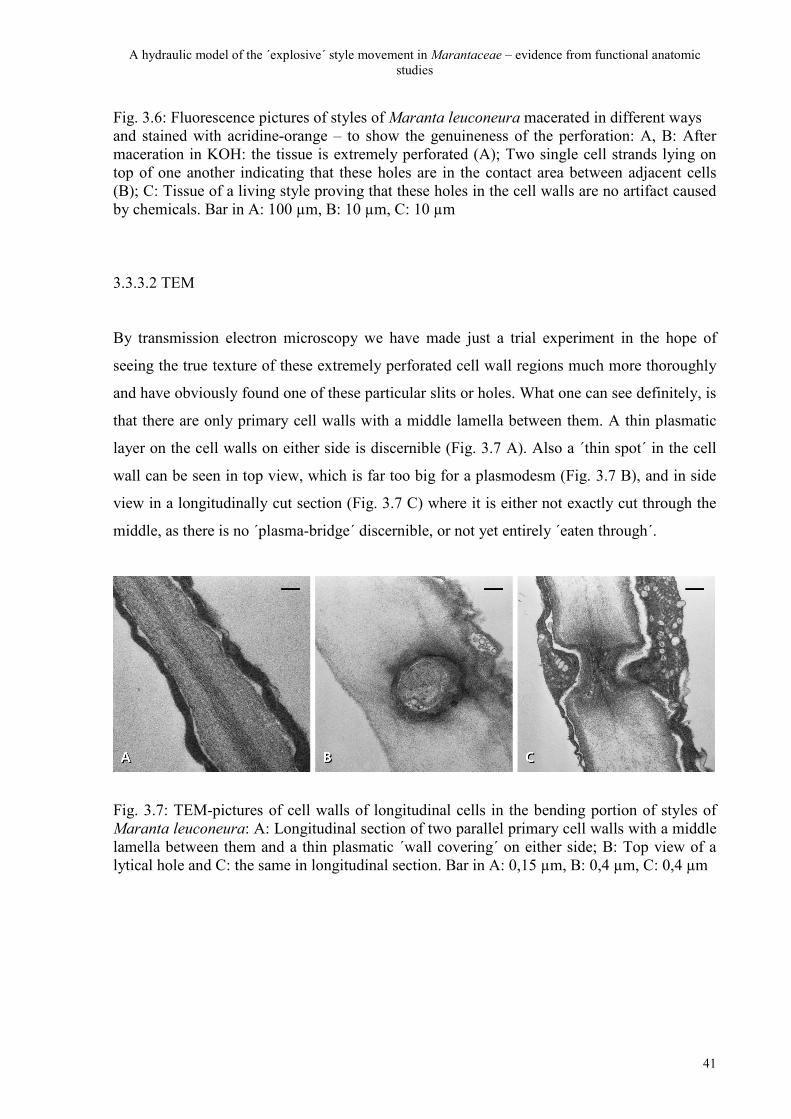
A hydraulic model of the ´explosive´ style movement in Marantaceae – evidence from functional anatomic studies
41
Fig. 3.6: Fluorescence pictures of styles of Maranta leuconeura macerated in different ways and stained with acridine-orange – to show the genuineness of the perforation: A, B: After maceration in KOH: the tissue is extremely perforated (A); Two single cell strands lying on top of one another indicating that these holes are in the contact area between adjacent cells (B); C: Tissue of a living style proving that these holes in the cell walls are no artifact caused by chemicals. Bar in A: 100 µm, B: 10 µm, C: 10 µm
3.3.3.2 TEM
By transmission electron microscopy we have made just a trial experiment in the hope of
seeing the true texture of these extremely perforated cell wall regions much more thoroughly
and have obviously found one of these particular slits or holes. What one can see definitely, is
that there are only primary cell walls with a middle lamella between them. A thin plasmatic
layer on the cell walls on either side is discernible (Fig. 3.7 A). Also a ´thin spot´ in the cell
wall can be seen in top view, which is far too big for a plasmodesm (Fig. 3.7 B), and in side
view in a longitudinally cut section (Fig. 3.7 C) where it is either not exactly cut through the
middle, as there is no ´plasma-bridge´ discernible, or not yet entirely ´eaten through´.
Fig. 3.7: TEM-pictures of cell walls of longitudinal cells in the bending portion of styles of Maranta leuconeura: A: Longitudinal section of two parallel primary cell walls with a middle lamella between them and a thin plasmatic ´wall covering´ on either side; B: Top view of a lytical hole and C: the same in longitudinal section. Bar in A: 0,15 µm, B: 0,4 µm, C: 0,4 µm
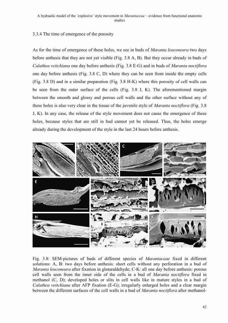
A hydraulic model of the ´explosive´ style movement in Marantaceae – evidence from functional anatomic studies
42
3.3.4 The time of emergence of the porosity
As for the time of emergence of these holes, we see in buds of Maranta leuconeura two days
before anthesis that they are not yet visible (Fig. 3.8 A, B). But they occur already in buds of
Calathea veitchiana one day before anthesis (Fig. 3.8 E-G) and in buds of Maranta noctiflora
one day before anthesis (Fig. 3.8 C, D) where they can be seen from inside the empty cells
(Fig. 3.8 D) and in a similar preparation (Fig. 3.8 H-K) where this porosity of cell walls can
be seen from the outer surface of the cells (Fig. 3.8 J, K). The aforementioned margin
between the smooth and glossy and porous cell walls and the other surface without any of
these holes is also very clear in the tissue of the juvenile style of Maranta noctiflora (Fig. 3.8
J, K). In any case, the release of the style movement does not cause the emergence of these
holes, because styles that are still in bud cannot yet be released. Thus, the holes emerge
already during the development of the style in the last 24 hours before anthesis.
Fig. 3.8: SEM-pictures of buds of different species of Marantaceae fixed in different solutions: A, B: two days before anthesis: short cells without any perforation in a bud of Maranta leuconeura after fixation in glutaraldehyde; C-K: all one day before anthesis: porous cell walls seen from the inner side of the cells in a bud of Maranta noctiflora fixed in methanol (C, D); developed holes or slits in cell walls like in mature styles in a bud of Calathea veitchiana after AFP fixation (E-G); irregularly enlarged holes and a clear margin between the different surfaces of the cell walls in a bud of Maranta noctiflora after methanol-
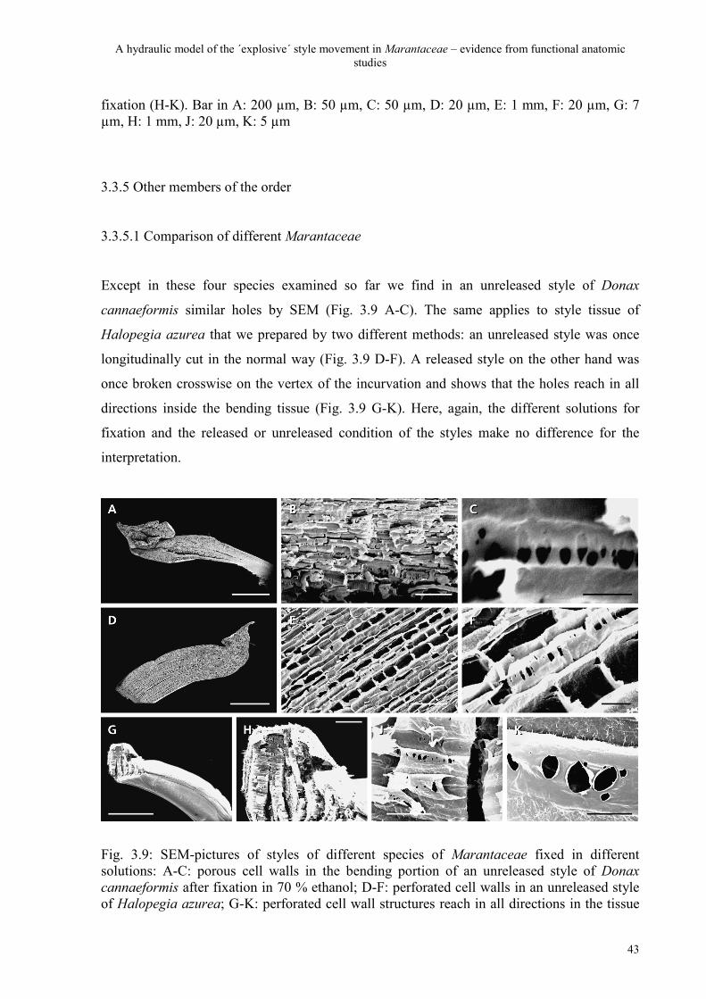
A hydraulic model of the ´explosive´ style movement in Marantaceae – evidence from functional anatomic studies
43
fixation (H-K). Bar in A: 200 µm, B: 50 µm, C: 50 µm, D: 20 µm, E: 1 mm, F: 20 µm, G: 7 µm, H: 1 mm, J: 20 µm, K: 5 µm
3.3.5 Other members of the order
3.3.5.1 Comparison of different Marantaceae
Except in these four species examined so far we find in an unreleased style of Donax
cannaeformis similar holes by SEM (Fig. 3.9 A-C). The same applies to style tissue of
Halopegia azurea that we prepared by two different methods: an unreleased style was once
longitudinally cut in the normal way (Fig. 3.9 D-F). A released style on the other hand was
once broken crosswise on the vertex of the incurvation and shows that the holes reach in all
directions inside the bending tissue (Fig. 3.9 G-K). Here, again, the different solutions for
fixation and the released or unreleased condition of the styles make no difference for the
interpretation.
Fig. 3.9: SEM-pictures of styles of different species of Marantaceae fixed in different solutions: A-C: porous cell walls in the bending portion of an unreleased style of Donax cannaeformis after fixation in 70 % ethanol; D-F: perforated cell walls in an unreleased style of Halopegia azurea; G-K: perforated cell wall structures reach in all directions in the tissue
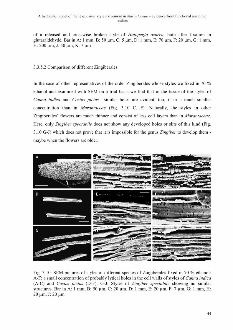
A hydraulic model of the ´explosive´ style movement in Marantaceae – evidence from functional anatomic studies
44
of a released and crosswise broken style of Halopegia azurea, both after fixation in glutaraldehyde. Bar in A: 1 mm, B: 50 µm, C: 5 µm, D: 1 mm, E: 70 µm, F: 20 µm, G: 1 mm, H: 200 µm, J: 50 µm, K: 7 µm
3.3.5.2 Comparison of different Zingiberales
In the case of other representatives of the order Zingiberales whose styles we fixed in 70 %
ethanol and examined with SEM on a trial basis we find that in the tissue of the styles of
Canna indica and Costus pictus similar holes are evident, too, if in a much smaller
concentration than in Marantaceae (Fig. 3.10 C, F). Naturally, the styles in other
Zingiberales´ flowers are much thinner and consist of less cell layers than in Marantaceae.
Here, only Zingiber spectabile does not show any developed holes or slits of this kind (Fig.
3.10 G-J) which does not prove that it is impossible for the genus Zingiber to develop them -
maybe when the flowers are older.
Fig. 3.10: SEM-pictures of styles of different species of Zingiberales fixed in 70 % ethanol: A-F: a small concentration of probably lytical holes in the cell walls of styles of Canna indica (A-C) and Costus pictus (D-F); G-J: Styles of Zingiber spectabile showing no similar structures. Bar in A: 1 mm, B: 50 µm, C: 20 µm, D: 1 mm, E: 20 µm, F: 7 µm, G: 1 mm, H: 20 µm, J: 20 µm
A hydraulic model of the ´explosive´ style movement in Marantaceae – evidence from functional anatomic studies
45
3.3.6 Summary of the results
We can safely conclude that these interesting holes and slits do not occur through the release
of the style. They are discernible in unreleased as well as released styles. They are present in
buds one day before anthesis. They can be found in the tissue of living styles and in tissues of
styles after four different chemical fixations and in macerated tissue. We have found them in
all examined 5 genera (6 species) of Marantaceae and even in representatives of two other
families of Zingiberales. They appear to be only in the thin, probably primary, cell walls and
between adjacent cells. We never found them in the thicker and more strongly textured cell
walls. It appears that they are no regular pits, but emerge through cell wall loosening.
3.4 Discussion
All results that we have to hand about differential growth rates and differential tissue tension
(see chapter 2) and anatomical and histological structures meet in the following interpretation
of both the development and function of the style: the maturing style is actively (i.e. energy-
demanding) ´pumped up´ with water under a strong osmotic potential during an enormous
elongational growth in the last hours before anthesis. This leads to the aforementioned
´straightjacket´ syndrome with the style being extremely turgescent and actually longer than
the hooded staminode in which it is jammed. The pumping-up process produces an
elongational pressure with largely extended cells on the ventral side. The shorter hooded
staminode on the other hand causes a counterpressure in the opposite direction. Hereby the
style becomes a little overstretched with ´compressed´ dorsal cells that are actually hindered
from expansion. The result is a differential turgor on the ventral and dorsal side (Fig. 3.11 A).
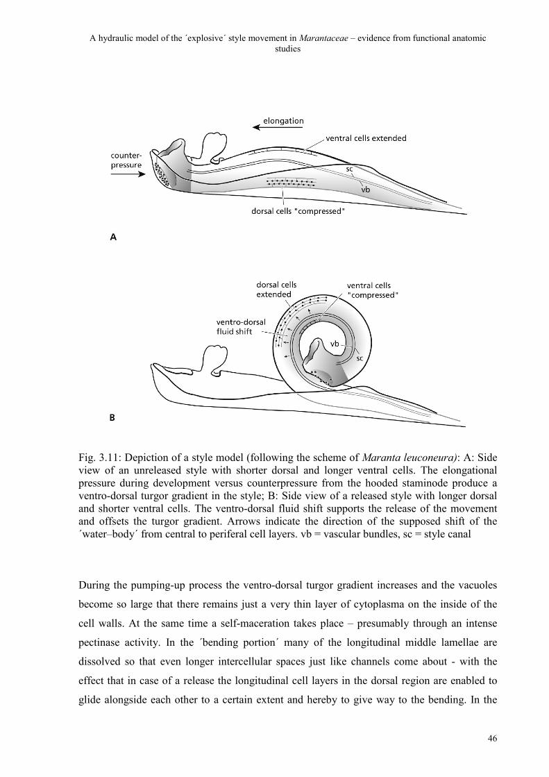
A hydraulic model of the ´explosive´ style movement in Marantaceae – evidence from functional anatomic studies
46
Fig. 3.11: Depiction of a style model (following the scheme of Maranta leuconeura): A: Side view of an unreleased style with shorter dorsal and longer ventral cells. The elongational pressure during development versus counterpressure from the hooded staminode produce a ventro-dorsal turgor gradient in the style; B: Side view of a released style with longer dorsal and shorter ventral cells. The ventro-dorsal fluid shift supports the release of the movement and offsets the turgor gradient. Arrows indicate the direction of the supposed shift of the ´water–body´ from central to periferal cell layers. vb = vascular bundles, sc = style canal
During the pumping-up process the ventro-dorsal turgor gradient increases and the vacuoles
become so large that there remains just a very thin layer of cytoplasma on the inside of the
cell walls. At the same time a self-maceration takes place – presumably through an intense
pectinase activity. In the ´bending portion´ many of the longitudinal middle lamellae are
dissolved so that even longer intercellular spaces just like channels come about - with the
effect that in case of a release the longitudinal cell layers in the dorsal region are enabled to
glide alongside each other to a certain extent and hereby to give way to the bending. In the
A hydraulic model of the ´explosive´ style movement in Marantaceae – evidence from functional anatomic studies
47
contact areas between adjacent longitudinal cells an extreme porosity of probably primary cell
walls is brought about through cell wall loosening - also through pectinase activity. The
question is whether these holes or slits derive either from primary pit fields in the juvenile cell
walls while in the course of maturation the individual plasmodesma are secondarily enlarged
to slits in the service of a quick water flow or whether they are just ´lytical holes´ that develop
only in a late phase of bud growth due to an intense pectinase activity. This cannot be
answered absolutely. The unusually great number of holes and slits that we have found
concurrently in the dorsal bending portion of different genera does not let us believe in normal
pits although especially in tissues where neighbouring cells must allow a mass flow (e.g. in
phloem-bundles, Strasburger 1999) they can appear with strongly increased denseness.
Nevertheless, as they are, first, only discernible in buds one day before anthesis and, secondly,
very irregularly shaped (see Fig. 3.4) we rather assume that the lysis through pectinase does
not necessarily begin with juvenile primary pit fields, but that in riper buds there may be extra
preformed spots in the cell walls (an idea also of H. Edelmann, pers. com.) in the contact area
of adjacent cells which are ´eaten through´ by pectinase. It appears that primary cell walls are
preserved in such places where cell-wall loosening is meant to start and that they may be
strengthened with secondary wall material (mainly cellulose) only in such areas where these
holes shall not emerge. Differential strengthening of cell walls may hereby develop in a
similar fashion as in a lamellar collenchyma. The style appears to make a compromise
between on one hand stability – to maintain an extremely high and even differential turgor –
and, on the other hand, pliability through differential cell-wall strengthening to endure the
bending without rupture.
Anyway, this unusual porosity may also support the litheness of the region. But the main
function of the perforation surely is not in first place pliability, but permeability. The theory
of a sudden loss (or change) of turgor through a runoff of water into the intercellular spaces
(Schumann 1902, Kunze 1984) in our opinion cannot explain the unusually high speed of the
movement sufficiently. Diffusion may have a share in the whole process but differential
turgor surely does not produce the movement alone. Our findings indicate that this unusual
perforation is in primary cell walls and between neighbouring cells (see Fig. 3.12).
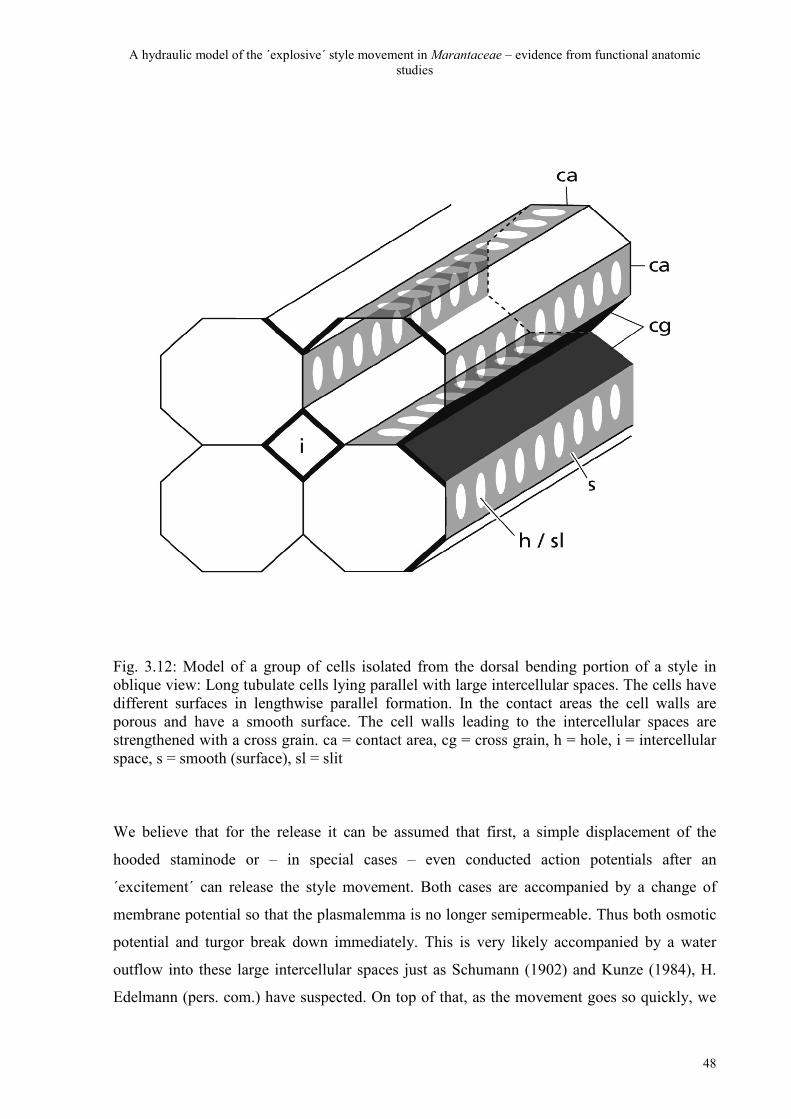
A hydraulic model of the ´explosive´ style movement in Marantaceae – evidence from functional anatomic studies
48
Fig. 3.12: Model of a group of cells isolated from the dorsal bending portion of a style in oblique view: Long tubulate cells lying parallel with large intercellular spaces. The cells have different surfaces in lengthwise parallel formation. In the contact areas the cell walls are porous and have a smooth surface. The cell walls leading to the intercellular spaces are strengthened with a cross grain. ca = contact area, cg = cross grain, h = hole, i = intercellular space, s = smooth (surface), sl = slit
We believe that for the release it can be assumed that first, a simple displacement of the
hooded staminode or – in special cases – even conducted action potentials after an
´excitement´ can release the style movement. Both cases are accompanied by a change of
membrane potential so that the plasmalemma is no longer semipermeable. Thus both osmotic
potential and turgor break down immediately. This is very likely accompanied by a water
outflow into these large intercellular spaces just as Schumann (1902) and Kunze (1984), H.
Edelmann (pers. com.) have suspected. On top of that, as the movement goes so quickly, we
A hydraulic model of the ´explosive´ style movement in Marantaceae – evidence from functional anatomic studies
49
think it even possible that, as an additional effect, the vacuoles in the region may break down.
The effect that we assume is that in one complete and very sudden shift of fluid from central
to periferal cell layers the entire ´water-body´ sweeps through and comes to a standstill (Fig.
3.11 B). This vast - and fast - ventro-dorsal shift of fluid goes through the whole symplastic
body in the middle of the style and outdoes the runoff of water through intercellular spaces by
far and may hereby explain the particularly high speed of the style movement. As the water
flow goes from central into the periferal (i.e. dorsal ) cell layers and not, as assumed by Kunze
(1984), in the ventral side of the style the dorsal longitudinal cells eventually become longer,
but not thinner which means that they increase their volume (Classen-Bockhoff and
Pischtschan 2000). This, again, explains the irreversibility of the movement. Consequently the
assumed break down of vacuoles in the middle part involves at least a partial dying of the
parenchyma of the style. This indeed would do no further harm because the pollen only needs
to migrate through the pollen canal in the next hours before the style withers completely.
There is an interesting observation at the genus Calathea described by Hildebrand (1870) and
Kennedy (2000) whereafter the style already during movement first turns brownish and later
violet-black. Both authors have assumed that as a result of this discolouring the insects
recognise that the respective flower is already done. One might perhaps assume that it is a
necrosis of the parenchyma as a result of the complete break down of the vacuoles in that
middle area of the style. Meanwhile, Hildebrand emphasises that the style when it dies
unreleased also discolours in such a way.
Concerning the strength or stability of the style we agree with Schumann (see chapter 2) that
the turgor is already built up before and not, as Kunze says, only at the moment of an
´excitement´ of the style via the basal plate.
The geometrical shape of the complete curvature after release is in all probability determined
by the course and position of the only supporting elements the style has, namely the three
vascular bundles. It may even be assumed – but still unproven by our methods - that these
vascular bundles during the ´pumping-up´ process in the last hours remain a little shorter in
comparison to the water-filled ´parenchymatic style´ in order to support and stabilise the
incurved form through their tension. In this sense we agree with Schumann (1902) that there
are no mechanical (i.e. antagonistic) tissues as they can be found in leaf bases of Dionaea and
Mimosa which explains again why in Marantaceae there is no reset of the movement.
Nevertheless, in the bending portion there is a tissue specialised in the service of elasticity and
permeability - a motile tissue producing a movement and which emerges in the last hours of
A hydraulic model of the ´explosive´ style movement in Marantaceae – evidence from functional anatomic studies
50
maturation through self-maceration. This may not be called a mechanical tissue, too, but
perhaps or at least a ´hydraulic tissue´. The style ´movement´ is thus facilitated once by
differential growth rates and differential tissue tensioning during development (see chapter 2)
and at the same time by differential cell-wall strengthening followed by enzymatic dissolution
of stabilising and damming structures in the parenchyma. In this sense also the demand of
Skotheim and Mahadevan (2005) for ´elastic instability´ is here not only fulfilled but
absolutely carried to extremes. It seems, as the investigation of other members of Zingiberales
has shown, that a certain predisposition for such a tissue instability may already be laid out in
the order, though being still functionless in the other families. Maybe that only Marantaceae
are able to take advantage of this ´design´ – particularly as their styles are much more
voluminous and consist of many more cell layers than those of the other families of
Zingiberales. The potential to make use of such a feature is anyway higher in Marantaceae
due to the fleshy consistency of the style. Altogether, even if we take the term of the ´elastic
instability´ as a basis, it must here be modified in the sense that the release of the style indeed
results not in an elastic and herewith reversible movement, but in a plastic and irreversible
reshaping of the organ. It is remarkable that the flowers of Marantaceae in the service of an
extremely precise pollination method obviously take advantage of self-maceration which is a
natural process related to ageing.
Diversity and systematic value of the cucullate staminodes in Marantaceae
51
4. Diversity and systematic value of the cucullate staminode in Marantaceae
Abstract
The cucullate staminode in Marantaceae plays a critical role in the pollination process and
shows a remarkable morphological diversity. Cucullate staminodes of 24 genera of Maran-
taceae are sorted according to a minimalistic selection of shape characters such as the trigger-
appendage and few others. Eight distinct morphological types are abstracted from the
resulting groups and mapped onto a maximally parsimonious tree by Prince and Kress (2006)
containing five major clades. An amazing correspondence is found between the mor-
phological types and the clades; especially the given Calathea-clade and the Sarcophrynium-
clade are absolutely consistent with the respective American and African types of hooded
staminodes. Furthermore, several sister-relationships are confirmed and for some uncertain
positions possible evolutionary pathways are discussed, as well as for the entire family. The
value of morphological characters in relation to the modern phylogenetical analysis is
discussed and conditions for the selection of morphological characters most meaningful for a
systematic grouping are proposed.
4.1 Introduction
While actually trying to clarify the mechanisms underlying the ´explosive´ style movement in
Marantaceae (see chapters 2 and 3) in years of flower preparation an intriguing ´side-
observation´ was made that eventually led to the impression that the hooded (cucullate)
staminodes in this family bear conspicuously diverse appendages and other morphological
features that might be of high systematic value. Expressed in simplified terms: only from a
hooded staminode alone one might make out which continent a specimen of Marantaceae
comes from and which systematic group it belongs to without even needing to investigate into
other floral or vegetative or reproductive characters – hereby being provided with a handy and
quick morphological key to the genera and their geographic position.
Why of all floral parts in Marantaceae the cucullate staminode is so conspicuously divers that
it alone may be of diagnostic value - about this we hypothesise as follows: the flowers and the
Diversity and systematic value of the cucullate staminodes in Marantaceae
52
pollination mechanism in Marantaceae are unique to the family and the cucullate staminode
has an essential share in it (see chapter 2). Therefore, a fundamental notion of an adaptive
relation between flowers and their pollinators seems to suggest itself. It is likely that these
flowers can only be pollinated by insects or other animals that can cope with the
biomechanical properties of flowers of a certain genus or species, depending on their size,
shape, physical force and maybe even weight. It is also possible that these flowers are more
exclusively specialised in their pollinators than the other way round so that a certain selective
pressure may be supposed which acts more stringently on the plants than on their pollinators.
This might explain why in particular the plant family has developed such specialisations and
not the euglossine bees that are the principal pollinators of Marantaceae in the New World
(Kennedy 1978). However, to make a more reliable statement about the adaptive effect of
these divers structures extensive observations of pollinators and their physical ´pollinating-
behaviour´ (see Kennedy 1978) - especially in connection with the mechanical properties of
the triggers (see below) - at natural habitats in the Old World are necessary. As ecological
investigations are still in progress at present, the question of the adaptive value and the
processes selective on these structures must remain open here. Furthermore, the varied
modifications of the cucullate staminodes might also provide much phylogenetic information,
so affording a better knowledge about the evolutionary paths of the family and the degree of
relationship amongst them.
The hooded staminode in Marantaceae is nowadays agreed to be an element of the epipetal,
inner androeceal whorl (Eichler 1884, Schumann 1902, Loesener 1930, Kunze 1984), together
with the anther and the callose staminode. Its function is the hold of the ´explosive´ style prior
to the visit of a pollinator alike the setting-up of the potential for the style movement after
release through a pollinator (Eichler 1884, Costerus 1918, Loesener 1930, Kennedy 1978; see
chapter 2) The release of the movement is predominantly believed to be caused by the touch
of the ´trigger´ which is a lateral appendage at the margin of the hooded staminode while the
´basal plate´, a swelling at the base of the trigger, transmits the stimulus from the trigger to the
style (Kunze 1984, Claßen-Bockhoff 1991, Yeo 1993). About the appendages of the hooded
staminodes there are already some early observations: Koernicke (1859) observed the
diversity of trigger-appendages and he was the first to describe the possible presence of a
second appendage or ´bulge´ in examples of Calathea, ´Monostiche´ and Ischnosiphon. On
top of that he considered the trigger in Thalia to be one appendage splitted in two ´ribbons´
Diversity and systematic value of the cucullate staminodes in Marantaceae
53
contrary to Andersson (1998) who saw Thalia with two appendages. For the development of a
hooded staminode there are first interpretations from Eichler (1883): one margin of the
staminode develops to the hooded part of it and the other margin forms 1-2 appendages (in:
Costerus 1918). Hereby in Maranta sanguinea (Eichler 1884) the margin of the staminode
elongates to a ´tooth-shaped appendage´ that later ´moves down´ (in the direction of the leaf
base, the authors). Kunze (1984) carried out ontogenetic investigations at hooded staminodes
of Maranta leuconeura, Ataenidia conferta and Calathea vaginata with special focus on the
course of the medianus or the midrib, respectively. His conclusions support the observations
of Eichler (1883) that the two main portions that are divided by the medianus eventually are
the hood and the trigger and that they start early to develop very differently. The early formed
indentation between the hood and the trigger-appendage is the actual leaf apex. Through
allometric growth on the trigger-side there develops a fold and at its base the basal plate. In
many species of Calathea and in Ataenidia he observed a swelling of the tissue in a place
proximal to the trigger and treats them as equivalent, at least as regards their function (Kunze
1984). For this the medianus takes a turn ´into the trigger´.
The first systematic subdivision of the family was carried out by Petersen (1889) dividing the
family into two tribes, namely the tribe Maranteae with one fertile locule in the 3-carpellate
ovary and the tribe Phryniae with three fertile locules in the ovary, hereby following
Koernicke´s (1858) first key to the genera. This subdivision was accepted by Schumann
(1902) and Loesener (1930) and others, but was criticised as artificial by Andersson (1981)
who presented a subdivision of the genera in five ´informal´ groups (1998), based on a great
number of morphological and reproductive characters relating to the entire plants, such as
branchings in the vegetative and floral part, metric data like the length of the corolla tube,
numeric data like the number of outer staminodes or fertile locules, fruit dehiscence, the form
and position of the trigger-appendages and many more. This grouping of the family by
Andersson (1998) must now be called into question, too. Andersson and Chase (2001)
presented a phylogenetic analysis based on combined morphological and molecular data,
hereby using plastid-DNA sequence data (rps 16 intron) representing 21 genera and resulting
in a reordering of the family. There was no support for the ´antique´ division in Phryniae and
Maranteae (Petersen 1889). Anderssons (1998) informal groups were only partly supported,
e.g. the monophyletic Calathea group including the genera Calathea, Pleiostachya,
Ischnosiphon and Monotagma. The newest phylogenetic analysis of the family was recently
Diversity and systematic value of the cucullate staminodes in Marantaceae
54
presented by Prince and Kress (2006) using plastid DNA sequence data (matK coding and 3´
intergenic spacer region and the trnl-F intergenic spacer region) representing 27 genera and
considering also some of the morphological characters used by Andersson and Chase (2001),
such as bracteoles, shape of the corolla tube, outer staminodes, appendages of the cucullate
staminode and fruit. This investigation resulted in five major clades, namely a Calathea-clade,
a Sarcophrynium-clade, a Donax-clade, a Maranta-clade and a Stachyphrynium-clade. Herein,
besides Calathea, the genera Schumannianthus, Phrynium, and Marantochloa turned out to be
non-monophyletic.
Although flowers in Marantaceae are on the whole morphologically very uniform and easy to
recognise (Kennedy 2000) this shows how difficult it is to make out the value of single
morphological characters and to decide which of them to use for a grouping. Starting from the
aforementioned impression of the conspicuous diversity of hooded staminodes we bring up in
this study the following three hypotheses:
• Characters of form and shape of the hooded staminode may be more meaningful for
the systematic grouping than vegetative, metric or numerical characters which were
predominantly used in previous subdivisions of the family (Petersen 1889, Andersson
1998).
• Rare characters are of higher significance than frequent characters.
• If the most suitable floral organs and characters are selected, even purely mor-
phological features are of a very high systematic value.
In the present study we make an attempt, only based on a small number of morphological
characters of hooded staminodes, to subdivide the Marantaceae into few types. These types
are then compared with the results of the two recent phylogenetic analyses by Prince and
Kress (2006) in first place, Andersson and Chase (2001) and Andersson´s informal grouping
(1998). Based on these comparisons we are going to judge the weight of our own
morphological characters for the systematics of the family.
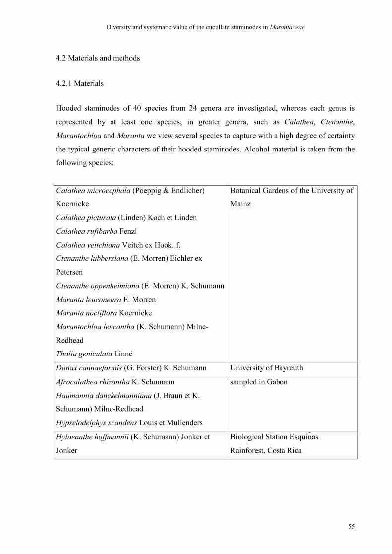
Diversity and systematic value of the cucullate staminodes in Marantaceae
55
4.2 Materials and methods
4.2.1 Materials
Hooded staminodes of 40 species from 24 genera are investigated, whereas each genus is
represented by at least one species; in greater genera, such as Calathea, Ctenanthe,
Marantochloa and Maranta we view several species to capture with a high degree of certainty
the typical generic characters of their hooded staminodes. Alcohol material is taken from the
following species:
Calathea microcephala (Poeppig & Endlicher)
Koernicke
Calathea picturata (Linden) Koch et Linden
Calathea rufibarba Fenzl
Calathea veitchiana Veitch ex Hook. f.
Ctenanthe lubbersiana (E. Morren) Eichler ex
Petersen
Ctenanthe oppenheimiana (E. Morren) K. Schumann
Maranta leuconeura E. Morren
Maranta noctiflora Koernicke
Marantochloa leucantha (K. Schumann) Milne-
Redhead
Thalia geniculata Linné
Botanical Gardens of the University of
Mainz
Donax cannaeformis (G. Forster) K. Schumann University of Bayreuth
Afrocalathea rhizantha K. Schumann
Haumannia danckelmanniana (J. Braun et K.
Schumann) Milne-Redhead
Hypselodelphys scandens Louis et Mullenders
sampled in Gabon
Hylaeanthe hoffmannii (K. Schumann) Jonker et
Jonker
Biological Station Esquin as
Rainforest, Costa Rica
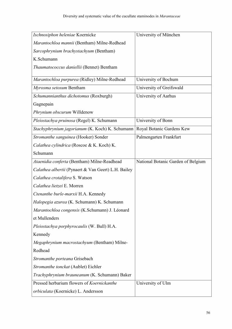
Diversity and systematic value of the cucullate staminodes in Marantaceae
56
Ischnosiphon heleniae Koernicke
Marantochloa mannii (Bentham) Milne-Redhead
Sarcophrynium brachystachyum (Bentham)
K.Schumann
Thaumatococcus daniellii (Bennet) Bentham
University of München
Marantochloa purpurea (Ridley) Milne-Redhead University of Bochum
Myrosma setosum Bentham University of Greifswald
Schumannianthus dichotomus (Roxburgh)
Gagnepain
Phrynium obscurum Willdenow
University of Aarhus
Pleiostachya pruinosa (Regel) K. Schumann University of Bonn
Stachyphrynium jagorianum (K. Koch) K. Schumann Royal Botanic Gardens Kew
Stromanthe sanguinea (Hooker) Sonder
Calathea cylindrica (Roscoe & K. Koch) K.
Schumann
Palmengarten Frankfurt
Ataenidia conferta (Bentham) Milne-Readhead
Calathea albertii (Pynaert & Van Geert) L.H. Bailey
Calathea crotalifera S. Watson
Calathea lietzei E. Morren
Ctenanthe burle-marxii H.A. Kennedy
Halopegia azurea (K. Schumann) K. Schumann
Marantochloa congensis (K.Schumann) J. Léonard
et Mullenders
Pleiostachya porphyrocaulis (W. Bull) H.A.
Kennedy
Megaphrynium macrostachyum (Bentham) Milne-
Redhead
Stromanthe porteana Grisebach
Stromanthe tonckat (Aublet) Eichler
Trachyphrynium brauneanum (K. Schumann) Baker
National Botanic Garden of Belgium
Pressed herbarium flowers of Koernickanthe
orbiculata (Koernicke) L. Andersson
University of Ulm
Diversity and systematic value of the cucullate staminodes in Marantaceae
57
4.2.2 Methods
Selection of characters:
Vegetative characters like those that Andersson (1998) took as a basis for his grouping are
entirely avoided. Metric floral characters such as the size of a flower, length and width of the
corolla tube, distances between single markings and all other floral parts like callose
staminodes, anthers a. s. o. are also excluded from the study. Numerical characters like the
number of outer staminodes or the number of ovules are avoided, too. All these ´traditional´
characters have proven to lead to an unsatisfying grouping which is little consistent with the
results of modern phylogenetical analysis. We are going to work by a minimalistic method
based only on hooded staminodes to fathom the diagnostic value of our own characters in
relation to genetic phylogeny. Functional features of hooded staminodes relating to the
pollination mechanism and the mutual dependence of flowers and their pollinators must be
disregarded here because we have only alcohol material or herbarium material to hand. The
only characters that we share with Andersson (1998) as well as Andersson and Chase (2001)
and Prince and Kress (2006) are the position and shape of the trigger-appendages.
Consequently, a very small number of distinctive characters of form and shape only at the
hooded staminodes are taken as a basis for the sorting. The most important characters for our
purpose are
• the presence of one or two lateral appendages on the trigger-side
• the presence or absence of a basal plate (sensu Kunze 1984) and its strength
• the basal plate that may be elongated in distal direction like a ´flag´
• the form of the appendages with swellings or callose thickenings
• the form of the appendages that may be rolled up or elongated
• the course of the midrib
• the area of the midrib that may have ledges or swellings
• the degree of support of the marginal and laminal growth of the hood
Pressed herbarium flowers of Koernickanthe orbiculata are first cooked in water at 80° C for
7 minutes and further put into 70 % alcohol for a day and eventually treated by the same
method as all others: the hooded staminodes of the pickled flowers are removed and stretched
on a black microscope slide so that the ´inner side´ of the hooded staminode (adaxial in
Diversity and systematic value of the cucullate staminodes in Marantaceae
58
relation to the axis of origin of the single flower) lies uppermost and affords a clear view of
the basal plate and trigger appendage. Only the hooded staminode of Phrynium obscurum is
not removed, but just bent away from the entire flower. They are photographed by means of
binoculars and a digital camera (Nikon coolpix 995). A selection of these photographs are
then drawn by hand in a slightly simplified way according to the abovementioned list of
characters, but still following the natural proportions, and then touched up by means of an
Illustrator 7 programme so that finally every genus in this study is represented by at least one
specimen and one sketch. Drawings are made of those that appear very typical of the
respective genus. As we have only alcohol material to hand, the positioning of these drawings
must be independent of the actual position and function of hooded staminodes in living plants
in order to secure an exclusively morphological observation and assessment uninfluenced by
any knowledge of real processes during pollination. These drawings of the hooded staminodes
are then sorted ´without prejudice´ according to the already mentioned characters into groups.
Finally a small number of types are abstracted from the groups and this entirely
morphological sorting is compared with the recent phylogenetic trees and discussed.
4.3 Results
In all hooded staminodes of the 24 genera investigated it is the midrib that does not take a
straight course to the leaf tip as in ´normal´ leaves (Fig. 4.1). Instead, it takes a turn of about
90°. The resulting leaf apex is in a lateral position and is clearly discernible because the main
vascular bundle is the thickest. Herewith the hooded staminode is generally divided into two
different ´halves´. On the adaxial side of the leaf (relating to the main axis of a pair of
flowers) there is the bigger ´half´ that forms the actual ´hood´ of the cucullate staminode. The
growth of the margin of the hood is always more or less supported. The hood can therefore
cover or even envelope the stigmatic orifice in some species or leave it entirely open in other
species.
The smaller, abaxial half forms the 1-2 appendages, one of which is considered the ´trigger´
for the release of the style movement. In the place where the midrib ends it reaches into a
small indentation between the hood and the first appendage – seen from the tip of the flower.
Here the appendage ´rises´ from the leaf margin of the cucullate staminode. In the place
where the appendage ´reenters´ the leaf margin there is often a fold that can have a callose
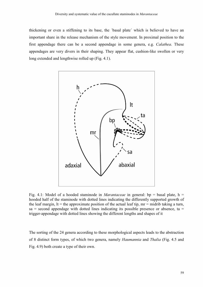
Diversity and systematic value of the cucullate staminodes in Marantaceae
59
thickening or even a stiffening to its base, the ´basal plate´ which is believed to have an
important share in the release mechanism of the style movement. In proximal position to the
first appendage there can be a second appendage in some genera, e.g. Calathea. These
appendages are very divers in their shaping. They appear flat, cushion-like swollen or very
long extended and lengthwise rolled up (Fig. 4.1).
Fig. 4.1: Model of a hooded staminode in Marantaceae in general: bp = basal plate, h = hooded half of the staminode with dotted lines indicating the differently supported growth of the leaf margin, lt = the approximate position of the actual leaf tip, mr = midrib taking a turn, sa = second appendage with dotted lines indicating its possible presence or absence, ta = trigger-appendage with dotted lines showing the different lengths and shapes of it
The sorting of the 24 genera according to these morphological aspects leads to the abstraction
of 8 distinct form types, of which two genera, namely Haumannia and Thalia (Fig. 4.5 and
Fig. 4.9) both create a type of their own.
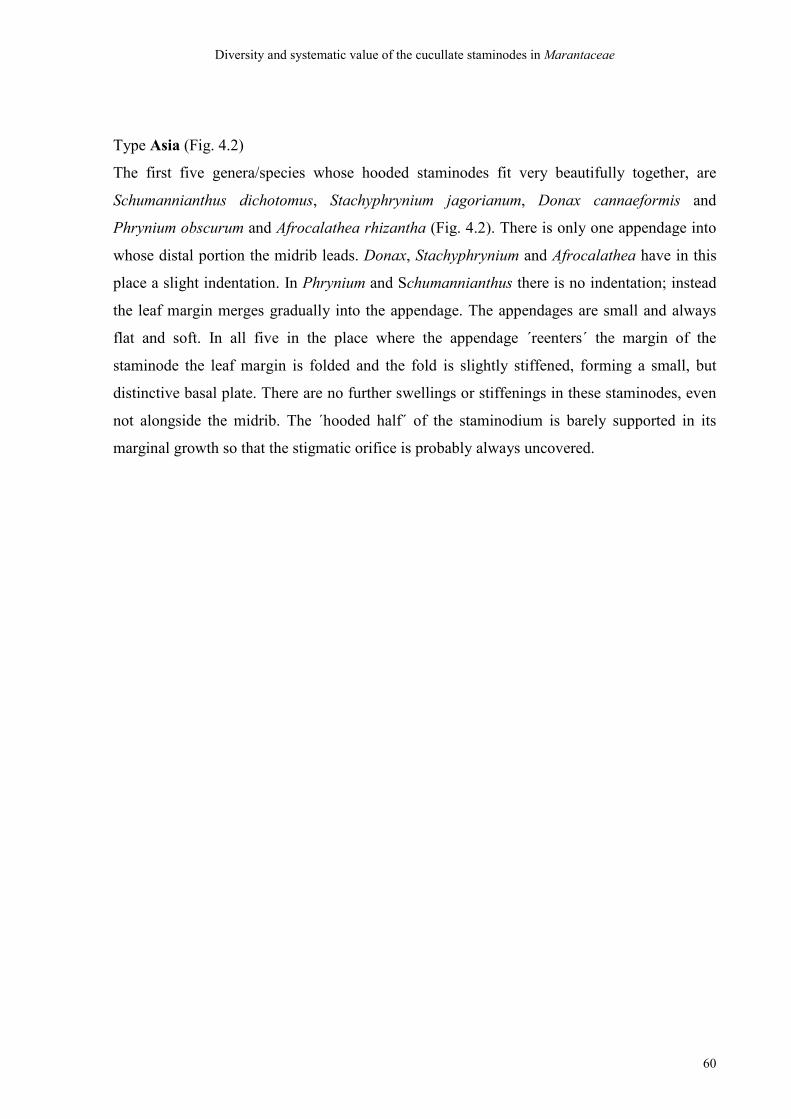
Diversity and systematic value of the cucullate staminodes in Marantaceae
60
Type Asia (Fig. 4.2)
The first five genera/species whose hooded staminodes fit very beautifully together, are
Schumannianthus dichotomus, Stachyphrynium jagorianum, Donax cannaeformis and
Phrynium obscurum and Afrocalathea rhizantha (Fig. 4.2). There is only one appendage into
whose distal portion the midrib leads. Donax, Stachyphrynium and Afrocalathea have in this
place a slight indentation. In Phrynium and Schumannianthus there is no indentation; instead
the leaf margin merges gradually into the appendage. The appendages are small and always
flat and soft. In all five in the place where the appendage ´reenters´ the margin of the
staminode the leaf margin is folded and the fold is slightly stiffened, forming a small, but
distinctive basal plate. There are no further swellings or stiffenings in these staminodes, even
not alongside the midrib. The ´hooded half´ of the staminodium is barely supported in its
marginal growth so that the stigmatic orifice is probably always uncovered.
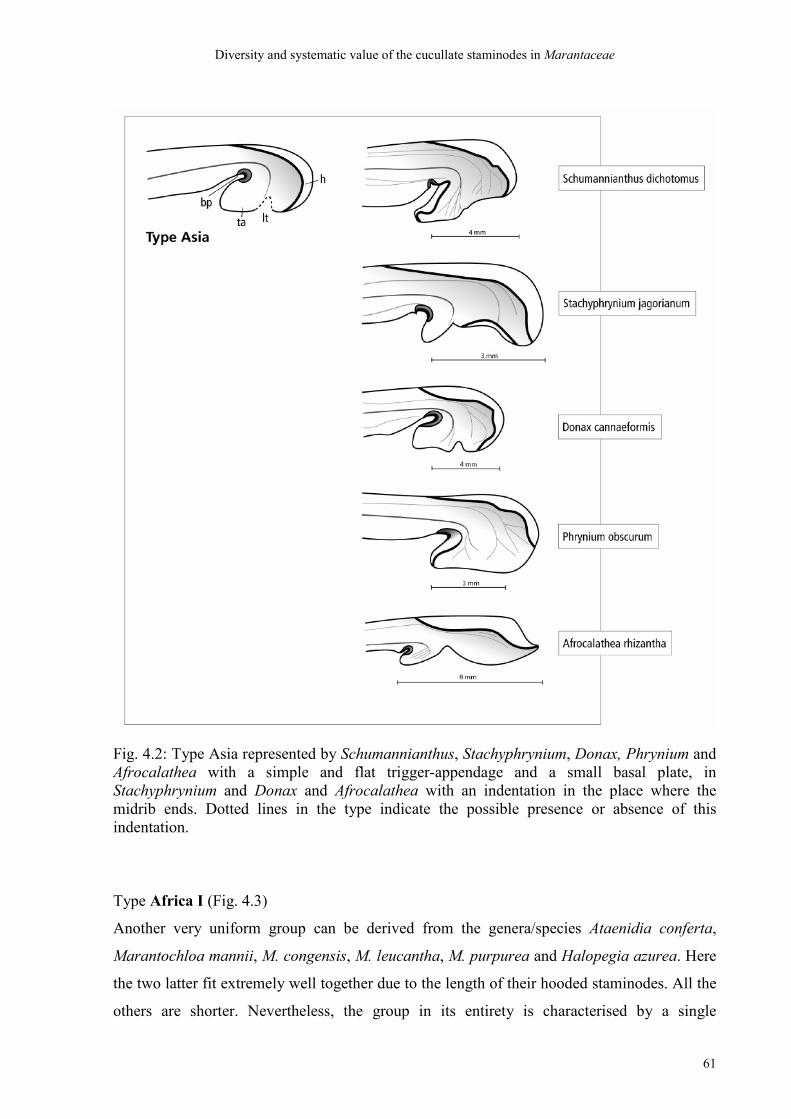
Diversity and systematic value of the cucullate staminodes in Marantaceae
61
Fig. 4.2: Type Asia represented by Schumannianthus, Stachyphrynium, Donax, Phrynium and Afrocalathea with a simple and flat trigger-appendage and a small basal plate, in Stachyphrynium and Donax and Afrocalathea with an indentation in the place where the midrib ends. Dotted lines in the type indicate the possible presence or absence of this indentation.
Type Africa I (Fig. 4.3)
Another very uniform group can be derived from the genera/species Ataenidia conferta,
Marantochloa mannii, M. congensis, M. leucantha, M. purpurea and Halopegia azurea. Here
the two latter fit extremely well together due to the length of their hooded staminodes. All the
others are shorter. Nevertheless, the group in its entirety is characterised by a single
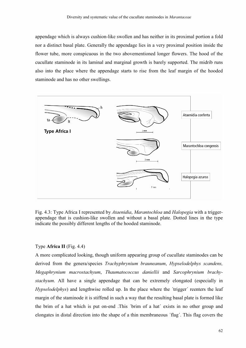
Diversity and systematic value of the cucullate staminodes in Marantaceae
62
appendage which is always cushion-like swollen and has neither in its proximal portion a fold
nor a distinct basal plate. Generally the appendage lies in a very proximal position inside the
flower tube, more conspicuous in the two abovementioned longer flowers. The hood of the
cucullate staminode in its laminal and marginal growth is barely supported. The midrib runs
also into the place where the appendage starts to rise from the leaf margin of the hooded
staminode and has no other swellings.
Fig. 4.3: Type Africa I represented by Ataenidia, Marantochloa and Halopegia with a trigger-appendage that is cushion-like swollen and without a basal plate. Dotted lines in the type indicate the possibly different lengths of the hooded staminode.
Type Africa II (Fig. 4.4)
A more complicated looking, though uniform appearing group of cucullate staminodes can be
derived from the genera/species Trachyphrynium brauneanum, Hypselodelphys scandens,
Megaphrynium macrostachyum, Thaumatococcus daniellii and Sarcophrynium brachy-
stachyum. All have a single appendage that can be extremely elongated (especially in
Hypselodelphys) and lengthwise rolled up. In the place where the ´trigger´ reenters the leaf
margin of the staminode it is stiffend in such a way that the resulting basal plate is formed like
the brim of a hat which is put on-end .This ´brim of a hat´ exists in no other group and
elongates in distal direction into the shape of a thin membraneous ´flag´. This flag covers the
Diversity and systematic value of the cucullate staminodes in Marantaceae
63
stigmatic orifice of the respective style as the marginal growth of the hooded half of the
staminodium is only slightly supported. Two of them, namely Trachyphrynium brauneanum
and Hypselodelphys scandens, bear a stiffening of the tissue along the midrib while
Megaphrynium macrostachyum has a swelling aside the midrib at the trigger-side.
Megaphrynium and Thaumatococcus have shorter appendages that are lengthwise rolled up,
too, and an almost identical course of vascular bundles. Trachyphrynium and Sarcophrynium
have the biggest hoods in this group.
Fig. 4.4: Type Africa II represented by Trachyphrynium, Hypselodelphys, Megaphrynium,Thaumatococcus and Sarcophrynium with elongated trigger-appendages, the shortest in Sarcophrynium, the longest in Hypselodelphys, that are always lengthwise rolled up. The basal plates are very stiff and put on-end and extended into a ´flag´ that can cover the
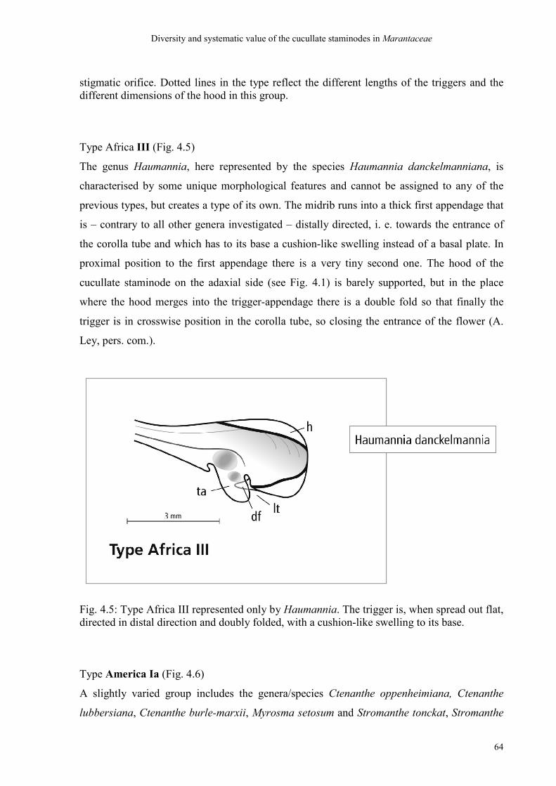
Diversity and systematic value of the cucullate staminodes in Marantaceae
64
stigmatic orifice. Dotted lines in the type reflect the different lengths of the triggers and the different dimensions of the hood in this group.
Type Africa III (Fig. 4.5)
The genus Haumannia, here represented by the species Haumannia danckelmanniana, is
characterised by some unique morphological features and cannot be assigned to any of the
previous types, but creates a type of its own. The midrib runs into a thick first appendage that
is – contrary to all other genera investigated – distally directed, i. e. towards the entrance of
the corolla tube and which has to its base a cushion-like swelling instead of a basal plate. In
proximal position to the first appendage there is a very tiny second one. The hood of the
cucullate staminode on the adaxial side (see Fig. 4.1) is barely supported, but in the place
where the hood merges into the trigger-appendage there is a double fold so that finally the
trigger is in crosswise position in the corolla tube, so closing the entrance of the flower (A.
Ley, pers. com.).
Fig. 4.5: Type Africa III represented only by Haumannia. The trigger is, when spread out flat, directed in distal direction and doubly folded, with a cushion-like swelling to its base.
Type America Ia (Fig. 4.6)
A slightly varied group includes the genera/species Ctenanthe oppenheimiana, Ctenanthe
lubbersiana, Ctenanthe burle-marxii, Myrosma setosum and Stromanthe tonckat, Stromanthe
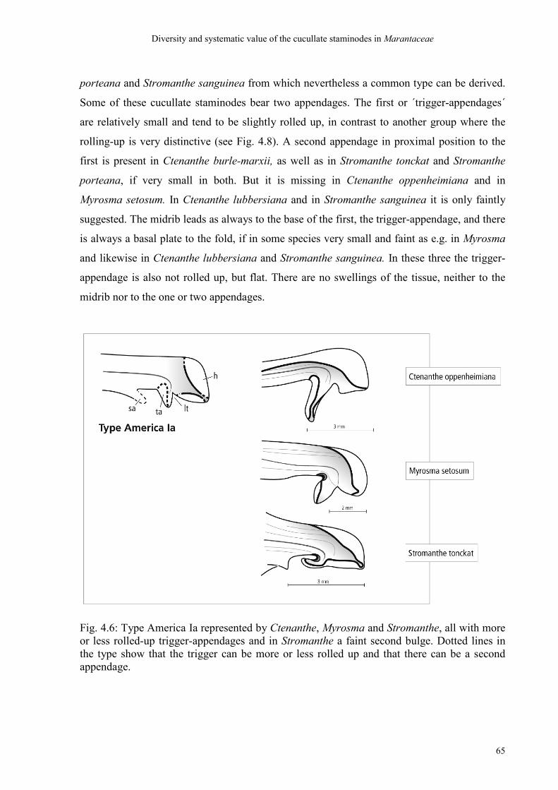
Diversity and systematic value of the cucullate staminodes in Marantaceae
65
porteana and Stromanthe sanguinea from which nevertheless a common type can be derived.
Some of these cucullate staminodes bear two appendages. The first or ´trigger-appendages´
are relatively small and tend to be slightly rolled up, in contrast to another group where the
rolling-up is very distinctive (see Fig. 4.8). A second appendage in proximal position to the
first is present in Ctenanthe burle-marxii, as well as in Stromanthe tonckat and Stromanthe
porteana, if very small in both. But it is missing in Ctenanthe oppenheimiana and in
Myrosma setosum. In Ctenanthe lubbersiana and in Stromanthe sanguinea it is only faintly
suggested. The midrib leads as always to the base of the first, the trigger-appendage, and there
is always a basal plate to the fold, if in some species very small and faint as e.g. in Myrosma
and likewise in Ctenanthe lubbersiana and Stromanthe sanguinea. In these three the trigger-
appendage is also not rolled up, but flat. There are no swellings of the tissue, neither to the
midrib nor to the one or two appendages.
Fig. 4.6: Type America Ia represented by Ctenanthe, Myrosma and Stromanthe, all with more or less rolled-up trigger-appendages and in Stromanthe a faint second bulge. Dotted lines in the type show that the trigger can be more or less rolled up and that there can be a second appendage.
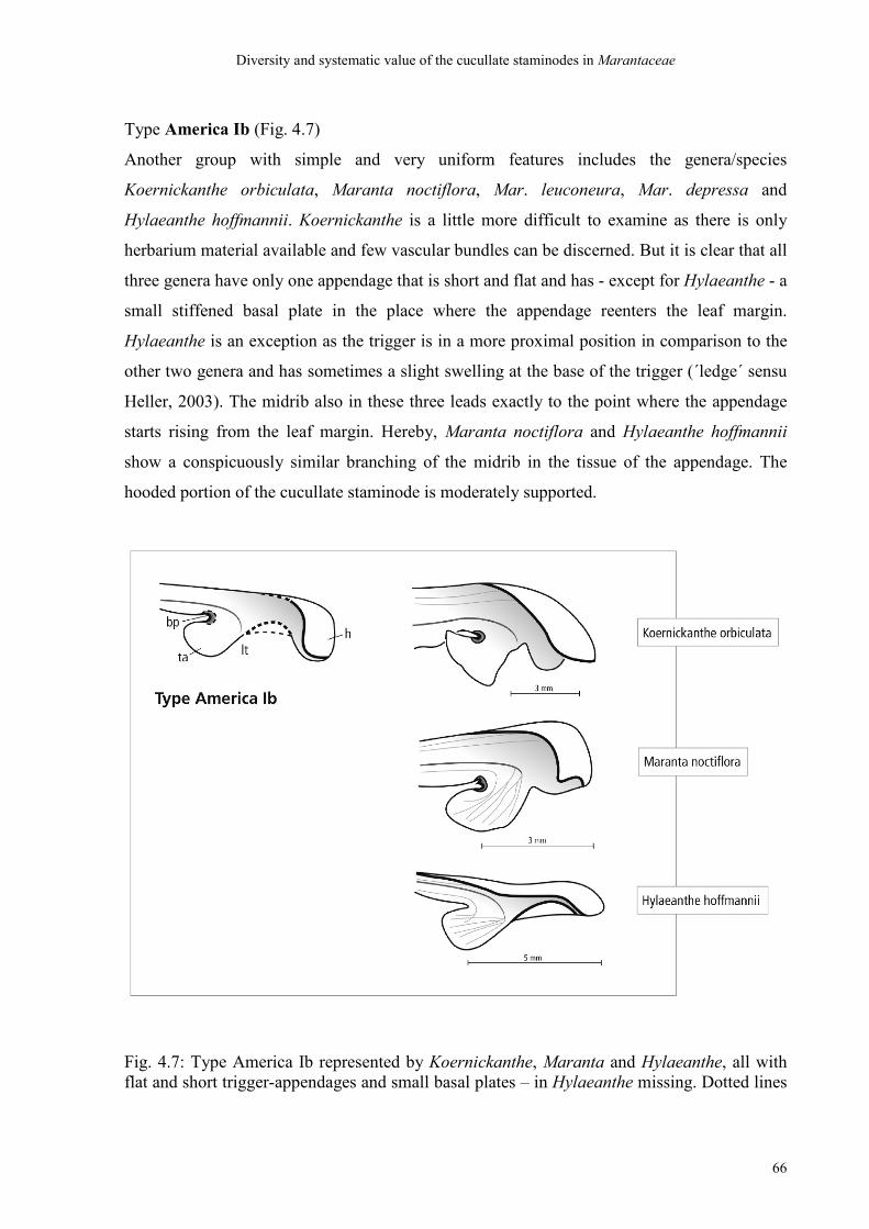
Diversity and systematic value of the cucullate staminodes in Marantaceae
66
Type America Ib (Fig. 4.7)
Another group with simple and very uniform features includes the genera/species
Koernickanthe orbiculata, Maranta noctiflora, Mar. leuconeura, Mar. depressa and
Hylaeanthe hoffmannii. Koernickanthe is a little more difficult to examine as there is only
herbarium material available and few vascular bundles can be discerned. But it is clear that all
three genera have only one appendage that is short and flat and has - except for Hylaeanthe - a
small stiffened basal plate in the place where the appendage reenters the leaf margin.
Hylaeanthe is an exception as the trigger is in a more proximal position in comparison to the
other two genera and has sometimes a slight swelling at the base of the trigger (´ledge´ sensu
Heller, 2003). The midrib also in these three leads exactly to the point where the appendage
starts rising from the leaf margin. Hereby, Maranta noctiflora and Hylaeanthe hoffmannii
show a conspicuously similar branching of the midrib in the tissue of the appendage. The
hooded portion of the cucullate staminode is moderately supported.
Fig. 4.7: Type America Ib represented by Koernickanthe, Maranta and Hylaeanthe, all with flat and short trigger-appendages and small basal plates – in Hylaeanthe missing. Dotted lines
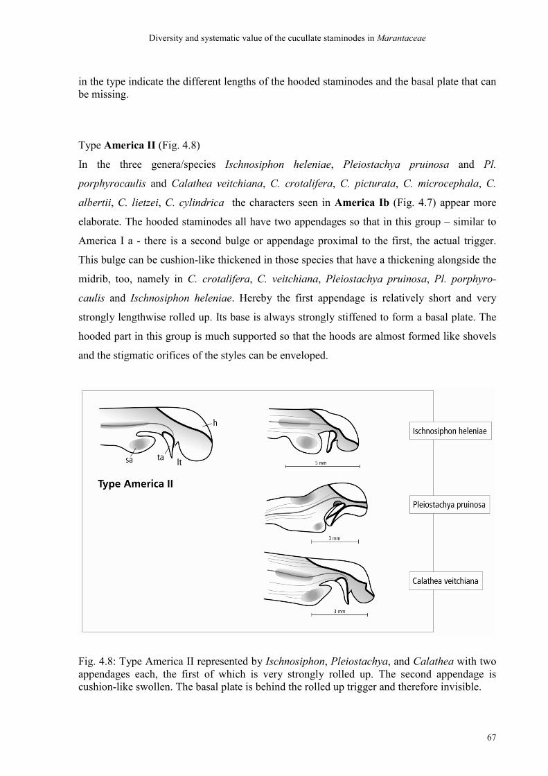
Diversity and systematic value of the cucullate staminodes in Marantaceae
67
in the type indicate the different lengths of the hooded staminodes and the basal plate that can be missing.
Type America II (Fig. 4.8)
In the three genera/species Ischnosiphon heleniae, Pleiostachya pruinosa and Pl.
porphyrocaulis and Calathea veitchiana, C. crotalifera, C. picturata, C. microcephala, C.
albertii, C. lietzei, C. cylindrica the characters seen in America Ib (Fig. 4.7) appear more
elaborate. The hooded staminodes all have two appendages so that in this group – similar to
America I a - there is a second bulge or appendage proximal to the first, the actual trigger.
This bulge can be cushion-like thickened in those species that have a thickening alongside the
midrib, too, namely in C. crotalifera, C. veitchiana, Pleiostachya pruinosa, Pl. porphyro-
caulis and Ischnosiphon heleniae. Hereby the first appendage is relatively short and very
strongly lengthwise rolled up. Its base is always strongly stiffened to form a basal plate. The
hooded part in this group is much supported so that the hoods are almost formed like shovels
and the stigmatic orifices of the styles can be enveloped.
Fig. 4.8: Type America II represented by Ischnosiphon, Pleiostachya, and Calathea with two appendages each, the first of which is very strongly rolled up. The second appendage is cushion-like swollen. The basal plate is behind the rolled up trigger and therefore invisible.
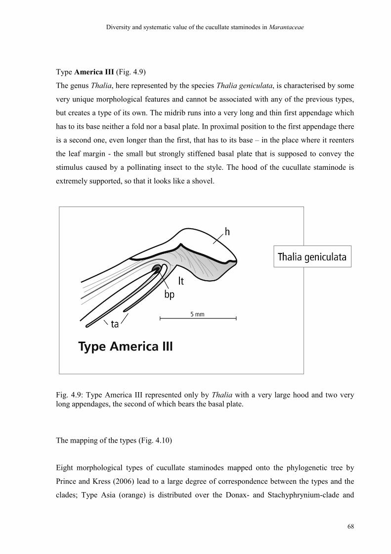
Diversity and systematic value of the cucullate staminodes in Marantaceae
68
Type America III (Fig. 4.9)
The genus Thalia, here represented by the species Thalia geniculata, is characterised by some
very unique morphological features and cannot be associated with any of the previous types,
but creates a type of its own. The midrib runs into a very long and thin first appendage which
has to its base neither a fold nor a basal plate. In proximal position to the first appendage there
is a second one, even longer than the first, that has to its base – in the place where it reenters
the leaf margin - the small but strongly stiffened basal plate that is supposed to convey the
stimulus caused by a pollinating insect to the style. The hood of the cucullate staminode is
extremely supported, so that it looks like a shovel.
Fig. 4.9: Type America III represented only by Thalia with a very large hood and two very long appendages, the second of which bears the basal plate.
The mapping of the types (Fig. 4.10)
Eight morphological types of cucullate staminodes mapped onto the phylogenetic tree by
Prince and Kress (2006) lead to a large degree of correspondence between the types and the
clades; Type Asia (orange) is distributed over the Donax- and Stachyphrynium-clade and
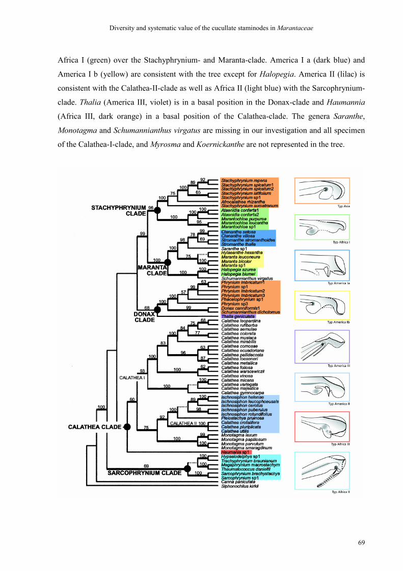
Diversity and systematic value of the cucullate staminodes in Marantaceae
69
Africa I (green) over the Stachyphrynium- and Maranta-clade. America I a (dark blue) and
America I b (yellow) are consistent with the tree except for Halopegia. America II (lilac) is
consistent with the Calathea-II-clade as well as Africa II (light blue) with the Sarcophrynium-
clade. Thalia (America III, violet) is in a basal position in the Donax-clade and Haumannia
(Africa III, dark orange) in a basal position of the Calathea-clade. The genera Saranthe,
Monotagma and Schumannianthus virgatus are missing in our investigation and all specimen
of the Calathea-I-clade, and Myrosma and Koernickanthe are not represented in the tree.
Diversity and systematic value of the cucullate staminodes in Marantaceae
70
Fig. 4.10: Types of hooded staminodes mapped onto the phylogenetic tree by Prince and Kress (2006). Type Asia (orange) is distributed over the Donax- and Stachyphrynium-clade and Africa I (green) over the Stachyphrynium- and Maranta-clade. America I a (dark blue) and America I b (yellow) are consistent with the tree except for Halopegia. America II (lilac) is consistent with the Calathea-II-clade as well as Africa II (light blue) with the Sarcophrynium-clade. Thalia (America III, violet) is in a basal position in the Donax-clade and Haumannia (Africa III, dark orange) in a basal position of the Calathea-clade.
4.4 Discussion
4.4.1 Discussion of the ´bauplan´ of cucullate staminodes
Fig. 4.1 shows a general ´bauplan´ of hooded staminodes that goes most likely – as we have
not investigated all of 31 genera - for the entire family. Ontogenetic observations on the
development of hooded staminodes of Eichler (1883, 1884 with Maranta sanguinea), Kunze
(1984 with Maranta leuconeura, Ataenidia conferta, Calathea vaginata) and Ley
(unpublished data) are in accord with the appearance of all mature hooded staminodes
investigated in this study: the midrib takes a turn by about a right angle so that finally the
actual leaf tip is at the abaxial side of the staminode, relating to the main axis of a flower pair.
This side eventually forms the later trigger part and the adaxial side forms the hood.
Comparative observations on our part have shown that the midrib is discernible already in
very small buds (2-4 mm) several days before anthesis and divides the growing staminode in
exactly these abovementioned ´halves´, one of which forms the later hood and the other the
trigger in all observed genera in the same fashion (A. Ley, with 20 genera, unpublished). The
diversity discussed here and taken as a basis for a systematic grouping is an expression of
variable proportions of exactly this basic model; the adaxial hood can be more or less
supported in its marginal and laminary growth. In addition, the margin of the hood can bear
lobes that are able to wrap the style in certain places. As a consequence in some genera (e. g.
Calatheas) the leaf blade of the hooded staminode can be so enlarged through such lobes that
the head of the style and the stigmatic orifice are completely enveloped. In others the surface
of the hood is short (e.g. Marantas) so that the stigma is left open. The abaxial trigger part can
bear 1-2 appendages, the first of which (seen from the leaf apex) is named the ´trigger´ that
releases the style movement and can – in most cases – bear at its base the basal plate or a
basal swelling. In extreme cases the triggers can be much elongated, some are lengthwise
rolled up or even lengthwise splitted in two, and some are simple and flat or cushion-like
Diversity and systematic value of the cucullate staminodes in Marantaceae
71
swollen. Also the basal plate can be very different in its solidity and shape and extension. The
second appendage - if one is formed - is in proximal position to the first and can also be with
or without swellings, ledges and differently shaped, but not as ´sophisticated´ as the first
appendage. And it is never a ´trigger´ to trigger off the style movement and never does the
midrib run into it. However different the actual trigger may be shaped, it is always the ´first
appendage´ that the midrib runs into and in this sense all appendages at the tip of the
medianus on the abaxial side of the leaf are considered homologous in their position. Any
lobe on the hooded part (adaxial side) of the staminode is in this study never called an
´appendage´ and has probably never to do with triggering off the style movement – rather it is
supposed to protect the stigmatic orifice and to hold the style in its unreleased position prior
to a pollinator´s visit. Another feature that is subject to diversity is the strength of all these
elements of hooded staminodes ( and also other floral parts) that can vary from very fragile
(e.g. Myrosma) to extremely solid (e.g. Megaphrynium) types, which may possibly be an
adaptation between the flowers and their pollinating animals and which remains to be
examined. Anyway, in this study characters of robustness are only taken in account for basal
plates as explained above.
4.4.2 Discussion of the types
Our simple looking Type Asia of hooded staminodes is in the analysis of Prince and Kress
(2006) distributed over two different clades, namely the Stachyphrynium-clade and the
Donax-clade. Schumannianthus dichotomus and Donax are sister-groups (which is consistent
with Andersson´s informal grouping of 1998) and together sister to Phrynium within the
Donax-clade; Schumannianthus virgatus which is in a quite different position in the tree
(Maranta-clade) we could unfortunately not examine here. Afrocalathea and Stachyphrynium,
on the other hand, are together in the Stachyphrynium-clade. Except for Afrocalathea they are
all Asian genera (see below).
The Type Africa I is only partly reflected by the tree of Prince and Kress (2006). The genera
Marantochloa and Ataenidia are unsurprisingly close regarding the shape of their hooded
staminodes which appear almost identical. The close relationship of these two was already
confirmed by the analysis of Andersson and Chase (2001) in contrast to Andersson´s informal
grouping of 1998 with Ataenidia in the Phrynium group and Marantochloa in the Maranta
Diversity and systematic value of the cucullate staminodes in Marantaceae
72
group. Prince and Kress (2006) even regarded Ataenidia as a ´highly modified member of
Marantochloa´ which was already strongly confirmed by Ley and Claßen-Bockhoff (2005,
2006). On the other hand, comparing Marantochloa purpurea with Halopegia azurea whose
cucullate staminodes are extremely similar the position of Halopegia in the Maranta-clade
seems surprising at first sight (see below). Likewise surprising is the remark of Prince and
Kress about the ´absence of a staminodial appendage´ in Ataenidia. From the course of the
midrib it is understandable which spot in the leaf margin forms the leaf tip. Proximal to it a
distinct appendage or a derivation of it can be determined. It is strongly cushion-like swollen
and sometimes a little rolled up. If in Ataenidia there were no appendage one would have to
regard the homologous positions in many flowers of the genus Marantochloa as vestigial, too.
This cushion-like swelling in the actual position of a trigger-appendage must be regarded as a
rare character. But it is not exclusive to the clade Marantochloa/Ataenidia as the position of
Halopegia, which has a cushion-like swelling in the same place, in the Maranta-clade proves.
Kunze (1984) equated the swelling of this spot in Ataenidia at least in a functional sense with
another swelling proximal to the trigger in many Calatheas. It is important to realise that in a
morphological sense these spots can not be homologous, because in the Calatheas the swollen
area shown in ontogenetic drawings of Calathea vaginata by Kunze is the second appendage
or bulge which was already described by Koernicke (1858). This is what we also found in
several species of Calathea in this study. The position of the first, the actual trigger-
appendage is clearly discernible from the medianus that leads to the leaf apex. The sister-
genera Ataenidia/Marantochloa belong to the Stachyphrynium-clade, together with Afro-
calathea and Stachyphrynium. The main difference between these two types of hooded
staminodes lies in the appendages that bear basal plates in the latter and a swelling with no
basal plate in the first. They are all African except for Stachyphrynium which is Asian.
The type Africa II is a group that is precisely and without exception reflected by the
Sarcophrynium-clade of Prince and Kress (2006). They all share a very striking character for
which Andersson (1998) created the term of a ´bilobed trigger´ which was taken over by
Prince and Kress. The term of course is only usable if one regards the basal plate as a part of
the trigger. For the ´flag´ that is extended in distal direction comes from the ´brimmed´ and
very stiff basal plate and not from the actual trigger-appendage which is very thin and frail.
Both the form and consistency of the basal plate and the membrane that comes out of it are
particularly rare characters which are found in no other group. This makes them highly
Diversity and systematic value of the cucullate staminodes in Marantaceae
73
meaningful and exclusive. The sister-relationship of Trachyphrynium / Hypselodelphys was
already described by Andersson (1998) and supported by the phylogenetic analyses of
Andersson and Chase (2001), Ley and Claßen-Bockhoff (2005, 2006) and Prince and Kress
(2006). The same is reported for the sister-genera Megaphrynium and Thaumatococcus that
were placed in very different positions by Andersson (1998). But considering the rarity and
value of the observed characters this newly found close relationship is not so very surprising
any more. Even if one takes solely the trigger appendages and their basal plates as a basis for
the grouping one can make out that they are African.
The type Africa III is formed by the genus Haumannia alone. In contrast to all other genera
whose triggers are always directed in proximal direction, i. e. towards the ovary, the trigger in
Haumannia is directed towards the flower entrance. Furthermore, it has two folds; one fold is
in the place where the appendage rises from the leaf margin, which is exceptional, and the
second fold is in the place where the appendage reenters the leaf margin as in general in the
family. Instead of a basal plate there is a cushion-like swelling similar to those in Africa I so
that a relation between them suggests itself. The consequence of the double fold is that the
appendage lies crosswise in the flower tube in a living flower (A. Ley, pers. com.).
Our Type America Ia corresponds with a part of the Maranta-clade of Prince and Kress (2006)
where Stromanthe and Ctenanthe are confirmed to be sister-groups (as proposed already by
Anderssson and Chase 2001). Features that are very elaborate in the Type America II appear
here modest and are not always very clearly developed, such as the second appendage which
can be present or absent in Ctenanthe and Stromanthe or the slight tendency of the triggers to
roll up. The close relationship of Myrosma to the sisters Stromanthe and Ctenanthe is also
supported by the analysis of Andersson and Chase (2001) and by Andersson´s Myrosma
group (1998).
Our type America Ib fits with another part of the Maranta-clade, namely the sister-groups
Maranta and Hylaeanthe according to the evaluation of Prince and Kress (2006). Another
close relationship between Maranta and Koernickanthe is reported by Andersson and Chase
(2001). Andersson (1998) included Maranta, Koernickanthe and Hylaeanthe in the Maranta-
group which is consistent with our type America Ib. The positioning of Halopegia in the
Maranta-clade after the analysis of Prince and Kress ( and together in the Myrosma-group of
Diversity and systematic value of the cucullate staminodes in Marantaceae
74
Andersson, 1998) seems surprising at first sight in comparison with our morphological
findings. But when one imagines the appendage at the hooded staminode in Halopegia
without its characteristic swelling and instead a small basal plate – or even none, as
sometimes in Hylaeanthe (also confirmed by our findings and by Heller 2003) – then also the
hooded staminode of Halopegia can look like a simple Type America Ib. Our decision to
place Halopegia among the African genera together with Marantochloa and Ataenidia was
primarily based on the presence of a cushion-like swollen appendage without a basal plate and
the striking similarity of the entire hooded staminodes between Halopegia and M. purpurea.
In both the appendage is in a very proximal position, i.e. deeper than normally in the flower
tube. Obviously is such a swelling not an exclusive character as we thought. Furthermore,
also Hylaeanthe has an appendage that is relatively deep in the flower tube, more proximal to
the ovary than in the sister-genera Maranta and Koernickanthe. This might also be an
adaptive character relating to the function of the staminodes during pollination, as we cannot
prove in this study.
The genera Calathea, Ischnosiphon and Pleiostachya form a very uniform type America II
that is exactly reflected by the Calathea-clade of Prince and Kress (2006) as well as the
Calathea-clade of Andersson and Chase (2001). Even Andersson´s Calathea-group (1998) is
consistent with this clade. However, in the recent analysis of Prince and Kress (2006) the
Calathea-clade turns out to be non-monophyletic. The species investigated all share the short
and stiff and strongly rolled-up trigger-appendage, in comparison with the soft and long
triggers in Africa II where only the basal plate is stiff. This is a striking character and a second
´American bulge´ whose function was discussed by Kunze (1984) at the example of Calathea
vaginata. Similarity between Calathea lutea, Calathea platystachya and Pleiostachya
pruinosa was already described by Heller (2003) – not only in a morphological but also
functional sense.
Our reasons to associate Thalia with the American types (America III) are, first, that most
species of Thalia are native to America and, secondly, because the American types tend to
develop a second bulge or appendage which at first sight appears to be a similar case here,
too. Furthermore, Thalia resembles no Asian or African type distinguished so far. Koernicke
(1858) regarded the appendage in Thalia as a single, but deeply lengthwise splitted
appendage. As the basal plate is at the base of the second ´ribbon´ this may be plausible. Also
Diversity and systematic value of the cucullate staminodes in Marantaceae
75
the course of the midrib supports this notion. This second ´ribbon´ sensu Koernicke looks less
supported than the first when one looks closely at the vascular bundles. Andersson (1998) on
the other hand spoke of ´two appendages´ in Thalia. But this would say that Thalia was the
only genus whose hooded staminode bears the basal plate in a different place than all other
genera, namely at a second appendage. A second appendage occurs often in American genera
(Type America I and II) and never bears a basal plate – just in some cases a swelling whose
function in species of Calathea was already discussed by Kunze (1984). Up to now all
observed genera and species were found to have their basal plates or equivalent derivations,
such as swellings or stiffenings, in the same place. It is the place in which the risen trigger-
appendage reenters the margin of the hooded staminode. Taking herewith a positional
homology for the basal plates in the family as a basis for all, it is impossible to accept
Andersson´s notion of two independent appendages, the second of which bears the basal plate.
Our findings strongly support the previous interpretation of Koernicke (1859) that in Thalia
there is one trigger-appendage splitted in two. Furthermore, the positioning of Thalia in the
Donax-clade ´with moderate support´ (Prince and Kress, 2006), or near the Donax-clade as
was already found out by Andersson and Chase (2001), makes any discussion about a second
´American appendage´ obsolete.
Altogether, we never found in this study a hooded staminode without a trigger-appendage,
contrary to the opinion of Prince and Kress about Ataenidia (see above, Type Africa I). A
number of these appendages provided most striking characters that enabled the separation of
distinct lineages without doubt or any exception. One of them is the rolled-up trigger with a
brim-shaped basal plate (´bilobed trigger´ sensu Andersson, 1998) in our Type Africa II or the
Sarcophrynium-clade of Prince and Kress, respectively. Another one is the stiff and rolled-up
trigger in our Type America II or the Calathea-clade of both Andersson and Chase (2001) and
Prince and Kress (2006). These features may now be regarded as the actual ´shape-characters´
which are obligatory to a group and must be fully expressed. The cushion-shaped appendage
in the sister-genera Ataenidia and Marantochloa (after Andersson and Chase 2001, Ley and
Claßen-Bockhoff 2005, and Prince and Kress 2006) is also conspicuous but not absolutely
exclusive to the clade and can therefore be misleading as it occurs also in Halopegia and more
or less also in Hylaeanthe (Heller 2003). The thickenings or swellings in the ´American
bulge´ in our type America II can also be more or less developed as well as stiffenings along
or aside the midrib. Perhaps one must conclude that swellings are no definite ´shape-

Diversity and systematic value of the cucullate staminodes in Marantaceae
76
characters´ but rather an optional and perhaps continuous feature relating to the function of an
organ (e.g. the guidance of a pollinating insect) and that there may be gradual transitions in
the expression of such a character. Even for the number of appendages there can be transitions
as we have seen in Myrosma and in members of Stromanthe where the second ´American
appendage´ is either very faint or can almost only be suspected. It is always the second
appendage that is facultative and that is not the trigger for the pollination mechanism. Also
obligatory to the respective group is the basal plate. It is either extremely elaborate, such as
the ´brim-shaped´ basal plate in type Africa II, even extending in a ´flag´ that can cover the
stigmatic orifice. Or it is simply a small, pointed stiffening in the fold of the leaf margin or in
some cases, such as Hylaeanthe or the Marantochloas, completely missing. A missing basal
plate is also a distinct and distinguishing character. The course of the midrib is always the
same and does therefore not enable any systematic grouping. But it can be helpful in some
difficult cases to find out the position of the leaf tip and herewith the first appendage,
inconspicuous though it may be, which is always the trigger-appendage. The degree of
support of the growth of the leaf margin of the hooded half of the staminode turns out in this
investigation not to provide a very distinctive morphological character. But it may turn out to
be an important ecological character as regards the protection of the stigmatic orifice and
herewith the regulation of cross pollination versus self pollination. Concluding, among the
eight characters selected for the study we found few really distinctive characters whose
systematic value was absolutely confirmed by the phylogenetical analyses, namely an
extremely elaborate shape of the trigger, an equally elaborate shape of the basal plate and a
clear markedness of a second ´American´ appendage, hereby fulfilling the second criterion of
homology (Remane, 1956). Where the triggers and basal plates and second appendages are
weakly shaped they have no such distinguishing effect, as well as tissue thickenings in some
places.
Based on this experience we propose generally for the morphological grouping to select
characters according to the following aspects: they must be characters of shape and must be
typical of a certain group that is to be investigated, e.g. genus, family, order. They should be
rather rare than frequent, rather discrete than continuous, and rather obligatory than
facultative. From such characters as few as possible should be taken for the grouping,
contrary to previous methods of grouping based on numerous characters (see above:
Andersson 1998).
Diversity and systematic value of the cucullate staminodes in Marantaceae
77
4.4.3 Discussion of evolutionary pathways
In the maximally parsimonious tree of Prince and Kress (2006) (Fig. 4.10) comprising five
big clades the first split divides up the family into the basal Sarcophrynium-clade and four
others, namely the Calathea-, Donax-, Maranta-, and Stachyphrynium-clade. The early lineage
consisting of Sarcophrynium as well as the sister-genera Hypselodelphys/Trachyphrynium and
Megaphrynium/Thaumatococcus is consistent with our morphological findings (Africa II, Fig.
4.4) showing striking synapomorphies - compared to the Cannaceae - in the shape of a
staminodium cucullatum with a rolled-up trigger and a basal plate that is brim-shaped and
extended in a flag-like fashion (the ´bilobed trigger´ of Andersson, 1998). The next split
detaches the Calathea-clade; herein the Calathea-II-clade is in accordance with our America II
(Fig. 4.8) with Calathea, Ischnosiphon and Pleiostachya and with the exception of
Haumannia (Africa III, Fig. 4.5) which is in a basal position here (see below). Unfortunately,
the Calathea-I-clade of Prince and Kress (2006) is not covered by our experimental plants
while most of our Calatheas, such as Calathea veitchiana, C. picturata, C. microcephala, C.
albertii, C. lietzei, and C. cylindrica are not included in their study so that we have only
Calathea crotalifera in common and must refer our results merely to the Calathea-II-clade in
the phylogenetical tree. Both together, the Sarcophrynium-clade and the Calathea-clade, share
the rolled-up trigger-appendage which appears to be the symplesiomorphic state of it, in
contrast to the entirety of the remaining three clades that look more derived.
The next split divides the Donax-clade from the two remaining. In this clade, which is partly
consistent with our Asia type (Fig. 4.2), we find as expected the genera Donax and Phrynium
and Schumannianthus dichotomus and surprisingly the genus Thalia (America III, Fig. 4.9),
which is nowadays American as well as African, in a basal position (Fig. 4.10). The last
division is continuous right through the Maranta- and Stachyphrynium-clade. In the Maranta-
clade the Hylaeanthe/Maranta-group can be comprehended after our morphological findings,
namely very simplified and flat trigger-appendages (America I b, Fig. 4.7) and the
membership of Koernickanthe, which is missing in the tree, is morphologically well-founded
(see also Andersson and Chase 2001). To a more basal group in the Maranta-clade belong
Schumannianthus virgatus, which we have not investigated, and Halopegia, that occurs in
Africa and some Asian habitats. Halopegia is conspicuous for its very apomorphic ´deep´
trigger with a tissue swelling instead of a basal plate that tempted us to include the genus first
of all in our Africa I (Fig. 4.3). Sister group to the Hylaeanthe/Maranta-group is the
Diversity and systematic value of the cucullate staminodes in Marantaceae
78
Ctenanthe/Stromanthe/Saranthe-group which – with the exception of Saranthe that we have
not investigated – is in accordance with our America I a (Fig. 4.6) including Myrosma which
is missing in the tree and whose membership is morphologically justified (see also Andersson
1998, and Andersson and Chase 2001). All these genera in the Maranta-clade are in fact
American. The remaining Stachyphrynium-clade divides itself in two lineages, namely the
African Ataenidia/Marantochloa-group, that shares a synapomorphic ´deep´ trigger and loss
of the basal plate and corresponds with our Africa I (Fig. 4.3) - with the exception of
Halopegia - and the very simplified Stachyphrynium/Afrocalathea-group which is in
accordance with the second part of our type Asia (Fig. 4.2), except that Afrocalathea is
African.
Considering the assumed course of the development given in the parsimonious tree of Prince
and Kress (2006) (see Fig. 4.10) it turns out that the two first clades, namely the
Sarcophrynium-clade and the Calathea-clade, are those that are conspicuous for their
lengthwise rolled-up trigger-appendages which we have named particularly ´elaborate´
triggers. The rolled-up shape of triggers appears to be a primal character shared by an African
and an American group of the family, a symplesiomorphy. When we start from the
assumption that the ancestral area of the family is Africa (see also Andersson 1981 and
Andersson and Chase 2001) then the American Calathea-clade must be the next relations.
While Africa is obviously a centre of diversity of genera, some of which are even monotypic
or very poor in species (Thaumatococcus, Trachyphrynium, Ataenidia, Afrocalathea,
Halopegia) the size of the Calathea-clade reflects an enormous radiation resulting in some
300 species of Calathea alone (Andersson and Chase 2001). So it suggests itself that a ´primal
Calathea´ came from Africa to America. The other American genera may have secondarily
´simplified´ their cucullate staminodes while the lineage Ctenanthe/Stromanthe/Myrosma/
(Saranthe), our America I a, still retains partly the old characters in the shape of small ´second
appendages´ und more or less rolled-up triggers (Fig. 4.6). The group
Maranta/Hylaeanthe/Koernickanthe, our America I b, is most strongly derived and simplified
with flat triggers and without second appendages (Fig. 4.7) where according to the tree also
Halopegia belongs. Also the group Ataenidia/Marantochloa (Africa I, Fig. 4.3) appears
secondarily simplified in comparison with Afrika II (Fig. 4.4). This may make our erroneous
assignment of Halopegia to Africa I, in particular considering a swelling at the trigger,
understandable. Both groups, America I b and Africa I, appear to be the most strongly
Diversity and systematic value of the cucullate staminodes in Marantaceae
79
advanced each in their continent and are therefore – at least as regards the morphology of the
cucullate staminodes - very similar.
The last section is the Asian (Fig. 4.2) which is also twice secondarily strongly simplified,
namely in the Donax-clade and the Stachyphrynium-clade in which likewise the cucullate
staminodes are very similar to each other in their simplicity. Here the basal position of Thalia
in an Asian clade is interesting – if with little support (Prince and Kress, 2006) – it seems to
connect all three continents (see below). Despite the doubtful situation of Thalia it seems to
be cogent to acknowledge the lengthwise splitted trigger-appendage as a strong apomorphy to
the genus alone.
Very interesting is also the basal position of Haumannia (Africa III, Fig. 4.5) in the Calathea-
clade (Fig. 4.7). Like that in Thalia the hooded staminode in Haumannia is morphologically
very peculiar in displaying even two apomorphies, such as a completely missing basal plate
and a doubly folded trigger-appendage. Haumannia that is actually endemic to Africa appears
morphologically African and genetically American and does not look actually like our virtual
´primal Calathea´ with a rolled-up trigger. But we do not know how strongly derived the
contemporary types Africa II and III and America II look nowadays in comparison to their
ancestral form. Also the modern Haumannia, basal though its position in the Calathea-clade
may be, appears very derived. It may be secondarily simplified in a way similar to the
Marantochloas and Ataenidia as regards the ´swollen´ trigger after a possible re-migration
from America back to Africa in a secondary dispersal event. This ´modern African
simplification´ might be an adaptation to certain African pollinators, involving perhaps a
simpler pollen transfer mechanism, which we cannot prove here. The position of
Afrocalathea, which is endemic to Africa, in the Asian Stachyphrynium-clade might be
interpreted either as an example of a convergent development in both continents due to the
aforementioned mutual dependence of plants and their pollinators or as a result of a secondary
dispersal event, too. Secondary migration must also be suspected for Halopegia, which is in
the Maranta-clade, but occurs in African and Asian habitats and for Thalia that occurs in
America and Africa (see also Andersson 1981).
Altogether, we are in agreement with previous authors (Andersson, 1981 and Andersson and
Chase, 2001) on the conclusion that most probably the ancestral area of the family is Africa.
Thence a ´primal Calathea´ may have settled in the New World – whether prior to the
continental drift or after (see also Andersson and Chase, 2001) cannot be proved here – giving
rise to an enormous radiation of species. The last migration is most likely the one to Asia
Diversity and systematic value of the cucullate staminodes in Marantaceae
80
where the cucullate staminodes developed to simple modern forms without any exception
found in this study. When looking at these assumed derivations one must always allow for the
interaction between flowers and pollinators which we know very little about at present.
Therefore, ecological investigations and experiments on the trigger-mechanism are doubtless
the most important in the next time.
General conclusions
81
5. General conclusions
All results of our investigations taken together meet in the conviction that the hypothesised
key innovation in Marantaceae can be confirmed and that it lies in the very close cooperation
of the style and the cucullate staminode, ranging from their mutual development in bud to the
hydraulic tissue of the style to the peculiar shapes of the cucullate staminodes.
In Marantaceae the style is thick and fleshy and consists of so many cell layers that under
´straitjacket-conditions´ it can be tensioned for the movement and during ageing a tissue of
hydraulic function can be established. Contrary to this there is in the sister group Cannaceae a
thin and flat petaloid style (Petersen 1981). Although a tendency to an increased porosity of
the style parenchyma is already laid out in Cannaceae the style tissue cannot make use of that
feature in the service of pollination.
The cucullate staminodes in Marantaceae with their unique ´bauplan´ and strikingly divers
appendages are able to keep a firm hold on the style prior to a pollinator´s visit until the
opportune moment for pollen transfer; the maturity of the style must be on one hand sufficient
to perform the full movement and on the other hand not over-mature because a wilting style
could not be released any more. And regarding the diversity of the trigger-appendages it
appears very likely that only particular pollinators can trigger off the movement in a certain
genus or species of the family – other insects may only visit the flowers without pollinating -
so that through the cucullate staminodes the styles are probably ´detained´ to wait for the best-
adapted pollinator, so as to avoid hybridisation. In summary, the cucullate staminodes have
the task to prepare the style for the movement, to control the time of pollination, possibly to
select pollinators, and they are supposed to have a certain share in the regulation of self-
pollination versus cross-pollination (Hildebrand 1870, Eichler 1884, Schumann 1902) And we
feel certain that they also control the release of the movement (see below). In comparison to
this in the sister Cannaceae there is in the homologous position of the cucullate staminode a
simple and flat staminode that does not enclose the style and can have none of the
aforementioned functions during pollen transfer. The great precision of the pollen transfer in
Marantaceae is probably also supported by the callose staminodes of the inner androeceal
whorl which we have not investigated in this study. As for the evolutionary ´success´ of this
precision, it appears likely that it lies less in the multiplying of individuals because despite the
elaborate pollen transfer seed set through cross-pollination is generally low (A. Ley, pers.
General conclusions
82
com.) and at least 44 species (8% of the family) help themselves with autogamy (Kennedy
2000) and vegetative propagation through rhizomes. The main aspect of success may rather
lie in the avoidance of hybridisation and, in consequence, in the ecological niche-finding. If
this could be confirmed, we would eventually be convinced that the great radiation of the
family was in fact an adaptive radiation as a result of a key innovation.
As for the release, in view of the size of the family we must allow for deviations from the
general scheme anyway. But at least, we tried to approach the physiological contributions to
the trigger-mechanism in the style-staminode-complex. For this, several electrophysiological
experiments were carried out in styles of Maranta leuconeura and in styles and cucullate
staminodes of Calathea veitchiana (unpublished data). These experiments turned out to be
extremely difficult. What we expected was at least a measurable alteration of a membrane
potential in one or both organs as a result of style release by hand manipulation. Unfortuna-
tely, the release of the movement always resulted either in a break of the electrode itself or –
through the mechanical inertia of the electrode - in a tear of the style tissue so that the
expected local potential changes or even conducted action potentials could not be detected.
We do not know at present how this problem might be solved by a better technical method.
We succeeded after all in detecting a resting potential of about –160 mV in both organs and
species (unpublished data). This, however, is not very meaningful because any plant or animal
cell membrane shows a resting potential. Therefore, the physiological part in the release must
remain open for the time being and subject to conjectures.
What remains to be done in future are observations of plant-pollinator-interactions at the
native habitats so to shed more light on the ecological aspects of the adaptive processes in the
family. The hypothesis concerning the avoidance of hybridisation should be tested by
crossing experiments and the examination of chromosome numbers in the family.
Furthermore, the share that other floral organs apart from the cucullate staminodes have in the
precision of the pollen-transfer, such as the callose staminodes and maybe even the styles
themselves, should be looked into.
References
83
References
Andersson, L. (1981). The neotropical genera of Marantaceae. Circumscription and relation-
ships. Nordic Journal of Botany 1: 218-245
Andersson, L. (1998). Marantaceae. In: Kubitzki, K. (ed.) The families and genera of vascu-
lar plants. Vol. IV Flowering plants, Monocotyledons, Alismatanae and Commelinanae
(except Gramineae) Pp. 278-293. Springer, Berlin
Andersson, L., Chase, M.W. (2001). Phylogeny and classification of Marantaceae. Botanical
Journal of the Linnean Society. 135: 275-287
Arns, K.Y., Mayo, S.J., Alves, M.V. (2002). Morfologia de Marantaceae ocorrente no estado
de Pernambuco, Brasil. Iheringia Sér. Botanica 57: 3-20
Claßen-Bockhoff, R. (1991). Untersuchungen zur Konstruktion des Bestäubungsapparates
von Thalia geniculata (Marantaceae). Botanica Acta 74: 183-193
Claßen-Bockhoff, R., Pischtschan, E. (2000). The Explosive Style in Marantaceae – Preli-
minary Results from Anatomic Studies. In: Spatz, H. and Speck Th. (eds.) Plant
Biomechanics 2000. Proceedings of the 3rd Plant Biomechanics Conference Freiburg
Badenweiler. Pp. 515-521. Thieme, Stuttgart.
Costerus, J.C. (1918). Die Übereinstimmung und der Unterschied im Bau der Blumen von
Canna und derjenigen der Marantaceen. Annales du Jardin Botanique de Bruitenzorg
11.15: 59-90
Delpino, F. (1869). Breve cenno sulle relazioni biologiche e genealogiche delle Marantacee.
Nuovo Giornale Botanico Italiano 1: 293-306
Eichler, A. (1884). Beiträge zur Morphologie und Systematik der Marantaceen. Abhandlun-
gen der Königlich Preussischen Akademie der Wissenschaften, Berlin
Gris, A. (1859). Observations sur la fleur des Marantées. Annales des Sciences naturelles IV,
12: 193-219
Guttenberg H. v. (1971). Bewegungsgewebe und Perzeptionsorgane. In: Zimmermann, W.,
Carlquist, S., Ozenda, P., Wulff, H.D. (eds.) Encyclopedia of Plant Anatomy, Handbuch
der Pflanzenanatomie, 2nd ed., V/5: 152-223 Bornträger, Stuttgart
Heller, A. (2003). Untersuchungen zur Griffelbewegung bei Marantaceen. diploma thesis,
University of Mainz, (unpublished)
References
84
Hildebrand, F. (1870). F. Delpino´s weitere Beobachtungen über die Dichogamie im Pflan-
zenreich. Botanische Zeitung 39: 617-625
Holttum, R.E. (1951). The Marantaceae of Malaya. Gardens´ Bulletin. Straits Settlements.
Singapore 13: 254-296
Howell G.J., Slater, A.T., Knox, R.B. (1993). Secondary pollen presentation in angiosperms
and its biological significance. Australian Journal of Botany 41: 417-438
Kennedy, H. (1978). Systematics and pollination of the ´closed-flowered´ species of Calathea
(Marantaceae). University of California Publication in Botany 71: 1-90
Kennedy, H. (2000). Diversification in pollination mechanisms in the Marantaceae. In: Wil-
son, K.L. and Morrison, D.A.(eds) Monocots: Systematics and Evolution. Pp. 335- 343.
CSIRO Melbourne
Koernicke, F. (1858). Beiträge zur Kenntnis der in unseren Gärten cultivierten Marantaceen.
Gartenflora 7: 66-89
Kress, W. J., Prince, L. M., Hahn, W. J., Zimmer, E. A. (2001). Unravelling the Evolutionary
Radiation of the Families of the Zingiberales using Morphological and Molecular
Evidence. Systematic Biology 50 (6): 926-944
Kunze, H. (1984). Vergleichende Studien an Cannaceen- und Marantaceenblüten. Flora175:
301-318
Ley, A., Claßen-Bockhoff, R. (2005). Floral Diversity of African Marantaceae. XVII. Inter-
national Botanical Congress, Vienna, Austria 17.-23.7.2005, Abstracts: 477 (poster
abstract)
Ley, A., Claßen-Bockhoff, R. (2006). Floral ecology and pollination in African Marantaceae.
19. Jahrestagung der GTÖ, Kaiserslautern
Lindley, J. (1819). Maranta zebrina. Botanical Register 5, t. 385
Lindley, J. (1826 a). Calathea flavescens. Botanical Register 11, t. 932Lindley, J. (1826 b).
Calathea violacea. Botanical Register 12, t. 962 text sub t. 961
Locatelli, E., Machado I.C., Medeiros P. (2004). Saranthe klotzschiana (Koer.) Eichl.
(Marantaceae) e seu mecanismo explosivo de polinização. Revista Brasileira de
botánica, São Paulo. V. 27, n.4: 757-765
Loesener, Th. (1930). Marantaceae. In: Engler A. and Prantl, K. (eds.) Die natürlichen Pflan-
zenfamilien. Ed.1,15a: 654-693. Engelmann, Leipzig
Metzner, H. (1982). Pflanzenphysiologische Versuche. Fischer, Stuttgart.
References
85
Petersen, O.G. (1981). Marantaceae. In: Hickey, M. and King, C. (eds.) 70 families of flowe-
ring plants. Pp. 528-533. Cambridge University Press
Prince, L.M., Kress, W.J. (2006). Phylogenetic relationships and classification in Maranta-
ceae: insights from plastid DNA sequences data. Taxon 55 (2): 281-296
Remane, A. (1956). Die Grundlagen des natürlichen Systems, der vergleichenden Anatomie
und der Phylogenetik. 2. Auflage Leipzig: Akademische Verlagsgesellschaft. Geest &
Portig.
Schumann, K. (1902). Marantaceae. In: Engler, A. (ed.) Das Pflanzenreich IV. 48 (11) Pp. 3-
29
Sitte, P. (1999). Bau und Feinbau der Zelle: Zellwände. In: Sitte, P., Ziegler, H., Ehrendorfer,
F., Bresinsky, A. (eds.) Strasburger, Lehrbuch der Botanik für Hochschulen, 34.
Auflage. Pp 89-99. 34 Spektrum Akademischer Verlag, Heidelberg
Skotheim, J. M., Mahadevan, L. (2005). Physical Limits and Design Principles for Plant and
Fungal Movements. Science 308: 1308-137
Tomlinson, P.B. (1961). Morphological and anatomical characteristics of the Marantaceae.
Journal of the Linnean Society. Botany. 58, 55-78
Yang, W., Lou, Ch. (1994). Electrochemical wave transmission and rapid coiling movement
in tendril of luffa. Science in China (Series B) 38 No.8: 944-953
Yeo, P. (1993). Secondary pollen presentation. Form, Function and Evolution. Plant Syste-
matics and Evolution, Supplement 6: 1-268