Embed Size (px)
Citation preview
NE35CH06-Grant ARI 14 May 2012 12:4
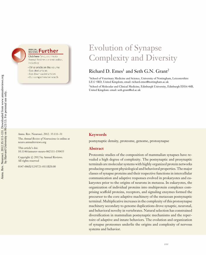
Evolution of SynapseComplexity and DiversityRichard D. Emes1 and Seth G.N. Grant2
1School of Veterinary Medicine and Science, University of Nottingham, LeicestershireLE12 5RD, United Kingdom; email: [email protected] of Molecular and Clinical Medicine, Edinburgh University, Edinburgh EH16 4SB,United Kingdom: email: [email protected]
Annu. Rev. Neurosci. 2012. 35:111–31
The Annual Review of Neuroscience is online atneuro.annualreviews.org
This article’s doi:10.1146/annurev-neuro-062111-150433
Copyright c© 2012 by Annual Reviews.All rights reserved
0147-006X/12/0721-0111$20.00
Keywords
postsynaptic density, proteome, genome, protosynapse
Abstract
Proteomic studies of the composition of mammalian synapses have re-vealed a high degree of complexity. The postsynaptic and presynapticterminals are molecular systems with highly organized protein networksproducing emergent physiological and behavioral properties. The majorclasses of synapse proteins and their respective functions in intercellularcommunication and adaptive responses evolved in prokaryotes and eu-karyotes prior to the origins of neurons in metazoa. In eukaryotes, theorganization of individual proteins into multiprotein complexes com-prising scaffold proteins, receptors, and signaling enzymes formed theprecursor to the core adaptive machinery of the metazoan postsynapticterminal. Multiplicative increases in the complexity of this protosynapsemachinery secondary to genome duplications drove synaptic, neuronal,and behavioral novelty in vertebrates. Natural selection has constraineddiversification in mammalian postsynaptic mechanisms and the reper-toire of adaptive and innate behaviors. The evolution and organizationof synapse proteomes underlie the origins and complexity of nervoussystems and behavior.
111
Ann
u. R
ev. N
euro
sci.
2012
.35:
111-
131.
Dow
nloa
ded
from
ww
w.a
nnua
lrev
iew
s.or
gby
Har
vard
Uni
vers
ity o
n 06
/21/
13. F
or p
erso
nal u
se o
nly.
NE35CH06-Grant ARI 14 May 2012 12:4
Contents
INTRODUCTION . . . . . . . . . . . . . . . . . . 112SYNAPSE PROTEOMICS AS A
BASIS FOR STUDYINGSYNAPSE EVOLUTION . . . . . . . . . 112
MOLECULAR COMPLEXITY INTHE SYNAPSE PROTEOME . . . . 113
GENOME EVOLUTION ANDITS ROLE IN SYNAPSEEVOLUTION . . . . . . . . . . . . . . . . . . . . 115
PROKARYOTE ORIGINS OFSYNAPSE PROTEINS ANDCORE PATHWAYS . . . . . . . . . . . . . . 116
EUKARYOTE INNOVATIONSAND THE PROTOSYNAPSE . . . . 119
COMPLEXITY AND DIVERSITYIN METAZOAN SYNAPSES . . . . . 121
INSIGHTS FROM THEHUMAN SYNAPSE . . . . . . . . . . . . . . 125
A MODEL FOR THE EVOLUTIONOF THE POSTSYNAPTICPROTEOME . . . . . . . . . . . . . . . . . . . . . 125
CONCLUSIONS . . . . . . . . . . . . . . . . . . . . 127
INTRODUCTION
The hallmark of the brain of humans and thatof most other species is its high degree ofcomplexity and diversity of neuronal morphol-ogy. Comparisons of the neuroanatomy of or-ganisms in different phyla point to the originof functional neuronal circuits ∼600 Mya inthe gelatinous Ctenophora and Cnidaria (re-viewed in Lichtneckert & Reichert 2009). De-spite the apparent anatomical simplicity of theseorganisms, their neurons possessed chemical(symmetrical and asymmetrical) and electricalsynapses, as well as chemical and peptidergicneurotransmitters. Considering that morpho-logical synapses are absent in Porifera (sponges)or earlier multicellular organisms, it is diffi-cult to envision a set of evolutionary pressuresthat would have selected for the evolution ofall the specialized molecular components thatconstruct a synapse in a single step. As with
the evolution of the vertebrate eye (Dawkins1986, 1994; Nilsson & Pelger 1994), it is farmore likely that the many molecular compo-nents of synapses had already existed in thoseearlier organisms that lacked neurons and thatthese components were reorganized into thevisibly distinct structure we know as the neu-ronal synapse.
In this review we focus on the molecularevolution of synapse proteins and their orga-nization. Akin to the neuroanatomical stain-ing methods that exposed the striking cellularcomplexity of the nervous system discoveredby anatomists in the nineteenth century, neu-roproteomic methods in the twenty-first cen-tury uncovered a far higher degree of molec-ular complexity in the protein composition ofsynapses than had been expected from ear-lier electrophysiological, biochemical, or ge-netic studies. It is from these proteomic datathat investigators can systematically explore themolecular evolution of all the individual pro-teins as well as their organization into networksand supramolecular structures. We first intro-duce the composition of synapse proteomes andgenome evolution and then review the evolu-tion of synapses.
SYNAPSE PROTEOMICS AS ABASIS FOR STUDYING SYNAPSEEVOLUTION
Because one can readily measure gross neu-roanatomy, cell number, and neuronal shape,there is an extensive literature on the evolu-tion of the nervous system based on these mea-surements (reviewed in Kaas 2009). By con-trast, there is a paucity of studies on synapseevolution, presumably because microscopic andelectrophysiological measurements are difficultto obtain and compare between species. Thishas been a major shortcoming in evolution-ary neuroscience since synapses were identi-fied, more than one century ago, as the basis forneuronal connections and information transferwithin neuronal circuits.
In the late 1980s and 1990s, the applica-tion of complementary DNA (cDNA) cloning
112 Emes · Grant
Ann
u. R
ev. N
euro
sci.
2012
.35:
111-
131.
Dow
nloa
ded
from
ww
w.a
nnua
lrev
iew
s.or
gby
Har
vard
Uni
vers
ity o
n 06
/21/
13. F
or p
erso
nal u
se o
nly.
NE35CH06-Grant ARI 14 May 2012 12:4
Presynapticproteome (PreSP):complete set ofidentified proteinsin the presynapticterminal of the synapse
Postsynapticproteome (PSP):complete set ofidentified proteinsin the postsynapticterminal of the synapse
methods identified the genes for a number ofion channel subunits and a relatively small num-ber of other synaptic proteins. At this pointgenome sequence data were sparse, and thus thescope of enquiry into the range of species andultimately the ancestry of the genes could notbe readily determined. Moreover, these weremostly single gene studies and did not explainthe evolution of the synapse itself: The synapseis a macromolecular subcellular structure thatis assembled from the protein products of manygenes, and it is necessary to examine the evolu-tion of its composite sets of proteins.
The year 2000 was a significant turningpoint for two reasons. First, proteomic methodswere applied to the study of synapses, which re-vealed unexpectedly large numbers of proteins(Husi et al. 2000). Proteomics has discoveredmore synapse proteins than has any other ap-proach and has provided the necessary startinginformation for molecular studies of synapseevolution. Second, the draft sequence of thehuman genome was released, and the ensuingexplosion of genome sequencing from many or-ganisms provided essential data for systematicstudies of the phylogeny of individual genes,sets of genes, and whole organisms. The com-bination of synapse proteomics and genomicshas been at the center of the first systematicstudies of synapse evolution.
MOLECULAR COMPLEXITY INTHE SYNAPSE PROTEOME
Catalogs of proteins found within mammaliansynapses have been derived from mass spec-trometry profiling of whole synapses, pre- andpostsynaptic fractions, and subcellular struc-tures including synaptic vesicles and signalingcomplexes (reviewed in Bayes & Grant 2009).In this review we separate discussion of thepresynaptic proteome (PreSP) from the postsy-naptic proteome (PSP) because most evolution-ary research has been performed on the PSP.This is also a useful distinction with regard tofunction because these proteomes have separateorigins in unicellular organisms: The presynap-tic release machinery is composed largely of the
vesicular release machinery used by unicellu-lar organisms to release chemicals or outputinformation into their environment, whereasthe postsynaptic machinery is the point on thecell surface at which information from the en-vironment is received, sensed, or input to thecell.
The mammalian PreSP comprises hundredsof proteins centered around the vesicular re-lease of the neurotransmitter, which occurs inresponse to the invasion of the action poten-tial into the presynaptic terminal. The ternarycomplexes formed by synaptobrevin, synapto-tagmin, and SNAP25 in mammals form a corestructure derived from invertebrate and eukary-otic proteins. Purification of the rodent PreSPcoupled with prediction of interacting partnersidentified 117 core proteins including 32 pro-teins involved in trafficking [including adapterprotein (AP)] complex, syntaxins, synapsins,and synaptotagmins), 22 signaling molecules(including G proteins and 14-3-3 proteins),and 23 cytoskeletal proteins (including actins,septins, and tubulins) (Abul-Husn et al. 2009).Proteomic analysis of the presynaptic activezone with docked synaptic vesicles identified240 proteins including many plasma membraneand synaptic vesicle proteins (Morciano et al.2009). Although much is known about the func-tion of many of the individual proteins in neu-rotransmitter release, by contrast with the PSPrelatively little is known about the organiza-tion of presynaptic molecular networks and howtheir complexity is functionally integrated. ThePreSP appears to be less complex than the PSP,perhaps because its function is simpler—to re-liably release signals when instructed by the ar-riving action potential—whereas the PSP needsto decode a wide variety of signals in the pat-terns of neurotransmitter release and other sig-nals from the extracellular environment.
More than 1000 proteins have been iden-tified in the PSP of mammalian brain excita-tory synapses (Bayes et al. 2010; Cheng et al.2006; Collins et al. 2006; Dosemeci et al. 2006,2007; Fernandez et al. 2009; Hahn et al. 2009;Jordan et al. 2004; Peng et al. 2004; Satoh et al.2002; Selimi et al. 2009; Trinidad et al. 2005,
www.annualreviews.org • Synapse Complexity and Diversity 113
Ann
u. R
ev. N
euro
sci.
2012
.35:
111-
131.
Dow
nloa
ded
from
ww
w.a
nnua
lrev
iew
s.or
gby
Har
vard
Uni
vers
ity o
n 06
/21/
13. F
or p
erso
nal u
se o
nly.
NE35CH06-Grant ARI 14 May 2012 12:4
Postsynaptic density(PSD): a densestructure observedbeneath thepostsynapticmembrane ofvertebrate synapsesusing electronmicroscopy
MASC:MAGUK-associatedsignaling complexcontaining ionotropicNMDA andmetabotropic subtypesof glutamate receptors
MAGUK:membrane-associatedguanylate kinase
2008; Walikonis et al. 2000; Yoshimura et al.2004). Less than 10% of these proteins are neu-rotransmitter receptors, which highlights thatthe majority of PSP proteins are not directlyinvolved in electrophysiological functions andinstead perform a plethora of signaling and reg-ulatory roles. It has therefore been of great im-portance to understand how this high numeri-cal complexity can be simplified, understood,and represented in a logical framework. To-ward this objective, tools that classify individualproteins into their respective functional typesby structure, protein domain composition, andorganization and interactions with other pro-teins have been used (Bayes et al. 2010; Emeset al. 2008; Pocklington et al. 2006a,b). Thesegenerate molecular networks that reveal an ar-chitecture or organization with several key fea-tures. From the membrane, where receptorsand channels reside and information is first re-ceived by the neuron, to the most downstreamof cytoplasmic signaling pathways is a hierar-chy (upstream to downstream) of highly com-plex networks (Coba et al. 2009). Within thesenetworks are pathways, modules or groupsof functionally similar proteins, and proteinsthat are highly connected (hub proteins), manyof which are scaffolding proteins that assem-ble other proteins into multiprotein signalingcomplexes.
This architecture suggests that activation ofa neurotransmitter receptor orchestrates a mul-titude of intracellular proteins via protein in-teractions and that these are in many classesof effectors: ion channels, receptors, and struc-tural, biosynthetic, metabolic, and signaling en-zymes. This network model of signaling wassupported by phosphoproteomic experimentssuch as those showing that the activation ofthe N-methyl-D-aspartate receptor (NMDAR)in mouse hippocampus slices produced simulta-neous changes in phosphorylation of more than130 postsynaptic density (PSD) proteins on>200 phosphorylation sites (Coba et al. 2009).Moreover, the detailed study of the phospho-rylation sites and their network relationshipsshowed seven types of phosphorylation build-ing blocks that are used in combination on
different proteins to perform regulatory roles(Coba et al. 2009). These studies emphasize thatthe PSP is a molecular system employing anelaborate multidimensional interrelationship ofhundreds of proteins that require mathemat-ical methods to represent their structure andfunction. Thus in considering the evolution ofthe synapse, one must draw attention not onlyto the origins of the proteins, their domains,and regulatory sites, but also to their organiza-tion and physical interrelationships into higher-order structures.
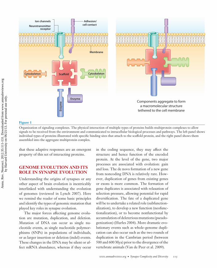
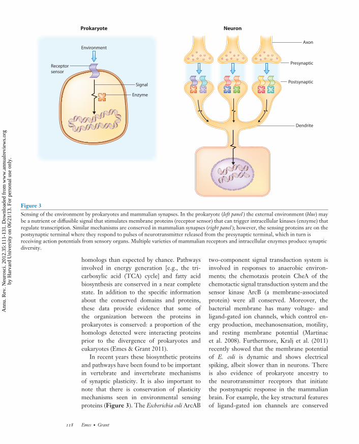
Examples of such structures found withinthe PSP are the signaling complexes, whichplay a central role in detecting and process-ing the information that arrives at the post-synaptic terminal (Figure 1). They have alsoserved as more manageable sets of proteinsfor experimental manipulation. The proto-type postsynaptic complex is known as MASC(MAGUK-associated signaling complex) com-prising ∼10% of all vertebrate PSP proteins.MASC can be physically isolated from the brainusing affinity purification methods (Fernandezet al. 2009, Husi et al. 2000). MAGUK pro-teins are scaffold proteins in the membrane-associated guanylate kinase family and have noenzymatic activity but contain protein-bindingdomains that allow receptors and enzymes toact in close proximity (Good et al. 2011, Nourryet al. 2003). MASC contains the principal post-synaptic machinery involved in synaptic trans-mission and synaptic plasticity: ionotropic andmetabotropic glutamate receptors, potassiumchannels, cell-adhesion proteins, and MAGUKand other scaffold proteins as well as their as-sociated signaling enzymes and structural pro-teins (Bayes et al. 2010; Collins et al. 2005,2006; Emes et al. 2008; Husi et al. 2000;Pocklington et al. 2006b). The functional im-portance of the prototypical MAGUK proteincalled postsynaptic density 95 (PSD-95) wasshown using knockout mice, which had impair-ments in synaptic plasticity, learning, and otherforms of behavioral adaptation (Migaud et al.1998). Mice carrying mutations in other MASCproteins also show impairments in synapticplasticity and adaptive behaviors, indicating
114 Emes · Grant
Ann
u. R
ev. N
euro
sci.
2012
.35:
111-
131.
Dow
nloa
ded
from
ww
w.a
nnua
lrev
iew
s.or
gby
Har
vard
Uni
vers
ity o
n 06
/21/
13. F
or p
erso
nal u
se o
nly.
NE35CH06-Grant ARI 14 May 2012 12:4
Ion channels
Neurotransmitterreceptor
Membrane
Adhesion/cell contact
ScaffoldCytoskeleton Cytoskeleton
Enzyme
Enzy
me Components aggregate to form
a macromolecular structure
tethered to the cell membrane
Figure 1Organization of signaling complexes. The physical interaction of multiple types of proteins builds multiprotein complexes to allowsignals to be received from the environment and communicated to intracellular biological processes and pathways. The left panel showsindividual types of proteins illustrated with specific binding sites that attach to the scaffold protein, and the right panel shows themassembled into the aggregate multiprotein complex.
that these adaptive responses are an emergentproperty of this set of interacting proteins.
GENOME EVOLUTION AND ITSROLE IN SYNAPSE EVOLUTION
Understanding the origins of synapses or anyother aspect of brain evolution is inextricablyinterlinked with understanding the evolutionof genomes (reviewed in Lynch 2007). Herewe remind the reader of some basic principlesand identify the types of genomic mutation thatplayed key roles in synapse evolution.
The major forces affecting genome evolu-tion are mutation, duplication, and deletion.Mutation of DNA can occur as single nu-cleotide events, as single nucleotide polymor-phisms (SNPs) in populations of individuals,or as larger insertion or deletion (indel) events.These changes in the DNA may be silent or af-fect mRNA abundance, whereas if they occur
in the coding sequence, they may affect thestructure and hence function of the encodedprotein. At the level of the gene, two majorprocesses are associated with evolution: gainand loss. The de novo formation of a new genefrom noncoding DNA is relatively rare. How-ever, duplication of genes from existing genesor exons is more common. The formation ofgene duplicates is associated with relaxation ofselection pressure, allowing potential for rapiddiversification. The fate of a duplicated genewill be to undertake a related role (subfunction-alization), to develop a new function (neofunc-tionalization), or to become nonfunctional byaccumulation of deleterious mutations (pseudo-genization) (Hurles 2004). More dramatic evo-lutionary events such as whole-genome dupli-cation can also occur such as the two rounds ofduplication in the Cambrian period (between500 and 600 Mya) prior to the divergence of thevertebrate animals (Van de Peer et al. 2009).
www.annualreviews.org • Synapse Complexity and Diversity 115
Ann
u. R
ev. N
euro
sci.
2012
.35:
111-
131.
Dow
nloa
ded
from
ww
w.a
nnua
lrev
iew
s.or
gby
Har
vard
Uni
vers
ity o
n 06
/21/
13. F
or p
erso
nal u
se o
nly.
NE35CH06-Grant ARI 14 May 2012 12:4
Synaptome: thecomplete proteincomplement of thesynapse. Thesynaptome is the sumof the PreSP and PSP
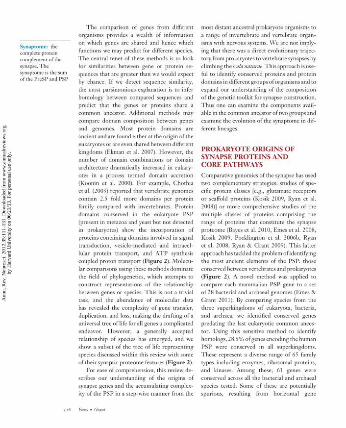
The comparison of genes from differentorganisms provides a wealth of informationon which genes are shared and hence whichfunctions we may predict for different species.The central tenet of these methods is to lookfor similarities between gene or protein se-quences that are greater than we would expectby chance. If we detect sequence similarity,the most parsimonious explanation is to inferhomology between compared sequences andpredict that the genes or proteins share acommon ancestor. Additional methods maycompare domain composition between genesand genomes. Most protein domains areancient and are found either at the origin of theeukaryotes or are even shared between differentkingdoms (Ekman et al. 2007). However, thenumber of domain combinations or domainarchitecture dramatically increased in eukary-otes in a process termed domain accretion(Koonin et al. 2000). For example, Chothiaet al. (2003) reported that vertebrate genomescontain 2.5 fold more domains per proteinfamily compared with invertebrates. Proteindomains conserved in the eukaryote PSP(present in metazoa and yeast but not detectedin prokaryotes) show the incorporation ofproteins containing domains involved in signaltransduction, vesicle-mediated and intracel-lular protein transport, and ATP synthesiscoupled proton transport (Figure 2). Molecu-lar comparisons using these methods dominatethe field of phylogenetics, which attempts toconstruct representations of the relationshipbetween genes or species. This is not a trivialtask, and the abundance of molecular datahas revealed the complexity of gene transfer,duplication, and loss, making the drafting of auniversal tree of life for all genes a complicatedendeavor. However, a generally acceptedrelationship of species has emerged, and weshow a subset of the tree of life representingspecies discussed within this review with someof their synaptic proteome features (Figure 2).
For ease of comprehension, this review de-scribes our understanding of the origins ofsynapse genes and the accumulating complex-ity of the PSP in a step-wise manner from the
most distant ancestral prokaryote organisms toa range of invertebrate and vertebrate organ-isms with nervous systems. We are not imply-ing that there was a direct evolutionary trajec-tory from prokaryotes to vertebrate synapses byclimbing the scala naturae. This approach is use-ful to identify conserved proteins and proteindomains in different groups of organisms and toexpand our understanding of the compositionof the genetic toolkit for synapse construction.Thus one can examine the components avail-able in the common ancestor of two groups andexamine the evolution of the synaptome in dif-ferent lineages.
PROKARYOTE ORIGINS OFSYNAPSE PROTEINS ANDCORE PATHWAYS
Comparative genomics of the synapse has usedtwo complementary strategies: studies of spe-cific protein classes [e.g., glutamate receptorsor scaffold proteins (Kosik 2009, Ryan et al.2008)] or more comprehensive studies of themultiple classes of proteins comprising therange of proteins that constitute the synapseproteome (Bayes et al. 2010, Emes et al. 2008,Kosik 2009, Pocklington et al. 2006b, Ryanet al. 2008, Ryan & Grant 2009). This latterapproach has tackled the problem of identifyingthe most ancient elements of the PSP: thoseconserved between vertebrates and prokaryotes(Figure 2). A novel method was applied tocompare each mammalian PSP gene to a setof 28 bacterial and archaeal genomes (Emes &Grant 2011). By comparing species from thethree superkingdoms of eukaryota, bacteria,and archaea, we identified conserved genespredating the last eukaryotic common ances-tor. Using this sensitive method to identifyhomologs, 28.5% of genes encoding the humanPSP were conserved in all superkingdoms.These represent a diverse range of 65 familytypes including enzymes, ribosomal proteins,and kinases. Among these, 61 genes wereconserved across all the bacterial and archaealspecies tested. Some of these are potentiallyspurious, resulting from horizontal gene
116 Emes · Grant
Ann
u. R
ev. N
euro
sci.
2012
.35:
111-
131.
Dow
nloa
ded
from
ww
w.a
nnua
lrev
iew
s.or
gby
Har
vard
Uni
vers
ity o
n 06
/21/
13. F
or p
erso
nal u
se o
nly.
NE35CH06-Grant ARI 14 May 2012 12:4
Prokaryotes
Voltage- and ligand-gated ion channels
Receptor–kinase–transcriptome signaling
Protein translation regulation
2,622# Mya
FungiProtosynapse organization of signaling
Complexity in signaling responses1,513 ± 66 Mya
ChoanoflagellatesScaffold proteins:
MAGUK, Shank, and Homer
1,450* Mya
Sponges
Glutamate and GABA receptors
K+ channels
Cell–cell contact signaling 1,351 ± 120 Mya
Cnidarians
Chemical and peptidergic synapses
NMDA and AMPA receptors
Neuronal circuits and ursynapse1,298 ± 74 Mya
Bilaterians
Molecules of PSP present
Limited repertoire of upstream
signaling molecules976 ± 97 Mya
550 MyaVertebrates
Expansion of key families and proteome complexity
Divergence of upstream signaling molecules
Synapse diversification
Two rounds of
genome duplication
Figure 2Cladogram of taxonomic groups and origins of PSP components. A generalized cladogram showing the groups discussed and the typesof molecules and functions found in the mammalian PSP are indicated (modified from Ryan & Grant 2009). These represent thegenetic toolkit for synapse construction. Dates indicate divergence time in millions of years +/− error estimates of divergence (Hedgeset al. 2004). ∗Estimate of divergence based on midpoint of adjacent nodes (Hedges et al. 2004). #Weighted average divergence time ofvertebrates and eubacteria as calculated by time tree (Hedges et al. 2006).
transfer (HGT) events between prokaryotesand eukaryotes (Koonin 2003, Lawrence &Hendrickson 2003) such as synthetase genes(Koonin et al. 2001, Wolf et al. 1999).However, ribosomal proteins, translationelongation factors, lyases, chaperone proteins,and the G protein OLA1 are monophyloge-netic without obvious evidence of HGT andare therefore candidates for PSP homologsshared since the last common ancestor ofprokaryotes and eukaryotes (Emes & Grant2011). To determine the conserved biologicalfunctions underlying the detection of homol-ogous genes, it is useful to investigate the
conservation of protein domains. Owing to thefunctional information known about conserveddomains (Finn et al. 2010, Marchler-Baueret al. 2005, Schultz et al. 2000), we can link do-mains to biological processes shared betweenprokaryotes and eukaryotes. The domainsconserved in all three superkingdoms reflectedbasic biological processes, such as translation,carbohydrate metabolic process, glycolysis, andtRNA aminoacylation for protein translation.Comparison of eukaryotic PSP biochemicalpathways with a representative prokaryoteidentified ten pathways that had a significantlygreater number of prokaryote-eukaryote
www.annualreviews.org • Synapse Complexity and Diversity 117
Ann
u. R
ev. N
euro
sci.
2012
.35:
111-
131.
Dow
nloa
ded
from
ww
w.a
nnua
lrev
iew
s.or
gby
Har
vard
Uni
vers
ity o
n 06
/21/
13. F
or p
erso
nal u
se o
nly.
NE35CH06-Grant ARI 14 May 2012 12:4
Prokaryote Neuron
Environment
Receptor
sensor
Enzyme
Signal
Axon
Presynaptic
Postsynaptic
Dendrite
Figure 3Sensing of the environment by prokaryotes and mammalian synapses. In the prokaryote (left panel ) the external environment (blue) maybe a nutrient or diffusible signal that stimulates membrane proteins (receptor sensor) that can trigger intracellular kinases (enzyme) thatregulate transcription. Similar mechanisms are conserved in mammalian synapses (right panel ); however, the sensing proteins are on thepostsynaptic terminal where they respond to pulses of neurotransmitter released from the presynaptic terminal, which in turn isreceiving action potentials from sensory organs. Multiple varieties of mammalian receptors and intracellular enzymes produce synapticdiversity.
homologs than expected by chance. Pathwaysinvolved in energy generation [e.g., the tri-carboxylic acid (TCA) cycle] and fatty acidbiosynthesis are conserved in a near completestate. In addition to the specific informationabout the conserved domains and proteins,these data provide evidence that some ofthe organization between the proteins inprokaryotes is conserved: a proportion of thehomologs detected were interacting proteinsprior to the divergence of prokaryotes andeukaryotes (Emes & Grant 2011).
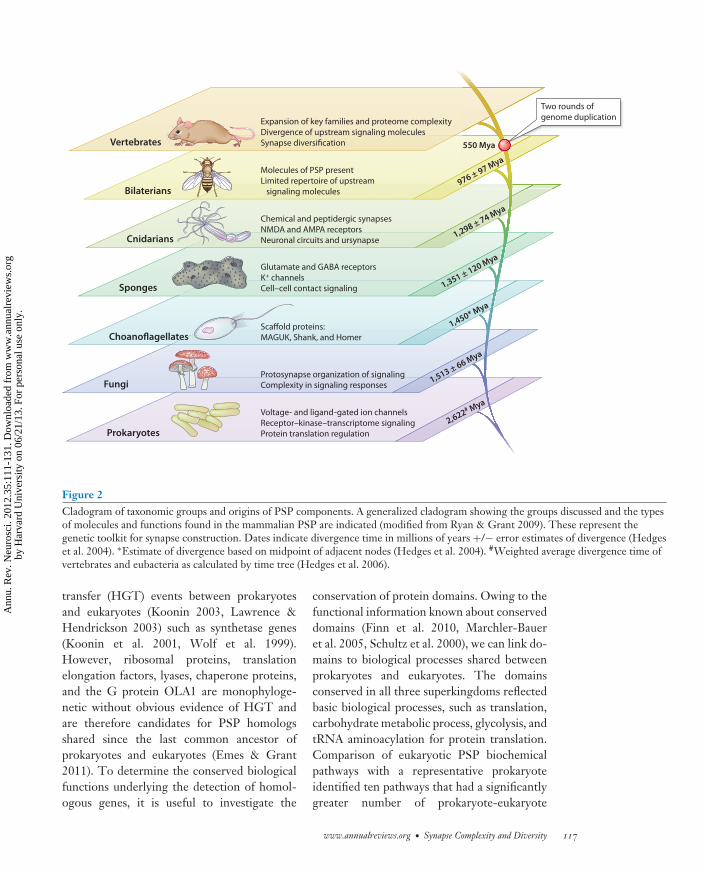
In recent years these biosynthetic proteinsand pathways have been found to be importantin vertebrate and invertebrate mechanismsof synaptic plasticity. It is also important tonote that there is conservation of plasticitymechanisms seen in environmental sensingproteins (Figure 3). The Escherichia coli ArcAB
two-component signal transduction system isinvolved in responses to anaerobic environ-ments; the chemotaxis protein CheA of thechemotactic signal transduction system and thesensor kinase ArcB (a membrane-associatedprotein) were all conserved. Moreover, thebacterial membrane has many voltage- andligand-gated ion channels, which control en-ergy production, mechanosensation, motility,and resting membrane potential (Martinacet al. 2008). Furthermore, Kralj et al. (2011)recently showed that the membrane potentialof E. coli is dynamic and shows electricalspiking, albeit slower than in neurons. Thereis also evidence of prokaryote ancestry tothe neurotransmitter receptors that initiatethe postsynaptic response in the mammalianbrain. For example, the key structural featuresof ligand-gated ion channels are conserved
118 Emes · Grant
Ann
u. R
ev. N
euro
sci.
2012
.35:
111-
131.
Dow
nloa
ded
from
ww
w.a
nnua
lrev
iew
s.or
gby
Har
vard
Uni
vers
ity o
n 06
/21/
13. F
or p
erso
nal u
se o
nly.
NE35CH06-Grant ARI 14 May 2012 12:4
(Bocquet et al. 2007, 2009; Nury et al. 2011),and in the case of the glutamate receptors,there is evidence of conservation of gluta-mate binding domains ( Janovjak et al. 2011,Nakanishi et al. 1990, Sprengel et al. 2001).
This conservation from receptor-to-transcriptome signaling is interesting toconsider in terms of environmental sensing andadaptive behaviors (Figure 3). In the case of theunicellular prokaryote, the sensing is initiatedat the cell’s surface, where it is in direct contactwith the environment. In the case of the brain,the environment of the outside world is de-tected by sensory end organs (e.g., eyes, ears),which convert information into patterns ofaction potentials that are transmitted by nerveconduction to the synapses in the brain. Theseaction potentials stimulate releases of pulses ofneurotransmitters into the local extracellularenvironment where the receptors and signalingsystems in the PSP are activated. The setsof synapse proteins comprising receptors andtheir signaling and biosynthetic pathways arosein prokaryotes, and their role in enabling theprokaryotic organisms to respond and adapt tochanging environments appears to be broadlythe same role they perform in the brain.
EUKARYOTE INNOVATIONSAND THE PROTOSYNAPSE
The emergence of eukaryotic cells was markedby larger and more complex genomes, lin-ear chromosomes requiring capping with telo-meres, and multiple replication origins. The in-crease in complexity of the transcriptome wasmarked by a shift from prokaryotic operons tosplicing and novel RNA regulator machineryprogramming the proteome of complex subcel-lular structures, including membrane-enclosedorganelles and the cytoskeleton. The vesicu-lar machinery (which later evolved into neu-rotransmitter release machinery in metazoans)allowed the active movement and engulfing ofmaterial by phagocytosis, and this was likelya key step in the origins of the eukaryote:Predation of aerobic bacteria by an ancestraleukaryote cell resulted in the symbiosis of the
genomes of engulfed bacteria to form the mi-tochondria as well as contributing expansionsto the genome of the stem eukaryote (Lynch2007). It is fascinating to consider that the vesic-ular mechanism that might have been respon-sible for eukaryotic genomic complexity, andthus the complex biology of eukaryotes, also un-derpinned the mechanisms of neurotransmittervesicle recycling, which is a central function ofthe presynaptic terminal.
A comparison of mouse PSP orthologswith 19 eukaryote species (fungi, invertebrates,nonmammalian vertebrates, and mammals) re-vealed extensive conservation of the compo-nents of the synapse across the eukaryota (Emeset al. 2008). Approximately 23% of genes testedhad a detectable ortholog in the yeast Saccha-romyces cerevisiae, and this rose to ∼45% havingdetectable orthologs in Caenorhabditis elegans orDrosophila melanogaster (these numbers shouldnot be directly compared with those reportedfor the prokaryote above because here we aredescribing identified orthologs and hence ex-pect to see a relatively lower percentage thanthat for all homologs detected by the methoddescribed above). With this finding it is evidentthat the genome of the common ancestor ofmammals and S. cerevisiae that obviously lackeda nervous system or morphological synapse har-bored many of the genes used to encode theconstituents of the functional synapse.
As with the conserved prokaryote genes,many conserved eukaryotic genes in the PSPencode environment-sensing mechanisms driv-ing signal transduction pathways and basic cel-lular functions such as protein synthesis anddegradation enzymes controlling turnover ofsynaptic proteins (Emes et al. 2008). The de-tection of yeast orthologs of NF1 (ira2), PKA(tpk2), Erk2 ( fus3), and GNB5 (ste4) membersof canonical pathways regulating transcription,cell morphology, and adhesion downstreamof nutrient- and pheromone-sensitive GPCRs(Elion et al. 2005; Erdman & Snyder 2001;Harashima et al. 2006; Palecek et al. 2002) sug-gests that these components of synaptic path-ways regulating protein synthesis and structuralplasticity in mammals conduct analogous roles
www.annualreviews.org • Synapse Complexity and Diversity 119
Ann
u. R
ev. N
euro
sci.
2012
.35:
111-
131.
Dow
nloa
ded
from
ww
w.a
nnua
lrev
iew
s.or
gby
Har
vard
Uni
vers
ity o
n 06
/21/
13. F
or p
erso
nal u
se o
nly.
NE35CH06-Grant ARI 14 May 2012 12:4
Protosynapse:complex of synapticproteins present inearly metazoanswithout a definednervous system
in unicellular responses to environmental cues(ions, nutrients) and cell-cell communication.
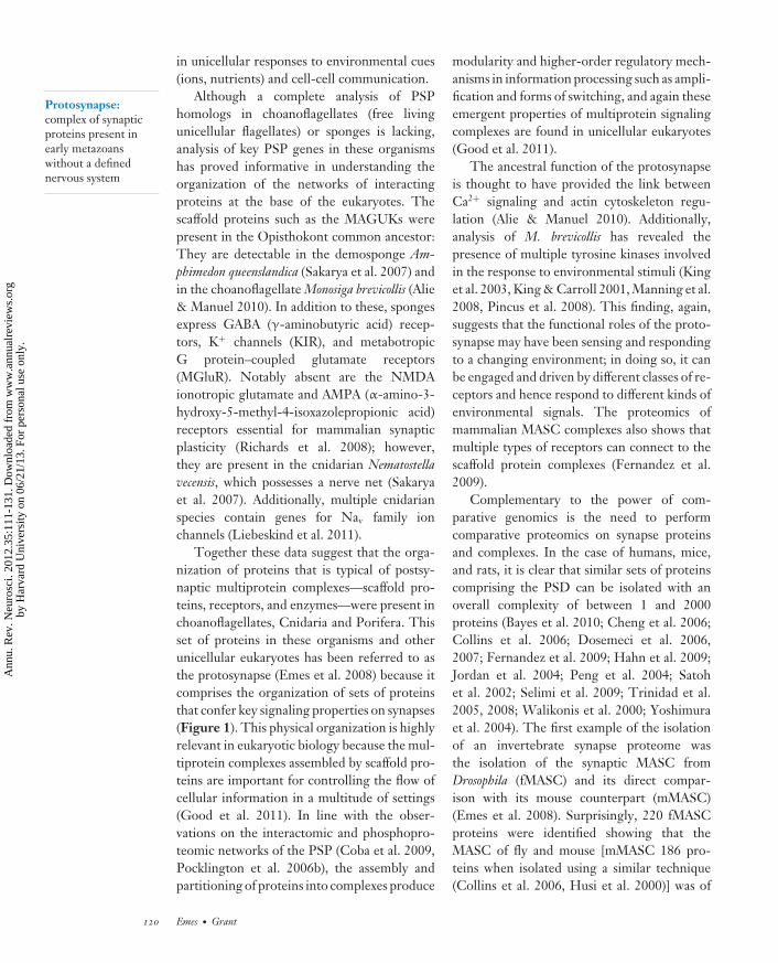
Although a complete analysis of PSPhomologs in choanoflagellates (free livingunicellular flagellates) or sponges is lacking,analysis of key PSP genes in these organismshas proved informative in understanding theorganization of the networks of interactingproteins at the base of the eukaryotes. Thescaffold proteins such as the MAGUKs werepresent in the Opisthokont common ancestor:They are detectable in the demosponge Am-phimedon queenslandica (Sakarya et al. 2007) andin the choanoflagellate Monosiga brevicollis (Alie& Manuel 2010). In addition to these, spongesexpress GABA (γ-aminobutyric acid) recep-tors, K+ channels (KIR), and metabotropicG protein–coupled glutamate receptors(MGluR). Notably absent are the NMDAionotropic glutamate and AMPA (α-amino-3-hydroxy-5-methyl-4-isoxazolepropionic acid)receptors essential for mammalian synapticplasticity (Richards et al. 2008); however,they are present in the cnidarian Nematostellavecensis, which possesses a nerve net (Sakaryaet al. 2007). Additionally, multiple cnidarianspecies contain genes for Nav family ionchannels (Liebeskind et al. 2011).
Together these data suggest that the orga-nization of proteins that is typical of postsy-naptic multiprotein complexes—scaffold pro-teins, receptors, and enzymes—were present inchoanoflagellates, Cnidaria and Porifera. Thisset of proteins in these organisms and otherunicellular eukaryotes has been referred to asthe protosynapse (Emes et al. 2008) because itcomprises the organization of sets of proteinsthat confer key signaling properties on synapses(Figure 1). This physical organization is highlyrelevant in eukaryotic biology because the mul-tiprotein complexes assembled by scaffold pro-teins are important for controlling the flow ofcellular information in a multitude of settings(Good et al. 2011). In line with the obser-vations on the interactomic and phosphopro-teomic networks of the PSP (Coba et al. 2009,Pocklington et al. 2006b), the assembly andpartitioning of proteins into complexes produce
modularity and higher-order regulatory mech-anisms in information processing such as ampli-fication and forms of switching, and again theseemergent properties of multiprotein signalingcomplexes are found in unicellular eukaryotes(Good et al. 2011).
The ancestral function of the protosynapseis thought to have provided the link betweenCa2+ signaling and actin cytoskeleton regu-lation (Alie & Manuel 2010). Additionally,analysis of M. brevicollis has revealed thepresence of multiple tyrosine kinases involvedin the response to environmental stimuli (Kinget al. 2003, King & Carroll 2001, Manning et al.2008, Pincus et al. 2008). This finding, again,suggests that the functional roles of the proto-synapse may have been sensing and respondingto a changing environment; in doing so, it canbe engaged and driven by different classes of re-ceptors and hence respond to different kinds ofenvironmental signals. The proteomics ofmammalian MASC complexes also shows thatmultiple types of receptors can connect to thescaffold protein complexes (Fernandez et al.2009).
Complementary to the power of com-parative genomics is the need to performcomparative proteomics on synapse proteinsand complexes. In the case of humans, mice,and rats, it is clear that similar sets of proteinscomprising the PSD can be isolated with anoverall complexity of between 1 and 2000proteins (Bayes et al. 2010; Cheng et al. 2006;Collins et al. 2006; Dosemeci et al. 2006,2007; Fernandez et al. 2009; Hahn et al. 2009;Jordan et al. 2004; Peng et al. 2004; Satohet al. 2002; Selimi et al. 2009; Trinidad et al.2005, 2008; Walikonis et al. 2000; Yoshimuraet al. 2004). The first example of the isolationof an invertebrate synapse proteome wasthe isolation of the synaptic MASC fromDrosophila (fMASC) and its direct compar-ison with its mouse counterpart (mMASC)(Emes et al. 2008). Surprisingly, 220 fMASCproteins were identified showing that theMASC of fly and mouse [mMASC 186 pro-teins when isolated using a similar technique(Collins et al. 2006, Husi et al. 2000)] was of
120 Emes · Grant
Ann
u. R
ev. N
euro
sci.
2012
.35:
111-
131.
Dow
nloa
ded
from
ww
w.a
nnua
lrev
iew
s.or
gby
Har
vard
Uni
vers
ity o
n 06
/21/
13. F
or p
erso
nal u
se o
nly.
NE35CH06-Grant ARI 14 May 2012 12:4
Ursynapse: lastcommon ancestorof all morphologicalsynapses from whichall extant synapsesevolved
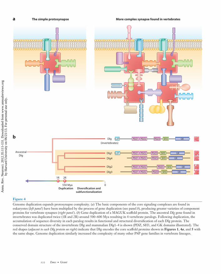
comparable size. However, although mMASCand fMASC are approximately equal in size,major differences in the types of proteins wereidentified. By functional annotation investiga-tors revealed that upstream signaling/structuralcomponents (receptors, scaffolds, signal trans-duction molecules) accounted for ∼25% ofthe fMASC proteome compared with >60%of the mMASC. When the composition ofthe fMASC was compared with yeast, 71%of fMASC genes were also found in the yeastS. cerevisiae, and hence only 29% appearedto be of metazoan origin. Thus the majorityof downstream components were present inyeast, while the upstream signaling/structuralcomponents of fMASC and mMASC showedlineage-specific functional expansions. Thusthe core functionality of the protosynapsemachinery that comprises MASC evolved inunicellular eukaryotes and lineage expansion ofupstream signaling molecules (such as recep-tors and their directly associated cytoplasmicproteins) in metazoans increased the molecularcomplexity of this machinery (Figures 1 and 4).Together the comparative proteomic and ge-nomic studies reveal that invertebrates evolvedsynapses with highly complex molecularcomposition built around the protosynapse.
COMPLEXITY AND DIVERSITYIN METAZOAN SYNAPSES
The molecular phylogeny described above in-dicates that the core functionality in pro-tein components, pathways, and organizationin mammalian synapses was present prior tothe first morphologically visible synapses, suchas synapses in Cnidaria that evolved ∼900–1400 Myr ago (Figure 2). The protosynapsemachinery with its specialized environmentalsensing capacity and regulation of transcrip-tome responses for adaptive changes evolvedbefore this and was incorporated into the ursy-napse. The simple nervous systems of Cnidariawere the precursors of the highly elaborate anddiverse nervous systems that characterize themultitude of invertebrates and vertebrates thatsubsequently arose. In these anatomically com-plex brains, some with enormous numbers of
synapses, there is considerable anatomical di-versity in the postsynaptic dendritic spines. Itis therefore of great interest to understand howdiversity in populations of synapses arose and ifthis was relevant to the molecular organizationof the protosynapse.
A major event in the diversification of bi-ological functions that characterize chordatesand thus all vertebrates was the 2 completegenome duplication events ∼550 Myr ago (Vande Peer et al. 2009) (Figure 4b). This periodcorresponded with the Cambrian explosion,when there was a dramatic increase in the diver-sity of animal life in the fossil record, which waspresumably accompanied by a diversification innervous system complexity. Comparison of 13vertebrate genomes (including primate, rodent,fish, chicken, and opossum genomes) showed astep-wise expansion from invertebrates in thenumber of conserved PSP homologs to ∼85–90% conservation (Emes et al. 2008). This PSPexpansion in complexity event coincides withthe predicted genome duplication, where thesetwo rounds of genome duplication are thoughtto have occurred prior to the divergence ofthe hagfish and lampreys (Holland 2009). Thetwo rounds of genome duplication typically ex-panded gene families to four copies; however,some gene families have lost copies and othergene families have gained further copies by in-dividual gene duplication events (Figure 4b)(Van de Peer et al. 2009). Further evidencefor the importance of gene duplication or generetention following whole genome duplicationcomes from the comparison of protein domains.The number of domain types did not increaseto the same extent that gene number did, whichsuggests that the synapse proteome expansionseen in vertebrate genomes does not represent arecruitment of proteins containing new domaintypes but rather the expansion of protein typesalready present in the PSP of early branchingmetazoans such as flies and worms.
Examining the expansion in the vertebratePSP further showed that the protein familiesthat had the greatest expansion were the up-stream signaling proteins (receptors, adhesionproteins in the membrane, and their proximal
www.annualreviews.org • Synapse Complexity and Diversity 121
Ann
u. R
ev. N
euro
sci.
2012
.35:
111-
131.
Dow
nloa
ded
from
ww
w.a
nnua
lrev
iew
s.or
gby
Har
vard
Uni
vers
ity o
n 06
/21/
13. F
or p
erso
nal u
se o
nly.
NE35CH06-Grant ARI 14 May 2012 12:4
a The simple protosynapse More complex synapse found in vertebrates
b
Diversification andsubfunctionalization
Duplication
1R 2R
550 Mya 0
Ancestral
Dlg
Dlg
(invertebrates)
Dlg1
Dlg4
Dlg2
Dlg3
L27 PDZ1 PDZ2 PDZ3 SH3 GK
L27 PDZ1 PDZ2 PDZ3 SH3 GK
PDZ1 PDZ2 PDZ3 SH3 GK
PDZ1 PDZ2 PDZ3 SH3 GK
PDZ1 PDZ2 PDZ3 SH3 GK
Figure 4Genome duplication expands protosynapse complexity. (a) The basic components of the core signaling complexes are found ineukaryotes (left panel ) have been multiplied by the process of gene duplication (see panel b), producing greater varieties of componentproteins for vertebrate synapses (right panel ). (b) Gene duplication of a MAGUK scaffold protein. The ancestral Dlg gene found ininvertebrates was duplicated twice (1R and 2R) around 500–600 Mya resulting in 4 vertebrate paralogs. Following duplication, theaccumulation of sequence diversity in each paralog results in functional and structural diversification of each Dlg protein. Theconserved domain structure of the invertebrate Dlg and mammalian Dlg1–4 is shown (PDZ, SH3, and GK domains illustrated). Thered shapes (adjacent to each Dlg protein on right) indicate that Dlg encodes the core scaffold proteins shown in Figures 1, 4a, and 5 withthe same shape. Genome duplication similarly increased the complexity of many other PSP gene families in vertebrate lineages.
122 Emes · Grant
Ann
u. R
ev. N
euro
sci.
2012
.35:
111-
131.
Dow
nloa
ded
from
ww
w.a
nnua
lrev
iew
s.or
gby
Har
vard
Uni
vers
ity o
n 06
/21/
13. F
or p
erso
nal u
se o
nly.
NE35CH06-Grant ARI 14 May 2012 12:4
MASC 1
MASC 2
MASC 3
Figure 5Synapse diversity in the mammalian brain is generated by combinations of protosynapse and MASC proteins. Three varieties of MASCcomplexes (labeled MASC1, 2, 3) comprising central scaffold proteins bound to receptors and adhesion protein, enzymes, andcytoskeleton are shown as in Figure 1. The variation in shapes of the components between the three complexes indicates that they areparalogs in expanded vertebrate gene families arising from duplication of the ancestral genes. The paralogs arising in early vertebrateevolution played a major role in diversifying neuroanatomical function.
associated proteins), and the families maintain-ing equal numbers of genes were those encod-ing the downstream cytoplasmic signaling pro-teins (Emes et al. 2008). This finding shows thatthe hierarchy of the PSP network was subject todifferential expansion, and these upstream pro-tein families were presumably retained and po-tentially diversified by sub- or neofunctional-ization since their duplication.
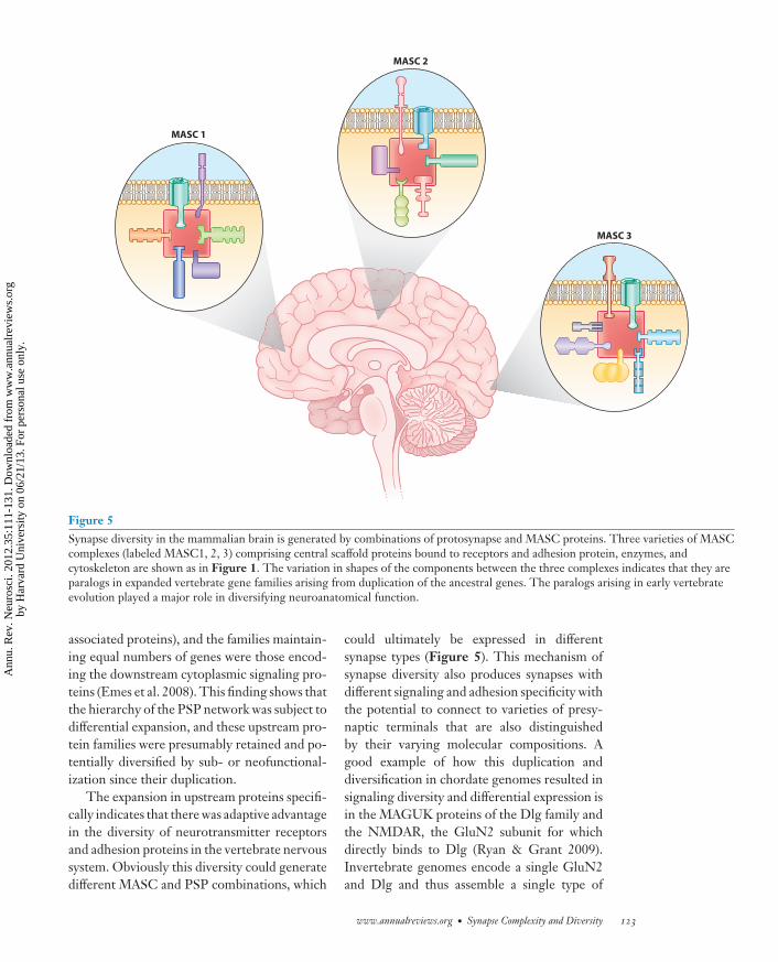
The expansion in upstream proteins specifi-cally indicates that there was adaptive advantagein the diversity of neurotransmitter receptorsand adhesion proteins in the vertebrate nervoussystem. Obviously this diversity could generatedifferent MASC and PSP combinations, which
could ultimately be expressed in differentsynapse types (Figure 5). This mechanism ofsynapse diversity also produces synapses withdifferent signaling and adhesion specificity withthe potential to connect to varieties of presy-naptic terminals that are also distinguishedby their varying molecular compositions. Agood example of how this duplication anddiversification in chordate genomes resulted insignaling diversity and differential expression isin the MAGUK proteins of the Dlg family andthe NMDAR, the GluN2 subunit for whichdirectly binds to Dlg (Ryan & Grant 2009).Invertebrate genomes encode a single GluN2and Dlg and thus assemble a single type of
www.annualreviews.org • Synapse Complexity and Diversity 123
Ann
u. R
ev. N
euro
sci.
2012
.35:
111-
131.
Dow
nloa
ded
from
ww
w.a
nnua
lrev
iew
s.or
gby
Har
vard
Uni
vers
ity o
n 06
/21/
13. F
or p
erso
nal u
se o
nly.
NE35CH06-Grant ARI 14 May 2012 12:4
MASC complex, whereas mammals have 4genes of each, potentially producing 16 typesof MASC complexes. If one considers the otherproteins that are in these complexes, includingthe many upstream proteins that were retainedwith the duplication events, an astronomicalnumber of MASC and PSP types are availableto diversify the synaptic types of vertebrates.
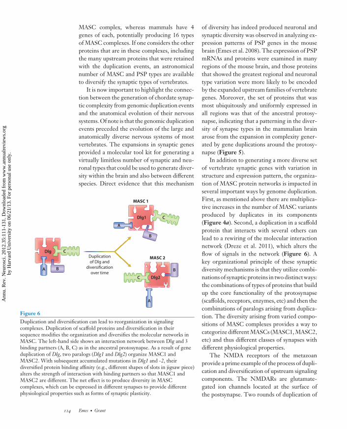
It is now important to highlight the connec-tion between the generation of chordate synap-tic complexity from genomic duplication eventsand the anatomical evolution of their nervoussystems. Of note is that the genomic duplicationevents preceded the evolution of the large andanatomically diverse nervous systems of mostvertebrates. The expansions in synaptic genesprovided a molecular tool kit for generating avirtually limitless number of synaptic and neu-ronal types that could be used to generate diver-sity within the brain and also between differentspecies. Direct evidence that this mechanism
Dlg
Dlg1
Dlg2
Duplication
of Dlg and
diversification
over timeA
A
A
B
B
B
C
C
C
MASC 1
MASC 2
Figure 6Duplication and diversification can lead to reorganization in signalingcomplexes. Duplication of scaffold proteins and diversification in theirsequence modifies the organization and diversifies the molecular networks inMASC. The left-hand side shows an interaction network between Dlg and 3binding partners (A, B, C) as in the ancestral protosynapse. As a result of geneduplication of Dlg, two paralogs (Dlg1 and Dlg2) organize MASC1 andMASC2. With subsequent accumulated mutations in Dlg1 and -2, theirdiversified protein binding affinity (e.g., different shapes of slots in jigsaw piece)alters the strength of interaction with binding partners so that MASC1 andMASC2 are different. The net effect is to produce diversity in MASCcomplexes, which can be expressed in different synapses to provide differentphysiological properties such as forms of synaptic plasticity.
of diversity has indeed produced neuronal andsynaptic diversity was observed in analyzing ex-pression patterns of PSP genes in the mousebrain (Emes et al. 2008). The expression of PSPmRNAs and proteins were examined in manyregions of the mouse brain, and those proteinsthat showed the greatest regional and neuronaltype variation were more likely to be encodedby the expanded upstream families of vertebrategenes. Moreover, the set of proteins that wasmost ubiquitously and uniformly expressed inall regions was that of the ancestral protosy-napse, indicating that a patterning in the diver-sity of synapse types in the mammalian brainarose from the expansion in complexity gener-ated by gene duplications around the protosy-napse (Figure 5).
In addition to generating a more diverse setof vertebrate synaptic genes with variation instructure and expression pattern, the organiza-tion of MASC protein networks is impacted inseveral important ways by genome duplication.First, as mentioned above there are multiplica-tive increases in the number of MASC variantsproduced by duplicates in its components(Figure 4a). Second, a duplication in a scaffoldprotein that interacts with several others canlead to a rewiring of the molecular interactionnetwork (Dreze et al. 2011), which alters theflow of signals in the network (Figure 6). Akey organizational principle of these synapticdiversity mechanisms is that they utilize combi-nations of synaptic proteins in two distinct ways:the combinations of types of proteins that buildup the core functionality of the protosynapse(scaffolds, receptors, enzymes, etc) and then thecombinations of paralogs arising from duplica-tion. The diversity arising from varied compo-sitions of MASC complexes provides a way tocategorize different MASCs (MASC1, MASC2,etc) and thus different classes of synapses withdifferent physiological properties.
The NMDA receptors of the metazoanprovide a prime example of the process of dupli-cation and diversification of upstream signalingcomponents. The NMDARs are glutamate-gated ion channels located at the surface ofthe postsynapse. Two rounds of duplication of
124 Emes · Grant
Ann
u. R
ev. N
euro
sci.
2012
.35:
111-
131.
Dow
nloa
ded
from
ww
w.a
nnua
lrev
iew
s.or
gby
Har
vard
Uni
vers
ity o
n 06
/21/
13. F
or p
erso
nal u
se o
nly.
NE35CH06-Grant ARI 14 May 2012 12:4
the NMDA receptors in the vertebrate lineageresulted in four extant sequences. With thisexpansion, a dramatic change in the intra-cellular C-terminus of the NMDA proteinsoccurred. The C-terminus is the location ofthe phosphorylation-dependent interactionwith scaffold and signaling molecules e.g.,Fyn, CamKII, P85 PI3K, and PSD95. ThisC-terminal region (which is encoded by asingle exon) is almost absent in invertebratehomologs, and hence so is the potential formultiple protein interactions in these species(Ryan et al. 2008). Thus the evolution ofthe intracellular portion of the NMDARsin the vertebrate lineages was likely a keystage in the link between sensing and cellularresponse to environmental stimuli.
INSIGHTS FROM THEHUMAN SYNAPSE
What sets humans apart from the rest of theanimals, and what is the basis of human dis-ease? The first comprehensive profiling of thehuman PSP and detailed study of its evolutionshowed that the genes of the PSP over the past100 Myr are evolving under very strong puri-fying selection compared with the rest of thegenome or other neuronal proteins and subcel-lular organelles (Bayes et al. 2010). This con-straint was observed in primate and rodent lin-eages and shown to correlate with structural,physiological, and behavioral functions. Themost conserved subset of the PSP was MASC,reinforcing its centrality in PSP function. Theconserved functions of mouse and human PSPproteins were identified by systematic pheno-type mapping of mutations and showed cog-nitive and motor functions, including learningand memory, and social functions were highlyconserved. These findings again highlight theimportance of adaptive behaviors as central andancestral functions of MASC and the PSP. An-other insight that arose from the proteomics ofthe human PSP was to identify that ∼200 genesare involved with Mendelian diseases of which130 were brain diseases (Bayes et al. 2010).Many of these diseases arose from gene du-
plication events, indicating that the cost of theevolution of paralogs was susceptibility to dis-ease. A number of the MASC proteins in hu-mans have been identified as mutated in patientswith schizophrenia and other cognitive disor-ders (Fernandez et al. 2009, Kirov et al. 2011).Linking the genetic disease phenotypes to theobserved constraint in vertebrate PSP evolutionindicates that reduced fitness from PSP muta-tions is observed as strong and pervasive puri-fying selection.
Less is presently known about the changesin the PSP that are unique to the human lineageafter it diverged from other primates around 6–8 Myr ago. Using methods to identify punctu-ated periods of adaptive evolution, eight genesof the PSP (Cybrd1, SirpA, Ank2, Ca2, Cox5A,Pclo, Ndufb6, and Psd3) show significant evi-dence of positive selection along the primate(including human) compared with the nonpri-mate lineage (R.D. Emes and S.G.N. Grant,manuscript in preparation). These genes maybe candidates that underlie clade-specific adap-tive evolution and may underpin more sub-tle differences in primate synapse function andhence in cognitive ability as well.
A MODEL FOR THE EVOLUTIONOF THE POSTSYNAPTICPROTEOME
Taken together, the comparative studiesto date suggest a consistent model for theevolution of the PSP and its contribution tosynaptic diversity and behavior. Elements ofenvironmental sensing from surface receptor totranscriptome and core components associatedwith basic cellular life such as translation,energy generation, and fatty acid biosynthesisidentified in bacteria and archaea were presentprior to the divergence of the common ances-tor of eukaryotes and prokaryotes. A numberof constituents are conserved in prokaryotesas interacting proteins, suggesting that thesewere co-opted into the protosynapse as aninteracting complex and remain in the extantPSP. In the fungi we observe homologs ofthe signal transduction pathways, including
www.annualreviews.org • Synapse Complexity and Diversity 125
Ann
u. R
ev. N
euro
sci.
2012
.35:
111-
131.
Dow
nloa
ded
from
ww
w.a
nnua
lrev
iew
s.or
gby
Har
vard
Uni
vers
ity o
n 06
/21/
13. F
or p
erso
nal u
se o
nly.

NE35CH06-Grant ARI 14 May 2012 12:4
increased repertoire of protein kinases and theimportant role of scaffold proteins assemblingand organizing signaling machinery. Thepresence of most types of synapse proteinsin unicellular eukaryotes such as fungi andchoanoflagellates and Porifera highlights thatthe evolution of the fundamental synapticcomponents predates the origins of identifiableneurons in metazoans. The ion channelsincorporated after the cnidarian–poriferandivergence therefore would interact with apreexisting scaffold of intracellular proteins.The expanding transmembrane receptorswould also plug into this preexisting networkto expand rapidly the signaling complexity ofthe synapse (Figure 4a). By comparing thepredicted PSP of invertebrate metazoan species(fly, worm, bee, and mosquito) to vertebratesand by directly isolating the fly MASC com-ponent of the PSP, it is clear that the majorityof protein classes were present in the inverte-brates. In addition, specialization and divisionof labor were expanded by differential geneexpression, providing combinations of proteinsin different synapses (Figure 5). Followingthe divergence of the vertebrates from otherdeuterostomes, the driving force was of rapidexpansion by duplication and diversification,particularly in the upstream signaling compo-nents such as receptors and signal transductionmolecules. The expansion of the synapse pro-teome, therefore, predates the developmentof anatomically enlarged brains. This modelin which the development of the synapse isa necessary step prior to the expansion anddevelopment of an enlarged nervous systemhas been proposed in the “synapse first” modelof brain evolution (Ryan & Grant 2009).
One prediction of this model is that thesynapse developed before axons and the branch-ing network of dendrites. The increase ofneuronal connectivity produced by neuronalbranching rapidly increases the number ofsynapses and multiplies the computationalpower and diversity of synapses and overallsignaling complexity. This may have driventhe form of the nervous system we see today.Support for the theory that gene repertoire
may predate the enlargement or increase in en-cephalization of the brain has recently emerged.Encephalization, the development of relativelyexcess brain size, is often measured by com-paring the encephalization quotient (EQ, thelog brain size versus log body size) and hasbeen proposed as a measure of information-processing capacity or intelligence ( Jerison1977, 1985). By using high-resolution X-raycomputed tomography, Rowe et al. (2011) re-cently showed that the particular enlargementof cerebral hemispheres and cerebellum asso-ciated with mammals occurred in a step-wisemanner and was associated with expansion ofkey gene families. Expansion of the brain, es-pecially the olfactory bulb and olfactory cortex,is seen in the skull of Morganucodon oehleri, abasal member of the mammaliaforms from theearly Jurassic (∼199–175 Mya). This postdatesthe expansion of the PSP we predict to haveoccurred in the ancestor of the vertebrates. Asecond wave of encephalization was seen withHadrocodium wui, where expansion of the ol-factory bulb and olfactory cortex accounts forthe increase in EQ to within that seen in ex-tant crown group mammals. Rowe et al. pro-pose that the first wave of EQ expansion wasdriven by increase in olfaction and tactile sensi-tivity. These were then further amplified by anolfactory expansion owing to expression of theexpanded olfactory receptor genome. We pro-pose that, like the olfactory receptors, the ex-pansion of gene repertoire in the PSP by geneduplication and expansion at the base of thevertebrates was a driver rather than a conse-quence of an increase in EQ. Like the explod-ing bubbles when uncorking champagne, mu-tations leading to change in brain size releasedthe potential of the expanded PSP repertoire.
With the model that additional proteininteractors plugged into an existing scaffold,why should the PSP complex expand byincreasing interacting partners? The selectiveadvantage of accumulating interacting pro-teins by scaffold protein binding proposes anintuitive adaptionist theory for the evolutionof the synapse. The scaffold proteins providea means to localize interacting proteins among
126 Emes · Grant
Ann
u. R
ev. N
euro
sci.
2012
.35:
111-
131.
Dow
nloa
ded
from
ww
w.a
nnua
lrev
iew
s.or
gby
Har
vard
Uni
vers
ity o
n 06
/21/
13. F
or p
erso
nal u
se o
nly.
NE35CH06-Grant ARI 14 May 2012 12:4
the multifarious soup of proteins in a cell andorchestrates the flow of information in a cell(Good et al. 2011). For example, the signalingcomplex of mating pheromones in S. cerevisiaeis tethered by a scaffold protein (Ste5) that actsto increase signal transduction efficiency. Atlow total protein concentration, the resultingcolocalization will increase local concentrationand hence the probability of interaction.Additionally, the scaffold proteins containingvarying architectures of domains promotingprotein-protein interactions (e.g., the PDZdomains of PSD-95) can act as a means to allowthe rapid evolution of new pathways by chang-ing binding specificity and hence interactingpartners. This type of universal port allowsdifferent interacting proteins to plug into apreexisting network of downstream effectors.
This theory suggests that adaptive evolu-tion by natural selection of beneficial mutationshas driven the aggregation of synapse proteinsand other protein complexes. This mechanismcould have expanded the MASC into the evengreater complexity of PSD. However, Fernan-dez & Lynch (2011) recently proposed a non-adaptive theory to explain the trend of proteincomplex development seen in eukaryotes. Theysuggested that the small population size seenin eukaryotes compared with the prokaryotesallows the accumulation of mildly deleteriousmutations by genetic drift in key proteins (driftis less dominant in larger populations owing tostronger selection coefficients). The accumula-tion of these mutations in turn drives the accu-mulation of protein complexes to stabilize in-dividual proteins (Fernandez & Lynch 2011).Therefore, the growth of the synapse proteomewith time may simply be due to the selectionpressure to maintain protein function follow-ing neutral mutations.
CONCLUSIONS
Studies of synapse proteomes have shed lighton the organization of molecular networks andmacromolecular complexes in synapses and en-abled the first systematic studies on synapseevolution. These studies reveal that synapses
THE EVOLUTION OF CELL-CELL ADHESION
The essence of multicellularity is the adhesion of cells via cell-cell junctions. The most basic of these is the adherens junctioncontaining the cadherin domains identified in sponge (Fahey &Degnan 2010) and choanoflagellate proteins (King et al. 2003,2008). Junctions that allow cell-cell communication by passingsmall molecules such as gap junctions are predicted to haveevolved later with cnidarian (for review, see Abedin & King2010). Cells mixed from two species of sponge will reform asspecies-specific clumps (Wilson 1910) via a proteoglycan lig-and for a cell-surface receptor (Dunham et al. 1983), suggest-ing cell-cell signaling coupled to cell adhesion is an ancientprocess. Cell-adhesion molecules, including cadherins, are keyto synapse formation and function. These molecules are notlimited to the neuronal synapse: the interaction of mammalianT-cells and antigen-presenting cells utilizes these proteins and isknown as the immunological synapse (Dustin 2009, Dustin et al.2010, Paul & Seder 1994). Moreover, presynaptic proteins suchas SNARE, VAMP, and SNAP proteins are found at the im-munological synapse (Griffiths et al. 2010), supporting a generalmodel for the evolution of synaptic mechanisms in the biology ofmany neuronal and nonneuronal cells.
evolved from humble beginnings in prokary-otes and the earliest forms of cellular life. Therealization that the primary role of the nervoussystem in sensing and responding to the envi-ronment arose in the organized protein archi-tecture of signaling complexes or protosynapsesin unicellular organisms, prior to the first neu-rons in any multicellular organism, opens newpaths to understand the origins of behavior andthe evolution of the behavioral repertoire of an-imals. It was the organization of this molecu-lar machinery, primarily through combinato-rial use of preexisting building blocks, that wasexploited to generate the remarkable synap-tic diversity found in invertebrates and verte-brates. These observations suggest that to un-derstand the function of the brain we shouldaim to understand the evolution in the com-plexity of synaptic molecular systems. How be-havioral diversity arose in organisms with largeand complex brains remains mysterious, and itmay be that the diversity or repertoire of their
www.annualreviews.org • Synapse Complexity and Diversity 127
Ann
u. R
ev. N
euro
sci.
2012
.35:
111-
131.
Dow
nloa
ded
from
ww
w.a
nnua
lrev
iew
s.or
gby
Har
vard
Uni
vers
ity o
n 06
/21/
13. F
or p
erso
nal u
se o
nly.
NE35CH06-Grant ARI 14 May 2012 12:4
adaptive behaviors was shaped by the evolution-ary mechanisms discovered in the synapse. Theframework of the evolution and composition of
the synaptome provides a path to investigatethese problems and perhaps lead to a truly uni-fied understanding of synapse biology.
DISCLOSURE STATEMENT
The authors are not aware of any affiliations, memberships, funding, or financial holdings thatmight be perceived as affecting the objectivity of this review.
ACKNOWLEDGMENTS
R.D.E. was supported by a Royal Society UK Grant, RG080388, and by the School of VeterinaryMedicine and Science, University of Nottingham. S.G.N.G. was supported by the WellcomeTrust Genes to Cognition Program, MRC, EU FP7 EUROSPIN, GENCODYS and SYNSYSprograms.
LITERATURE CITED
Abedin M, King N. 2010. Diverse evolutionary paths to cell adhesion. Trends Cell Biol. 20:734–42Abul-Husn NS, Bushlin I, Moron JA, Jenkins SL, Dolios G, et al. 2009. Systems approach to explore compo-
nents and interactions in the presynapse. Proteomics 9:3303–15Alie A, Manuel M. 2010. The backbone of the post-synaptic density originated in a unicellular ancestor of
choanoflagellates and metazoans. BMC Evol. Biol. 10:34Bayes A, Grant SG. 2009. Neuroproteomics: understanding the molecular organization and complexity of the
brain. Nat. Rev. Neurosci. 10:635–46Bayes A, van de Lagemaat LN, Collins MO, Croning MD, Whittle IR, et al. 2010. Characterization of the
proteome, diseases and evolution of the human postsynaptic density. Nat. Neurosci. 14:19–21Bocquet N, Nury H, Baaden M, Le Poupon C, Changeux JP, et al. 2009. X-ray structure of a pentameric
ligand-gated ion channel in an apparently open conformation. Nature 457:111–14Bocquet N, Prado de Carvalho L, Cartaud J, Neyton J, Le Poupon C, et al. 2007. A prokaryotic proton-gated
ion channel from the nicotinic acetylcholine receptor family. Nature 445:116–19Cheng D, Hoogenraad CC, Rush J, Ramm E, Schlager MA, et al. 2006. Relative and absolute quantification of
postsynaptic density proteome isolated from rat forebrain and cerebellum. Mol. Cell Proteomics 5:1158–70Chothia C, Gough J, Vogel C, Teichmann SA. 2003. Evolution of the protein repertoire. Science 300:1701–3Coba MP, Pocklington AJ, Collins MO, Kopanitsa MV, Uren RT, et al. 2009. Neurotransmitters drive
combinatorial multistate postsynaptic density networks. Sci. Signal. 2:ra19Collins MO, Husi H, Yu L, Brandon JM, Anderson CN, et al. 2006. Molecular characterization and compari-
son of the components and multiprotein complexes in the postsynaptic proteome. J. Neurochem. 97(Suppl.1):16–23
Collins MO, Yu L, Coba MP, Husi H, Campuzano I, et al. 2005. Proteomic analysis of in vivo phosphorylatedsynaptic proteins. J. Biol. Chem. 280:5972–82
Dawkins R. 1986. The Blind Watchmaker. London: PenguinDawkins R. 1994. Evolutionary biology. The eye in a twinkling. Nature 368:690–91DeFelipe J. 2010. From the connectome to the synaptome: an epic love story. Science 330:1198–201Dosemeci A, Makusky AJ, Jankowska-Stephens E, Yang X, Slotta DJ, Markey SP. 2007. Composition of the
synaptic PSD-95 complex. Mol. Cell Proteomics 6:1749–60Dosemeci A, Tao-Cheng JH, Vinade L, Jaffe H. 2006. Preparation of postsynaptic density fraction from
hippocampal slices and proteomic analysis. Biochem. Biophys. Res. Commun. 339:687–94Dreze M, Carvunis AR, Charloteaux B, Galli M, Pevzner SJ, et al. 2011. Evidence for network evolution in
an Arabidopsis interactome map. Science 333:601–7Dunham P, Anderson C, Rich AM, Weissmann G. 1983. Stimulus-response coupling in sponge cell aggrega-
tion: Evidence for calcium as an intracellular messenger. Proc. Natl. Acad. Sci. USA 80:4756–60
128 Emes · Grant
Ann
u. R
ev. N
euro
sci.
2012
.35:
111-
131.
Dow
nloa
ded
from
ww
w.a
nnua
lrev
iew
s.or
gby
Har
vard
Uni
vers
ity o
n 06
/21/
13. F
or p
erso
nal u
se o
nly.
NE35CH06-Grant ARI 14 May 2012 12:4
Dustin ML. 2009. Modular design of immunological synapses and kinapses. Cold Spring Harb. Perspect. Biol.1:a002873
Dustin ML, Chakraborty AK, Shaw AS. 2010. Understanding the structure and function of the immunologicalsynapse. Cold Spring Harb. Perspect. Biol. 2:a002311
Ekman D, Bjorklund AK, Elofsson A. 2007. Quantification of the elevated rate of domain rearrangements inmetazoa. J. Mol. Biol. 372:1337–48
Elion EA, Qi M, Chen W. 2005. Signal transduction. Signaling specificity in yeast. Science 307:687–88Emes RD, Grant SG. 2011. The human postsynaptic density shares conserved elements with proteomes of
unicellular eukaryotes and prokaryotes. Front. Neurosci. 5:44Emes RD, Pocklington AJ, Anderson CN, Bayes A, Collins MO, et al. 2008. Evolutionary expansion and
anatomical specialization of synapse proteome complexity. Nat. Neurosci. 11:799–806Erdman S, Snyder M. 2001. A filamentous growth response mediated by the yeast mating pathway. Genetics
159:919–28Fahey B, Degnan BM. 2010. Origin of animal epithelia: insights from the sponge genome. Evol. Dev. 12:601–17Fernandez A, Lynch M. 2011. Non-adaptive origins of interactome complexity. Nature 474:502–5Fernandez E, Collins MO, Uren RT, Kopanitsa MV, Komiyama NH, et al. 2009. Targeted tandem affinity
purification of PSD-95 recovers core postsynaptic complexes and schizophrenia susceptibility proteins.Mol. Syst. Biol. 5:269
Finn RD, Mistry J, Tate J, Coggill P, Heger A, et al. 2010. The Pfam protein families database. Nucleic AcidsRes. 38:D211–22
Good MC, Zalatan JG, Lim WA. 2011. Scaffold proteins: hubs for controlling the flow of cellular information.Science 332:680–86
Griffiths GM, Tsun A, Stinchcombe JC. 2010. The immunological synapse: a focal point for endocytosis andexocytosis. J. Cell Biol. 189:399–406
Hahn CG, Banerjee A, Macdonald ML, Cho DS, Kamins J, et al. 2009. The post-synaptic density of hu-man postmortem brain tissues: an experimental study paradigm for neuropsychiatric illnesses. PLoS One4:e5251
Harashima T, Anderson S, Yates JR 3rd, Heitman J. 2006. The kelch proteins Gpb1 and Gpb2 inhibit Rasactivity via association with the yeast RasGAP neurofibromin homologs Ira1 and Ira2. Mol. Cell 22:819–30
Hedges SB, Blair JE, Venturi ML, Shoe JL. 2004. A molecular timescale of eukaryote evolution and the riseof complex multicellular life. BMC Evol. Biol. 4:2
Hedges SB, Dudley J, Kumar S. 2006. TimeTree: a public knowledge-base of divergence times among or-ganisms. Bioinformatics 22:2971–72
Holland LZ. 2009. Chordate roots of the vertebrate nervous system: expanding the molecular toolkit. Nat.Rev. Neurosci. 10:736–46
Hurles M. 2004. Gene duplication: the genomic trade in spare parts. PLoS Biol. 2:E206Husi H, Ward MA, Choudhary JS, Blackstock WP, Grant SG. 2000. Proteomic analysis of NMDA receptor-
adhesion protein signaling complexes. Nat. Neurosci. 3:661–69Janovjak H, Sandoz G, Isacoff EY. 2011. A modern ionotropic glutamate receptor with a K(+) selectivity
signature sequence. Nat. Commun. 2:232Jerison HJ. 1977. The theory of encephalization. Ann. N. Y. Acad. Sci. 299:146–60Jerison HJ. 1985. Animal intelligence as encephalization. Philos. Trans. R. Soc. Lond. B Biol. Sci. 308:21–35Jordan BA, Fernholz BD, Boussac M, Xu C, Grigorean G, et al. 2004. Identification and verification of novel
rodent postsynaptic density proteins. Mol. Cell Proteomics 3:857–71Kaas J, ed. 2009. Evolutionary Neuroscience. Oxford: AcademicKing N, Carroll SB. 2001. A receptor tyrosine kinase from choanoflagellates: molecular insights into early
animal evolution. Proc. Natl. Acad. Sci. USA 98:15032–37King N, Hittinger CT, Carroll SB. 2003. Evolution of key cell signaling and adhesion protein families predates
animal origins. Science 301:361–63King N, Westbrook MJ, Young SL, Kuo A, Abedin M, et al. 2008. The genome of the choanoflagellate
Monosiga brevicollis and the origin of metazoans. Nature 451:783–88
www.annualreviews.org • Synapse Complexity and Diversity 129
Ann
u. R
ev. N
euro
sci.
2012
.35:
111-
131.
Dow
nloa
ded
from
ww
w.a
nnua
lrev
iew
s.or
gby
Har
vard
Uni
vers
ity o
n 06
/21/
13. F
or p
erso
nal u
se o
nly.
NE35CH06-Grant ARI 14 May 2012 12:4
Kirov G, Pocklington AJ, Holmans P, Ivanov D, Ikeda M, et al. 2011. De novo CNV analysis implicatesspecific abnormalities of postsynaptic signalling complexes in the pathogenesis of schizophrenia. Mol.Psychiatry 17:142–53
Koonin EV. 2003. Horizontal gene transfer: the path to maturity. Mol. Microbiol. 50:725–27Koonin EV, Aravind L, Kondrashov AS. 2000. The impact of comparative genomics on our understanding of
evolution. Cell 101:573–76Koonin EV, Makarova KS, Aravind L. 2001. Horizontal gene transfer in prokaryotes: quantification and
classification. Annu. Rev. Microbiol. 55:709–42Kosik KS. 2009. Exploring the early origins of the synapse by comparative genomics. Biol. Lett. 5:108–11Kralj JM, Hochbaum DR, Douglass AD, Cohen AE. 2011. Electrical spiking in Escherichia coli probed with a
fluorescent voltage-indicating protein. Science 333:345–48Lawrence JG, Hendrickson H. 2003. Lateral gene transfer: When will adolescence end? Mol. Microbiol. 50:739–
49Lichtneckert R, Reichert H. 2009. Origin and evolution of the first nervous system. See Kaas 2009, pp. 51–78Liebeskind BJ, Hillis DM, Zakon HH. 2011. Evolution of sodium channels predates the origin of nervous
systems in animals. Proc. Natl. Acad. Sci. USA 108:9154–59Lynch M. 2007. The Origins of Genome Architecture. Sunderland, MA: SinauerManning G, Young SL, Miller WT, Zhai Y. 2008. The protist, Monosiga brevicollis, has a tyrosine kinase
signaling network more elaborate and diverse than found in any known metazoan. Proc. Natl. Acad. Sci.USA 105:9674–79
Marchler-Bauer A, Anderson JB, Cherukuri PF, DeWeese-Scott C, Geer LY, et al. 2005. CDD: a ConservedDomain Database for protein classification. Nucleic Acids Res. 33:D192–96
Martinac B, Saimi Y, Kung C. 2008. Ion channels in microbes. Physiol. Rev. 88:1449–90Migaud M, Charlesworth P, Dempster M, Webster LC, Watabe AM, et al. 1998. Enhanced long-term
potentiation and impaired learning in mice with mutant postsynaptic density-95 protein. Nature 396:433–39
Morciano M, Beckhaus T, Karas M, Zimmermann H, Volknandt W. 2009. The proteome of the presynap-tic active zone: from docked synaptic vesicles to adhesion molecules and maxi-channels. J. Neurochem.108:662–75
Nakanishi N, Shneider NA, Axel R. 1990. A family of glutamate receptor genes: evidence for the formationof heteromultimeric receptors with distinct channel properties. Neuron 5:569–81
Nilsson DE, Pelger S. 1994. A pessimistic estimate of the time required for an eye to evolve. Proc. Biol. Sci.256:53–58
Nourry C, Grant SG, Borg JP. 2003. PDZ domain proteins: plug and play! Sci. STKE 2003:re7Nury H, Van Renterghem C, Weng Y, Tran A, Baaden M, et al. 2011. X-ray structures of general anaesthetics
bound to a pentameric ligand-gated ion channel. Nature 469:428–31Palecek SP, Parikh AS, Kron SJ. 2002. Sensing, signalling and integrating physical processes during Saccha-
romyces cerevisiae invasive and filamentous growth. Microbiology 148:893–907Paul WE, Seder RA. 1994. Lymphocyte responses and cytokines. Cell 76:241–51Peng J, Kim MJ, Cheng D, Duong DM, Gygi SP, Sheng M. 2004. Semiquantitative proteomic analysis of rat
forebrain postsynaptic density fractions by mass spectrometry. J. Biol. Chem. 279:21003–11Pincus D, Letunic I, Bork P, Lim WA. 2008. Evolution of the phospho-tyrosine signaling machinery in
premetazoan lineages. Proc. Natl. Acad. Sci. USA 105:9680–84Pocklington AJ, Armstrong JD, Grant SG. 2006a. Organization of brain complexity—synapse proteome form
and function. Brief Funct. Genomics Proteomics 5:66–73Pocklington AJ, Cumiskey M, Armstrong JD, Grant SGN. 2006b. The proteomes of neurotransmitter recep-
tor complexes form modular networks with distributed functionality underlying plasticity and behaviour.Mol. Syst. Biol. 2:E1–14
Richards GS, Simionato E, Perron M, Adamska M, Vervoort M, Degnan BM. 2008. Sponge genes providenew insight into the evolutionary origin of the neurogenic circuit. Curr. Biol. 18:1156–61
Rowe TB, Macrini TE, Luo ZX. 2011. Fossil evidence on origin of the mammalian brain. Science 332:955–57Ryan TJ, Emes RD, Grant SG, Komiyama NH. 2008. Evolution of NMDA receptor cytoplasmic interaction
domains: implications for organisation of synaptic signalling complexes. BMC Neurosci. 9:6
130 Emes · Grant
Ann
u. R
ev. N
euro
sci.
2012
.35:
111-
131.
Dow
nloa
ded
from
ww
w.a
nnua
lrev
iew
s.or
gby
Har
vard
Uni
vers
ity o
n 06
/21/
13. F
or p
erso
nal u
se o
nly.

NE35CH06-Grant ARI 14 May 2012 12:4
Ryan TJ, Grant SG. 2009. The origin and evolution of synapses. Nat. Rev. Neurosci. 10:701–12Sakarya O, Armstrong KA, Adamska M, Adamski M, Wang IF, et al. 2007. A post-synaptic scaffold at the
origin of the animal kingdom. PLoS ONE 2:e506Satoh K, Takeuchi M, Oda Y, Deguchi-Tawarada M, Sakamoto Y, et al. 2002. Identification of activity-
regulated proteins in the postsynaptic density fraction. Genes Cells 7:187–97Schultz J, Copley RR, Doerks T, Ponting CP, Bork P. 2000. SMART: a web-based tool for the study of
genetically mobile domains. Nucleic Acids Res. 28:231–34Selimi F, Cristea IM, Heller E, Chait BT, Heintz N. 2009. Proteomic studies of a single CNS synapse type:
the parallel fiber/Purkinje cell synapse. PLoS Biol. 7:e83Sprengel R, Aronoff R, Volkner M, Schmitt B, Mosbach R, Kuner T. 2001. Glutamate receptor channel
signatures. Trends Pharmacol. Sci. 22:7–10Trinidad JC, Thalhammer A, Specht CG, Lynn AJ, Baker PR, et al. 2008. Quantitative analysis of synaptic
phosphorylation and protein expression. Mol. Cell Proteomics 7:684–96Trinidad JC, Thalhammer A, Specht CG, Schoepfer R, Burlingame AL. 2005. Phosphorylation state of
postsynaptic density proteins. J. Neurochem. 92:1306–16Van de Peer Y, Maere S, Meyer A. 2009. The evolutionary significance of ancient genome duplications. Nat.
Rev. Genet. 10:725–32Walikonis RS, Jensen ON, Mann M, Provance DW Jr, Mercer JA, Kennedy MB. 2000. Identification of
proteins in the postsynaptic density fraction by mass spectrometry. J. Neurosci. 20:4069–80Wilson HV. 1910. Development of sponges from dissociated tissue cells. Bull. Bur. Fish. 30:1–30Wolf YI, Aravind L, Grishin NV, Koonin EV. 1999. Evolution of aminoacyl-tRNA synthetases—analysis of
unique domain architectures and phylogenetic trees reveals a complex history of horizontal gene transferevents. Genome Res. 9:689–710
Yoshimura Y, Yamauchi Y, Shinkawa T, Taoka M, Donai H, et al. 2004. Molecular constituents of the post-synaptic density fraction revealed by proteomic analysis using multidimensional liquid chromatography-tandem mass spectrometry. J. Neurochem. 88:759–68
RELATED RESOURCES
Bayes A, Grant SG. 2009. Neuroproteomics: understanding the molecular organization and com-plexity of the brain. Nat. Rev. Neurosci. 10:635–46
Grant SG. 2009. A general basis for cognition in the evolution of synapse signaling complexes.Cold Spring Harb. Symp. Quant. Biol. 74:249—57
Ryan TJ, Grant SG. 2009. The origin and evolution of synapses. Nat. Rev. Neurosci. 10:701–12Genes to Cognition. http://www.Genes2Cognition.org. Web site for synapse proteome and
related data.
www.annualreviews.org • Synapse Complexity and Diversity 131
Ann
u. R
ev. N
euro
sci.
2012
.35:
111-
131.
Dow
nloa
ded
from
ww
w.a
nnua
lrev
iew
s.or
gby
Har
vard
Uni
vers
ity o
n 06
/21/
13. F
or p
erso
nal u
se o
nly.
NE35-FrontMatter ARI 21 May 2012 11:24
Annual Review ofNeuroscience
Volume 35, 2012Contents
The Neural Basis of EmpathyBoris C. Bernhardt and Tania Singer � � � � � � � � � � � � � � � � � � � � � � � � � � � � � � � � � � � � � � � � � � � � � � � � � � � � � � � � 1
Cellular Pathways of Hereditary Spastic ParaplegiaCraig Blackstone � � � � � � � � � � � � � � � � � � � � � � � � � � � � � � � � � � � � � � � � � � � � � � � � � � � � � � � � � � � � � � � � � � � � � � � � � � � � � �25
Functional Consequences of Mutations in Postsynaptic ScaffoldingProteins and Relevance to Psychiatric DisordersJonathan T. Ting, Joao Peca, and Guoping Feng � � � � � � � � � � � � � � � � � � � � � � � � � � � � � � � � � � � � � � � � � �49
The Attention System of the Human Brain: 20 Years AfterSteven E. Petersen and Michael I. Posner � � � � � � � � � � � � � � � � � � � � � � � � � � � � � � � � � � � � � � � � � � � � � � � � � � �73
Primary Visual Cortex: Awareness and BlindsightDavid A. Leopold � � � � � � � � � � � � � � � � � � � � � � � � � � � � � � � � � � � � � � � � � � � � � � � � � � � � � � � � � � � � � � � � � � � � � � � � � � � � �91
Evolution of Synapse Complexity and DiversityRichard D. Emes and Seth G.N. Grant � � � � � � � � � � � � � � � � � � � � � � � � � � � � � � � � � � � � � � � � � � � � � � � � � � � 111
Social Control of the BrainRussell D. Fernald � � � � � � � � � � � � � � � � � � � � � � � � � � � � � � � � � � � � � � � � � � � � � � � � � � � � � � � � � � � � � � � � � � � � � � � � � � 133
Under Pressure: Cellular and Molecular Responses During Glaucoma,a Common Neurodegeneration with AxonopathyRobert W. Nickells, Gareth R. Howell, Ileana Soto, and Simon W.M. John � � � � � � � � � � � 153
Early Events in Axon/Dendrite PolarizationPei-lin Cheng and Mu-ming Poo � � � � � � � � � � � � � � � � � � � � � � � � � � � � � � � � � � � � � � � � � � � � � � � � � � � � � � � � � � 181
Mechanisms of Gamma OscillationsGyorgy Buzsaki and Xiao-Jing Wang � � � � � � � � � � � � � � � � � � � � � � � � � � � � � � � � � � � � � � � � � � � � � � � � � � � � � 203
The Restless Engram: Consolidations Never EndYadin Dudai � � � � � � � � � � � � � � � � � � � � � � � � � � � � � � � � � � � � � � � � � � � � � � � � � � � � � � � � � � � � � � � � � � � � � � � � � � � � � � � � 227
The Physiology of the Axon Initial SegmentKevin J. Bender and Laurence O. Trussell � � � � � � � � � � � � � � � � � � � � � � � � � � � � � � � � � � � � � � � � � � � � � � � � 249
Attractor Dynamics of Spatially Correlated Neural Activity in theLimbic SystemJames J. Knierim and Kechen Zhang � � � � � � � � � � � � � � � � � � � � � � � � � � � � � � � � � � � � � � � � � � � � � � � � � � � � � 267
Neural Basis of Reinforcement Learning and Decision MakingDaeyeol Lee, Hyojung Seo, and Min Whan Jung � � � � � � � � � � � � � � � � � � � � � � � � � � � � � � � � � � � � � � � � � 287
vii
Ann
u. R
ev. N
euro
sci.
2012
.35:
111-
131.
Dow
nloa
ded
from
ww
w.a
nnua
lrev
iew
s.or
gby
Har
vard
Uni
vers
ity o
n 06
/21/
13. F
or p
erso
nal u
se o
nly.
NE35-FrontMatter ARI 21 May 2012 11:24
Critical-Period Plasticity in the Visual CortexChristiaan N. Levelt and Mark Hubener � � � � � � � � � � � � � � � � � � � � � � � � � � � � � � � � � � � � � � � � � � � � � � � � � 309
What Is the Brain-Cancer Connection?Lei Cao and Matthew J. During � � � � � � � � � � � � � � � � � � � � � � � � � � � � � � � � � � � � � � � � � � � � � � � � � � � � � � � � � � 331
The Role of Organizers in Patterning the Nervous SystemClemens Kiecker and Andrew Lumsden � � � � � � � � � � � � � � � � � � � � � � � � � � � � � � � � � � � � � � � � � � � � � � � � � � � 347
The Complement System: An Unexpected Role in Synaptic PruningDuring Development and DiseaseAlexander H. Stephan, Ben A. Barres, and Beth Stevens � � � � � � � � � � � � � � � � � � � � � � � � � � � � � � � � 369
Brain Plasticity Through the Life Span: Learning to Learn and ActionVideo GamesDaphne Bavelier, C. Shawn Green, Alexandre Pouget, and Paul Schrater � � � � � � � � � � � � � 391
The Pathophysiology of Fragile X (and What It Teaches Us aboutSynapses)Asha L. Bhakar, Gul Dolen, and Mark F. Bear � � � � � � � � � � � � � � � � � � � � � � � � � � � � � � � � � � � � � � � � � � 417
Central and Peripheral Circadian Clocks in MammalsJennifer A. Mohawk, Carla B. Green, and Joseph S. Takahashi � � � � � � � � � � � � � � � � � � � � � � � � 445
Decision-Related Activity in Sensory Neurons: Correlations AmongNeurons and with BehaviorHendrikje Nienborg, Marlene R. Cohen, and Bruce G. Cumming � � � � � � � � � � � � � � � � � � � � � � 463
Compressed Sensing, Sparsity, and Dimensionality in NeuronalInformation Processing and Data AnalysisSurya Ganguli and Haim Sompolinsky � � � � � � � � � � � � � � � � � � � � � � � � � � � � � � � � � � � � � � � � � � � � � � � � � � � 485
The Auditory Hair Cell Ribbon Synapse: From Assembly to FunctionSaaid Safieddine, Aziz El-Amraoui, and Christine Petit � � � � � � � � � � � � � � � � � � � � � � � � � � � � � � � � 509
Multiple Functions of Endocannabinoid Signaling in the BrainIstvan Katona and Tamas F. Freund � � � � � � � � � � � � � � � � � � � � � � � � � � � � � � � � � � � � � � � � � � � � � � � � � � � � � � 529
Circuits for Skilled Reaching and GraspingBror Alstermark and Tadashi Isa � � � � � � � � � � � � � � � � � � � � � � � � � � � � � � � � � � � � � � � � � � � � � � � � � � � � � � � � � � 559
Indexes
Cumulative Index of Contributing Authors, Volumes 26–35 � � � � � � � � � � � � � � � � � � � � � � � � � � � 579
Cumulative Index of Chapter Titles, Volumes 26–35 � � � � � � � � � � � � � � � � � � � � � � � � � � � � � � � � � � � 583
Errata
An online log of corrections to Annual Review of Neuroscience articles may be found athttp://neuro.annualreviews.org/
viii Contents
Ann
u. R
ev. N
euro
sci.
2012
.35:
111-
131.
Dow
nloa
ded
from
ww
w.a
nnua
lrev
iew
s.or
gby
Har
vard
Uni
vers
ity o
n 06
/21/
13. F
or p
erso
nal u
se o
nly.