Embed Size (px)
Citation preview

EVALUATION OF VISUAL EVOKED POTENTIAL AND BRAINSTEM
AUDITORY EVOKED POTENTIAL IN MIGRAINE
Dissertation submitted to
THE TAMILNADU Dr.M.G.R. MEDICAL UNIVERSITY
In partial fulfillment of the
Regulations for the award of the degree of
M.D.(PHYSIOLOGY )
BRANCH – V
Thanjavur Medical College and Hospital
The Tamil Nadu Dr.M.G.R. Medical university
Chennai, India
April 2015

CERTIFICATE
This is to certify that this Dissertation entitled “ Evaluation of Visual Evoked
Potential and Brainstem Auditory Evoked Potential in Migraine ” is a bonafied
work done by Dr. R.Sowmiya , under my guidance and supervision in the
Department of Physiology , Thanjavur Medical College , Thanjavur during her Post
graduate course from 2012 to 2015.
Dr.K.MAHADEVAN,M.S, Dr.R.VINODHA,M.D.,
The Dean , Professor and HOD,
Thanjavur Medical College, Thanjavur Medical College,
Thanjavur – 613004. Thanjavur – 613004.

CERTIFICATE
This is to certify that this Dissertation entitled “ Evaluation of Visual Evoked
Potential and Brainstem Auditory Evoked Potential in Migraine ” is a bonafied
work done by Dr. R.Sowmiya , under my guidance and supervision in the
Department of Physiology , Thanjavur Medical College , Thanjavur during her Post
graduate course from 2012 to 2015.
Dr.R.VINODHA,M.D.,
Professor and HOD,
Thanjavur Medical College,
Thanjavur – 613004

DECLARATION
I solemnly declare that this Dissertation “ Evaluation of Visual Evoked
Potential and Brainstem Auditory Evoked Potential in Migraine ” was done
by me in the Department of Physiology , Thanjavur Medical College and Hospital ,
Thanjavur under the guidance and supervision of my Professor
Dr.R.VINODHA,M.D., Department of Physiology ,Thanjavur Medical College ,
Thanjavur between 2012 and 2015.
This Dissertation is submitted to the TamilNadu Dr.MGR Medical University
, Chennai in partial fulfillment of University requirements for the award of M.D.
Degree (Branch – V ) in Physiology.
Dr.R.Sowmiya,
Postgraduate student ,
Thanjavur Medical College,
Thanjavur – 4.

ANTI PLAGIARISM – ORIGINALITY REPORT

ACKNOWLEDGEMENT
I express my sincere gratitude and thanks to my Professor & HOD
Dr.R.VINODHA,M.D., Thanjavur Medical College , Thanjavur , for the constant
unfathomable guidance, immense support, constructive suggestions and for being a
great source of inspiration throughout the period of study.
I would like to thank the Dean , Thanjavur Medical College , Thanjavur , for
granting me permission to conduct this research study at Thanjavur Medical College &
Hospital , Thanjavur.
I sincerely thank Head of Department of Neuromedicine for permitting me to carry
out the study in the outpatient of Department of Neuromedicine.
I express my thanks and gratitude to all my patients who co-operated to
undergo the study.

CONTENTS
S.NO
TITLE PAGE NO.
1
INTRODUCTION
1
2
AIM AND OBJECTIVES
4
3
REVIEW OF LITERATURE
5
4
MATERIALS AND METHODS
50
5
RESULTS
61
6
DISCUSSION
95
7
CONCLUSION
100
8
BIBILOGRAPHY
9
ANNEXURES

ABSTRACT
Evaluation of Visual Evoked Potential and Brainstem Auditory Evoked Potential In
Migraine
Aim : The present study was undertaken to investigate the visual and brainstem auditory
functions in Migraine patients.
Materials And Methods : The subjects were recruited from Out patient Clinic of
Department of Neuromedicine, Thanjavur Medical College & Hospital , Thanjavur
based on International Headache Society classification for Migraine. Subjects with
history suggestive of other types of headache, TTH , cluster headache , sinusitis and
subjects with Visual field defects , Auditory deficits are excluded from the study . Forty
subjects ( 16 with Aura & 24 cases – Migraine without aura ) in the mean age group of 19
to 52 yrs & forty age / sex matched controls with no history of Headache were selected
for the study. Informed written consent was obtained from the subject. Ethical
committee approval was obtained from the Institution before commencing the study. A
detailed history of Headache duration, frequency and history suggestive of aura and
history to rule out other types of headache were noted. Ophthalmologic examination was
done to determine visual acuity, Field of Vision, extraocular movements and pupillary
diameter. The results were analysed statistically using student „t‟ test .
Results : There was significant prolongation of P100 & N145 latency (p<0.05) in both
Migraine with aura and without aura compared with controls. BAEP recording shows
significant prolongation of latency of Wave I, III & V and the Interpeak latency I-III, III-
V & I-V in Migraine with aura. In Migraine without aura, there was significant
prolongation of Wave I, III & V and only III-V IPL & I-V IPL. (p<0.05).
Keywords : Migraine , Aura , Visual evoked potential , Brainstem auditory evoked
potential.

INTRODUCTION

INTRODUCTION
Headache is one of the most frequently encountered Neurological symptom.(1)
Headache is caused by irritation of pain sensitive Intracranial structures like Dural
sinuses , intracranial portions of Trigeminal , Glossopharyngeal ,Vagus and upper
Cervical nerves ;large arteries and venous sinuses. The structures which are insensitive to
pain are Brain parenchyma, Ependymal lining of ventricles and the Choroid plexus.(2)
Painful stimuli arising from the brain tissue above the Tentorium cerebelli
are transmitted via Trigeminal nerve whereas impulses from posterior fossa are conveyed
by Glossopharyngeal ,vagus and upper two cervical nerves.(2)
Headache disorders can be classified into
1. Primary Headache disorder
2. Headache secondary to structural brain disease
Primary Headaches are disorders in which headache and associated features occur in
the absence of exogenous cause. Migraine, Tension type headache and Cluster headache
are most common Primary headache syndromes. (3)
Migraine is the disorder of the brain characterized by complex sensory
dysfunction.(4)
It is an Episodic headache disorder and second most common type of
primary headache.(2,3)
Migraine occurs at any age either at childhood , adolescent and
adult life , more common in Females than Males in the ratio of 3:1. 60% of patients have
positive Family history.(5)

Migraine has a great impact on mental , physical , functional and socioeconomic
aspects of patient „s life.(6)
Migrainous have higher lifetime risk of Depressive
disorder, Panic disorder, OCD , Generalised Anxiety disorder , phobias and Suicide
attempts than the normal subjects.(7)
The Diagnosis of Migraine was based on headache characteristics and associated
symptoms which is subjective.(2)
Routine Clinical Examination and Testing for Visual
function also appears to be normal in Migraine patients. So, Electrophysiological and
Psychophysical tests have been carried out in Migraine patients.(6)
The Migraineous brain is hyperexcitable not only during the attack but also in
between attack i.e., the interictal phase. There is specific involvement of visual system in
Migraine patients. Migraineous aura is visual in about 82 to 90% of cases.(6)
Due to
frequent occurrence of visual symptoms and due to impairment of Visual processing in
Migraine many studies are oriented towards evaluation of VEP changes in Migraine
patients which is a simple and Non-invasive test .(8)
Migraine attacks also originate due to abnormal Nociceptive Neuromodulator
centers especially the Monoaminergic sensory control systems located in the Brainstem.
Neuro-otological symptoms like vertigo, phonophobia, tinnitus, unsteadiness and hearing
loss are also common in Migraine. There is a mild bilateral and reversible auditory &
vestibular hypofunction during Migraine attack. So, BAEP can be done to assess the
function of Brainstem structures traversed by auditory pathways.
Functional and Electrophysiological alterations in cortical functioning also found,
an association between Cognitive impairment and Migraine attack.(7)

Hence, Electrophysiological tests like Visual Evoked Potential and Brainstem
Auditory Evoked Potential are done in Migraine patients to better understand the
pathogenesis of Migraine and to utilize these tests for Diagnosis and Effective
management of Migraine.

AIM AND OBJECTIVES

AIMS AND OBJECTIVES
1. To investigate and compare the visual function and Auditory function
in Migraine patients and healthy controls.
2. This study was undertaken to evaluate Electrophysiological parameters
VEP and BAEP in Migraine patients with and without Aura compared
with controls.
3. To evaluate the role of VEP and BAEP in the diagnosis of Migraine.

REVIEW OF LITERATURE

REVIEW OF LITERATURE
Migraine is worldwide common, chronic, incapacitating Neurovascular
disorder, characterized by attacks of severe headache, Autonomic nervous system
dysfunction and an Aura involving neurologic symptoms. Individuals with Migraine
appear to process Auditory and Visual information differently from those without
Migraine.( 7)
HISTORY:
It was Arateus of Cappadocia in the 2nd
century AD first identified many of the
features of Migraine. Galen in 200 AD named illness using the Greek , as Hemicrania
to describe periodic disorder that comprises paroxysmal and blinding hemicranial pain
from which the term corrupted to latin “Hemigranea and Migranea” and finally the
French translation “Migraine” was obtained in the 18th
century.(9)
In 1988, the
International Headache Society published Guidelines for classification of Headache
types. Then, Stewart and Lipton published the American Migraine study.(10)
EPIDEMIOLOGY:
The Lifetime prevalence of Migraine is about 20% in Females and 6% in
Males. Migraine may begin at any Age, from early Childhood, although peak age of
onset is Adolescent and early Adulthood. (2)
Over 90% of sufferers have their first attack
by 30yrs of age and commonly affects the working individuals in the population. (1)
This
chronic illness is widespread among the population, 10% diagnosed & 5 % undiagnosed
with seriousness varying from mild distress to chronic daily headache.

ETIOLOGY:
Etiology of Migraine is largely unknown. Proposed causes are
1. Family history suggests Genetic predisposition, positive history is present in 60
to 90 % of cases. One type of Migraine called Familial Hemiplegic Migraine
is due to mutation in a gene for P/Q type calcium channel on Chr.19 and a gene
encoding Sodium – Potassium ion pump on Chr.1. The calcium channels
cause serotonin release in the Midbrain. So, dysfunction of these channels,
impair serotonin release and predispose to Migraine attack. The release of
CGRP , a Neuropeptide also play a role in Migraine as it is a potent
vasodilator.(2)
2. Female preponderance suggests Hormonal influence. Some women have
Migraine only around the Menstrual period called Menstrual Migraine which is
more intense and last longer.(11)
3. Dietary precipitants – diet such as cheese , chocolate and red wine can
precipitate an attack of Migraine.(5)
4. Stress - often trigger an attack in about 70% of cases. Stress may be of
Endogenous (eg.Hormone), Exogenous, psychological stress like Emotional,
socioeconomic and social stress. These repeated stressors lead to changes in
the brain state , characterized by Cortical Excitability , changes in brain
Morphology and changes in Behavior.(11)
5. Headache can be initiated or amplified by various triggers like glare , bright
lights , sounds or other afferent stimulation ; Hunger, Excess stress , physical
exertion , stormy weather or barometric pressure changes , excess sleep or
lack of sleep , alcohol or other chemical stimulation can precipitate an
attack.(3)
Acute Migraine attack depends upon the individual„s inherent level of
Vulnerability to an attack. The lower the threshold, the greater the
vulnerability for Migraine.
CLASSIFICATION :(12)
Several varieties of Migraine are present.
1. Migraine without Aura ( common Migraine )
2. Migraine with Aura ( classic Migraine)
3. Complicated Migraine
4. Hemiplegic Migraine
5. Ophthalmoplegic Migraine
6. Basilar Migraine
7. Facioplegic Migraine
8. Retinal Migraine
1. COMMON MIGRAINE:
Migraine without Aura accounts for 80% of cases. A prodrome may occur
24hrs preceding the headache. Prodromal features are hunger, thirst, euphoria, mania,
depression, psychomotor slowing or irritability.(10)
In General pain in Migraine is Unilateral, Pulsating, Fronto temporal in
distribution usually accompanied by anorexia , Nausea and vomiting.

Patients are intolerant to
Light ( photophobia)
Sound ( phonophobia )
Odours ( osmophobia )(2)
In Children Migraine is characterized by episodic abdominal pain, motion
sickness and sleep disturbances.(2)
2. CLASSIC MIGRAINE:
Migraine with Aura is seen in only about 15 to 25% of cases. Migraine auras are
focal Neurologic symptoms that precede, accompany or follow an attack. Aura usually
evolves over 15 – 20 mins lasts less than 60 mins and involves visual, sensorimotor,
language or brainstem disturbances.(2)
Aura symptoms present as
1. FORTIFICATION SPECTRA:
In the Early phase, there is isolated Paracentral Scotomas which slowly expands
in to C shape scotoma. It appears colored as the scotoma moves towards the periphery
of the involved half of the visual field then gradually it disappears over the horizon of the
peripheral vision. This process occurs for 20 – 25 mins. This phenomenon called
Fortification Spectra, is due to cerebral structural anomaly.(9,10)
2. Positive visual symptoms such as Shimmering ( wavy lines ) , tiechopsia and
metemorphopsia – distortion of form ,size ,position of objects or environment in part
of visual field.(13)
3. Positive sensory symptoms like tingling and dysphasia or aphasia.(13)

4. Weakness, confusion, dysphagia, tinnitus and hemiparesis.
Headache may begin before or simultaneously with aura.(10)
There is an
association between Migraine especially with Aura and development of Stroke. Migraine
is an independent risk factor for stroke. The mechanism involved in developing stroke is
Hyperaggregability of the platelet and reduction in the cerebral blood flow in Migraine
with aura.
Thus, patients with Migraine are found to develop stroke with increased motor deficit on
the basis of Rankin Disability scale which was found to be significantly worse in
Migraine patients.
2. COMPLICATED MIGRAINE :
This term refers to Migraine attacks with major neurologic dysfunction i.e.,
Migraine with hemiplegia or coma which is different from an attack of Aura. In these
patients neurologic dysfunction outlasts the headache by hours to 1 or 2 days. Structural
brain disease should be ruled out.(2)
3. HEMIPLEGIC MIGRAINE :
Hemiplegic Migraine is a rare Autosomal dominant disorder , mutation in a gene
for P/Q type calcium channel located in chromosome 19 is characterized by hemiparesis
during the prodromal phase of Migraine which resolves in 20 to 30 mins then
contralateral headache begins.(10)
A serious form appears as hemiplegia, affecting same
side, that persists for days to weeks after head pain subsides.
Dysarthria and aphasia present in 50% of patients. Hemihypesthesia is present
in all cases. CSF analysis shows pleocytosis and elevated CSF protein concentration.(9)

4. BASILAR MIGRAINE:
The incidence of Basilar Migraine is common in Children in whom severe episodic
headache is preceded by signs of bilateral occipital lobe, brainstem or cerebellar
dysfunction like diplopia, ataxia, vertigo and dysarthria. Bilateral sensory and motor
disturbances can occur. (2)
The episodes begin with total blindness which is accompanied
or followed by other symptoms. Symptoms persist for 30mins then followed by occipital
headache. Sensorial alterations like confusional states will be present for 5 days which
may be mistaken for psychotic reactions.(9)
5. FACIOPLEGIC MIGRAINE:
Facioplegic migraine also called Lower half headache or Facial Migraine pain is
restricted to nose, cheek, gums and teeth on one side of the face. Sometimes transient
facial palsy seen. Episodic throbbing Pain often turning into sharp, continuous, deep and
dull aching type that last for minutes to hours is characteristic of Facioplegic Migraine.
Tenderness and pulsations over the carotid artery and swelling of the soft tissues over the
carotid are present on the side of the pain.(9,12)
6. OPHTHALMOPLEGIC MIGRAINE:
In Ophthalmoplegic Migraine in addition to attacks of headache there is
unilateral third nerve palsy associated with ptosis , dilated pupil and diplopia. Patient
presents with periorbital pain for 1 to 4 days as the pain subsides ptosis and palsy
occurs. This ophthalmoplegia persist for several days to even 2 months.(9)
7. RETINAL MIGRAINE:
It is a rare variant of Migraine. There is sudden transient monocular blindness
followed by retro- orbital headache.(12)
PATHOGENESIS OF MIGRAINE:
Migraine can be initiated either by Central mechanisms - by afferent
stimulation from central centers in cortex , thalamus and the hypothalamus or by
peripheral afferent stimulation via Trigeminal nerve or C1 – C3 nerve roots. Local
defects in the Endogenous pain control system prevents inhibition of pain stimulation
in the spinal nucleus of Trigeminal nerve.
Mechanism of Migraine can be divided into three phases: Brainstem generation is
the first phase ; the second phase is Vasomotor activation in which arteries within and
outside the brain contract or dilate ; Third phase is the Activation of the cells of the
Trigeminal nucleus caudalis which release vasoactive neuropeptides at the terminations
of Trigeminal nerve on blood vessels resulting in soft tissue swelling and tenderness
of blood vessels during Migraine attacks.(9)
CONCEPT OF NERVE STORMS:
In 1873, Liveing published the concept of Nerve Storms. According to him the
clinically apparent circulatory phenomena of Migrainous attacks were caused by
repeated cerebral discharges or Nerve storms.(9)
VASCULAR HYPOTHESIS OF MIGRAINE:
In 1930, Graham & Wolff put forward this hypothesis, they proposed that the

headache phase of Migraine attacks was caused by extracranial vasodilatation and
that Neurologic symptoms are produced by intracranial vasoconstriction.(9)
The basis for pain in Migraine is the activation of trigeminal vascular system.
First division of Trigeminal nerve innervating the large intracranial vessels and the
dura forms the Trigeminal vascular system.(13)
TRIGEMINALVACULAR SYSTEM (TVS):
The TVS arises from the dense plexus of Nociceptors, that innervate the cranial
vasculature and the duramater. The central projections from these plexus travel via the
trigeminal ganglion which synapse on second order neurons in the dorsal horn that form
Trigeminal Cervical Complex (TCC) (14)
which is illustrated in Fig 1.
Activation of these sensory afferents releases neuropeptides that act on
cerebrovasculature and the spinalcord. The TCC has ascending connections with
brainstem areas like Locus coeruleus and Periaqueductal grey, thalamus and the
hypothalamus through there ascending tracts. In addition to these the projections are
connected to parasympathetic system via superior salivatory nucleus and sphenopalatine
ganglion. Activation of this system causes neuronal activation in pontine and brainstem
regions.(10)
TRIGEMINAL VASCULAR REFLEX:(10)
Parasympathetic dilation of Internal & External carotid arteries
↓
Stimulation of pain centers in spinal nucleus of Trigeminal nerve.

Activation of cells in the Trigeminal nucleus releases vasoactive neuropeptides like
CGRP , Neurokinin A , substance P and 5-HT serotonin that causes painful meningeal
inflammation and vasodilation. These neuropeptides also activates serotonin receptors
and nerve endings on small dural arteries, resulting in neurogenic inflammation. These
processes in turn stimulate perivascular nerve endings, with resultant orthodromic
stimulation of the trigeminal nerve and pain is referred to its distribution.(2)
Fig .1 TRIGEMINAL CERVICAL COMPLEX

CORTICAL SPREADING DEPRESSION THEORY:
The pathophysiology of Aura in Migraine can be explained by cortical spreading
theory of Leao. According to this theory, the underlying mechanism for aura is the
disturbance of the cerebral cortex.(15)
CSD is a short lasting depolarization wave that moves across the cortex at
a rate of 3-5 mm /min. A brief phase of excitation heralds the reaction which is
immediately followed by prolonged nerve cell depression. Aura represents a
spreading front of electrical excitation followed by depression of activity of the
cortical cells. Thus, CSD is a wave of neuronal depolarization followed by
depressed activity spreading slowly anteriorly across the cerebral cortex from the
occipital cortex (14,15)
as shown in the figure no.2.
In CSD there is change in the local pathway, cellular environment that produces
subtle functional changes in neural performance. Precortical deficits in Migraine group
are due to changes in ocular vasoregulation, that results in functional abnormalities either
due to hypoperfusion or changes in local cellular environment.
During attacks of Classic Migraine , studies of regional cerebral blood flow
showed cortical hypoperfusion which averaged 25 to 30% and progresses anteriorly
in a wave like fashion . This hypoperfusion persists for 4 to 6 hrs does not cross the
central sulcus and progresses to the frontal lobe via insula. Perfusion in the
subcortical region was found to be normal. In few patients focal ischemia was sufficient
to cause symptoms.(9)

In attacks of common Migraine , there is no abnormalities in the regional
cerebral blood flow . So, these changes in the blood flow are due to alterations in
the cerebral neuronal function . Brainstem acts as a Generator for these cortical
events.(9)
ROLE OF POTASSIUM:
Potassium plays a central role for CSD that any disturbance in K+
homeostasis
predispose the brain to CSD. In the Brain K+ homeostasis or clearance system
depends on the capacity of glial cells . In humans , the lowest glial neuronal cell
ratio is in the primary visual cortex , therefore CSD starts in the visual cortex and
most of the auras in Migraine are visual.(15)

OTHER MECHANISMS:
Other systems like Serotonergic , Nor adrenergic and Dopaminergic pathways;
Hypothalamic and deep brainstem structures and hormones like estrogen are also
involved in the expression of Migraine.
Thus, Migraine can also occur due to neuronal sensitization , neurogenic
inflammation leading to many neurochemical changes.(10)
There is dysfunction of neuromodulatory structures in the brainstem such as
Locus coeruleus, dorsal raphe nucleus and peri aqueductal grey matter. Locus coeruleus ,
the major Nor adrenergic nucleus is activated during the acute attack of Migraine. Locus
coeruleus dysfunction lead to distractibility and anxiety in Migraine patients.(4)

Fig 2 CORTICAL SPREADING DEPRESSION
1. Cortical spreading depression
2. Release of neuropeptides
3. Activation of brainstem
4. Perception of pain.

Electrical stimulation near dorsal raphe nucleus provoke Migraine. Projections from
these nucleus terminate on the cerebral arteries and alter cerebral blood flow. The Dorsal
raphe also projects to the neurons in the lateral geniculate body, superior colliculus ,
retina and visual cortex. This explains the anatomical and physiological basis of
circulatory and visual characteristics of Migraine. In addition to this the dorsal raphe
nucleus stops firing during sleep so sleep ameliorates Migraine.(9)
Dysfunction of
brainstem not only account for somatosensory component of Migraine but also for the
auditory , olfactory and visual components.(10)
X – Y IMBALANCE IN MIGRAINE:
There are two parallel visual pathways, Contrast processing – X system and
Luminance Processing Y system. In Migraine patients there is an imbalance between
these two pathways with predominance of Luminance processing Y system. X - system
receives input mainly from the foveal and perifoveal areas via parvocellular pathway
whereas Y – system receives from the retinal periphery via magnocellular pathway .(16)
Abnormality of these two pathways is due to impairment of GABAergic
inhibitory interneurons or Dopaminergic transmission. Rieke Oelkers et.al using Pattern
reversal VEP, proposed that the P1 component is attributed by the X- system and the N2
component by the Y – system. In Migraine patients, there is prolongation of N2 latency
which is due to imbalance between these two pathways which play a role in Migraine
pathophysiology.(17)
Interictal Y predominance increases the sensitivity to visual stimuli in Migraine
patients. This imbalance also plays a role in habituation behavior i.e. defective
habituation in Migraine patients during long periods of pattern reversal stimulation.(18)
MIGRAINE AND COGNITIVE FUNCTION:
Migraine was found to be associated with cognitive impairment which was
demonstrated on tests of perception, psychomotor ability, attention and verbal memory.
Mini Mental State Examination :( MMSE)
There is significant difference between patients and controls as regards MMSE.
Controls perform better than the Migraine patients. This was explained due to cumulative
effects of Migraine attacks which result in subtle CNS dysfunction that occur due to
repeated vascular insult.(7)
COGNITIVE RESERVE THEORY:
This theory states that brain injury or damage result in cognitive deficit, after a
certain threshold of damage has been achieved. Thus, cognitive deficits are more
apparent after a long history of Migraine.(7)
Event related potential (ERP) Component P300 was considered as a cognitive
Neuro -electrical indicator of CNS activity. They reported longer P300 latency and lower
P300 amplitude which suggests that Migraine is a central disorder, which has deficit in
attention or memory.(7)
In another study by Karen E.Waldie et.al. proposed that there is cognitive
impairment specific to Migraineurs but from Early age .So, they suggest that Migraine is

associated with Atypical Neurodevelopment in utero or in infancy.(19)
Thus , Migraine
children have poor Academic performance and generalized impairment in attention.
DIAGNOSTIC CRITERIA :(10)
Based on International Headache Society Classification, Migraine can be
diagnosed as:
Criteria for Migraine without Aura( Common Migraine)
A. Atleast 5 attacks lasting 5- 72 hrs
B. Headache has atleast two of the following characteristics ,
Unilateral location
Pulsating quality
Moderate or severe intensity
Aggravation by routine physical activity
C. Atleast one of the following during headache :
Nausea or vomiting
Photophobia
Phonophobia
CRITERIA FOR MIGRAINE WITH AURA (CLASSIC MIGRAINE ):
A. Atleast two attacks
B. Aura must exhibit atleast three of the following characteristics:
Fully reversible and indicative of focal cerebral, cortical or brainstem dysfunction

Gradual onset
Lasts less than 60 mins
Followed by headache with a free interval of less than 1 hour
Headache may begin before or simultaneously with the aura.
MIDAS (Migraine Disability Assessment Score)(20)
MIDAS is used to assess the extent of patient„s severity and disability.
MIDAS QUESTIONNARIE
INSTRUCTIONS: Please answer the following questions about ALL headaches
you had over the last 3 months. Write zero if you did not do the activity in the last 3
months.
1. On how many days in the last 3 months did you miss work or school because of
your headache?_____________ days
2. How many days in the 3 months was your productivity at work or school reduced
by half or more because of your headache ?_______________days
3. On how many days in the 3 months did you not do household work because of
your headache? _______________days
4. How many days in the 3 months was your productivity in household work
reduced by half or more because of your headache ?_______________days
5. On how many days in the 3 months did you miss family, social or leisure
activities because of your headache? ____________days.

A. On how many days in the 3 months did you have a headache ?________days
B. On a scale of 0 – 10, on average how painful were these
headaches?___________.
Migraine Disability Assesment Score(2)
Grade I – Minimal or No disability:0- 5
Grade II - Mild or Infrequent disability : 6 – 10
Grade III – Moderate disability : 11- 20
Grade IV – Severe disability : >2
GENERAL EXAMINATION:
General Examination of the subjects including the vital parameters like Pulse
rate, respiratory rate, blood pressure have been done. In addition to this Visual acuity,
Examination of Eye, Ear, Nose and Paranasal Sinuses are carried out.
NEUROLOGICAL EXAMINATION:
Neurological Examination performed in the subjects to rule out the
secondary causes for headache.
ELECTROPHYSIOLOGICAL STUDY:
EVOKED POTENTIAL:
Evoked potential studies are actually an extension of neurological
Examination. Evoked potentials are electrical signals generated by the nervous system
in response to visual, Auditory, tactile or any other peripheral stimuli.(21)
These studies
reveal the existence and location of neurological lesions. A slow in conduction reflects
inflammation or demyelination in the respective pathway. Evoked potential studies also
detect clinically silent lesions. These studies like EEG are tests of function and are not
Etiologically specific.(22,23)
Visual evoked potential, Brainstem auditory evoked potential and
somatosensory evoked potentials belong to afferent evoked potentials whereas Motor
evoked responses to electrical or magnetic stimulation reflect multiple synapses between
cortex and peripheral muscle are efferent evoked potentials. (23)
Neurophysiological studies can also be used to assess the integrity of reflex
functions and their central connections. Thus, Evoked potential techniques are non-
invasive have better resolution, that permits to study the functional changes in the CNS.
VISUAL EVOKED POTENTIAL (VEP):
Visual Evoked Potential are electrical potential differences recorded from
scalp in response to visual stimuli. VEPs are the summed electrical signals generated
by Occipital areas 17 , 18 and 19 in response to visual stimulation.(24)
VEPs depend on
the functional integrity of the central vision at any level of the Visual pathway , so it
assess the integrity of the entire visual system.(25)
Physiological Basis of VEPs:
Retinal ganglion cells classified into X, Y & W cells. X cells are small cells that
mediate the colour vision i.e., the cone function. They have small receptive field
concentrated in the central retina (central visual field). X ganglion cells provide substrate
for pattern VEPs and exhibit lateral inhibition via geniculate pathway.
Y ganglion cells are large mediate rod function. They have large receptive field and
concentrated in the peripheral retina. These cells provide substrate for Flash VEPs via
extra geniculate pathway. Activities from the peripheral retina reach the deeper regions
of the visual cortex whereas stimulation from the central vision reach the surface of the
occipital cortex.
VEPs are elicited by a temporal change in the Visual stimulation. Three types of
stimuli are used to record VEP:
1. Luminance (Light ) Flashes
2. Pattern Onset / Offset
3. Pattern Contrast Reversal (23)
TRANSIENT LUMINANCE FLASH VEPs:
Flash VEPs are recorded in response to a strobe light or a flashing light - emitting
diode (LED) Display or goggles. Recent studies suggest that flash VEPs primarily reflect
the activity of striate and extrastriate cortex.(26)
They are elicited by a brief flash that
subtends a visual field of atleast 20 degree in a dim illuminated room . Flash VEPs are
more variable than the Pattern reversal VEP.(25)
Thus Flash VEPs are recommended only in patients with dense media opacities,
poor visual acuity, poor fixation. Infants are usually tested with Flash VEP.(27)

PATTERN ONSET/OFFSET:
In Pattern Onset / Offset, the checkerboard pattern is exchanged with a diffuse
gray background. The luminance of diffuse background and the checkerboard pattern
must be equal.(21)
Pattern Onset / Offset VEPs show greater inter-subject variability than
the Pattern reversal VEP. This stimulus pattern is effective for the detection of
Malingering and for evaluation of patients with Nystagmus.(25)
PATTERN REVERSAL VEP:
Pattern Reversal VEP is the most sensitive and reliable method of recording VEP.
The stimulus is change of Black squares to white and white squares to black repeatedly at
a specified number of reversals per second. VEP is generated by Foveal and Parafoveal
components. Monocular full-field stimulation is used. (22)
PRVEP is less variable than
the pattern onset /offset and flash VEPs.(26)
WAVEFORM OF VEP:
The Waveform consists of N75, P100 and N145 peaks. The peaks are designated as
Negative and Positive followed by the mean peak time.P100 waveform is generated in
the occipital cortex due to activation of primary visual cortex and thalamocortical fibre
discharge. N145 reflects the activity of the visual association areas 18 and 19. N75- P100
peak to peak amplitude and P100 amplitude are measured. The mean luminance of the
pattern should remain constant throughout the contrast reversals. (28)
Waveform of VEP depends upon the temporal frequency of the stimulus. At rapid
rates of stimulation i.e., when the light flashes or pattern reversals are repeated frequently
at regular intervals (at 10 Hz or higher) a simpler waveform is obtained. This is called
Steady state response.
If the light flash, pattern onset /offset or contrast reversals occurs infrequently (at 1
Hz or less) the entire waveform is obtained. This is called a Transient Response.(23)
MULTIFOCAL VEPs:
Multifocal VEP measurement is a new technique to evaluate the visual function.
Here , VEPs are measured simultaneously from many areas of the visual field .The
patient is instructed to view a display containing about 60 sectors each sector with a
checker board pattern. These VEPs are used to assess disease progression in optic neuritis
and multiple sclerosis and to exclude Non – organic visual loss. Multifocal VEPs
compared with Multifocal ERG to differentiate diseases of retina from disease of
ganglion cell and optic nerve.
Factors affecting VEP: (24)
Factors that affect VEP are:
1. Age :
Amplitude of the VEP is stable in adult life. In the first decade, the amplitude is
larger and the mean amplitude is double the adult value. These changes are due to
age related changes in both retina and in the visual pathway.
2. Gender :
P100 latency is longer in adult males than the females. This is due to longer

head size and low body temperature in males. P100 amplitude is greater in
females than in males. These changes are thought to have a hormonal influence.
3. Eye dominance :
The P100 wave in the dominant eye is shorter and the amplitude is greater than
in the Non dominant eye. The amplitude is greater in Right hemifield stimulation
in Right handed persons. This is due to neuroanatomic asymmetries in the striate
cortex.
4. Eye movement :
Eye movement reduces the P100 amplitude. Latency is not affected by eye
movement.
5. Visual acuity: The VEP amplitude reduces with reduction in visual acuity.
Latency of P100 wave was found to be normal.
6. Drugs :
Drugs that cause constriction of the pupil increases the latency of P100 wave
whereas mydriatics decreases the latency.
CLINICAL APPLICATION OF VEP(22)
VEPs are used in the clinical assessment of
Demyelinating disease like Multiple sclerosis
Ischemic optic neuritis
Spinocerebellar degenerations
Nutritional and toxic amblyopias
Optic nerve tumours.

VEPs are used to assess the integrity or maturation of the visual pathway in
premature infants and preverbal children. Thus, VEPs are most sensitive in diagnosing
Optic nerve lesions anterior to the optic chiasm but also detect Retro-chiasmatic or
postchiasmatic abnormalities.
AUDITORY EVOKED POTENTIAL:
Auditory evoked potentials are signals produced in the Auditory nerve and the
Brainstem after acoustic stimulus. It was Jeweet and colleagues who correctly described
the sequence of ABR waveform components.(29)
Auditory evoked potentials can be divided into
1. Short latency component
2. Middle latency AEPs
3. Long latency AEPs
SHORT LATENCY AEPs:
These are series of Neurogenic potentials recorded within 10ms after the stimuli is
provided. Short latency AEPs are commonly called Brainstem Auditory Evoked Potential
or Far field electrococleography.(30)
Relatively easy to record and the waveforms and latencies are not highly variable
among subjects. BAEP are unaffected by subject„s degree of attention and identical in
both waking and sleep state.(30)
MIDDLE LATENCY RESPONSE: Middle latency AEPs are recorded within 80 to
100ms and the peak latencies range from 12 to 65ms .Generators of this response are

located in the thalamus and Auditory cortex. Middle latency AEPs depends on the
attention and arousal of the subject.(30)
LONG LATENCY RESPONSE:
These responses are generated by Postsynaptic potentials within areas of cerebral
cortex. They are used as probes for cognitive process. Long latency AEPs are used
especially in patients who are uncooperative and in legal cases where Non-organic loss
should be excluded.(29)
BRAINSTEM AUDITORY EVOKED POTENTIAL (BAEP):
BAEPs are the potentials recorded from the Ear and scalp in response to a brief
auditory stimuli that asses the conduction through the auditory pathway from the auditory
nerve up to Midbrain.(31)
Transduction of acoustic stimulus by the ear cells create an
electrical signal that appear as evoked potential and is carried through the auditory
pathway to the brainstem and from there to the cerebral cortex. (32)
BAEPs are recorded within 10ms after the acoustic stimulus. Stimulus is delivered to
one ear via headphones while the contralateral ear is masked with continuous white noise
at an intensity of 30 to 40db which is below that of the BAEP stimulus.(28)
The stimulus is usually a square wave pulse. If the electrical square pulse causes the
diaphragm of the earphone to move toward the patient„s ear then condensation click is
produced. Reversing the polarity produces a rarefaction click .The amplitude of the
waveforms are affected by the type of the stimulus. (30)

WAVES OF BAEP:
Five to seven waveforms are recorded within 10ms of auditory stimulus. Wave I
originates from the peripheral portion of the eighth cranial nerve adjacent to the
cochlea, Wave II arises from cochlear nucleus, Wave III from superior olivary
nucleus, Wave IV originates from the lateral lemniscus and Wave V from inferior
colliculi. (32)
Wave I is the first prominent upgoing peak in the ipsilateral ear recording channel.
It is reduced or absent from the contralateral ear recording channel where as Wave II is
more prominent in the contralateral channel recording. Wave III is the prominent upgoing
peak, Wave IV is a small wave, sometimes it may be absent or appear as a bifid wave
along with Wave V which is the most prominent wave in the recording of BAEP. (32)
Interpeak latencies: (28 )
The interpeak latencies measured are I-III, III- V & I- V. They are measured as the
distance between the peaks of both waves.
I - V IPL measures conduction from the proximal part of 8th
cranial nerve up to the
Midbrain. Normal value is 4ms. Prolonged in conditions like demyelination and
degenerative diseases. I-III IPL represents conduction from the eighth nerve into the
core of the lower pons. Normal value is 2.5ms. prolonged in conditions like tumours of
the eighth nerve and at pontomedullary junction. III - V IPL represents conduction from
the lower pons to the midbrain. Normal range is 1.9 – 2.4ms.

Factors affecting BAEP: (31)
Factors that affect BAEP are:
1. AGE: Age has an distinct effect on BAEP Waveform latency . Till the age of
18months BAEP is age dependent. Older adults have slightly prolonged I-V IPL
than the younger individuals.
2. GENDER :
Females have shorter latency and larger amplitude than males. It is due to
higher core body temperature and shorter length of the brainstem auditory
pathway.
3. BAEP waveforms are resistant to the effect of various Drugs.
CLINICAL APPLICATIONS OF AUDITORY EVOKED POTENTIAL: (28,33)
Used to assess conduction through the auditory pathway up to midbrain.
To assess severity of hearing deficits.
To assess functioning of middle portion of brainstem and localization of
brainstem dysfunction.
Identification and quantification of hearing loss in children who are
uncooperative for audiometry.
Used for intra operative monitoring of auditory nerve.
Other findings in evoked potentials are cortical evoked potential and nociceptive
blink reflex in Migraine patients with and without aura demonstrate lack of habituation
during repeated stimulation due to CNS dysfunction.(34)

FINDINGS OF EVOKED POTENTIALS IN MIGRAINE PATIENTS:
VISUAL EVOKED POTENTIAL:
EL- Shater et al.,(35)
Studied, PRVEP in 30 Migraine patients (11 patients with aura & 19 patients
without aura) P100 latency was significantly prolonged in Migraine with aura cases not
in without aura patients when compared with controls. There is no significant difference
in P100 amplitude between patients and controls. They demonstrated that there is subtle
neuronal damage within the visual system of migraine patients especially in patients with
aura. This may be due to recurrent cerebral hypoperfusion and due to cortical
hyperexcitability between attacks.
Laila EL Mosly et al.,(7)
Evaluated the effect of Migraine on quality of life in females and associated
changes in evoked potentials . They recorded VEP in 30 Migrainous females and reported
that P100 latency was prolonged in Migraine patients but there was no significant
difference in P100 amplitude. The prolongation was due to occipital cortex dysfunction
that plays a role in the pathogenesis of Migraine and found to have a structural basis.
Pedro.F.Moreira Filho , Adalmir M.Dantas (36)
Studied PRVEP in 27 Migraine patients without aura. The study revealed that there
is a significant increase in the latency of P100 wave when compared with controls. On
this basis they concluded that VEP–PR can be used for investigation in Migraine patients
without aura.

Bockowski.L et al., (37)
Measured VEP in 51 children , 12 with aura and 30 patients without aura and 9 with
other variants of Migraine .They showed that P100 latency was significantly prolonged
than in the controls and Amplitudes N1 – P100 and P100 – N2 were significantly larger.
These abnormalities were related to cortical spreading depression and due to alterations
in the central neurotransmission.
N.Ashjazadeh . B.Varavipour (38)
Done VEP in 53 Migraine cases (27 with aura, 26 are with common Migraine). They
demonstrated that only in patients with classic Migraine P100 latency was significantly
prolonged when compared with controls whereas there is no significant difference in
P100 – N140 peak to peak amplitude between patients and controls reported it to be due
to hyper excitability of the brain and due to synaptic delay.
Nofal M Khalil , Nigel J Legg ,Duncan J Anderson (6)
Studied PRVEP in 92 Migraine patients. The mean latency of P100 wave was
increased significantly in both Migraine with aura and without aura patients. P100
amplitude was increased by 23 % in both Migraine with aura and without aura patients of
short duration while it was decreased by 21% for a duration of 30yrs or more. This
decline is due to subtle neuronal damage in the visual system from repeated transient
ischemia.

Akbar Hamzei Moghaddam et al.,(8)
Recorded VEP in 30 classic Migraine patients and revealed that there was reduction
in the amplitude of P100 and increase in P100 latency during an attack as well as after
aura. This reduction in amplitude is due to spreading depression wave and that hypo
perfusion wave is continued for 4- 6 hrs after an aura. So it persists after an attack also.
Boylu E et al., (39)
Studied VEP in 41 Migraine patients and reported that the latencies of N75, P100 and
N145 were significantly prolonged than the controls whereas the N75 – P100 peak to
peak amplitude in the study group were lower when compared with controls. So, they
suggest that there is persisting dysfunction of pre cortical visual processing which may be
relevant in understanding the pathogenesis of Migraine.
Kennard et al.,(40)
Studied VEP in Migraine patients and reported longer P100 latency in Migraine patients
with aura. They suggested that prolonged latency have a structural basis due to ischemic
damage from repeated attacks. Hyperexcitability of the brain is the cause for changes in
P100 latency.
Khalil et al.,(41)
Reported that there is prolonged P100 latency but the study suggested that prolonged
latencies are constitutional, perhaps due to synaptic delay.

Bramanti et al .,(42)
Studied about positive correlation of prolonged latency with disease duration and
frequency of the attack in Migraine patients. The study reported that P100 latency altered
significantly in patients with numerous attacks and longer duration of Migraine, they
suggested that prolongation is due to ischemic damage from repeated attacks.
Polich et al., & Schoenen et al .,(43)
Reported that the alteration of P100 latency in Migraine patients was found mainly in
Migraine patients with aura and it was significantly correlated with prolonged aura.
Golla & Winter(44)
Was the first to reveal the changes in VEP to repetitive flash stimulus in Migraine
patients. They demonstrated VEP in 113 Migraine without aura cases and found
significant difference between patients and controls.
Richey et al.,(45)
Measured VEP in 50 patients (29 without aura & 21 with aura). They reported that there
is no alteration in the latencies and amplitude was decreased in Migraine patients.
Lehtonen et al., (46)
Studied, VEP to flash stimuli in 33 patients (19 without aura, 14 with aura) they reported
that latencies were greater in Migraine patients than the control group.
Mariati et al .,(47)
Obtained VEP - PR in 20 Migraine patients with visual aura and without aura. They
observed an increase in the P100 latency in Migraine patients where as amplitude results
are quite dispersed among patients and controls. They suggested that alterations in the
monoamine neuromediators occur during the interictal period.
Benna P et al ., (48)
Measured VEP in Migraine petients .VEP parameters are normal in Migraine patients
when compared with the patients with Vertebro basilar TIA. Thus, PRVEPs are useful in
evaluating the damage caused by any noxa, but it cannot clearly emphasize factors
predisposing to specific pathology.
Drake ME et al ., (49)
Recorded VEP in 50 patients with common Migraine. The waves N1, P1 and N2
latencies were longer in Migraine patients than in controls and VEP amplitudes were
minimally greater. No significant differences were found between patients and
controls.
BRAINSTEM AUDITORY EVOKED POTENTIAL:
D Kaushal , S Sanjay Munjal , M Modi , N Panda (50)
Evaluated BAEPs in 25 Migraine patients. They reported prolongation of latencies
of Waves I, III & V and I-III & I- V interpeak latencies. These findings suggest

involvement of Brainstem structures as well as activation of brainstem in Migraine
patients.
Firat Y et al ., (51)
Measured auditory brainstem responses in pediatric population during the period
of an attack and asymptomatic period of Migraine. There is prolongation of wave V and
I –V Interpeak latency during the attack in Migraineurs. This indicate that there is a
transient impairment of auditory brainstem function in Migraine patients.
Schlake HP et al ., (52)
Reported that in Migraine patients the peak latencies are pathologically delayed
but there was no significant difference between patients and controls. However side
differences of all the peaks were significantly increased in Migraine patients than the
controls. These results indicate that there is permanent impairment in the brainstem
function in Migraine patients.
Anil K Dash et al ., (53)
Studied audiovestibular functions in cases of Migraine patients with and without
vertigo. BAEP revealed that there is significant prolongation of latencies of wave I,
wave III & V and interpeak latencies I- III , III-V & I- V. The study concluded that
BAEP abnormalities are the earliest indicator of impending auditory involvement in
patients with Migraine.
Laila EL Mosly et al., (7)
Evaluated the effect of Migraine on quality of life in females
and associated changes in evoked potentials. They measured BAEP in 30 Migraine

patients and reported that there was prolongation of wave III & wave V latency and I-
III and I- V interpeak latency but no significant change in III – V interpeak latency
both during an attack and in the interictal phase.
Bayazit Y et al ., (54)
Studied BAEP in Migraine patients , they reported abnormal BAEP findings with
increased latency of waves I , III & V and the interpeak latency III-V.
Yang Y ,Li P , Ye HC (55)
Explored personality test and BAEPs in 30 Migraine patients. They reported that
the latencies of wave I , III & V and the Interpeak latencies of III- V are prolonged and
related this prolongation to brainstem dysfunction.
Zgorzalewicz M et al ., (56)
The study evaluated BAEP in children with primary headaches. They reported significant
prolongation of latencies of wave III & IV in Migraine children when compared with
TTH. This study suggests that brainstem contributes to the pathophysiology of Migraine.
Sherifa A Hamed , Amal Mohammed Elatter (57)
Evaluated vestibular function in 58 Migraine patients and reported prolonged
latency of wave III and I-III, III -V& I - V interpeak latencies. This study suggests that
in cases chronic Migraine, there is permanent vestibular damage either peripheral or
central vestibular pathways.

Drake ME et al ., (49)
In this study BAEP was measured in 50 common Migraine cases. They found that there
was significant prolongation of I – V and III - V interpeak latency in Migraine
patients. This study suggests dysfunction of brainstem centers possibly related to
endorphin or serotonin neurotransmission.
Benna et al ., (48)
Studied BAEP in 20 Migraine patients in which they reported no alterations in BAEP
Parameters in Migraine Patients.
HABITUATION IN MIGRAINE: (24)
It has been reported that PRVEP recording in Migraine patients, based on
periods of stimulation, the normal habituation pattern was found to be replaced by
potentiation in the Interictal period. Defective habituation was present not only for visual
processing but also for auditory evoked responses and event related potentials.
This lack of Habituation depend on chemical connections from the brainstem that
include serotonin, dopamine, Noradrenaline, acetylcholine and histamine.These
transmitters diffusely innervates the layer IV pyramidal cells and the interneurons in the
sensory cortices and have regular , tonic , pacemaker activity. Low pre activation level in
the sensory cortex and low activity in the raphe cortical serotonergic pathway causes
increased thresholds and a supra threshold activation before reaching a saturation or
„ceiling effect ‟. This is responsible for the initial low amplitude waves then followed by
high amplitude curves during long periods of stimulation.

Defective habituation had a deleterious effect on metabolic homeostasis in the brain
parenchyma. However habituation was thought to protect the cerebral cortex against
sensory overload.
OTHER IVESTIGATIONS:
CT / MRI: (58)
CT / MRI scans are often done in Migraine patients to rule out the secondary causes of
headache. CT scan in Migraine patients especially with complicated Migraine reveal
cerebral infarction or occlusion commonly in the Posterior cerebral artery is involved
producing a deep Parietotemporal infarct. In some cases, Frontotemporal infarct in the
territory of Middle cerebral artery has also been reported. The prognosis, functional
recovery was found to be good. CT findings in other forms of Migraine are normal.
MRI: (59)
MRI in Migraine patients demonstrated White matter lesions (Fig 3). Anatomical
changes are reported in both white matter and to some extent in the gray matter also.
These cannot be used as a reliable marker for the diagnosis of Migraine. WMLs are not
specific, it may be due to Ischemia, Demyelination and Connective tissue diseases.
Significance of presence of WMLs is that Migraine patients are more prone to develop
Ischemic stroke especially with Deep white matter lesions (DWMLs) .
Pavese et al ., studied T2 weighted MRI in Migraine patients and found that 19.3% of
Migraneurs had DWMLs , So this study documented that Migraine is a risk factor for
Cerebral ischemia.

Fig . 3 DWMLs in cerebral cortex
Auditory aura in patients with Migraine manifest in the form of auditory
hallucinations but the condition is very rare. A patient with auditory aura was evaluated
with MRI and found that there was a lesion in the lateral lemniscus along the auditory
pathway. This peduncular lesion represent a migrainous infarct due to vasoconstriction.
This study revealed that either damage to the ascending reticular systems or loss of
brainstem control of descending cortical process may be responsible for such changes in
Migraine patients.(60)
PET STUDY :(61)
PET Study in Migraine patients revealed significant brainstem activation
particularly in the dorsal Pons and Rostral medulla. Other areas which appear activated

Fig 4 PET STUDY IN MIGRAINE
are anterior cingulate, bilateral Insula , bilateral Cerebellar hemispheres , prefrontal
cortex and putamen as shown in Figure 4. PET study taken following treatment with
Sumatriptan, showed that dorsal pons remained active even after the treatment. There is
an imbalance in activity between brainstem nuclei regulating anti nociception and
vascular control.
REGIONAL CEREBRAL BLOOD FLOW ( rCBF ): (62)
Cerebral blood flow falls by about 20% during attack in Migraine with aura reaching
40 – 50 ml /100g/min. This was explained as due to reduced level of metabolic activity
during CSD. In the least perfused areas, the flow has been decreased to 16-23 ml / 100g
/min. This degree of hypoperfusion, definitely produces transient hypoxia and
neurological deficits due to damage to that area from repeated ischemia. The final
pathway for this cell damage follows Neurotoxicity, from the action of Excitatory
aminoacids that results in neuronal damage through prolonged activation of NMDA
receptors.(6)
In 1980, Olesen and his coworkers used Xenon blood flow techniques to study
the changes that occur during an aura. The study revealed that rCBF reduced mainly in
the posterior regions of the brain, hypoperfusion was also seen in Frontal cortex. This
decreased blood flow persisted for one hour. After one hour rCBF either normalized or
remain decreased in the focal regions. This decrease in blood flow was not sufficient to
cause ischemia and was termed just as spreading oligemia.
rCBF was measured by using SPECT during Migraine attacks. Patients with
common Migraine showed normal cerebral blood flow pattern during an attack. In
patients with classic Migraine most patients displayed unilateral hypoperfusion and few
revealed normal perfusion. These changes are not related to distribution of cerebral
arteries but to cerebral metabolism with increase in the lactate levels.(59)
Studies of
cerebral blood flow suggests that there are areas of focal ischemia surrounded by
hyperemia during Migraine attack. They also demonstrated elevated GABA and cAMP
after an Migraine attack which represents cerebral ischemia occurs during an attack.
ELECTRO NYSTAGMOGRAPHY (ENG): (63)
ENG reveals horizontal deviations, indicating impairment of vigilance in Basilar
type of Migraine in whom vestibule cochlear functions are impaired. In other types of
Migraine ENG was found to be normal.

CSF ANALYSIS IN MIGRAINE :(64,65)
CSF in Migraine patients, in classic and common Migraine reveal normal cell
count and normal CSF pressure both during and in between attacks but the total protein
content was lower than in the controls. The CSF 5-hydroxy indole acetic acid (5- HIAA)
was found to be higher in Migraine patients during attacks. 5–HIAA is the end product of
serotonin metabolism, this suggests that serotonin plays an important role in the
pathophysiology of Migraine.
FUNCTIONAL NEURO IMAGING: (66)
Perfusion weighted Imaging (PWI)–Gadolinium based fMRI technique was applied
to study about Migraine aura. PWI is sensitive to microvascular changes, minimally
Invasive and has higher spatial resolution than radionuclide based techniques. fMRI
evaluates both hemodynamic changes and metabolic parameters at the same time.
They proposed that relative cerebral blood volume and relative cerebral blood flow
decreased in Migraine patients on average of 35% and 19% respectively while the
mean transit time was increased by 32% in the gray matter of the occipital cortex
opposite to the affected visual hemifield. Hemodynamic changes are not seen in other
areas like thalamus, frontal & temporal cortex or brainstem during an attack of aura.
Additional information was obtained from Magnetization Transfer Imaging (MTI)
and Diffusion Tensor imaging (DTI).(59)

MTI in Migraine patients showed that the WMLs are due to ischemia with
relative gliosis, there is no infarction. There is no significant difference between patients
and controls using this technique.(59)
DTI detect ischemic tissue with higher resolution and sensitivity than the
conventional MRI. Here the histograms of the average mean diffusivity (D) of the normal
appearing white matter (NAWM) Was lower in Migraine patients than the controls. This
damage may be due to recurrent ischemia or secondary to Wallerian degeneration of
axons projecting into NAWM from the WMLs. They also suggested that cortical
reorganization occurs due to deep WMLs. Though, White matter lesions are most
commonly seen anatomical changes in Migraine, it cannot be considered as a bio- marker
for the diagnosis of Migraine.(59)
Complications of Migraine:
Chronic Migraine
Status Migrainosus
Persistent aura without Migraine
Migrainous Infarction
Migraine triggered Seizures.
TREATMENT: (2)
The aim in Migraine treatment is to reduce the severity, frequency and duration
of an attack and to improve the life quality of the patient.
Non – pharmacologic treatment
Management of Acute attack
Preventive therapy.
NON –PHARMACOLOGIC TREATMENT: (10)
This includes Identification and avoidance of headache triggers, regulated
lifestyle, stress management that includes Yoga, Meditation, Hypnosis, balanced diet and
Biofeedback relaxation. All these measures decrease neuronal irritability and reduce the
frequency of attacks. If these measures fail, pharmacologic treatments are necessary.
MANAGEMENT OF ACUTE ATTACK: (2,3)
Acute treatment should begin as soon as possible after the onset of headache.
Repeated use of analgesics should be avoided because it exacerbates the pain. Mode of
treatment depends on the severity of attacks. Mild attacks need oral therapy that yields 50
to 70% efficacy. During attacks the Gastrointestinal motility will be reduced so, drug
absorption will be impaired. If the oral agents fail then rectal administration of
Ergotamine, subcutaneous triptans are given. In Classic Migraine, Ergotamine is given
either sublingually or rectally to abort the attack during the prodromal phase. Severe
attacks require Parenteral therapy.
Three major classes of drugs used are
a. Anti inflammatory drugs
b. 5 HT receptor agonists
c. Dopamine receptor antagonists

Anti inflammatory drugs:
NSAIDs reduce both severity and duration of Migraine attack. For
effectiveness it should be taken early during an attack. A combination of Acetaminophen,
Aspirin and Caffeine can be used for effective therapy. Major side effects are dyspepsia
and gastrointestinal irritation.(3)
5 HT RECEPTOR AGONISTS:(67)
These drugs can be administrated via oral, nasal or parenteral depending upon the
severity of the attack. Triptans act on 5HT1b receptors and produces constriction of
cranial vessels, through 5HT1D receptors they inhibit peripheral trigeminal afferents that
innervate the vessels and the duramater. Peripheral inhibition also involves inhibition of
CGRP release thereby reducing the trigeminally mediated inflammatory process.
ORAL ROUTE :
Ergotamine and Dihydroergotamine are non selective 5 HT receptor agonists.
Triptans are selective agonists. Triptans commonly used are Sumatriptan , Naratriptan ,
Zolmitriptan and Frovatriptan . Monotherapy with triptans are not effective so, co-
administration of NSAIDs with triptans are recommended. Triptans are avoided when
there is any vascular disease and Ergotamine group of drugs are contraindicated in
pregnancy, hypertension, ischemic heart disease and peripheral vascular disorders. (3)
NASAL ROUTE:
Nasal formulations available are Dihydroergotamine and Zolmitriptan, they
provide faster and more effective relief.

PARENTERAL:
Parenteral administration of Dihydroergotamine and sumatriptan were
approved. Following Administration, peak plasma levels reach by 3mins via I.V. route ,
30mins after I.M. and 45mins after subcutaneous route.(3)
DOPAMINE ANTAGONISTS:
Dopamine antagonists are used as adjunctive in Migraine therapy. Addition of
dopamine antagonists like Metaclopramide increase gastric absorption of other drugs
also. Parenteral drugs like Chlorpromazine, Prochlorperazine and Metaclopramide
provide acute relief from Migraine.(13)
CGRP antagonists are effective in acute
treatment eg. Telcagepant.
OTHER MEDICATIONS: (3)
The combination of Acetaminophen, dichloralphenazon and isometheptene can be
given orally. Butorphanol nasal preparation can be used in selected group of persons.
Narcotics are beneficial in acute management of pain. The main drawback is that it
causes craving or withdrawal and can aggravate the attack. So they are limited to patients
with severe headache not responding to other therapies.
PREVENTIVE THERAPY: (3)
Preventive therapy is initated in patients with increasing frequency of Migraine
attacks or in those who are not responding to the current treatment. Drugs used are
Amitriptyline , Propranolol , Timolol , Sodium valporate , Topiramate and
Methysergide. These drugs should be taken daily and have the capacity to stabilize

Migraine. The probability of success with single Anti – Migraine drug is 50- 75 %. Thus,
these drugs can alter the natural history of Migraine.
Relaxation technique in Migraine: (68)
Lifestyle and behavior modification programmes can significantly lower the
frequency as well as the severity of Migraine attacks. Meditation along with deep
breathing can be used as adjuncts to routine anti Migraine treatment.
PROGNOSIS:
Prognosis in Migraine patients is highly variable. Severity decreases with age,
whereas in patients with aura without headache severity increases with age. Modification
of trigger factors can improve the condition. In some cases Migraine can turn in to
chronic, defined type with 15 days headache/month particularly in obesity and low socio
economic state condition.

MATERIALS AND METHODS

MATERIALS AND METHODS
This study, a case control study was conducted in the Research laboratory,
Department of Physiology, Thanjavur Medical College & Hospital, Thanjavur. The study
period extended from August 2013 to June 2014. The subjects were recruited from the
Out-patient clinic of Department of Neuromedicine. The study group comprises of 40
Migraine patients who are subdivided in to 16 patients – Migraine with Aura and 24
patients – Migraine without Aura , 4 males and 36 females of age group 19 to 52 yrs
were selected according to International Headache Society Diagnostic Criteria for
Migraine.
Patients with history suggestive of other types of headache, TTH, cluster
headache, sinusitis and subjects with Visual field defects, auditory deficits are excluded
from the study.
Out of 40 controls, 6 males and 34 females of age group of 19 to 55yrs with mean
age of 35.45 ± 9.9 years with no history of headache, healthy controls were included in
the study.
INCLUSION CRITERIA:
Patients in the age group of 19 to 52 yrs diagnosed as Migraine with episodes of
headache for atleast 2 yrs and atleast 2 attacks per month in the last quarter year were
included in the study.
EXCLUSION CRITERIA:
Subjects with
Neurological diseases
Ophthalmic diseases
ENT & Systemic diseases
Visual and Auditory deficits.
Ethical Committee approval was obtained from the institution before
commencing the study. The nature of the study was explained to the subjects, an
informed written consent was obtained from the subjects prior to the study.
A detailed history of Headache duration, frequency and history suggestive of
aura and history to rule other types of headache were noted. Ophthalmologic examination
was done to determine visual acuity, Field of Vision, extraocular movements and
pupillary diameter. All patients had Visual acuity 6/6 or corrected with optical lenses and
none had any visual disorder.
ELECTROPHYSIOLOGICAL ASSESSMENT:
The following elctrophysiological parameters are studied in cases & controls.
Visual Evoked Potential (VEP )
Brainstem Auditory Evoked Potential (BAEP )
All the parameters ( Latency & Amplitude ) were recorded using Four channel
polygraph , Digital intex colour monitor , 17 ” model no: IT – 173 SB.

Method of recording VEP: (32)
Electrodes are placed using 10-20 electrode placement system.
Visual evoked potential:
The subjects were informed about the procedure of the VEP test and informed
consent was obtained from the subject.
Pre test instructions: (28)
The subject is asked to avoid applying hair oil or spray after the last hair
wash.
If the subject uses optical lenses, glasses should be worn during the test.
The subject is instructed not to use any miotics or mydriatics 12hrs before
the test.
The room should be quite and comfortable.
Procedure:
The patternshift Visual evoked potential was measured separately for Right
eye and Left eye . It involves the following steps:
The skin is prepared by abrading and degreasing before applying the
electrodes for proper contact of electrodes.

ACTIVE ELECTRODE:
Placed at Oz - 10% from the inion using EEG paste as per 10-20
international system for electrode placement.
REFERENCE ELECTRODE : Reference electrode is placed at FPz
position.
GROUND ELECTRODE: Ground electrode is placed at vertex Cz as
shown in Fig no.7.
The subject is instructed or made to sit at a distance of 1m from the VEP
screen. ( Zebronics CRT monitor showing pattern reversal stimuli in
checker board pattern with reversal rate 2/sec , contrast 50-80 % , check
size 28-32 of arc and average number of trials – 100 )
A waveform is obtained. VEP latencies (N75, P100 & N145 in ms) are
marked as in Fig 5.

RECORDING OF VEP
(Instrument setting for VEP)
Settings VEP
Sweep 20msec
Sensitivity 10µv
Low cut 2Hz
High cut 200Hz
Pulse 1/sec
Pulse width 0.1msec
Notch Off
Recordings
100 average was recorded using
checker board pattern stimulus

Fig 5 Normal VEP Waveform
Parameters
Mean value
P100 latency(ms) 96.9 ±3.6
Amplitude(µv) 1.5 ± 0.5
Duration(ms) 55.9 ± 7.7

Fig 6 Normal BAEP Waveform
WAVEFORM
GENERATORS
I VIII Nerve
II
Cochlear nucleus
III
Superior olivary nucleus
IV
Lateral lemniscus
V
Inferior colliculi

Brainstem auditory evoked potential:
The subject was explained about the procedure of the test and informed consent
was obtained.
Pre test instructions: (31)
The subject is asked to avoid applying hair oil or spray after the last hair wash.
Examination of external ear and hearing tests (Rinne„s & Weber test) are
done.
Room should be quite and comfortable.
Procedure:
The procedure is explained to the subject. It involves the following steps..
The skin is prepared by abrading and degreasing before applying the
electrodes for proper contact.
Electrodes are placed according to 10 – 20 electrode placement system.
Channel 1 is placed at Cz - Ai ( ipsilateral ear )
Channel 2 is placed at Cz - Ac ( contralateral ear )
Ground electrode is placed 20% from the Nasion – Fz position.
Auditory stimulus in the form of click sound is delivered through the
headphones.
Clicks are delivered at a rate of 8-10 /sec
Intensity of the stimulus is set at 60db
About 100 averages were recorded as in Fig 8.
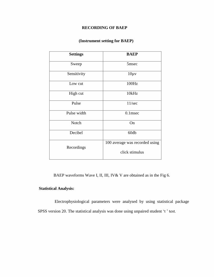
RECORDING OF BAEP
(Instrument setting for BAEP)
Settings BAEP
Sweep 5msec
Sensitivity 10µv
Low cut 100Hz
High cut 10kHz
Pulse 11/sec
Pulse width 0.1msec
Notch On
Decibel 60db
Recordings
100 average was recorded using
click stimulus
BAEP waveforms Wave I, II, III, IV& V are obtained as in the Fig 6.
Statistical Analysis:
Electrophysiological parameters were analysed by using statistical package
SPSS version 20. The statistical analysis was done using unpaired student „t ʼ test.

VEP RECORDING
Fig 7

BAEP RECORDING
Fig 8

RESULTS

RESULTS
The study group comprises of 40 Migraine patients who are subdivided in to 16
patients – Migraine with Aura and 24 patients – Migraine without Aura , 4 males and 36
females of age group 19 to 52 yrs were selected according to International Headache
Society Diagnostic Criteria for Migraine. Out of 40 controls, 6 males and 34 females
of age group of 19 to 55 yrs with mean age of 35.45 ± 9.9 years with no history of any
headache were included in the study.
The mean values and their standard deviation for the control group and study
group - Migraine patients with aura and without aura were tabulated. Various
electrophysiological parameters in VEP and BAEP are compared between the two study
groups and the control group.
The results were analysed using student t test. P value less than 0.05 was
considered as statistically significant.
Findings of evoked potential study in Migraine with Aura : (Table 3 )
Visual Evoked Potential :
Our study results showed that there was a significant prolongation of P100
and N145 Latency with P value < 0.05 whereas P100 amplitude value was
increased in Migraine with aura patients when compared with controls but it
was not statistically significant with P value 0.654.
Brainstem Auditory Evoked Potential :Study results showed a significant
prolongation in the Latencies of wave I , III & V and the Interpeak Latencies
I - III , III – V & I - V ( p < 0.05 ) as compared to control group.

Visual Evoked Potential : (Table 4 )
VEP N75 Latency: The mean value of N75 in Migraine with Aura was
found to be 71.5438± 3.41818 and in the control group it is 70.5750 ±
4.20058 with P value 0.416 and the difference was found to be insignificant.
P100 Latency: P100 Latency mean value in Migraine with Aura was
observed to be 102.0625 ± 5.70782 and in the control group it is 95.1625 ±
3.56512 with P value 0.000 and the difference was found to be statistically
significant.
N145 Latency: The mean value of N145 in Migraine with Aura was found
to be 136.2188 ± 10.45302 and in the control group it is 129.9875±8.61460
with P value 0.025 and found to be statistically significant.
P100 Amplitude: P100 Amplitude mean value in Migraine with Aura was
observed to be 6.2344 ± 3.28791 and in the control group it is 5.7975 ±
3.27955 with P value 0.654 and the difference was found to be statistically
insignificant.
Brainstem Auditory Evoked Potential : (Table 4 )
Latencies of Wave I, III & V and the Interpeak latencies I-III, III – V & I-V
were measured.
WAVE I: The mean value of wave I latency in Migraine with Aura was
observed to be 1.2725±0.29322 and in the control group 1.1222±0.22525
with P value 0.044 and found to be statistically significant.

WAVE III: Wave III latency in Migraine with Aura was observed to be
3.9381±0.62159 and in the control group 3.4323±0.63872 with P value
0.009 and found to be statistically significant.
WAVE V: The mean value of wave V latency in Migraine with Aura was
observed to be 6.3444±1.39867 and in the control group 5.6510±0.91618
with P value 0.033 and found to be statistically significant.
I - III IPL: I – III Interpeak latency in Migraine with Aura was observed to
be 2.6625±0.57633 and in the control group 2.2797±0.57041 with P value
0.028 and found to be statistically significant.
III – V IPL: III – V Interpeak latency in Migraine with Aura was observed
to be 2.5906±0.57130 and in the control group 2.2420±0.45662 with P
value 0.020 and found to be statistically significant.
I – V IPL: The mean value of I-V IPL in Migraine with Aura was observed
to be 5.2556±1.04950 and in the control group 4.4888±0.81594 with P
value 0.005 and found to be statistically significant.
Findings of evoked potential study in Migraine without Aura: (Table 2)
Visual Evoked Potential :
Our study results showed that there was a significant prolongation of
P100 and N145 Latency with P value < 0.05. P100 amplitude did not differ
significantly when compared with controls with P value 0.732.
Brainstem Auditory Evoked Potential: Study results showed a significant
prolongation in the Latencies of wave I, III & V and the Interpeak latencies

III – V & I - V ( p < 0.05 ) as compared to control group. I - III IPL was not
significantly prolonged.
Visual Evoked Potential : (Table 5 )
VEP N75 Latency: The mean value of N75 in Migraine without Aura was
found to be 71.4896± 4.62653 and in the control group it is 70.5750 ±
4.20058 with P value 0.420 and the difference was found to be insignificant.
P100 Latency: P100 Latency mean value in Migraine without Aura was
observed to be 99.9792 ± 5.06261 and in the control group it is 95.1625 ±
3.56512 with P value 0.000 and the difference was found to be statistically
significant.
N145 Latency: The mean value of N145 in Migraine without Aura was
found to be 134.5833 ± 5.45369 and in the control group it is 129.9875 ±
8.61460 with P value 0.022 and found to be statistically significant.
P100 Amplitude: P100 Amplitude mean value in Migraine without Aura was
observed to be 5.5371 ± 2.23703 and in the control group it is 5.7975 ±
3.27955 with P value 0.732 and the difference was found to be statistically
insignificant.
Brainstem Auditory Evoked Potential: (Table 5)
Latencies of Wave I ,III & V and the Interpeak latencies I-III , III – V & I-V
were measured.
WAVE I : The mean value of wave I latency in Migraine without Aura
was observed to be 1.3900±0.35864 and in the control group

1.1222±0.22525 with P value 0.000 and found to be statistically significant.
WAVE III : Wave III latency in Migraine without Aura was observed to
be 3.7850 ± 0.56300 and in the control group 3.4323 ± 0.63872 with P
value 0.029 and found to be statistically significant .
WAVE V: The mean value of wave V latency in Migraine without Aura
was observed to be 6.3321±0.81817 and in the control group
5.6510±0.91618 with P value 0.004 and found to be statistically significant.
I - III IPL :
I – III Interpeak latency in Migraine without Aura was observed to be
2.3908 ± 0.41921 and in the control group 2.2797 ± 0.57041 with P value
0.411 and found to be statistically insignificant.
III – V IPL:
III – V Interpeak latency in Migraine without Aura was observed to be
2.5567±0.52122 and in the control group 2.2420 ± 0.45662 with P value
0.014 and found to be statistically significant.
I – V IPL :
The mean value of I - V IPL in Migraine without Aura was observed
to be 4.9529 ± 0.67163 and in the control group 4.4888 ± 0.81594 with P
value 0.022 and found to be statistically significant.
Comparsion of Parameters Between Migraine With Aura And Without Aura:
(Table 6)
When Electrophysiological parameters(VEP N75,P100 & N145 latency and P100
amplitude, BAEP Wave I,III & V and IPL I- III , III- V & I - V ) were compared

between two groups Migraine with aura and without aura , Our study results did not show
any significance between two groups but when the individual parameters were
compared , the values of Migraine with aura are larger than Migraine without aura.
Visual Evoked Potential :
VEP N75 Latency: The mean value of N75 in Migraine with Aura was
found to be 71.5438± 3.41818 and in the Migraine without aura it is 71.4896±
4.62653 with P value 0.968 and the difference was found to be insignificant.
P100 Latency: P100 Latency mean value in Migraine with Aura was
observed to be 102.0625 ± 5.70782 and in the Migraine without aura it is
99.9792 ± 5.06261 with P value 0.233 and the difference was found to be
statistically insignificant.
N145 Latency: The mean value of N145 in Migraine with Aura was found
to be 136.2188 ± 10.45302 and in the Migraine without aura it is134.5833 ±
5.45369 with P value 0.521 and found to be statistically insignificant.
P100 Amplitude: P100 Amplitude mean value in Migraine with Aura was
observed to be 6.2344 ± 3.28791 and in the Migraine without aura it is
5.5371 ± 2.23703 with P value 0.429 and the difference was found to be
statistically insignificant.
Brainstem Auditory Evoked Potential:
Latencies of Wave I, III & V and the Interpeak latencies I-III, III – V & I-V
were measured.

WAVE I: The mean value of wave I latency in Migraine with Aura was
observed to be 1.2725±0.29322 and in Migraine without aura it is
1.3900±0.35864 with P value 0.283 and found to be statistically
insignificant.
WAVE III: Wave III latency in Migraine with Aura was observed to be
3.9381 ± 0.62159 and in Migraine without aura 3.7850 ± 0.56300 with P
value 0.424 and found to be statistically insignificant.
WAVE V: The mean value of wave V latency in Migraine with Aura was
observed to be 6.3444 ±1.39867 and in the Migraine without aura
6.3321±0.81817 with P value 0.972 and found to be statistically
insignificant.
I - III IPL :
I – III Interpeak latency in Migraine with Aura was observed to be
2.6625 ± 0.57633 and in Migraine without aura 2.3908 ± 0.41921 with P
value 0.092 and found to be statistically insignificant.
III – V IPL:
III – V Interpeak latency in Migraine with Aura was observed to be
2.5906 ± 0.57130 and in Migraine without aura 2.5567±0.52122 with P
value 0.847 and found to be statistically insignificant.
I – V IPL :
The mean value of I - V IPL in Migraine with Aura was observed to
be 5.2556 ± 1.04950 and in Migraine without aura 4.9529 ± 0.67163
with P value 0.272 and found to be statistically insignificant.
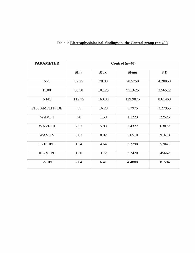
Table 1: Electrophysiological findings in the Control group (n= 40 )
PARAMETER Control (n=40)
Min. Max. Mean S.D
N75 62.25 78.00 70.5750 4.20058
P100 86.50 101.25 95.1625 3.56512
N145 112.75 163.00 129.9875 8.61460
P100 AMPLITUDE .55 16.29 5.7975 3.27955
WAVE I .70 1.50 1.1223 .22525
WAVE III 2.33 5.83 3.4322 .63872
WAVE V 3.63 8.02 5.6510 .91618
I - III IPL 1.34 4.64 2.2798 .57041
III - V IPL 1.30 3.72 2.2420 .45662
I -V IPL 2.64 6.41 4.4888 .81594

Table 2 : Electrophysiological findings in the Study group Migraine without aura
( n = 24)
Parameters Min Max Mean Std
deviation
N75 63.00 83.00 71.4896 4.62653
P100 90.00 109.50 99.9792 5.06261
N145 125.50 149.25 134.5833 5.45369
P100 AMPLITUDE 1.13 9.22 5.5371 2.23703
WAVE I .61 2.09 1.3900 .35864
WAVE III 2.76 4.93 3.7850 .56300
WAVE V 4.74 8.45 6.3321 .81817
I - III IPL 1.63 3.24 2.3908 .41921
III - V IPL 1.86 4.08 2.5567 .52122
I -V IPL 3.80 6.36 4.9529 .67163

Table 3 : Electrophysiological findings in the Study group Migraine with Aura
( n = 16 )
Parameters Min Max Mean Std deviation
N75 66.50 78.25 71.5437 3.41818
P100 93.75 114.25 102.0625 5.70782
N145 121.00 153.00 136.2187 10.45302
P100
AMPLITUDE 1.07 13.08 6.2344 3.28791
WAVE I .57 1.72 1.2725 .29322
WAVE III 3.03 5.11 3.9381 .62159
WAVE V 3.01 8.21 6.3444 1.39867
I - III IPL 1.93 3.53 2.6625 .57633
III - V IPL 1.91 3.52 2.5906 .57130
I -V IPL 4.01 6.63 5.2556 1.04950
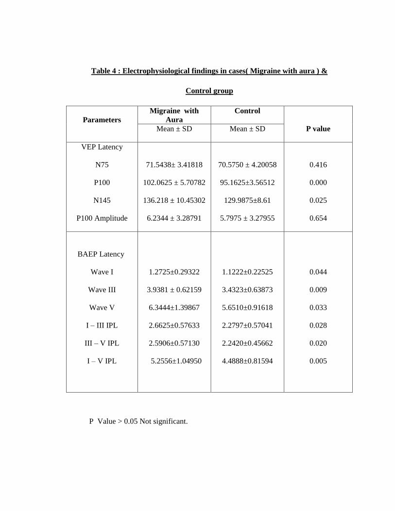
Table 4 : Electrophysiological findings in cases( Migraine with aura ) &
Control group
Parameters
Migraine with
Aura
Control
P value Mean ± SD Mean ± SD
VEP Latency
N75
P100
N145
P100 Amplitude
71.5438± 3.41818
102.0625 ± 5.70782
136.218 ± 10.45302
6.2344 ± 3.28791
70.5750 ± 4.20058
95.1625±3.56512
129.9875±8.61
5.7975 ± 3.27955
0.416
0.000
0.025
0.654
BAEP Latency
Wave I
Wave III
Wave V
I – III IPL
III – V IPL
I – V IPL
1.2725±0.29322
3.9381 ± 0.62159
6.3444±1.39867
2.6625±0.57633
2.5906±0.57130
5.2556±1.04950
1.1222±0.22525
3.4323±0.63873
5.6510±0.91618
2.2797±0.57041
2.2420±0.45662
4.4888±0.81594
0.044
0.009
0.033
0.028
0.020
0.005
P Value > 0.05 Not significant.

Table 5 : Electrophysiological findings in cases( Migraine without aura ) &
Control group
Parameters
Migraine without
Aura
Control
P value Mean ± SD Mean ± SD
VEP Latency
N75
P100
N145
P100 Amplitude
71.4896± 4.62653
99.9792± 5.06261
134.5833 ± 5.45369
5.5371 ± 2.23703
70.5750 ± 4.20058
95.1625±3.55126
129.9875±8.61
5.7975 ± 3.27955
0.420
0.000
0.022
0.732
BAEP Latency
Wave I
Wave III
Wave V
I – III IPL
III – V IPL
I – V IPL
1.3900 ± 0.35864
3.7850 ± 0.56300
6.3321 ± 0.81817
2.3908 ± 0.41921
2.5567 ± 0.52122
4.9529 ±0.67163
1.1222±0.22525
3.4323±0.63873
5.6510±0.91618
2.2797±0.57041
2.2420±0.45662
4.4888±0.81594
0.000
0.029
0.004
0.411
0.014
0.022

Table 6 : Electrophysiological findings in Migraine with aura &
Migraine without aura group
Parameters
Migraine with
Aura (n= 16)
Migraine without
aura (n= 24)
P value Mean ± SD Mean ± SD
VEP Latency
N75
P100
N145
P100 Amplitude
71.5438± 3.41818
102.0625 ± 5.70782
136.218 ± 10.45302
6.2344 ± 3.28791
71.4896± 4.62653
99.9792± 5.06261
134.5833 ± 5.45369
5.5371 ± 2.23703
0.968
0.233
0.521
0.429
BAEP Latency
Wave I
Wave III
Wave V
I – III IPL
III – V IPL
I – V IPL
1.2725±0.29322
3.9381 ± 0.62159
6.3444±1.39867
2.6625±0.57633
2.5906±0.57130
5.2556±1.04950
1.3900 ± 0.35864
3.7850 ± 0.56300
6.3321 ± 0.81817
2.3908 ± 0.41921
2.5567 ± 0.52122
4.9529 ±0.67163
0.283
0.424
0.972
0.092
0.847
0.272

MIGRAINE WITH AURA
VEP N75 Latency: The mean value of N75 in Migraine with Aura was found to
be 71.5438± 3.41818 and in the control group it is 70.5750 ± 4.20058 with P value
0.416 and the difference was found to be insignificant.
N145 Latency: The mean value of N145 in Migraine with Aura was found to be
136.2188 ± 10.45302 and in the control group it is 129.9875±8.61460 with P value 0.025
and found to be statistically significant.( Fig 9 )
Fig 9: VEP N75 & N145 Latency in Migraine with aura
0
20
40
60
80
100
120
140
160
N75 N145
MIGRAINE WITH AURA
CONTROL

VEP P100 Latency: P100 Latency mean value in Migraine with Aura was observed to
be 102.0625 ± 5.70782 and in the control group it is 95.1625 ± 3.56512 with P value
0.000 and the difference was found to be statistically significant. (Fig 10)
Fig 10 . VEP P100 Latency in Migraine with aura
90
92
94
96
98
100
102
104
MIGRAINEWITH AURA
CONTROL
P100 LATENCY
P100 LATENCY

P100 Amplitude:
P100 Amplitude mean value in Migraine with Aura was observed to be 6.2344 ±
3.27955 and in the control group it is 5.7975 ± 3.27955 with P value 0.654 and the
difference was found to be statistically insignificant.(Fig 11 )
Fig 11 P100 amplitude in Migraine with aura
5.5
5.6
5.7
5.8
5.9
6
6.1
6.2
6.3
MIGRAINEWITH AURA
CONTROL
P100 AMPLITUDE
P100 AMPLITUDE

BAEP:
WAVE I: The mean value of wave I latency in Migraine with Aura was observed
to be 1.2725±0.29322 and in the control group 1.1222±0.22525 with P value 0.044 and
found to be statistically significant.
WAVE III: Wave III latency in Migraine with Aura was observed to be
3.9381±0.62159 and in the control group 3.4323±0.63872 with P value 0.009 and found
to be statistically significant.
WAVE V: The mean value of wave V latency in Migraine with Aura was
observed to be 6.3444±1.39867 and in the control group 5.6510±0.91618 with P value
0.033 and found to be statistically significant. (Fig 12)
Fig 12 BAEP Wave I, III, V Latencies in Migraine with aura
0
1
2
3
4
5
6
7
WAVE I WAVE III WAVE V
MIGRAINE WITH AURA
CONTROL
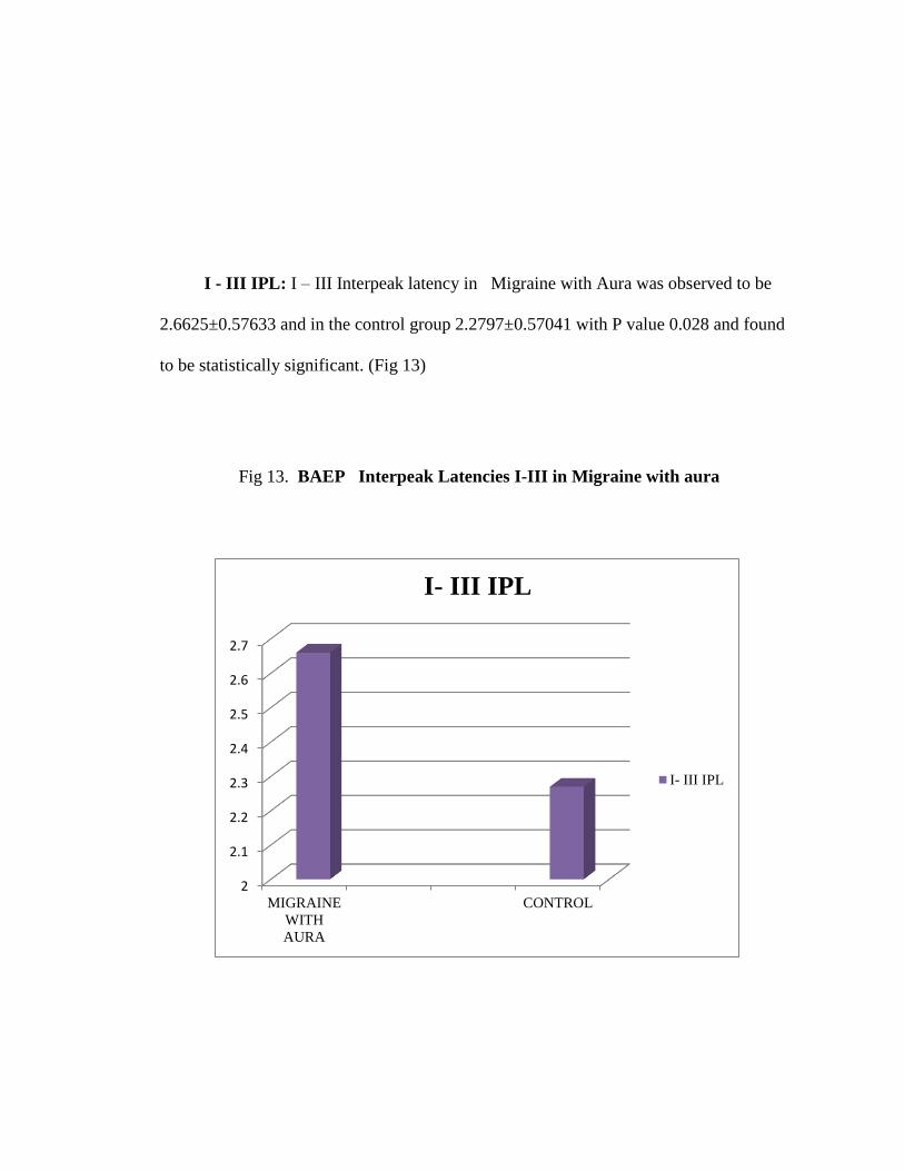
I - III IPL: I – III Interpeak latency in Migraine with Aura was observed to be
2.6625±0.57633 and in the control group 2.2797±0.57041 with P value 0.028 and found
to be statistically significant. (Fig 13)
Fig 13. BAEP Interpeak Latencies I-III in Migraine with aura
2
2.1
2.2
2.3
2.4
2.5
2.6
2.7
MIGRAINE
WITH
AURA
CONTROL
I- III IPL
I- III IPL

III – V IPL: III – V Interpeak latency in Migraine with Aura was observed to be
2.5906±0.57130 and in the control group 2.2420±0.45662 with P value 0.020 and found
to be statistically significant. (Fig 14)
Fig 14. BAEP Interpeak Latencies III- V in Migraine with aura
2
2.1
2.2
2.3
2.4
2.5
2.6
MIGRAINEWITH AURA
CONTROL
III- V IPL
III- V IPL

I – V IPL: The mean value of I - V IPL in Migraine with Aura was observed to be
5.2556±1.04950 and in the control group 4.4888±0.81594 with P value 0.005 and found
to be statistically significant. (Fig 15)
Fig 15. Interpeak Latency I - V in Migraine with aura
4
4.2
4.4
4.6
4.8
5
5.2
5.4
MIGRAINEWITH AURA
CONTROL
I-V IPL
I-V IPL
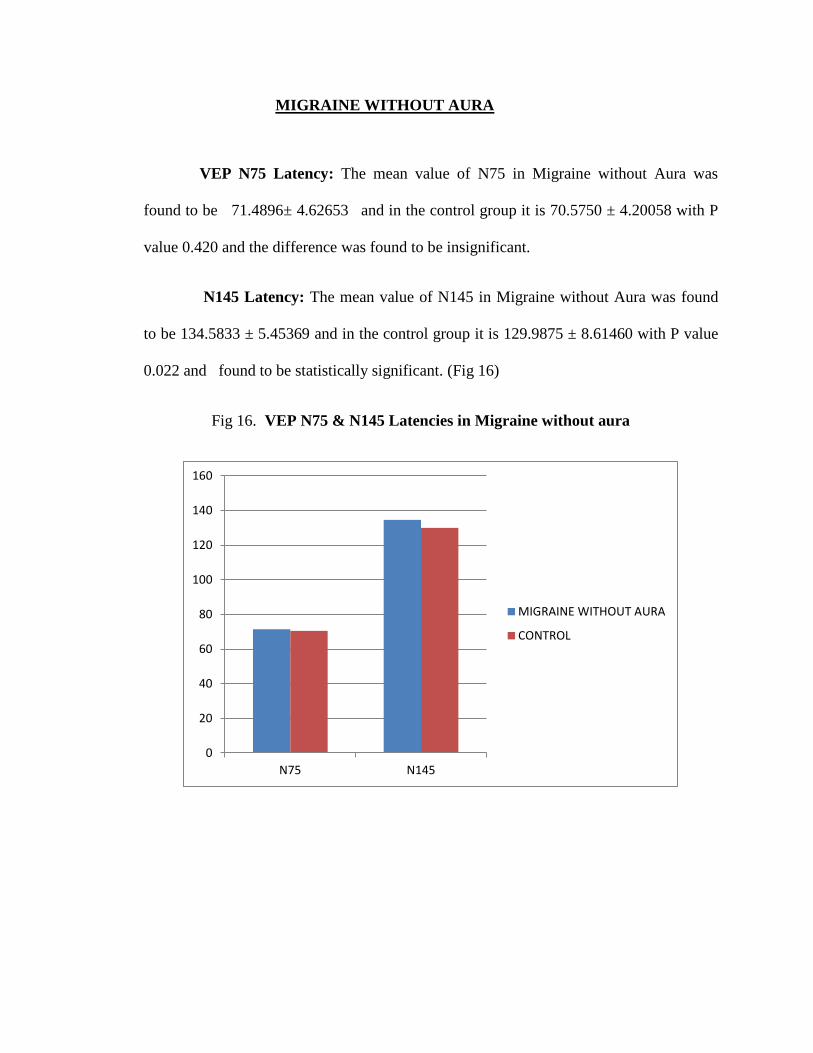
MIGRAINE WITHOUT AURA
VEP N75 Latency: The mean value of N75 in Migraine without Aura was
found to be 71.4896± 4.62653 and in the control group it is 70.5750 ± 4.20058 with P
value 0.420 and the difference was found to be insignificant.
N145 Latency: The mean value of N145 in Migraine without Aura was found
to be 134.5833 ± 5.45369 and in the control group it is 129.9875 ± 8.61460 with P value
0.022 and found to be statistically significant. (Fig 16)
Fig 16. VEP N75 & N145 Latencies in Migraine without aura
0
20
40
60
80
100
120
140
160
N75 N145
MIGRAINE WITHOUT AURA
CONTROL

P100 Latency: P100 Latency mean value in Migraine without Aura was
observed to be 99.9792 ± 5.06261 and in the control group it is 95.1625 ± 3.56512 with P
value 0.000 and the difference was found to be statistically significant. (Fig 17)
Fig 17. P100 Latency in Migraine without aura
92
93
94
95
96
97
98
99
100
101
MIGRAINEWITHOUT
AURA
CONTROL
P100 LATENCY
P100 LATENCY

P100 Amplitude: P100 Amplitude mean value in Migraine without Aura was
observed to be 5.5371 ± 2.23703 and in the control group it is 5.7975± 3.27955 with P
value 0.732 and the difference was found to be statistically insignificant. (Fig 18)
Fig 18. P100 Amplitude in Migraine without aura
5.4
5.45
5.5
5.55
5.6
5.65
5.7
5.75
5.8
5.85
MIGRAINEWITHOUT
AURA
CONTROL
P100 AMPLITUDE
P100 AMPLITUDE
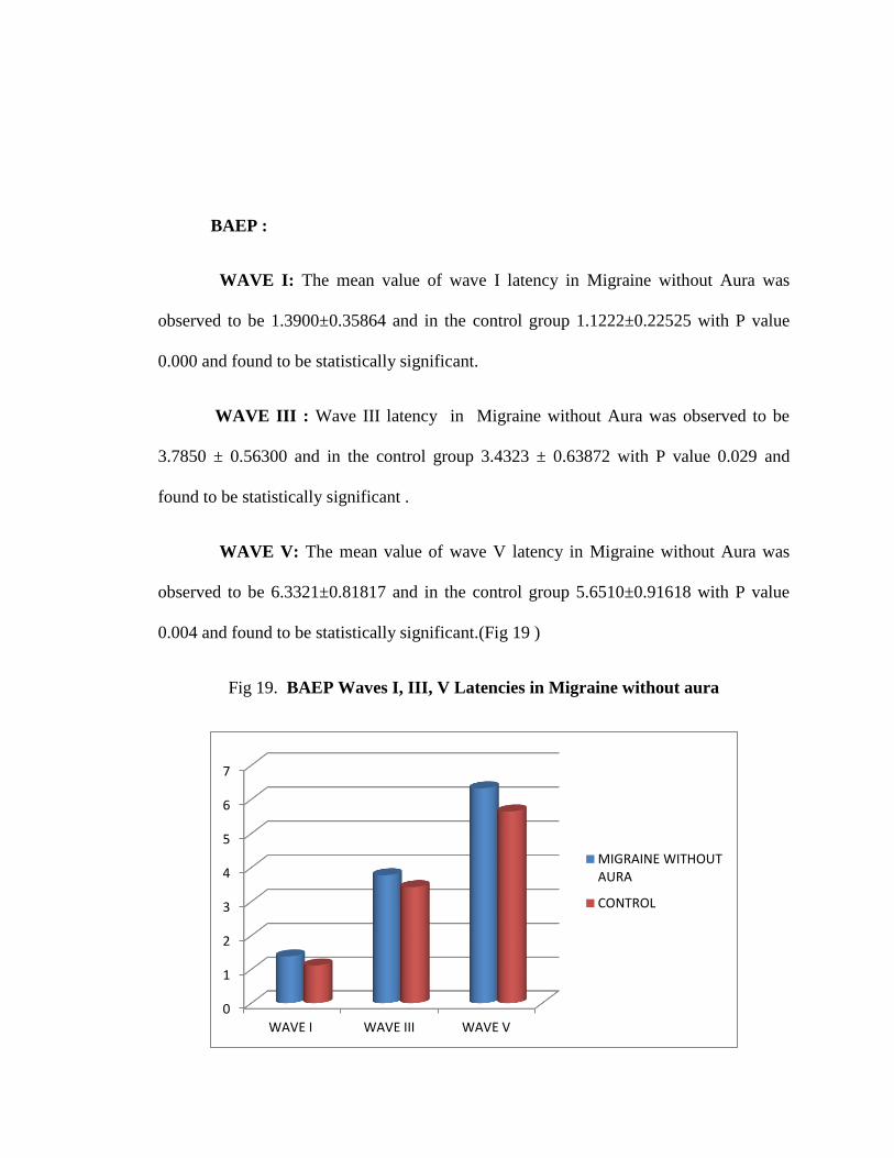
BAEP :
WAVE I: The mean value of wave I latency in Migraine without Aura was
observed to be 1.3900±0.35864 and in the control group 1.1222±0.22525 with P value
0.000 and found to be statistically significant.
WAVE III : Wave III latency in Migraine without Aura was observed to be
3.7850 ± 0.56300 and in the control group 3.4323 ± 0.63872 with P value 0.029 and
found to be statistically significant .
WAVE V: The mean value of wave V latency in Migraine without Aura was
observed to be 6.3321±0.81817 and in the control group 5.6510±0.91618 with P value
0.004 and found to be statistically significant.(Fig 19 )
Fig 19. BAEP Waves I, III, V Latencies in Migraine without aura
0
1
2
3
4
5
6
7
WAVE I WAVE III WAVE V
MIGRAINE WITHOUTAURA
CONTROL

I - III IPL: I – III Interpeak latency in Migraine without Aura was observed to be
2.3908 ± 0.41921 and in the control group 2.2797 ± 0.57041 with P value 0.411 and
found to be statistically insignificant. (Fig 20)
Fig 20. BAEP Interpeak Latency I-III in Migraine without aura
2.2
2.22
2.24
2.26
2.28
2.3
2.32
2.34
2.36
2.38
2.4
MIGRAINEWITHOUT
AURA
CONTROL
I- III IPL
I-III IPL

III – V IPL: III – V Interpeak latency in Migraine without Aura was observed to
be 2.5567±0.52122 and in the control group 2.2420 ± 0.45662 with P value 0.014 and
found to be statistically significant. (Fig 21)
Fig 21. Interpeak Latency III – V in Migraine without aura
2.05
2.1
2.15
2.2
2.25
2.3
2.35
2.4
2.45
2.5
2.55
MIGRAINEWITHOUT
AURA
CONTROL
III - V IPL
III - V IPL

I – V IPL: The mean value of I - V IPL in Migraine without Aura was observed to be
4.9529 ± 0.67163 and in the control group 4.4888 ± 0.81594 with P value 0.022 and
found to be statistically significant. (Fig 22)
Fig 22. Interpeak Latency I – V in Migraine without aura
4.2
4.3
4.4
4.5
4.6
4.7
4.8
4.9
5
MIGRAINEWITHOUT
AURA CONTROL
I-V IPL
I-V IPL

MIGRAINE WITH AURA & MIGRAINE WITHOUT AURA
VEP N75 Latency: The mean value of N75 in Migraine with Aura was found to be
71.5438± 3.41818 and in the Migraine without aura it is 71.4896 ± 4.62653 with P value
0.968 and the difference was found to be insignificant.
N145 Latency: The mean value of N145 in Migraine with Aura was found to be
136.2188 ± 10.45302 and in the Migraine without aura it is134.5833 ± 5.45369 with P
value 0.521 and found to be statistically insignificant. (Fig 23)
Fig 23. VEP N75 & N145 in study group
0
20
40
60
80
100
120
140
160
N75 N145
MIGRAINE WITHAURA
MIGRAINE WITHOUTAURA

P100 Latency:
P100 Latency mean value in Migraine with Aura was observed to be 102.0625 ±
5.70782 and in the Migraine without aura it is 99.9792 ± 5.06261 with P value 0.233 and
the difference was found to be statistically insignificant. (Fig 24)
Fig 24. P100 latency in study group
98.5
99
99.5
100
100.5
101
101.5
102
102.5
MIGRAINE WITH AURA MIGRAINE WITHOUT AURA
P100 LATENCY
P100 LATENCY

P100 Amplitude:
P100 Amplitude mean value in Migraine with Aura was observed to be 6.2344 ±
3.28791 and in the Migraine without aura it is 5.5371 ± 2.23703 with P value 0.429 and
the difference was found to be statistically insignificant. (Fig 25)
Fig 25. P100 Amplitude in study group
5
5.2
5.4
5.6
5.8
6
6.2
6.4
MIGRAINE WITH AURAMIGRAINE WITHOUT AURA
P100 AMPLITUDE
P100 AMPLITUDE

BAEP:
WAVE I: The mean value of wave I latency in Migraine with Aura was
observed to be 1.2725±0.29322 and in Migraine without aura it is
1.3900±0.35864 with P value 0.283 and found to be statistically
insignificant.
WAVE III: Wave III latency in Migraine with Aura was observed to be
3.9381 ± 0.62159 and in Migraine without aura 3.7850 ± 0.56300 with P
value 0.424 and found to be statistically insignificant.
WAVE V: The mean value of wave V latency in Migraine with Aura was
observed to be 6.3444 ±1.39867 and in the Migraine without aura
6.3321±0.81817 with P value 0.972 and found to be statistically
insignificant. (Fig 26 )
Fig 26. Wave I, III & V Latency
0
1
2
3
4
5
6
7
WAVE I WAVE III WAVE V
MIGRAINE WITHAURA
MIGRAINE WITHOUT
AURA

I - III IPL :
I – III Interpeak latency in Migraine with Aura was observed to be 2.6625 ±
0.57633 and in Migraine without aura was 2.3908 ± 0.41921 with P value 0.092 and
found to be statistically insignificant .( Fig 27 )
Fig 27. I-III IPL in study group
2.25
2.3
2.35
2.4
2.45
2.5
2.55
2.6
2.65
2.7
MIGRAINE WITH AURAMIGRAINE WITHOUT AURA
I-III IPL
I-III IPL

III – V IPL:
III – V Interpeak latency in Migraine with Aura was observed to be 2.5906 ± 0.57130
and in Migraine without aura was 2.5567±0.52122 with P value 0.847 and found to be
statistically insignificant.( Fig 28 )
Fig 28. III-V IPL in study group
2.53
2.54
2.55
2.56
2.57
2.58
2.59
2.6
MIGRAINE WITH AURAMIGRAINE WITHOUT AURA
III - V IPL
III - V IPL

I – V IPL:
The mean value of I - V IPL in Migraine with Aura was observed to be 5.2556 ±
1.04950 and in Migraine without aura 4.9529 ± 0.67163 with P value 0.272 and found
to be statistically insignificant. (Fig 29)
Fig 29. I- V IPL in study group
4.8
4.85
4.9
4.95
5
5.05
5.1
5.15
5.2
5.25
5.3
MIGRAINE
WITH
AURA
MIGRAINE
WITHOUT
AURA
I - V IPL
I - V IPL

DISCUSSION

DISCUSSION
In the present study, the Electrophysiological parameters are evaluated in
Migraine patients with and without Aura and in 40 control group healthy volunteers.
Migraine, the most common Neurological disorder is associated with substantial
functional impairment, involving both physical and emotional ramifications. Migraine
can best be explained as a „Brain state ʼ in which the cellular and vascular functional
changes occur at the same time due to dysfunction of subcortical structures, brainstem
and diencephalic nuclei that modulate sensory inputs. These nuclei act as a „Migraine
Mediatorʼ whose dysfunction will lead to abnormal perception and activation of TVS
which then activate the central structures.
Thus, Migraine is mainly due to TVS activation generated within the brain
without a peripheral sensory input. Migraine is the central sensory processing disorder,
there is dysfunction of descending brainstem pain modulatory system. The
hyperexcitability of the nociceptive circuitry downstream is responsible for the central
sensitization in Migraine patients.(69)
In our present study, patients with Migraine either with aura or without aura
differ significantly in various electrophysiological parameters.
Our VEP study results showed that P100 and N145 latencies were significantly
prolonged with p < 0.05 in both Migraine with and without Aura when compared with
controls whereas P100 Amplitude was increased in study group especially in cases with
Aura than the control group but it was not statistically significant P=0.654 .

The results of the present study agreed with EL Shater et al., Laila EL Mosly et al .,
Kennard et al ., Khalil et al ., Mariani et al ., Drake et al ., Bockowski L et al .,
In the study conducted by EL Shater et al.,(35)
showed significant prolongation of
P100 latency only in Migraine with aura patients and there was no significant difference
in P100 Amplitude between patients and controls. These changes were found to have a
structural basis, due to repeated attacks resulting in ischemia and proposed that there is
subtle neuronal damage in the visual system in Migraine with aura patients. These results
were consistent with our present study.
Kennard et al., (40)
also reported prolonged P100 latency in Migraine patients and
explained it to have a structural basis. Khalil et al., also found the same results but
suggested the prolonged latencies are constitutional due to synaptic delay. Similar results
were found in our study.
Laila EL Mosly et al .,(7)
also found significant prolongation of P100 latency with no
change in P100 amplitude, these changes were explained to be due to Occipital cortex
dysfunction in Migraine patients. These results are in accordance with our present study.
Drake et al .,(49)
found significant prolongation of P100 & N145 latencies in
Migraine without aura cases due to dysfunction of brainstem centers probably related to
endorphin or serotonin neurotransmission. Mariani et al ., (47)
reported an increase in
P100 latency in Migraine with & without aura whereas Amplitudes were quite dispersed
among patients and controls. Suggested the alterations in the monoamine neuromediators
are responsible for these changes. Similar findings were observed in our study.

Bockowski L et al ., (37)
found significant prolongation of P100 latency whereas
amplitudes N1-P100 & P100-N2 were prolonged in Migraine children than the children
with tension type headache. They also reported that these amplitudes were lower in
Migraine with aura when compared with Migraine without aura patients. These changes
were due to visual dysfunction that might be secondary to a loss of inhibitory GABAergic
interneurons in the visual cortex from repeated parenchymal insults. P100 latency showed
similar findings in our study.
N.Ashjazadeh , B Varavipour (38)
reported significant prolongation of P100
latency in classic Migraine patients with no change in the Amplitude due to subtle
neuronal damage in the visual system of Migraine patients from repeated transient
ischemia , probably due to constitutional change. These results were consistent with our
present study.
Nofal M Khalil , Nigel J Legg , Duncan J Anderson (6)
found similar results due
to synaptic delay . Research work also showed positive correlation between disease
duration and changes in amplitude of P100. The results of P100 latency were consistent
with our present study.
Pedro.F Moreira Filho , Adalmir M. Dantas (34)
found significant prolongation in
P100 latency in Migraine without aura patients due to alterations in Monoamine
neuromediators. Similar results were found in our study.
Thus, Migraine patients with and without aura had significant prolongation of P100
& N145 latency due to hyperexcitability of the cortex in Migraine patients even between
attacks and due to synaptic delay.
Our BAEP study reports showed significant prolongation of latencies of Wave I,
III & V with P <0.05 in Migraine patients when compared with controls. Also, the
interpeak latencies I- III, III- V & I- V were significantly prolonged in study group,
Migraine with aura than the controls. In Migraine without Aura III- V & I- V were
significantly prolonged.
The results of present study were consistent with D Koushal ,S Sanjay Munjal , M Modi;
N Panda , Firat Y et al., Schalke HP et al ., Anil K Dash et al ., Laila EL Mosly et al .,
Drake et al ., Bayazit et al .,
D Koushal ,S Sanjay Munjal , M Modi , N Panda (50)
studied BAEP in Migraine
patients and found significant prolongation of latencies of wave I , III & V and in the IPL
I - III & III- V due to involvement of brainstem structures as well as due to activation
of brainstem in Migraine patients . Similar results were found in our study.
Schalke HP et al ., (52)
found the same results and reported that there is a permanent
impairment of brainstem function in Migraine patients. These findings are consistent with
our present study.
Firat Y et al., (51)
studied in paediatric Migraine patients and found prolongation of
wave V latency & I- V IPL in study group due to transient impairment of brainstem
function in Migraine patients.
Anil K Dash et al ., (53)
found significant prolongation of latencies of Wave I ,
III, V & I-III IPL, III- V IPL & I – V IPL . The study concluded that there is brainstem
involvement in Migraine and BAEP are the earliest indicator of impending auditory

involvement in patients with Migraine. These results are in accordance with our present
study.
Laila EL Mosly et al ., (7)
found prolongation in wave III & V and the I-III , I – V
IPL due to hyperexcitability of the cerebral cortex. Bayazit Y et al., (55)
found
prolongation of latency of wave I, III & V and the IPL III- V. Similar results were found
in our study.
Sheriya A Hamed , Amal Mohammed Elatter (53)
found prolongation of wave III
latency and the IPL I-III , III – V & I – V. This study suggests that there is permanent
vestibular damage involving either central or peripheral pathways. Similar results were
observed in our study.
Thus , Migraine patients with and without aura show significant prolongation
of latency in Electrophysiological studies probably due to subtle neuronal damage within
the visual system especially in patients with aura due to recurrent cerebral hypoperfusion
and due to cortical hyperexcitability. These findings suggest dysfunction of neuronal
excitability due to defective neurotransmitter signaling and cerebral bioelectrical
dysrhythmia.

CONCLUSION

CONCLUSION
The present study results show that there is involvement of the central nervous
system Visual pathway and the Brainstem in Migraine patients. The Migraine patients
show prolongation of latency in Electrophysiological studies.
Migraine patients with aura and without aura show significant prolongation of
P100, N145 Latency with no change in P100 amplitude and in BAEP the latencies of
waves I , III & V and the IPLs I-III , III- V & I-V are prolonged. These findings suggest
there is defect in the central processing of visual function and BAEP alterations suggest
involvement of Brainstem structures during Migraine attack.
Thus, VEP and Auditory brainstem evoked responses are considered as useful,
non-invasive, reliable & diagnostic techniques for better understanding the
Neurophysiological processes involved in Migraine patients which aid in the selection of
adequate, effective treatment in Migraine subjects.
However, further studies are needed to compare the duration of the disease with
the changes in the Electrophysiological study and to explain the role of
Neuromodulatory centers in the brainstem in pathophysiology of Migraine. Then, the role
of pattern reversal check size on VEP parameters in Migraine patients and lack of
habituation during prolonged stimulation should be evaluated.

BIBLIOGRAPHY

BIBLIOGRAPHY
1. Nicholas A.Boon, Nicki R.Colledge, Brian R. Walker, John A.A. Hunter.,
Davidson „s Principles & Practice of Medicine . 20th
edition Elsevier pg..1160-
1163.
2. Andreoli and Carpenter „ s Cecil Essentials of Medicine., Charles C.J.Carpenter ,
Robert C.Griggs, Ivor J. Benjamin . 7th
edition saunders Elsevier p: 1069- 1073.
3. Dan L.Longo , Antony S. Fauci , Dennis L. Kasper, J.Larry Jameson , Stephen L.
Hauser, Joseph Loscalzo., Harrison „s Principles of Internal Medicine 18th
edition
vol 1., McGraw Hill Medical p.114- 120.
4. Till Sprenger and Peter J Goadsby Minireview..Migraine pathogenesis and state
of pharmacological treatment options. BMC Medicine 2009, 7:71.
5. S N Chugh , Ashima chugh Textbook of Medicine for Medicine.. Delhi Arya
publications pg ..596 – 599.
6. Nofal MKhalil , Nigel J Legg , Duncan J Anderson Long term decline of P100
amplitude in Migraine with Aura. J Neurol Neurosurg Psychiatry 2000;69:507 –
511.
7. Laila El Mosly , Azza Bayoumy , Hoda Massoud ,Mahmoud Abdel Moty , Manal
Hafez , Taghreed Elshafie and Rasha El Bialy Impact of Migraine Headache on
Quality of Life in a group of Female patients using Neurophysiological
Assessment. AAMJ ,Vol.10,N.2, April,2012,suppl.
8. Akbar Hamzei Moghaddam ,Rostam Syfaldiny , Farhad Iranmanesh , Hamid
Mahadavy., Visual Evoked Potentials in patientswith Classic Migraine. ZJRMS
2013; 15 (4) :25-28.
9. Merrit „s Textbook of Neurology 9th
edition p:837-842.
10. Marschall S. Runge , M. Andrew Greganti , Frank H.Netter., Netter‟s Internal
Medicine 2nd
edition . Elsevier p:873-881.

11. Nasim Maleki , Lino Becerra and David Borsook Migraine: Maladaptive Brain
responses to stress. Headache 2012 ;52 ( suppl 2 ) : 102-106.
12. Dr. S.N. Khosla Essentials of Medicine..CBS Publishers p .643-646.
13. . Parveen Kumar, Dr Michael Clark „s Clinical Medicine 8th
edition .Elsevier
p:1107- 1111.
14. Marten Scheffer , Albert Van den Berg , Michel D . Ferrari Migraine strikes as
Neuronal Excitability reaches a Tipping point. PLoS ONE 88(8) ;e72514: march
17 , 2013.
15. Martin Lauritzen Pathophysiology of the Migraine aura, The spreading
Depression Theory. Brain 1994, 117, 199- 210.
16. Andrew Prescot , Lino Becerra , Gautam Pendse , Shannon Tully,Eric Jensen ,
Richard Hargreaves,PerryRenshaw,Rami Burstein and David Borsook. Excitatory
neurotransmitters in brain regions in Interictal Migraine patients. BiolMed central
Molecular pain 2009,5 : 34.
17. Rieke Oelkers , Konard Grosser , Eberhard Lang, Gerd Geisslinger, Gerd Kobal,
Kay Brune and Jorn Lotsch. Visual evoked potentials in Migraine patients:
alterations depend on pattern spatial frequency. Brain 1999,122, 1147-1155.
18. Judit Afra,Alberto Proietti Cocchini , Victor De Pasqua , Adelin Albert and Jean
Schoenen Visual evoked potentials during long periods of pattern reversal
stimulation in Migraine. Brain 1998, 121, 233-241.
19. Karen E. Waldie , Markus Hausmann , Barry J Milne, Richie Poulton Migraine
and cognitive function .Neurology 2002 ;59 :904- 908.
20. Michael Donaghy Brain„s diseases of the Nervous system. Oxford university. 12th
edition.

21. Randolph W. Evans Diagnostic Testing in Neurology p: 249-284.
22. Walter G. Bradley , Robert B. Daroff, Gerald M. Fenichel , Joseph Jankovic
Neurology in Clinical Practice Vol 1. Principles of Diagnosis and Management .
5th
edition Elsevier p.471- 476.
23. Michael J. Aminoff , Electrodiagnosis in Clinical Neurology, Visual evoked
potentials 5th
edition Elsevier p:453-468.
24. U K Misra , J Kalita Clinical Neurophysiology Visual evoked potentials. 3rd
edition p:310-324.
25. J. Vernon Odom Michael Bach Mitchell Brigell Graham E. HolderDaphne L. Mc
culloch ISCEV standard for clinical Visual evoked potentials. Doc Ophthal
2010,120 :111- 119.
26. Eileen E. Birchand Rain G. Bosworth Electrodiagnosis in Clinical Neurology,
Visual evoked potentials in Infants and children. 5th
edition Elsevier p:473-483
27. Neil R.Miller , Nancy J. Newman , Valerie Biousse , John B. Kerrison Walsh and
Hoyt „s Clinical Neuro- Ophthalmology.. Lippincott Williams & Wilkins 6th
edition p:140-144.
28. Dr.A K Jain Manual of Practical Physiology., p: 301-309.
29. Samuel R. Atcherson , Tina M. Stoody Auditory Electrophysiology A Clinical
Guide. P.67- 81.al Application.
30. Alan D. Legatt Electrodiagnosis in Clinical Neurology, Brainstem Auditory
Evoked Potantials :Methology, Interpretation and Clinic al Application. P : 490-
519.
31. U K Misra , J Kalita Clinical Neurophysiology Brainstem Auditory Evoked
Potential .3rd
edition p : 330- 344.
32. G K Pal , Pravati Pal Textbook of Practical Physiology.3rd
edition p: 298 – 303.

33. Jack Katz , Larry Medwetsky , Robert Burkard , Linda J . Hood Handbook of
Clinical Audiology. 6th
edition Lippincott Williams & Wilkins p: 222 – 240.
34. Michael Donaghy Brain„s diseases of the Nervous system. Oxford university .
12th
edition .
35. M.F.EL Snater, Y.Abo El Naga , A. Gaber, A.Hazzo , A.Nassif Migraine with
and without aura : Clinical and Electrophysiological changes.. Egyptian Journal of
Neurology, Psychiatry and Neurosurgery July 2006 Vol .43 Issue :2.
36. Pedro.F Moreira Filho , Adalmir M. Dantas Pattern Reversal Visual evoked
potentials in Migraine subjects without aura. ArqNeuropsiquiatr 1994, 52 (4) :
484- 488.
37. Bockowski L , Sobaniec W , Smigielska Kuzia , Kulak W, Solowiej E The pattern
reversal visual evoked potentials in children with Migraine with aura and without
aura. Roczniki Akademii Medycznej w Bialymstoku vol 48, 2003.
38. N. Ashjazadeh , B Varavipour Abnormalities of Visual evoked potentials in
Migraine patients. IJMS Vol 28, No.2, June 2003.
39. Professor Peter J. Goadsby serotonin 5 HT 1B/1D Receptor agonists in Migraine .
40. Kennard C , Gawel M and Rudolf Nde M. Visual evoked potentials in Migraine
subjects. Res Clin Study Headache 1987; 6 : 73-80.
41. American academy of Neurology the Electroencephalogram in the evauation of
headache. Neurology 1995;45 :1411-13.
42. Bramanti P , Grugno R, Vitetta A , Di Bella P , Muscara N and Nappi G Migraine
with and without aura:electrophysiological and functional neuroimaging evidence
. Functional Neurology 2005 ;20 (1) :29- 32.

43. Polich J Ehlers Cl and Dalassio DJ. Pattern shift visual evoked responses and
EEG in Migrane. Headache 1986 ; 26 (9) : 451-6.
44. Golla FL, Winter AL. Analysis of cerebral responses to flicker in patients
complaining of episodic headache . Electroencephalogr Clin Neurophysiol 1959,
11:539- 549.
45. Richey ET , Kooi KA , Waggoner RW. Visually evoked responses in Migraine.
Electroencephalogr Clin Neurophysiol 1966, 21:23- 27.
46. Lehtonen JB. visual evoked cortical potentials in man. Studies on recording and
stimulation technique and findings in Migraine patients. Thesis . University of
Turku, 1972.
47. Mariani E , Moschini V , Pastorino G , Rizzi F , Severgnini A , Tiengo M .
Pattern shift visual evoked potentials in Migraine subjects with visual aura.
Headache 1990 ; 30 :435- 438.
48. Bena P, Bianco C , Costa P , Piazza D , Bergamasco B Visual evoked potentials
and Brainstem Auditory Evoked Potential in Migraine and Transient ischemic
attacks. Cephalalgia 1985 May ; 5 suppl 2 : 53 – 8.
49. Drake ME, Pakalnis A , Hietter SA , Padamadan H. Visual and Auditory evoked
potentials in Migraine . Electromyogr Clin Neurophysiol 1990 Feb – Mar ; 30 (2)
:77 – 81
50. D Koushal ,S Sanjay Munjal , M Modi , N Panda Auditory brainstem evoked
responses in Migraine patients. The Internet Journal of Neurology 2008 Vol.12
number 1.
51. Firat Y , Ozturan O , Bicak U , Yakinci C, Akarcay M Auditory brainstem
response in pediatric Migraine : during the attack and asymptomatic period.
International journal of Pediatric Otorhinolaryngology 2006, 70 (8) : 1431-1438.

52. Bayazit Y ,Yilmaz M , Mumbuc S , Kanlikama M. (2001) .Assessment of
Migraine -related cochleovestibular symptoms. Rev Laryngol Otol Rhinol ( Bord)
; 122 (2) :85-8.
53. Anil K. Dash , Naresh Panda , Gaurav Khandelwal , Vivek Lal , Sherbaj
S.Mann.Migraine and audiovestibular dysfunction : is there a correlation?
American Journal of Otolaryngology – head and Neck Medicine and surgery 29 (
2008 ) 295 – 299.
54. Boylu E , Domac FM , KocerA , Unal Z , Tanridaq , Us O . visual evoked
potentials abnormalities in Migraine patients. Electroencephalogr Clin
Neurophysiol 2010 sep – oct; 50 (6) :303-8.
55. Yang Y , Li P , Ye HC Brainstem auditory evoked potentials and assessment of
personality test in patients with Migraine. Hunan medical university 2000, 25 ( 1)
: 63 – 64.
56. Zgorzalewicz M The study of Early auditory evoked potentials in primary
headaches in children and adolescents and their pathogenetic implications.
Neurologia 2005, 39 (4suppl 1):s17 – 25.
57. Sherifa A. Hamed , Amal Mohamad Elattar Peripheral and central vestibular
function in patients with Migraine . Jurnal ofNeurology and NeuroscienceVol 3,
No.1 2012.
58. Leslie J. Dorfman, M.D., William H. Marshall, M.D. and Dieter R. Enzmann,
M.D. Cerebral infarction and migraine. Neurology March 1979 vol. 29 no. 3 ,317
59. Shaheen E. Lakhan, MD, PhD, MEd, MS, Mihaela Avramut, MD, PhD, Stewart J.
Tepper, MD Structural and Functional Neuroimaging in Migraine .
Headache. 2013; 53(1):46-66.
60. Y.L.Lo , S.Hamed, H.Rumpel , L.L.Chan Auditory hallucinations and Migraine
of possible Brainstem origin. J Headache pain 2011, 12:573-575.

61. Tedeschi G, Russo A, Tessitore A Relevance of functional neuroimaging studies
for understanding migraine mechanisms. Expert Rev Neurother. 2013
Mar;13(3):275-85. doi: 10.1586/ern.13.20.
62. Regional Cerebral Blood Flow During Migraine Attacks By Xenon-133
Inhalation And Emission Tomography ,
Martin Lauritzen And Jes Olesen
63. Schalke HP, Hoffeberth B , Grotemeyer KH ,Husstedt IW
Electronystagmographic investigations in migraine and cluster
headache during the pain-free interval. Cephalalgia. 1989 Dec;9(4):271-5.
64. F. Sicuteri , A.Testi and B. Anselmi Biochemical investigations in Headache
.General Medical clinic of Forensic University. Int Arch Allergy 19 ; 55- 58 (
1961)
65. Katalin Kovacs et al , Laszlo Bors , Laszlo Tothfausi , Jl na Jelensik , Gyorgy B
ozsik , Loszlo , Kerenyi and Samuel Komoly . Cephalalgia Vol 9 ,, Issue 1 , p:53-
57 , mar 1989.
66. Curtrer FM , O‟ Donnell A , Sanchez del Rio M Functional neuroimaging
:enhanced understanding of Migraine pathophysiology. Neurology 2000;55 ( 9
suppl 2 ) :S36-45.
67. Baburhan Guldiken Triptans in the treatment of Migraine Clinical Medicine :
Therapeutics 2009 :1711- 713.
68. Dalia A .Biswas , Meghana A. Gaikwad A study of Visual evoked potentials and
effect of relaxation technique in patients with Migraine. Int J Adv Med 2014 may
;1 (1) : 24-33.
69. Eric A.Moulten , Rami Burstein , Shannon Tully , Richard Hargreaves ,Lino
Becerra, David Borsook Interictal dysfunction of a brainstem descending
modulatory center in Migraine patients. PLoS ONE 3(11):e3799.

ANNEXURES

ABBREVIATIONS
VEP : Visual evoked potential
BAEP : Brainstem auditory evoked potential
OCD : Obsessive compulsive disorder
IHS : International headache society
MIDAS : Migraine disability assessment score
MMSE : Mini mental state examination
CSD : Cortical spreading depression
TVS: Trigeminal Vascular system
TCC : Trigeminal cervical component
ERP : Event related potential
EP : Evoked potential
IPL : Interpeak latency
ABR : Auditory brainstem response

INFORMED CONSENT
Dr. R.Sowmiya, Post graduate student in the Department of Physiology,
Thanjavur Medical College, Thanjavur is doing the study entitled „Evaluation of Visual
evoked potential and Auditory evoked potential in Migraine‟. The procedure of the
study has been explained to me in my own language. I understand that there is no risk
involved in the above procedure and it is purely non- invasive. I hereby give my consent
to participate in this study. The data obtained here may be used for research and
publication.
Station: Signature of the subject
Date:
Signature of the Investigator

PROFORMA
EVALUATION OF VISUAL EVOKED POTENTIAL & BRAINSTEM
AUDITORY EVOKED POTENTIAL IN MIGRAINE
NAME: AGE: SEX:
ADDRESS: OCCUPATION:
PRESENT HISTORY:
H/O Headache :
Duration :
Frequency:
HISTORY SUGGESTIVE OF AURA:
H/O Numbness / Tingling
H/O Double vision
H/O Flash of light
H/O Tinnitus
H/O Speech disturbances
H/O Floaters
H/O Nausea / Vomiting / Giddiness
H/O Ear discharge
H/O Visual disturbances
PAST HISTORY:
H/O Hypertension / Diabetes / Thyrotoxicosis

PERSONAL HISTORY:
FAMILY HISTORY:
MENSTRUAL HISTORY:
DRUG HISTORY:
GENERAL EXAMINATION:
Pallor: Icteric: Cyanosis: Clubbing: Lymphadenopathy:
VITAL SIGNS:
Pulse: Respiratory rate:
BP: Vision:
Examination of CVS:
Examination of RS:
Examination of Abdomen:
Examination of CNS:
Examination of Eye: Right: Left:
Examination of Ear: Right: Left:
Examination of Nose& Paranasal sinuses:
Examination of Teeth & Oropharynx:
DIAGNOSIS: Migraine with aura / Migraine without aura
ROUTINE INVESTIGATIONS:
Blood sugar :
Urea :

VEP :
N75
Latency
P100
Latency
N145
Latency
P100
Amplitude
LEFT EYE
RIGHT EYE
BAER:
Wave I Wave III Wave V I – III
IPL
III – V
IPL
I – V IPL
LEFT EAR
RIGHT
EAR

MASTER CHART

Visual Evoked Potential and Brainstem Auditory Evoked Potential in Migraine with Aura Cases
S.No
Age/
Sex
VEP N75
Latency(ms)
VEP P100
Latency (ms)
VEP N145
Latency (ms)
VEP P100
Amplitude(µv )
BAEP LT
BAEP RT
LT RT LT RT LT RT LT RT I III V I-III III-V I-V I III V I-III III-V I-V
1 31/F 73.5 64.5 110 111 154 152 8.62 8.93 1.75 3.82 6.12 2.07 2.3 4.37 0.98 3.2 5.48 2.22 2.28 4.5
2 44/F 64.5 70 115.5 113 144.5 134 6.15 6.96 1.2 3.25 5.2 2.05 1.95 4 0.85 2.82 4.88 1.97 2.06 4.03
3 30/M 77 70.5 103.5 100.5 136 132.5 5.71 7.88 1.4 3.82 8.07 2.42 4.25 6.67 1.32 3.82 6.62 2.5 2.8 5.3
4 43/F 70.5 75.5 105 96 148 133.5 1.8 0.34 1.4 4.4 6.18 3 1.78 4.78 1.05 2.42 4.92 1.37 2.5 3.87
5 19/F 66 71 98 101 145.5 152 2.51 1.53 0.8 4.05 6.42 3.25 2.37 5.62 0.85 4.65 8.43 3.8 3.78 7.58
6 19/F 70.5 76 110.5 111 139.5 141 12 14.17 1.72 5.7 .78 3.98 3.08 7.06 1.42 4.53 7.65 3.08 3.12 6.2
7 27/F 79 68 93 104.5 139 136 6.98 6.17 0.35 4.55 6.4 4.2 1.85 6.05 0.8 3.58 7.4 2.78 3.82 6.6
8 45/F 75.5 70.5 105.5 103 128 124 6.44 7.11 0.78 2.58 4.85 1.8 2.27 4.07 1.5 4.53 7.2 3.03 2.67 5.7
9 42/M 71 72.5 96 99 125 134 6.14 3 1.3 3.25 5.1 1.95 1.85 3.8 1.23 3.15 5.5 1.92 2.35 4.27
10 47/F 77.5 79 100.5 100.5 121 121 6.23 2.38 1.65 3.75 5.78 2.1 2.03 4.13 1.23 3.35 5.2 2.12 1.85 3.97
11 29/F 66 67 98.5 96.5 126 126.5 7.06 10.87 1.2 3.88 5.88 2.68 2 4.68 1.45 2.98 4.8 1.53 1.82 3.35
12 49/F 73.5 74 100 101.5 126 121.5 8.64 3.12 1.1 4.62 6.32 3.52 1.7 5.22 1.82 4.15 6.42 2.33 2.27 4.6
13 42/M 75 78.5 91.5 96 116.5 135.5 4.6 1.33 1.4 3.78 7.02 2.38 3.24 5.62 2.05 5.85 9.2 3.8 3.35 7.15
14 49/F 66 70.5 100.5 97 133 133 12.53 11.61 1.38 3.32 5.7 1.94 2.38 4.32 1.1 4.85 9.2 3.75 4.35 8.1
15 19/F 60 77 103.5 109.5 150 145.5 5.07 4.03 1.05 4.42 6.85 3.37 2.43 5.8 1.4 3.3 5.68 1.9 2.38 4.28
16 30/F 72.5 63.5 97.5 94.5 156.5 124.5 9.68 14.9 0.88 4.32 7.5 3.44 3.18 6.62 2.38 5.42 8.35 3.04 2.93 5.97

Visual Evoked Potential and Brainstem Auditory Evoked Potential in Migraine without Aura Cases
S.No
Age/
Sex
VEP N75
Latency(ms)
VEP P100
Latency (ms)
VEP N145
Latency (ms)
VEP P100
Amplitude(µv )
BAEP LT
BAEP RT
LT RT LT RT LT RT LT RT I III V I-III III-V I-V I III V I-III III-V I-V
1 36/F 60 69.5 90.5 102 121 131 3.57 2.33 0.82 3.35 6 2.53 2.65 5.18 1.45 3.65 5.22 2.2 1.57 3.77
2 38/M 76 70 92 92 129 132 1.68 2.01 1.65 3.12 4.68 1.47 1.56 3.03 1.85 3.98 6.45 2,15 2.47 4.6
3 33/F 73 67 103.5 93 133.5 132.5 3.53 6.3 1.8 4.85 7.58 3.05 2.73 5.78 0.9 2.82 4.58 1.92 1.76 3.68
4 33/F 68.5 70 95 95 125.5 137 6.37 8.57 0.75 3.32 9.15 2.57 5.83 8.4 2.33 3.82 6.15 1.49 2.33 3.82
5 30/F 66.5 65.5 104 100.5 131 131 7.51 7.26 1.05 3.88 6.75 2.83 2.87 5.7 1.32 4.78 8.25 3.46 3.47 6.93
6 34/F 74.5 70 103.5 98 141.5 145 8.53 9.92 1.35 3.68 7.95 2.33 4.27 6.6 0.9 3.5 5.03 2.6 1.53 4.13
7 19/F 73 71 101.5 101 128.5 142.5 9.74 8.59 0.85 3.02 5 2.17 1.98 4.15 1.42 3.8 5.8 2.38 2 4.38
8 52/M 82 74.5 107 102.5 134.5 137.5 6.1 4.86 0.6 2.75 4.68 2.15 1.93 4.08 0.62 3 4.8 2.38 1.8 4.18
9 19/F 74 70 87.5 92.5 137 139.5 5.81 4.42 0.65 2.8 5.05 2.15 2.25 4.4 1.5 3.7 5.9 2.2 2.2 4.4
10 19/F 70.5 69.5 105 99.5 146 131.5 4.53 5.08 1.15 2.72 5.18 1.57 2.46 4.03 1.15 3.35 5.68 2.2 2.33 4.53
11 33/F 75 73.5 98.5 94 136.5 132.5 5.5 5.77 1 3.72 6.02 2.72 2.3 5.02 1.27 4.15 6.62 2.88 2.47 5.35
12 23/F 76 73.5 109 103 137 135 10.92 2.56 1.7 5.25 7.55 3.55 2.3 5.85 1.85 4.58 6.22 2.73 1.64 4.37
13 28/F 74.5 65.5 97 97 136.5 149.5 0.27 8.66 1.52 3.65 5.98 2.13 2.33 4.46 1.92 4 6.95 2.08 2.95 5.03
14 32/F 65 76 99 106 129 137 7.29 9.57 0.78 2.95 5.22 2.17 2.27 4.44 1.4 5.72 8.02 4.32 2.3 6.62
15 33/F 59.5 66.5 95.5 95.5 123 128 3.85 6.05 1.58 3.82 7.08 2.24 3.26 5.5 1.68 4.1 6.42 2.42 2.32 4.74
16 44/F 68.5 69 96 95 130.5 130.5 9.57 8.28 1.98 3.98 7.72 2 3.74 5.74 2.2 5.88 9.18 3.68 3.3 6.98
17 33/M 72 68 100.5 102.5 137.5 141.5 10.82 3.46 0.92 2.98 5.1 2.06 2.12 4.18 1.3 3.5 7 2.2 3.5 5.7
18 36/F 77.5 85.5 102 105.5 136 129 3.56 4.85 1.08 2.7 5.68 1.62 2.98 4.6 1.18 2.82 5.48 1.64 2.66 4.3
19 26/F 76 65 91 93.5 139 123 4.99 2.47 1.55 4.58 7.52 3.03 2.94 5.97 0.95 3.25 5.95 2.3 2.7 5
20 36/F 83 83 105.5 103 125.5 137 1.06 1.21 1.58 3.68 6.1 2.1 2.42 4.52 1.9 3.95 6.48 2.05 2.53 4.58
21 29/F 66 68 94 111.5 147.5 151 6.53 3.43 1.82 3.62 6.08 1.8 2.46 4.26 1.5 4.62 7.82 3.12 3.2 6.32
22 42/F 75.5 67 101.5 107.5 134 134 2.29 3.09 1.88 4.05 6.35 2.17 2.3 4.47 1.02 3.3 5.52 2.28 2.22 4.5
23 39/F 80 65 110 109 132.5 139 8.23 6.3 2.6 4.92 7.35 2.32 2.43 4.75 1.52 3.92 6.52 2.4 2.6 5
24 33/F 76 66.5 105.5 105.5 135 126.5 11.09 0.12 1.2 4.08 5.92 2.88 1.84 4.72 1.8 4.18 6.92 2.38 2.74 5.12

Visual Evoked Potential and Brainstem Auditory Evoked Potential in Controls
S.No
Age/
Sex
VEP N75
Latency(ms)
VEP P100
Latency (ms)
VEP N145
Latency (ms)
VEP P100
Amplitude(µv )
BAEP LT
BAEP RT
LT RT LT RT LT RT LT RT I III V I-III III-V I-V I III V I-III III-V I-V
1 40/F 64.5 64.5 90.5 89.5 143.5 126 10.2 11.87 1.35 3.4 5.42 2.05 2.02 4.07 1.7 3.95 5.75 2.25 1.8 4.05
2 40/F 71 73 91.5 91 132.5 130.5 9.77 9.55 1.65 3.85 7.88 2.2 4.03 6.23 1 5.08 7.05 4.08 1.97 6.05
3 31/M 66 68.5 89 90.5 112.5 113 6.51 5.63 1.82 6.15 8.05 4.33 1.9 6.23 0.57 5.52 8 4.95 2.48 7.43
4 21/F 68.5 67.5 98 92 128 124 5.47 0.97 1 2.58 5.82 1.58 3.24 4.82 0.85 2.45 4.05 1.6 1.6 3.2
5 44/F 79 60.5 98.5 92 131.5 134.5 3.56 6.63 0.8 2.7 4.28 1.9 1.58 3.48 0.8 2.65 4.5 1.85 1.85 3.7
6 40/F 76.5 77 98 98.5 136.5 137 0.26 1.89 0.6 2.6 4.6 2 2 4 1.4 3.3 5.42 1.9 2.12 4.02
7 40/F 74 77 98 102.5 137 138.5 6.29 6.28 0.85 2.98 5.98 2.13 3 5.13 1.05 3.38 5.05 2.33 1.67 4
8 54/M 76 80 89 92.5 119 122.5 0.75 1.3 0.78 2.08 3.38 1.3 1.3 2.6 1.2 2.58 3.88 1.38 1.3 2.68
9 42/F 68.5 66 99.5 96 129 123.5 3.24 7.09 1.2 2.6 3.92 1.4 1.32 2.72 0.82 3 6.4 2.18 3.4 5.58
10 55/F 65 68.5 98 97 128 124 6.37 9.05 1.05 3.6 5.62 2.55 2.02 4.57 0.95 3.6 7.4 2.73 3.72 6.45
11 23/F 70.5 70.5 97 93.5 127.5 123 8.57 6.36 1.48 5.48 8.65 4 3.17 7.17 1.02 3.9 6 2.88 2.1 4.98
12 19/F 84 64 95 102 126 143 14.09 2.24 0.68 3.18 6.25 2.5 3.07 5.57 0.72 3.72 6.92 3 3.2 6.2
13 27/F 71.5 68 97.5 103.5 146 141.5 7.42 10.88 0.9 3.18 5.55 2.28 2.37 4.65 1 5.82 9.02 4.82 3.2 8.02
14 35/F 66.5 68.5 97 98.5 143.5 130.5 16.11 16.47 1.02 3.12 4.9 2.1 1.78 3.88 1.23 3.2 5.2 1.97 2 3.97
15 32/F 72.5 70.5 96 95 129.5 121.5 5.8 3.5 1.3 3.45 5.88 2.15 2.43 4.58 0.6 3.25 6.02 2.65 2.77 5.42
16 27/M 79 73.5 102 97.5 133 125.5 3.13 7.03 1.18 3.38 5.7 2.2 2.32 4.52 0.62 2.5 4.62 1.88 2.12 4
17 20/M 76 77.5 100 102.5 117 143 1.76 3.87 0.57 3.62 6.7 3.05 3.08 6.13 1.25 3.58 7.95 2.33 4.37 6.7
18 47/F 70.5 73.5 100.5 98.5 130.5 135 6.93 1.51 1.25 3.88 6.5 2.63 2.62 5.25 1.7 3.12 4.47 1.42 1.35 2.77
19 39/F 70 74 95 93.5 136 132 8.44 4.52 1.55 4.5 7.42 2.95 2.92 5.87 1.12 3.18 5.12 2.06 1.94 4
20 37/F 67.5 73.5 92.5 95.5 133 123.5 3.94 0.4 1.35 3.9 5.72 2.55 1.82 4.37 1.1 3.1 5.88 2 2.78 4.78

Visual Evoked Potential And Brainstem Auditory Evoked Potential in Controls
S.N0
Age/
Sex
VEP N75
Latency(ms)
VEP P100
Latency (ms)
VEP N145
Latency (ms)
VEP P100
Amplitude(µv )
BAEP LT
BAEP RT
LT RT LT RT LT RT LT RT I III V I-III III-V I-V I III V I-III III-V I-V
21 38/F 63 63.5 92 93 131.5 130.5 6.83 3.9 0.78 2.98 5.22 2.2 2.24 4.44 0.9 2.98 4.95 2.08 1.97 4.05
22 44/F 79 64 101 96 121.5 126 1.66 9.39 1.05 2.78 5.03 1.73 2.25 3.98 1.92 4.08 5.5 2.16 1.42 3.58
23 25/F 72.5 75 92.5 97 132 134.5 1.03 4.57 1.52 3.75 6 2.23 2.25 4.48 1.05 3.9 5.52 2.85 1.62 4.47
24 31/F 66.5 75.5 93.5 96.5 129.5 128.5 0.34 0.76 1.68 3.2 4.75 1.52 1.55 3.07 1.42 3.75 5.25 2.53 1.5 3.83
25 39/F 67 64 93.5 92 120.5 123.5 9.68 1.89 1.62 3.48 5.22 1.86 1.74 3.6 0.98 3.85 5.78 2.87 1.93 4.8
26 27/F 74 78 100 92 144.5 141 4.02 10.33 1.75 3.82 6.2 2.07 2.38 4.45 0.95 3.12 5.35 2.17 2.23 4.4
27 29/F 69 74 96.5 104 167.5 158.5 1.77 1.57 1.4 3.3 5.42 1.9 2.12 4.02 1.23 3.52 6.02 2.29 2.5 4.79
28 40/M 65.5 69 83 90 131 136 1.95 2.99 1.82 3.8 6.08 1.98 2.28 4.26 0.98 3.05 5.35 2.07 2.3 4.37
29 45/F 71.5 73.5 92.5 92 127 121 2.41 5.06 0.9 2.65 4.72 1.75 2.07 3.82 1.05 3.55 3.85 2.5 2.3 4.8
30 26/F 75.5 73.5 99.5 98 146.5 136 6.84 5.17 1.23 2.58 3.8 1.35 1.22 2.57 0.88 3.45 5.98 2.57 2.53 5.1
31 25/F 65.5 69 95 100 126.5 132 14.29 6.08 0.78 2.48 4.95 1.7 2.47 4.17 1.45 3.68 5.75 2.23 2.07 4.3
32 25/F 70 77 91 91 136.5 120.5 8.59 6.18 1.82 4.32 6.65 2.5 2.33 4.83 0.7 3.18 4.85 2.48 1.67 4.15
33 50/M 65 59.5 99 96 134.5 123 4.65 6.39 1.52 3.85 5.78 2.33 1.93 4.26 1.35 2.55 4.4 1.2 1.85 3.05
34 43/F 68.5 68.5 92.5 98 123 127 5.8 5.07 0.78 1.92 5.45 1.14 3.53 4.67 0.8 3.45 5.25 2.65 1.8 4.45
35 29/F 75.5 77.5 93 97.5 120 118.5 3.09 4.58 1.05 3.15 5.55 2.1 2.4 4.5 0.95 4.7 7.72 3.75 3.02 6.77
36 44/F 63.5 70.5 93.5 91 125.5 117.5 10.17 3.25 1.58 3.78 5.98 2.2 2.2 4.4 0.98 3.38 5.6 2.4 2.22 4.62
37 19/F 80 63 91 83.5 122 112.5 5.17 5.81 0.82 2.52 5.1 1.7 2.58 4.28 1.32 2.95 4.65 1.63 1.7 3.33
38 34/F 72 77 95 89 126 120 3.28 2.17 1.82 4.03 6.35 2.21 2.32 4.53 0.45 3.12 4.9 2.67 1.78 4.45
39 39/F 64 61.5 96.5 104 127 126 8.59 0.12 1.92 3.7 5.22 1.86 1.44 3.3 0.72 2.42 4.8 1.7 2.38 4.08
40 53/F 65 67 97 96 126 133.5 4.35 1.35 1.3 3.75 5.28 2.45 1.53 3.98 0.75 3.02 5.2 2.27 2.18 4.45