Embed Size (px)
Citation preview
BIOAUTOMATION, 2009, 13 (3), 57-72
57
Brainstem Auditory Evoked Potentials in Patients
with Subarachnoid Haemorrhage
Lyubomir Haralanov
1, Mikhail Matveev
2, *, Elena Mermeklieva
3
1National Heart Hospital, Department of Neurology, Sofia, Bulgaria
E-mail: [email protected]
2Centre of Biomedical Engineering, Bulgarian Academy of Sciences, Sofia, Bulgaria
E-mail: [email protected]
3Alexandrovska University Hospital, Medical Faculty, Medical University, Sofia, Bulgaria
E-mail: [email protected]
*Corresponding author
Received: September 9, 2009 Accepted: September 15, 2009
Published: October 14, 2009
Abstract: Objective. The aim of the present study is to typify BAEPs configurations of
patients with different location of lesions caused by subarachnoid haemorrhage (SAH) and
the ensuing complications, in view of assessing the auditory-brainstem system disturbance.
Methods. The typization was performed by comparing BAEPs with standard patterns from
two sets of types of BAEPs by ipsilateral and binaural stimulation and by cross-stimulation.
Results. 94 BAEPs were used for collection of normal referential values: for the absolute
latencies and the absolute amplitudes of waves I, II, III, IV and V; for inter-peak latencies I-
III, II-III, III-V, I-V and II-V; for amplitude ratios I/V and III/V. 146 BAEPs of patients with
mild SAH and 55 from patients with severe SAH, were typified. In 5 types of BAEPs out of a
total of 11, the percentage of the potentials in patients with mild SAH and severe SAH
differed significantly (p < 0.01).
Conclusions. The use of sets of types of BAEPs by ipsilateral, binaural and cross-stimulation
correctly classifies the potentials in patients with mild and severe SAH.
Keywords: Brainstem auditory evoked potentials (BAEPs), Subarachnoid haemorrhage,
Typification of BAEPs.
Introduction Recording and analysis of brainstem auditory evoked potentials (BAEPs) is an objective
electrophysiological method, which allows assessment of the functional state of brain stem
following primary or secondary damage. Unlike the cortical auditory brain potentials in
patients with severe cerebral lesions, BAEPs have much greater information value, owing to
their waveform stability and to the fact that they are less influenced by various exogenous and
endogenous factors [2, 11, 13, 15, 17, 20, 21, 23].
Few studies [15, 16, 22] have attempted to systematize BAEPs in the case of subarachnoid
hemorrhage (SAH). This is due to the difficulties in conducting the study, in particular its
duration. For some severely ill SAH patients, BAEPs studies have to be performed in an
intensive care unit. Filling of the 4th
ventricle leads to additional microcirculatory disorders in
the tegmentum of the brainstem, which affects the reticular formation and the ascending
auditory routes localized there. Consequently, systematization is needed for tracing the
transition from norm to pathology and for identifying abnormalities of BAEPs in SAH and
their secondary complications. This motivated us to apply our system for classification of
BAEPs, which we introduced for patients with cerebrovascular pathology [6, 7], to investigate
BIOAUTOMATION, 2009, 13 (3), 57-72
58
BAEPs in patients with different location of lesions caused by SAH and their complications.
Methods
Clinical Sample The study was performed on 58 individuals, divided as follows:
First (control) group: 19 healthy subjects (10 women and 9 men), mean age 38.1±16.1, used
for collection of normal referential BAEP values.
Second group: 37 patients (26 women and 11 men), mean age 42.7±12.0. Depending on the
severity of SAH, this group was divided into two subgroups using the Hunt and Hess scale
[12].
The subgroup of “mild SAH” has a total of 29 patients, 21 of them with 1st degree by the Hunt
and Hess scale and 8 with 2nd
degree. Two of them were with clinical evidence for SAH and
unilateral lesion of the oculomotor nerve. One had evidence about combined otoneurological
syndrome. One was with proved peripheral unilateral otoneurological syndrome and SAH in
the vertebro-basilar system, due to a rupture of collateral vessel, developed with the subclavia
style syndrome. One had unilateral pre-morbid deafness. Two patients of the group died of
rapidly developing brain edema with tonsillar herniation.
The “severe SAH” subgroup consists of 12 patients, with 3rd
to 5th
degrees by the Hunt and
Hess scale, of which 11 died and one was in coma vigil. Four patients had control study with
BAEPs after deterioration of their neurological status and changed from “mild SAH” to
“severe SAH”. Two other patients from the group have been studied twice after changes in
their neurological status. Three patients had an aneurysm in the vertebral-basilar system. Five
had evidence of combined otoneurological syndrome, and one was with peripheral unilateral
otoneurological syndrome.
The total number of valid patterns of the first group was 94 BAEPs, used for collection of
normal referential BAEP values. A total of 201 validated BAEP patterns were recorded from
43 studies of patients from the second group, from one to three BAEPs were obtained with the
same stimulation type. Of all 43 studies of BAEPs, 39 were conducted with ipsilateral
bilateral stimulation, 36 with cross-lateral bilateral stimulation, and 26 with binaural bilateral
stimulation. The potentials from some of the studies were removed from the study due to
suspected presence of artifacts. Table 1 presents the distribution of the patterns according to
group and type of stimulation.
Instrumentation BAEPs were studied in a specialised functional laboratory of an intensive care unit equipped
with acoustic and electrical isolation, oxygen sources (above 5 atm.) and vacuum-aspiration
system.
A hardware and software instrumentation complex was developed for real time and off-line
investigation of BAEPs, as well as for their storage in a database. The complex comprises of a
generator of click-stimuli of 100 ms duration and alternating polarity, with a frequency of the
12 s-1
, intensity of the 90 dB above the level of the individual click-threshold, and with 50 dB
white noise given to the opposite ear. The cerebral electrical activity was initiated by needle
electrodes allowing repeated chemical sterilization. In accordance with the American
Electroencephalographic Society Guidelines [1], the position of the electrodes was mentioned
as the positive electrode was always placed on the vertex (Cz position of the 10-20 system),
BIOAUTOMATION, 2009, 13 (3), 57-72
59
the reference electrodes were placed on both mastoid points (M1 and M2 position of the 10-
20 system) and the ground electrode was placed frontally on the median line, at 3 cm in front
of Fz (position of the 10-20 system). The resistance of all electrodes was kept below 3 kOm.
The evoked cerebral activity was amplified approximately 100000 times and displayed on
computer screen with 10 msec sweep. The full amplitude/division scale of the screen contains
10 divisions with voltage range from 0.1 to 1.0 µV/div. Analogue and subsequent digital
filtering was used to select the frequency band of 156 to 2031 Hz. At least two separate
averages of 2000 clicks were superimposed. Cz-M1 or Cz-M2 was consequently recorded
with ipsilateral, cross-lateral, and binaural stimulation. With ipsilateral stimulation, electrical
activity is conducted to a reference electrode on the ipsi-mastoid and an active electrode on
the vertex. With cross-lateral stimulation, electrical activity is conducted from a reference
electrode on the mastoid and an active electrode on the vertex. With binaural stimulation, the
active electrode remains on the vertex, and the reference electrode is in mastoid position at the
side of the preceding ipsilateral stimulation. The two sides are studied consequently, placing
the reference electrode on the left and right mastoid, respectively.
Table 1. BAEP patterns for patient groups and stimulation types
Examination methods The groups of subjects were investigated using: otoneurological study with suprathreshold
test for perception of click-stimuli; Doppler sonography; EEG; computer tomography;
cerebral panangiography; basic blood parameters; liquor and urine study; clinical-
pathoanatomical verification.
Different examination methods were used for patients in coma. They were subjected to
various treatment procedures for acute cerebral circulation disorders, as well as all
reanimation measures for patients with disturbed vital functions. Continuous pulmonary
ventilation was applied during the BAEP study. Respiratory equipment was used, operating
on the volume principle and set in motion by the inhaled gases.
Patients having qualitative consciousness disorders, such as delirium, were not suitable for
studies during the excitation state. In these patients, benzodiazepine sedation failed to
guarantee the state of rest needed for the study. In the patients with quantitative consciousness
disorders it was possible to study BAEPs using benzodiazepine sedation, when necessary.
Non-depolarizing myorelaxants were used for the patients in stupor or coma on artificial
pulmonary ventilation. These myorelaxants have a sufficiently prolonged action to eliminate
muscle artifacts and the unconscious movements of the patients during the actual study [16].
Side of Stimulation
Group Ipsilateral Binaural Crossed
Number of
Patterns
Normal 37 28 29 94
SAH (total) 77 51 73 201
Mild SAH 56 35 55 146
Severe SAH 21 16 18 55
BIOAUTOMATION, 2009, 13 (3), 57-72
60
Typification of BAEPs The absolute latencies (L) and the absolute peak amplitudes (A) of the main waves I, II, III,
IV and V were measured. The inter-peak latencies (IPL) I-III, III-V and I-V, as well as the
amplitude ratios (AR) I/V, I/III and III/V were measured in ipsilateral and binaural
stimulation. In contralateral stimulation (cross-stimulation), IPL II-III, III-V and II-V, as well
as III/V AR were measured.
Table 2. Reference upper limits (UL) of ipsilateral, binaural and crossed stimulation indices;
for latencies: UL = mean + 2.5xSD, for amplitudes and amplitude ratios:
UL = mean + 3.0xSD.
An earlier study of ours [5] proved by discriminant analysis that the following are of highly
informative value: L of I, II, III and V waves; IPL I-III, III-V, I-V; AR I/V, III/V, in cases of
ipsilateral and binaural stimulation. In contralateral stimulation the informative indices are: L
of II, III, V waves; IPL II-III, III-V, II-V; AR III/V.
According to the American Electroencephalographic Society Guidelines [1], the reference
upper limits (UL) of three types of stimulation for the latencies were determined using mean
+ 2.5 SD, and for amplitudes and for AR using mean + 3.0 SD (Table 2).
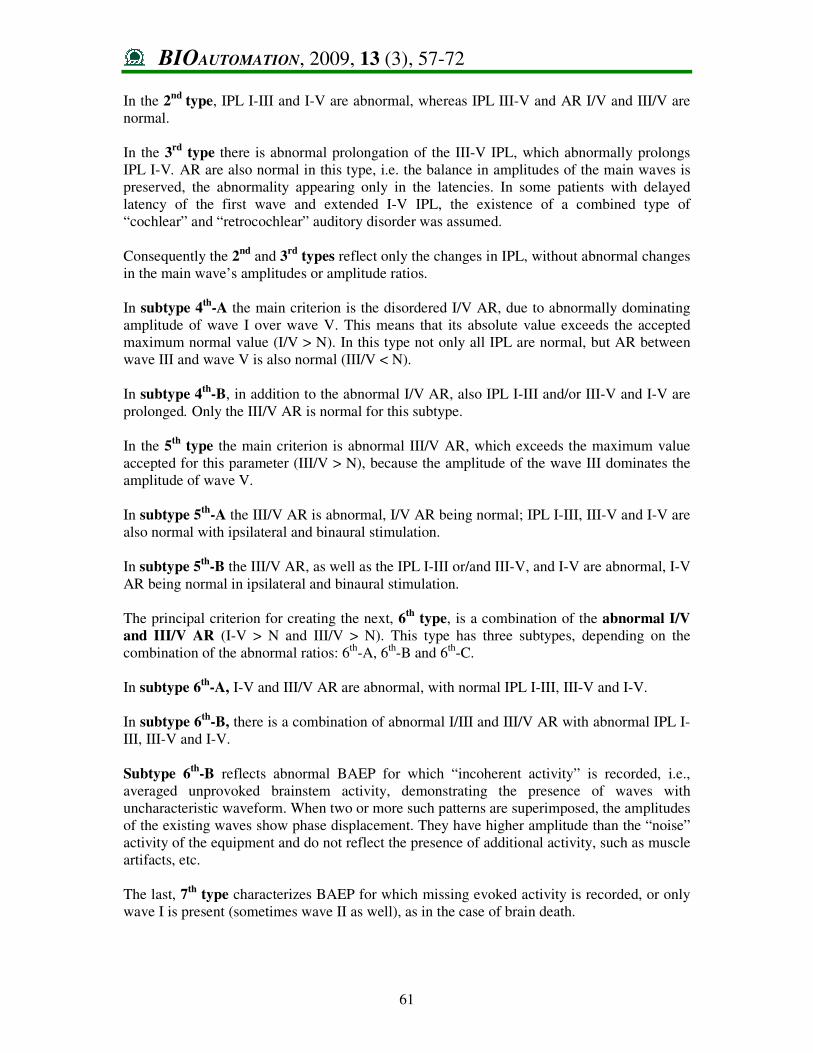
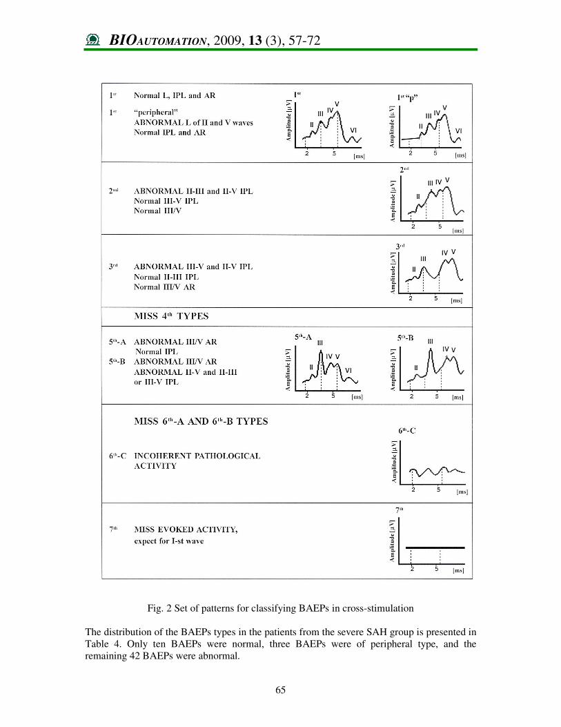
Fig. 1 presents a set of types of ipsilateral and binaural stimulation BAEPs, which comprises
all possible variants: normal and abnormal BAEP patterns, including patterns with no evoked
activity (in the case of cerebral death or total deafness).
In the case of the 1st type of BAEP, the parameters L, IPL and AR are statistically not
different from those in the group of individuals with normal hearing.
When the latencies of wave I and wave V are above normal and IPL and AR are normal, the
BAEP are typified as 1st “peripheral” (1
st “p”
) type.
Indices
Latencies (msec)
Absolute Inter-peak
Amplitude
(µV)
Amplitude
ratio
Stimulation
I II III V I-
III
II-
III
III-
V I-V
II-
V I III V I/V III/V
mean 1.35 2.44 3.44 5.27 2.08 - 1.83 3.92 - 0.51 0.42 1.11 0.48 0,4
SD 0.14 0.17 0.19 0.19 0.17 - 0.13 0.17 - 0.23 0.24 0.36 0.23 0,2 Ipsilateral (90
dB)
UL 1.70 2.87 3.92 5.75 2.51 - 2.16 4.35 - 1.20 1.14 2.19 1.17 1.00
mean 1.36 2.53 3.39 5.29 2.03 - 1.9 3.92 - 0.65 0.55 1.33 0.48 0.42
SD 0.15 0.16 0.14 0.17 0.15 - 0.10 0.18 - 0.33 0.3 0.39 0.2 0.2 Binaural
(90 dB)
UL 1.74 2.93 3.74 5.72 2.41 - 2.15 4.37 - 1.64 1.45 2.50 1.08 1.02
mean - 2.52 3.38 5.31 - 0.86 1.93 - 2.8 - 0.47 1.0 - 0.47
SD - 0.16 0.25 0.20 - 0.26 0.22 - 0.18 - 0.31 0.33 - 0.25 Crossed
(90 dB)
UL - 2.92 4.01 5.75 - 1.51 2.48 - 3.25 - 1.40 1.93 - 1.19
BIOAUTOMATION, 2009, 13 (3), 57-72
61
In the 2nd
type, IPL I-III and I-V are abnormal, whereas IPL III-V and AR I/V and III/V are
normal.
In the 3rd
type there is abnormal prolongation of the III-V IPL, which abnormally prolongs
IPL I-V. AR are also normal in this type, i.e. the balance in amplitudes of the main waves is
preserved, the abnormality appearing only in the latencies. In some patients with delayed
latency of the first wave and extended I-V IPL, the existence of a combined type of
“cochlear” and “retrocochlear” auditory disorder was assumed.
Consequently the 2nd
and 3rd
types reflect only the changes in IPL, without abnormal changes
in the main wave’s amplitudes or amplitude ratios.
In subtype 4th
-A the main criterion is the disordered I/V AR, due to abnormally dominating
amplitude of wave I over wave V. This means that its absolute value exceeds the accepted
maximum normal value (I/V > N). In this type not only all IPL are normal, but AR between
wave III and wave V is also normal (III/V < N).
In subtype 4th
-B, in addition to the abnormal I/V AR, also IPL I-III and/or III-V and I-V are
prolonged. Only the III/V AR is normal for this subtype.
In the 5th
type the main criterion is abnormal III/V AR, which exceeds the maximum value
accepted for this parameter (III/V > N), because the amplitude of the wave III dominates the
amplitude of wave V.
In subtype 5th
-A the III/V AR is abnormal, I/V AR being normal; IPL I-III, III-V and I-V are
also normal with ipsilateral and binaural stimulation.
In subtype 5th
-B the III/V AR, as well as the IPL I-III or/and III-V, and I-V are abnormal, I-V
AR being normal in ipsilateral and binaural stimulation.
The principal criterion for creating the next, 6th
type, is a combination of the abnormal I/V
and III/V AR (I-V > N and III/V > N). This type has three subtypes, depending on the
combination of the abnormal ratios: 6th
-A, 6th
-B and 6th
-C.
In subtype 6th
-A, I-V and III/V AR are abnormal, with normal IPL I-III, III-V and I-V.
In subtype 6th
-B, there is a combination of abnormal I/III and III/V AR with abnormal IPL I-
III, III-V and I-V.
Subtype 6th
-B reflects abnormal BAEP for which “incoherent activity” is recorded, i.e.,
averaged unprovoked brainstem activity, demonstrating the presence of waves with
uncharacteristic waveform. When two or more such patterns are superimposed, the amplitudes
of the existing waves show phase displacement. They have higher amplitude than the “noise”
activity of the equipment and do not reflect the presence of additional activity, such as muscle
artifacts, etc.
The last, 7th
type characterizes BAEP for which missing evoked activity is recorded, or only
wave I is present (sometimes wave II as well), as in the case of brain death.
BIOAUTOMATION, 2009, 13 (3), 57-72
62
Fig. 1 Set of patterns for classifying BAEPs in ipsilateral and binaural stimulation
When in ipsilateral and binaural stimulation prolonged absolute L wave І with prolonged І-V
IPL is registered, in combination with/without abnormal І/V and/or ІІІ/V AR or “incoherent
activity” or “missing evoked activity”, this type of pattern is associated with the so-called
BIOAUTOMATION, 2009, 13 (3), 57-72
63
“combined type” of abnormal BAEP – peripheral and brainstem, as in combined
otoneurological syndrom.
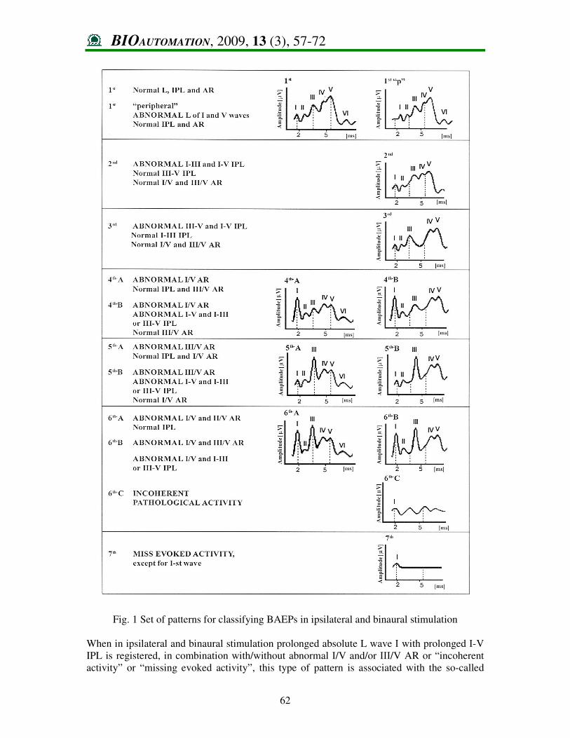
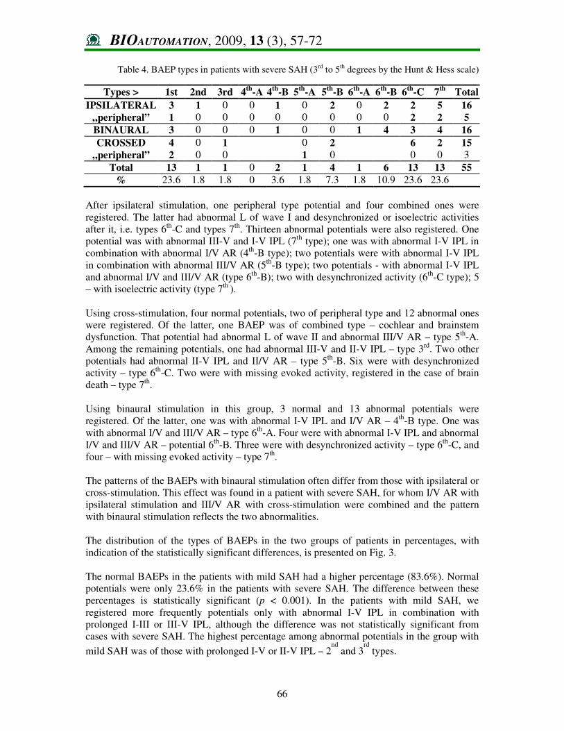
Unlike the set for classifying BAEPs in ipsilateral and binaural stimulation, the set of BAEP
types due to cross-stimulation uses wave II instead of wave I. The criteria are IPL II-III, III-
V and II-V, as well as the AR III/V. This set of patterns is convenient for reflecting changes
in the waveform, especially in BAEP obtained by cross-stimulation. Wave I is not recorded as
a positive peak in the contralateral mastoid. Fig. 2 presents a set of patterns for categorizing
BAEPs in cross-stimulation. In the case of prolonged L of wave ІІ and normal remaining
parameters, there is a “peripheral type” of BAEPs with cross-stimulation. In the case of
prolonged absolute L of wave ІІ and prolonged ІІ-V IPL in combination with or without
abnormal ІІІ/V AR, the pattern is considered as “combined type" of abnormal BAEP.
Results Of all 29 studies conducted with the three types of stimulation on “mild SAH” patients, 16
had bilaterally normal BAEP (classified as 1st type). Six studies registered unilaterally normal
potentials, while in 7 there were bilaterally abnormal potentials.
With “severe SAH” patients, 2 of the 14 studies exhibited bilaterally normal potentials; the
rest had bilaterally abnormal potentials with some type of stimulation.
The distribution of the types of BAEPs in the patients with mild SAH is presented in Table 3.
Table 3. BAEP types in patients with mild SAH (1 and 2 degrees by the Hunt & Hess scale)
With ispilateral stimulation, 46 normal potentials were registered. Normal potentials were
found in patients with clinical signs of SAH and one-side oculomotor nerve damage. Two
potentials were of peripheral type (1st“p”
), registered in the patients with evidence of unilateral
peripheral otoneurological syndrome. They had prolonged L of waves І and V on the side of
the peripheral damage. One of these 1st “p”
types was found unilaterally in the patient with
subclavia style syndrome and SAH in the vertebro-basilar system. Another potential was
recorded in a patient with SAH and vertigo.
The eight abnormal potentials were registered in the patients with clinical evidence of mild
manifestation of brainstem dysfunction. Two of them were only with abnormal IPL, i.e., they
are of 2nd
and 3rd
type. Four BAEPs were with abnormal AR, but with normal IPL. In three of
them, only І/V AR was abnormal (4th
-A type). Abnormal BAEPs classified as 4th-A type was
found only after ipsilateral stimulation on left ear in the patients with mild SAH and clinical
evidence of unilateral lesion of the left oculomotor nerve. It suggested a mild disturbance of
either nuclear or intrabrainstem part of oculomotor nerve. One unilateral abnormal 4th
-A type
Types > 1st 2nd 3rd 4th
-A 4th
-B 5th
-A 5th
-B 6th
-A 6th
-B 6th
-C 7th
Total
IPSILATERAL 46 1 1 3 0 1 0 0 0 1 0 53
”peripheral” 2 0 0 0 1 0 0 0 0 0 0 3
BINAURAL 27 2 4 1 0 0 0 1 0 0 0 35
CROSSED 46 1 4 2 0 1 0 54
”peripheral” 1 0 0 0 0 0 0 1
Total 122 4 9 4 1 3 0 1 0 2 0 146
% 83.6 2.7 6.16 2.7 0.7 2.1 0 0.7 0 1.4 0
BIOAUTOMATION, 2009, 13 (3), 57-72
64
potential was found in the one patient with SAH and vestibular syndrome. Another abnormal
potential of 4th
-A type was recorded on the side of development of hemispheric secondary
focal ischemic dysfunction. A patient with 2 degree by Hunt and Hess had a potential with
abnormally prolonged І-st wave L and І-V wave IPL, as well as abnormal І/V AR, classified
as 4th
-B“p”. One potential with abnormal ІІІ/V (5
th-A type) was found in one patient with mild
SAH and complaint about dizziness.
One BAEP was with severe configurational changes, but it was registered in a patient with
pre-morbid reduction of hearing in one ear: 6th
-В type. In one patient, with proved combined
otoneurological syndrome, a “combined type” potential was registered, whereby L of wave І
is prolonged: prolonged IPL І-V and abnormal ІІІ/V AR, i.e., “type 5th
-B”.
In the case of cross-stimulation, the normal potentials were 46 and one was of “peripheral
type”. The latter was registered in a patient with peripheral otoneurological syndrome. BAEP
was with prolonged L of wave ІІ in the case of normal IPL and AR - type 1st “p”
.
The eight abnormal potentials with this stimulation were in the same patients who also had
the abnormal potentials with the ipsilateral one. However, not all of them had potential
patterns as with ipsilateral stimulation. Five of them had abnormal ІІ-V IPL in combination
with abnormal ІІ-ІІІ or ІІІ-V IPL: 2nd
and 3rd
types. Two potentials were abnormal only
according to AR ІІІ/V: type 5th
-A. One was with configurational changes due to stimulated
ear with pre-morbid strong reduction of hearing: 6th
-C type.
With binaural stimulation, 27 BAEPs were normal and 8 were abnormal. Six of the abnormal
potentials had abnormal І-V and ІІ-V IPL, with normal AR (2nd
and 3rd
types). Of the
remaining two, one was with abnormal І/V AR - type 4th
-A. The other one was with abnormal
І/V and ІІІ/V AR with normal IPL - type 6th
-A. This was the result of summing of the
abnormal І/V AR (4th
-А type) with ipsilateral stimulation with abnormal ІІІ/V AR (5th
-А
type) with cross-stimulation.
The patterns of the BAEPs with binaural stimulation often are the same types as one of
unilateral stimulation and differ from the other one. In two patients with SAH, one with
oculomotor nerve damage and another with vestibular syndrome, the potentials were 1st type,
the same as for cross-stimulation. In one patient with SAH without brainstem dysfunction the
potentials were 3rd
types for binaural stimulation, the same as for cross-stimulation.
In some patients the patterns of the BAEPs with binaural stimulation differ from the potentials
recorded for ipsilateral and cross- stimulation. In one patient with mild SAH the potentials
were 6th
-A types for binaural stimulation, but for ipsilateral and cross-stimulation the
potentials were 5th
-A types. In another patient with mild SAH without brainstem dysfunction
the potentials for binaural stimulation were 3rd
type, but potentials for ipsi- and cross-
stimulation were normal on right hemisphere side. On the other hemispherical side in same
patient the potentials were: 1st“p”
type for ipsilateral stimulation; 2nd
type for cross-stimulation
and normal (1st type) for binaural stimulation.
BIOAUTOMATION, 2009, 13 (3), 57-72
65
Fig. 2 Set of patterns for classifying BAEPs in cross-stimulation
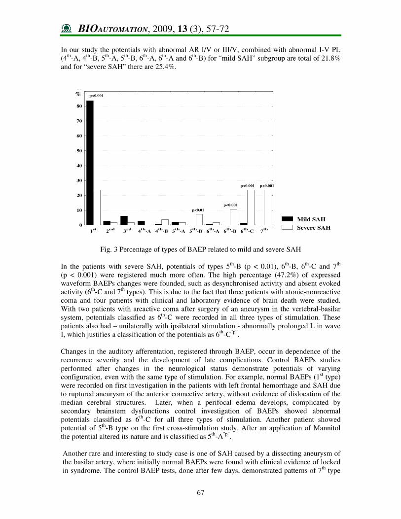
The distribution of the BAEPs types in the patients from the severe SAH group is presented in
Table 4. Only ten BAEPs were normal, three BAEPs were of peripheral type, and the
remaining 42 BAEPs were abnormal.
BIOAUTOMATION, 2009, 13 (3), 57-72
66
Table 4. BAEP types in patients with severe SAH (3rd to 5th degrees by the Hunt & Hess scale)
After ipsilateral stimulation, one peripheral type potential and four combined ones were
registered. The latter had abnormal L of wave I and desynchronized or isoelectric activities
after it, i.e. types 6th
-C and types 7th
. Thirteen abnormal potentials were also registered. One
potential was with abnormal ІІІ-V and І-V IPL (7th
type); one was with abnormal І-V IPL in
combination with abnormal І/V AR (4th
-B type); two potentials were with abnormal І-V IPL
in combination with abnormal ІІІ/V AR (5th
-B type); two potentials - with abnormal І-V IPL
and abnormal І/V and ІІІ/V AR (type 6th
-B); two with desynchronized activity (6th
-C type); 5
– with isoelectric activity (type 7th
).
Using cross-stimulation, four normal potentials, two of peripheral type and 12 abnormal ones
were registered. Of the latter, one BAEP was of combined type – cochlear and brainstem
dysfunction. That potential had abnormal L of wave II and abnormal III/V AR – type 5th
-A.
Among the remaining potentials, one had abnormal ІІІ-V and ІІ-V IPL – type 3rd
. Two other
potentials had abnormal ІІ-V IPL and ІІ/V AR – type 5th
-B. Six were with desynchronized
activity – type 6th
-C. Two were with missing evoked activity, registered in the case of brain
death – type 7th
.
Using binaural stimulation in this group, 3 normal and 13 abnormal potentials were
registered. Of the latter, one was with abnormal І-V IPL and І/V AR – 4th
-В type. One was
with abnormal І/V and ІІІ/V AR – type 6th
-А. Four were with abnormal І-V IPL and abnormal
І/V and ІІІ/V AR – potential 6th
-В. Three were with desynchronized activity – type 6th
-C, and
four – with missing evoked activity – type 7th
.
The patterns of the BAEPs with binaural stimulation often differ from those with ipsilateral or
cross-stimulation. This effect was found in a patient with severe SAH, for whom І/V AR with
ipsilateral stimulation and ІІІ/V AR with cross-stimulation were combined and the pattern
with binaural stimulation reflects the two abnormalities.
The distribution of the types of BAEPs in the two groups of patients in percentages, with
indication of the statistically significant differences, is presented on Fig. 3.
The normal BAEPs in the patients with mild SAH had a higher percentage (83.6%). Normal
potentials were only 23.6% in the patients with severe SAH. The difference between these
percentages is statistically significant (p < 0.001). In the patients with mild SAH, we
registered more frequently potentials only with abnormal І-V IPL in combination with
prolonged І-ІІІ or ІІІ-V IPL, although the difference was not statistically significant from
cases with severe SAH. The highest percentage among abnormal potentials in the group with
mild SAH was of those with prolonged I-V or II-V IPL – 2nd
and 3rd
types.
Types > 1st 2nd 3rd 4th
-A 4th
-B 5th
-A 5th
-B 6th
-A 6th
-B 6th
-C 7th
Total
IPSILATERAL 3 1 0 0 1 0 2 0 2 2 5 16
„peripheral” 1 0 0 0 0 0 0 0 0 2 2 5
BINAURAL 3 0 0 0 1 0 0 1 4 3 4 16
CROSSED 4 0 1 0 2 6 2 15
„peripheral” 2 0 0 1 0 0 0 3
Total 13 1 1 0 2 1 4 1 6 13 13 55
% 23.6 1.8 1.8 0 3.6 1.8 7.3 1.8 10.9 23.6 23.6
BIOAUTOMATION, 2009, 13 (3), 57-72
67
In our study the potentials with abnormal AR I/V or III/V, combined with abnormal I-V PL
(4th
-A, 4th
-B, 5th
-A, 5th
-B, 6th
-A, 6th
-A and 6th
-B) for “mild SAH” subgroup are total of 21.8%
and for “severe SAH” there are 25.4%.
Fig. 3 Percentage of types of BAEP related to mild and severe SAH
In the patients with severe SAH, potentials of types 5th
-B (p < 0.01), 6th
-B, 6th
-C and 7th
(p < 0.001) were registered much more often. The high percentage (47.2%) of expressed
waveform BAEPs changes were founded, such as desynchronised activity and absent evoked
activity (6th
-C and 7th
types). This is due to the fact that three patients with atonic-nonreactive
coma and four patients with clinical and laboratory evidence of brain death were studied.
With two patients with areactive coma after surgery of an aneurysm in the vertebral-basilar
system, potentials classified as 6th
-C were recorded in all three types of stimulation. These
patients also had – unilaterally with ipsilateral stimulation - abnormally prolonged L in wave
І, which justifies a classification of the potentials as 6th
-C“p”
.
Changes in the auditory afferentation, registered through BAEP, occur in dependence of the
recurrence severity and the development of late complications. Control BAEPs studies
performed after changes in the neurological status demonstrate potentials of varying
configuration, even with the same type of stimulation. For example, normal BAEPs (1st type)
were recorded on first investigation in the patients with left frontal hemorrhage and SAH due
to ruptured aneurysm of the anterior connective artery, without evidence of dislocation of the
median cerebral structures. Later, when a perifocal edema develops, complicated by
secondary brainstem dysfunctions control investigation of BAEPs showed abnormal
potentials classified as 6th
-C for all three types of stimulation. Another patient showed
potential of 5th
-B type on the first cross-stimulation study. After an application of Маnnitol
the potential altered its nature and is classified as 5th
-A"p"
.
Another rare and interesting to study case is one of SAH caused by a dissecting aneurysm of
the basilar artery, where initially normal BAEPs were found with clinical evidence of locked
in syndrome. The control BAEP tests, done after few days, demonstrated patterns of 7th
type
Mild SAH
Severe SAH0
10
20
30
40
50
60
70
80
90
1st
2nd
3rd
4th
-A 4th
-B 5th
-A 5th
-B 6th
-A 6th
-B 6th
-C 7th
% p<0.001
p<0.01
p<0.001
p<0.001 p<0.001
BIOAUTOMATION, 2009, 13 (3), 57-72
68
for all three types of stimulations, which corresponded to the clinical symptoms of brain
death.
Discussion This study claims that the use of classified BAEPs patterns in patients with different degrees
of SAH enhances the comparison of potentials. Comparison is necessary between potentials
of the same patient in the course of the disease as well as between potentials of patients with
different degrees of SAH. The two pattern sets facilitate the investigation of the dynamics of
the potentials, from normal to brain death, that occur in the various types of stimulation. The
Hunt and Hess scale is a convenient method of discerning patients with different degrees of
SAH. Such an in-depth study of BAEP in patients with SAH, and their comparison with the
Scale of Hunt and Hess, can only be found in [9] and [10]. Unlike their work, we made use of
binaural stimulation in addition to ipsilateral and cross types of stimulation when looking for
changes in BAEPs patterns specific to particular degree of SAH severity. The patterns of the
BAEPs with binaural stimulation often differ from those with ipsilateral or cross-stimulation.
This is due to the summing of abnormality of differing gravity with the two types of
monaural stimulation [11]. Significant deviations in potential patterns with monaural
stimulation remain also with binaural stimulation. We registered a case of retaining an
abnormal potential component present in one of the two monaural stimulations in the
configuration of the binaural stimulation. Another case showed however that a component of
abnormal type but close to the normal pattern (IPL or AR) existing in one of the monaural
stimulations, is not found in the binaural stimulation. We consider this to be the reason why
Chiappa [2] generalises this masking phenomenon and does not recommend the application
of binaural stimulation alone.
A third case of special combination was found in a mild-SAH patient: the monaural
stimulation potentials were normal but close to the upper limit, while the binaural stimulation
potentials were abnormal. This is due to summing patterns close to normal with monaural
stimulation, that are abnormal with binaural stimulation. We think that the presence of
abnormality in binaural stimulation, together with abnormality in one of the monaural
stimulations, is a manifestation of decompensation in hearing afferentation. Normal
potentials in binaural stimulation together with abnormality in one of the monaural
stimulations, is characteristic of compensation. Therefore, monaural stimulations may be
used to obtain a precise estimation of the status of cross and direct auditory brainstem
pathways, and binaural stimulation serves to investigate the status of compensatory
mechanisms. Intact compensatory mechanisms are featured by abnormal potentials but
missing clinical signs for brainstem impairment. In one of the studied cases, for example,
abnormal I/V AR only with ipsilateral stimulation on the side of the lesion in patients with
SAH and hemispheric secondary focal ischemic dysfunction indicates affected brainstem
auditory structures with missing clinical manifestation of this.
Some authors [3, 9, 10] assume that the reason for the abnormal BAEP is the increased
intracranial pressure as a result of SAH. Wada et al. [23] believe that brainstem secondary
ischemia, resulting from increased intracranial pressure, affects the III/V AR. Although the
experimental studies [22] had not detected any influence of the increasing intracranial
pressure on the BAEP components. In our opinion, the intracranial pressure has an effect on
the BAEP patterns, due to the fact that abnormal potentials are found not only in patients
with brainstem clinical signs.
BIOAUTOMATION, 2009, 13 (3), 57-72
69
Although the BAEPs could be initially normal, there were a high percentage of potentials
combining abnormal IPL and AR from the control study, as a result of an acute development
of cerebral edema and intracranial hypertensive syndrome and exchanged cerebral perfusion
leading to brainstem dysfunction. In contrast, Lebedev et al. [18] have found mainly
prolonged І-V IPL in such a group of patients. Hashimoto et al. [8] describe such a
prolongation of the “brainstem conduction time” intraoperatively. Similarly, we used BAEP
to prove whether auditory brainstem structures were affected after surgery of an aneurysm in
the vertebral-basilar system. Prolonged I-V IPL was not found in our patients subjected to
postoperative tests. Our data had poor prognostic value for the effect of the surgical
intervention.
The registered 1st“p”
patterns were characteristic of peripheral otoneurological syndrome,
which is probably due to spasm of the auditory artery of the affected inner ear.
In our SAH patients there were some potentials demonstrating combined abnormality at the
peripheral and the brainstem level. In these cases, combined-type potentials were registered,
such as 4th
-B“p”
, 5th
-A“p”
, 6th
-C“p”
, 7th“p”
. We regard the reason for this to be, apart from the
cochlea, a probable spasm both of the auditory artery and of the circumference arteries of the
basilar artery in the brainstem, which impaired the blood supply to the tegmentum.
The use of a set of patterns for categorized BAEP with cross-stimulation improves the
analysis of information about the involvement of crossed pathways in patients with
quantitative consciousness disturbances. One of the criteria for this disturbance is amplitude
abnormality of AR III/V. While the I-III, III-V and I-V IPL and I/V AR are recommended in
ipsilateral and binaural stimulation in cases with brainstem damage, III/V AR is missing [1].
For this reason, most authors do not use it.
The absence of evoked activity when BAEP were recorded in patients who had developed
atonic and apneic coma is a criterion for brain death. The recordings demonstrate incoherent
noise activity with low amplitude and absence of I wave bilaterally in case of ipsilateral and
binaural stimulation. Many studies leading to similar results have been devoted to this
problem. For example, Goldie et al. [4] and Machado et al. [19] have not recorded any wave
in more than 70% of the patients investigated. According to [14], the absence of I wave in
patients with verified brain death is due to complete destruction of the labyrinth, as a result of
interrupted perfusion of the inner ear. Our experience has shown that BAEP analysis is
among the most informative methods for objective evaluation of the brainstem dysfunction -
a method which continues to demonstrate its advantages over the routine EEG-investigations,
especially for SAH patients.
Conclusion We have shown that normal BAEPs are registered in the patients with mild SAH, whereas the
abnormal types are relatively uniformly represented. In the group of patients studied, there is
a frequent occurrence of abnormal ІІІ/V AR, especially with cross-stimulation. In the patients
with SAH we demonstrated that the indices II-III and II-V IPL, and of the III/V AR, are
connected with violation of the crossed auditory pathways from the auditory nuclei to colliculi
inferiores of tectum mesencephali in brainstem lesions. The existence of a peripheral type of
BAEP suggests auditory disorders caused by ischemisation of the inner ear or by direct
damage to the SAH part of the auditory nerve. In the patients with combined peripheral-
brainstem damage, it is attributed to the abnormal latency of the I wave, in combination with
or without abnormal I-V IPL and I/V and III/V AR.
BIOAUTOMATION, 2009, 13 (3), 57-72
70
On the basis of the results obtained and the analysis of the data of the clinical and paraclinical
investigations, compared to the BAEP results in our experiments, we believe that BAEP
analysis should be applied to patients with brainstem lesions, irrespective of its severity and
volume, caused by vascular incidents of varying etiology. The analysis of BAEPs according
to the side of the stimulation - ipsilateral or contralateral - shows that the study of crossed and
non-crossed auditory pathways is informative to the same degree. A set of patterns of cross-
stimulation fills a gap in the analysis of the configurational disturbances, which is often
omitted or avoided by most authors.
References 1. American Electroencephalographic Society Guidelines for Clinical Evoked Potential
Studies “Recommended Standards for short-latency auditory evoked potentials” (1984), 1
(1), 32-40.
2. Chiappa K. H. (1989). Principles of Evoked Potentials, In: Evoked Potentials in Clinical
Medicine, 6th
ed. New York: Raven Press, 1-25.
3. Docherty T. B., A. G. Herbout, E. M. Sedgwick (1987). Brainstem Auditory Evoked
Potential Abnormalities in Mielomeningocoele in the Older child, Journal Neurol.
Neurosurg. Psychiatr., 50, 1318-1322.
4. Goldie W. D, K. H. Chiappa, R. R. Young, E. G. Brooks (1981). Brainstem Auditory
Evoked Potentials and Short-latency Somatosensory Evoked Potentials in Brain Death,
Neurology, 31, 248-256.
5. Haralanov L, M. Matveev (1991). Mathematic Statistical Method for Classification of
Normal and Pathological Auditory Brainstem Evoked Potentials (BAEP), Proceedings of
the 14th
Annual Meeting of the Societies of EEG, EMG and Clinical Neurophysiology of
the East European countries, Sofia, 22-24.
6. Haralanov L., M. Matveev, E. Mermeklieva (1996). System for Analysis of Brainstem
Auditory Evoked Potentials (BAEP) in Patients with Acute Disorders if brain blood
Circulation (ADBBC), Electroenceph Clin Neurophysiol, 99(4), 351.
7. Haralanov L., M. Matveev, N. Mudrov, T. Trifonov, E. Mermeklieva (1997). Method for
Analysis of Auditory Brainstem Evoked Potential in Patients with Acute Disturbances of
brain blood Circulation, Neurologia Balkanica, 3-4, 18-19.
8. Hashimoto I., Y. Ishiyata, G. Totzuka, H. Mizutani (1980). Monitoring Brainstem
Function During Posterior Fossa Surgery with Brainstem Auditory Evoked Potentials, In:
Berber C, (Ed.), Evoked Potentials, Baltimore, University Park Press, 377-390.
9. Haupt W. F., C. Hojer, G. Pawlik (2005). Prognostic Value of Evoked Potentials and
Clinical Grading in Primary SAH, Acta Neurochirurgica, 137, 146-150.
10. Haupt W. F., G. Pawlik (1998). Contribution of Initial Median-nerve Somatosensory
Evoked Potentials and Brainstem Auditory Evoked Potentials to Prediction of Clinical
Outcome in Cerebrovascular Critical Care Patients: A Statistical Evaluation, J. Clin
Neurophysiol, 5, 154-158.
11. Hechinashvili S. X., Z. S. Kevanishvili (1985). Human Auditory Evoked Potentials,
Tbilisi, Sobchota Sakartvelo.
12. Hunt W. E., R. M. Hess (1968). Surgical Risk as Related to Time of Intervention in the
Repair of Intracranial Aneurysms, Journal of Neurosurgery, 28(1), 14-20.
13. Johkura K., S. Matsumoto, O. Hasegawa, Y. Kuroiwa (1998). Defective Auditory
Recognition after Small Haemorrhage in the Inferior Colliculi, J. Neurol. Sci., 161, 91-96.
14. Kaga K., K. Uebo, H. Sakata, J. Suzuki (1993). Auditory and Vestibular Pathology in
Brainstem Death Revealed by Auditory Brainstem Response, Acta Oto-Laryngologica
(Suppl), 503, 99-103.
BIOAUTOMATION, 2009, 13 (3), 57-72
71
15. Kalkman C. J. (1994). Monitoring the Central Nervous System, Anesthesiology Clinics of
North America; 12, 173-196.
16. Karadimov D., L. Haralanov, E. Mermeklieva (1997). Anaestesiological Method using
during Investigation of Neurological Patients with Brainstem Auditory Evoked Potentials,
Anesthesiology and Intensive Care, ХХІV(4), 29-31.
17. Klein A. J., D. C. Teas (1978). Acoustically Dependent Latency Shifts of Brainstem
Auditory Evoked Responses (Wave V) in Man, J. Acoust. Soc. Amer., 63, 1887-1895.
18. Lebedev V., L. Sumskii, M. Miatchin, V Krjlov, A Zakharov (1992). The Assisment of
Brain-stem Function in the Acute Period of Aneurysm Rupture, Zh Vopr Neirokhir
Burdenko, 6, 5-7.
19. Machado C., P. Valdes, J. Garcia-Tigera, T. Virues, R. Biscay, J. Miranda, P. Coutin, J.
Roman, O. Garcia (1991). Brain-stem Auditory Evoked Potentials and Brain Death,
Electroenceph Clin Neurophysiol, 80, 392-398.
20. Meoller A. R. (1992). Use of Brainstem Auditory Evoked Potentials in Intraoperative
Neurophysiologic Monitoring, In: Kartush J. M., K. R. Bouchard (editors),
Neuromonitoring in Otology and Head and Neck Surgery, New York, Raven Press, 199-
214.
21. Picton T. W. (1990). Auditory Evoked Potentials, In: Daly DD, TA Pedley (editors).
Current practice of clinical electroencephalography, New York, Raven Press, 626-678.
22. Sutton L. N., B. K. Cho, J. Jaggi, P. M. Joseph, D. A. Bruce (1986). Effects of
Hydrocephalus and Increased Intracranial Pressure on Auditory and Somatosensory
Evoked Responses, Neurosurgery, 18, 756-761.
23. Thornton C. (1991). Evoked Potentials in Anaesthesia, Europ. J. Anaest., 8, 89-107.
24. Wada S.-I., S. Matsuoka, E.-I. Urazaki, C. Yadomic (1988). Quantitative Analysis of
Reversible Dysfunction of Brain-stem Midline Structures Caused by Disturbance of
Basilar Artery Blood Flow with the Auditory Brainstem Responses, Electroenceph Clin
Neurophysiol, 69, 148-159.
Assoc. Prof. Lyubomir Haralanov, M.D., Ph.D.
E-mail: [email protected]
Assoc. Prof. Dr. Lyubomir Haralanov, Specialist of Neurology, is a
Head of Clinic of Neurology, National Heart Hospital. He is a Member
of board of the Neurological Society of the Bulgaria, a Member of the
Science and Curricula Council, Institute of Neurology, Psychiatry and
Neurosurgery, a Member of Pan-European Society of Neurology. He has
more than 100 papers published in scientific journals and conference
proceedings.
BIOAUTOMATION, 2009, 13 (3), 57-72
72
Prof. Mikhail Matveev, Ph.D. E-mail: [email protected]
Professor of Biomedical Engineering Mikhail Matveev is Director (Head)
of Centre of Biomedical Engineering, Bulgarian Academy of Sciences. He
holds the degrees of Master of Science in Nuclear Physics, Computing
Mathematics and Ph.D. in area of Medicine.
His research interests are in the medical microprocessor systems and
devices; biomedical data and signal processing and analysis; basic sciences
in cardiology; pattern recognition; medical decision support systems; risk
assessment and medical decision making; classification procedures in
medicine. His publications are in the fields of biomedical engineering in cardio-vascular
diseases and electrocardiology, neurology, anaesthesiology, resuscitation and intensive care,
emergency medicine, pulmonary diseases, pediatrics and neonatology, self-controlling
biological systems, methods for optimization of the diagnostic and therapeutic process,
autonomic cardiac control, information technologies in medicine etc.
Prof. Matveev is a member of several professional societies: European Society for Computing
and Technology in Anaesthesia and Intensive Care, International Federation of Medical and
Biological Engineering, European Society of Cardiology, European Hearth Rhythm
Association, European Association for Cardiovascular Prevention and Rehabilitation,
American Heart Association etc.
He enjoys reading, hunting, and travelling.
Elena Atanassova Mermeklieva-Haralanova, M.D. E-mail: [email protected]
Dr. Elena Mermeklieva-Haralanova is an Ophthalmologist in
Alexandrovska University Hospital, Department of Ophthalmology,
Medical University – Sofia.
Her professional interests are oculoplastics, cornea, cataract, vitreoretinal
surgery, plastic and reconstructive surgery of eyelids, orbital surgery and
surgery of dacryolacrimal system, laser treatment, electrophysiology,
neuroophthalmology. She is a member of Bulgarian Medical Association,
Bulgarian Association of Ophthalmology, Union of Bulgarian Ophthalmologists, National
Association of Ophthalmologists from Outpatient Care and Glaucoma Association.


![Road Traffic Noise and its Effect on Brain Stem Auditory ... · [2]. Among all objective methods of hearing evaluation, brainstem auditory evoked potential is considered the most](https://img.pdfslide.us/doc/110x75/5f5d38a1dfd4a155386e996d/road-traffic-noise-and-its-effect-on-brain-stem-auditory-2-among-all-objective.jpg)