Embed Size (px)
Citation preview
P.O. Box 2925 Riyadh – 11461KSATel: +966 1 2520088 ext 40151Fax: +966 1 2520718Email: [email protected]: www.sha.org.sa
FULL
LEN
GTH
ART
ICLE
Disclosure: Authors have nothing to disclose with regard to commercialsupport.
Received 20 July 2015; revised 3 September 2015; accepted 16 September2015.Available online 26 September 2015
⇑ Corresponding author at: Department of Cardiology, Cairo Univer-sity, Cairo 002020, Egypt.
E-mail address: [email protected] (N. Hassanin).Evaluation of pulmonary artery pressureand resistance by pulsed Dopplerechocardiography in patients with end-stagerenal disease on dialysis therapy
1016–7315 � 2015 The Authors. Production and hosting by Elsevier B.V. on behalf of King Saud University. This is an open access article under th
NC-ND license (http://creativecommons.org/licenses/by-nc-nd/4.0/).
Peer review under responsibility of King Saud University.
URL: www.ksu.edu.sa
http://dx.doi.org/10.1016/j.jsha.2015.09.002Production and hosting by Elsevier
Noha Hassanin a,⇑, Alkhateeb Alkemary b
aDepartment of Cardiology, Cairo University, CairobDepartment of Internal Medicine and Nephrology, Cairo University, Cairo
a,b Egypt
Background: Pulmonary hypertension (PH) is one of the most important comorbidities in patients undergoinghemodialysis (HD). The goal of the present work is to determine the possible etiologic factors for its occurrence.Methods: The prevalence of PH was estimated by Doppler echocardiography in a cohort of 100 patients aged
49.3 � 13.9 years on regular HD. Mean pulmonary artery pressure was estimated from pulmonary acceleration timeby Mahan’s regression equation. Pulmonary vascular resistance and pulmonary capillary wedge pressure were cal-culated. We focused on the effect of HD on left and right ventricle diastolic and systolic function. Right ventriclesystolic function was assessed by tricuspid annular systolic excursion and pulsed Doppler myocardial performanceindex. Since impaired endothelial function was postulated as an underlying cause of PH, we studied the effects ofHD on brachial artery endothelial function.Results: The current study found that pulmonary hypertension was prevalent in 70% of patients on dialysis. Left
atrium diameter, left ventricle mass indexed to body surface area, and mitral E/E0 were increased in the dialysisgroup (4.4 � 0.2 cm, 126.5 � 24.6 g/m2, and 16.9 � 4.4, respectively, p < 0.001 for all). Pulmonary artery systolic pres-sure was positively correlated to duration of dialysis and negatively correlated to glomerular filtration rate(p < 0.001 and r = �0.991). Pulmonary vascular resistance was significantly increased in dialysis patients (1.9 � 0.2Wood units vs. 1.2 Wood units in controls, p < 0.001). Endothelial dysfunction, defined as brachial artery flow medi-ated dilatation <6%, was found in 46% of dialysis group.Conclusion: Increased pulmonary artery systolic pressure in the HD population could be attributed to left atrium
dilatation and left ventricle diastolic dysfunction. Pulmonary vascular resistance was significantly increased in dial-ysis group. This might be explained by impaired endothelial nitric oxide synthesis that not only caused systemicvasoconstriction but also affected the pulmonary vasculature.
� 2015 The Authors. Production and hosting by Elsevier B.V. on behalf of King Saud University. This is an openaccess article under the CC BY-NC-ND license (http://creativecommons.org/licenses/by-nc-nd/4.0/).
Keywords: Hemodialysis, End stage renal disease, Pulmonary artery pressure, Pulmonary vascular resistance,Pulsed Doppler echocardiography
e CCBY-
Abbreviations
PH pulmonary hypertensionHD hemodialysisPA pulmonary arteryPAP pulmonary artery pressurePVR pulmonary vascular resistanceTRV tricuspid regurgitant velocityTVI RVOT right ventricular outflow tract time-velocity
integralPCWP pulmonary capillary wedge pressureFMD flow mediated dilatationLV left ventricleGFR glomerular filtration rateMDRD formula Modification of Diet in Renal Disease
formulaWHO FC World Health Organization functional classLVEF left ventricular ejection fractionLVMI left ventricular mass indexEACVI European Association of Cardiovascular ImagingASE American Society of EchocardiographyLA left atriumLAVI left atrium volume indexRV right ventricleTDI tissue Doppler imagingTAPSE tricuspid annular plane systolic excursionMPAP mean pulmonary artery pressureAT pulmonary acceleration timeSPAP pulmonary artery systolic pressureRAP right atrium pressureTTE transthoracic echocardiographyLVH left ventricle hypertrophy
FULL LEN
GTH
ARTIC
LE
102 HASSANIN, ALKEMARYPULSED DOPPLER ECHOCARDIOGRAPHY IN ESRD PATIENTS
J Saudi Heart Assoc2016;28:101–112
Introduction
Pulmonary hypertension (PH) is a complex
hemodynamic alteration that may result fromdisparate causes. In the 2008 classification by theWorld Health Organization and in more recentguidelines by the European Society of Cardiology,for the first time attention was given to PH in dial-ysis patients classified in the fifth category gather-ing various forms of PH with unclear etiology [1].The high prevalence of PHwas attributed to high
cardiac output secondary to the presence ofarteriovenous fistula, anemia, and/or and to leftventricular (LV) disorders [2]. Moreover, sleepapnea, accumulation of endogenous inhibitors ofnitric oxide synthase, insult to pulmonarymicrocir-culation attributable to exposure to dialysismembranes is likely to contribute to the uniquepropensity of dialysis patients to PH [3]. Our aimwas to study the contribution of left and rightventricle dysfunction to pulmonary hypertension.The second aim was to show that simple echocar-diographic equations can be used to assess systolic,mean pulmonary artery pressure and pulmonaryvascular resistance that might help in evaluationand risk stratification of patients on dialysis
MPI myocardial performance indexAVF arteriovenous fistulaTPG transpulmonary pressure gradientRHC right heart catheterizationCKD chronic kidney disease
Patients and methods
Forty healthy controls and 100 adult patientsaged 49.3 � 13.9 years on regular hemodialysis(HD) for at least 12 months (range, 12–80 months)were referred from the Nephrology Department ofCairo University, Cairo, Egypt for echocardiogra-phy. Written consent was given by all the partici-pants. The study protocol was approved by theEthics Committee at Cairo University Hospital.Inclusion criteria were: adults aged P18 years,
stage 4 or 5 chronic kidney disease (CKD) definedas serum creatinineP2.26 mg/dL or glomerular fil-tration rate (GFR)630 mL/min/1.73 m2 assessed byMDRD4-formula [4] on HD, and in World HealthOrganization functional class P II with dyspneaunexplained by other causes. Exclusion criteriawere: pregnancy; LV ejection fraction (EF) <50%;mitral or aortic regurgitation > Grade 2; myocardi-tis; endocarditis; pericarditis; severe chronicobstructive pulmonary disease; lung fibrosis; andknown pulmonary artery hypertension-reducingmedication with prostanoids, endothelin receptorantagonists, or phosphodiesterase-5 inhibitors.Patients were subjected to history taking, phys-
ical examination, and demographic parameters,including age, sex, and body mass index.
Echocardiography studies were performedusing a commercial scanner (iE33; Philips MedicalSystem, Andover, MA, USA) according to therecommendations of the American Society ofEchocardiography [5].
M-mode echocardiographyLV dimensions, wall thickness and functions
were studied. LV mass was calculated using theDevereux formula [6]:
LV massðgÞ ¼ 0:8� 1:04� ½LVIDþ PWTþ IVST�3
� ½LVID�3 þ 0:6 g; ð1Þ
where LVID is the left ventricle internal dimension,PWT is the posterior wall thickness, IVST is the inter-ventricular septal thickness, 1.04 is the specific gravityof the myocardium, and 0.8 is the correction factor.The LV mass index (LVMI, g/m2) was defined as LVmass divided by body surface area (m2). The referenceranges used to define LV hypertrophy (LVH) was LVMI>115 g/m2 and 95 g/m2 for men and women, respectively[5]. We tried refinements in image processing according

Figure 1. Pulmonary acceleration time (a) 70 ms in dialysis patient and (b) 141 ms in a control participant.
FULL
LEN
GTH
ART
ICLE
J Saudi Heart Assoc2016;28:101–112
HASSANIN, ALKEMARY 103PULSED DOPPLER ECHOCARDIOGRAPHY IN ESRD PATIENTS
to the current American Society of EchocardiographyASE/European Association of Cardiovascular Imaging(EACVI) guidelines in order to measure the actualvisualized thickness of the ventricular septum andother chamber dimensions as defined by the actualtissue–blood interface, rather than the distance betweenthe leading edge echoes, which had previously beenrecommended [7].
Left atrium studyLinear dimension of the left atrium (LA) is the
anteroposterior diameter in the parasternallong-axis view using M-mode echocardiography.From the apical four chamber views maximum
left atrium volumes were calculated using thearea–length method according to the guidelinesof the American Society of Echocardiography.Maximum volumes were divided by body surfacearea to calculate the LA volume index (LAVI) [5]
Right ventricle studyOur study was designed to evaluate the effect of
HD on right ventricular (RV) diastolic function andsystolic functions. Distal RV outflow diameter wasstudied using the inner-edge-to-inner-edgemethod according to the recent guidelines justproximal to the pulmonary valve at end-diastole[5]. Tricuspid inflow velocities were recorded usingpulsed wave Doppler with the sample volumeplaced at the tip of the tricuspid valve tips fromthe apical four-chamber view. Peak E and A wave
velocities and E/A ratio were studied. The cut offfor diagnosis of impaired RV diastolic function is<0.8 [5]. The average early diastolic velocity of thetricuspid annulus E0 was obtained by tissueDoppler imaging (TDI) of the septal and lateralsides of the tricuspid annulus. Right-sided E/E0
was used to assess filling pressure of the RV.RV systolic function was assessed by RV
myocardial performance index (Tei index) usingpulsed Doppler method and tricuspid annularplane systolic excursion (TAPSE) using M-modeechocardiography.
Pulsed Doppler echocardiography
Mitral inflow velocities were recorded usingpulsed wave Doppler with the sample volumeplaced at the tip of the mitral valve tips fromthe apical four-chamber view. From the mitralvalve inflow velocity curve, the followingmeasurements were made: peak E, A wave veloc-ities, and E/A ratio. The average early diastolicvelocity of mitral annulus E0 was obtained byTDI of the septal and lateral sides of the mitralannulus. Pulmonary capillary wedge pressure(PCWP) was calculated according to the formula:
1:24� ðE=E0Þ þ 1:9 ½8�: ð2Þ
Color Doppler flow propagation velocityColor Doppler M-mode imaging can be applied
in the apical views to assess the velocity and rate
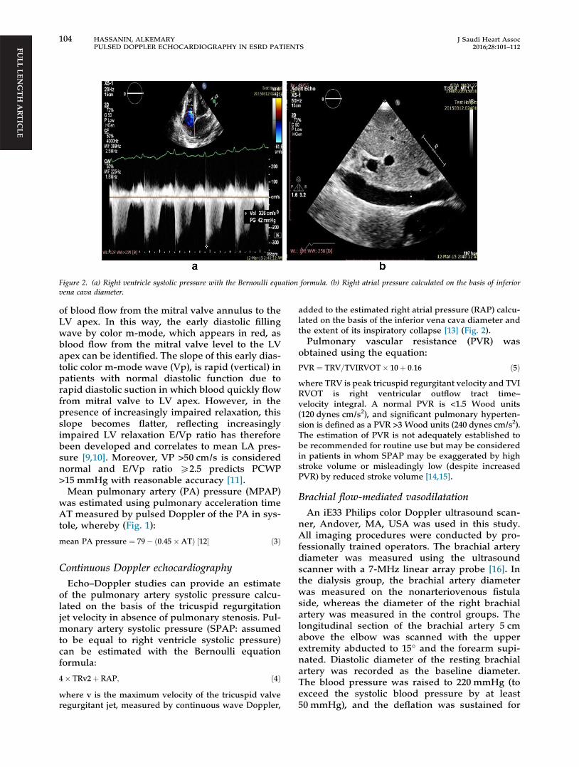
Figure 2. (a) Right ventricle systolic pressure with the Bernoulli equation formula. (b) Right atrial pressure calculated on the basis of inferiorvena cava diameter.
FULL LEN
GTH
ARTIC
LE
104 HASSANIN, ALKEMARYPULSED DOPPLER ECHOCARDIOGRAPHY IN ESRD PATIENTS
J Saudi Heart Assoc2016;28:101–112
of blood flow from the mitral valve annulus to theLV apex. In this way, the early diastolic fillingwave by color m-mode, which appears in red, asblood flow from the mitral valve level to the LVapex can be identified. The slope of this early dias-tolic color m-mode wave (Vp), is rapid (vertical) inpatients with normal diastolic function due torapid diastolic suction in which blood quickly flowfrom mitral valve to LV apex. However, in thepresence of increasingly impaired relaxation, thisslope becomes flatter, reflecting increasinglyimpaired LV relaxation E/Vp ratio has thereforebeen developed and correlates to mean LA pres-sure [9,10]. Moreover, VP >50 cm/s is considerednormal and E/Vp ratio P2.5 predicts PCWP>15 mmHg with reasonable accuracy [11].Mean pulmonary artery (PA) pressure (MPAP)
was estimated using pulmonary acceleration timeAT measured by pulsed Doppler of the PA in sys-tole, whereby (Fig. 1):
mean PA pressure ¼ 79� ð0:45�ATÞ ½12� ð3Þ
Continuous Doppler echocardiography
Echo–Doppler studies can provide an estimateof the pulmonary artery systolic pressure calcu-lated on the basis of the tricuspid regurgitationjet velocity in absence of pulmonary stenosis. Pul-monary artery systolic pressure (SPAP: assumedto be equal to right ventricle systolic pressure)can be estimated with the Bernoulli equationformula:
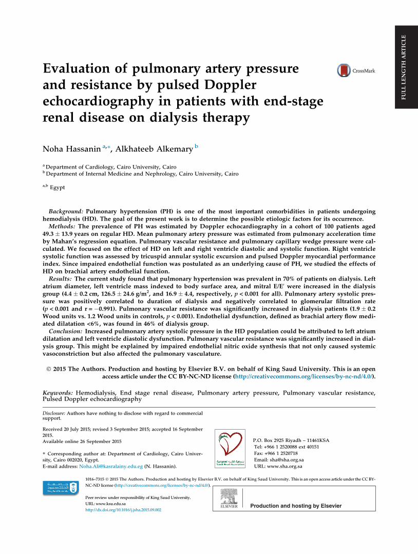
4� TRv2þ RAP; ð4Þwhere v is the maximum velocity of the tricuspid valveregurgitant jet, measured by continuous wave Doppler,
added to the estimated right atrial pressure (RAP) calcu-lated on the basis of the inferior vena cava diameter andthe extent of its inspiratory collapse [13] (Fig. 2).Pulmonary vascular resistance (PVR) was
obtained using the equation:
PVR ¼ TRV=TVIRVOT� 10þ 0:16 ð5Þwhere TRV is peak tricuspid regurgitant velocity and TVIRVOT is right ventricular outflow tract time–velocity integral. A normal PVR is <1.5 Wood units(120 dynes cm/s2), and significant pulmonary hyperten-sion is defined as a PVR >3 Wood units (240 dynes cm/s2).The estimation of PVR is not adequately established tobe recommended for routine use but may be consideredin patients in whom SPAP may be exaggerated by highstroke volume or misleadingly low (despite increasedPVR) by reduced stroke volume [14,15].
Brachial flow-mediated vasodilatationAn iE33 Philips color Doppler ultrasound scan-
ner, Andover, MA, USA was used in this study.All imaging procedures were conducted by pro-fessionally trained operators. The brachial arterydiameter was measured using the ultrasoundscanner with a 7-MHz linear array probe [16]. Inthe dialysis group, the brachial artery diameterwas measured on the nonarteriovenous fistulaside, whereas the diameter of the right brachialartery was measured in the control groups. Thelongitudinal section of the brachial artery 5 cmabove the elbow was scanned with the upperextremity abducted to 15� and the forearm supi-nated. Diastolic diameter of the resting brachialartery was recorded as the baseline diameter.The blood pressure was raised to 220 mmHg (toexceed the systolic blood pressure by at least50 mmHg), and the deflation was sustained for

Table 1. Baseline characteristics.
Clinical parameters Patients on hemodialysis (n = 100) Control (n = 40) p
Age (y) 49.3 � 13.9 49.3 � 14 NSSex 52F/47 M 20F/20 M NSBody mass index (kg/m2) 25.0 � 1.6 26 � 7.2 NSWaist circumference (cm) 114.4 � 14.5 90.6 � 11.7 <0.001Systolic blood pressure (mmHg) 142.9 � 9.5 129.4 � 8.0 <0.001Diastolic blood pressure (mmHg) 90.1 � 7.8 78.4 � 6.9 <0.001Duration of dialysis (mo) 38.7 � 20 — —
Underlying renal disease, n (%)Chronic glomerulonephritis 10 (10)Diabetic nephropathy 33 (33.3)Hypertensive nephrosclerosis 16 (16.7)Polycystic kidney syndrome 13 (13.3)Others 26 (26.7)
Laboratory dataHemoglobin (g/dL) 9.7 � 0.9 14.5 � 0.3 <0.001Serum creatinine (mg/dL) 7.8 � 1.1 0.6 � 0.0 <0.001Serum potassium (mEq/L) 5.4 � 0.2 3.9 � 0.2 <0.001Serum calcium (mg/dL) 8.3 � 0.8 9.3 � 0.1 <0.001Residual GFR (mL/min/1.73 m2) 21.9 � 4.9 96.8 � 2.5 <0.001
Continuous data are expressed as mean � standard deviation.GFR = glomerular filtration rate; NS = not significant.
FULL
LEN
GTH
ART
ICLE
J Saudi Heart Assoc2016;28:101–112
HASSANIN, ALKEMARY 105PULSED DOPPLER ECHOCARDIOGRAPHY IN ESRD PATIENTS
4 minutes. Flow mediated dilatation was calcu-lated as:
ðdiameter after arterial occlusion
� baseline diameterÞ=baseline diameter� 100: ð6ÞNormal endothelial functions were defined as
flow mediated dilatation (FMD) >6%; thus, FMD66% was considered to indicate endothelial dys-function [17].
Statistical analysisData were analyzed using IBM SPSS advanced
statistics version 22 (SPSS Inc., Chicago, IL,USA). Numerical data were expressed as meanand standard deviation or median and range asappropriate. Qualitative data were expressed asfrequency and percentage. Chi-square test wasused to examine the relation between qualitativevariables. For quantitative data, comparisonbetween two groups was done using Student ttest. Pearson product–moment was used to esti-mate correlation between numerical variables.All tests were two-tailed. A p value <0.05 was con-sidered significant.
Results
From September 2014 to June 2015, 100 consecu-tive patients with severe CKD stage 4 or 5 on reg-ular HD and 40 age- and sex-matched controlswere screened by transthoracic echocardiography
for study participation. Baseline characteristics ofthe study population are shown in Table 1.Laboratory investigations showed a significant
increase in serum creatinine and potassium anddecrease in blood hemoglobin, calcium and GFRin the HD group compared with the control group(p < 0.001 for all).Of the 100 patients, 66 patients had E/E0 ratios
>15 (which identifies an abnormally elevated LVfilling pressure) [18]. Using Pearson correlationan elevated E/E0 ratio >15 was negatively corre-lated to GFR (correlation coefficient rvalue = �0.991), positively correlated to SPAPand LA diameter (p < 0.001, for all). ColorM-mode propagation velocity was significantlydecreased in dialysis group (45.3 � 3.2 cm/s vs.66 � 2.3 cm/s in control, p < 0.001), E/Vp ratiowas 1.6 and 1.1, p < 0.001 in dialysis and controlsrespectively. Moreover, E/Vp was P1.5 in 52% ofdialysis population (Fig. 3).Seventy percent of the dialysis group had
LVH. LVMI was positively correlated tosystolic blood pressure (p < 0.001) and negativelycorrelated to hemoglobin level (r = �0.6). Leftatrium diameter and indexed maximum volumeswere significantly increased in dialysis group(4.3 � 0.5 cm and 33.5 � 3.0 mL/m2 vs.3.4 � 0.3 cm and 24.2 � 2.4 mL/m2 in controls(p < 0.001 for both; Fig. 4). Other echocardio-graphic parameters are presented in Table 2.Pulmonary artery systolic and mean pressure
values were significantly elevated in dialysis
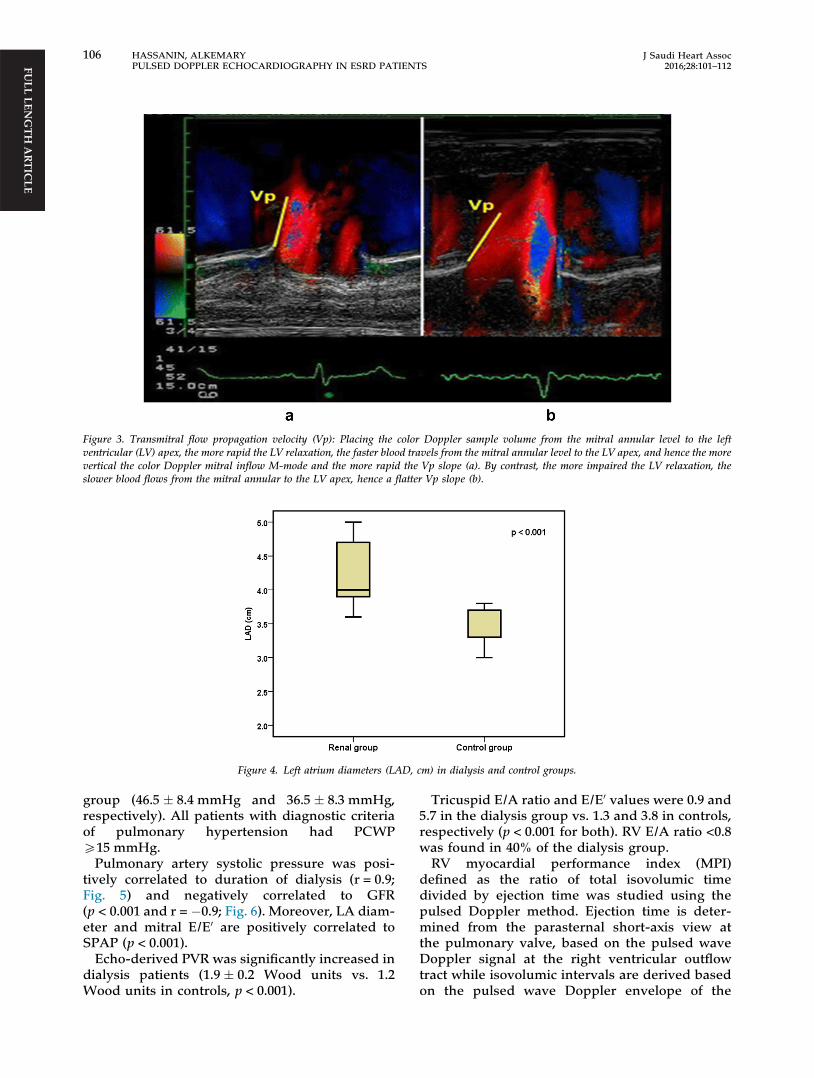
Figure 3. Transmitral flow propagation velocity (Vp): Placing the color Doppler sample volume from the mitral annular level to the leftventricular (LV) apex, the more rapid the LV relaxation, the faster blood travels from the mitral annular level to the LV apex, and hence the morevertical the color Doppler mitral inflow M-mode and the more rapid the Vp slope (a). By contrast, the more impaired the LV relaxation, theslower blood flows from the mitral annular to the LV apex, hence a flatter Vp slope (b).
FULL LEN
GTH
ARTIC
LEFigure 4. Left atrium diameters (LAD, cm) in dialysis and control groups.
106 HASSANIN, ALKEMARYPULSED DOPPLER ECHOCARDIOGRAPHY IN ESRD PATIENTS
J Saudi Heart Assoc2016;28:101–112
group (46.5 � 8.4 mmHg and 36.5 � 8.3 mmHg,respectively). All patients with diagnostic criteriaof pulmonary hypertension had PCWPP15 mmHg.Pulmonary artery systolic pressure was posi-
tively correlated to duration of dialysis (r = 0.9;Fig. 5) and negatively correlated to GFR(p < 0.001 and r = �0.9; Fig. 6). Moreover, LA diam-eter and mitral E/E0 are positively correlated toSPAP (p < 0.001).Echo-derived PVR was significantly increased in
dialysis patients (1.9 � 0.2 Wood units vs. 1.2Wood units in controls, p < 0.001).
Tricuspid E/A ratio and E/E0 values were 0.9 and5.7 in the dialysis group vs. 1.3 and 3.8 in controls,respectively (p < 0.001 for both). RV E/A ratio <0.8was found in 40% of the dialysis group.RV myocardial performance index (MPI)
defined as the ratio of total isovolumic timedivided by ejection time was studied using thepulsed Doppler method. Ejection time is deter-mined from the parasternal short-axis view atthe pulmonary valve, based on the pulsed waveDoppler signal at the right ventricular outflowtract while isovolumic intervals are derived basedon the pulsed wave Doppler envelope of the

Table 2. Echocardiographic parameters of the study groups.
Echocardiographic parameters Dialysis group (n = 100) Control group (n = 40) p
M-mode & 2DLVEDD (cm) 5.1 � 0.5 4.6 � 0.4 <0.001LV EF (%) 54.3 � 1.5 63 � 0.7 <0.001LVMI (g/m2) 126.5 � 24.6 81.8 � 11 <0.001LADs (cm) 4.3 � 0.5 3.4 � 0.3 <0.001LAVI max (mL/m2) 33.5 � 3.0 24.2 � 2.4 <0.001
Mitral inflow velocitiesE, cm/s 69.9 � 19.8 73.9 � 3.6 0.056A, cm/s 72 � 14.6 60.2 � 1.6 <0.001E/A ratio 0.9 � 0.1 1.2 � 0.02 <0.001
LV diastolic filling pressureE/E0 ratio 16.9 5.5 <0.001
Data are presented as mean � standard deviation.A = late diastolic transmitral flow velocity; E = early diastolic transmitral inflow velocity; E/A = ratio of early to late transmitral flow velocity;EDD = end-diastolic diameter; EF = ejection fraction; LADs = left atrial diameter at end-systole; LAVI = left atrium maximum volume indexed to bodysurface area; LVMI = left ventricle mass index; 2D = two dimensional.
FULL
LEN
GTH
ART
ICLE
0
10
20
30
40
50
60
70
0 20 40 60 80 100
PASP
Dura�on of dialysis
Figure 5. Duration of dialysis in months and pulmonary arterysystolic pressure (PASP) in mm Hg among dialysis patients.
J Saudi Heart Assoc2016;28:101–112
HASSANIN, ALKEMARY 107PULSED DOPPLER ECHOCARDIOGRAPHY IN ESRD PATIENTS
tricuspid flow (Fig. 7). The problem with thismethod is that the measurements are taken fromtwo cardiac cycles and care must be taken tochoose beats with similar R–R intervals for a moreaccurate assessment of the MPI. Right ventricleMPI was 0.42 in dialysis group versus 0.2 incontrols (p < 0.001).
0
10
20
30
40
50
60
70
0 5 10 15 2
Figure 6. Glomerular filtration rate (GFR) in ml/min/1.73 m2 and pulmon
TAPSE was <20 mm in 65% of the dialysis group.TAPSE was 23 mm and 19 mm in controls anddialysis groups respectively (p < 0.001; Fig. 8).
Endothelial dysfunction
Endothelial dysfunction, defined as FMD 66%was present in 46 of the 100 patients (46%). Thebaseline, postocclusion diameters of the brachialartery, and FMD were 3.9 � 0.8 mm,4.1 � 0.8 mm, and 5.1%, respectively in dialysispatients versus 3.8 � 0.7 mm, 4.5 � 0.8 mm, and18.4%, respectively, in the control group with nor-mal endothelial function (Fig. 9).
Discussion
PA pressure may be increased by high cardiacoutput resulting from the arteriovenous fistula(AVF) and/or concomitant renal anemia, as wellas from fluid overload [2,19,20]. Critics of this
0 25 30 35
ary artery systolic pressure (PASP) in mm Hg among dialysis group.

Figure 7. Right ventricle myocardial performance index by pulsed Doppler envelope. TCO-ET/ET where TCO is tricuspid valve closure-to-opening time and ET is ejection time.
FULL LEN
GTH
ARTIC
LE
Figure 8. (a) Normal tricuspid annular plane systolic excursion; (b) tricuspid annular plane systolic excursion in a dialysis patient.
108 HASSANIN, ALKEMARYPULSED DOPPLER ECHOCARDIOGRAPHY IN ESRD PATIENTS
J Saudi Heart Assoc2016;28:101–112
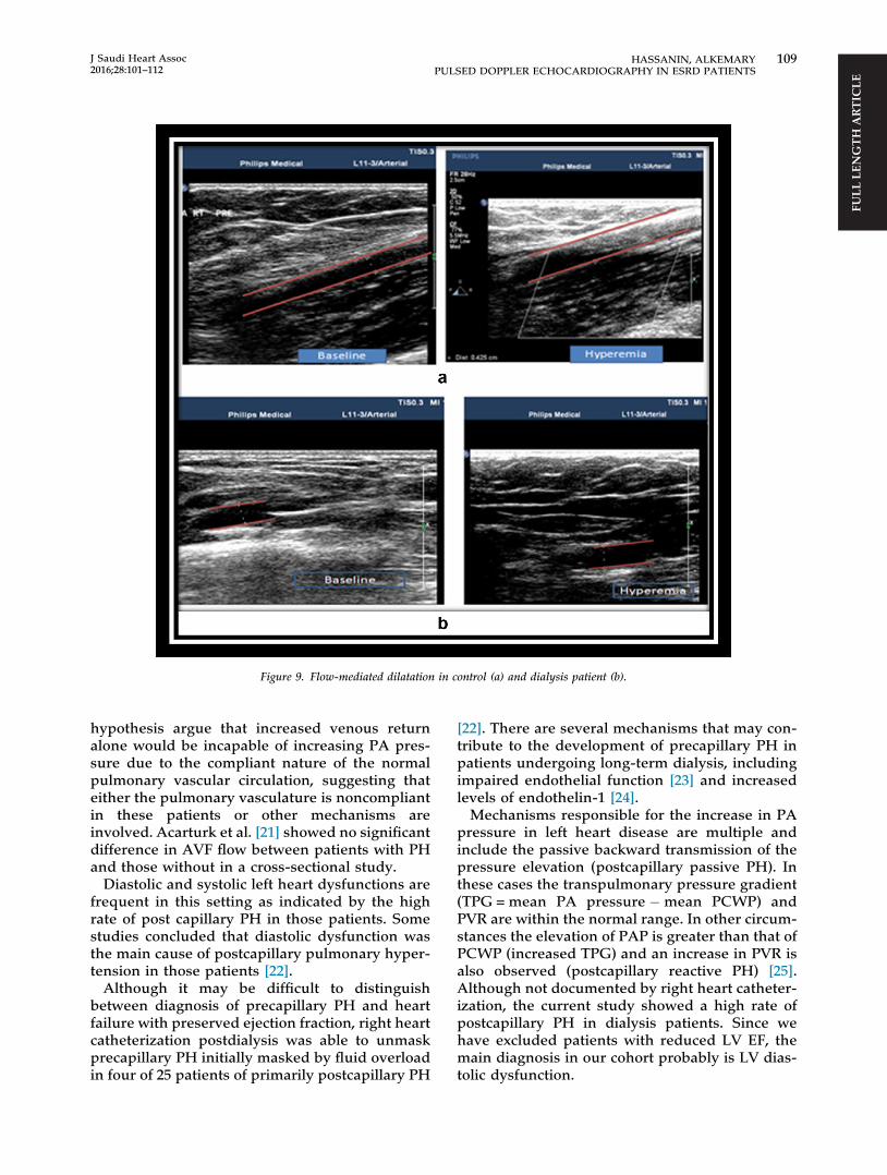
Figure 9. Flow-mediated dilatation in control (a) and dialysis patient (b).
FULL
LEN
GTH
ART
ICLE
J Saudi Heart Assoc2016;28:101–112
HASSANIN, ALKEMARY 109PULSED DOPPLER ECHOCARDIOGRAPHY IN ESRD PATIENTS
hypothesis argue that increased venous returnalone would be incapable of increasing PA pres-sure due to the compliant nature of the normalpulmonary vascular circulation, suggesting thateither the pulmonary vasculature is noncompliantin these patients or other mechanisms areinvolved. Acarturk et al. [21] showed no significantdifference in AVF flow between patients with PHand those without in a cross-sectional study.Diastolic and systolic left heart dysfunctions are
frequent in this setting as indicated by the highrate of post capillary PH in those patients. Somestudies concluded that diastolic dysfunction wasthe main cause of postcapillary pulmonary hyper-tension in those patients [22].Although it may be difficult to distinguish
between diagnosis of precapillary PH and heartfailure with preserved ejection fraction, right heartcatheterization postdialysis was able to unmaskprecapillary PH initially masked by fluid overloadin four of 25 patients of primarily postcapillary PH
[22]. There are several mechanisms that may con-tribute to the development of precapillary PH inpatients undergoing long-term dialysis, includingimpaired endothelial function [23] and increasedlevels of endothelin-1 [24].Mechanisms responsible for the increase in PA
pressure in left heart disease are multiple andinclude the passive backward transmission of thepressure elevation (postcapillary passive PH). Inthese cases the transpulmonary pressure gradient(TPG = mean PA pressure �mean PCWP) andPVR are within the normal range. In other circum-stances the elevation of PAP is greater than that ofPCWP (increased TPG) and an increase in PVR isalso observed (postcapillary reactive PH) [25].Although not documented by right heart catheter-ization, the current study showed a high rate ofpostcapillary PH in dialysis patients. Since wehave excluded patients with reduced LV EF, themain diagnosis in our cohort probably is LV dias-tolic dysfunction.
FULL LEN
GTH
ARTIC
LE
110 HASSANIN, ALKEMARYPULSED DOPPLER ECHOCARDIOGRAPHY IN ESRD PATIENTS
J Saudi Heart Assoc2016;28:101–112
There are notable limitations of this study.PCWP may be a poor indicator of LV end-diastolic pressure; in one study about half of thepatients diagnosed with PH based on PCWP<15 mmHg had elevated LV end-diastolic pres-sures on left heart catheterization [26], In accor-dance with other studies [27], we found that PHis prevalent in 70% of dialysis patients with signif-icant relationship between duration of dialysisand PAP. By contrast, Mousavi and colleagues’[28] study on 62 patients, found that prevalenceof PH was 51.6%, and reduction in serum albuminand anemia were considered as contributingcauses. Moreover, they failed to reach a significantrelationship between duration of dialysis and PApressure [28]. Concordant with others [29] LAdiameter and GFR were independent determi-nants (r = 0.991, p < 0.001 for all) of PA pressure.The current study found that PVR was signifi-cantly increased in the dialysis group. This mightbe explained by impaired endothelial nitric oxidesynthesis and elevated levels of endothelin thatcause systemic vasoconstriction but are also likelyto affect the pulmonary vasculature [30].Methods of assessing vascular endothelial func-
tion can be classified as invasive, noninvasive, ormicrovascular. Sonography is the most widelyused noninvasive method for clinical assessmentof flow-mediated dilatation [16]. It was found thatFMD of brachial artery on the nonarteriovenousfistula side of dialysis patients may closely reflectperipheral vascular endothelial function. More-over, uremic patients have evidence of endothelialdysfunction that is accelerated by HD and theeffects were associated with the duration of HD[31]. The current study showed significantlyreduced FMD in dialysis patients compared tocontrols (5.1% vs. 18.4% in controls, p < 0.001).Impaired endothelial function might be theunderlying cause of elevated PVR in dialysispatients. Although not verified in dialysis patientsit was found that a significant inverse correlationbetween brachial FMD and pulmonary vascularresistance studied using right heart catheteriza-tion in patients with heart failure with preservedEF [32].There are conflicting data concerning the LV EF
in patients on HD. Paneni et al. [33] found that theEF was significantly lower in HD patients than inperitoneal dialysis patients and controls(56.1 � 8.6%, 62.4 � 9.8%, and 68.3 � 5.7%,p = 0.001, respectively). However Said et al. [34]found no difference in the LV EF between theHD group and the control group (62.50 � 11.6%and 64.54 � 15.20%, p = 0.525, respectively). The
current study found that EF was significantlylower in HD (54.3 � 1.5 vs. 63 � 0.7, p < 0.001).The LVH is highly prevalent in CKD and is associ-ated to a clearly unfavorable prognosis. More thantwo-thirds of the patients undergoing dialysiswith LVH die of congestive heart failure or suddendeath [35]. Prevalence of LVH varies from 16% to31% in individuals with CKD and glomerular fil-tration >30 mL/min 60–75% in those starting renalsubstitution therapy and up to 70–90% in patientsundergoing regular dialysis therapy [36]. Concor-dantly, we found that LVH was prevalent in 80%of patients on regular HD.As LA diameter is a strong predictor of diastolic
dysfunction, the findings suggest that, in thisgroup of patients, diastolic dysfunction may be amore relevant mechanism for PH. Recent direc-tives recommend that the adequate quantificationof the LA size be obtained by the estimate of thechamber volume in the two-dimensional modeand not by the traditional measurement of theanteroposterior diameter in the M-mode [5,37].In the ASE/EAE guidelines, the upper normal
limit for max LAVI is 34 mL/m2. This referencevalue is not only derived from population studies,but it is also based upon an estimation of riskrelated to chamber size, and from expert opinion[5]. We found that 40% of dialysis group had LAVIP34 mL/m2. It was found that indexed LA volume>32 mL/m2 provided complementary informationto the traditional clinical and echocardiographicdata, including EF, E/E0 ratio, and LV mass [38].As the LV diastolic function seems to be chroni-cally compromised in most patients undergoingHD, even in those who are asymptomatic, theLA volume can offer the opportunity to identifythe individuals at higher risk to present with heartfailure, atrial arrhythmias, and poor clinical evolu-tion [38].Palecek et al. [39] had previously found that TDI
E0 appears to be superior to Vp in the detection ofmild to moderate LV diastolic dysfunction. How-ever, color M-mode propagation velocity Vp wassignificantly decreased in the dialysis group(45.3 � 3.2 cm/s vs. 66 � 2.3 cm/s in control,p < 0.001), E/Vp ratio was 1.6 � 0.2 and 1.1 in dial-ysis and controls, respectively. Moreover, E/Vpwas P1.5 in 52% of the dialysis population. Thestudy did not compare values before and afterdialysis as the load dependency of propagationvelocity was previously reported by some studiesthat reported that TDI E0 maximal velocity mea-sured at the lateral portion of the mitral annulusappeared to be relatively preload independentand reproducible. In contrast to E0, septal velocity
FULL
LEN
GTH
ART
ICLE
J Saudi Heart Assoc2016;28:101–112
HASSANIN, ALKEMARY 111PULSED DOPPLER ECHOCARDIOGRAPHY IN ESRD PATIENTS
and Vp both appeared sensitive to abrupt preloadreduction [40]. Ie and Zietse [41] reported thatcolor M-mode Doppler is a time-consumingassessment and less sensitive in detecting milderdegrees of diastolic dysfunction in dialysispatients.It was reported that patients on HD have two
peculiar clinical features: anemia and AVF; bothfactors lead to an increased preload on the rightheart chambers [42]. Although both factors arepresent in our study group, no significant changeson right heart volumes were noticed because vol-umes return to normal ranges at the end of dialy-sis treatment due to reduction of extracellularfluid volumes others. Di Lullo et al. [43] found thatHD led to a reduction in TAPSE in patients withAVF as opposed to central venous catheters dueto the effect of preload increase operated byAVF. The current study enrolled patients withAVF and found that RV systolic function assessedby MPI and TAPSE was impaired in dialysispatients, concordant with Floccari et al. [44], whofound that TAPSE was reduced <23 mm in 43%of CKD patients.
Conclusion
The mechanisms underlying development ofpostcapillary PH in patients on dialysis are com-plex. Further, prospective study utilizing rightheart catheterization should be conducted to char-acterize patients with intrinsic increases in pul-monary vascular resistance. Increased SPAP inthe HD population could be attributed to leftatrium dilatation, LV diastolic dysfunction, andAVF, which causes volume overload.
Acknowledgments
The authors wish to thank the dialysis Staff of CairoUniversity Hospital for their cooperation.
References
[1] Galiè N, Hoeper M, Humbert M, Torbicki A, Vachiery J,Barbera J, et al. Guidelines for the diagnosis andtreatment of pulmonary hypertension. Eur Heart J2009;30:2493–537.
[2] Nakhoul F, Yigla M, Gilman R, Reisner S, Abassi Z. Thepathogenesis of pulmonary hypertension in haemodialysispatients via arteriovenous access. Nephrol Dial Transplant2005;20:1686–92.
[3] Agarwal R. Prevalence Determinants and prognosis ofpulmonary hypertension among hemodialysis patients.Nephrol Dial Transplant 2012;27:3908–14.
[4] Levey AS, Bosch JP, Lewis JB, Greene T, Rogers N, Roth D.A more accurate method to estimate glomerular filtrationrate from serum creatinine: a new prediction equation.Modification of diet in renal disease study group. AnnIntern Med 1999;130:461–70.
[5] Lang RM, Badano LP, Mor-Avi V, Afilalo J, Armstrong A,Ernande L, et al. Recommendations for cardiac chamberquantification by echocardiography in adults: an updatefrom the American Society of Echocardiography and theEuropean Association of Cardiovascular Imaging. J AmSoc Echocardiogr 2015;28:1–39.
[6] Devereux R, Reichek N. Echocardiographic determinationof left ventricular mass in man. Anatomic validation of themethod. Circulation 1977;55:613–8.
[7] Marwick TH, Gillebert TC, Aurigemma G, Chirinos J,Derumeaux G, Galderisi M, et al. Recommendations onthe use of echocardiography in adult hypertension: areport from the European Association of CardiovascularImaging (EACVI) and the American Society ofEchocardiography. Eur Heart J Cardiovasc Imaging2015;16:577–605.
[8] Nagueh S, Middleton K, Kopelen H, Zoghbi W, QuinonesM. Doppler tissue imaging: a noninvasive technique forevaluation of left ventricular relaxation and estimation offilling pressures. J Am Coll Cardiol 1997;30:1527–33.
[9] Rivas-Gotz C, Manolios M, Thohan V, Nagueh SF. Impactof left ventricular ejection fraction on estimation of leftventricular filling pressures using tissue Doppler and flowpropagation velocity. Am J Cardiol 2003;91:780–4.
[10] Dokainish H. Left ventricular diastolic function anddysfunction: central role of echocardiography. GlobalCardiol Sci Pract 2015;3. http://dx.doi.org/10.5339/gcsp.2015.3.
[11] Nagueh SF, Appleton CP, Gillebert TC, Marino PN, Oh JK,Smiseth OA, et al. Recommendations for the evaluation ofleft ventricular diastolic function by echocardiography.Eur J Echocardiogr 2009;10:165–93.
[12] Dabestani A, Mahan G, Gardin JM, Takenaka K, Burn C,Allfie A, et al. Evaluation of pulmonary artery pressureand resistance by pulsed Doppler echocardiography. Am JCardiol 1987;59:662–8.
[13] Rudski LG, Lai WW, Afilalo J, Hua L, HandschumacherMD, Chandrasekaran K, et al. Guidelines for theechocardiographic assessment of the right heart inadults: a report from the American Society ofEchocardiography endorsed by the European Associationof Echocardiography, a registered branch of the EuropeanSociety of Cardiology, and the Canadian Society ofEchocardiography. J Am Soc Echocardiogr 2010;23:685–713.
[14] Abbas AE, Fortuin FD, Schiller NB, Appleton CP, MorenoCA, Lester SJ. A simple method for noninvasive estimationof pulmonary vascular resistance. Am Coll Cardiol2003;41:1021–7.
[15] Rajagopalan N, Simon M, Suffoletto M, Shah H, EdelmanK, Mathier M, et al. Noninvasive estimation of pulmonaryvascular resistance in pulmonary hypertension.Echocardiography 2009;26:489–94.
[16] Celermajer DS, Sorensen KE, Gooch VM, Spiegelhalter DJ,Miller OI, Sullivan ID, et al. Non-invasive detection ofendothelial dysfunction in children and adults at risk ofatherosclerosis. Lancet 1992;340:1111–5.
[17] Vogel RA. Measurement of endothelial function bybrachial artery flow mediated vasodilatation. Am JCardiol 2001;88:31E–4E.
[18] Ommen S, Nishimura R, Appleton C, Miller F, Oh J,Redfield M, et al. Clinical utility of Dopplerechocardiography and tissue Doppler imaging in theestimation of left ventricular filling pressures. Circulation2000;102:1788–94.
[19] Unal A, Tasdemir K, Oymak S, Duran M, Kocyigit I, OguzF, et al. The long- term effects of arteriovenous fistulacreation on the development of pulmonary hypertensionin hemodialysis patients. Hemodial Int 2010;14:398–402.
[20] Simonneau G, Robbins I, Beghetti M, Channick R,Delcroix M, Denton C, et al. Updated clinicalclassification of pulmonary hypertension. J Am CollCardiol 2009;54:S43–54.
[21] Acarturk G, Albayrak R, Melek M, Yuksel S, Uslan I, AtliH, et al. The relationship between arteriovenous fistula

FULL LEN
GTH
ARTIC
LE
112 HASSANIN, ALKEMARYPULSED DOPPLER ECHOCARDIOGRAPHY IN ESRD PATIENTS
J Saudi Heart Assoc2016;28:101–112
blood flow rate and pulmonary artery pressure inhemodialysis patients. Int Urol Nephrol 2008;40:509–13.
[22] Pabst S, Hammerstingl C, Hundt F, Gerhardt T, Grohé C,Nickenig G, et al. Pulmonary hypertension in patientswith chronic kidney disease on dialysis and withoutdialysis: results of the PEPPER-study. PLoS One 2012;7:e35310.
[23] Thambyrajah J, Landray M, McGlynn F, Jones H, WheelerD, Townend J. Abnormalities of endothelial function inpatients with pre-dialysis renal failure. Heart 2000;83:205–9.
[24] Tomic M, Galesic K, Markota I. Endothelin-1 and nitricoxide in patients on chronic hemodialysis. Ren Fail 2008;30:836–42.
[25] Oudiz R. Pulmonary hypertension associated with left-sided heart disease. Clin Chest Med 2007;28:233–41.
[26] Halpern S, Taichman D. Misclassification of pulmonaryhypertension due to reliance on pulmonary capillarywedge pressure rather than left ventricular end-diastolicpressure. Chest 2009;136:37–43.
[27] Abbas F, Homayoun K, Bahador B, Fatemeh H, Bahar T.Prevalence of pulmonary hypertension in patientsundergoing hemodialysis. Iran J Kidney Dis 2013;7:60–3.
[28] Mousavi S, Mahdavi-Mazdeh M, Yahyazadeh H, Azadi M,Rahimzadeh N, Yoosefnejad H, et al. Pulmonaryhypertension and predisposing factors in patientsreceiving hemodialysis. Iran J Kidney Dis 2008;2:29–33.
[29] Yang Q, Bao X. Pulmonary hypertension in patients withstage 1–3 chronic kidney disease. Genet Mol Res2014;13:5695–703.
[30] Dhaun N, Goddard J, Webb D. The endothelin system andits antagonism in chronic kidney disease. J Am Soc Nephrol2006;17:943–55.
[31] Li X, Li L, Fang S, Xu Y. Effects of hemodialysis on brachialartery endothelial function. J Ultrasound Med 2012;31:1783–7.
[32] Farrero M, Blanco I, Batlle M, Santiago E, Cardona M,Vidal B, et al. Pulmonary hypertension is related toperipheral endothelial dysfunction in heart failure withpreserved ejection fraction. Circ Heart Fail 2014;7:791–8.
[33] Paneni F, Gregori M, Ciavarella G, Sciarretta S, De Biase L,Marino L, et al. Right ventricular dysfunction in patientswith end-stage renal disease. Am J Nephrol 2010;32:432–8.
[34] Said K, HassanM, Baligh E, Zayed B, Sorour K. Ventricularfunction in patients with end-stage renal disease startingdialysis therapy: a tissue Doppler imaging study.Echocardiography 2012;29:1054–9.
[35] Levin A, Singer J, Thompson CR, Ross H, Lewis M.Prevalent left ventricular hypertrophy in the pre dialysispopulation: identifying opportunities for intervention. AmJ Kidney Dis 1996;27:347–54.
[36] London GM, Pannier B, Guerin AP, Blacher J, Marchais SJ,Darne B, et al. Alterations of left ventricular hypertrophyin and survival of patients receiving hemodialysis: follow-up of an interventional study. J Am Soc Nephrol 2001;12:2759–67.
[37] Tsang TS, Abhayaratna W, Barnes M, Miyasaka Y, GershB, Bailey K, et al. Prediction of cardiovascular outcomeswith left atrial size: is volume superior to area or diameter?J Am Coll Cardiol 2006;47:1018–23.
[38] Barberato S, Pecoits R. Prognostic value of left atrialvolume index in hemodialysis patients. Arq Bras Cardiol2007;88:643–50.
[39] Palecek T, Linhart A, Bultas J, Aschermann M.Comparison of early diastolic mitral annular velocity andflow propagation velocity in detection of mild to moderateleft ventricular diastolic dysfunction. Eur J Echocardiogr2004;5:196–204.
[40] Vignon P, Allot V, Lesage J, Martaillé JF, Aldigier JC,François B, et al. Diagnosis of left ventricular diastolicdysfunction in the setting of acute changes in loadingconditions. Crit Care 2007;11:R43.
[41] Ie EH, Zietse R. Evaluation of cardiac function in thedialysis patient—a primer for the non-expert. NephrolDial Transplant 2006;21:1474–81.
[42] Yigla M, Nakhoul F, Sabag A, Tov N, Gorevich B, Abassi Z,et al. Pulmonary hypertension in patients with end-stagerenal disease. Chest 2003;123:1577–82.
[43] Di Lullo L, Floccari F, Polito P. Right ventricular diastolicfunction in dialysis patients could be affected by vascularaccess. Nephron Clin Pract 2011;118:c257–61.
[44] Floccari F, Granata A, Rivera R, Marrocco F, Santoboni A,Malaguti M, et al. Echocardiography and rightventricular function in NKF stage III chronic kidneydisease: ultrasound nephrologists’ role. J Ultrasound2012;15:252–6.