Embed Size (px)
Citation preview
73https://icjournal.org
ABSTRACT
Scrub typhus is an acute febrile disease caused by Orientia tsutsugamushi, and the clinical course varies from mild and self-limiting to fatal. We describe a rare case of a pulmonary artery thrombosis associated with scrub typhus. Vasculitis via endothelial dysfunction caused by scrub typhus could be considered as the main mechanism.
Keywords: Scrub typhus; Pulmonary artery; Thrombosis; Vasculitis
INTRODUCTION
Scrub typhus, or tsutsugamushi disease is an acute febrile illness caused by bacteria from the Rickettsiaceae family, named Orientia tsutsugamushi. This illness is characterized by a fever, rash, and lymphadenopathy. The clinical illness varies from mild and self-limiting to fatal. We describe a rare case of a pulmonary artery thrombosis (PAT) associated with scrub typhus.
This study was approved by the Institutional Review Board of the Yeungnam University Medical Center of Korea with a waiver of informed consent (Subject number: 2018-12-038).
CASE REPORT
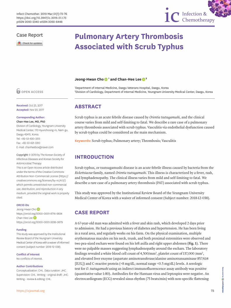
A 67-year-old man was admitted with a fever and skin rash, which developed 2 days prior to admission. He had a previous history of diabetes and hypertension. He has been living in a rural area, and regularly works on his farm. On the physical examination, multiple erythematous macules on his neck, trunk, and both proximal extremities were observed and two pea-sized eschars were found on his left axilla and right upper abdomen (Fig. 1). There were no palpable masses suggesting lymphadenopathy around the eschars. The laboratory findings revealed a white blood cell count of 4,500/mm3, platelet count of 137,000 /mm3, and elevated liver enzyme (aspartate aminotransferase/alanine aminotransaminase 857/614 [IU/L]) and C-reactive protein (>8 mg/dL; normal range <0.5 mg/dL) levels. A serologic test for O. tsutsugamushi using an indirect immunofluorescence assay antibody was positive (quantitative value 1:80). Antibodies for the Hantaan virus and leptospira were negative. An electrocardiogram (ECG) revealed sinus rhythm (75 beats/min) with non-specific flattening
Infect Chemother. 2019 Mar;51(1):73-76https://doi.org/10.3947/ic.2019.51.1.73pISSN 2093-2340·eISSN 2092-6448
Case Report
Received: Oct 25, 2017Accepted: Nov 20, 2017
Corresponding Author: Chan-Hee Lee, MD, PhDDivision of Cardiology, Yeungnam University Medical Center, 170 Hyunchoong-ro, Nam-gu, Daegu 42415, Korea. Tel: +82-53-620-3313Fax: +82-53-621-3310E-mail: [email protected]
Copyright © 2019 by The Korean Society of Infectious Diseases and Korean Society for Antimicrobial TherapyThis is an Open Access article distributed under the terms of the Creative Commons Attribution Non-Commercial License (https://creativecommons.org/licenses/by-nc/4.0/) which permits unrestricted non-commercial use, distribution, and reproduction in any medium, provided the original work is properly cited.
ORCID iDsJeong-Hwan Cho https://orcid.org/0000-0001-8716-9834Chan-Hee Lee https://orcid.org/0000-0001-9338-0679
FundingThis study was approved by the Institutional Review Board of the Yeungnam University Medical Center of Korea with a waiver of informed consent (subject number: 2018-12-038).
Conflict of InterestNo conflicts of interest.
Author ContributionsConceptualization: CHL. Data curation: JHC. Supervision: CHL. Writing - original draft: JHC. Writing - review & editing: CHL.
Jeong-Hwan Cho 1 and Chan-Hee Lee 2
1Department of Internal Medicine, Daegu Veterans Hospital, Daegu, Korea2Division of Cardiology, Department of Internal Medicine, Yeungnam University Medical Center, Daegu, Korea
Pulmonary Artery Thrombosis Associated with Scrub Typhus
of the T waves in the inferior leads. His clinical symptoms and laboratory findings gradually improved with oral doxycycline (200 mg/day), and he was discharged on the 5th hospital day. Oral doxycycline was additionally administered for 2 more days after discharge (7 days, totally).
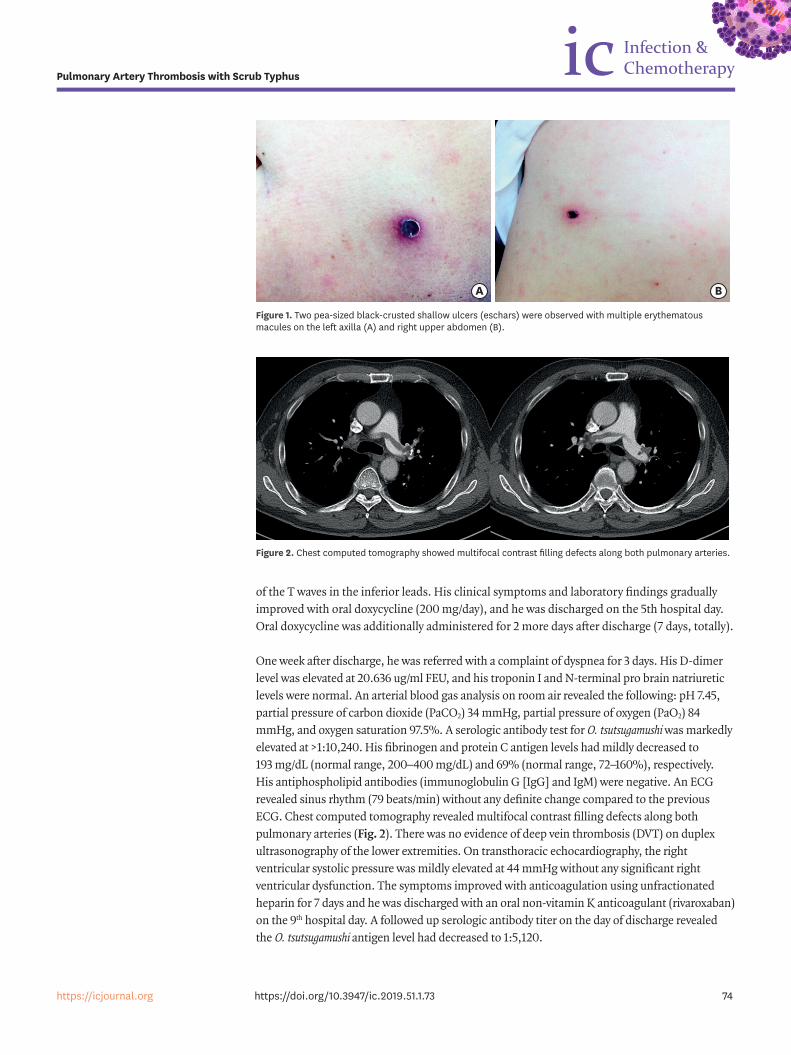
One week after discharge, he was referred with a complaint of dyspnea for 3 days. His D-dimer level was elevated at 20.636 ug/ml FEU, and his troponin I and N-terminal pro brain natriuretic levels were normal. An arterial blood gas analysis on room air revealed the following: pH 7.45, partial pressure of carbon dioxide (PaCO2) 34 mmHg, partial pressure of oxygen (PaO2) 84 mmHg, and oxygen saturation 97.5%. A serologic antibody test for O. tsutsugamushi was markedly elevated at >1:10,240. His fibrinogen and protein C antigen levels had mildly decreased to 193 mg/dL (normal range, 200–400 mg/dL) and 69% (normal range, 72–160%), respectively. His antiphospholipid antibodies (immunoglobulin G [IgG] and IgM) were negative. An ECG revealed sinus rhythm (79 beats/min) without any definite change compared to the previous ECG. Chest computed tomography revealed multifocal contrast filling defects along both pulmonary arteries (Fig. 2). There was no evidence of deep vein thrombosis (DVT) on duplex ultrasonography of the lower extremities. On transthoracic echocardiography, the right ventricular systolic pressure was mildly elevated at 44 mmHg without any significant right ventricular dysfunction. The symptoms improved with anticoagulation using unfractionated heparin for 7 days and he was discharged with an oral non-vitamin K anticoagulant (rivaroxaban) on the 9th hospital day. A followed up serologic antibody titer on the day of discharge revealed the O. tsutsugamushi antigen level had decreased to 1:5,120.
74https://icjournal.org https://doi.org/10.3947/ic.2019.51.1.73
Pulmonary Artery Thrombosis with Scrub Typhus
A B
Figure 1. Two pea-sized black-crusted shallow ulcers (eschars) were observed with multiple erythematous macules on the left axilla (A) and right upper abdomen (B).
Figure 2. Chest computed tomography showed multifocal contrast filling defects along both pulmonary arteries.
DISCUSSION
It is known that the main pathologic findings in scrub typhus are systemic vasculitis and perivasculitis, which are caused by a proliferation of O. tsutsugamushi in endothelial cells of the microvascular system [1]. A previous report suggested the concept that acute infections are associated with a transient increase in the risk of vascular events [2]. However, the mechanism by which an acute infection or inflammation affect the risk of vascular events is uncertain.
The so called ‘rickettsial vasculitis’ can affect the skin, lungs, liver, kidneys, central nervous system, and skeletal and cardiac muscles. Injury to endothelial cells in the lungs and brain results in the most severe manifestations including non-cardiogenic pulmonary edema, interstitial pneumonia, acute respiratory distress syndrome, meningoencephalitis, seizures, and comas [3]. The manifestations of the cardiovascular system are mainly due to endothelial cell damage, vasculitis, inflammatory cell infiltration, new endothelial cell proliferation, thrombosis, and ischemia [4]. Myocardial infarctions [5] as well as DVTs [6], retinal vein occlusions [7], and central venous sinus thromboses [8] have been reported. Although the exact pathogenesis of the PAT in our patient with scrub typhus was unclear, it is possible that vasculitis or a vascular occlusion may have been involved via endothelial dysfunction.
Considering the involvement of the pulmonary vasculature without any evidence of an embolic source, the scrub typhus may have affected the procoagulability via inflammation. Specific infections can result in thrombo-hemorrhagic syndromes associated with bacterial as well as non-bacterial pathogens [9]. Recently, a study demonstrated the coagulation changes in scrub typhus, which are characterized by an inflammation-induced coagulopathy and coagulation activation [10]. A previous study reported a hypofibrinogenetic coagulopathy associated with thrombophlebitis in scrub typhus [11]. Further, disseminated intravascular coagulation caused by O. tsutsugamushi has also been reported [12]. The fibrinogen level was mildly decreased in the current patient, and an infection-induced activation of coagulation could have been associated.
We described a rare case of PAT associated with scrub typhus. The vasculitis with endothelial dysfunction caused by the scrub typhus could be considered as the main pathologic mechanism. Further studies are needed to clarify this issue in terms of the epidemiological and pathological aspects in the future.
REFERENCES
1. Levine HD. Pathologic study of thirty-one cases of scrub typhus fever with especial reference to the cardiovascular system. Am Heart J 1946;31:314-28. PUBMED | CROSSREF
2. Smeeth L, Thomas SL, Hall AJ, Hubbard R, Farrington P, Vallance P. Risk of myocardial infarction and stroke after acute infection or vaccination. N Engl J Med 2004;351:2611-8. PUBMED | CROSSREF
3. Valbuena G, Walker DH. Infection of the endothelium by members of the order Rickettsiales. Thromb Haemost 2009;102:1071-9. PUBMED | CROSSREF
4. Settle EB, Pinkerton H, Corbett AJ. A pathologic study of tsutsugamushi disease (scrub typhus) with notes on clinicopathologic correlation. J Lab Clin Med 1945;30:639-61.
75https://icjournal.org https://doi.org/10.3947/ic.2019.51.1.73
Pulmonary Artery Thrombosis with Scrub Typhus
5. Kim DG, Kim JW, Choi YS, Kim SH, Kim SM, Park CG, Seo HS, Oh DJ. Acute myocardial infarction following scrub typhus infection. Int J Cardiol 2007;114:e18-20. PUBMED | CROSSREF
6. Natarajan A, Rozario A, Santhosh AO, Kanth CN. Scrub typhus: a rare cause of multiorgan failure in a surgical patient presenting with deep venous thrombosis. Trop Doct 2002;32:115-6. PUBMED | CROSSREF
7. Nagaki Y, Hayasaka S, Kadoi C, Matsumoto M, Sakagami T. Branch retinal vein occlusion in the right eye and retinal hemorrhage in the left in a patient with classical tsutsugamushi disease. Jpn J Ophthalmol 2001;45:108-10. PUBMED | CROSSREF
8. Jena SS, Mathew A, Sanjith A, Ajith S, Nair BR, Prakash J. Cerebral venous sinus thrombosis presentation in severe scrub typhus infection: a rare entity. Neurol India 2014;62:308-10. PUBMED | CROSSREF
9. Levi M, Keller TT, van Gorp E, ten Cate H. Infection and inflammation and the coagulation system. Cardiovasc Res 2003;60:26-39. PUBMED | CROSSREF
10. Paris DH, Chansamouth V, Nawtaisong P, Löwenberg EC, Phetsouvanh R, Blacksell SD, Lee SJ, Dondorp AM, van der Poll T, Newton PN, Levi M, Day NP. Coagulation and inflammation in scrub typhus and murine typhus--a prospective comparative study from Laos. Clin Microbiol Infect 2012;18:1221-8. PUBMED | CROSSREF
11. Chernof D. Hypofibrinogenemia in scrub typhus. Report of a case. N Engl J Med 1967;276:1195-6. PUBMED | CROSSREF
12. Ono Y, Ikegami Y, Tasaki K, Abe M, Tase C. Case of scrub typhus complicated by severe disseminated intravascular coagulation and death. Emerg Med Australas 2012;24:577-80. PUBMED | CROSSREF
76https://icjournal.org https://doi.org/10.3947/ic.2019.51.1.73
Pulmonary Artery Thrombosis with Scrub Typhus