Embed Size (px)
Citation preview

Chronic Hypoxia Promotes Pulmonary ArteryEndothelial Cell Proliferation throughH2O2-Induced 5-LipoxygenaseKristi M. Porter, Emory UniversityBum-Yong Kang, Emory UniversitySherry E. Adesina, Emory UniversityTamara C. Murphy, Emory UniversityMichael Hart, Emory UniversityRoy Sutliff, Emory University
Journal Title: PLoS ONEVolume: Volume 9, Number 6Publisher: Public Library of Science | 2014, Pages e98532- e98532Type of Work: Article | Final Publisher PDFPublisher DOI: 10.1371/journal.pone.0098532Permanent URL: http://pid.emory.edu/ark:/25593/ghmtp
Final published version:http://www.plosone.org/article/info%3Adoi%2F10.1371%2Fjournal.pone.0098532
Copyright information:© 2014 Porter et al.This is an Open Access article distributed under the terms of the CreativeCommons Attribution 4.0 International License (http://creativecommons.org/licenses/by/4.0/), which permits distribution ofderivative works, making multiple copies, distribution, public display, andpublicly performance, provided the original work is properly cited. This licenserequires copyright and license notices be kept intact, credit be given tocopyright holder and/or author.
Accessed November 26, 2021 6:28 PM EST
Chronic Hypoxia Promotes Pulmonary Artery EndothelialCell Proliferation through H2O2-Induced 5-LipoxygenaseKristi M. Porter, Bum-Yong Kang, Sherry E. Adesina, Tamara C. Murphy, C. Michael Hart, Roy L. Sutliff*
Emory University School of Medicine/Atlanta Veterans Affairs Medical Center, Department of Pulmonary, Allergy and Critical Care Medicine, Atlanta, Georgia, United States
of America
Abstract
Pulmonary Hypertension (PH) is a progressive disorder characterized by endothelial dysfunction and proliferation. Hypoxiainduces PH by increasing vascular remodeling. A potential mediator in hypoxia-induced PH development is arachidonate 5-Lipoxygenase (ALOX5). While ALOX5 metabolites have been shown to promote pulmonary vasoconstriction and endothelialcell proliferation, the contribution of ALOX5 to hypoxia-induced proliferation remains unknown. We hypothesize thathypoxia exposure stimulates HPAEC proliferation by increasing ALOX5 expression and activity. To test this, humanpulmonary artery endothelial cells (HPAEC) were cultured under normoxic (21% O2) or hypoxic (1% O2) conditions for 24-,48-, or 72 hours. In a subset of cells, the ALOX5 inhibitor, zileuton, or the 5-lipoxygenase activating protein inhibitor, MK-886, was administered during hypoxia exposure. ALOX5 expression was measured by qRT-PCR and western blot and HPAECproliferation was assessed. Our results demonstrate that 24 and 48 hours of hypoxia exposure have no effect on HPAECproliferation or ALOX5 expression. Seventy two hours of hypoxia significantly increases HPAEC ALOX5 expression, hydrogenperoxide (H2O2) release, and HPAEC proliferation. We also demonstrate that targeted ALOX5 gene silencing or inhibition ofthe ALOX5 pathway by pharmacological blockade attenuates hypoxia-induced HPAEC proliferation. Furthermore, ourfindings indicate that hypoxia-induced increases in cell proliferation and ALOX5 expression are dependent on H2O2
production, as administration of the antioxidant PEG-catalase blocks these effects and addition of H2O2 to HPAEC promotesproliferation. Overall, these studies indicate that hypoxia exposure induces HPAEC proliferation by activating the ALOX5pathway via the generation of H2O2.
Citation: Porter KM, Kang B-Y, Adesina SE, Murphy TC, Hart CM, et al. (2014) Chronic Hypoxia Promotes Pulmonary Artery Endothelial Cell Proliferation throughH2O2-Induced 5-Lipoxygenase. PLoS ONE 9(6): e98532. doi:10.1371/journal.pone.0098532
Editor: Philip Michael Bauer, University of Pittsburgh School of Medicine, United States of America
Received August 2, 2013; Accepted May 5, 2014; Published June 6, 2014
Copyright: � 2014 Porter et al. This is an open-access article distributed under the terms of the Creative Commons Attribution License, which permitsunrestricted use, distribution, and reproduction in any medium, provided the original author and source are credited.
Funding: This work was supported by HL070892 to RLS, National Science Foundation Award #0450303 Subaward # I-66-606-63 to Emory University,Pharmacological Sciences Training Grant TM GM 008602, National Institute of Allergy and Infectious Diseases 1-F31-AI084460 to KMP, and HL 102167 to RLS andCMH. The funders had no role in study design, data collection and analysis, decision to publish, or preparation of the manuscript.
Competing Interests: The authors have declared that no competing interests exist.
* E-mail: [email protected]
Introduction
Pulmonary Hypertension (PH) is a progressive disorder
characterized by sustained increases in pulmonary arterial
pressures and vascular remodeling. Although the mechanisms
underlying PH remain unknown, hypoxia induces PH in
experimental models and is believed to contribute to disease
development [1,2] by impairing endothelial cell function [3,4] as
evidenced by abnormal endothelial cell growth in lung sections
and pulmonary artery endothelial cells from PH patients [5,6].
Altered expression of arachidonate 5-lipoxygenase (ALOX5), the
enzyme that catalyzes the production of vasoactive leukotrienes
from arachidonic acid, is associated with endothelial proliferation
and PH development. Previous studies demonstrate that patients
with idiopathic pulmonary hypertension exhibit increased ALOX5
expression in lung tissue, particularly in small pulmonary artery
endothelial cells. Also, inhibition of ALOX5 or its required
cofactor, 5-lipoxygenase activating protein (FLAP) attenuates
hypoxia- or monocrotaline (MCT)-induced PH [7,8], whereas
ALOX5 overexpression accelerates and exacerbates PH in MCT-
treated rats [9]. ALOX5 metabolites, such as the cysteinyl
leukotrienes (CysLT) are suggested to mediate these effects as
they induce vasoconstriction in the distal segments of pulmonary
arteries [10]. Moreover, inhibitors of CysLT production attenuate
proliferation of pulmonary artery endothelial cells [11].
ALOX5 activity and leukotriene production are regulated by
numerous signaling pathways. Primarily, ALOX5 requires the
presence of FLAP for leukotriene synthesis [12,13]. Yet, ALOX5 is
also activated in conditions that promote lipid peroxidation [14]
particularly following glutathione depletion [15,16]. Studies also
indicate that increases in endogenous reactive oxygen species
(ROS) release stimulate ALOX5 expression [14] and cause an
almost 4-fold increase in leukotriene formation [17]. These studies
strongly suggest that ROS may induce ALOX5 expression.
However, the connection between hypoxia-induced PH, ROS
and endothelial ALOX5 is not completely understood. In this
study, we investigate whether chronic hypoxia exposure alters
endothelial ALOX5 expression, the effects of hypoxia-induced
ALOX5 expression on endothelial cell proliferation and the role of
hypoxia-induced ROS.
Materials and Methods
ReagentsTrypan blue, fetal bovine serum (FBS), dimethyl sulfoxide
(DMSO), PEG-Catalase, and gelatin were obtained from Sigma-
PLOS ONE | www.plosone.org 1 June 2014 | Volume 9 | Issue 6 | e98532
Aldrich (St. Louis, MO). Zileuton was obtained from Patheon
Pharmaceuticals (Cincinnatti, OH). MK-886 was purchased from
Calbiochem (San Diego, CA). Scrambled and silencing RNA
(siRNA) for ALOX5 was obtained from Qiagen (Valencia, CA).
Cell CultureHuman pulmonary artery endothelial cells (HPAEC) were
obtained from Lonza Clonetics (Walkersville, MD). HPAEC were
grown in EGM-2 medium (Lonza), which contains basic growth
medium (EBM-2), fetal bovine serum (FBS), and antibiotics,
ascorbic acid, vascular endothelial growth factor (VEGF), human
fibroblast growth factor (hFGF-B), hydrocortisone, human epider-
mal growth factor (hEGF), R3-IGF-1 (insulin-like growth factor),
GA-1000 (gentamicin, amphotericin B), and heparin. Unless
otherwise stated, cells were maintained in a 37uC incubator at 5%
CO2.
Hypoxia ExposureHPAEC, passages 3-8, were exposed to hypoxia in a Biospherix
exposure chamber (Lacona, NY) as previously described [18,19].
For normoxic conditions, HPAEC were placed into a standard
incubator maintained at 37uC and 5% CO2 levels. For hypoxic
conditions, HPAEC were placed in a hypoxia chamber main-
tained at 37uC, 1% oxygen, and 5% CO2 levels. Human
pulmonary artery endothelial cells (HPAEC) were cultured in
normoxic or hypoxic conditions for 24-, 48-, or 72 hours. To
investigate the contribution of ROS levels, PEG-Catalase
(1,000 U/mL) was administered during the final 24 hours of the
72 hours of hypoxia exposure. The role of 5-Lipoxygenase in
HPAEC proliferation was studied by exposing cells to 10 mM
concentrations of Zileuton or MK-886 either throughout the
entire exposure period or during the final 24 hours.
ALOX5 and FLAP mRNA AnalysisALOX5 and FLAP mRNA levels were determined by
quantitative real-time PCR (qRT-PCR) using the iCycler system
(Bio-Rad, Hercules, CA). Total RNA was extracted from HPAEC
using RNA-Bee. RNA concentrations were measured using the
ND-1000 Spectrophotometer (NanoDrop Technology, Wilming-
ton, DE). RNA (1.5 micrograms) was combined with random
nanomer primers (Ambion), dNTPs (New England Bio-Labs) and
nuclease-free water for reverse transcription. cDNA templates
were amplified with gene-specific primer sets. All transcripts were
detected using SYBR Green I (Molecular Probes, Inc). Transcripts
were normalized to the housekeeping gene, b-Globin. Values are
expressed as percent of control. Expression changes were
determined using the 22DDCt method.
RNA Interference and HPAEC TransfectionHuman ALOX5 siRNA (NM_000698), siRNA duplexes (59-
GGCAGGAAGACCTGATGTT -39, target region 333–351)
were designed using BLOCK-it RNAi Designer (Invitrogen).
siRNA targeted to a specific noncoding gene was employed as a
scrambled RNA control. At 40–50% confluence, HPAECs were
transfected with scrambled or ALOX5 siRNA using GeneSilencer
(Genlantis, San Diego, CA) transfection reagent according to
manufacturer’s instructions. After transfection for 6 hours, the
transfection media was replaced with EGM containing 10% FBS.
HPAECs were exposed to normoxia (NOR, 21% O2) or hypoxia
(HYP, 1% O2) for 72 h. HPAEC lysates were then harvested and
examined for ALOX5 levels using qRT-PCR and Western blots.
In selected studies, HPAEC proliferation was determined using
MTT assays.
Western Blot AnalysesFollowing normoxia or hypoxia exposure, HPAEC lysates were
subjected to Western Blot analysis as reported [18]. Primary
antibodies for ALOX5 and GAPDH were purchased from
Cayman Chemical Company (Ann Arbor, Michigan) and
Sigma-Aldrich respectively. Proteins were visualized using fluo-
rescent anti-goat or anti-rabbit secondary antibodies using the
Licor system. Bands for protein of interest were quantified by
densitometry and normalized to GAPDH levels within the same
lane.
Cell Proliferation AssayCell proliferation was assessed using the MTT (ATCC,
Manassas, VA) Cell Proliferation Assay. Briefly, proliferating cells
reduce the tetrazolium MTT resulting in intracellular formazan.
Detergent reagent was added to cell to solubilize formazan.
Supernatants were then collected and quantified using a spectro-
photometer at 562 nm. Cell proliferation was further confirmed
by cell counting using a trypan blue dye exclusion assay. Briefly,
following hypoxia exposure, cells were trysinized and resuspended
in medium. Cells were mixed with PBS and trypan blue at a 1:8:1
ratio. The trypan blue-negative cells were counted using a
hemacytometer.
Hydrogen Peroxide AnalysisHydrogen Peroxide (H2O2) release was quantified using the
Amplex Red Assay. Cells were incubated in a solution containing
the Amplex Red reagent (Molecular Probes), horseradish perox-
idase and a buffer solution for 30 minutes at 37uC. Supernatants
were then collected and fluorescence measured at 560 nm. H2O2
concentrations were determined through standard curve extrap-
olation normalized to cellular protein concentration. Overall
reactive oxygen species (ROS) and reactive nitrogen species were
detected using the ROS-sensitive fluorescent probe 29, 79-
dihydrodichlorofluorescein diacetate (DCF-DA; Invitrogen, Carls-
bad, CA). Confluent HPAEC monolayers were loaded with
25 mg/mL DCF-DA for 1 hour at 37uC in Krebs–Ringer
Phosphate Buffer (KRPG; 145 mM NaCl, 5.7 mM KH2PO4,
4.86 mM KCl, 0.54 mM CaCl2, 1.22 mM MgSO4, and 5.5 mM
glucose, pH 7.35). A laser-scanning confocal microscope (Olym-
pus, Center Valley, PA) and fluorimeter were used to detect DCF
fluorescence at excitation and emission wavelengths of 488 nm
and 520 nm, respectively.
Statistical AnalysisA student’s t-test analysis was used for comparison of two
groups. One-way ANOVA with Tukey’s posttest was used for the
comparison of multiple groups. All experiments using cell cultures
were repeated at least twice on different cell lines, and samples
were run in duplicate or triplicate. Statistical significance was
defined as P, 0.05, and all graphs are expressed as mean 6 SEM.
All statistical analyses were performed using GraphPad Prism
software (La Jolla, CA).
Results
Prolonged Hypoxia Exposure Promotes Endothelial CellProliferation
Hypoxia is associated with significant endothelial alterations
which are thought to contribute to PH development and
progression [3,20]. Pulmonary arteries [4] and lung sections of
PH patients demonstrate abnormal endothelial cell growth [5,6].
To determine whether hypoxia alters HPAEC function in vitro, we
Hypoxia, 5-Lipoxygenase, and Endothelial Cells
PLOS ONE | www.plosone.org 2 June 2014 | Volume 9 | Issue 6 | e98532
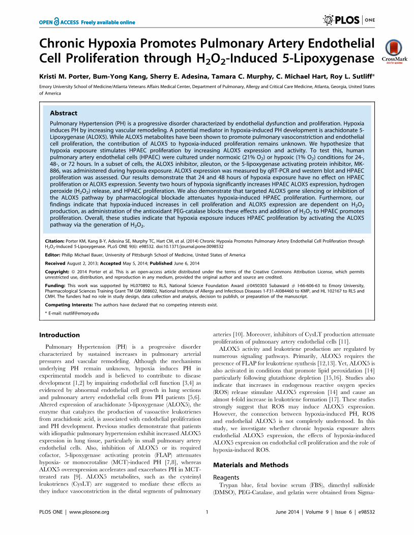
Figure 1. Prolonged hypoxia exposure increases endothelial cell proliferation. Seventy two hours of hypoxia exposure significantlystimulates endothelial cell proliferation when compared to all other groups. Human pulmonary artery endothelial cells (HPAEC) were exposed tonormoxic or hypoxic (1% O2) conditions for 24-, 48-, or 72 hours (n = 4). Following exposure, cell proliferation was assessed by MTT (Figure 1A) assayand Trypan Blue Dye Exclusion Assay (Figure 1B). * p,0.0001.doi:10.1371/journal.pone.0098532.g001
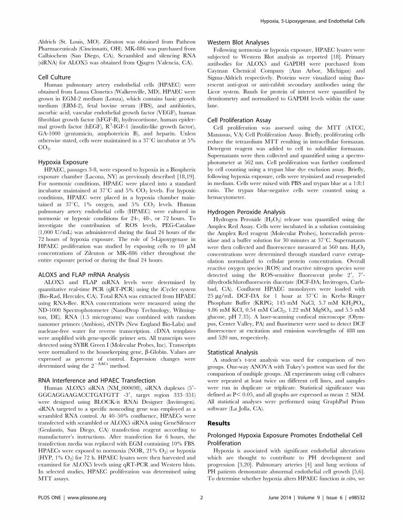
Figure 2. Chronic hypoxia exposure increases endothelial ALOX5 expression. Seventy two hours of hypoxia exposure significantlystimulates endothelial ALOX5 expression when compared to all other groups. HPAEC were exposed to normoxic or hypoxic conditions for 24-, 48-, or72 hours. Following exposure, cells were collected, and total RNA and protein were isolated for expression analyses via quantitative real time PCR andWestern blot respectively. Results indicate that ALOX5 mRNA levels are significantly increased following hypoxia exposure (A, n = 5). Chronic hypoxiaexposure also causes a 3-fold elevation in ALOX5 protein expression levels (B, n = 4). Endothelial FLAP expression is also increased when compared toall other groups (C, n = 5–7). Values are expressed as percent of control. * p,0.001 when compared to all other groups.doi:10.1371/journal.pone.0098532.g002
Hypoxia, 5-Lipoxygenase, and Endothelial Cells
PLOS ONE | www.plosone.org 3 June 2014 | Volume 9 | Issue 6 | e98532
assessed HPAEC proliferation following 24, 48, and 72 hours of
hypoxia exposure. While 24 and 48 hours of hypoxia exposure
had no effect on cellular proliferation, 72 hours of hypoxia
increases cellular proliferation when measured by MTT assay
(Figure 1A) and Trypan Blue Dye Exclusion Assay (Figure 1B).
Chronic Hypoxia Exposure Stimulates HPAEC5-Lipoxygenase Expression
In vivo studies demonstrate that both hypoxia exposure and
MCT administration upregulate 5-lipoxygenase (ALOX5) [9,21].
To specifically investigate the effect of hypoxia on pulmonary
artery endothelial cell ALOX5, HPAEC were exposed to hypoxia
for 24-, 48-, or 72-hours. Our results indicate that 72 hours of
hypoxia exposure significantly increases HPAEC ALOX5 mRNA
(Figure 2A) when analyzed by qRT-PCR and ALOX5 protein
expression (Figure 2B) as measured by western blot. Prolonged
hypoxia exposure also stimulates a significant increase in the
expression of the ALOX5 required cofactor, FLAP when
measured by qRT-PCR (Figure 2C).
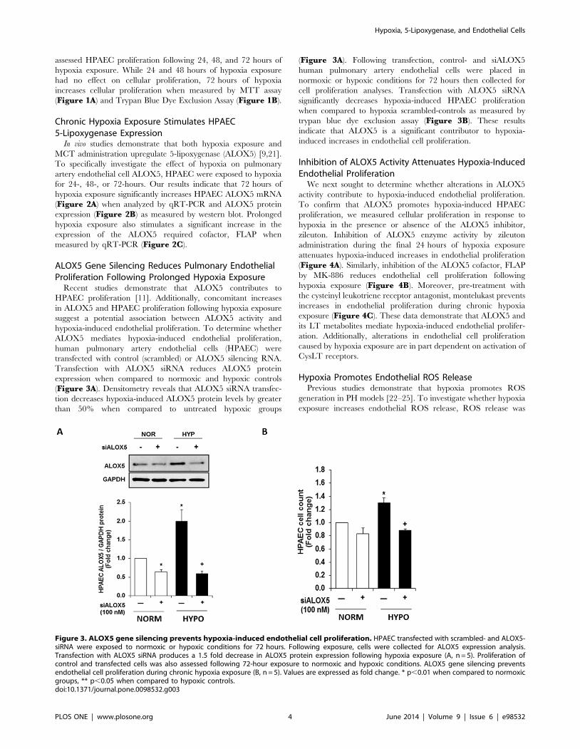
ALOX5 Gene Silencing Reduces Pulmonary EndothelialProliferation Following Prolonged Hypoxia Exposure
Recent studies demonstrate that ALOX5 contributes to
HPAEC proliferation [11]. Additionally, concomitant increases
in ALOX5 and HPAEC proliferation following hypoxia exposure
suggest a potential association between ALOX5 activity and
hypoxia-induced endothelial proliferation. To determine whether
ALOX5 mediates hypoxia-induced endothelial proliferation,
human pulmonary artery endothelial cells (HPAEC) were
transfected with control (scrambled) or ALOX5 silencing RNA.
Transfection with ALOX5 siRNA reduces ALOX5 protein
expression when compared to normoxic and hypoxic controls
(Figure 3A). Densitometry reveals that ALOX5 siRNA transfec-
tion decreases hypoxia-induced ALOX5 protein levels by greater
than 50% when compared to untreated hypoxic groups
(Figure 3A). Following transfection, control- and siALOX5
human pulmonary artery endothelial cells were placed in
normoxic or hypoxic conditions for 72 hours then collected for
cell proliferation analyses. Transfection with ALOX5 siRNA
significantly decreases hypoxia-induced HPAEC proliferation
when compared to hypoxia scrambled-controls as measured by
trypan blue dye exclusion assay (Figure 3B). These results
indicate that ALOX5 is a significant contributor to hypoxia-
induced increases in endothelial cell proliferation.
Inhibition of ALOX5 Activity Attenuates Hypoxia-InducedEndothelial Proliferation
We next sought to determine whether alterations in ALOX5
activity contribute to hypoxia-induced endothelial proliferation.
To confirm that ALOX5 promotes hypoxia-induced HPAEC
proliferation, we measured cellular proliferation in response to
hypoxia in the presence or absence of the ALOX5 inhibitor,
zileuton. Inhibition of ALOX5 enzyme activity by zileuton
administration during the final 24 hours of hypoxia exposure
attenuates hypoxia-induced increases in endothelial proliferation
(Figure 4A). Similarly, inhibition of the ALOX5 cofactor, FLAP
by MK-886 reduces endothelial cell proliferation following
hypoxia exposure (Figure 4B). Moreover, pre-treatment with
the cysteinyl leukotriene receptor antagonist, montelukast prevents
increases in endothelial proliferation during chronic hypoxia
exposure (Figure 4C). These data demonstrate that ALOX5 and
its LT metabolites mediate hypoxia-induced endothelial prolifer-
ation. Additionally, alterations in endothelial cell proliferation
caused by hypoxia exposure are in part dependent on activation of
CysLT receptors.
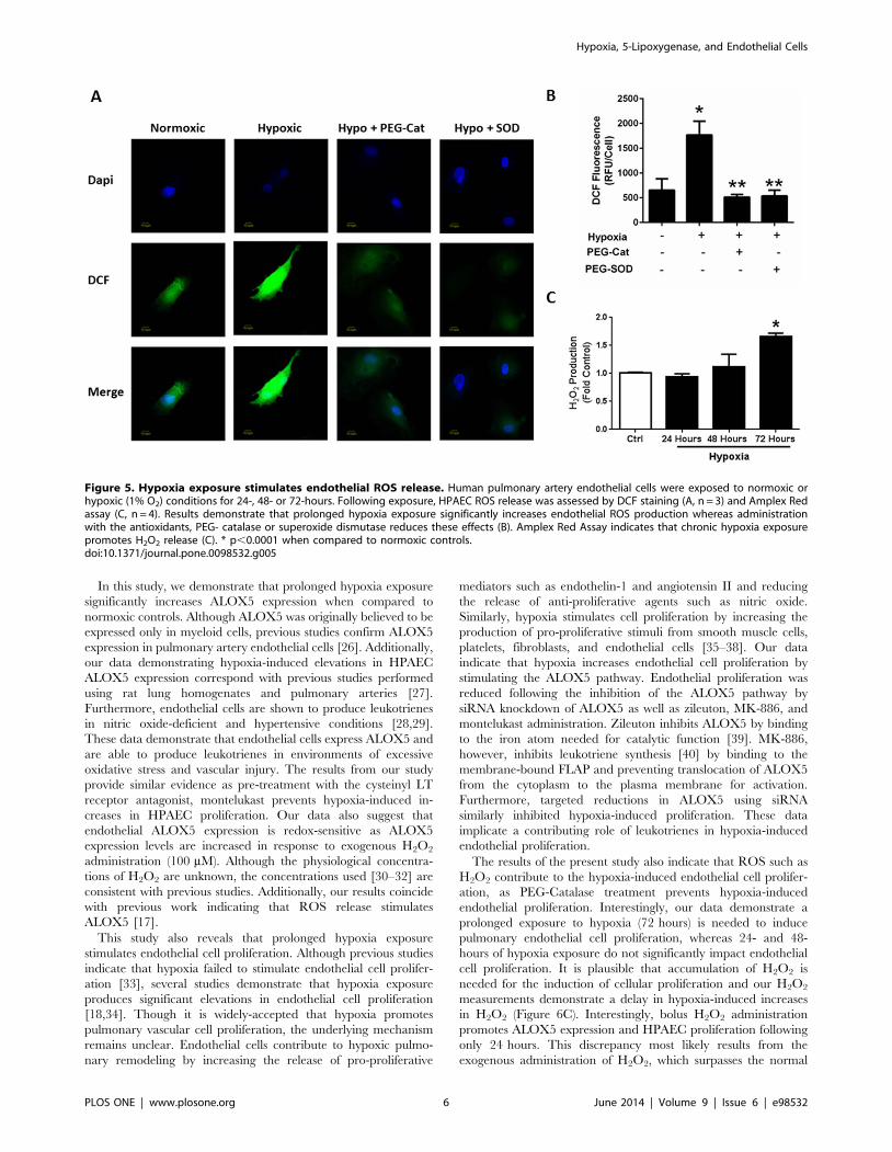
Hypoxia Promotes Endothelial ROS ReleasePrevious studies demonstrate that hypoxia promotes ROS
generation in PH models [22–25]. To investigate whether hypoxia
exposure increases endothelial ROS release, ROS release was
Figure 3. ALOX5 gene silencing prevents hypoxia-induced endothelial cell proliferation. HPAEC transfected with scrambled- and ALOX5-siRNA were exposed to normoxic or hypoxic conditions for 72 hours. Following exposure, cells were collected for ALOX5 expression analysis.Transfection with ALOX5 siRNA produces a 1.5 fold decrease in ALOX5 protein expression following hypoxia exposure (A, n = 5). Proliferation ofcontrol and transfected cells was also assessed following 72-hour exposure to normoxic and hypoxic conditions. ALOX5 gene silencing preventsendothelial cell proliferation during chronic hypoxia exposure (B, n = 5). Values are expressed as fold change. * p,0.01 when compared to normoxicgroups, ** p,0.05 when compared to hypoxic controls.doi:10.1371/journal.pone.0098532.g003
Hypoxia, 5-Lipoxygenase, and Endothelial Cells
PLOS ONE | www.plosone.org 4 June 2014 | Volume 9 | Issue 6 | e98532

analyzed following 24, 48, and 72-hours of hypoxia exposure.
DCF staining indicates that chronic hypoxia exposure increases
ROS production in endothelial cells (Figure 5A). This increase in
ROS release was attenuated by the administration of PEG-
catalase and superoxide dismutase (Figure 5B). Furthermore,
Amplex Red assay analysis of hydrogen peroxide (H2O2) release
also demonstrates that 72 hours of hypoxia exposure is required
for increased in H2O2 (Figure 5C). These data suggest that
chronic exposure to hypoxic conditions increases endothelial
H2O2.
H2O2 Mediates Hypoxia-Induced Increases in HPAECALOX5 Expression and Cell Proliferation
Since elevated ROS production is associated with increased
ALOX5 activity [14,15,17], we next sought to determine whether
H2O2 mediates hypoxia-induced increases in endothelial ALOX5.
HPAECs exposed to 0–200 mM concentrations of H2O2 for
24 hours revealed that 100 mM and 200 mM H2O2 concentrations
increase endothelial ALOX5 mRNA levels (Figure 6A). Similarly,
H2O2 exposure stimulates ALOX5 protein expression when
assessed by Western blot (Figure 6B). These data indicate that
ALOX5 expression levels are altered by increased ROS. To
determine whether hypoxia-induced ROS mediate alterations in
HPAEC ALOX5 levels and cell proliferation, we administered the
antioxidant, PEG-catalase during the final 24 hours of the 72 hour
hypoxia exposure period and determined ALOX5 mRNA levels
by qRT-PCR and cell proliferation by MTT Assay. Results
demonstrate that catalase administration significantly reduces
hypoxia-induced elevations in endothelial ALOX5 gene expres-
sion (Figure 6C). Catalase treatment during the final 24 hours of
the 72-hour hypoxia exposure also prevents HPAEC proliferation
(Figure 6D) in a concentration-dependent manner when
compared to untreated hypoxic groups.
Discussion
Our studies indicate that hypoxia exposure promotes pulmo-
nary artery endothelial proliferation by stimulating key catalytic
molecules in the LT pathway, ALOX5 and its required co-factor,
FLAP. We also demonstrate that hypoxia mediates these events by
increasing ROS particularly H2O2. Overall, these studies suggest
that ALOX5 contributes to hypoxia-induced endothelial prolifer-
ation in a ROS-dependent manner.
Figure 4. Pharmacological inhibition of ALOX5 signaling attenuates hypoxia-induced endothelial proliferation. ALOX5 blockade byzileuton administration reduces hypoxia-induced endothelial proliferation when measured by MTT Assay (A, n = 4). FLAP inhibition by MK-886attenuates endothelial proliferation following hypoxia exposure (B, n = 4) Pre-treatment with the cysteinyl leukotriene receptor antagonist,montelukast prevents endothelial proliferation during prolonged hypoxia exposure (C, n = 6) HPAEC were exposed to normoxic or hypoxic conditionsfor 72 hours. ALOX5 inhibitors, zileuton (10 mM) and MK-886 (0.5 mM) were administered during the final 24 hours of normoxia or hypoxia exposure.Cell proliferation was then assessed by MTT assay. * p,0.05 when compared to normoxic groups. ** p,0.05 when compared to untreated hypoxicgroups.doi:10.1371/journal.pone.0098532.g004
Hypoxia, 5-Lipoxygenase, and Endothelial Cells
PLOS ONE | www.plosone.org 5 June 2014 | Volume 9 | Issue 6 | e98532
In this study, we demonstrate that prolonged hypoxia exposure
significantly increases ALOX5 expression when compared to
normoxic controls. Although ALOX5 was originally believed to be
expressed only in myeloid cells, previous studies confirm ALOX5
expression in pulmonary artery endothelial cells [26]. Additionally,
our data demonstrating hypoxia-induced elevations in HPAEC
ALOX5 expression correspond with previous studies performed
using rat lung homogenates and pulmonary arteries [27].
Furthermore, endothelial cells are shown to produce leukotrienes
in nitric oxide-deficient and hypertensive conditions [28,29].
These data demonstrate that endothelial cells express ALOX5 and
are able to produce leukotrienes in environments of excessive
oxidative stress and vascular injury. The results from our study
provide similar evidence as pre-treatment with the cysteinyl LT
receptor antagonist, montelukast prevents hypoxia-induced in-
creases in HPAEC proliferation. Our data also suggest that
endothelial ALOX5 expression is redox-sensitive as ALOX5
expression levels are increased in response to exogenous H2O2
administration (100 mM). Although the physiological concentra-
tions of H2O2 are unknown, the concentrations used [30–32] are
consistent with previous studies. Additionally, our results coincide
with previous work indicating that ROS release stimulates
ALOX5 [17].
This study also reveals that prolonged hypoxia exposure
stimulates endothelial cell proliferation. Although previous studies
indicate that hypoxia failed to stimulate endothelial cell prolifer-
ation [33], several studies demonstrate that hypoxia exposure
produces significant elevations in endothelial cell proliferation
[18,34]. Though it is widely-accepted that hypoxia promotes
pulmonary vascular cell proliferation, the underlying mechanism
remains unclear. Endothelial cells contribute to hypoxic pulmo-
nary remodeling by increasing the release of pro-proliferative
mediators such as endothelin-1 and angiotensin II and reducing
the release of anti-proliferative agents such as nitric oxide.
Similarly, hypoxia stimulates cell proliferation by increasing the
production of pro-proliferative stimuli from smooth muscle cells,
platelets, fibroblasts, and endothelial cells [35–38]. Our data
indicate that hypoxia increases endothelial cell proliferation by
stimulating the ALOX5 pathway. Endothelial proliferation was
reduced following the inhibition of the ALOX5 pathway by
siRNA knockdown of ALOX5 as well as zileuton, MK-886, and
montelukast administration. Zileuton inhibits ALOX5 by binding
to the iron atom needed for catalytic function [39]. MK-886,
however, inhibits leukotriene synthesis [40] by binding to the
membrane-bound FLAP and preventing translocation of ALOX5
from the cytoplasm to the plasma membrane for activation.
Furthermore, targeted reductions in ALOX5 using siRNA
similarly inhibited hypoxia-induced proliferation. These data
implicate a contributing role of leukotrienes in hypoxia-induced
endothelial proliferation.
The results of the present study also indicate that ROS such as
H2O2 contribute to the hypoxia-induced endothelial cell prolifer-
ation, as PEG-Catalase treatment prevents hypoxia-induced
endothelial proliferation. Interestingly, our data demonstrate a
prolonged exposure to hypoxia (72 hours) is needed to induce
pulmonary endothelial cell proliferation, whereas 24- and 48-
hours of hypoxia exposure do not significantly impact endothelial
cell proliferation. It is plausible that accumulation of H2O2 is
needed for the induction of cellular proliferation and our H2O2
measurements demonstrate a delay in hypoxia-induced increases
in H2O2 (Figure 6C). Interestingly, bolus H2O2 administration
promotes ALOX5 expression and HPAEC proliferation following
only 24 hours. This discrepancy most likely results from the
exogenous administration of H2O2, which surpasses the normal
Figure 5. Hypoxia exposure stimulates endothelial ROS release. Human pulmonary artery endothelial cells were exposed to normoxic orhypoxic (1% O2) conditions for 24-, 48- or 72-hours. Following exposure, HPAEC ROS release was assessed by DCF staining (A, n = 3) and Amplex Redassay (C, n = 4). Results demonstrate that prolonged hypoxia exposure significantly increases endothelial ROS production whereas administrationwith the antioxidants, PEG- catalase or superoxide dismutase reduces these effects (B). Amplex Red Assay indicates that chronic hypoxia exposurepromotes H2O2 release (C). * p,0.0001 when compared to normoxic controls.doi:10.1371/journal.pone.0098532.g005
Hypoxia, 5-Lipoxygenase, and Endothelial Cells
PLOS ONE | www.plosone.org 6 June 2014 | Volume 9 | Issue 6 | e98532

cellular processes necessary for H2O2 production during hypoxia
exposure. Moreover, bolus H2O2 administration may also
stimulate ALOX5 expression and HPAEC proliferation through
the activation of downstream signaling pathways. Recent reports
indicate that H2O2 activates endothelial p38 MAPK [41,42] and
NF-kappaB activity [43,44], two mediators associated with
increases in ALOX5 expression and activity. Furthermore, the
addition of PEG-catalase is shown to double endogenous
endothelial catalase activity within 4 hours [45]. Taken together,
these findings are consistent with previous reports demonstrating
that the highly reactive oxidant, peroxynitrite stimulates pulmo-
nary artery endothelial cell proliferation [46] and catalase inhibits
cell proliferation [47]. The mechanism responsible for an
ALOX5-mediated increase in proliferation is unclear however, It
is possible that ALOX5 promotes cell proliferation via its nuclear
localization or through interaction with cytoskeletal proteins
[48,49]. Nonetheless, the excessive proliferation of pulmonary
endothelial cells is thought to contribute to the obliteration of the
pulmonary artery vessel lumen. Therefore, it is likely that hypoxia-
induced pulmonary artery endothelial proliferation contributes to
pulmonary hypertension (PH) development and progression.
Previous research demonstrates that chronic hypoxia signifi-
cantly increases endothelial cell proliferation. In vivo studies
indicate that endothelial cells in the main pulmonary artery and
in the small muscular arteries are increased in chronically hypoxic
rats [50,51]. Endothelial proliferation is also increased in neonatal
calves following exposure to 8% oxygen for 14 days [52].
Additionally, excessive endothelial proliferation leads to plexiform
lesion formation in idiopathic PAH patients [53]. Our research,
performed in vitro, similarly demonstrates that hypoxia promotes
endothelial proliferation. We also show that hypoxia-induced
endothelial proliferation is ROS-dependent as the administration
of PEG-catalase attenuates these events. These hypoxia-induced
ROS are likely produced by NADPH oxidases (Noxes) as previous
studies indicate that Nox4 expression is elevated in low oxygen
environments [25]. Interestingly, previous studies also suggest that
Figure 6. ROS mediate hypoxia-induced increases in endothelial ALOX5 expression and cell proliferation. Human pulmonary arteryendothelial cells (HPAEC) were exposed to 0, 10, 100, and 200 mM hydrogen peroxide (H2O2) for 24 hours. Following exposure, supernatants werecollected to assess cell toxicity by adenylate kinase release. Results demonstrate no significant changes in cell death as indicated by adenylate kinaserelease (n = 4–6; data not shown). HPAEC were collected and total RNA was isolated for quantitative real-time PCR gene expression analysis. ALOX5was normalized to the housekeeping gene b-globin. Relative expression was calculated using the Delta-Delta CT method and values were expressedas percent of control (A, n = 4–5). * p,0.05 when compared to untreated controls. H2O2 exposure stimulates HPAEC ALOX5 protein levels as analyzedby western blot (B, n = 4). PEG-Catalase (10U - 1000 U/ml) administration during the final 24 hours of the 72 hour hypoxia exposure prevents hypoxia-induced elevations in endothelial ALOX5 expression (C, n = 5) and cell proliferation (D, n = 6). * p,0.01 when compared to normoxic groups. ** p,0.05 when compared to untreated hypoxia controls.doi:10.1371/journal.pone.0098532.g006
Hypoxia, 5-Lipoxygenase, and Endothelial Cells
PLOS ONE | www.plosone.org 7 June 2014 | Volume 9 | Issue 6 | e98532

NADPH oxidase activity is required for endothelial cell prolifer-
ation [54]. Additionally, studies by our group and others have
implicated Nox4 in the hypoxia-induced proliferation of human
pulmonary artery smooth muscle cells [19,55]. Hypoxia-induced
increases in ROS may also result from reductions in antioxidant
availability, as hypoxia exposure significantly decreases glutathi-
one levels in porcine pulmonary artery endothelial cells [56].
Although our evidence indicates that hypoxia increases ALOX5
in a redox-sensitive manner, the mechanisms underlying these
events remain unclear. Two major pathways are implicated in
regulating ALOX5 expression: promoter methylation and tran-
scription factor activation [57]. Research demonstrates that
leukocyte cell lines that have methylated promoters, U-937 and
HL-60TB, do not express ALOX5, while the HL-60 cell line,
which is unmethylated expresses ALOX5 [58,59]. Moreover, the
treatment of the U-937 and HL-60TB cell lines with the
demethylating agent, 5-aza-2’deoxycytidine resulted in the resto-
ration of ALOX5 expression [58]. There are also 5 GC boxes that
bind SP1 and have been shown to be important in regulating
expression of ALOX5 [60] however, ablation of all GC-boxes only
reduces ALOX5 promoter activity 47%, implicating other
transcription factors such as hypoxia- inducible factor (HIF) in
ALOX5 regulation. The oxygen-sensing molecule, HIF-1a [61]
regulates the adaptive response by activating genes associated with
energy metabolism, erythropoiesis, vasomotor tone, and angio-
genesis [62], and recent work demonstrates that HIF-1a mediates
hypoxia-induced FLAP expression in human pulmonary micro-
vascular endothelial cells [63]. HIF-2a also contributes to the
vascular response to chronic hypoxia including the expression of
genes involved in pulmonary vascular cell proliferation [64].
Hypoxia may also modulate ALOX5 via the early growth
response protein-1, or Egr-1. This hypoxia-inducible transcription
factor [65] has a binding site within the ALOX5 promoter region
[66] and is expressed in a variety of pulmonary vascular cells,
including endothelial cells [67]. Research also demonstrates that
Egr-1 acts in a redox-sensitive manner, as Egr-1 is upregulated by
ROS release and attenuated following antioxidant overexpression
[68,69]. In addition to HIF and Egr-1, the ALOX5 promoter
region also contains binding sites for TGF-b [70] and NF-kB
[71,72], which are also implicated in the endothelial dysfunction
[73,74] and vascular remodeling [75] in PH pathogenesis [76,77].
In summary, the current study demonstrates that hypoxia-
induced ROS plays a critical role regulating ALOX5 and
endothelial proliferation. Our data suggests that ALOX5 mediates
hypoxia-induced endothelial cell proliferation and implicates
increased ALOX5 expression and activity in vascular remodeling
in experimental and clinical PH. Overall, these results suggest that
ALOX5 inhibition merits additional investigation as a therapeutic
target for the prevention or treatment of PH.
Author Contributions
Conceived and designed the experiments: KMP RLS. Performed the
experiments: KMP BYK SEA TCM. Analyzed the data: KMP BYK SEA
TCM CMH RLS. Contributed reagents/materials/analysis tools: RLS.
Wrote the paper: KMP CMH RLS.
References
1. Wright JL, Lawson L, Pare PD, Hooper RO, Peretz DI, et al. (1983) The
structure and function of the pulmonary vasculature in mild chronic obstructivepulmonary disease. The effect of oxygen and exercise. Am Rev Respir Dis 128:
702–707.
2. Orr R, Smith LJ, Cuttica MJ (2012) Pulmonary hypertension in advancedchronic obstructive pulmonary disease. Curr Opin Pulm Med 18: 138–143.
3. Tuder RM, Groves B, Badesch DB, Voelkel NF (1994) Exuberant endothelial
cell growth and elements of inflammation are present in plexiform lesions ofpulmonary hypertension. Am J Pathol 144: 275–285.
4. Sakao S, Tatsumi K, Voelkel N (2009) Endothelial cells and pulmonary arterial
hypertension: apoptosis, proliferation, interaction and transdifferentiation.Respiratory Research 10: 95.
5. Masri F, Xu W, Comhair S, Asosingh K, Koo M, et al. (2007) Hyperprolifera-
tive apoptosis-resistant endothelial cells in idiopathic pulmonary arterialhypertension. Am J Physiol Lung Cell Mol Physiol 293: 548–554.
6. Rabinovitch M, Bothwell T, Hayakawa BN, Williams WG, Trusler GA, et al.
(1986) Pulmonary artery endothelial abnormalities in patients with congenitalheart defects and pulmonary hypertension. A correlation of light with scanning
electron microscopy and transmission electron microscopy. Lab Invest 55: 632–653.
7. Morganroth ML, Stenmark KR, Morris KG, Murphy RC, Mathias M, et al.
(1985) Diethylcarbamazine inhibits acute and chronic hypoxic pulmonaryhypertension in awake rats. Am Rev Respir Dis 131: 488–492.
8. Stenmark KR, Morganroth ML, Remigio LK, Voelkel NF, Murphy RC, et al.
(1985) Alveolar inflammation and arachidonate metabolism in monocrotaline-induced pulmonary hypertension. Am J Physiol 248: H859–866.
9. Jones JE, Walker JL, Song Y, Weiss N, Cardoso WV, et al. (2004) Effect of 5-
lipoxygenase on the development of pulmonary hypertension in rats. Am J Physiol
Heart Circ Physiol 286: H1775–1784.
10. Friedman Z, Lunyong VE, Courtney J, Smith H, Berkowitz P, et al. (1984)
Prostaglandin formation in the isolated human ductus arteriosus, aorta,
pulmonary and umbilical arteries. Prostaglandins Leukot Med 14: 279–286.
11. Walker JL, Loscalzo J, Zhang YY (2002) 5-Lipoxygenase and human pulmonary
artery endothelial cell proliferation. Am J Physiol Heart Circ Physiol 282: H585–
593.
12. Miller DK, Gillard JW, Vickers PJ, Sadowski S, Leveille C, et al. (1990)
Identification and isolation of a membrane protein necessary for leukotriene
production. Nature 343: 278–281.
13. Dixon RA, Diehl RE, Opas E, Rands E, Vickers PJ, et al. (1990) Requirement of
a 5-lipoxygenase-activating protein for leukotriene synthesis. Nature 343: 282–
284.
14. Riendeau D, Denis D, Choo LY, Nathaniel DJ (1989) Stimulation of 5-
lipoxygenase activity under conditions which promote lipid peroxidation.
Biochem J 263: 565–572.
15. Hatzelmann A, Ullrich V (1987) Regulation of 5-lipoxygenase activity by the
glutathione status in human polymorphonuclear leukocytes. Eur J Biochem 169:
175–184.
16. Hatzelmann A, Schatz M, Ullrich V (1989) Involvement of glutathione
peroxidase activity in the stimulation of 5-lipoxygenase activity by glutathione-
depleting agents in human polymorphonuclear leukocytes. Eur J Biochem 180:
527–533.
17. Werz O, Szellas D, Steinhilber D (2000) Reactive oxygen species released from
granulocytes stimulate 5-lipoxygenase activity in a B-lymphocytic cell line.
Eur J Biochem 267: 1263–1269.
18. Kang B-Y, Kleinhenz JM, Murphy TC, Hart CM (2011) The PPAR-gamma
ligand rosiglitazone attenuates hypoxia-induced endothelin signaling in vitro and
in vivo. American Journal of Physiology - Lung Cellular and Molecular
Physiology 301: L881–L891.
19. Green DE, Murphy TC, Kang BY, Kleinhenz JM, Szyndralewiez C, et al.
(2012) The Nox4 Inhibitor, GKT137831, Attenuates Hypoxia-Induced
Pulmonary Vascular Cell Proliferation. Am J Respir Cell Mol Biol.
20. Schaefer CA, Kuhlmann CR, Weiterer S, Fehsecke A, Abdallah Y, et al. (2006)
Statins inhibit hypoxia-induced endothelial proliferation by preventing calcium-
induced ROS formation. Atherosclerosis 185: 290–296.
21. Voelkel NF, Tuder RM, Wade K, Hoper M, Lepley RA, et al. (1996) Inhibition
of 5-lipoxygenase-activating protein (FLAP) reduces pulmonary vascular
reactivity and pulmonary hypertension in hypoxic rats. The Journal of Clinical
Investigation 97: 2491–2498.
22. Fresquet F, Pourageaud F, Leblais V, Brandes RP, Savineau JP, et al. (2006)
Role of reactive oxygen species and gp91phox in endothelial dysfunction of
pulmonary arteries induced by chronic hypoxia. Br J Pharmacol 148: 714–723.
23. Wang X, Tong M, Chinta S, Raj JU, Gao Y (2006) Hypoxia-induced reactive
oxygen species downregulate ETB receptor-mediated contraction of rat
pulmonary arteries. Am J Physiol Lung Cell Mol Physiol 290: L570–578.
24. Weerackody RP, Welsh DJ, Wadsworth RM, Peacock AJ (2009) Inhibition of
p38 MAPK reverses hypoxia-induced pulmonary artery endothelial dysfunction.
Am J Physiol Heart Circ Physiol 296: H1312–1320.
25. Nisbet RE, Bland JM, Kleinhenz DJ, Mitchell PO, Walp ER, et al. (2009)
Rosiglitazone Attenuates Chronic Hypoxia-Induced Pulmonary Hypertension in
a Mouse Model. Am J Respir Cell Mol Biol 42: 482–490.
26. Zhang Y-Y, Walker JL, Huang A, Keaney JF, Clish CB, et al. (2002) Expression
of 5-lipoxygenase in pulmonary artery endothelial cells. Biochem J 361: 267–
276.
27. Burke DL, Frid MG, Kunrath CL, Karoor V, Anwar A, et al. (2009) Sustained
hypoxia promotes the development of a pulmonary artery-specific chronic
inflammatory microenvironment. American Journal of Physiology - Lung
Cellular and Molecular Physiology 297: L238–L250.
Hypoxia, 5-Lipoxygenase, and Endothelial Cells
PLOS ONE | www.plosone.org 8 June 2014 | Volume 9 | Issue 6 | e98532
28. Stanke-Labesque F, Devillier P, Veitl S, Caron F, Cracowski JL, et al. (2001)
Cysteinyl leukotrienes are involved in angiotensin II-induced contraction ofaorta from spontaneously hypertensive rats. Cardiovasc Res 49: 152–160.
29. Stanke-Labesque F, Hardy G, Caron F, Cracowski JL, Bessard G (2003)
Inhibition of leukotriene synthesis with MK-886 prevents a rise in blood pressureand reduces noradrenaline-evoked contraction in L-NAME-treated rats.
Br J Pharmacol 140: 186–194.30. Ishii Y, Partridge CA, Del Vecchio PJ, Malik AB (1992) Tumor necrosis factor-
alpha-mediated decrease in glutathione increases the sensitivity of pulmonary
vascular endothelial cells to H2O2. J Clin Invest 89: 794–802.31. Breton-Romero R, Lamas S (2013) Hydrogen Peroxide Signaling Mediator in
the Activation of p38 MAPK in Vascular Endothelial Cells. Methods Enzymol528: 49–59.
32. Jin BY, Lin AJ, Golan DE, Michel T (2012) MARCKS protein mediateshydrogen peroxide regulation of endothelial permeability. Proc Natl Acad
Sci U S A 109: 14864–14869.
33. Yu L, Hales C (2011) Hypoxia does neither stimulate pulmonary arteryendothelial cell proliferation in mice and rats with pulmonary hypertension and
vascular remodeling nor in human pulmonary artery endothelial cells. J VascRes 48: 465–475.
34. Toby IT, Chicoine LG, Cui H, Chen B, Nelin LD (2010) Hypoxia-induced
proliferation of human pulmonary microvascular endothelial cells depends onepidermal growth factor receptor tyrosine kinase activation. Am J Physiol Lung
Cell Mol Physiol 298: L600–606.35. Kourembanas S, Hannan RL, Faller DV (1990) Oxygen tension regulates the
expression of the platelet-derived growth factor-B chain gene in humanendothelial cells. J Clin Invest 86: 670–674.
36. Kourembanas S, Marsden PA, McQuillan LP, Faller DV (1991) Hypoxia
induces endothelin gene expression and secretion in cultured humanendothelium. J Clin Invest 88: 1054–1057.
37. Humar R, Kiefer FN, Berns H, Resink TJ, Battegay EJ (2002) Hypoxiaenhances vascular cell proliferation and angiogenesis in vitro via rapamycin
(mTOR)-dependent signaling. FASEB J 16: 771–780.
38. Mukhopadhyay D, Tsiokas L, Zhou XM, Foster D, Brugge JS, et al. (1995)Hypoxic induction of human vascular endothelial growth factor expression
through c-Src activation. Nature 375: 577–581.39. Carter GW, Young PR, Albert DH, Bouska J, Dyer R, et al. (1991) 5-
lipoxygenase inhibitory activity of zileuton. J Pharmacol Exp Ther 256: 929–937.
40. Rouzer CA, Ford-Hutchinson AW, Morton HE, Gillard JW (1990) MK886, a
potent and specific leukotriene biosynthesis inhibitor blocks and reverses themembrane association of 5-lipoxygenase in ionophore-challenged leukocytes.
J Biol Chem 265: 1436–1442.41. Breton-Romero R, Gonzalez de Orduna C, Romero N, Sanchez-Gomez FJ, de
Alvaro C, et al. (2012) Critical role of hydrogen peroxide signaling in the
sequential activation of p38 MAPK and eNOS in laminar shear stress. FreeRadic Biol Med 52: 1093–1100.
42. Usatyuk PV, Vepa S, Watkins T, He D, Parinandi NL, et al. (2003) Redoxregulation of reactive oxygen species-induced p38 MAP kinase activation and
barrier dysfunction in lung microvascular endothelial cells. Antioxid RedoxSignal 5: 723–730.
43. Lee YJ, Kang IJ, Bunger R, Kang YH (2004) Enhanced survival effect of
pyruvate correlates MAPK and NF-kappaB activation in hydrogen peroxide-treated human endothelial cells. J Appl Physiol (1985) 96: 793–801; discussion
792.44. Ungvari Z, Orosz Z, Labinskyy N, Rivera A, Xiangmin Z, et al. (2007)
Increased mitochondrial H2O2 production promotes endothelial NF-kappaB
activation in aged rat arteries. Am J Physiol Heart Circ Physiol 293: H37–47.45. Beckman JS, Minor RL Jr, White CW, Repine JE, Rosen GM, et al. (1988)
Superoxide dismutase and catalase conjugated to polyethylene glycol increasesendothelial enzyme activity and oxidant resistance. J Biol Chem 263: 6884–
6892.
46. Agbani EO, Coats P, Mills A, Wadsworth RM (2011) Peroxynitrite stimulatespulmonary artery endothelial and smooth muscle cell proliferation: involvement
of ERK and PKC. Pulm Pharmacol Ther 24: 100–109.47. Zanetti M, Katusic ZS, O’Brien T (2002) Adenoviral-mediated overexpression
of catalase inhibits endothelial cell proliferation. Am J Physiol Heart Circ Physiol283: H2620–2626.
48. Provost P, Doucet J, Hammarberg T, Gerisch G, Samuelsson B, et al. (2001) 5-
Lipoxygenase interacts with coactosin-like protein. J Biol Chem 276: 16520–16527.
49. Lepley RA, Fitzpatrick FA (1994) 5-Lipoxygenase contains a functional Srchomology 3-binding motif that interacts with the Src homology 3 domain of
Grb2 and cytoskeletal proteins. J Biol Chem 269: 24163–24168.
50. Howell K, Preston RJ, McLoughlin P (2003) Chronic hypoxia causesangiogenesis in addition to remodelling in the adult rat pulmonary circulation.
J Physiol 547: 133–145.51. Meyrick B, Reid L (1979) Hypoxia and incorporation of 3H-thymidine by cells
of the rat pulmonary arteries and alveolar wall. Am J Pathol 96: 51–70.
52. Stiebellehner L, Belknap JK, Ensley B, Tucker A, Orton EC, et al. (1998) Lung
endothelial cell proliferation in normal and pulmonary hypertensive neonatalcalves. Am J Physiol 275: L593–600.
53. Voelkel NF, Tuder RM (1997) Cellular and molecular biology of vascular
smooth muscle cells in pulmonary hypertension. Pulm Pharmacol Ther 10: 231–241.
54. Abid MR, Kachra Z, Spokes KC, Aird WC (2000) NADPH oxidase activity isrequired for endothelial cell proliferation and migration. FEBS Lett 486: 252–
256.
55. Ismail S, Sturrock A, Wu P, Cahill B, Norman K, et al. (2009) NOX4 mediateshypoxia-induced proliferation of human pulmonary artery smooth muscle cells:
the role of autocrine production of transforming growth factor-{beta}1 and
insulin-like growth factor binding protein-3. Am J Physiol Lung Cell Mol Physiol296: L489–499.
56. Bhat GB, Tinsley SB, Tolson JK, Patel JM, Block ER (1992) Hypoxia increases
the susceptibility of pulmonary artery endothelial cells to hydrogen peroxideinjury. J Cell Physiol 151: 228–238.
57. Radmark O, Samuelsson B (2005) Regulation of 5-lipoxygenase enzyme activity.
Biochem Biophys Res Commun 338: 102–110.
58. Uhl J, Klan N, Rose M, Entian KD, Werz O, et al. (2002) The 5-lipoxygenase
promoter is regulated by DNA methylation. J Biol Chem 277: 4374–4379.
59. Katryniok C, Schnur N, Gillis A, von Knethen A, Sorg BL, et al. (2010) Role ofDNA methylation and methyl-DNA binding proteins in the repression of 5-
lipoxygenase promoter activity. Biochim Biophys Acta 1801: 49–57.
60. Dishart D, Schnur N, Klan N, Werz O, Steinhilber D, et al. (2005) GC-rich
sequences in the 5-lipoxygenase gene promoter are required for expression inMono Mac 6 cells, characterization of a novel Sp1 binding site. Biochim Biophys
Acta 1738: 37–47.
61. Semenza GL (2007) Life with oxygen. Science 318: 62–64.
62. Semenza GL (2003) Targeting HIF-1 for cancer therapy. Nat Rev Cancer 3:
721–732.
63. Gonsalves CS, Kalra VK (2010) Hypoxia-mediated expression of 5-lipoxygen-ase-activating protein involves HIF-1alpha and NF-kappaB and microRNAs
135a and 199a-5p. J Immunol 184: 3878–3888.
64. Tuder RM, Flook BE, Voelkel NF (1995) Increased gene expression for VEGF
and the VEGF receptors KDR/Flk and Flt in lungs exposed to acute or tochronic hypoxia. Modulation of gene expression by nitric oxide. J Clin Invest 95:
1798–1807.
65. Banks MF, Gerasimovskaya EV, Tucker DA, Frid MG, Carpenter TC, et al.(2005) Egr-1 antisense oligonucleotides inhibit hypoxia-induced proliferation of
pulmonary artery adventitial fibroblasts. J Appl Physiol 98: 732–738.
66. Silverman ES, Le L, Baron RM, Hallock A, Hjoberg J, et al. (2002) Cloning and
functional analysis of the mouse 5-lipoxygenase promoter. Am J Respir Cell MolBiol 26: 475–483.
67. Fu M, Zhu X, Zhang J, Liang J, Lin Y, et al. (2003) Egr-1 target genes in human
endothelial cells identified by microarray analysis. Gene 315: 33–41.
68. Nozik-Grayck E, Suliman HB, Majka S, Albietz J, Van Rheen Z, et al. (2008)
Lung EC-SOD overexpression attenuates hypoxic induction of Egr-1 andchronic hypoxic pulmonary vascular remodeling. Am J Physiol Lung Cell Mol
Physiol 295: L422–430.
69. Hartney T, Birari R, Venkataraman S, Villegas L, Martinez M, et al. (2011)
Xanthine oxidase-derived ROS upregulate Egr-1 via ERK1/2 in PA smoothmuscle cells; model to test impact of extracellular ROS in chronic hypoxia. PLoS
One 6: e27531.
70. Sorg BL, Klan N, Seuter S, Dishart D, Radmark O, et al. (2006) Analysis of the5-lipoxygenase promoter and characterization of a vitamin D receptor binding
site. Biochim Biophys Acta 1761: 686–697.
71. Samuelsson B, Hoshiko S, Radmark O (1991) Characterization of the promoter
of the human 5-lipoxygenase gene. Adv Prostaglandin Thromboxane LeukotRes 21A: 1–8.
72. Hoshiko S, Radmark O, Samuelsson B (1990) Characterization of the human 5-
lipoxygenase gene promoter. Proc Natl Acad Sci U S A 87: 9073–9077.
73. Birukova AA, Adyshev D, Gorshkov B, Birukov KG, Verin AD (2005) ALK5
and Smad4 are involved in TGF-beta1-induced pulmonary endothelialpermeability. FEBS Lett 579: 4031–4037.
74. Lu Q, Harrington EO, Jackson H, Morin N, Shannon C, et al. (2006)
Transforming growth factor-beta1-induced endothelial barrier dysfunctioninvolves Smad2-dependent p38 activation and subsequent RhoA activation.
J Appl Physiol 101: 375–384.
75. Botney MD, Bahadori L, Gold LI (1994) Vascular remodeling in primary
pulmonary hypertension. Potential role for transforming growth factor-beta.Am J Pathol 144: 286–295.
76. Richter A, Yeager ME, Zaiman A, Cool CD, Voelkel NF, et al. (2004) Impaired
transforming growth factor-beta signaling in idiopathic pulmonary arterial
hypertension. Am J Respir Crit Care Med 170: 1340–1348.
77. Harrison RE, Berger R, Haworth SG, Tulloh R, Mache CJ, et al. (2005)Transforming growth factor-beta receptor mutations and pulmonary arterial
hypertension in childhood. Circulation 111: 435–441.
Hypoxia, 5-Lipoxygenase, and Endothelial Cells
PLOS ONE | www.plosone.org 9 June 2014 | Volume 9 | Issue 6 | e98532
![Uterine artery blood flow, fetal hypoxia and fetal growth · approximately 800 ml min21 bilateral UtA blood flow (270 ml min 21kg newborn weight) is required [20]. (c) Importance](https://img.pdfslide.us/doc/110x75/5d49918488c993e10e8b9228/uterine-artery-blood-flow-fetal-hypoxia-and-fetal-growth-approximately-800.jpg)