Embed Size (px)
Citation preview

Slide 1 / 113
Eukaryotes

Slide 2 / 113
prokaryotes: pro: before karyon: kernel/seed (nucleus)
Prokaryotes and Eukaryotes
eukaryote: eu: truekaryon: kernel/seed (nucleus)
So prokaryote = "before a nucleus"And eukaryote = "true nucleus"

Slide 3 / 113
A eukaryotic cell contains a true nucleus, as well as other membrane bound "organelles" (parts of a cell).
But what is a nucleus?Where did these "organelles" come from?
EukaryotesOrganelles
Nucleus

Slide 4 / 113
The nucleus from chemistry with protons and neutrons is not the same nucleus involved with cells.
The biological nucleus is usually, but not always, in the center of a cell and it is sometimes referred to as the "control center" of the cell.
We will come back to this in a little bit when we start looking at all of these cell parts to see what they do. For now, just know that in a eukaryotic cell, most of the cell's DNA is found in the nucleus.
The Biological Nucleus
BiologicalNucleus

Slide 5 / 113
1 Cells that contain a "true nucleus" and other membrane bound organelles are _______________.
A archaea.
B bacteria.
C eukaryotes.
D prokaryotes.

Slide 6 / 113
One key difference between prokaryotic and eukaryotic cells is that eukaryotic cells are partitioned into functional compartments called organelles.
All eukaryotic cells, whether they belong to animals, plants, fungi, or protists are fundamentally similar to one another and very different from prokaryotic cells.
At the end of this chapter we will discuss how eukaryotes are thought to have evolved from prokaryotes!
All Cells

Slide 7 / 113
Are surrounded by a plasma membrane (or cell membrane).
Contain a semifluid substance called the cytosol/cytoplasm.
Contain structures called chromosomes, which carry the cell's genes.
Have ribosomes, which assemble amino acids into proteins.
All Cells

Slide 8 / 113
Review of Prokaryotic Structure
Slide 9 / 113
Eukaryotic cells have their chromosomes (structures in which their DNA is configured) in a nucleus that is bound by a membranous nuclear envelope.
Eukaryotic cells have many membrane-bound organelles.
Eukaryotic cells are generally much larger than prokaryotic cells. Even still, the logistics of carrying out cellular metabolism sets limits on the size of cells.
Eukaryotes are different because...
Slide 10 / 113
2 Which of the following are prokaryotic cells?
A Plants
B Fungi
C Bacteria
D Animals

Slide 11 / 113
3 Which is NOT a basic feature of all cells?
A All cells are surrounded by a plasma membrane.
B Al cells contain a semifluid substance called the cytoplasm.
C All cells contain structures called chromosomes, which are contained in the nucleus.
D All cells have ribosomes.

Slide 12 / 113
4 Where is the DNA of a prokaryote found?
A Nucleus
B Nucleolus
C Nucleoid region
D Mitochondria

Slide 13 / 113
Diversity of eukaryotes
Eukaryotes range from single-celled Protists to 100-meter tall redwood trees.

Slide 14 / 113
Diversity of Eukaryotes
Protists: The first eukaryotic cells. Protists are single-celled eukaryotes. They range from protozoans to algae.
Fungi: These organisms evolved second in time along with plants. Examples include mushrooms, molds, and mildews.
Plants: Plants vary in type from the first plants called mosses to the modern flowering plants.
Animals: Animals were the last eukaryotes to evolve. Animals range from ancient sponges and hydra to primates.

Slide 15 / 113
Surface Area to Volume Ratio
At the time when prokaryotic cells were evolving, there were most likely different sizes of cells. The smaller cells were more efficient than larger ones. They had an increased surface area to volume ratio.
This meant that the small cell had lots of cell membrane (therefore lots of surface area) to service the smaller volume inside the cell.
The smaller cell could get substances it needed in faster and get waste out faster because the substances only needed to travel a short distance from anywhere inside the cell to the cell membrane and vice versa.

Slide 16 / 113
Smaller cells are able to have a more efficient metabolism compared to the larger cells. These smaller cells out-competed the larger ones and were able to pass this small size to their offspring.
So, if it is good for a cell to be small, why didn't cells evolve to be even smaller than they are?
Smaller Cell = More efficient metabolism?

Slide 17 / 113
We know that cells need to be small enough so that they have an increased surface area to volume ratio, but be large enough tofit the parts of the cell inside.
least efficient most efficient
The smaller the cell, the larger its surface area and the smaller its volume.
Limits of Cell Size
The bigger the cell, the smaller the surface area is compared to its large volume inside.

Slide 18 / 113
Animal Cell (Eukaryote)
Bacterium (Prokaryote)
Eukaryotic cells are, on average, much larger than prokaryotic cells. The average diameter of most prokaryotic cells is between 1 and 10µ. By contrast, most eukaryotic cells are between 5 to 100µ in diameter.
Eukaryotic vs. Prokaryotic Cells

Slide 19 / 113
What could have been a potential problem as these first cells began to grow in diameter?
Hint: think of the cell's energy and nutritional requirements
Eukaryotic vs. Prokaryotic Cells

Slide 20 / 113
Diffusion allows nutrients and other molecules, such as ATP, to get to where they are needed in a prokaryote.
Prokaryotes are small enough for diffusion to be an effective transport mechanism. In fact, the size of these cells is probably limited by the distance that molecules need to travel inside the cell.
Eukaryotes are much larger.
Eukaryotic vs. Prokaryotic Cells

Slide 21 / 113
The problem for larger cells is that ions and small molecules (ATP, amino acids, nucleotides, etc.) cannot diffuse quickly across a large volume.
If they are needed to go the other side of a cell, it could take a long time to get there. This would be detrimental to the cell.
Eukaryotic vs. Prokaryotic Cells

Slide 22 / 113
Eukaryotic Problem of Diffusion
Eukaryotic cells are comprised of many bacterium-sized parts known as organelles.
Organelles subdivide the cell into specialized compartments.
The advantage of this is the molecules required for specific chemical reactions are often located within a certain compartment and do not need to diffuse long distances to be useful.

Slide 23 / 113
Main Advantage for Compartmentalization
Each compartment or organelle can specialize at what it does. Diffusion of nutrients and substances is easier in a larger cell because substances needed to perform reactions (like reactants and enzymes) within the organelle either travel a short distance from another organelle or are stored in the organelle itself.
This is why compartmentalization in the eukaryote makes this type of cell very efficient, despite its larger size.
Separating incompatible chemical reactions increases their efficiency by keeping substrates and their enzymes in close proximity.

Slide 24 / 113
5 How did eukaryotes solve the problem of diffusion?
A By remaining the same size as prokaryotes.
B By using a nucleus.
C Compartmentalization.
D They haven't solved the problem.

Slide 25 / 113
6 Which is NOT an advantage of compartmentalization?
A It allows incomaptible chemical reactions to be separated.
B It increases the efficiency of chemical reactions.
C It decreases the speed of reactions since reactants have to travel farther.
D Substrates required for particular reactions can be localized and maintained at high concentrations within organelles.

Slide 26 / 113
Organelles making up Eukaryotic cells include:
Nucleus Lysosomes
Ribosomes Peroxisomes
Rough ER Vacuoles Smooth ER Chloroplasts
Golgi Apparatus Mitochondria
Organelles

Slide 27 / 113
Using a technique known as cell fractionation, the cell components can be separated and each organelle can be studied individually.
Cell Fractionation
Cell Fractionation involves splitting cells open in a test tube and getting the organelles to spill out.
When put in a centrifuge, the different organelles will then settle out and make layers according to their size and weight. The heaviest settle to the bottom of the test tube.

Slide 28 / 113
Nucleus
The nucleus contains a blueprint for all of the functions necessary for that cell's survival.
The nucleus contains DNA, the genetic material of the cell.
The "directions" are in the DNA's genes. Genes are configured into structures called chromosomes.
The nucleus controls the cell's activities by directing protein synthesis from DNA.

Slide 29 / 113
Inside the Nucleus
The nucleus is enclosed by a double cell membrane structure called the nuclear envelope.
The nuclear envelope has many openings called nuclear pores. Nuclear pores help the nucleus "communicate" with other parts of the cell.
Inside the nucleus is a dense region known as the nucleolus.The nucleolus is where rRNA is made and ribosomes are assembled. They then exit through the nuclear pores.

Slide 30 / 113
Prokaryotic Nucleoid
Unlike the eukaryotic cell, the prokaryotic cell has a nucleoid where the genetic material is found that is without a nuclear membrane.
Recall that the prokaryote genetic material is double-stranded and circular. Eukaryotic genetic material is usually found in the form of chromatin, a tightly coiled mass of DNA and associated proteins.
Slide 31 / 113
3 Main Functions of the Nucleus
1. To keep and contain a safe copy of all chromosomes (DNA) and pass them on to daughter cells in cell division.
2. To assemble ribosomes (specifically in the nucleolus).
3. To copy DNA instructions into RNA (via transcription).

Slide 32 / 113
7 How does the nucleus control the activities of the cell?
A By making DNA.
B By directing protein synthesis.
C By allowing DNA to leave the nucleus to make proteins.
D By sending instructions to the mitochondria.

Slide 33 / 113
8 What is the importance of nuclear pores?
A They allow the nucleus to communicate with other parts of the cell.
B They allow DNA to leave the nucleus in order to direct protein synthesis.
C They allow RNA to leave the nucleus and become functional in the cytoplasm.
D They allow single stranded DNA molecules to enter the nucleus and assemble into the double helix.

Slide 34 / 113
Ribosomes
Large subunit
Small subunit
Recall that the ribosome is made of rRNA and proteins. This is where translation occurs.
Ribosomes consist of two subunits, a small and a large. Each subunit consists of proteins and rRNA. The two subunits come together when proteins are needed to be made.

Slide 35 / 113
Ribosomes
Recall ribosomes make peptide bonds between amino acids in translation.
The instructions for making ribosomes are in the DNA. From DNA, rRNA is made. Some of the rRNA is structural and other rRNA holds the code from the DNA to make the ribosomal proteins from mRNA.
DNA mRNA Proteintranscription translation

Slide 36 / 113
9 Where are ribosomal subunits made in the cell?
A Cytoplasm
B Nucleus
C Nucleolus
D On the Plasma membrane

Slide 37 / 113
10 What do ribosomes consist of?
A proteins and DNA
B proteins and rRNA
C proteins only
D DNA only

Slide 38 / 113
The Endomembrane System
The endomembrane system is exclusive to eukaryotic cells only.
Several organelles, some made up mainly of membranes, form a type of assembly line in the cell. They make a product, then process and ship it to its final destination whether that be inside or outside the cell. Organelles included in this system include the nucleus, rough and smooth ER, golgi, and lysosomes.
Collectively, we refer to them as the endomembrane system.
Note: The nuclear envelope and plasma membrane also are considered part of this system

Slide 39 / 113
The Endomembrane System

Slide 40 / 113
11 Which of following are parts of the endomembrane system? (more than one answer)
A smooth ER
B rough ER
C nucleus
D lysosome

Slide 41 / 113
12 The endomembrane system serves to
A ship cell products to places in and out of the cell
B assemble DNA
C give directions to other organelles
D create pathways for organelles to travel

Slide 42 / 113
Endoplasmic Reticulum
The Endoplasmic reticulum is a network within the cytoplasm (reticulum comes from the latin word for little net). This organelle is a series of membrane-bound sacs and tubules. It is continuous with the outer membrane of the nuclear envelope.
There are two types of Endoplasmic Reticulum: Rough and Smooth

Slide 43 / 113
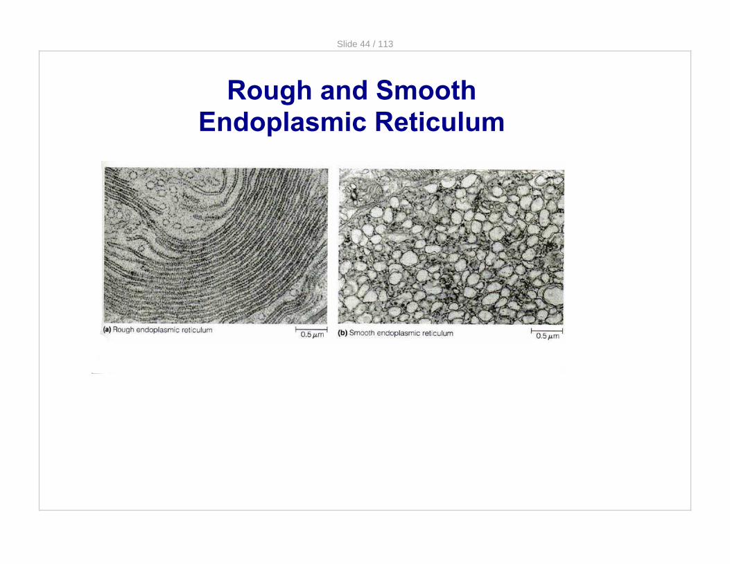
Rough and Smooth Endoplasmic Reticulum
Slide 44 / 113
Rough and Smooth Endoplasmic Reticulum
Slide 45 / 113
Smooth Endoplasmic Reticulum
This type of E.R. is called Smooth because it lacks ribosomes on its surface. (it looks smooth compared to rough ER)
There are a variety of functions of this organelle, which include:· making lipids.· processing certain drugs and poisons absorbed by the cell.· storing calcium ions (for example, in muscle cells).
Note: The liver is an organ that detoxifies substances that are brought into the body. Therefore, liver cells have huge amounts of Smooth E.R.

Slide 46 / 113
Rough Endoplasmic Reticulum
Rough E.R. has ribosomes attached to its membrane (thus a rough appearance).
These ribosomes synthesize proteins that will be used in the plasma membrane, secreted outside the cell or shipped to another organelle called a lysosome.
As proteins are made by the ribosomes, they enter the lumen (opening) of the E.R. where they are folded and processed.

Slide 47 / 113
Rough Endoplasmic Reticulum
Once the proteins are processed, short chains of sugars are sometimes linked to these proteins, which are then known as glycoproteins. These glycoproteins serve as "zip codes" that will tell the protein where it will go. Most secretory proteins have glycoproteins.
When the molecule is ready to be exported out of the E.R., it gets packaged into a transport vesicle. This vesicle is made of membranes from the E.R. itself. The transport vesicle travels to another organelle known as the Golgi apparatus.

Slide 48 / 113
Insulin is a protein hormone made by certain cells of the pancreas that enable cells to take glucose (sugar) in from the blood.
Insulin is made in the rough E.R. because it is a secretory protein. Specifically, it is secreted out of the pancreas cells into the blood stream.
Insulin - a product of the Rough Endoplasmic Reticulum

Slide 49 / 113
13 Which organelle is involved in making proteins?
A Smooth E.R.
B Ribosomes
C DNA
D Nuclear membrane

Slide 50 / 113
14 What determines if we classify endoplasmic reticulum as smooth or rough?
A presence or absence of nuclear pores
B presence or absence of genetic material
C presence or absence of ribosomes
D presence of absence of DNA

Slide 51 / 113
15 Where in the cell are lipids made?
A Nucleus
B Ribosomes
C Rough endoplasmic reticulum
D Smooth endoplasmic reticulum

Slide 52 / 113
Golgi Apparatus
The main function of this organelle is to finish, sort, and ship cell products. It works like the postal department of the cell.
Structurally, the golgi consists of stacked flattened sacs (sort of looks like a stack of pita bread).
Slide 53 / 113
The Golgi is located near the cell membrane. The Golgi works closely with the E.R. of a cell.
It receives and modifies substances manufactured by the E.R. Once the substances are modified, they are shipped out to other areas of the cell.
One key difference between the Golgi apparatus and endoplasmic reticulum is that the sacs comprising the Golgi are not interconnected.
Golgi Apparatus

Slide 54 / 113
The Golgi receives transport vesicles that bud off from the E.R. and contain proteins. It takes the substances contained in these vesicles and modifies them chemically in order to mark them and sort them into different batches depending on their destination.
The finished products are then packaged into new transport vesicles which will then move to lysosomes, or will be inserted into the plasma membrane or dumped out of the cell if the protein is a secretory protein.
The Golgi Apparatus & the E.R.
http://www.youtube.com/watch?v=rvfvRgk0MfAVideo onProtein Traffickingthrough the Golgi click

Slide 55 / 113
16 A difference between the Golgi Apparatus and the E.R. is that
A The ER takes the vesicles from the Golgi to transport
B The sacs making the Golgi are not interconnected
C The Golgi has ribosomes, the ER does not
D There is no difference, they are part of the same organelle

Slide 56 / 113
17 Which organelle receives and modifies substances from the endoplasmic reticulum?
A Nucleus
B Ribosomes
C Lysosomes
D Golgi Bodies
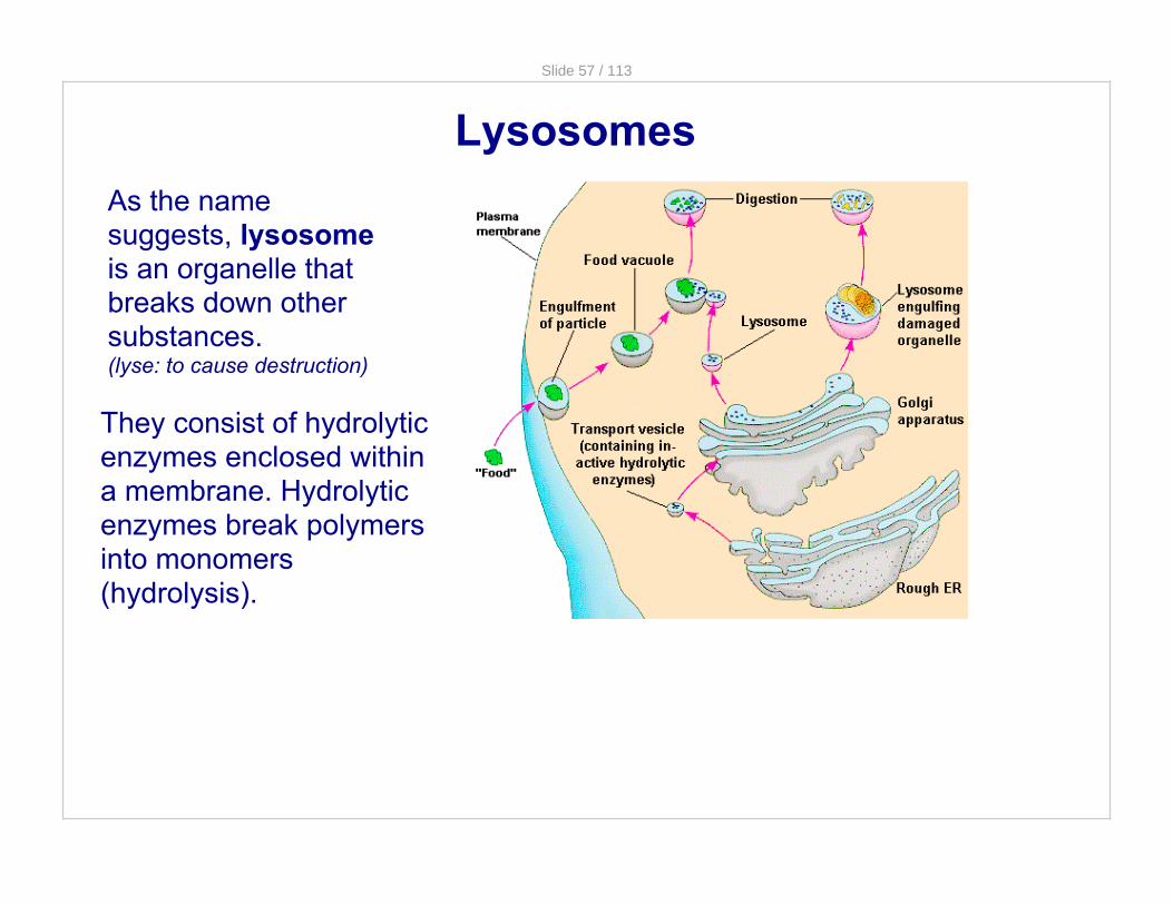
Slide 57 / 113
LysosomesAs the name suggests, lysosome is an organelle that breaks down other substances.(lyse: to cause destruction)
They consist of hydrolytic enzymes enclosed within a membrane. Hydrolytic enzymes break polymers into monomers (hydrolysis).

Slide 58 / 113
Lysosomes
Lysosomes may fuse with vacuoles containing food particles and then the enzymes digest the food, releasing nutrients into the cell. Protists do this.
Damaged organelles may become enclosed within a membranous vesicle which then fuses with a lysosome.
The organic molecules from the breakdown process are recycled and reused by the cell.

Slide 59 / 113
18 Which is not a function of lysosomes?
A aiding the cell in creating ribosomes
B fusing with vacuoles to digest food
C breaking polymers into monomers
D recycling worn out cell parts

Slide 60 / 113
19 Which organelle contains hydrolytic enzymes that break down other substances?
A Endoplasmic Reticulum
B Golgi Bodies
C Lysosomes
D Vacuoles

Slide 61 / 113
A peroxisome is a specific lysosome that forms and breaks down hydrogen peroxide (H2O2) which is toxic to cells.
In all cells, hydrogen peroxide forms constantly (from the combining of hydrogen and oxygen as bi-products of metabolism) and needs to be broken down quickly.
Important note: Peroxisomes are not part of the endomembrane system.
Peroxisomes

Slide 62 / 113
VacuolesVacuoles are also membranous sacs and they come in different shapes and sizes and have a variety of functions.
Central Vacuole
PLANTCELL
PROTIST

Slide 63 / 113
Types of Vacuoles
Central Vacuole
Contractile vacuoles
Food Vacuoles

Slide 64 / 113
Central VacuolesCentral Vacuole in plants stores water. Absorbing water makes a plant cell more turgid, or having more pressure inside - leading to strength and rigidity.
Central vacuoles that are full will take over most of the cytoplasm and literally push the organelles to the sides of the cell. It can also store vital chemicals, pigments and waste products.

Slide 65 / 113
Increased turgor pressure results from the central vacuole being full with water. It presses out on the cell membrane which then presses out on the cell wall.
Increased Turgor Pressure
"Turgid" cells are synonomous with fresh fruits and veggies.
The plant cell will not explode or lose its shape like an animal cell would in a hypotonic environment.

Slide 66 / 113
Decreased turgor pressure results when the central vacuole is not full with water. The central vacuole pulls away from the cell membrane which pulls away from the cell wall.
Decreased Turgor Pressure
When this happens the cell is limp and droopy. This is associated with wilted, limp lettuce, as well as droopy flowers.
However, the plant cell will not lose its shape. Only the central vacuole shrinks.

Slide 67 / 113
Contractile Vacuoles
Contractile vacuoles can be found in certain single-celled organisms. These act as a pump to expel excess water from the cell. This is especially helpful to those organisms living in a freshwater environment to keep the cell from exploding.

Slide 68 / 113
Food Vacuoles
Food Vacuoles are mainly found in protists.
The protist ingests food particles. The particles then fuse with a lysosome. The lysosome contains hydrolytic enzymes that break the food down. Paramecium fed dyed food showing vacuoles.

Slide 69 / 113
20 An organelle found in plant cells that stores water as well as other important substances is called the ___________.
A Lysosome
B Contractile Vacuole
C Central Vacuole
D Golgi bodies
Slide 70 / 113
21 Food vacuoles are primarily found in which organisms?
A Plants
B Animals
C Protists
D Bacteria

Slide 71 / 113
Energy-Converting Organelles
Chloroplasts reside in plant cells only and convert solar radiation into energy stored in the cell for later use.
Mitochondria reside in plant and animal cells and convert chemical energy from glucose into ATP.
Interestingly, both chloroplasts and mitochondria have their own DNA, separate from that found in the nucleus of the cell. They also have a double cell membrane.

Slide 72 / 113
Chloroplasts
These organelles convert solar energy to chemical energy through photosynthesis. Chloroplasts are partitioned into three major compartments by internal membranes.
Remember that during photosynthesis it is on the thylakoid that the Light Dependant Reactions take place.
In prokaryotes, thylakoids are areas of highly folded membranes.In eukaryotes, they are stacked in the chloroplasts.
eukaryotic chloroplast

Slide 73 / 113
MitochondriaMitochondria are sometimes referred to as the "powerhouses" of the cell. They convert chemical energy(glucose) into a more usable and regenerative form of chemical energy(ATP).
The mitochondrion only has two compartments as opposed to three in the chloroplast.
The mitochondrion is also partitioned like the chloroplast.

Slide 74 / 113
Remember cell respiration must take place near a membrane so that a proton gradient can be built in a "membrane space" that is separate from the rest of the cell. Thus, the membrane would separate the inner volume, with a deficit of protons, from the outside, with an excess.
In prokaryotes, the "inter- membrane space" is between the cell membrane and the cell wall.
In eukaryotes, that membrane is the Inter- Membrane Space of the Mitochondria in between the inner membrane and outer membrane.
Mitochondria and Respiration

Slide 75 / 113
Since mitochondrial DNA is not in the cell nucleus, it is only passed along from mother to child; animals, including you, inherit your mitochondria from your mother only.
This is because the egg from our mothers contained her organelles. (Dad's sperm only contains the chromosomes, none of his organelles usually).
All of our organelles we inherited from our mothers. Mitochondrial DNA is a way to trace maternal heritage through a family or through a species. The "Mitochondrial Eve" is the first human female that gave rise to all humans. In theory, we can trace all humans back to her through our mitochondrial DNA.
The Mitochondrial Eve

Slide 76 / 113
22 Which organelle converts solar energy into chemical energy in plants and other photosynthetic organisms?
A Nucleus
B Chloroplast
C Mitochondrion
D Golgi

Slide 77 / 113
23 Which organelle converts food energy into chemical energy that the cell can use?
A Nucleus
B Chloroplast
C Mitochondrion
D Golgi

Slide 78 / 113
Cytoskeleton
Cytoskeleton is a network of fibers within the cytoplasm.
Three types of fibers collectively make up the cytoskeleton:· Microfilaments· Intermediate filaments· Microtubules
These fibers provide structural support and are also involved in various types of cell movement and motility.
Slide 79 / 113
24 Cells can be described as having a cytoskeleton of internal structures that contribute to the shape, organization, and movement of the cell. All of the following are part of the cytoskeleton except
A the nuclear envelope.
B microtubules.
C microfilaments.
D intermediate filaments.

Slide 80 / 113
25 Which of the following is not a known function of the cytoskeleton?
A to maintain a critical limit on cell size
B to provide mechanical support to the cell
C to maintain the characteristic shape of the cell
D to hold mitochondria and other organelles in place within the cytosol

Slide 81 / 113
Remember the plasma membrane is a phospholipid bilayer with proteins and other molecules interspersed throughout.
Plasma Membrane
· Selective Permeability · Protection· Structural support
The 3 mainfunctions of theplasma membrane:

Slide 82 / 113
26 Which of the following statements about the role of phospholipids in forming membranes is correct?
A they are completely insoluble in water
B they form a single sheet in water
C they form a structure in which the hydrophobic portion faces outward
D they form a selectively permeable structure

Slide 83 / 113
But what if the substance that needs to pass through the cell membrane is too big for a protein carrier or intregal protein?
Then, the substance uses other ways of getting into or out of a cell by fusing with the cell membrane.
Large Molecules and the Plasma Membrane
There are several special functions of the membrane as larger substances enter and exit the cell.

Slide 84 / 113
The vesicles that enclose the proteins fuse with the plasma membrane and the vesicles then open up and spill their contents outside of the cell. This process is known as exocytosis. The vesicle will become part of the cell membrane
ExocytosisExocytosis
The proteins the cell makes are too large to diffuse through the phospholipid bilayer.
This is how secretory proteins from the Golgi exit the cell. This is true for insulin in the pancreas.

Slide 85 / 113
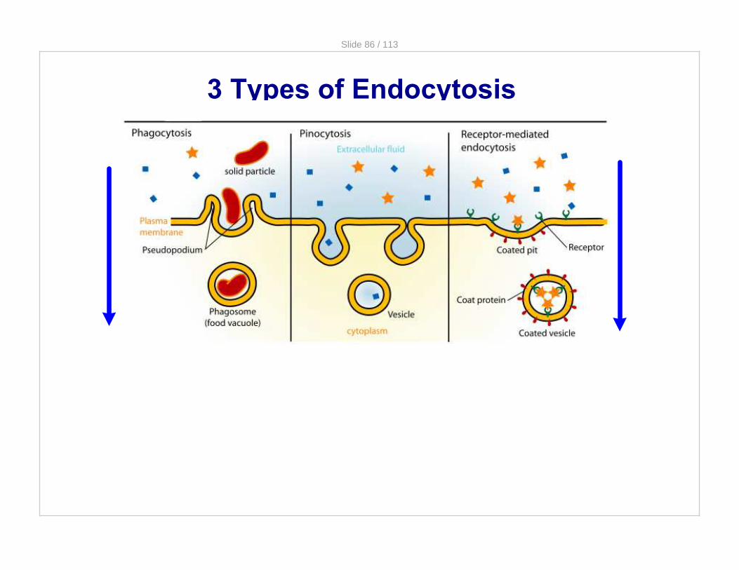
The opposite of exocytosis is endocytosis.
In this process, the cell takes in macromolecules or other particles by forming vesicles or vacuoles from its plasma membrane.
Endocytosis
This is how many protists ingest food particles
Slide 86 / 113
3 Types of Endocytosis

Slide 87 / 113
Phagocytosis Is for taking in solid particles. ("phago" mean to eat)
Pinocytosis Is for taking in liquids. However what the cell wants is not the liquid itself, but the substances that are dissolved in the liquid. ("pino" means to drink)
Receptor-mediated endocytosis requires the help of a protein coat and receptor on the membrane to get through.
3 Types of Endocytosis

Slide 88 / 113
27 The process by which a cell ingests large solid particles, therefore it is known as "cell eating".
A Pinocytosis
B Phagocytosis
C Exocytosis
D Osmoregulation

Slide 89 / 113
28 Protein coated vesicles move through the plasma membrane via this process:
A Phagocytosis
B Active Transport
C Receptor-Mediated Endocytosis
D Pinocytosis

Slide 90 / 113
29 After a vesicle empties its contents outside a cell, the vesicle becomes part of:
A the Golgi
B the plasma membrane
C another vesicle
D the extracellular fluid

Slide 91 / 113
Passive transport is the movement of substances from an area of high concentration to an area of low concentration without the requirement an energy input. Types include diffusion, osmosis, and facilitated diffusion.
Membrane Transport - review
Active transport is the movement of substances from an area of low concentration to an area of high concentration and requires an input of energy.
Passive Transport
Active Transport
(REQUIRES ENERGY)

Slide 92 / 113
30 Active transport moves molecules
A with their concentration gradients without the use of energy
B with their concentration gradients using energy
C against their concentration gradients without the use of energy
D against their concentration gradients using energy

Slide 93 / 113
31 Which of the following processes includes all others?
A passive transport
B facilitated diffusion
C diffusion of a solute across a membrane
D osmosis

Slide 94 / 113
Cell wallThe cell wall is an outer layer in addition to the plasma membrane, found in fungi, algae, and plant cells.
The composition of the cell wall varies among species and even between cells in the same individual.All cell walls have carbohydrate fibers embedded in a stiff matrix of proteins and other carbohydrates.
Plant cell walls are made of the polysaccharide cellulose. Fungal cell walls are made of the polysaccharide chitin.

Slide 95 / 113
Outside the Plasma Membrane - Extracellular Matrix
The extracellular matrix (ECM) found surrounding cells provides structural support to eukaryotic cells in addition to providing various other functions such as anchorage, cellular healing, separating tissues from one another and regulating cellular communication.
The ECM is primarily composed of an interlocking mesh of proteins and carbohydrates.

Slide 96 / 113
Cell Surfaces and Junctions
Cell surfaces protect, support, and join cells.
Cells interact with their environments and each other via their surfaces. Cells need to pass water, nutrients, hormones, and many, many more substances to one another. The way that cells that are adjacent to one another communicate and pass substances to one another are called Cell Junctions.
Animal and plant cells have different types of cell junctions. This is mainly because plants have cell walls and animal cells do not.

Slide 97 / 113
Plant cells are supported by rigid cell walls made largely of cellulose.
They connect by plasmodesmata which are channels that allow them to share water, food, and chemical messages.
Junctions specific to plant cells

Slide 98 / 113
Tight junctions
Adhering junctions
Communicating (Gap) junctions
Animal Cell Junctions

Slide 99 / 113
tight junction
Tight Junctions
Tight junctions can bind cells together into leakproof sheets
Example: the cells of the lining of the stomachor any epitheliallining where leaking of substances is not good.

Slide 100 / 113
Adhering Junctions
Adhering junctions fasten cells together into strong sheets. They are somewhat leakproof.
Example: actin is held together in muscle.

Slide 101 / 113
Communicating (Gap) JunctionsGap junctions allow substances to flow from cell to cell. They are totally leaky. They are the equivalent of plasmadesmata in plants.
Example: important in embryonic development. Nutrients like sugars, amino acids, ions, and other molecules pass through.

Slide 102 / 113
Organelles in Animal and Plant Cells
cell wall
chloroplastscentral vacuole
plasma membrane
mitochondria
Only Animal
Only Plant Both
rough ER
smooth ER
lysosomesgolgi
apparatus
ribosomesnucleus

Slide 103 / 113
The endosymbiotic theory states that eukaryotic cells arose as a result of a symbiotic relationship between different prokaryotic cells.
Endosymbiotic Theory

Slide 104 / 113
Endosymbiotic Theory
This idea has been best explained by the "Theory of Endosymbiosis" by Lynn Margulis in 1970.
She used 2 very special eukaryotic organelles to explain: · the mitochondria · the chloroplast

Slide 105 / 113
Remember how we said the mitochondria and chloroplast are different from other eukarytoic organelles because they have their own DNA, their own ribosomes, and have a double cell membrane.
Using these facts, she explained that the mitochondria and chloroplast were once free-living prokaryotes that got taken up (or "eaten") by another prokaryote.
The mitochondria was a bacteria that could make its own ATP. The chloroplast was a bacteria that could make its own food.
The Evolution of Eukaryotes

Slide 106 / 113
When they got taken up by another prokaryote, they dragged the one prokaryote's cell membrane around theirs, thus the double cell membrane. This now allowed the "new" eukaryote to make its own ATP or be able to do photosynthesis and make its own food. Thus the evolution of eukaryotes.
The nucleus and flagella could also have the same possible roots although they are not as heavily supported with evidence as the mitochondria and chloroplast.
The Evolution of Eukaryotes

Slide 107 / 113
Endosymbiosis

Slide 108 / 113
Evidence for SymbiosisBoth mitochondria and chloroplasts can arise only from preexisting mitochondria and chloroplasts. They cannot be formed in a cell that lacks them.
Both mitochondria and chloroplasts have their own DNA and it resembles the DNA of bacteria not the DNA found in the nucleus.
Both mitochondrial and chloroplast genomes consist of a single circular molecule of DNA, just like in prokaryotes.
Both mitochondria and chloroplasts have their own protein-synthesizing machinery, and it more closely resembles that of bacteria than that found in the cytoplasm of eukaryotes.

Slide 109 / 113
Evidence for SymbiosisBoth mitochondria and chloroplasts can arise only from preexisting mitochondria and chloroplasts. They cannot be formed in a cell that lacks them.
Both mitochondria and chloroplasts have their own DNA and it resembles the DNA of bacteria not the DNA found in the nucleus.
Both mitochondrial and chloroplast genomes consist of a single circular molecule of DNA, just like in prokaryotes.
Both mitochondria and chloroplasts have their own protein-synthesizing machinery, and it more closely resembles that of bacteria than that found in the cytoplasm of eukaryotes.

Slide 110 / 113
Evidence for SymbiosisBoth mitochondria and chloroplasts can arise only from preexisting mitochondria and chloroplasts. They cannot be formed in a cell that lacks them.
Both mitochondria and chloroplasts have their own DNA and it resembles the DNA of bacteria not the DNA found in the nucleus.
Both mitochondrial and chloroplast genomes consist of a single circular molecule of DNA, just like in prokaryotes.
Both mitochondria and chloroplasts have their own protein-synthesizing machinery, and it more closely resembles that of bacteria than that found in the cytoplasm of eukaryotes.

Slide 111 / 113
Evidence for SymbiosisBoth mitochondria and chloroplasts can arise only from preexisting mitochondria and chloroplasts. They cannot be formed in a cell that lacks them.
Both mitochondria and chloroplasts have their own DNA and it resembles the DNA of bacteria not the DNA found in the nucleus.
Both mitochondrial and chloroplast genomes consist of a single circular molecule of DNA, just like in prokaryotes.
Both mitochondria and chloroplasts have their own protein-synthesizing machinery, and it more closely resembles that of bacteria than that found in the cytoplasm of eukaryotes.

Slide 112 / 113
32 Which of the following does NOT provide evidence for the endosymbiotic theory?
A Mitochondria and chloroplasts both have their own DNA.
B Mitochondria and chloroplasts both come from pre-existing mitochondria and chloroplasts.
C The DNA of mitochondria and chloroplasts resembles the DNA found in nuclei.
D The DNA of mitochondria and chloroplasts resembles that of bacteria.

Slide 113 / 113
http://www.ce llsa live.com/ce lls /3dcell.htm
Plant and Animal Cell Organelle Review