Embed Size (px)
Citation preview
Esco1 and Esco2 regulate distinct cohesin functionsduring cell cycle progressionReem M. Alomera,1, Eulália M. L. da Silvab,1, Jingrong Chenb, Katarzyna M. Piekarzb, Katherine McDonaldb,Courtney G. Sansamb, Christopher L. Sansama,b, and Susannah Rankina,b,2
aDepartment of Cell Biology, University of Oklahoma Health Sciences Center, Oklahoma City, OK 73104; and bProgram in Cell Cycle and Cancer Biology,Oklahoma Medical Research Foundation, Oklahoma City, OK 73104
Edited by Douglas Koshland, University of California, Berkeley, CA, and approved July 31, 2017 (received for review May 19, 2017)
Sister chromatids are tethered together by the cohesin complex fromthe time they are made until their separation at anaphase. The abilityof cohesin to tether sister chromatids together depends on acetyla-tion of its Smc3 subunit by members of the Eco1 family of cohesinacetyltransferases. Vertebrates express two orthologs of Eco1, calledEsco1 and Esco2, both of which are capable of modifying Smc3, buttheir relative contributions to sister chromatid cohesion are unknown.We therefore set out to determine the precise contributions of Esco1and Esco2 to cohesion in vertebrate cells. Herewe show that cohesionestablishment is critically dependent upon Esco2. Although mostSmc3 acetylation is Esco1 dependent, inactivation of the ESCO1 genehas little effect on mitotic cohesion. The unique ability of Esco2 topromote cohesion is mediated by sequences in the N terminus of theprotein. We propose that Esco1-dependent modification of Smc3 regu-lates almost exclusively the noncohesive activities of cohesin, such asDNA repair, transcriptional control, chromosome loop formation, and/orstabilization. Collectively, our data indicate that Esco1 and Esco2 con-tribute to distinct and separable activities of cohesin in vertebrate cells.
chromosome biology | sister chromatid cohesion | Esco enzymes | cell cycle
Cohesin is a multisubunit protein complex first identifiedbased on its role in tethering together sister chromatids in M
phase cells. Since that time, cohesin has also been shown to playcritical roles in certain kinds of DNA repair and, in higher eu-karyotes, in chromosome structure. All of cohesin’s activitiesdepend on its ability to entrap or tether chromatin: in the case ofsister chromatid cohesion, cohesin tethers together the twoidentical products of DNA replication as they emerge from thereplication fork; in its structural role, cohesin is proposed tostabilize chromosome loops (1–5).The stability of the interaction between cohesin and chro-
matin is controlled in part by acetylation of the head domainof the Smc3 subunit of the complex. This acetylation inhibits openingof the cohesin ring by the protein Wapl, thereby stabilizing cohesion(6, 7). In budding yeast, cohesin is acetylated by the Eco1 acetyl-transferase (8–10). Vertebrates express two related acetyltransferaseenzymes, called Esco1 and Esco2, but their relative contributions tocohesin regulation are not clear. In embryonic extracts, the two Escoenzymes are not functionally redundant. Depletion of Esco2from Xenopus egg extracts results in loss of cohesion. Supple-mentation of extracts with recombinant Esco1, which is not nor-mally expressed in the early frog embryo, rescues Smc3 acetylation,but does not restore sister chromatid tethering (11). In contrast,some reports using cultured somatic cells have suggested that bothEsco1 and Esco2 contribute to sister chromatid cohesion, as si-multaneous depletion of both enzymes resulted in cohesion defectsthat were more severe than either single depletion (12).Esco1 and Esco2 have distinct patterns of expression relative
to cell cycle progression. While Esco1 is present at nearly con-stant levels throughout the cell cycle, Esco2 is a substrate of theanaphase promoting complex/cyclosome (APC/C), an E3 ubiquitinligase that is activated at mitotic exit (11–13). Thus, Esco2 levelsare low in G1, and only increase as APC activity drops duringS phase.
Finally, chromatin immunoprecipitation experiments in somaticcells indicate that Esco1 and Esco2 have distinct chromosomaladdresses. Colocalization of Esco1 with the insulator proteinCTCF and cohesin at the base of chromosome loops suggests thatEsco1 promotes normal chromosome structure (14, 15). Consistentwith this, depletion of Esco1 in somatic cells results in dysregulatedtranscriptional profiles (15). In contrast, Esco2 is localized to dis-tinctly different sites, perhaps due to association with the CoRESTrepressive complex (15, 16).Here, using a combination of siRNA-mediated depletion,
rescue, and CRISPR/Cas9-mediated genome editing, we definethe contributions of Esco1 and Esco2 to sister chromatid cohesionand Smc3 acetylation during cell cycle progression. We show thatthe majority of Smc3 acetylation is due to the activity of Esco1,while cohesion establishment during S phase requires Esco2. In-activation of the ESCO1 gene has insignificant impact on mitoticcohesion. We propose that cohesin acetylation by Esco1 promotesnormal chromosome structure throughout interphase and providesepigenetic memory during cell division by ensuring cohesin stabi-lization at appropriate loci upon mitotic exit. In contrast Esco2-dependent cohesin modification is essential during DNA replica-tion for the establishment of cohesion between sister chromatids.
ResultsThe Contributions of Esco1 and Esco2 to Sister Chromatid Cohesion.Like the founding member of the family, budding yeast Eco1, thevertebrate Esco enzymes both contain a PCNA interacting
Significance
Sister chromatids are tethered together by the cohesincomplex from the time they are made until cell division.Acetylation of the Smc3 subunit of cohesin stabilizes its as-sociation with chromatin, and is critical for sister chromatidcohesion. In vertebrates, cohesin is acetylated by two relatedenzymes: Esco1 and Esco2. We show here that Esco1 is re-sponsible for most Smc3 acetylation but has very little effecton sister cohesion. Esco1 is active throughout the cell cycle,while Esco2 modifies cohesin only during S phase, whensister chromatid cohesion is established. We propose thattwo distinct pathways regulate cohesin in vertebrates: one isdedicated to cohesion between sister chromatids, and onepromotes other functions of cohesin, such as maintenance ofchromosome structure.
Author contributions: R.M.A., E.M.L.d.S., K.M.P., and S.R. designed research; R.M.A., E.M.L.d.S.,J.C., K.M.P., K.M., C.G.S., and S.R. performed research; E.M.L.d.S., J.C., K.M.P., K.M., and S.R.contributed new reagents/analytic tools; R.M.A., E.M.L.d.S., K.M.P., K.M., C.G.S., C.L.S., and S.R.analyzed data; and R.M.A. and S.R. wrote the paper.
The authors declare no conflict of interest.
This article is a PNAS Direct Submission.1R.M.A. and E.M.L.d.S. contributed equally to this work.2To whom correspondence should be addressed. Email: [email protected].
This article contains supporting information online at www.pnas.org/lookup/suppl/doi:10.1073/pnas.1708291114/-/DCSupplemental.
9906–9911 | PNAS | September 12, 2017 | vol. 114 | no. 37 www.pnas.org/cgi/doi/10.1073/pnas.1708291114
Dow
nloa
ded
by g
uest
on
Apr
il 23
, 202
0

protein (PIP) box, a C2H2 zinc finger, and a catalytic region atthe C terminus (12, 17). In contrast to the yeast protein, both Esco1and Esco2 contain long N-terminal extensions, whose functions arepoorly characterized. These regions show no obvious sequence orstructural similarities between them (Fig. 1A).To define the contributions of Esco1 and Esco2 to sister
chromatid cohesion, we scored mitotic cohesion in HeLa cellsthat had been depleted of Esco1 and Esco2, either singly or to-gether, by siRNA-mediated depletion (Fig. 1B). Chromosomespreads were scored as representing one of four states of cohesion:
I–IV (Fig. 1C). Spreads that fell into categories III and IV wereconsidered defective in cohesion. Depletion of the cohesion reg-ulator Sororin was used as positive control for loss of cohesion(18). Depletion of Esco2 caused loss of cohesion in mitotic cells,with ∼30% of mitotic spreads showing defective cohesion (Fig. 1 Dand E). In contrast, acute depletion of Esco1 did not cause sig-nificant loss of cohesion, consistent with the analysis of a geneticknockout (see below). Codepletion of Esco1 with Esco2 caused anincrease in defective cohesion compared with cells depleted of Esco2only (Fig. 1E). In some experiments,>65% of mitotic spreads showeddefective cohesion when depleted of both enzymes. We concludethat mitotic cohesion requires Esco2-dependent cohesin modification.
Mitotic Progression Is Delayed in Cells Depleted of Esco2 but NotEsco1. We observed little effect of Esco1 depletion on mitoticcohesion (Fig. 1), although loss of cohesion had previously beenreported in Esco1 RNAi experiments (8, 12, 14). We thereforesought more sensitive methods to detect subtle cohesion defects.Because reduced cohesion can lead to activation of the spindlecheckpoint, a signal transduction pathway that prevents mitoticexit in the presence of unattached chromosomes, we tested theeffects of Esco1 and/or Esco2 depletion on mitotic progression.HeLa cells stably expressing red fluorescent protein (RFP)-histoneH2B were transfected with siRNAs and analyzed by time-lapsemicroscopy. The duration of mitosis in each of the cell pop-ulations from nuclear envelope breakdown (NEBD) until segre-gation of chromosomes into two masses was measured. Controlcells progressed through mitosis in ≤40 min (mean = 34.2 min; n =50), as did the cells depleted of Esco1 (mean = 33.7 min; n = 54)(Fig. 2A). Cells depleted of Esco2 frequently showed sustainedmitotic arrest (mean = 185 min; n = 102), as did cells that weredepleted of both Esco1 and Esco2 (mean = 367 min; n = 79). Themitotic arrest in these cells was similar to that seen in the samplesdepleted of Sororin (18). These results are broadly consistent withthe analysis of chromosome spreads (Fig. 1); loss of cohesion wasevident in the absence of Esco2, and this effect was exacerbated inthe absence of Esco1. Depletion of Esco1 alone had no significanteffect on mitotic cohesion or progression through mitosis.As an independent means of determining whether depletion of
the Esco enzymes results in an increased mitotic index, we an-alyzed the Esco1- and/or Esco2-depleted cells by flow cytometryusing phosphorylated histone H3 (pH3) as a marker for M phasecombined with DNA content analysis. Control and Esco1-depleted cells had similar percents of cells in mitosis, 4.37%and 4.24% (Fig. 2B). In contrast, depletion of Esco2, codepletionof Esco1 and Esco2, or depletion of Sororin all caused an in-crease of the percentage of cells in M phase compared withcontrols (Fig. 2B). We conclude that Esco2 activity is sufficientto promote full cohesion, and that Esco1’s ability to promotecohesion is only detectable when Esco2 levels are reduced.
Esco1 Knockout Cell Lines Have Essentially Normal Mitotic Cohesion.We did not detect an impact of Esco1 depletion on sister chro-matid cohesion, in contrast to previous reports (11, 12). Becauseincomplete depletion following RNAi is particularly problematicwhen manipulating expression of enzymes as small amounts ofcatalytic activity can suffice, we used CRISPR/Cas9 technologyto inactivate the ESCO1 gene in HeLa cells. The Cas9 nuclease wastargeted to two different regions of the Esco1 gene using gRNAsequences unique in the human genome. Individual clones werescreened by immunoblot for loss of Esco1 expression, and inacti-vation of the gene was confirmed by sequence analysis of genomicDNA. Two independent lines were isolated containing inactivatingmutations in the ESCO1 gene (Fig. S2). Perhaps surprisingly, theESCO1KO cells grew indistinguishably from their parental coun-terparts under standard culture conditions, and bulk DNA replica-tion appeared unaffected (Fig. S3). Consistent with its crucial function
Fig. 1. Contributions of Esco1 and Esco2 to mitotic cohesion. (A) Schematic ofSaccharomyces cerevisiae Eco1p and the vertebrate Esco1 and Esco2 enzymeswith conserved domains indicated. The catalytic acetyltransferase domain isshown in blue, and the conserved zinc fingers and PCNA interacting protein (PIP)box are shown in black and gold, respectively. Nonhomologous N-terminal ex-tensions in Esco1 and Esco2 are shown in green and light blue, respectively.(B) HeLa cells were transfected with siRNA against the indicated proteins, andwhole cell lysates were analyzed by immunoblot for the indicated proteins. Thecytosolic scaffold protein Nck was used as a loading control. * indicates back-ground band detected by Esco2 antibody (see Fig. S1). (C) Chromosome spreadswere prepared from the same samples shown in B and cohesion phenotypeswere classified as one of four states of cohesion, I–IV, as shown. (I) Unresolvedsister chromatid arms; (II) resolved arms, with tight centromeres, (III) sepa-rated sisters that remain near to each other; and (IV) scattering of individual sisterchromatids. (Scale bar, 20 μm.) (D) The percentage of cells with defective cohesionin categories III (separated) and IV (scattered) was determined for cellsdepleted of the indicated proteins. Data are presented as the percentageof mitotic spreads with the indicated phenotype, n ≥ 100 for all samples(E ). Total loss of cohesion (categories III and IV): cumulative data from six sepa-rate experiments that were scored as in D. Boxes: 25–75% data range; whiskers:total data range. ****P < 0.0001; **P = 0.0021; n.s., not significant; ANOVA,n ≥ 100 all samples.
Alomer et al. PNAS | September 12, 2017 | vol. 114 | no. 37 | 9907
CELL
BIOLO
GY
Dow
nloa
ded
by g
uest
on
Apr
il 23
, 202
0

for cohesion, repeated attempts to knock out both ESCO2 allelesusing a similar strategy were unsuccessful (Discussion).The ESCO1KO cells were used to determine the contributions
of Esco1 and Esco2 to Smc3 acetylation. Cells were synchronizedin M phase by sequential thymidine-nocodazole arrest, released toresume cell cycle progression, and assayed for Smc3 acetylation onK105/106 and DNA content (Fig. 3 A and B). Progression throughS phase was similar in the parental and ESCO1KO cells (Fig. 3B). Inmitotic cells (t = 0), Smc3 acetylation was relatively low, as seenpreviously (14). Smc3 acetylation in the parental cells rose early inG1, well before DNA replication was evident (Fig. 3A). In contrast,in the ESCO1KO cell line, Smc3 acetylation remained low until bulk
DNA replication occurred, ∼8 h after nocodazole release. Thus,Esco1-dependent acetylation occurs throughout interphase, bothbefore and during DNA replication, and at all time points themajority of Smc3 acetylation on K105/106 is dependent uponEsco1 (Fig. 3C), while Esco2-dependent acetylation occurs onlycoincident with DNA replication.Consistent with our siRNA experiments (Fig. 1), we found that
cohesion was largely unaffected by inactivation of the ESCO1gene alone (Fig. 4A). Depletion of Esco2 from the parentalHeLa cell line resulted in significant loss of cohesion (∼45%).Strikingly, depletion of Esco2 from the ESCO1KO cell line re-sulted in catastrophic loss of cohesion, similar to depletion of theessential cohesion regulator Sororin. The mobility of Sororinwas reduced in these samples, consistent with mitotic arrest (Fig.4B). These data indicate that Esco1 enhances cohesion when Esco2is present, but does not normally contribute to sister chromatidcohesion.Although ESCO1 gene inactivation alone caused no obvious
loss of cohesion, it remained possible that cohesion was reduced,but not sufficiently to activate the spindle checkpoint. To explorethis possibility, we measured the distance between sister centro-meres, comparing parental and ESCO1KO cells that were stainedwith calcinosis, Reynaud’s phenomenon, esophageal dysmotility,sclerodactyly, and telangiectasia (CREST) serum to label centro-meric proteins. The intercentromere distance in parental andESCO1KO cells was not significantly different (Fig. 4C), indicatingthat cohesion at the centromere region is not greatly affected byloss of Esco1 function.
Fig. 2. Mitotic progression is delayed in cells depleted of Esco2. HeLa cellsstably expressing RFP-H2B were transfected with siRNA against the indicatedproteins and time-lapse images were collected starting 24 h after trans-fection. (A) The duration of mitosis was measured starting 24 h after siRNAtransfection. Each blue line represents a single mitotic event, with the lengthof the line indicating the duration of mitosis from nuclear envelope break-down until the separation of two RFP-positive DNA masses, shown on x axis.Data from one representative experiment are shown. (B) Parallel sampleswere harvested 48 h after siRNA transfection, fixed, and stained for phos-phorylated histone H3 (pH3) as a marker of mitosis, and analyzed by flowcytometry for both DNA content (x axis) and pH3 signal (y axis). The per-centages of cells in mitosis based on pH3 staining (boxed) are indicated foreach sample. Significant differences in pH3-positive cells compared withcontrol cells are indicated: ****P < 0.0001; n.s., not significant; χ2 with Yatescorrection.
Fig. 3. Esco1-dependent Smc3 acetylation occurs throughout interphase. Pa-rental and ESCO1KO cells were synchronized in M phase by sequential thymi-dine and nocodazole treatment. Following washout, samples were collected atthe indicated times for immunoblot analysis (A) and to measure DNA contentby flow cytometry (B). All samples were run on the same gel, and blots were cuthorizontally (Fig. S4). The relative amount of Smc3Ac (normalized to total Smc3)in parental and ESCO1KO cells was quantified by comparing chemiluminescentsignal intensities in A and is presented in C. As, asynchronously growing cells.T = 0 samples were collected before nocodazole washout. * indicates back-ground band detected by Esco2 antibody (see Fig. S1).
9908 | www.pnas.org/cgi/doi/10.1073/pnas.1708291114 Alomer et al.
Dow
nloa
ded
by g
uest
on
Apr
il 23
, 202
0
During sustained mitotic arrest, cohesion between sister chro-matids eventually fails, and the rate at which this failure occurs isinversely correlated with the overall level of mitotic cohesion (19,20). We measured “cohesion fatigue” in the ESCO1KO cells com-pared with parental controls. The ESCO1KO cells showed slightlyelevated rate of fatigue, suggesting that Esco1 makes a measurablecontribution to the strength of mitotic cohesion established by Esco2activity (Fig. 4D).
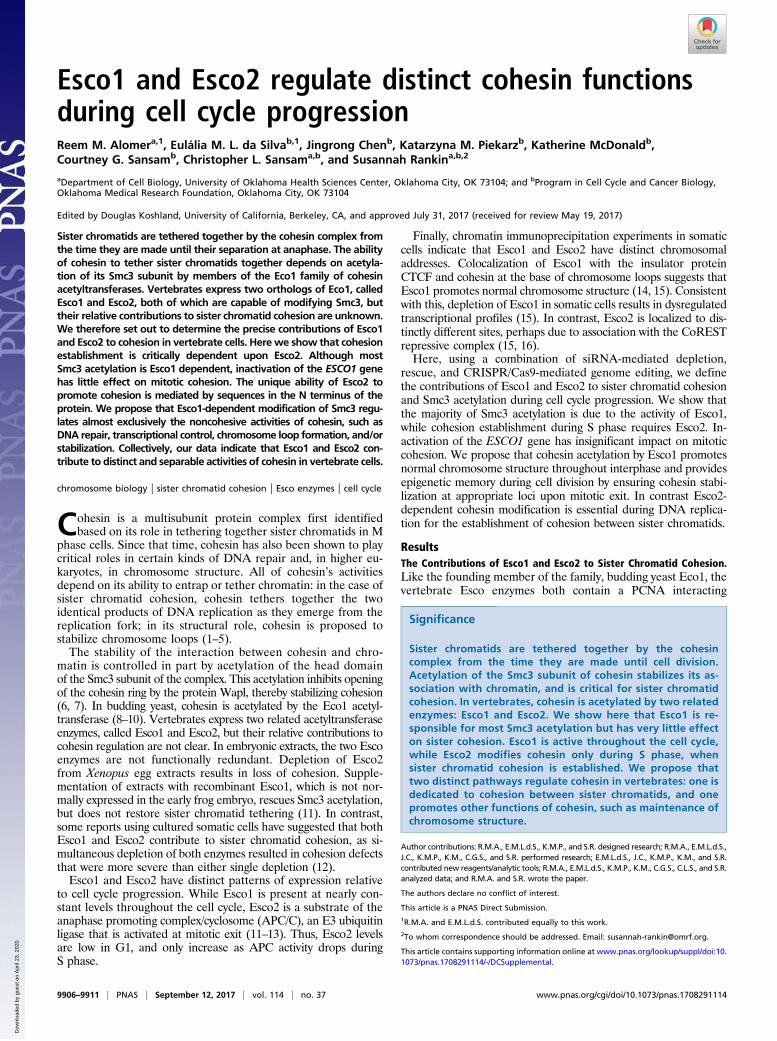
Esco Activity Depends on N-Terminal Sequences. To determinewhether the ability of Esco2 to promote cohesion is dependentupon the unique sequences in its N terminus, we performed rescueexperiments using chimeric fusion proteins. The N terminus ofEsco1 was fused at the PIP box to the C terminus of Esco2 togenerate a fusion protein called Esco1-2 (Fig. 5A). The conversecDNA fusion, Esco2-1, was also constructed (Fig. S5A). The geneswere integrated into the ESCO1KO cell line in a manner thatallowed transgene expression in a tetracycline-inducible manner(Fig. 5B). Expression of the transgenes was induced with doxycy-cline, and cells were transfected with siRNA directed against the
endogenous Esco2. To avoid depletion of the transgenes beingtested, siRNAs were chosen to target the regions of the endogenousEsco2 transcript not present in the transgene. Analyzing chromo-some spreads as in Fig. 1, we found that Esco2-1 was able to pro-mote cohesion, while Esco1-2 was not (Fig. 5C). Thus, the uniquecapacity of Esco2 to promote sister chromatid cohesion lies in its Nterminus, which cannot be substituted with the N terminus of Esco1.The level of Smc3 acetylation was lower in the cells expressing
Fig. 4. ESCO1KO cells have largely normal sister chromatid cohesion.(A) Cohesion assay. Chromosome spreads from parental and ESCO1KO cells werescored for sister chromatid cohesion following transfection with the in-dicated siRNAs. Shown are the mean and SD of three replicate experiments.(B) Immunoblot showing levels of the indicated proteins from a represen-tative experiment included in A. Both histone H3 and Nck were used asloading controls. * indicates background band. (C ) Intercentromere dis-tance. The distance between sister centromeres was measured in parentaland ESCO1KO cells stained with CREST serum. Error bars represent minimum/maximum, box includes 25th–75th percentiles. Unpaired t test P = 0.06 n =116 (parental) and 112 (ESCO1KO). n.s., not significant. (D) Cohesion fatigue.Analysis of cohesion fatigue in parental and ESCO1KO cell lines. The spindle-dependent separation of sister chromatids during M phase arrest wasassessed by comparing samples treated with MG132 (blue bars) to cellstreated with both MG132 and nocodazole (green bars). The graph indicatespercent of spreads with scattered chromatids at 0, 4, and 6 h of treatment.n ≥ 100 for all samples. **P = 0.0137 Fisher’s exact.
Fig. 5. The N terminus of Esco2 promotes cohesion establishment. ChimericcDNAs were generated to express fusions between the Esco1 (green) andEsco2 (blue) proteins. (A) Cartoon illustrating the wild-type proteins, as wellas chimeric derivatives, exchanged precisely at the PIP boxes (shown inblack), as shown (dotted lines). Bars below each cartoon indicate the regionrecognized by antibodies against Esco2 (dark green) and Esco1 (dark blue).(B) Immunoblot analysis of expression of chimeric proteins. ESCO1KO cellswere engineered to express Esco1, Esco1-2, or Esco2-1 in a doxycycline-inducible manner. The expression of each transgene following treatmentwith doxycycline was assessed using appropriate antibodies as indicated inA. Samples were all blotted together from the same gel; dotted line wasadded for clarity. To assess the ability of the chimeras to promote sisterchromatid cohesion, cells were also treated with one of two siRNAs, selectedto react only against the endogenous Esco2 gene. These siRNAs, “a” (Nterminal) and “b” (C terminal) are illustrated in A; “c” = control siRNA.(C) The samples probed in B were assayed for sister chromatid cohesion andscored as described in Fig. 1. (D) ESCO1KO cells expressing the indicatedtransgenes were synchronized in S phase by treatment with thymidine for20 h and analyzed by immunoblot for protein expression. Shown also issubcellular fractionation analysis of endogenous Esco1 and Esco2 in cellsgrowing asynchronously (E) or arrested in M phase with nocodazole (F). N,chromatin associated; S, cytosolic supernatant; T, total cell lysate. TheEsco2 band in M phase cells is difficult to detect due to the presence of abackground band (*) (see also Fig. S1).
Alomer et al. PNAS | September 12, 2017 | vol. 114 | no. 37 | 9909
CELL
BIOLO
GY
Dow
nloa
ded
by g
uest
on
Apr
il 23
, 202
0
Esco1-2 compared with Esco2-1. This may reflect nearly completecohesion failure and mitotic arrest in the Esco1-2 cells, as in Mphase, the overall acetylation is relatively low (Fig. 3). Alternatively,this result may suggest that Esco1-2 is intrinsically unable to pro-mote Smc3 acetylation. To distinguish between these two possibil-ities, we synchronized cells in S phase and found that Esco1-2chimera still did not promote Smc3 acetylation, while Esco2-1, whichwas able to rescue cohesion (Fig. 5D), did. Additional Esco1-2clones had the same phenotype (Fig. S5B). Thus, the C-terminal,catalytic domain of Esco1 is functional for both Smc3 acetylationand cohesion establishment when fused to the N terminus of Esco2.We do not yet fully understand why the Esco1-2 fusion is inactive;perhaps dimerization, possibly important for Esco function (21, 22),is critically impaired in the Esco1-2 chimera.To further characterize the differences between Esco1 and
Esco2 we analyzed their localization in fractionated cells. Inasynchronously growing cells, both enzymes were associated withthe chromatin fraction (Fig. 5E). In contrast, although Esco1 re-cruitment to chromatin is dependent upon the cohesin complex (6,7, 15), Esco1 remained associated with chromatin in M phase,when cohesin is largely dispersed (Fig. 5F), as noted previously (8–10, 12). Esco2 was difficult to detect in M phase samples (see alsoFig. 3 and Fig. S1), suggesting that it may be at least partiallydegraded before metaphase (Fig. 5E). Importantly, Esco1 is re-tained on chromosomes in M phase where it can promote Smc3acetylation directly upon mitotic exit. These data are consistentwith a model in which Esco1 regulates cohesin beginning in earlytelophase.
DiscussionAcetylation of the Smc3 subunit of cohesin by members of theEco1 family of acetyltransferases stabilizes the interaction ofcohesin with chromatin, and is essential for cohesion establishment.The advent of CRISPR-based genome editing has allowed us toassess unambiguously the relative contributions of the two verte-brate Eco enzymes, Esco1 and Esco2, to mitotic tethering of sisterchromatids. Our results suggest that Esco1 is only able to promotecohesion when cohesion has already been established by the actionof Esco2 during S phase. Loss of Esco1, either acutely, by siRNA-mediated depletion, or genetically, by gene inactivation, did notcause overt loss of mitotic cohesion. This observation is reminiscentof G2 cohesion establishment in response to DNA damage inbudding yeast, which is only effective in tethering sister chromatidsif replicative establishment has already occurred (23). VertebrateEsco1 then, may contribute to or strengthen sister chromatid co-hesion, but is not dedicated to replicative cohesion establishment.In contrast, depletion of Esco2 alone caused significant loss of co-hesion. Importantly, in the absence of Esco1, depletion of Esco2 ledto complete loss of cohesion. These data are consistent with earlierwork, in which we showed that Esco1 does not promote cohesion inEsco2-depleted Xenopus egg extracts (13). In fact Esco1 is notexpressed in the early frog embryo until the onset of zygotic tran-scription. Thus, Esco2 is sufficient to promote proper tethering ofsister chromatids during early embryonic development. Our workhere contradicts previous work suggesting that Esco1 makes criticalcontributions to mitotic sister chromatid cohesion in somatic cells(12). This prior result may reflect off-target effects of siRNAs,which were used at higher concentration than used here, or perhapsdifferences among strains of HeLa cells. The consistency of our datafrom both ESCO1 knockout cells and siRNA-mediated depletionexperiments makes us confident in our conclusions.If the contribution of Esco1 to mitotic sister chromatid tethering is
minimal in both embryonic and somatic cells, what then is its func-tion? Esco1 acetylates cohesin throughout interphase, even well be-fore DNA replication is evident. As cohesin plays critical roles information or maintenance of interphase chromosome structure (2, 3,14, 15, 24–29), one attractive model is that Esco1 contributes to theseevents, perhaps through impacts on chromosome loop formation
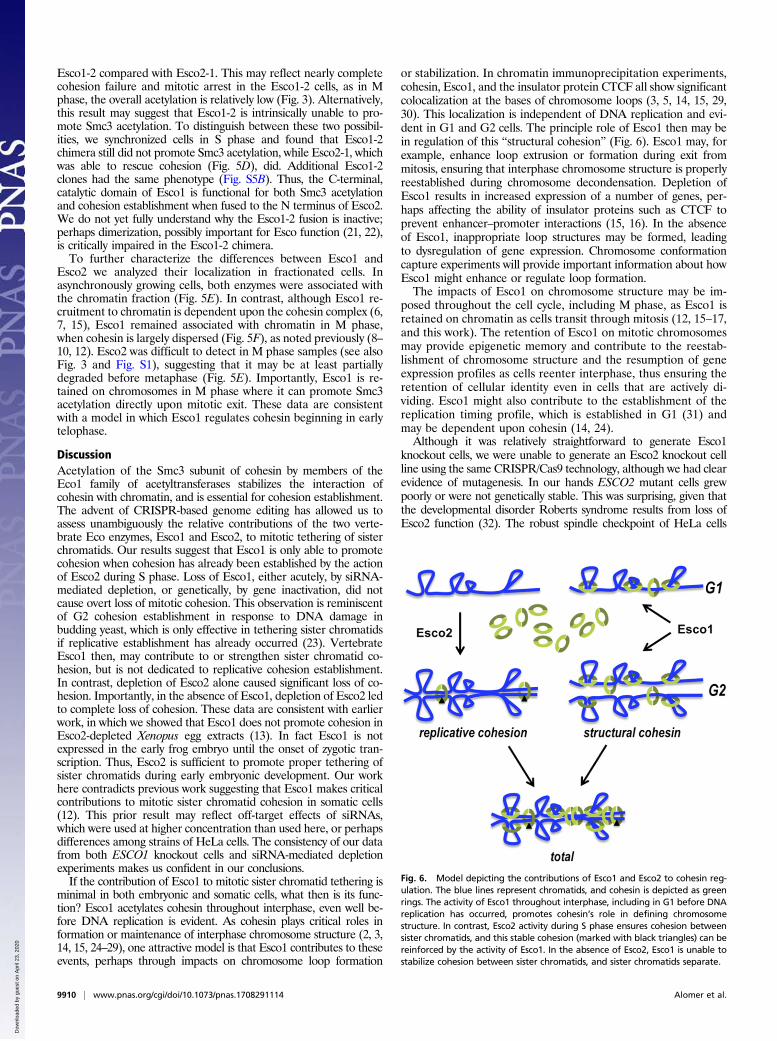
or stabilization. In chromatin immunoprecipitation experiments,cohesin, Esco1, and the insulator protein CTCF all show significantcolocalization at the bases of chromosome loops (3, 5, 14, 15, 29,30). This localization is independent of DNA replication and evi-dent in G1 and G2 cells. The principle role of Esco1 then may bein regulation of this “structural cohesion” (Fig. 6). Esco1 may, forexample, enhance loop extrusion or formation during exit frommitosis, ensuring that interphase chromosome structure is properlyreestablished during chromosome decondensation. Depletion ofEsco1 results in increased expression of a number of genes, per-haps affecting the ability of insulator proteins such as CTCF toprevent enhancer–promoter interactions (15, 16). In the absenceof Esco1, inappropriate loop structures may be formed, leadingto dysregulation of gene expression. Chromosome conformationcapture experiments will provide important information about howEsco1 might enhance or regulate loop formation.The impacts of Esco1 on chromosome structure may be im-
posed throughout the cell cycle, including M phase, as Esco1 isretained on chromatin as cells transit through mitosis (12, 15–17,and this work). The retention of Esco1 on mitotic chromosomesmay provide epigenetic memory and contribute to the reestab-lishment of chromosome structure and the resumption of geneexpression profiles as cells reenter interphase, thus ensuring theretention of cellular identity even in cells that are actively di-viding. Esco1 might also contribute to the establishment of thereplication timing profile, which is established in G1 (31) andmay be dependent upon cohesin (14, 24).Although it was relatively straightforward to generate Esco1
knockout cells, we were unable to generate an Esco2 knockout cellline using the same CRISPR/Cas9 technology, although we had clearevidence of mutagenesis. In our hands ESCO2 mutant cells grewpoorly or were not genetically stable. This was surprising, given thatthe developmental disorder Roberts syndrome results from loss ofEsco2 function (32). The robust spindle checkpoint of HeLa cells
Fig. 6. Model depicting the contributions of Esco1 and Esco2 to cohesin reg-ulation. The blue lines represent chromatids, and cohesin is depicted as greenrings. The activity of Esco1 throughout interphase, including in G1 before DNAreplication has occurred, promotes cohesin’s role in defining chromosomestructure. In contrast, Esco2 activity during S phase ensures cohesion betweensister chromatids, and this stable cohesion (marked with black triangles) can bereinforced by the activity of Esco1. In the absence of Esco2, Esco1 is unable tostabilize cohesion between sister chromatids, and sister chromatids separate.
9910 | www.pnas.org/cgi/doi/10.1073/pnas.1708291114 Alomer et al.
Dow
nloa
ded
by g
uest
on
Apr
il 23
, 202
0

may prevent their proliferation in the absence of Esco2, while othercell lines might not arrest so strongly. Alternatively, Roberts syn-drome mutations may be hypomorphic, or there may be mo-saicism in affected individuals.We have shown that it is the N terminus of Esco2 that specifies
its distinct ability to promote sister chromatid cohesion. TheC-terminal catalytic region, which contains the PIP box and zincfinger motif, may not contribute to the mechanistic specificity ofthe enzymes, as the C terminus of Esco1 is a functional surrogatewhen fused to the Esco2 N terminus. Prior work has shown thatthe C-terminal PIP box is essential for Esco2 function (13), butthe purpose of the conserved PIP box in Esco1 remains myste-rious, especially considering that Esco1 is active outside of S phase.In both Esco1 and Esco2, short conserved stretches within their Ntermini have previously been shown to promote normal functionbut the mechanistic basis for this is not known (12, 13, 15, 33).Interestingly, the N termini of both Esco1 and Esco2 are sufficientto promote their association with chromatin, independently of theC-terminal domain (12, 14). In budding yeast, the Eco1 PIP boxwas shown to be sufficient for binding to chromatin (17, 19, 20).The identity of the chromatin-associated binding partners of Esco1and Esco2 will provide important clues about how these enzymesare differentially regulated to contribute to cohesin’s distinct func-tions. The ability of Esco1 to partially compensate when Esco2 isdepleted may suggest that Esco1 competes for interaction with thesame chromatin-associated factor, although perhaps inefficiently.
MethodsPlease see SI Methods for additional experimental details.
Cell Culture, RNAi, and Flow Cytometry. HeLa cells were cultured in Dulbecco’sModified Eagle Medium (Corning) supplemented with 10% FBS, and trans-fected with Lipofectamine 2000 (for DNA) or Lipofectamine RNAiMAX (forRNA) (both from Invitrogen) following the manufacturer’s guidelines. HeLacells were synchronized in M phase by sequential treatment with 2 mMthymidine for 24 h, 4 h release, and 100 ng/mL nocodazole for 12 h. Flowcytometry data were acquired using FACSCalibur (BD Biosciences) and an-alyzed with FloJo software (Tree Star). Statistical analyses were done withPrism (GraphPad).
Cohesion Assays and Microscopy. Cohesion assays were prepared as previouslydescribed (34). Loss-of-cohesion phenotypes, scored blind, were assigned tospreads in which ≥10 chromosomes showed the indicated morphology.These data were removed during the revision process. Cohesion fatigue wasassayed by incubation of asynchronously growing cells with 10 μM MG132,both with and without 100 ng/mL nocodazole.
ACKNOWLEDGMENTS. We thank K. Shirahige for the anti-Smc3Ac antibodyand members of Cell Cycle & Cancer Biology at the Oklahoma Medical Re-search Foundation for many helpful discussions. E.M.L.d.S. was supported bya fellowship from the National Council for Scientific and Technological De-velopment of Brazil. This work was supported by NIH Grant R01GM101250and the Oklahoma Center for Adult Stem Cell Research (both to S.R.) andNIH Grant R01GM121703 (to C.L.S.).
1. Hadjur S, et al. (2009) Cohesins form chromosomal cis-interactions at the de-velopmentally regulated IFNG locus. Nature 460:410–413.
2. Parelho V, et al. (2008) Cohesins functionally associate with CTCF on mammalianchromosome arms. Cell 132:422–433.
3. Wendt KS, et al. (2008) Cohesin mediates transcriptional insulation by CCCTC-bindingfactor. Nature 451:796–801.
4. Kagey MH, et al. (2010) Mediator and cohesin connect gene expression and chromatinarchitecture. Nature 467:430–435.
5. Rao SSP, et al. (2014) A 3D map of the human genome at kilobase resolution revealsprinciples of chromatin looping. Cell 159:1665–1680.
6. Beckouët F, et al. (2016) Releasing activity disengages cohesin’s Smc3/Scc1 interface ina process blocked by acetylation. Mol Cell 61:563–574.
7. Çamdere G, Guacci V, Stricklin J, Koshland D (2015) The ATPases of cohesin interfacewith regulators to modulate cohesin-mediated DNA tethering. Elife 4:e11315.
8. Zhang J, et al. (2008) Acetylation of Smc3 by Eco1 is required for S phase sisterchromatid cohesion in both human and yeast. Mol Cell 31:143–151.
9. Unal E, et al. (2008) A molecular determinant for the establishment of sister chro-matid cohesion. Science 321:566–569.
10. Rolef Ben-Shahar T, et al. (2008) Eco1-dependent cohesin acetylation during estab-lishment of sister chromatid cohesion. Science 321:563–566.
11. Lafont AL, Song J, Rankin S (2010) Sororin cooperates with the acetyltransferaseEco2 to ensure DNA replication-dependent sister chromatid cohesion. Proc Natl AcadSci USA 107:20364–20369.
12. Hou F, Zou H (2005) Two human orthologues of Eco1/Ctf7 acetyltransferases are bothrequired for proper sister-chromatid cohesion. Mol Biol Cell 16:3908–3918.
13. Song J, et al. (2012) Cohesin acetylation promotes sister chromatid cohesion only inassociation with the replication machinery. J Biol Chem 287:34325–34336.
14. Minamino M, et al. (2015) Esco1 acetylates cohesin via a mechanism different fromthat of Esco2. Curr Biol 25:1694–1706.
15. Rahman S, Jones MJK, Jallepalli PV (2015) Cohesin recruits the Esco1 acetyltransferasegenome wide to repress transcription and promote cohesion in somatic cells. ProcNatl Acad Sci USA 112:11270–11275.
16. Kim BJ, et al. (2008) Esco2 is a novel corepressor that associates with various chro-matin modifying enzymes. Biochem Biophys Res Commun 372:298–304.
17. Moldovan GL, Pfander B, Jentsch S (2006) PCNA controls establishment of sisterchromatid cohesion during S phase. Mol Cell 23:723–732.
18. Rankin S, Ayad NG, Kirschner MW (2005) Sororin, a substrate of the anaphase-promotingcomplex, is required for sister chromatid cohesion in vertebrates. Mol Cell 18:185–200.
19. Daum JR, et al. (2011) Cohesion fatigue induces chromatid separation in cells delayedat metaphase. Curr Biol 21:1018–1024.
20. Stevens D, Gassmann R, Oegema K, Desai A (2011) Uncoordinated loss of chromatidcohesion is a common outcome of extended metaphase arrest. PLoS One 6:e22969.
21. Kouznetsova E, et al. (2016) Sister chromatid cohesion establishment factorESCO1 operates by substrate-assisted catalysis. Structure 24:789–796.
22. Onn I, Guacci V, Koshland DE (2009) The zinc finger of Eco1 enhances its acetyl-transferase activity during sister chromatid cohesion. Nucleic Acids Res 37:6126–6134.
23. Ström L, et al. (2007) Postreplicative formation of cohesion is required for repair andinduced by a single DNA break. Science 317:242–245.
24. Guillou E, et al. (2010) Cohesin organizes chromatin loops at DNA replication facto-ries. Genes Dev 24:2812–2822.
25. Rubio ED, et al. (2008) CTCF physically links cohesin to chromatin. Proc Natl Acad SciUSA 105:8309–8314.
26. Günal-Sadık G, et al. (2014) Stage-specific binding profiles of cohesin in resting andactivated B lymphocytes suggest a role for cohesin in immunoglobulin class switchingand maturation. PLoS One 9:e111748.
27. Sofueva S, et al. (2013) Cohesin-mediated interactions organize chromosomal domainarchitecture. EMBO J 32:3119–3129.
28. Schmitt AD, et al. (2016) A compendium of chromatin contact maps reveals spatiallyactive regions in the human genome. Cell Rep 17:2042–2059.
29. Degner SC, et al. (2011) CCCTC-binding factor (CTCF) and cohesin influence the ge-nomic architecture of the Igh locus and antisense transcription in pro-B cells. Proc NatlAcad Sci USA 108:9566–9571.
30. Majumder P, Boss JM (2010) CTCF controls expression and chromatin architecture ofthe human major histocompatibility complex class II locus.Mol Cell Biol 30:4211–4223.
31. Dimitrova DS, Gilbert DM (1999) The spatial position and replication timing ofchromosomal domains are both established in early G1 phase. Mol Cell 4:983–993.
32. Vega H, et al. (2005) Roberts syndrome is caused by mutations in ESCO2, a humanhomolog of yeast ECO1 that is essential for the establishment of sister chromatidcohesion. Nat Genet 37:468–470.
33. Higashi TL, et al. (2012) The prereplication complex recruits XEco2 to chromatin topromote cohesin acetylation in Xenopus egg extracts. Curr Biol 22:977–988.
34. Wu FM, Nguyen JV, Rankin S (2011) A conserved motif at the C terminus of sororin isrequired for sister chromatid cohesion. J Biol Chem 286:3579–3586.
35. Nishiyama T, et al. (2010) Sororin mediates sister chromatid cohesion by antagonizingWapl. Cell 143:737–749.
36. Sivakumar S, Daum JR, Tipton AR, Rankin S, Gorbsky GJ (2014) The spindle andkinetochore-associated (Ska) complex enhances binding of the anaphase-promotingcomplex/cyclosome (APC/C) to chromosomes and promotes mitotic exit. Mol Biol Cell25:594–605.
37. Méndez J, Stillman B (2000) Chromatin association of human origin recognitioncomplex, cdc6, and minichromosome maintenance proteins during the cell cycle:Assembly of prereplication complexes in late mitosis. Mol Cell Biol 20:8602–8612.
38. Sansam CG, Goins D, Siefert JC, Clowdus EA, Sansam CL (2015) Cyclin-dependent ki-nase regulates the length of S phase through TICRR/TRESLIN phosphorylation. GenesDev 29:555–566.
39. Montague TG, Cruz JM, Gagnon JA, Church GM, Valen E (2014) CHOPCHOP: A CRISPR/Cas9 and TALEN web tool for genome editing. Nucleic Acids Res 42:W401–W407.
40. Cong L, et al. (2013) Multiplex genome engineering using CRISPR/Cas systems. Science339:819–823.
41. Gibson DG, et al. (2009) Enzymatic assembly of DNA molecules up to several hundredkilobases. Nat Methods 6:343–345.
42. Macville M, et al. (1999) Comprehensive and definitive molecular cytogenetic char-acterization of HeLa cells by spectral karyotyping. Cancer Res 59:141–150.
Alomer et al. PNAS | September 12, 2017 | vol. 114 | no. 37 | 9911
CELL
BIOLO
GY
Dow
nloa
ded
by g
uest
on
Apr
il 23
, 202
0