Embed Size (px)
Citation preview
Eosinophilic Granuloma With Arachnoid Cyst:A Case Report
Araknoid Kist He Birlikte Eozinofilik Graniilom: Vaka Sunumu
FATiH ERSAY DENiz, BUR<::AK BiLGiNER, PINAR OZI$IK
Gaziosmanpa~a University Faculty of Medicine, Department of Neurosurgery, Tokat-Turkey (FED)
Hacettepe University Faculty of Medicine, Department of Neurosurgery, Ankara-Turkey (BB, PO)
Short running title: Eosinophilic granuloma with arachnoid cyst
Abstract: A 9 - year- old boy was admitted with a right
parietal local swelling and tenderness. Plain X-rays andcomputerized tomography scan showed an osteolyticlesion at the right parietal cranium and a giant arachnoidcyst at the left temporoparietal region. The lesion wasremoved, histological examination revealed aneosinophilic granuloma. Immunohistochemical analysisof the specimen were positive for the S 100 protein andCD 1a cell surface molecule. No further surgery wasperformed for the arachnoid cyst, as each pathologiccondition is considered apart. Also to our knowledge,neither co-existance of eosinophilic granuloma andarachnoid cyst nor eosiniphilic granulome of the skullsecondary to aracnoid cyst have not been describedpreviously.
Key Words: Eosinophilic granuloma, arachnoid cyst,co-existance
118
Ozet: Sag parietal bolgesinde lokal ~i~lik ve hassasiyet~ikayeti ile ba~vuran 9 ya~mdaki erkek <;:ocugunyapllan
direkt grafi ve bilgisayarlI tomografi tetkiklerisonucunda, sag parietal cranium bolgesinde osteolitiklezy on ve sol temporoparietal dev araknoid kist tespitedildi. Lezyon eksize edildi ve yapllan histolojik veimmiin histokimyasal inceleme sonucu eosinofilik
graniilom oldugu tespit edildi. Her iki patolojikdurumun birbirinden baglmslz oldugu ve tesadiifibirlikteligin saptandlgl dii~iiniildiigii i<;:inaraknoid kisti<;:incerrahi giri~im uygulanmadl. Aynca bildigirnizkadan ile araknoid kist ve eosinofilik graniilom
birlikteligi veya araknoid kiste sekonder geli~eneosinofilik graniilom durumu daha once literallirdebildirilmemi~tir.
Anahtar Kelimeler: Eosinofilik graniilom, araknoid kist,birliktelik
Turkish Neurosurgery 13: 118-121, 2003
INTRODUCTION
Langerhans' cell histiocytosis (LCH) is a raredisorder affecting predominantly children. Thisterm encompasses a spectrum of clinicalconditions, ranging from a single, sometimes selflimited osteolytic bone lesion to a fulminant,disseminated process (12).
The etiology of this disorder is unknown (8).Historically, three clinical entities were described.The triad of calvarial defects, exophthalmus anddiabetes insipidus is known as Hand-SchiillerChristian disease. A fulminant progressiveproliferative disease of infancy is known asLetterer-Siwe disease. And a solitary osteolyticlesion of bone is known as eosinophilic granuloma(EG) (1,6,8). The predominant presentingmanifestations are bone pains, swellings and lyticlesions on radiology (1,4). The diagnosis of LCH isconfirmed by demonstration of langerhans cells inthe lesions (2,4,6).
Arachnoid cysts (AC) are benigndevelopmental cysts that occur in the
cerebrospinal axis in relation to the arachnoidmembrane (5). The cysts mostly contain clear,colorless fluid resembling normal CSF (10).Computed tomography. (CT) scan, magneticresonance imaging, cisternograms and / orventriculograms are the methods of evaluation.Despite numerous pathologic studies, themechanism of formation of these cysts is notcompletely understood.
Most arachnoid cysts that becomesymptomatic do so in early childhood. Thepresentation varies with location of the cyst. Manyauthors recommend not treating arachnoid cyststhat do not cause mass effect or symptoms,regardless of their size and location (5,7).Association between EG and AC is not described.
To our knowledge this is the first case reported.
CASE REPORT
A 9- year- old boy visited our departmentcomplaining of right parietal local tenderness. No
Delliz: EosillophiJic Grnlll/loma With Arachlloid Cyst: A Case Report
other symptoms or physical findings were foundon the initial evaluation. His neurologicalexamination found to be normal. The results of
laboratory tests including serum blood chemistryprofiles, sedimentation rate, leukocyte count,hematocrit concentration, serum and urine
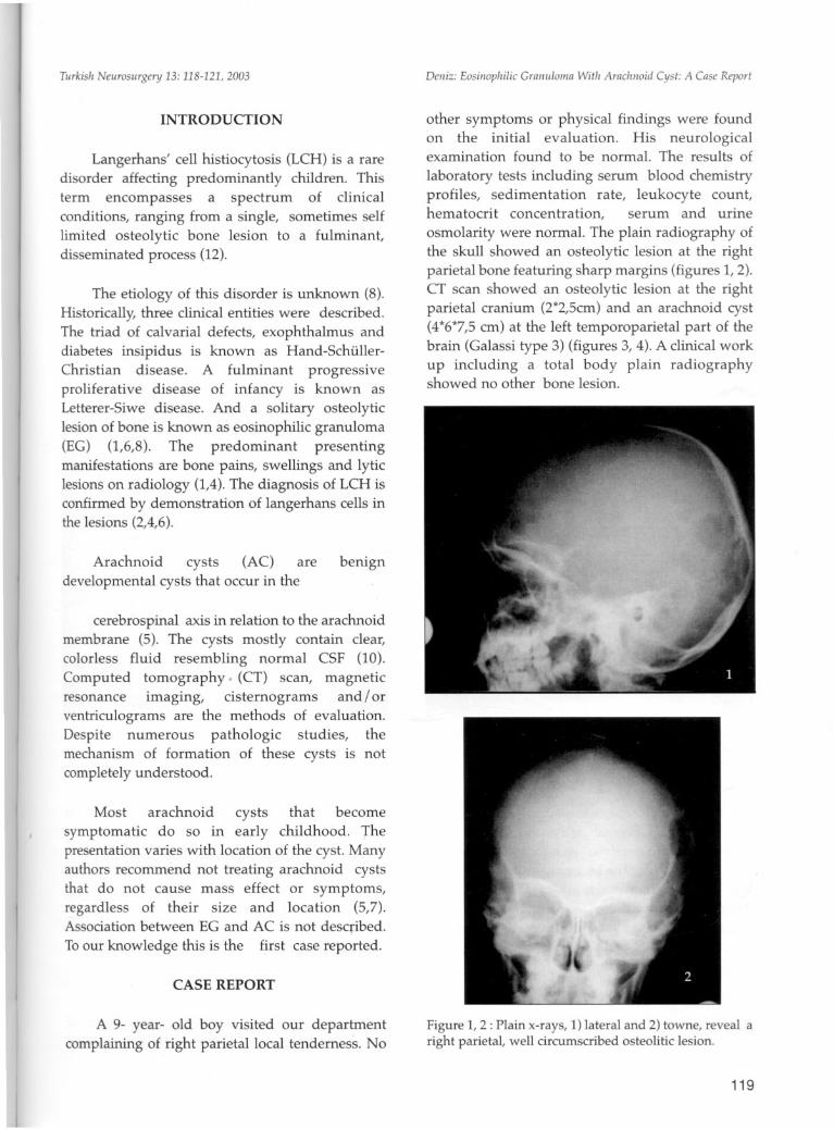
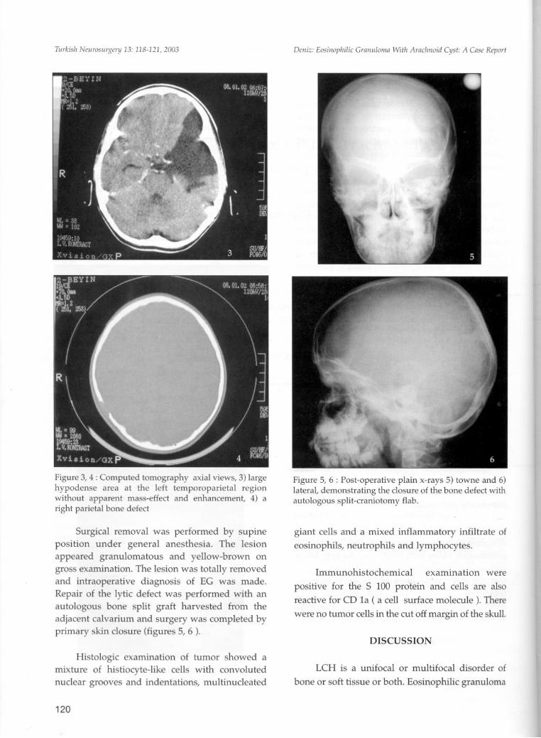
osmolarity were normal. The plain radiography ofthe skull showed an osteolytic lesion at the rightparietal bone featuring sharp margins (figures I, 2).CT scan showed an osteolytic lesion at the rightparietal cranium (2*2,5cm) and an arachnoid cyst(4*6*7,5cm) at the left temporoparietal part of thebrain (Galassi type 3) (figures 3, 4). A clinical workup including a total body plain radiographyshowed no other bone lesion.
Figure I, 2 : Plain x-rays, 1) lateral and 2) towne, reveal aright parietal, well circumscribed osteolitic lesion.
119
TlIrkish Nell1'Osllrgery 13: 118-121, 2003
Figure 3, 4: Computed tomography axial views, 3) largehypodense area at the left temporoparietal regionwithout apparent mass-effect and enhancement, 4) aright parietal bone defect
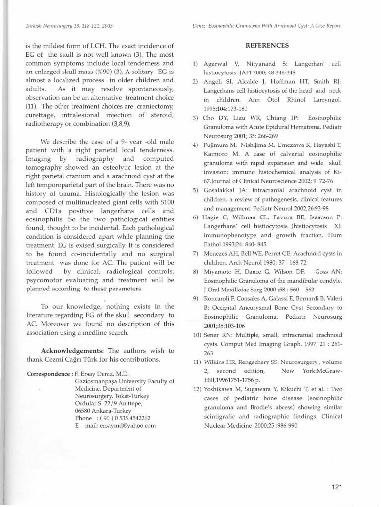
Surgical removal was performed by supineposition under general anesthesia. The lesionappeared granulomatous and yellow-brown ongross examination. The lesion was totally removedand intraoperative diagnosis of EG was made.Repair of the lytic defect was performed with anautologous bone split graft harvested from theadjacent calvarium and surgery was completed byprimary skin closure (figures 5, 6 ).
Histologic examination of tumor showed amixture of histiocyte-like cells with convolutednuclear grooves and indentations, multinucleated
120
Delli:: Eosillopllilic Gmmllolllil Witll Arachnoid Cyst: A Cilse Report
Figure 5, 6 : Post-operative plain x-rays 5) towne and 6)lateral, demonstrating the closure of the bone defect withautologous split-craniotomy flab.
giant cells and a mixed inflammatory infiltrate of
eosinophils, neutrophils and lymphocytes.
Immunohistochemical examination were
positive for the S 100 protein and cells are alsoreactive for CD 1a ( a cell surface molecule ). There
were no tumor cells in the cut off margin of the skull.
DISCUSSION
LCH is a unifocal or multifocal disorder of
bone or soft tissue or both. Eosinophilic granuloma
Turkish Neurosurgery 13: 118-121, 2003
is the mildest form of LCH. The exact incidence of
EG of the skull is not well known (3). The most
common symptoms include local tenderness andan enlarged skull mass (%90) (3). A solitary EG isalmost a localized process in older children andadults. As it may resolve spontaneously,observation can be an alternative treatment choice
(11). The other treatment choices are craniectomy,curettage, intralesional injection of steroid,radiotherapy or combination (3,8,9).
We describe the case of a 9- year -old malepatient with a right parietal local tenderness.Imaging by radiography and computedtomography showed an osteolytic lesion at theright parietal cranium and a arachnoid cyst at theleft temporoparietal part of the brain. There was nohistory of trauma. Histologically the lesion wascomposed of multinucleated giant cells with 5100and CD1a positive langerhans cells andeosinophilis. So the two pathological entitiesfound, thought to be incidental. Each pathologicalcondition is considered apart while planning thetreatment. EG is exised surgically. It is consideredto be found co-incidentally and no surgicaltreatment was done for AC. The patient will befollowed by clinical, radiological controls,psycomotor evaluating and treatment will beplanned according to these parameters.
To our knowledge, nothing exists m theliterature regarding EG of the skull secondary toAC. Moreover we found no description of thisassociation using a medline search.
Acknowledgements: The authors wish tothank Cezmi Cagn Turk for his contributions.
Correspondence: F. Ersay Deniz, M.D.Gaziosmanpa~a University Faculty ofMedicine, Department ofNeurosurgery, Tokat-TurkeyOrdular S. 22/9 Aruttepe,06580 Ankara-TurkeyPhone : ( 90 ) 0 535 4542262E-mail: [email protected]
Deniz: Eosinophilic Granuloma With Arachnoid Cyst: A Case Report
REFERENCES
1) Agarwal V, Nityanand S: Langerhan' cell
histiocytosis: JAPI 2000; 48:346-348
2) Angeli SI, Alcalde J, Hoffman HT, Smith RJ:
Langerhans cell histiocytosis of the head and neck
in children. Ann Otol Rhinol Larryngol.1995;104:173-180
3) Cho DY, Liau WR, Chiang IP: Eosinophilic
Granuloma with Acute Epidural Hematoma. Pediatr
Neurosurg 2001; 35: 266-269
4) Fujimura M, Nishijima M, Umezawa K, Hayashi T,
Kaimoro M. A case of calvarial eosinophilic
granuloma with rapid expansion and wide skull
invasion: immuno histochemical analysis of Ki67.Journal of Clinical Neuroscience 2002; 9: 72-76
5) Gosalakkal JA: Intracranial arachnoid cyst in
children: a review of pathogenesis, clinical features
and management. Pediatr Neural 2002;26:93-98
6) Hagie C, Willman CL, Favura BE, Isaacson P:
Langerhans' cell histiocytosis (histiocytosis X):
immunophenotype and growth fraction. HumPathoI1993;24: 840- 845
7) Menezes AH, Bell WE, Perret GE: Arachnoid cysts inchildren. Arch Neuro11980; 37 : 168-72
8) Miyamoto H, Dance G, Wilson DF, Goss AN:
Eosinophilic Granuloma of the mandibular condyle.
J Oral Maxillofac Surg 2000 ;58 : 560 - 5629) Roncaroli F, Consales A, Galassi E, Bernardi B, Valeri
B: OCCipital Aneurysmal Bone Cyst Secondary to
Eosinophilic Granuloma. Pediatr Neurosurg2001;35:103-106
10) Sener RN: Multiple, small, intracranial arachnoid
cysts. Com put Med Imaging Graph. 1997; 21 : 261263
11) Wilkins HR, Rengachary SS: Neurosurgery, volume2, second edition, New York:McGraw-
Hill,19961751-1756 p.
12) Yoshikawa M, Sugawara Y, Kikuchi T, et al. : Two
cases of pediatric bone disease (eosinophilic
granuloma and Brodie's a~cess) showing similar
scintigrafic and radiographic findings. ClinicalNuclear Medicine 2000;25 :986-990
121


![Repair of Tegmen Tympani Defect Presenting with ...€¦ · aberrant arachnoid granulations [3, 8]. According to the arachnoid theory, some arachnoid granulations may not find venous](https://img.pdfslide.us/doc/110x75/606db78183041435125f357b/repair-of-tegmen-tympani-defect-presenting-with-aberrant-arachnoid-granulations.jpg)