Embed Size (px)
Citation preview
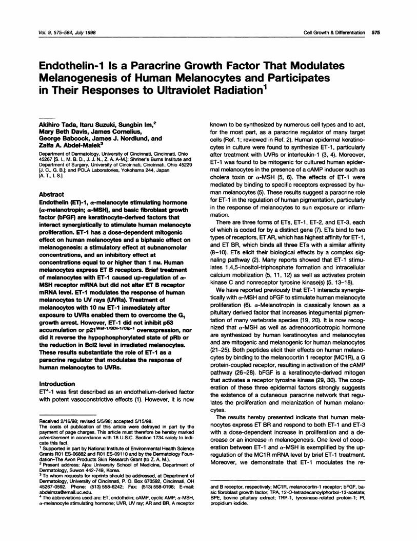
Vol. 9, 575-584, July 1998 Cell Growth & Differentiation 575
Endothelin-1 Is a Paracrine Growth Factor That ModulatesMelanogenesis of Human Melanocytes and Participatesin Their Responses to Ultraviolet Radiation’
Akihiro Tada, Itaru Suzuki, Sungbin lm,2Mary Beth Davis, James Cornelius,George Babcock, James J. Nordlund, andZalfa A. Abdel-MaIek�
Department of Dermatology, University of Cincinnati, Cincinnati, Ohio45267 [S. I., M. B. 0., J. J. N., Z. A. A-M.]; Shriner’s Burns Institute andDepartment of Surgery, University of Cincinnati, Cincinnati, Ohio 45229[J. C., G. B.]; and POLA Laboratories, Yokohama 244, Japan[A.T., l.S.]
Abstract
Endothelin (ET)-1, a-melanocyte stimulating hormone(a-melanotropin; a-MSH), and basic fibroblast growth
factor (bFGF) are keratinocyte-derived factors thatinteract synergistically to stimulate human melanocyteproliferation. El-I has a dose-dependent mitogeniceffect on human melanocytes and a biphasic effect onmelanogenesis: a stimulatory effect at subnanomolarconcentrations, and an inhibitory effect atconcentrations equal to or higher than I n�. Humanmelanocytes express ET B receptors. Brief treatmentof melanocytes with ET-1 caused up-regulation of a-
MSH receptor mRNA but did not alter ET B receptormRNA level. ET-1 modulates the response of humanmelanocytes to UV rays (UVRs). Treatment ofmelanocytes with 10 flM ET-1 immediately afterexposure to UVRs enabled them to overcome the G1growth arrest. However, ET-1 did not inhibit p53accumulation or p21 WCfI/5D11/C1P1 overexpression, nordid it reverse the hypophosphorylated state of pRb orthe reduction in Bc12 level in irradiated melanocytes.These results substantiate the role of ET-1 as aparacnne regulator that modulates the response ofhuman melanocytes to UVRs.
Introduction
E.r�-i was first described as an endothelium-denved factorwith potent vasoconstrictive effects (1). However, it is now
Received 2/16/98; revised 5/5/98; accepted 5/15/98.The costs of publication of this article were defrayed in part by thepayment of page charges. This article must therefore be hereby markedadvertisement In accordance with 18 U.S.C. Section 1734 solely to mdi-cete this fact.1 Supported in part by National Institute of Environmental Health ScienceGrants AOi ES-06882 and AOl ES-091 10 and by the Dermatology Foun-dation-The Avon Products Skin Research Grant (to 1 A. M.).2 Present address: Ajou University School of Medicine, Department ofOermatoiogy, Suwon 442-749, Korea.3 To whom requests for reprints should be addressed, at Department ofDermatology, University of Cincinnati. P. 0. Box 670592, Cincinnati, OH45267-0592. Phone: (513) 558-6242; Fax: (513) 558-0198; E-mail:[email protected] The abbreviations used are: Er, endothelin; cAMP, cyclic AMP; a-MSH,a-melanocyte stimulating hormone; UVR, UV ray; AR and BA, A receptor
known to be synthesized by numerous cell types and to act,for the most part, as a paracrine regulator of many targetcells (Ref. i ; reviewed in Ref. 2). Human epidermal keratino-cytes in culture were found to synthesize ET-i , particularly
after treatment with UVRs or interleukin-i (3, 4). Moreover,ET-i was found to be mitogenic for cultured human epider-mal melanocytes in the presence of a cAMP inducer such ascholera toxin or a-MSH (5, 6). The effects of Er-i weremediated by binding to specific receptors expressed by hu-man melanocytes (5). These results suggest a paracrine rolefor Er-i in the regulation of human pigmentation, particularly
in the response of melanocytes to sun exposure or inflam-mation.
There are three forms of ETs, ET-1 , ET-2, and ET-3, eachof which is coded for by a distinct gene (7). ETs bind to two
types of receptors, ET AR, which has highest affinity for ET-i,
and ET BR, which binds all three ETs with a similar affinity(8-10). ETs elicit their biological effects by a complex sig-naling pathway (2). Many reports showed that ET-i stimu-
lates i ,4,5-inositol-triphosphate formation and intracellularcalcium mobilization (5, i i , i 2) as well as activates protein
kinase C and nonreceptor tyrosine kinase(s) (5, i 3-i 8).We have reported previously that ET-i interacts synergis-
tically with a-MSH and bFGF to stimulate human melanocyteproliferation (6). a-Melanotropin is classically known as apituitary derived factor that increases integumental pigmen-tation of many vertebrate species (1 9, 20). It is now recog-nized that a-MSH as well as adrenocorticotropic hormoneare synthesized by human keratinocytes and melanocytesand are mitogenic and melanogenic for human melanocytes(2i-25). Both peptides elicit their effects on human melano-cytes by binding to the melanocortin i receptor (MCi A), a Gprotein-coupled receptor, resulting in activation of the cAMPpathway (26-28). bFGF is a keratinocyte-derived mitogenthat activates a receptor tyrosine kinase (29, 30). The coop-
eration of these three epidermal factors strongly suggeststhe existence of a cutaneous paracrine network that regu-lates the proliferation and melanization of human melano-cytes.
The results hereby presented indicate that human mela-nocytes express ET BR and respond to both ET-i and ET-3
with a dose-dependent increase in proliferation and a de-crease or an increase in melanogenesis. One level of coop-eration between ET-i and a-MSH is exemplified by the up-regulation of the MCi A mRNA level by brief ET-i treatment.Moreover, we demonstrate that ET-i modulates the re-
and B receptor, respectively; MCi A, melanocortln-i receptor bFGF, ba-sic fibroblast growth factor WA, i2-O-tetradecanoylphorbol-13-acetate;BPE, bovine pituitary extract; TRP-l , tyrosinase-related protein-i ; P1,propidium iodide.
A
w(1)94
0
B
�LLi:�
a)L.
(so.� 0
‘-0>. 0
Contra a-MSH ET-1 ET-1+a-MSH � a-M5H ET-3 �J-3+a4,jSH
mM 1r�M mM mM mM mM mM mM
Experiment I Experiment II
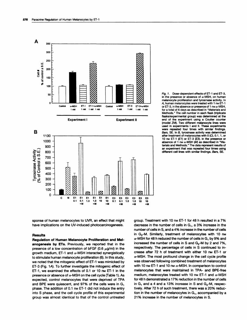
Fig. 1. Dose-dependent effects of El-i and ET-3,in the presence or absence of a-MSH, on humanmelanocyte proliferation and tyrosinase activity. InA, human melanocytes were treated wIth 1 nM ET-ior ET-3, in the absence or presence of 1 nM a-MSH,for a total of 6 days as described in “Materials andMethods.” The cell number in each flask (triplicateflasks/experimental group) was determined at theend of the experiment using a Coulter counter(model ZM). Two different melanocyte lines wereused in experiments I and II. These experimentswere repeated four times with similar findings.Bars, SE. In B, tyrosinase activity was determinedafter treatment of melanocytes with 0 (C), 0.1, 1, or10 nM ET-1 (El) or ET-3 (E3), in the presence orabsence of 1 nM a-MSH (M) as described in “Ma-terials and Methods.” The data represent results ofan experiment that was repeated four times usingdifferent cell lines with similar findings. Bars, SE.
1100 -
1000 -
900 -
800 -
700 -
600 -
500 -
400 -
300 -
200 -
100 -
0-
I
i n1h
‘1 JLifl�C M El El El El El El E3 E3 E3 E3 E3 E3
0.1 0.1 1.0 1.0 10 10 0.1 0.1 1.0 1.0 10 10+M +M +M +M +M +M
576 Paracrine Regulation of Human Melanocytes by ET-i
sponse of human melanocytes to UVR, an effect that mighthave implications on the UV-induced photocarcinogenesis.
ResultsRegulation of Human Melanocyte Proliferation and Mel-anogenesis by ETs. Previously, we reported that in the
presence of a low concentration of bFGF (0.6 �g/ml) in thegrowth medium, ET-1 and a-MSH interacted synergistically
to stimulate human melanocyte proliferation (6). In this study,
we noted that the mitogenic effect of ET-i was mimicked byET-3 (Fig. 1A). To further investigate the mitogenic effect ofET-i , we examined the effects of 0.1 or i 0 nt�i ET-i in thepresence or absence of a-MSH on the cell cycle (Table i). Asexpected, control melanocytes that were deprived of TPAand BPE were quiescent, and 97% of the cells were in G1phase. The addition of 0.1 n� ET-i did not induce the entry
into S phase, and the cell cycle profile of this experimentalgroup was almost identical to that of the control untreated
group. Treatment with i 0 nM ET-i for 48 h resulted in a 7%
decrease in the number of cells in G1, a 3% increase in thenumber of cells in 5, and a 4% increase in the number of cellsin G2-M. Similarly, treatment of melanocytes with 1 0 nti
a-MSH for 48 h reduced the number of cells in G1 by 9% andincreased the number of cells in S and G2-M by 2 and 7%,
respectively. The percentage of cells in S continued to in-crease after 72 h of treatment with either 1 0 flM ET-1 ora-MSH. The most profound change in the cell cycle profilewas observed following combined treatment of melanocyteswith i 0 nM ET-i and i 0 n�.i a-MSH. In comparison to controlmelanocytes that were maintained in TPA- and BPE-free
medium, melanocytes treated with i 0 nM ET-i and a-MSHfor 48 h demonstrated a i 7% reduction in the number of cellsin G1 and a 4 and a 13% increase in S and G2-M, respec-tively. After 72 h of such treatment, there was a 20% reduc-tion in the number of melanocytes in G1 , accompanied by a21 % increase in the number of melanocytes in S.
Cell Growth & Differentiation 577
Table 1 Dose-dependent effects of ET-1 in the absence or presenceof a-MSH on the cell cycle profile of human melanocytes
Melanocytes in TPA- and BPE-free medium were treated 48 h afterplating with 10 nM a-MSH, 0.1 or 10 nM ET-i , 0.1 nM ET-i + 10 n� a-MSH,or 10 nM ET-i + 10 np.i a-MSH. The medium was replaced, and theappropriate dose of hormone(s) was added every other day. Flow cyto-metric analysis of the cell cycle was carried out 24, 48, and 72 h aftertreatment, as described in “Materials and Methods.” Data represent thepercentage of the total population of melanocytes in G1 , 5, or G2-M.Similar results were obtained in two independent experiments.
% in G1 % in S % in G2-M
24 h
Control 96.7 0.6 2.710 nM a-MSH 96.4 0.9 2.70.1 nM ET-i 97.2 0.1 2.70.1 nM ET-i + 10 nM a-MSH 96.9 0.4 2.710 nM ET-i 96.8 0.5 2.8i0 nM ET-i + iO nM a-MSH 96.6 0.8 2.6
48 h
Control 97.i 0.6 2.310 nM a-MSH 88.3 2.7 8.90.1 nM Er-i 97.3 0.i 2.60.i nM ET-i + 10 nM a-MSH 91.5 1.3 7.2io n� ET-i 90.4 3.1 6.5io n� El-i + 10 nM a-MSH 79.8 5.3 i4.9
72 hControl 97.1 0.9 2.0io n� a-MSH 90.0 8.2 1.80.i nM ET-i 96.9 1.2 1.90.i nM ET-i + 10 nM a-MSH 90.0 8.4 1.610 nM ET-i 91.7 6.5 i.810 nM ET-i + 10 nM a-MSH 76.7 22.4 1.0
Tyrosinase is the rate-limiting enzyme in the melanin syn-thetic pathway (3i). We found that ET-i at i or 10 nM sig-nificantly reduced tyrosinase. activity by 30-40%. More ex-tensive dose-response experiments revealed that ET-i at 0.1
nM increased tyrosinase activity up to 350% of control (Fig.1B). Also, like ET-1 , ET-3 had a biphasic effect on tyrosinaseactivity, a stimulatory effect at subnanomolar concentra-tions, and an inhibitory effect at concentrations higher than 1
nM. Treatment with a-MSH in combination with either 0.1 nii
ET-i or ET-3 had an additive effect on tyrosinase activity(Fig. iB). The inhibitory effect of 10 nt�i ET-i on tyrosinase
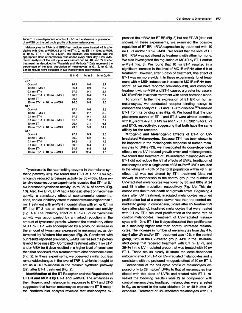
activity was accompanied by a marked reduction in theamount of tyrosinase protein, whereas the stimulatory effect
of 0.1 nM ET-i was accompanied by a profound increase inthe amount of tyrosinase expressed in melanocytes, as de-termined by Western blot analysis (Fig. 2). Consistent with
our results reported previously, a-MSH increased the protein
level of tyrosinase (25). Combined treatment with 0.1 nM ET-i
and a-MSH for 6 days resulted in a higher level of tyrosinasethan that observed after treatment with either hormone alone
(Fig. 2). In these experiments, we observed similar but less
remarkable changes in the level of TAP-i ,which is thought to
act as a DOPA-oxidase in the melanin synthetic pathway(32), after ET-i treatment (Fig. 2).
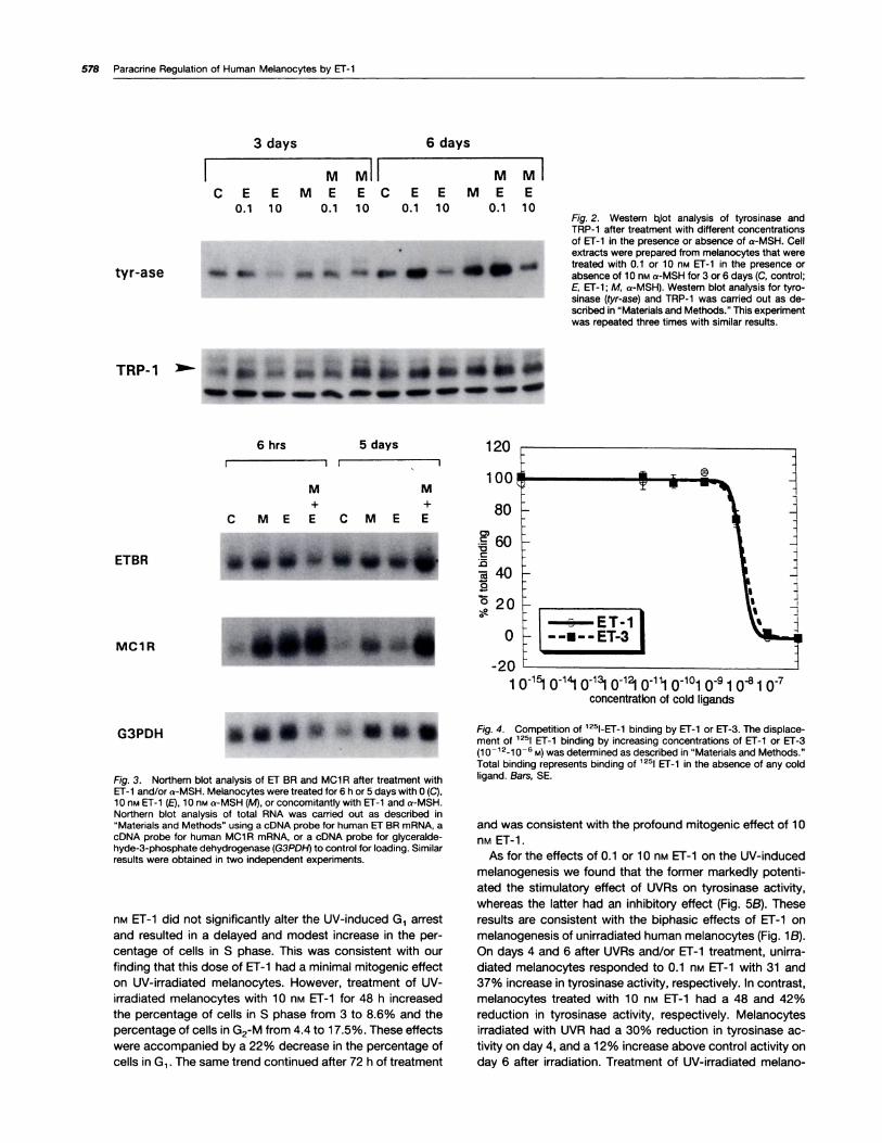
Identification of the Er Receptor and the Regulation ofEr BR and MCI R by ET-I and a-MSH. The similarities in
the mitogenic and melanogenic responses to ET-i and ET-3
suggested that human melanocytes express the Er B recep-tor. Northern blot analysis revealed that melanocytes ex-
pressed the mRNA for ET BR (Fig. 3) but not ET AR (data not
shown). In these experiments, we examined the possibleregulation of ET BR mRNA expression by treatment with 10
nM Er-i and/or 10 nM a-MSH. We found that the level of ET
BR mRNA was not altered by treatment with either hormone.We also investigated the regulation of MCi A by ET-i and/ora-MSH (Fig. 3). We found that 10 n� ET-i resulted in asignificant increase in the level of MCi A mRNA after 6 h of
treatment. However, after 5 days of treatment, this effect ofET-i was no more evident. In these experiments, brief treat-ment with a-MSH induced an increase in MCi A mANA tran-script, as we have reported previously (28), and combinedtreatment with a-MSH and ET-1 caused a greater increase in
MCi R mRNA level than treatment with either hormone alone.
To confirm further the expression of Er BR on human
melanocytes, we conducted receptor binding assays to
compare the ability of ET-i and ET-3 to displace 1251-labeled
Er-i from its binding sites (Fig. 4). We found that the dis-
placement curves of ET-i and ET-3 were almost identical,
with lCscs of 1 .479 ± 0.1 49 nM and 1 .757 ± 0.202 nM for ET-i
and ET-3, respectively, suggesting that both have the same
affinity for the receptor.Mitogenic and Melanogenic Effects of ET-1 on UV-
irradiated Melanocytes. Because ET-i has been shown tobe important in the melanogenic response of human mela-
nocytes to UVRs (33), we investigated its dose-dependent
effects on the UV-induced growth arrest and melanogenesis.
We found that treatment of UV-irradiated melanocytes with
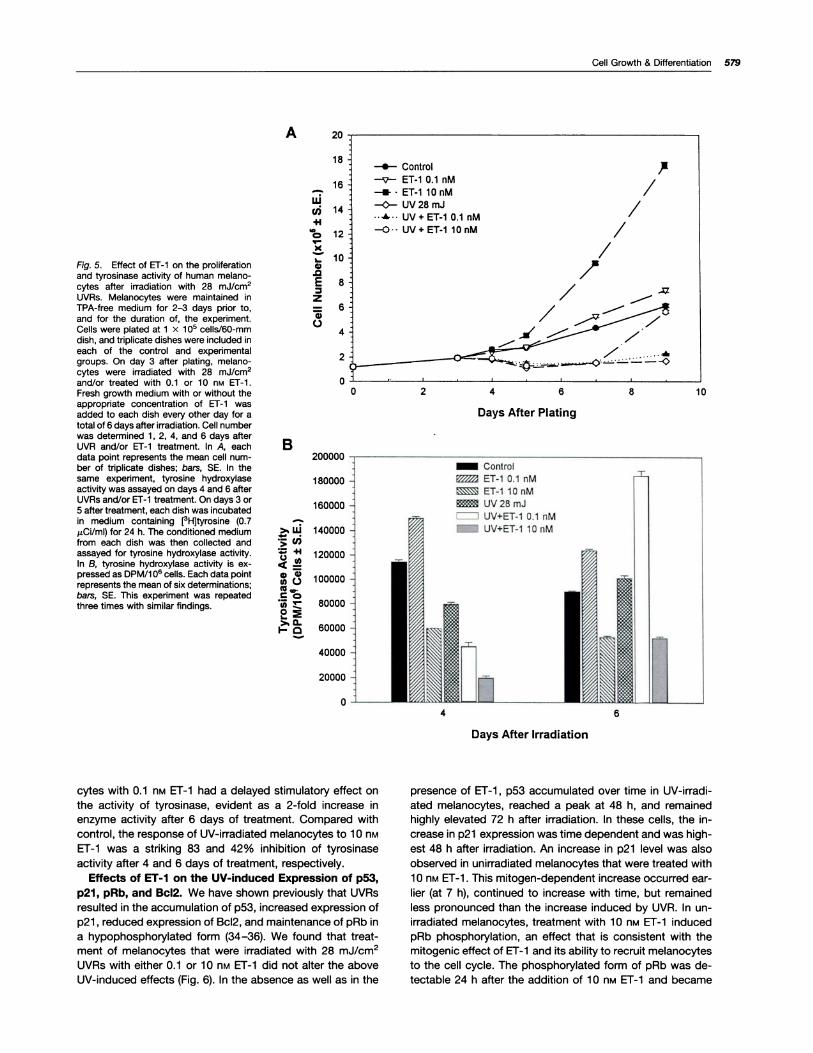
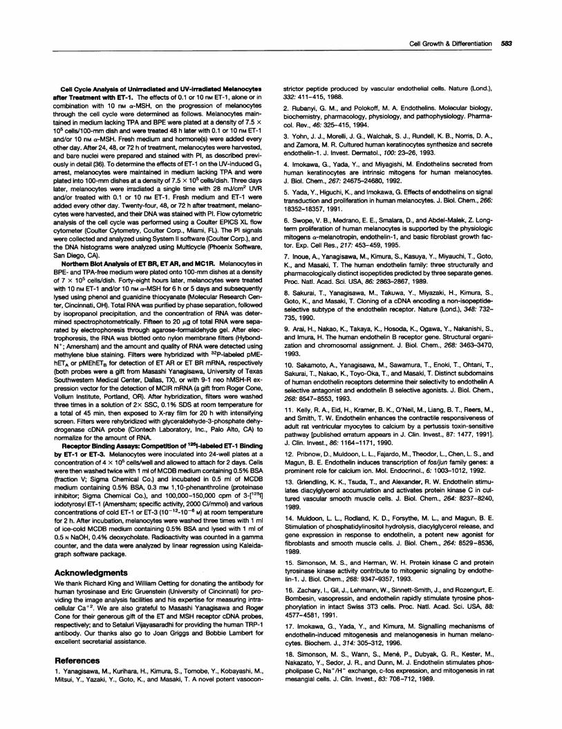
ET-i did not reduce the lethal effects of UVAs. Irradiation ofmelanocytes with a single dose of 28 mJ/cm2 UVAs resultedin the killing of -40% of the total population within 48 h, an
effect that was not altered by ET-i treatment (data notshown). In comparison to the control group, the number ofUV-irradiated melanocytes was lower by 30 and 60% at 24
and 48 h after irradiation, respectively (Fig. 5A). This de-crease was due to cell death and growth arrest. Beginning 4days after UV treatment, irradiated melanocytes resumed
proliferation but at a much slower rate than the control un-irradiated group. In comparison, 6 days after UV treatment (9
days after plating), irradiated melanocytes that were treated
with 0.1 nM ET-i resumed proliferation at the same rate as
control melanocytes. Treatment of UV-irradiated melano-cytes with i 0 nM Er-i for 6 days stimulated their proliferation
at a markedly higher rate than control untreated melano-cytes. The increase in number of melanocytes from day 4 to
day 6 after UV and/or Er-i treatment was 40% in the controlgroup, 12% in the UV-treated group, 44% in the UV-irradi-ated group that received treatment with 0.1 nM ET-i , and364% in the UV-irradiated group that was treated with i 0 n�ET-1 . These results clearly illustrate the dose-dependentmitogenic effect of ET-1 on UV-irradiated melanocytes and isconsistent with the profound mitogenic effect of 10 nM ET-i.
Comparison of the cell cycle profile of melanocytes ex-posed only to 28 mJ/cm2 .UVRs to that of melanocytes irra-diated with this dose of UVRS and treated with ET-i , re-vealed the following results (Table 2). In comparison withcontrol melanocytes, irradiated melanocytes were arrestedin G1 , as evident in the data obtained 24 or 48 h after UVexposure. Treatment of UV-irradiated melanocytes with 0.1
3 days 6 days
I MMII MMI
C E E ME EC E E ME E
0.1 10 0.1 10 0.1 10 0.1 10
i� �ii�tyr-ase � �
120
ETBR
80
; ET-�10-
MC 1 R � � � 0
G3PDH
Fig. 3. Northern blot analysis of ET BR and MCi R after treatment withEr-i and/or a-MSH. Melanocytes were treated for 6 h or 5 days with 0 (C),10 nM Er-i (E), 10 nM a-MSH (M), or concomitantly with ET-i and a-MSH.Northern blot analysis of total RNA was carried out as described in“Materials and Methods” using a cDNA probe for human ET BR rnRNA, acDNA probe for human MCi R rnRNA, or a cDNA probe for glyceralde-hyde-3-phosphate dehydrogenase (G3PD!-1) to control for loading. Similarresults were obtained in two independent experiments.
-201 01�1 01�1 01�1 0�1 0�’1 0�#{176}1� 1 0�8 1 �
concentration of cold Iigands
Fig. 4. Competition of 1251-Er-i binding by ET-i or ET-3. The displace-rnent of 1251 El-i binding by increasing concentrations of ET-i or ET-3(i0 12i0_6 M) was determined as described in “Materials and Methods.”Total binding represents binding of 1251 ET-i in the absence of any coldligand. Bars, SE.
578 Paracrine Regulation of Human Melanocytes by ET-i
TRP-1 �“-
� � � � � - � � - � �
6 hrs 5 days
F II �
M M+ +
C ME E C M E EC)
.� 60
....a,... �;40
0 20
nM ET-1 did not significantly alter the UV-induced G1 arrest
and resulted in a delayed and modest increase in the per-
centage of cells in S phase. This was consistent with our
finding that this dose of ET-1 had a minimal mitogenic effect
on UV-irradiated melanocytes. However, treatment of UV-irradiated melanocytes with 1 0 nM ET-1 for 48 h increased
the percentage of cells in S phase from 3 to 8.6% and thepercentage of cells in G2-M from 4.4 to 1 7.5%. These effectswere accompanied by a 22% decrease in the percentage ofcells in G1 . The same trend continued after 72 h of treatment
Fig. 2. Western bjot analysis of tyrosinase andTAP-i after treatment with different concentrationsof El-i in the presence or absence of a-MSH. Cellextracts were prepared from melanocytes that weretreated with 0.i or 10 n� ET-i in the presence orabsence of 10 nM a-MSH for 3 or 6 days (C, control;E, ET-i ; M, a-MSH). Western blot analysis for tyro-sinase (tyr-ase) and TRP-i was carried out as de-scribed in “Materials and Methods.” This experimentwas repeated three times with similar results.
and was consistent with the profound mitogenic effect of 10
nM ET-i.
As for the effects of 0.1 or 10 nM ET-i on the UV-induced
melanogenesis we found that the former markedly potenti-ated the stimulatory effect of UVRs on tyrosinase activity,whereas the latter had an inhibitory effect (Fig. 5B). Theseresults are consistent with the biphasic effects of ET-1 on
melanogenesis of unirradiated human melanocytes (Fig. 1 B).On days 4 and 6 after UVRs and/or ET-1 treatment, unirra-diated melanocytes responded to 0.i n� ET-1 with 31 and
37% increase in tyrosinase activity, respectively. In contrast,
melanocytes treated with 1 0 nM ET-i had a 48 and 42%reduction in tyrosinase activity, respectively. Melanocytes
irradiated with UVR had a 30% reduction in tyrosinase ac-
tivity on day 4, and a 12% increase above control activity onday 6 after irradiation. Treatment of UV-irradiated melano-
A 20
18
16
14
12
10
8
6
4
2
0
-.- Control
-v-- ET-i 0.1 nM
-.- . ET-1 10 nM
-0-- UV 28 mJ
�UV+ET-10.1nM-0#{149}-UV+ET-llOnM /
//
/
0 2 4 6 8 10
Days After Plating
B
>�LLi
‘�clj=4’
a,Q)(no(eta-� C.; �:
200000
180000
160000
140000
120000
100000
80000
60000
40000
20000
04 6
Cell Growth & Differentiation 579
Fig. 5. Effect of ET-1 on the proliferationand tyrosinase activity of human rnelano-cytes after irradiation with 28 rnJ/crn2UVRs. Melanocytes were maintained inWA-free medium for 2-3 days prior to,and for the duration of, the experiment.Cells were plated at 1 x i O� cells/60-rnmdish, and triplicate dishes were included in
each of the control and experimentalgroups. On day 3 after plating, melano-cytes were irradiated with 28 rnJ/cm2and/or treated with 0.i or i 0 n� ET-i.Fresh growth medium with or without theappropriate concentration of ET-i wasadded to each dish every other day for atotal of 6 days after irradiation. Cell numberwas determined 1 , 2, 4, and 6 days afterUVR and/or ET-i treatment. In A, eachdata point represents the mean cell nurn-ber of triplicate dishes; ba,�, SE. In thesame experiment, tyrosmne hydroxylaseactivity was assayed on days 4 and 6 afterUVR5 and/or Er-i treatment. On days 3 or5 after treatment, each dish was incubatedin medium containing [�H]tyrosine (0.7pCi/mI) for 24 h. The conditioned mediumfrom each dish was then collected andassayed for tyrosmne hydroxylase activity.In B, tyrosine hydroxylase activity is ex-pressed as DPM/i 06 cells. Each data pointrepresents the mean of six determinations;bars, SE. This experiment was repeatedthree times with similar findings.
Liiui4’
C
a,.0Ez
a,0
cytes with 0.1 nM ET-1 had a delayed stimulatory effect on
the activity of tyrosinase, evident as a 2-fold increase in
enzyme activity after 6 days of treatment. Compared with
control, the response of UV-irradiated melanocytes to 1 0 nM
ET-1 was a striking 83 and 42% inhibition of tyrosinaseactivity after 4 and 6 days of treatment, respectively.
Effects of ET-I on the UV-induced Expression of p53,p21 , pRb, and Bc12. We have shown previously that UVRsresulted in the accumulation of p53, increased expression of
p21 , reduced expression of Bc12, and maintenance of pRb in
a hypophosphorylated form (34-36). We found that treat-
ment of melanocytes that were irradiated with 28 mJ/cm2
UVRs with either 0.1 or 10 nM Er-i did not alter the above
UV-induced effects (Fig. 6). In the absence as well as in the
Days After Irradiation
presence of ET-1 , p53 accumulated over time in UV-irradi-
ated melanocytes, reached a peak at 48 h, and remained
highly elevated 72 h after irradiation. In these cells, the in-
crease in p2i expression was time dependent and was high-est 48 h after irradiation. An increase in p21 level was alsoobserved in unirradiated melanocytes that were treated with
1 0 nM ET-i . This mitogen-dependent increase occurred ear-
her (at 7 h), continued to increase with time, but remained
less pronounced than the increase induced by UVR. In un-irradiated melanocytes, treatment with 1 0 nM ET-1 induced
pRb phosphorylation, an effect that is consistent with the
mitogenic effect of ET-1 and its ability to recruit melanocytes
to the cell cycle. The phosphorylated form of pRb was de-
tectable 24 h after the addition of 1 0 nM ET-1 and became
5&, Paracrine Regulation of Human Melanocytes by ET-i
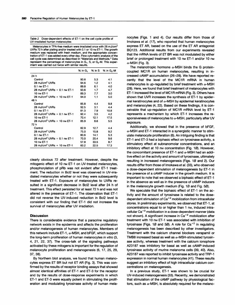
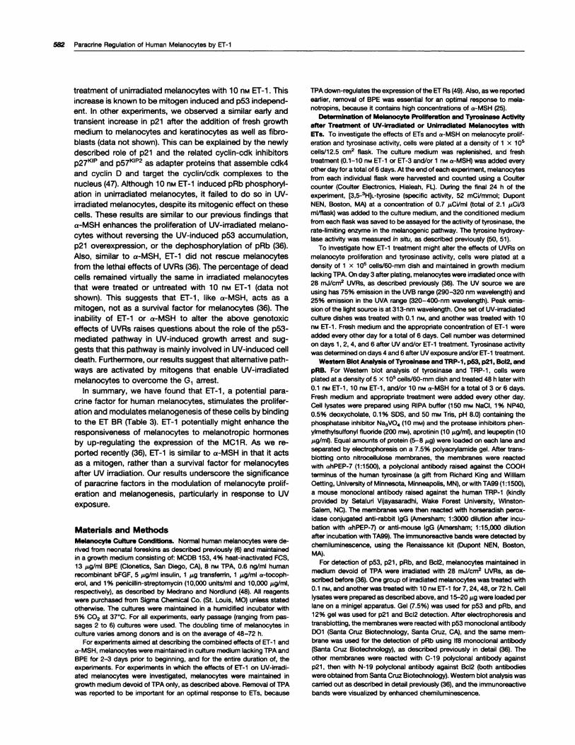
Table 2 Dose-dependent effects of Er-i on the cell cycle profile ofUV-irradiated human melanocytes
Melanocytes in TPA-free medium were irradiated once with 28 mJ/cm2UVRs 72 h after plating and/or treated with 0.i or iO n� ET-i . The growthmedium was replaced with fresh medium, and the appropriate concen-tration of ET-1 was added every other day. Flow cytornetric analysis of thecell cycle was determined as described in “Materials and Methods.” Datarepresent the percentage of melanocytes in G1 , 5, or G2-M. This exper-iment was darned out twice with similar results.
% in G1 % in S % in G2-M
24 hControl 90.6 5.3 4.i
28 rnJ/crn2 UVRs 94.6 1.2 4.i0_i nM Er-i 89.8 6.i 4.028 mJ/cm2 UVRs + 0.i n�i ET-i 93.6 1 .7 4.7io n� El-i 89.3 7.7 3.028 rnJ/cm2 UVRs + iO np�i ET-1 94.0 i.i 5.0
48 h
Control 85.8 4.4 9.828 mJ/cm2 UVRs 92.5 3.i 4.40.1 nM ET-i 8i.3 7.9 iO.828 mJ/cm2 UVRs + 0_i nr�i ET-i 92.4 3.5 4.2
10 nM ET-i 70.4 i2.i i7.528 mJ/crn2 UVRs + iO n� ET-i 85.9 8.6 5.5
72 h
Control 82.9 i2.7 4.428 mJ/cm2 UVRs 75.0 i5.8 9.20_i nM Er-i 80.6 i4.i 5.328 mJ/crn2 UVRs + 0.1 n� ET-i 70.0 20.4 9.6iO nM ET-1 57.8 33.5 8.728 rnJ/crn2 UVRs + 10 n� ET-i 60.2 22.5 i7.3
clearly obvious 72 after treatment. However, despite themitogenic effect of 10 n� ET-i on UV-treated melanocytes,phosphorylation of pRb was not evident after ET-1 treat-ment. The reduction in Bcl2 level was observed in UV-irra-diated melanocytes whether or not they were subsequentlytreated with ET-i . Exposure of melanocytes to UVRs re-
suIted in a significant decrease in Bc12 level after 24 h oftreatment. This effect persisted for at least 72 h and was notaltered in the presence of ET-i . The observation that ET-1did not reverse the UV-induced reduction in Bc12 level isconsistent with our finding that ET-i did not increase thesurvival of melanocytes after UV irradiation.
DiscussionThere is considerable evidence that a paracrine regulatorynetwork exists in the epidermis and affects the proliferationand/or melanogenesis of human melanocytes. Members ofthis network include Er-i ,a-MSH, and bFGF, which supportthe long-term proliferation of human melanocytes in vitro (3,4, 21 , 22, 37). The cross-talk of the signaling pathwaysactivated by these mitogens is important for the regulation ofmelanocyte proliferation and melanogenesis (6, 17, 25, 29,37, 38).
By Northern blot analysis, we found that human melano-cytes express ET BA but not ET AR (Fig. 3). This was con-firmed by the results of receptor binding assays that showed
almost identical affinities of Er-i and ET-3 for the receptorand by the results of dose-response experiments in whichET-i and ET-3 were equally potent in stimulating the prolif-eration and modulating tyrosinase activity of human mela-
nocytes (Figs. 1 and 4). Our results differ from those of
lmokawa et a!. (17), who reported that human melanocytesexpress ET AR, based on the use of the ET AR antagonistBQ1 23. Additional results from our experiments revealedthat the mRNA levels of ET BR was not modulated by eitherbrief or prolonged treatment with 10 nM ET-i and/or 10 nr�ia-MSH (Fig. 3).
The melanotropic hormone a-MSH binds the G protein-coupled MCi A on human melanocytes, resulting in in-creased cAMP accumulation (26-28). We have reported re-cently that the level of the MCi A mRNA in humanmelanocytes is up-regulated by brief treatment with a-MSH(28). Here, we found that brief treatment of melanocytes withET-i increased the level of MCi A mRNA(Fig. 3). Others haveshown that UVR increases the synthesis of ET-i by epider-mal keratinocytes and of a-MSH by epidermal keratinocytesand melanocytes (4, 22). Based on these findings, it is con-ceivable that up-regulation of MCi A mRNA level by ET-i
represents a mechanism by which ET-1 increases the re-sponsiveness of melanocytes to a-MSH, particularly after UVexposure.
Additionally, we showed that in the presence of bFGF,
a-MSH and ET-i interacted in a synergistic manner to stim-ulate melanocyte proliferation (6). An intriguing finding is thatET-i and ET-3 had a biphasic effect on tyrosinase activity, astimulatory effect at subnanomolar concentrations, and aninhibitory effect at 1 0 nM concentration (Fig. iB). However,the concomitant presence of ET-1 and a-MSH had an addi-tive effect on the activity and amount of tyrosinase, ultimatelyresulting in increased melanogenesis (Figs. i B and 2). Ourresults differ from those of lmokawa et a!. (33), who observeda dose-dependent stimulation of melanogenesis by ET-1 inthe presence of a cAMP inducer in the growth medium. It isimportant to note that we observed a biphasic effect of ET-i
in the absence as well as in the presence of cAMP inducersin the melanocyte growth medium (Fig. lB and Fig. 5B).
We speculate that the biphasic effect of Er-i on the ac-tivity and the amount of tyrosinase is related to the dose-dependent stimulation of Ca�2 mobilization from intracellularstores. In preliminary experiments, we observed that ET-i ,atconcentrations equal to or higher than 1 n�, induced intra-cellular Ca�2 mobilization in a dose-dependent manner (datanot shown). A significant increase in Ca�2 mobilization aftertreatment with 1 0 nM ET-i was associated with inhibition oftyrosinase (Figs. lB and 5B). A role for Ca�2 in regulatingmelanogenesis has been described by other investigators.Treatment with the calcium channel blockers verapamil orTMB8 increased basal as well as a-MSH-stimulated tyrosin-ase activity, whereas treatment with the calcium ionophoreA23i 87 was inhibitory for basal as well as cAMP-inducedtyrosinase activity of murine melanoma cells (39, 40). Also,A23i 87 was reported to inhibit tyrosinase activity and TAP-iexpression in normal human melanocytes (41). These resultssuggest an inhibitory effect of high intracellular calcium con-centrations on melanogenesis.
In a previous study, ET-i was shown to be crucial forUV-induced melanogenesis (33). Recently, we demonstratedthat stimulation of the cAMP pathway by physiological fac-tors, such as a-MSH, is absolutely required for the melano-
7 Hours 24 Hours 48 Hours 72 Hours
+
1 � � 11
0
C)
Cell Growth & Differentiation 581
detected. However, the response of irradiated melanocytes
-
�0 > � �
U � c� -
-
�� � - >
U � 0 � �J
I-,w 114
-
�� � - 0 >C�) J � - �
- 0 >� c; - �
p53 � � � - - - - - - � � � - - � - � - � �
p21-� .� � � � � � . �
pRb -� � .�-�-----------�- �
Bc12 � � �.- � .-. 1� �
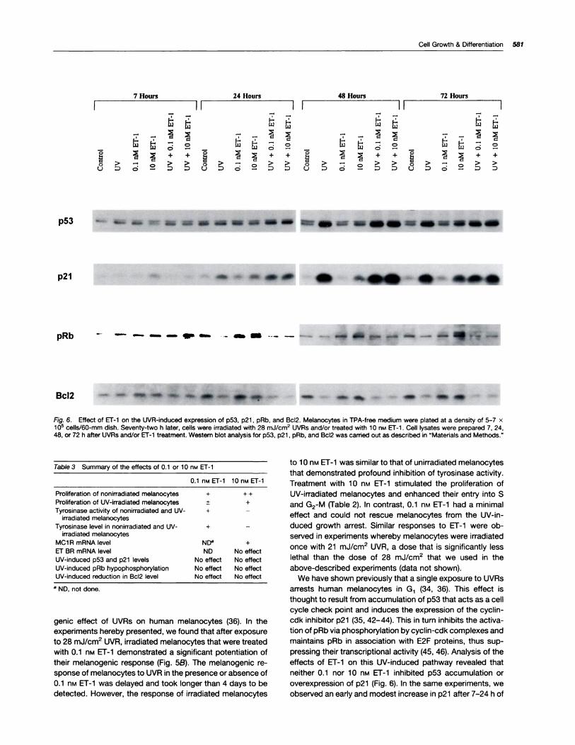
Fig. 6. Effect of Er-i on the UVR-induced expression of p53, p21, pRb, and Bc12. Melanocytes in TPA-free medium were plated at a density of 5-7 x1 o� cells/60-rnrn dish. Seventy-two h later, cells were irradiated with 28 mJ/crn2 UVRs and/or treated with 10 n� ET-1 . Cell lysates were prepared 7, 24,48, or 72 h after UVRs and/or Er-i treatment. Western blot analysis for p53, p21 , pRb, and Bd12 was camed out as described in “Materials and Methods.”
Table 3 Summary of the effects of 0.1 or iO nM ET-i
0.1 nM ET-i 10 nM ET-1
Proliferation of nonirradiated melanocytes + + +
Proliferation of UV-irradiated melanocytes ± +
Tyrosinase activity of nonirradiated and UV- + -
irradiated melanocytes
Tyrosinase level in nonirradiated and UV- + -
irradiated melanocytesMC1R rnRNA level NDC +
EF BR rnRNA level ND No effect
UV-induced p53 and p21 levels No effect No effect
UV-induced pRb hypophosphorylation No effect No effectUV-induced reduction in Bc12 level No effect No effect
a ND, not done.
genic effect of UVRs on human melanocytes (36). In theexperiments hereby presented, we found that after exposureto 28 mJ/cm2 UVR, irradiated melanocytes that were treatedwith 0.1 nM ET-1 demonstrated a significant potentiation oftheir melanogenic response (Fig. 5B). The melanogenic re-sponse of melanocytes to UVR in the presence or absence of0.i nM Er-i was delayed and took longer than 4 days to be
to i 0 nM ET-i was similar to that of unirradiated melanocytesthat demonstrated profound inhibition of tyrosinase activity.
Treatment with 10 nM Er-i stimulated the proliferation of
UV-irradiated melanocytes and enhanced their entry into Sand G2-M (Table 2). In contrast, 0.1 n�i ET-i had a minimaleffect and could not rescue melanocytes from the UV-in-duced growth arrest. Similar responses to ET-1 were ob-
served in experiments whereby melanocytes were irradiated
once with 21 mJ/cm2 UVR, a dose that is significantly less
lethal than the dose of 28 mJ/cm2 that we used in the
above-described experiments (data not shown).
We have shown previously that a single exposure to UVRsarrests human melanocytes in G1 (34, 36). This effect isthought to result from accumulation of p53 that acts as a cell
cycle check point and induces the expression of the cyclin-cdk inhibitor p21 (35, 42-44). This in turn inhibits the activa-
tion of pRb via phosphorylation by cyclin-cdk complexes and
maintains pRb in association with E2F proteins, thus sup-
pressing their transcriptional activity (45, 46). Analysis of the
effects of ET-i on this UV-induced pathway revealed thatneither 0.1 nor 10 nM ET-i inhibited p53 accumulation or
overexpression of p21 (Fig. 6). In the same experiments, weobserved an early and modest increase in p21 after 7-24 h of
582 Paracrine Regulation of Human Melanocytes by ET-i
treatment of unirradiated melanocytes with 10 n� ET-1 . Thisincrease is known to be mitogen induced and p53 independ-ent. In other experiments, we observed a similar early and
transient increase in p21 after the addition of fresh growthmedium to melanocytes and keratinocytes as well as fibro-blasts (data not shown). This can be explained by the newlydescribed role of p21 and the related cyclin-cdk inhibitorsp27�’ and p57�’2 as adapter proteins that assemble cdk4and cyclin D and target the cyclin/cdk complexes to thenucleus (47). Although 10 nt�i ET-i induced pAb phosphoryl-ation in unirradiated melanocytes, it failed to do so in UV-irradiated melanocytes, despite its mitogenic effect on thesecells. These results are similar to our previous findings thata-MSH enhances the proliferation of UV-irradiated melano-cytes without reversing the UV-induced p53 accumulation,p21 overexpression, or the dephosphorylation of pAb (36).Also, similar to a-MSH, ET-i did not rescue melanocytesfrom the lethal effects of UVAs (36). The percentage of deadcells remained virtually the same in irradiated melanocytesthat were treated or untreated with 10 nM Er-i (data notshown). This suggests that ET-1 , like a-MSH, acts as amitogen, not as a survival factor for melanocytes (36). Theinability of Er-i or a-MSH to alter the above genotoxiceffects of UVAs raises questions about the role of the p53-mediated pathway in UV-induced growth arrest and sug-gests that this pathway is mainly involved in UV-induced celldeath. Furthermore, our results suggest that alternative path-ways are activated by mitogens that enable UV-irradiatedmelanocytes to overcome the G1 arrest.
In summary, we have found that ET-i , a potential para-crine factor for human melanocytes, stimulates the prolifer-ation and modulates melanogenesis of these cells by bindingto the ET BR (Table 3). ET-i potentially might enhance theresponsiveness of melanocytes to melanotropic hormonesby up-regulating the expression of the MCi R. As we re-ported recently (36), Er-i is similar to a-MSH in that it actsas a mitogen, rather than a survival factor for melanocytesafter UV irradiation. Our results underscore the significanceof paracrine factors in the modulation of melanocyte prolif-eration and melanogenesis, particularly in response to UVexposure.
Materials and MethodsMelanocyte cutture Condftlons. Normal human melanocytes were de-
rived from neonatal foreskins as described previously (6) and maintainedin a growth medium consisting of: MCDB i53, 4% heat-inactivated FCS,13 �g/ml BPE (Clonetics. San Diego. CA), 8 n� WA, 0.6 ng/ml humanrecombinant bFGF, 5 �g/ml insulin, 1 �g transferrin, i �g/ml a-tocoph-
erol, and 1% penicillin-streptomycin (10,000 units/mI and 10,000 �g/ml,respectively), as described by Medrano and Nordlund (48). All reagentswere purchased from Sigma Chemical Co. (St. Louis, MO) unless statedotherwise. The cultures were maintained in a humidified incubator with5% CO2 at 37#{176}C.For all experiments, early passage (ranging from pas-sages 2 to 6) cultures were used. The doubling time of melanocytes inculture varies among donors and is on the average of 48-72 h.
For experiments aimed at describing the combined effects of ET-1 anda-MSH, melanocytes were maintained in culture medium lacking WA andBPE for 2-3 days prior to beginning, and for the entire duration of, theexperiments. For experiments in which the effects of ET-i on UV-irradi-ated melanocytes were investigated, melanocytes were maintained ingrowth medium devoid of WA only, as described above. Removal of WAwas reported to be important for an optimal response to ETs, because
WA down-regulates the expression of the ET As (49). Also, as we reported
earlier, removal of BPE was essential for an optimal response to mela-notropins, because it contains high concentrations of a-MSH (25).
DetermInation of Melanocyte Proliferation and Tyrosinase Activity
after Treatment of UV-irradiated or Unirradlated Melanocytes with
ETa. To investigate the effects of ETs and a-MSH on melanocyte prolif-eration and tyrosmnase activity, cells were plated at a density of i x 1 O�
cells/12.5 cm2 flask. The culture medium was replenished, and freshtreatment (0.1-iO n,i ET-1 or ET-3 and/or 1 flM a-MSH) was added everyother day for a total of 6 days. At the end of each experiment, melanocytesfrom each individual flask were harvested and counted using a Coulter
counter (Coulter Electronics, Hialeah, FL). During the final 24 h of theexperiment, [3,5-3H]L-tyrosine (specific activity, 52 mCVmmol; DupontNEN, Boston, MA) at a concentration of 0.7 �&CVml (total of 2.1 pCV3
mI/flask) was added to the culture medium, and the conditioned mediumfrom each flask was saved to be assayed for the activity of tyrosinase, therate-limiting enzyme in the melanogenic pathway. The tyrosine hydroxy-lase activity was measured in situ, as described previously (50, 51).
To investigate how Er-i treatment might alter the effects of UVAs on
melanocyte proliferation and tyrosinase activity, cells were plated at a
density of i x io� cells/60-mm dish and maintained in growth mediumlacking WA. On day 3 after plating, melanocytes were irradiated once with28 mJ/cm2 UVRs, as described previously (36). The UV source we areusing has 75% emission in the UVB range (290-320 nm wavelength) and
25% emission in the UVA range (320-400-nm wavelength). Peak emis-sion of the light source is at 313-nm wavelength. One set of UV-irradiated
culture dishes was treated with 0.1 nM, and another was treated with 10nM ET-1 . Fresh medium and the appropriate concentration of ET-1 wereadded every other day for a total of 6 days. Cell number was determinedon days i , 2, 4, and 6 after UV and/or ET-i treatment. Tyrosinase activitywas determined on days 4 and 6 after UV exposure and/or ET-1 treatment.
western BlotAnalysis ofTyrosinase and TRP-1, p53, p21, Bcl2, andpRB. For Western blot analysis of tyrosinase and TAP-i , cells wereplated at a density of 5 x io� cells/60-mm dish and treated 48 h later with
0.1 nM ET-1, 10 nM ET-i, and/or 10 n� a-MSH for a total of 3 or 6 days.Fresh medium and appropriate treatment were added every other day.Cell lysates were prepared using AIPA buffer (150 m.i NaCI, 1% NP4O,0.5% deoxycholate, 0.1 % SOS, and 50 m� Tris, pH 8.0) containing thephosphatase inhibitor Na3VO4 (1 0 mM) and the protease inhibitors phen-ylmethylsulfonyl fluoride (200 mM), aprotinin (10 �g/m�, and leupoptin (10�g/m�. Equal amounts of protein (5-8 gig) were loaded on each lane andseparated by electrophoresis on a 7.5% polyacrylamide gel. After trans-blothng onto nitrocellulose membranes, the membranes were reacted
with ahPEP-7 (1 :1500), a polydlonal antibody raised against the COOHterminus of the human tyrosinase (a gift from Richard King and WilliamOetting, University of Minnesota, Minneapolis, MN), orwith TA99 (1:1500),a mouse monoclonal antibody raised against the human TRP-1 (kindly
provided by Setaluri Vijayasaradhi, Wake Forest University, Winston-
Salem, NC). The membranes were then reacted with horseradish perox-idase conjugated anti-rabbit lgG (Amersham; 1 :3000 dilution after Incu-
bation with ahPEP-7) or anti-mouse IgG (Amersham; 1:15,000 dilution
after incubation with TA99). The immunoreactive bands were detected by
chemiluminescence, using the Renaissance kit (Dupont NEN, Boston,MA).
For detection of p53, p21 , pAb, and Bcl2, melanocytos maintained in
medium devoid of WA were irradiated with 28 mJ/cm2 UVAs, as de-
scribed before (36). One group of irradiated melanocytes was treated with0.1 nM, and another was treated with 10 n,�i ET-1 for 7, 24, 48, or 72 h. Celllysates were prepared as described above, and i 5-20 �g were loaded per
lane on a minigel apparatus. Gel (7.5%) was used for p53 and pAb, and12% gel was used for p21 and Bc12 detection. After electrophoresis and
transblothng, the membranes were reacted with p53 monoclonal antibodyOoi (Santa Cruz Biotechnology, Santa Cruz, CA), and the same mem-brane was used for the detection of pAb using 1f8 monoclonal antibody(Santa Cruz Biotechnology), as described previously in detail (36). Theother membranes were reacted with C-19 polydlonal antibody against
p2i , then with N-19 polyclonal antibody against Bc12 (both antibodieswere obtained from Santa Cruz Biotechnology). Western blot analysis was
carried out as described in detail previously (36), and the immunoreactivebands were visualized by enhanced chemiluminescence.
Cell Growth & Differentiation 593
1 . Vanagisawa, M., Kurihara, H., Kimura, S., Tomobe, V., Kobayashi, M.,Mitsui, V., Vazaki, V., Goto, K., and Masaki, T. A novel potent vasocon-
Cell Cycle Analysis of Unirradlated and UV-irradlated Melanocytes
after Treatment wIth ET-1 . The effects of 0.1 or 1 0 nim ET-1 , alone or incombination with 10 nM a-MSH, on the progression of melanocytesthrough the cell cycle were determined as follows. Melanocytes main-
tamed in medium lacking WA and BPE were plated at a density of 7.5 xio5 cells/iOO-mm dish and were treated 48 h later with 0.1 or 10 n� ET-i
and/or 10 nM a-MSH. Fresh medium and hormone(s) were added everyother day. After 24, 48, or 72 h of treatment, melanocytes were harvested,and bare nuclei were prepared and stained with P1, as described previ-
ously in detail(36). To determine the effects of ET-1 on the UV-induced G1arrest, melanocytes were maintained in medium lacking TPA and wereplated into 100-mm dishes at a density of 7.5 x i0� cells/dish. Three dayslater, melanocytes were irradiated a single time with 28 mJ/cm2 UVA
and/or treated with 0.i or 10 nr�i ET-1 . Fresh medium and ET-i wereadded every other day. Twenty-four, 48, or 72 h after treatment, melano-
cytes were harvested, and their DNA was stained with P1. Flow cytometricanalysis of the cell cycle was performed using a Coulter EPICS XL flowcytometer (Coulter Cytometry, Coulter Corp., Miami, FL). The P1 signals
were collected and analyzed using System II software(Coulter Corp.), andthe DNA histograms were analyzed using Multicydle (Phoenix Software,San Diego, CA).
Northern Blot Analysis of ET BR, ETAR, and McIR. Melanocytes in
BPE- and TPA-free medium were plated onto 100-mm dishes at a densityof 7 x 1 o� cells/dish. Forty-eight hours later, melanocytes were treatedwith 10 nM El-i and/or 10 n� a-MSH for 6 h or 5 days and subsequently
lysed using phenol and guanidine thiocyanate (Molecular Research Cen-ter, Cincinnati, OH). Total ANA was purified by phase separation, followedby isopropanol precipitation, and the concentration of ANA was deter-mined spectrophotometrically. Fifteen to 20 �g of total RNA were sepa-rated by electrophoresis through agarose-formaldehyde gel. After elec-trophoresis, the ANA was blotted onto nylon membrane filters (Hybond-
N�; Amersham) and the amount and quality of ANA were detected usingmethylene blue staining. Filters were hybridized with 32P-labeled pME-hETA or PMEhETB for detection of ET AR or ET BR mRNA, respectively
(both probes were a gift from Masashi Vanagisawa, University of Texas
Southwestern Medical Center, Dallas, TX), or with 9-1 nec hMSH-R ex-pression vector for the detection of MCIR mRNA (a gift from Roger Cone,Vollum Institute, Portland, OR). After hybridization, filters were washedthree times in a solution of 2x SSC, 0.i % SDS at room temperature for
a total of 45 mm, then exposed to X-ray film for 20 h with intensifyingscreen. Filters were rehybridized with glyceraldehyde-3-phosphate dehy-drogenase cDNA probe (Clontech Laboratory, Inc., Palo Alto, CA) tonormalize for the amount of RNA.
Receptor Binding Assays: Competition of 1�I-Iabeled ET-l Bindingby ET-1 or ET-3. Melanocytes were inoculated into 24-well plates at aconcentration of 4 x i 0� cells/well and allowed to attach for 2 days. Cellswerethen washed twice with 1 ml of MCDB medium containing 0.5% BSA(fraction V; Sigma Chemical Co.) and incubated in 0.5 ml of MCDBmedium containing 0.5% BSA, 0.3 m�i 1,iO-phenanthroline (proteinaseinhibitor; Sigma Chemical Co.), and 100,000-150,000 cpm of 3..[125l]
iodotyrosyl ET-1 (Amersham; specific activity, 2000 CVmmol) and variousconcentrations of cold ET-i or ET-3 (1012�i 0_6 M) at room temperaturefor 2 h. After incubation, melanocytes were washed three times with 1 ml
of ice-cold MCDB medium containing 0.5% BSA and lysed with i ml of0.5 N NaOH, 0.4% deoxycholate. Radioactivity was counted in a gammacounter, and the data were analyzed by linear regression using Kaleida-
graph software package.
AcknowledgmentsWe thank Richard King and William Oethng for donating the antibody forhuman tyrosinase and Eric Gruenstein (University of Cincinnati) for pro-
viding the image analysis facilities and his expertise for measuring intra-cellular Ca�2. We are also grateful to Masashi Yanagisawa and Roger
Cone for their generous gift of the El and MSH receptor cDNA probes,
respectively; and to Setaluri Vijayasaradhi for providing the human TAP-iantibody. Our thanks also go to Joan Griggs and Bobbie Lambert forexcellent secretarial assistance.
References
strictor peptide produced by vascular endothelial cells. Nature (Lond.),332: 4ii-4i5, i988.
2. Rubanyi, G. M., and Polokoff, M. A. Endothelins. Molecular biology,biochemistry, pharmacology, physiology, and pathophysiology. Pharma-
col. Rev., 46: 325-4i5, 1994.
3. Yohn, J. J., Morelli, J. G., Waichak, S. J., Rundell, K. B., Norris, D. A.,and Zamora, M. A. Cultured human keratinocytes synthesize and secreteendothelin-i . J. Invest. Dermatol., 100: 23-26, i993.
4. Imokawa, G., Vada, V., and Miyagishi, M. Endothelins secreted fromhuman keratinocytes are intrinsic mitogens for human melanocytes.J. Biol. Chem., 267: 24675-24680, i992.
5. Vada, V., Higuchi, K., and Irnokawa, G. Effects of endothelins on signaltransduction and proliferation in human melanocytes. J. Biol. Chem., 266:
18352-18357, i99i.
6. Swope, V. B., Medrano, E. E., Smalara, D., and Abdel-Malek, Z. Long-term proliferation of human melanocytes is supported by the physiologicmitogens a-melanotropin, endothelin-i , and basic fibroblast growth fad-tor. Exp. Cell Res., 217: 453-459, 1995.
7. lnoue, A., Vanagisawa, M., Kimura, S., Kasuya, V., Miyauchi, T., Goto,K., and Masaki, T. The human endothelin family: three structurally andpharmacologically distinct isopeptides predicted bythree separate genes.Proc. NatI. Acad. Sd. USA, 86: 2863-2867, 1989.
8. Sakurai, T., Vanagisawa, M., Takuwa, V., Miyazaki, H., Kimura, S.,Goto, K., and Masaki, T. Cloning of a cDNA encoding a non-isopeptide-selective subtype of the endothelin receptor. Nature (Lond.), 348: 732-735, 1990.
9. Arai, H., Nakao, K., Takaya, K., Hosoda, K., Ogawa, V., Nakanishi, S.,and Imura, H. The human endothelin B receptor gene. Structural organi-zation and chromosomal assignment. J. Biol. Chem., 268: 3463-3470,i993.
10. Sakamoto, A., Vanagisawa, M., Sawamura, T., Enoki, T., Ohtani, T.,
Sakurai, T., Nakao, K., Toyo-Oka, T., and Masaki, T. Distinct subdomainsof human endothelin receptors determine their selectivity to endothelin Aselective antagonist and endothelin B selective agonists. J. Biol. Chem.,
268: 8547-8553, 1993.
i 1 . Kelly, A. A., Eid, H., Kramer, B. K., O’Neil, M., Uang, B. T., Reers, M.,and Smith, T. W. Endothelin enhances the contractile responsiveness ofadult rat ventricular myocytes to calcium by a pertussis toxin-sensitive
pathway [published erratum appears in J. Clin. Invest., 87: i477, i991].J. Clin. Invest., 86: ii64-ii7i, i990.
i2. Pribnow, D., Muldoon, L L, Fajardo, M., Theodor, L, Chen, L S., andMagun, B. E. Endothelin induces transcription of fos/jun family genes: a
prominent role for calcium ion. Mol. Endocrinol., 6: i003-iOi2, 1992.
13. Griendling, K. K., Tsuda, T., and Alexander, A. W. Endothelin stimu-lates diacylglycerol accumulation and activates protein kinase C in cul-tured vascular smooth muscle cells. J. Biol. Chern., 264: 8237-8240,
i989.
14. Muldoon, L L, Rodland, K. D., Forsythe, M. L, and Magun, B. E.
Stimulation of phosphatidylinositol hydrolysis, diacylglycerol release, andgene expression in response to endothelin, a potent new agonist forfibroblasts and smooth muscle cells. J. Biol. Chem., 264: 8529-8536,1989.
i5. Sirnonson, M. S., and Herman, w. H. Protein kinase C and protein
tyrosinase kinase activity contribute to mitogenic signaling by endothe-in-i. J. Biol. Chem., 268: 9347-9357, i993.
16. Zachary, I., Gil, J., Lehmann, W., Sinnett-Srnith, J., and Rozengurt, E.
Bombesin, vasopressin, and endothelin rapidly stimulate tyrosine phos-
phorylation in intact Swiss 3T3 cells. Proc. NatI. Acad. Sd. USA, 88:4577-4581, i99i.
i7. lmokawa, G., Vada, V., and Kimura, M. Signalling mechanisms ofendothelin-induced mitogenesis and melanogenesis in human melano-
cytes. Biochem. J., 314: 305-3i2, i996.
i8. Simonson, M. S., Wann, S., Men#{233},P., Dubyak, G. A., Kester, M.,Nakazato, V., Sedor, J. R., and Dunn, M. J. Endothelin stimulates phos-pholipase C, Na�/H� exchange, c-fos expression, and mitogenesis in ratmesangial cells. J. Clin. Invest., 83: 708-712, i989.
584 Paracrine Regulation of Human Melanocytes by ET-i
34. Barker, D., Dixon, K., Medrano, E. E., Smalara, D., Im, S., Mitchell, D.,Babcock, G., and Abdel-Malek, 1 A. Comparison of the responses of
19. Sawyer, T. K., Hruby, V. J., Hadley, M. E., and Engel, M. H.a-Melanocyte stimulating hormone: chemical nature and mechanism ofaction. Am. Zool., 23: 529-540, i983.
20. Sherbrooke, W. C., Hadley, M. E., and Castrucci, A. M. L Melano-
tropic peptides and receptors: an evolutionary perspective in vertebratephysiologic color change. In: M. E. Hadley (ed), Melanotropic Peptides,Vol. II, pp. 175-190. Washington: CRC Press, i988.
2i . Schauer, E., Traulinger, F., Kock, A., Schwarz, A, BhardWaj, A., Simon,M., Meal, J. C., Schwarz, T., and Luger, T. k Proopiomelanocor�n-derived
peptkies are synthesized and released by human keratinocytes. J. Clin.Invest., 93: 2258-2262, 1994.
22. Chakraborty, A. K., Funasaka, V., Slominski, A., Ermak, G., Hwang, J.,Pawelek, J. M., and Ichihashi, M. Production and release of proopiomela-nocortin (POMC) derived peptides by human melanocytes and keratino-
cytes in culture: regulation by ultraviolet B. Biochim. Biophys. Acta, 1313:130-138, 1996.
23. De Luce, M., Siegrist, W., Bondanza, S., Mathor, M., Cancedda, A.,
and Eberle, A. N. cx-Melanocyte stimulating hormone (aMSH) stimulates
normal human melanocyte growth by binding to high-affinity receptors.J. Cell Sci., 105: i079-1084, 1993.
24. Hunt, G., Todd, C., Cresswell, J. E., and Thody, A. J. a-Melanocytestimulating hormone and its analogue Nle4DPhe7a-MSH affect morphol-ogy, tyrosinase activity and melanogenesis in cultured human melano-cytes. J. Cell Sci., 107: 205-211, i994.
25. Abdel-Malek, 1, Swope, V. B., Suzuki, I., Akcali, C., Harriger, M. D.,Boyce, S. T., Urabe, K., and Hearing, V. J. Mitogenic and melanogenic
stimulation of normal human melanocytes by melanotropic peptides.Proc. Natl. Aced. So. USA, 92: i 789-i793, i995.
26. Chhajlani, V., and Wikberg, J. E. S. Molecular cloning and expressionof the human melanocyte stimulating hormone receptor cDNA. FEBSLeft., 309: 417-420, 1992.
27. Mountjoy, K. G., Robbins, L S., Mortrud, M. T., and Cone, A. 0. Thecloning of a family of genes that encode the melanocortin receptors.
Science (Washington DC), 257: i248-i25i , i992.
28. Suzuki, I., Cone, A., lm, S., Nordlund, J., and Abdel-Malek, Z. Bindingcapacity and activation of the MCi receptors by melanotropic hormones
correlate directly with their mitogenic and melanogenic effects on humanmelanocytes. Endocrinology, 137: 1627-1633, 1996.
29. Halaban, A., Langdon, A., Birchall, N., Cuono, C., Baird, A., Scott, G.,
Moellmann, G., and McGuire, J. Basic fibroblast growth factor from hu-man keratinocytes is a natural mitogen for melanocytes. J. Cell Biol., 107:i6ii-i619, 1988.
30. Coughlin, S. A., Barr, P. J., Cousens, L S., Fretto, L J., and Williams,L T. Acidic and basic fibroblast growth factors stimulate tyrosine kinase
activity in vivo. J. Biol. Chem., 263: 988-993, i988.
31 . Hearing, V. J., and Tsukamoto, K. Enzymatic control of pigmentation
in mammals. FASEB J., 5: 2902-2909, 1991.
32. Kobayashi, T., Urabe, K., Winder, A. J., Jim#{233}nez-Cervantes, C.,Imokawa, G., Brewington, T., Solano, F., GarcIa-Borr#{243}n, J. C., andHearing, V. J. Tyrosinase-related protein 1 (TAP1) functions as a DHICA
oxidase in melanin biosynthesis. EMBO J., 13: 58i8-5825, 1994.
33. Imokawa, G., Miyagishi, M., and Vada, V. Endothelin-1 as a newmelanogen: coordinated expression of its gene and the tyrosinase genein UVB-exposed human epidermis. J. Invest. Dermatol., 105: 32-37,i995.
human melanocytes with different melanin contents to ultraviolet B irra-
diation. Cancer Res., 55: 4041-4046, 1995.
35. Medrano, E. E., Im, S., Vang, F., and Abdel-Malek, Z. UVB lightinduces G1 arrest in human melanocytes by prolonged inhibition of pAb
phosphorylation associated with long term expression of the proteinp21Wafl��c1Pl protein. Cancer Aes., 55: 4047-4052, 1995.
36. Im, S., Moro, 0., Peng, F., Medrano, E. E., Cornelius, J., Babcock, G.,Nordlund, J., and Abdel-Malek, Z. Activation of the cAMP pathway bya-melanotropin mediates the response of human melanocytes to UVBlight. Cancer Aes., 58: 47-54, 1998.
37. Halaban, A., Ghosh, S., Duray, P., Kirkwood, J. M., and Lerner, A. B.Human melanocytes cultured from nevi and melanomas. J. Invest. Der-
matol., 87: 95-101 , 1986.
38. Abdel-Malek, Z., Swope, V. B., Pallas, J., Krug, K., and Nordlund, J. J.Mitogenic, melanogenic and cAMP responses of cultured neonatal human
melanocytes to commonly used mitogens. J. Cell. Physiol., 150: 416-425,1992.
39. Buffey, J. A., Edgecombe, M., and MacNeil, S. Calcium plays acomplex role in the regulation of melanogenesis in murine Bi 6 melanoma
cells. Pigment Cell Res., 6: 385-393, 1993.
40. Fuller, B. B. Inhibition of tyrosinase activity and protein synthesis inmelanoma cells by calcium ionophore A23187. Pigment Cell Aes., 1:
176-i80, 1987.
41 . Maeda, K., Tomita, V., Fukuda. M., and Tagami, H. Effects of stau-rosporine, PMA and A23187 on human melanocyte cultures with dibutyrylcyclic AMP. Br. J. Dermatol., 126: 1 18-124, 1992.
42. UlIrich, S. J., Anderson, C. W., Mercer, W. E., and Appella, E. The p53tumor suppressor protein, a modulator of cell proliferation. J. Biol. Chem.,267: 15259-15262, 1992.
43. El-Deity, W. S., Tokino, T., Velculescu, V. E., Levy, D. B., Parsons, A.,Trent, J. M., tin, D., Mercer, W. E., Kinzler, K. W., and Vogelstein, B.
WAF1 , a potential mediator of p53 tumor suppression. Cell, 75: 81 7-825,1993.
44. Harper, J. W., Adami, G. A., Wei, N., Keyomarsi, K., and Elledge, S. J.
The p21 Cdk-interacting protein Cipi is a potent inhibitor of Gi cyclin-dependent kinases. Cell, 75: 805-816, 1993.
45. Hinds, P. W. The retinoblastoma tumor suppressor protein. Curr.Opin. Genet. Dev., 5: 79-83, 1995.
46. Dynlacht, B. 0. Regulation of transcription by proteins that control the
cell cycle. Nature (Lond.), 289: 149-152, 1997.
47. LaBaer, J., Garrett, M. D., Stevenson, L F., Slingerland, J. M.,Sandhu, C., Chou, H. S., Fattaey, A., and Harlow, E. New functionalactivities for the p21 family of CDK inhibitors. Genes Dev., 1 1: 847-862,
1997.
48. Medrano, E E, and Nordlund, J. J. Successful culture of adult humanmelanocytes from normal and vitiligo donors. J. Invest Dermatol., 95:
441-445, 1990.
49. Aesink, T. J., Scott-Burden, T., Weber, E., and B#{252}hler,F. A. Phorbol
ester promotes a sustained down-regulation of endothelin receptors andcellular responses to endothelin in human vascular smooth muscle cells.Biochem. Biophys. Aes. Commun., 166: 1213-1219, 1990.
50. Pomerantz, S. H. L-Tyrosine-3,5-3H assay for tyrosinase developmentin skin of newborn hamsters. Science (Washington DC), 164: 838-839,
1969.
51 . Abdel-Malek, Z. k, Swope, V. B., Trinkle, L S., and Nordlund, J. J.Stimulation of Cloudman melanoma tyrosinase activity occurs predomi-nantly in G2 phase of the cell cycle. Exp. Cell Aes., 180: 198-208, 1989.