Embed Size (px)
Citation preview
International Journal of Clinical Medicine, 2014, 5, 1147-1154 Published Online October 2014 in SciRes. http://www.scirp.org/journal/ijcm http://dx.doi.org/10.4236/ijcm.2014.519147
How to cite this paper: Inoue, Y., Fujino, Y., Onodera, M., Kikuchi, S., Sato, M., Sato, H., Kojika, M. and Endo, S. (2014) En-doscopic Hemostasis of Nonvariceal Gastrointestinal Bleeding. International Journal of Clinical Medicine, 5, 1147-1154. http://dx.doi.org/10.4236/ijcm.2014.519147
Endoscopic Hemostasis of Nonvariceal Gastrointestinal Bleeding Yoshihiro Inoue*, Yasuhisa Fujino, Makoto Onodera, Satoshi Kikuchi, Masayuki Sato, Hisaho Sato, Masahiro Kojika, Shigeatsu Endo Department of Critical Care Medicine, School of Medicine, Iwate Medical University, Morioka, Japan Email: *[email protected] Received 23 August 2014; revised 15 September 2014; accepted 5 October 2014
Copyright © 2014 by authors and Scientific Research Publishing Inc. This work is licensed under the Creative Commons Attribution International License (CC BY). http://creativecommons.org/licenses/by/4.0/
Abstract We investigated 2668 patients with non-variceal upper gastrointestinal bleeding and 660 patients with lower gastrointestinal bleeding treated between 1987 and 2011 in our hospital. Upper ga-strointestinal bleeding was associated with gastric ulcer, duodenal ulcer, Mallory-Weiss syndrome, esophageal disease, and others. Endoscopic hemostasis was performed in approximately 67% of all cases with upper gastrointestinal bleeding and approximately 90% of cases with ulcer. The hemostasis success rate was over 90% for ulcer bleeding, and was also generally high for other diseases. The total number of patients with lower gastrointestinal bleeding was lower, with it be-ing approximately 20% of those with upper gastrointestinal bleeding. Endoscopic hemostasis was performed in approximately 30% of the patients with lower gastrointestinal bleeding. The he-mostasis success rate was generally high, but treatment switch to surgery or interventional radi-ology (IVR) was observed in some cases with colorectal diverticular bleeding.
Keywords Gastrointestinal Bleeding, Endoscopic Hemostasis, Heater Probe Coagulation
1. Introduction Endoscopic therapy has been the first line of treatment for upper gastrointestinal bleeding since the 1980s. Our hospital has adopted the endoscopic therapy since its establishment in November, 1980 up to the present. En-doscopic hemostasis of non-variceal upper gastrointestinal bleeding in our hospital used to be performed by lo-cal injections of hyper saline-epinephrine solution (HSE) [1] when we started. Then, microwave coagulation [2] was introduced in 1983, followed by adoption of heater probe coagulation (HP) [3] in 1986, which has been uti-
*Corresponding author.

Y. Inoue et al.
1148
lized until now. In addition, endoscopic diagnosis and therapy for lower gastrointestinal bleeding are being ac-tively performed in the recent times. Thus, we reviewed endoscopic hemostasis of upper and lower gastrointes-tinal bleeding performed in our hospital from 1987 to 2011, for approximately 25 years.
2. Material and Methods We investigated 2668 patients with non-variceal upper gastrointestinal bleeding and 660 patients with lower ga-strointestinal bleeding treated between 1987 and 2011, a duration of approximately 25 years, in our hospital.
Upper gastrointestinal bleeding was associated with gastric ulcer (1152 patients), duodenal ulcer (483 pa-tients), Mallory-Weiss syndrome (MWS) (285 patients), esophageal disease (274 patients), acute gastric mucos-al lesion (AGML) (214 patients), malignant tumor (93 patients), stomal ulcer (69 patients), iatrogenic disease (55 patients), and others (43 patients) (Table 1).
Lower gastrointestinal bleeding was associated with colorectal diverticulum (113 patients), hemorrhoids (85 patients), ischemic colitis (80 patients), acute hemorrhagic rectal ulcer (AHRU) (75 patients), hemorrhagic coli-tis (57 patients), colorectal cancer (51 patients), iatrogenic disease (43 patients), and others (Table 2). The items investigated included the number of cases with endoscopic hemostasis, re-bleeding, hemostasis success, and/or death. Endoscopic hemostasis of non-variceal upper gastrointestinal bleeding was mostly performed by HP. In
Table 1. The cases of non-variceal upper gastrointestinal bleeding.
Disease Cases (%)
Gastric ulcer 1152 (43.2)
Duodenal ulcer 483 (18.1)
Mallory-Weiss syndrome 285 (10.7)
Esophageal disease 274 (10.3)
AGML 214 (8.0)
Malignant tumor 93 (3.5)
Stomal ulcer 69 (2.6)
Iatrogenic disease 55 (2.0)
Others 43 (1.6)
Total 2668 (100)
AGML: acute gastric mucosal legion.
Table 2. The cases of lower gastrointestinal bleeding.
Disease Cases (%)
Colorectal diverticulum 113 (17.1)
Hemorrhoid 85 (12.9)
Ischemic colitis 80 (12.1)
AHRU 75 (11.3)
Hemorrhagic colitis 57 (8.6)
Colonic cancer 51 (7.7)
Iatrogenic disease 43 (6.5)
Small intestinal disease 38 (5.8)
Colonic polyp 36 (5.5)
Colonic ulcer 34 (5.2)
Others 48 (7.3)
Total 660 (100)
AHRU: acute hemorrhagic rectal ulcer.

Y. Inoue et al.
1149
contrast, endoscopic hemostasis of lower gastrointestinal bleeding was mainly performed by HP for AHRU, po-lypectomy for polyp, endoscopic hemorrhoidal ligation (EHL) or endoscopic injection sclerotherapy for he-morrhoid, and clipping for other bleeding.
Our hospital is an advanced critical care and emergency center that treats severe cases on a 24-hour basis. Pa-tients with gastrointestinal bleeding are not the exception in our 24-hour medical care. When a patient with ga-strointestinal bleeding is brought to our hospital after office hours, an outpatient physician provides initial treatment, and calls a full-time gastroenterological endoscopy physician, who takes over the patient’s care on ar-rival. After preparation for endoscopy, the full-time physician carries out an endoscopic examination and treat-ment; then intravenously administers a proton pump inhibitor (PPI) if ulcer is detected. In case of upper ga-strointestinal bleeding, second-look endoscopy is performed on Day 2 of admission, and additional treatment is provided if necessary. If serious underlying disease or complications are absent, oral feeds will be started fol-lowing completion of the second-look endoscopy. For patients with ulcer bleeding, PPI will be orally adminis-tered. If no re-bleeding is observed, even patients with ulcer bleeding will be discharged approximately within a week. Four full-time gastroenterological endoscopy physicians attending our hospital are all certified by the Ja-pan Gastroenterological Endoscopy Society, and three of them are qualified supervisors.
3. Results Figure 1 and Figure 2 show the annual trends of the number of patients with upper gastrointestinal bleeding and those with gastric and duodenal ulcer bleeding, respectively. The number of both groups of patients gradually increased up to around the year 2000 and gradually decreased afterward. Endoscopic hemostasis was performed in 1763 of 2625 patients (67.2%), excluding 43 patients with “other” diseases. These 1763 patients were com-posed of 1026 patients with gastric ulcer (90.6%); 432 patients with duodenal ulcer (89.4%); 118 patients with MWS (41.4%); 60 patients with stomal ulcer (86.9%); 50 patients with iatrogenic disease (90.9%); 36 patients with malignant tumor (38.7%); 27 patients with esophageal disease, such as esophageal inflammation and eso-phageal ulcer (9.9%); and 14 patients with AGML (6.5%) (Table 3). Table 4 shows the classification of ulcer bleeding and the hemostasis success rate. The hemostasis success rate is defined as the proportion of patients who achieve endoscopic hemostasis regardless of the presence/absence of re-bleeding. The hemostasis rate ex-ceeded 90% in all the bleeding types according to the Forrest Classification [4]; e.g. 99.7% of patients with gas-tric ulcer and 94.0% of patients with duodenal ulcer. Since 1995, no patients with gastric ulcer has undergone treatment conversion to surgery or IVR, etc., for uncontrolled bleeding. Table 5 shows the re-bleeding rate in
Figure 1. The cases of nonvaricear upper gastrointestinal bleeding from A.D. 1987 to 2011.

Y. Inoue et al.
1150
Figure 2. The cases of gastroduodenal ulcer from A.D. 1987 to 2011.
Table 3. The cases of endoscopic hemostasis for upper gastro- intestinal bleeding.
Disease Hemostatic cases/total cases (%) Gastric ulcer 1026/1152 (90.6)
Duodenal ulcer 432/483 (89.4) MWS 118/285 (41.4)
Stomal ulcer 60/69 (86.9) Iatrogenic disease 50/55 (90.9) Malignant tumor 36/93 (38.7)
Esophageal disease 27/274 (9.9) AGML 14/214 (6.5) Total 1763/2625 (67.2)
AGML: acute gastric mucosal legion. Table 4. The successful cases of endoscopic hemostasis with forrest classification.
Forrest classification Successful cases/total cases (%)
Gastric ulcer (%) Duodenal ulcer (%) Total (%) Ia 185/188 (98.4) 56/77 (72.7) 241/265 (90.9) Ib 114/114 (100) 83/84 (98.8) 198/197 (99.5) IIa 665/665 (100) 209/213 (98.1) 874/878 (99.5) IIb 59/59 (100) 58/58 (100) 117/117 (100)
Total 1023/1026 (99.7) 406/432 (94.0) 1429/1458 (98.0)
Table 5. The rebleeding cases after endoscopic hemostasis.
Disease Rebleeding cases/total cases (%) Gastric ulcer 70/1026 (6.8)
Duodenal ulcer 33/432 (7.6) Total 103/1458 (7.1)
Y. Inoue et al.
1151
ulcer patients. Re-bleeding occurred in 6.8% and 7.6% of patients with gastric ulcer and duodenal ulcer, respec-tively. Table 6 shows the death rate in patients with ulcer bleeding. Death occurred in 1.9% and 4.4% of patients with gastric ulcer and duodenal ulcer, respectively. The main cause of death was multiple organ failure after endoscopic therapy.
Endoscopic hemostasis was performed in 118 of 285 patients with MWS. Re-bleeding occurred in six patients, for whom repeated endoscopic hemostasis was successfully achieved. Death occurred in two patients with he-patic cirrhosis; the cause of death was hepatic failure in both patients.
Endoscopic hemostasis was performed in 60 of 69 patients with stomal ulcer. Re-bleeding occurred in seven patients, of whom surgery was performed in three patients. Death occurred in two patients.
Endoscopic hemostasis was performed in 36 of 93 patients with malignant tumor for bleeding. Uncontrolled bleeding occurred in four patients, for which surgery and trans-catheter embolization using IVR were performed in two patients each. Death occurred in six patients.
Endoscopic hemostasis was performed in 50 of 55 patients with iatrogenic disease for bleeding associated with endoscopic biopsy or treatment including endoscopic mucosal resection and endoscopic sub-mucosal dis-section (ESD). Surgery was performed for uncontrolled bleeding following papillotomy. Re-bleeding occurred in two patients, for whom repeated endoscopic hemostasis was successfully achieved. No death occurred.
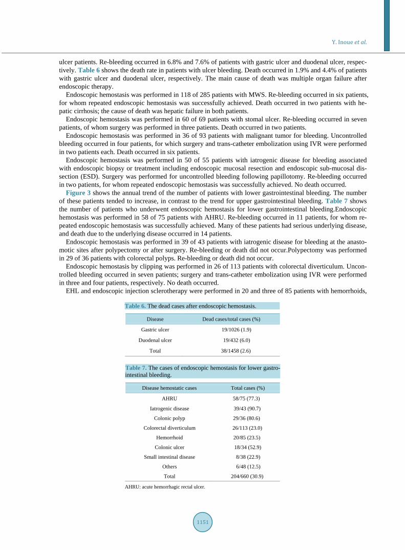
Figure 3 shows the annual trend of the number of patients with lower gastrointestinal bleeding. The number of these patients tended to increase, in contrast to the trend for upper gastrointestinal bleeding. Table 7 shows the number of patients who underwent endoscopic hemostasis for lower gastrointestinal bleeding.Endoscopic hemostasis was performed in 58 of 75 patients with AHRU. Re-bleeding occurred in 11 patients, for whom re-peated endoscopic hemostasis was successfully achieved. Many of these patients had serious underlying disease, and death due to the underlying disease occurred in 14 patients.
Endoscopic hemostasis was performed in 39 of 43 patients with iatrogenic disease for bleeding at the anasto-motic sites after polypectomy or after surgery. Re-bleeding or death did not occur.Polypectomy was performed in 29 of 36 patients with colorectal polyps. Re-bleeding or death did not occur.
Endoscopic hemostasis by clipping was performed in 26 of 113 patients with colorectal diverticulum. Uncon-trolled bleeding occurred in seven patients; surgery and trans-catheter embolization using IVR were performed in three and four patients, respectively. No death occurred.
EHL and endoscopic injection sclerotherapy were performed in 20 and three of 85 patients with hemorrhoids,
Table 6. The dead cases after endoscopic hemostasis.
Disease Dead cases/total cases (%)
Gastric ulcer 19/1026 (1.9)
Duodenal ulcer 19/432 (6.0)
Total 38/1458 (2.6)
Table 7. The cases of endoscopic hemostasis for lower gastro- intestinal bleeding.
Disease hemostatic cases Total cases (%)
AHRU 58/75 (77.3)
Iatrogenic disease 39/43 (90.7) Colonic polyp 29/36 (80.6)
Colorectal diverticulum 26/113 (23.0) Hemorrhoid 20/85 (23.5)
Colonic ulcer 18/34 (52.9) Small intestinal disease 8/38 (22.9)
Others 6/48 (12.5) Total 204/660 (30.9)
AHRU: acute hemorrhagic rectal ulcer.
Y. Inoue et al.
1152
Figure 3. The cases of lower gastrointestinal bleeding from A.D. 1987 to 2011.
respectively. Although hemostasis was obtained temporarily, bleeding occurred recurrently in two patients under endoscopic injection sclerotherapy; both patients died eventually.
4. Discussion Endoscopic hemostasis of non-variceal upper gastrointestinal bleeding in our hospital used to be performed by local injection of HSE when we started. Then, microwave coagulation was introduced in 1983, followed by adoption of HP in 1986, which has been utilized until now. Endoscopic hemostasis of non-variceal bleeding in-cludes local injection therapy using HSE or ethanol [5], thermo-coagulation therapy using high frequency waves [6], HP or microwaves, etc., and mechanical grasping methods using clips [7] or indwelling snares, etc. Our hospital exclusively adopts thermo-coagulation therapy with HP because of its convenient procedures as follows: First, thermo-coagulation is attained by simple contact of the bleeding site with the probe tip; thus, tangential le-sions can be treated without difficulty. Second, the probe sprays water; thus, the bleeding site can be easily iden-tified. Third, thermo-coagulation and water delivery are controlled by footswitches; thus, hemostasis can be managed by one operator. Finally, the amounts of heat and water delivery are adjusted in a stepwise manner; thus, a wide variety of bleeding patterns from arterial bleeding to capillary bleeding can be treated [8]. Clipping hemostasis is not effective in some cases for treatment of tangential lesions, and it is not suitable for blood spurts from the capillary vessels. Local injection hemostasis or clipping hemostasis requires not only an endos-cope operator but also assistant(s) who manipulate(s) an injection needle(s) or clip(s). Endoscopes integrated with water delivery function were recently developed; however before that, water delivery and treatment had not been available in one route at the same time. By resolving these difficulties, HP makes it possible to control he-mostasis in an easier manner compared to the other treatment methods. Note that hemostatic forceps using high- frequency waves are also being actively performed; this method utilizes soft coagulation that is used for hemos-tasis during ESD [9] [10].
The hemostatic rate of gastric ulcer with HP was 99.7% and duodenal ulcer was 94.0%. The hemostatic rate with other hemostatic methods were as follows: “clipping, 95.0% [11]; pure ethanol injection, 92.0% [12]; HSE injection, 95.0% [13]; high frequency waves, 91.6% [14]; and microwaves, 95.0% [15].”
Second-look endoscopy is endoscopy performed within approximately 24 hours after endoscopic hemostasis for upper gastrointestinal bleeding [16]. If the second-look endoscopy is performed without fail and additional treatment is given as required, the incidence of re-bleeding can be decreased [17] [18], which eventually will improve the hemostasis success rate [19]. The second-look endoscopy is currently recommended for patients with increased bleeding risk [20]; this is highly likely due to involvement of cost-effectiveness. Gastrointestinal endoscopy and treatment costs in Japan are several times less in comparison with those in Europe and the United States; therefore, the repeated endoscopy and treatment are more cost-effective in Japan than those in the regions.
Y. Inoue et al.
1153
In addition, in order to maintain the high hemostasis success rate, more patients must undergo second-look en-doscopy in an effort to decrease re-bleeding as much as possible.
In our hospital, over a long period of time, hemostasis of gastric ulcer bleeding has been accomplished only by using an endoscopic method. The possible reasons for our success include the following factors: First, opera-tors for endoscopic hemostasis have been settled in the long term in our hospital. Out of the four full-time ga-stroenterological endoscopy physicians, one has been attending our hospital for over 25 years, two for over 15 years, and one for over 8 years. Long-term engagement in medical care in a single hospital leads to the accumu-lation of expertise and treatment skills, which is believed to be reflected in treatment outcomes. Second, ther-mo-coagulation therapy by HP is adopted as an exclusive method for hemostasis in our hospital. Not surprising-ly as the situation requires, clipping, HSE injection therapy, or pure ethanol injection therapy may be performed, but utilization of HP is our basic stance. As described above, HP is easy-to-use as a hemostasis procedure, and it is expected that being expert in a uniform treatment will improve the outcomes. Third, second-look endoscopy is actively performed in our hospital. After emergency treatment for ulcers including that for the treatment of va-rices and MWS, second-look endoscopy is actively performed and additional treatment is provided if necessary. As described above, these tactics is effective not only for reduction in re-bleeding but also for improvement in treatment outcomes.
The incidence of lower gastrointestinal bleeding is 8% - 10%, lower than that of upper gastrointestinal bleed-ing [21]. Gastrointestinal endoscopy is useful for identification of the source of bleeding; however, the anatomi-cal forms of the colon (with acute angles of flexions and haustra) can make it difficult to identify the source. Although small-intestinal endoscopy for small-intestinal bleeding is routinely performed at present, this method is not indicated for emergency treatment under some circumstances [22]; thus, other measures such as CT and angiography are often taken. Only few diseases are subject to endoscopic hemostasis; in addition, the thin intes-tinal wall often prevents thermo-coagulation therapy and ethanol injection therapy, which may cause strong tis-sue damage, from being chosen as hemostasis procedures. Instead, clipping is generally used for these cases [23]. The main target diseases for hemostasis include colorectal diverticulum, rectal ulcers, polyps, iatrogenic diseas-es, and hemorrhoids. AHRU is an irregular-shaped ulcer formed immediately above the dentate line of the anus, and it is accompanied by painless, acute massive bleeding in elderly patients with severe underlying disease. Endoscopic hemostasis is appropriate for the disease and thermo-coagulation avails against bleeding. Bleeding from polyp usually resolves spontaneously, and elective polypectomy is performed. Iatrogenic disease is effec-tively managed by hemostasis using clips. Diverticular bleeding usually resolves spontaneously, but bleeding persists in some cases and requires endoscopic hemostasis using clips. In the event that the bleeding point is not accurately clipped, embolization may be performed under angiographic guidance. Hemorrhoidal bleeding usually resolves spontaneously; however, when bleeding persists, EHL may be performed. In cases when en-doscopic hemostasis does not succeed or endoscopy fails to identify the source of bleeding, etc., IVR with lower invasiveness is selected by an increasing number of hospitals rather than emergency surgery [24].
5. Conclusion We reported the current state of endoscopic therapy for gastrointestinal bleeding in our hospital. Endoscopic di-agnosis and therapy are superior for treatment of upper gastrointestinal bleeding with no treatment switch in most cases; however, massive duodenal bleeding may require treatment conversion to IVR in some cases. En-doscopic diagnosis and therapy are also effective for lower gastrointestinal bleeding; nevertheless, surgery or IVR is chosen in more cases compared to upper gastrointestinal bleeding. If small-intestinal endoscopy is further performed on a routine basis, it is expected that the number of cases treated by endoscopic therapy will increase in the future.
Acknowledgements The authors declare no conflict of interest. Part of the article was presented at a symposium in the 48th Annual Congress of Japanese Society for Abdominal Emergency Medicine in 2012.
References [1] Hirao, M., Yamazaki, H., Masuda, K., Kobayashi, T., Yamaguchi, S., Kawachi, H. and Sato, F. (1980) Hyper Saline-
Epinephrine Solution Injection Therapy. Stomach and Intestine, 15, 751-755.

Y. Inoue et al.
1154
[2] Tabuse, K., Katsumi, S., Kobayashi, Y., Nagai, Y., Noguchi, H. and Aoyama, O. (1982) Endoscopic Microwave-Tis- sue-Coagulation Method. Gastroenterological Endoscopy, 24, 1526-1535.
[3] Protell, R.L., Rubin, C.E., Auth, D.C., Silverstein, F.E., Terou, F., Dennis, M. and Piercey, J.R. (1978) The Heater Probe: A New Endoscopic Method for Stopping Massive Gastrointestinal Bleeding. Gastroenterology, 74, 257-262.
[4] Forrest, J.A., Finlayson, N.D. and Shearman, D.J. (1974) Endoscopy in Gastrointestinal Bleeding. Lancet, 2, 394-397. http://dx.doi.org/10.1016/S0140-6736(74)91770-X
[5] Asaki, S., Nishimura, T., Iwai, S., Kitamura, T., Masuda, Y., Hazama, K., Sato, M., Shibuki, S., Hanzawa, K., Sato, A., Ohkata, T. and Goto, Y. (1981) Tissue Solidification in Coping with Digestive Tract Bleeding: Hemostatic Effect of Local Injection of 99.5% Ethanol. Gastroenterological Endoscopy, 23, 792-799.
[6] Maruyama, M. (1992) Endoscopic Electrocoagulation Therapy. Endoscopia Digestiva, 4, 911-917. [7] Hachisu, T., Yamada, H. and Sato, S. (1996) The Knack of Clipping Hemostasis. Endoscopia Digestiva, 8, 1205-1207. [8] Inoue, Y., Fujino, Y. and Onodera, M. (2002) Heater Probe Method. In: Kitajima, M., Ed., The New Treatment of Ga-
stroenterological Endoscopy, The Institution of Advanced Medicine, Tokyo, 57-61. [9] Sugiyama, T., Dozaiku, T., Toyonaga, T., Hirooka, T., Ueda, C., Iwata, Y., Ono, W., Sugiyama, R. and Hirooka, T.
(2006) The Usefulness of 4 + 1 Contact Method Using Soft Coagulation for Bleeding Gastric Ulcers. Gastroentero-logical Endoscopy, 48, 204-211.
[10] Oyama, T., Miyata, Y., Tomori, A., Hotta, K., Yoneminato, T. and Morita, C. (2004) Selecting the Method of a High Frequency Unit for Endoscopic Submucosal Dissection. Endoscopia Digestiva, 16, 723-724.
[11] Nagayama, K., Tazawa, J., Sakai, Y., Miyasaka, Y., Yu, S.H., et al. (1999) Efficacy of Endoscopic Clipping for Bleeding Gastroduodenal Ulcer: Comparison with Topical Ethanol Injection. American Journal of Gastroenterology, 94, 2897-2901. http://dx.doi.org/10.1111/j.1572-0241.1999.01455.xb
[12] Nishiaki, M., Tada, M., Yanai, H., Tokiyama, H., Nakamura, H. and Okita, K. (2000) Endoscopic Hemostasis for Blee- ding Peptic Ulcer Using a Hemostatic Clip or Pure Ethanol Injection. Hepato-Gastroenterology, 47, 1042-1044.
[13] Hirao, M., Imai, Y. and Yamaguchi, S. (1997) Hyper Saline-Epinephrine Solution Injection Therapy. In: Saigenji, K., Ed., Endoscopic Handbook for Gastrointestinal Hemorrhage, Chugai Medical Co., Tokyo, 132-138.
[14] Maruyama, M. (1992) Endoscopic Electrocoagulation Therapy. Endoscopia Digestiva, 4, 911-917. [15] Nagai, Y., Takifuji, K., Shioji, Y., Ota, K., Hamanaka, H., Funabiki, S. and Tanimura, H. (2000) Endoscopic Micro-
wave Coagulation Therapy. Endoscopia Digestiva, 12, 770-771. [16] Inoue, Y., Fujino, Y. and Onodera, M. (2012) Emergency Endoscopy for Upper Gastrointestinal and Non-Variceal
Bleeding. Gastroenterology, 55, 545-552. [17] Chiu, P.W., Lam, C.Y., Lee, S.W., Kwong, K.H., Lam, S.H., Lee, D.T. and Kwok, S.P. (2003) Effect of Scheduled
Second Therapeutic Endoscopy on Peptic Ulcer Re-Bleeding: A Prospective Randomized Trial. Gut, 52, 1403-1407. http://dx.doi.org/10.1136/gut.52.10.1403
[18] Marmo, R., Rotondano, G., Bianco, M.A., Piscopo, R., Piscopo, A. and Cipolletta, L. (2003) Outcome of Endoscopic Treatment for Peptic Ulcer Bleeding: Is a Second Look Necessary? A Meta-Analysis. Gastroenterological Endoscopy, 57, 62-67. http://dx.doi.org/10.1067/mge.2003.48
[19] Yamaguchi, A., Uchizono, H., Taniguchi, Y., Iwakiri, Y., Utsunomiya, T., Shimokawara, H., Fujita, H., Nagota, T. and Ito, T. (2002) Internal Medical Treatment of Upper Gastrointestinal Hemorrhage. Japanese Journal of Clinical Internal Medicine, 17, 28-34.
[20] Bakun, A.N., Bardou, M., Kuipers, E.J., Sung, J., Hunt, R.H., Martel, M. and Sinclair, P. (2010) International Consen-sus Recommendations on the Management of Patients with Nonvariceal Upper Gastrointestinal Bleeding. Annual of Internal Medicine, 152, 101-113. http://dx.doi.org/10.7326/0003-4819-152-2-201001190-00009
[21] Inoue, Y., Kikuchi, S., Onodera, M., Fujino, Y., Kojika, M., Sato, N. and Endo, S. (2007) Primary Disease and Endos-copic Hemostasis of the Lower Digestive Tract Hemorrhage. Journal of Abdominal Emergency Medicine, 27, 923-928.
[22] Abe, T., Mizutani, M., Kawai, N., Kawano, J. and Sato, N. (2007) Double Balloon Enteroscopy for Diagnosis and Treatment of Small Intestinal Hemorrhage. Journal of Abdominal Emergency Medicine, 27, 949-956.
[23] Sugiyama, H. (2007) Endoscopic Hemostasis and Interventional Radiology for Lower Gastrointestinal Bleeding. Journal of Abdominal Emergency Medicine, 27, 929-935.
[24] Suzuki, S., Harada, N., Hayashi, T. and Suzuki, M. (2007) Selection of Non-Operative Hemostatic Therapeutic Strate-gies for Lower Gastrointestinal Bleeding. Journal of Abdominal Emergency Medicine, 27, 937-940.