Embed Size (px)
Citation preview
INTRODUCTION
Nonvariceal upper gastrointestinal bleeding (NVUGIB) is one of the most common reasons for hospitalization and a major cause of morbidity and mortality worldwide.1 Despite improvements in the management of this condition, the mor-tality rate has remained unchanged, possibly due to a longer life expectancy and the corresponding higher number of co-morbidities. International consensus recommendations on the management of patients with NVUGIB were published in 2010.2 In addition, an Asia-Pacific working group produced its own consensus recommendations taking into account some regional characteristics of NVUGIB. These include a high prevalence of Helicobacter pylori infection and a potential dif-ference in drug metabolism in Asia-Pacific populations as compared to other populations worldwide.3 Recently, new en-doscopic devices and apparatuses have been used for endo-scopic hemostasis along with the increasingly widespread
Clin Endosc 2015;48:96-101
96 Copyright © 2015 Korean Society of Gastrointestinal Endoscopy
use of the endoscopic submucosal dissection (ESD) technique. In this review article, we will focus on the state of the art man-agement of NVUGIB with a view to optimizing treatment ef-ficacy for individual patients.
PRE-ENDOSCOPIC MANAGEMENT
Patients presenting with upper gastrointestinal (GI) bleed-ing are at risk of hemodynamic shock and airway compromise. The priority is to assess the adequacy of the airway and cir-culation, and intravenous access should be secured with a large-bore cannula. Pulse oximetry, cardiac activity, and auto-mated blood pressure measurements should also be moni-tored. It is important to identify any use of nonsteroidal anti-inflammatory drugs, anticoagulants such as warfarin, and antiplatelet medicines since these treatments can cause or ex-acerbate upper GI bleeding. Coagulation time and hemoglo-bin levels should be checked as well. All patients should also be blood typed and cross-matched for an appropriate num-ber of units of packed red blood cells if necessary.4 Patients who have serious comorbid conditions may require hospital-ization regardless of the severity of their bleeding.5 Existing well-validated scoring systems can help categorize patients with upper GI bleeding as having either a low or high risk of rebleeding and mortality. The most widely applied scoring sys-tems are the Rockall and Blatchford scores.6,7 A primary aim
Endoscopic Management of Nonvariceal Upper Gastrointestinal Bleeding: State of the Art
Naoki Muguruma, Shinji Kitamura, Tetsuo Kimura, Hiroshi Miyamoto and Tetsuji TakayamaDepartment of Gastroenterology and Oncology, Institute of Health Biosciences, The University of Tokushima Graduate School, Tokushima, Japan
Nonvariceal upper gastrointestinal (GI) bleeding is one of the most common reasons for hospitalization and a major cause of morbidity and mortality worldwide. Recently developed endoscopic devices and supporting apparatuses can achieve endoscopic hemostasis with greater safety and efficiency. With these advancements in technology and technique, gastroenterologists should have no concerns re-garding the management of acute upper GI bleeding, provided that they are well prepared and trained. However, when endoscopic he-mostasis fails, endoscopy should not be continued. Rather, endoscopists should refer patients to radiologists and surgeons without any delay for evaluation regarding the appropriateness of emergency interventional radiology or surgery.
Key Words: Hemorrhage; Hemostasis; Equipment and supplies
Open Access
Received: January 5, 2015 Accepted: January 17, 2015Correspondence: Naoki MugurumaDepartment of Gastroenterology and Oncology, Institute of Health Biosciences, The University of Tokushima Graduate School, 3-18-15, Kuramoto-cho, Tokushi-ma 770-8503, JapanTel: +81-88-633-7124, Fax: +81-88-633-9235E-mail: [email protected] This is an Open Access article distributed under the terms of the Creative Commons Attribution Non-Commercial License (http://creativecommons.org/licenses/by-nc/3.0) which permits unrestricted non-commercial use, distribution, and reproduction in any medium, provided the original work is properly cited.
Print ISSN 2234-2400 / On-line ISSN 2234-2443
http://dx.doi.org/10.5946/ce.2015.48.2.96
FOCUSED REVIEW SERIES: Endoscopic Management of Upper Gastrointestinal Bleeding
Muguruma N et al.
97
Endoscopic hemostatic devices
Mechanical devicesThe hemoclip system is among the most popular endo-
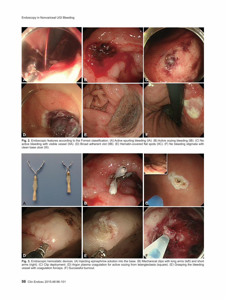
scopic mechanical modalities for achieving hemostasis. Clips have also been applied to the closure of mucosal defects, fis-tulas, and perforation holes.14 Endoscopic clips are deployed over a bleeding site including visible vessels and commonly fall off within days to weeks. If the bleeding site is located in the gastric fundus or the posterior wall of the duodenal bulb, ef-fective hemoclipping is relatively difficult. The deployment of hemoclips on a hard or fibrotic ulcer base is often challenging.15 Currently, hemoclip devices are rotatable and can be adjusted in the optimal direction by endoscopists. Hemoclips also come in various lengths, and shorter clips appear more effec-tive on hard or fibrotic areas because they attach to these le-sions more firmly than longer clips (Fig. 3A, B).16 Endoscopic band ligation devices, which have been used commonly for variceal bleeding, are also useful for treating NVUGIB by pro-ducing mechanical compression.17 The hood-like device of the ligation kit, which applies suction to varices, is useful for clearly visualizing the bleeding point. However, the effective-ness of this device depends on whether sufficient suction can be applied to a given lesion.
Cautery devicesArgon plasma coagulation (APC) is a noncontact thermal
method whereby ionized argon gas delivers a monopolar elec-trical current that effectively coagulates tissues. The most ad-vantageous aspect of APC is its familiarity to the endoscopy unit in that endoscopists and nurses are able to employ this equipment in most situations, especially vascular ectasia and
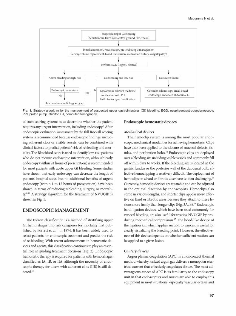
of such scoring systems is to determine whether the patient requires any urgent intervention, including endoscopy.4 After endoscopic evaluation, assessment by the full Rockall scoring system is recommended because endoscopic findings, includ-ing adherent clots or visible vessels, can be combined with clinical factors to predict patients’ risk of rebleeding and mor-tality. The Blatchford score is used to identify low-risk patients who do not require endoscopic intervention, although early endoscopy (within 24 hours of presentation) is recommended for most patients with acute upper GI bleeding. Some studies have shown that early endoscopy can decrease the length of patients’ hospital stays, but no additional benefits of urgent endoscopy (within 1 to 12 hours of presentation) have been shown in terms of reducing rebleeding, surgery, or mortali-ty.8-11 A strategy algorithm for the treatment of NVUGIB is shown in Fig. 1.
ENDOSCOPIC MANAGEMENT
The Forrest classification is a method of stratifying upper GI hemorrhages into risk categories for mortality first pub-lished by Forrest et al.12 in 1974. It has been widely used to select patients for endoscopic treatment and predict the risk of re-bleeding. With recent advancements in hemostatic de-vices and agents, this classification continues to play an essen-tial role in guiding treatment decisions (Fig. 2). Endoscopic hemostatic therapy is required for patients with hemorrhages classified as IA, IB, or IIA, although the necessity of endo-scopic therapy for ulcers with adherent clots (IIB) is still de-bated.13
Fig. 1. Strategy algorithm for the management of suspected upper gastrointestinal (GI) bleeding. EGD, esophagogastroduodenoscopy; PPI, proton pump inhibitor; CT, computed tomography.
Suspected upper GI bleeding(hematemesis, tarry stool, coffee-ground-like emesis)
Initial assessment, resuscitation, pre-endoscopic management(airway, volume replacement, blood transfusion, medication history, coagulopathy)
Perform EGD (urgent, elective)
No bleeding and low risk
Discontinue relevant medicine medication with PPIHelicobacter pylori eradication
No source found
Consider colonoscopy, small bowel endoscopy, enhanced abdominal CT
Active bleeding or high-risk
Endoscopic hemostasis
Interventional radiology surgery
NoYes
98 Clin Endosc 2015;48:96-101
Endoscopy in Nonvariceal UGI Bleeding
A
D
B
E
C
F Fig. 2. Endoscopic features according to the Forrest classification. (A) Active spurting bleeding (IA). (B) Active oozing bleeding (IB). (C) No active bleeding with visible vessel (IIA). (D) Broad adherent clot (IIB). (E) Hematin-covered flat spots (IIC). (F) No bleeding stigmata with clean base ulcer (III).
A
D
B
E
C
F Fig. 3. Endoscopic hemostatic devices. (A) Injecting epinephrine solution into the base. (B) Mechanical clips with long arms (left) and short arms (right). (C) Clip deployment. (D) Argon plasma coagulation for active oozing from telangiectasia (square). (E) Grasping the bleeding vessel with coagulation forceps. (F) Successful burnout.
Muguruma N et al.
99
oozing from superficial lesions. In APC, a flexible catheter passed through the working channel of the scope is placed against the target area, ideally in a vertical position. The press-ing of a foot pedal then triggers a synchronized flow of argon gas and electrical current from the generator, producing tissue coagulation (Fig. 3C). The depth of cauterization is thought to be limited because the coagulated tissue will gain increased resistance to the current. Various catheter types and operation modes are available with the APC device, including forced, pulsed, and precise. The variables that determine the cauteriza-tion effect can be set by endoscopists and nurses; these include the power setting (W), argon gas flow rate (L/min), distance to the tissue, and duration.18 In Korea and Japan, ESD has been widely performed for early gastric carcinoma, and the development of various hemostatic devices has accompanied its more widespread use. It has been reported that the use of hemostatic forceps together with soft coagulation is a safe and effective method of controlling upper GI ulcer bleeding (Fig. 3D, E).19 This type of device is preferred for the purpose of stopping spurting bleeding and ablating visible vessels.
Injection devicesEpinephrine injection is a common and effective method
of achieving endoscopic hemostasis owing to its low risk, low cost, and simplicity (Fig. 3F). Epinephrine injection alone is
superior to medical management alone, but is inferior to all other methods and should no longer be employed as a sole en-doscopic treatment when other options are available.20 Other injectable agents such as thrombin, fibrin, cyanoacrylate glues, polidocanol, and ethanolamine are not commonly used in the treatment of NVUGIB.5 It should be kept in mind that arterial injection of adrenaline may cause severe hypertension dur-ing emergency gastroscopy.21 The most commonly used injec-tors have diameters of 23 or 25 G, while the needle length typi-cally ranges from 3 to 5 mm. A special needle that is length-adjustable between 2 and 7 mm is also available.
Supporting apparatuses
Water jet-equipped endoscopeGiven the difficulty in identifying the bleeding point dur-
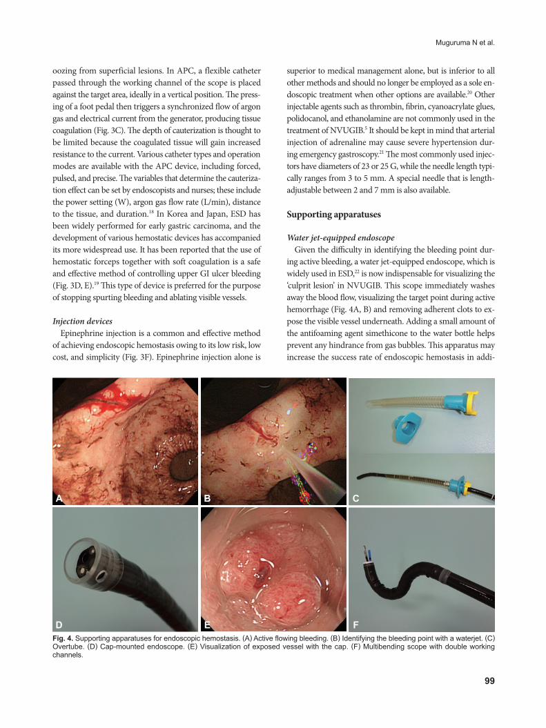
ing active bleeding, a water jet-equipped endoscope, which is widely used in ESD,22 is now indispensable for visualizing the ‘culprit lesion’ in NVUGIB. This scope immediately washes away the blood flow, visualizing the target point during active hemorrhage (Fig. 4A, B) and removing adherent clots to ex-pose the visible vessel underneath. Adding a small amount of the antifoaming agent simethicone to the water bottle helps prevent any hindrance from gas bubbles. This apparatus may increase the success rate of endoscopic hemostasis in addi-
A
D
B
E
C
F Fig. 4. Supporting apparatuses for endoscopic hemostasis. (A) Active flowing bleeding. (B) Identifying the bleeding point with a waterjet. (C) Overtube. (D) Cap-mounted endoscope. (E) Visualization of exposed vessel with the cap. (F) Multibending scope with double working channels.
100 Clin Endosc 2015;48:96-101
Endoscopy in Nonvariceal UGI Bleeding
tion to reducing operation times.
Carbon dioxide insufflatorsA benefit of carbon dioxide (CO2) is its rapid absorption
from the intestinal lumen into the blood stream. Thus, CO2 insufflation during endoscopy can significantly decrease ab-dominal pain and bloating both during and after the endo-scopic procedure.23 CO2 insufflation is preferred for endoscop-ic hemostasis procedures with a longer duration. CO2 use may permit the administration of lower doses of sedative medica-tions, which is apparently safer for patients in critical condi-tion, and can also lead to faster recovery times from high-de-pendency care. However, CO2 insufflation may place patients with respiratory disorders, sleep apnea, morbid obesity, or known CO2 retention at risk of ventilator compromise. Thus, the use of conventional room air is preferred in these cases.23 CO2 insufflation is beneficial in cases of perforation due to ex-cessive electrocautery, although efforts must be made to avoid this complication.
OvertubeAspiration pneumonia may occur in the setting of hemateme-
sis because of massive bleeding, and this may lead to mortali-ty. An overtube is a sleeve-like device designed to facilitate en-doscopy (Fig. 4C). All overtubes have an inner diameter that is larger than the diameter of an endoscope, providing a conduit for the passage of the device into the digestive tract.24 Overtubes limit the risk of aspiration during endoscopic hemostasis. They are also beneficial for repeated intubation and withdrawal in allowing adhered mucus or clots to be wiped off the endo-scope lens. In certain cases, the bleeding vessel is located in either the upper gastric body or the gastric fornix, which may not be visible owing to a large amount of blood clots. In such cases, it is necessary to switch from the conventional observa-tion position to the right lateral decubitus position. However, many endoscopists are uncomfortable using this reversed po-sition when performing hemostasis. Mori and colleagues25 (2013) invented an inverted overtube that can help endosco-pists perform an emergency endoscopy in their conventional standing position relative to patients who are rotated to the right lateral decubitus position without changing the positions of the endoscopy unit and light source. This technique allows endoscopists to dislodge blood clots and food residue by grav-ity and facilitates the rapid identification of exposed vessels, resulting in a higher rate of success for hemostasis.
Cap applicatorEndoscopic caps are commonly used in both diagnostic and
therapeutic endoscopy, including mucosal resections. A vari-ety of endoscopic caps are currently available in clinical prac-
tice, and they are effective when selected appropriately.26 It has been suggested that the application of a transparent cap to the tip of the endoscope can facilitate the approach to a tan-gential angle and stabilize the lesion during hemostasis (Fig. 4D, E).27 A non-randomized prospective study compared the success rates of achieving bleeding control using hemoclips with and without the aid of a transparent cap.28 In this study, hemo-clipping with the cap allowed for the clipping of lesions that were situated too tangentially to be clipped without the use of the cap. Movements resulting from the patient’s rapid respira-tion during emergency hemostasis often destabilize the posi-tioning of the scope and hemostatic devices. In this frustrating situation, control can be achieved by pressing the cap against the lumen wall.
Multibending scopePerforming endoscopic hemostasis is challenging in certain
locations such as the lesser curvature or posterior wall of the gastric body or the cardia and the lesser curvature of the an-trum. A multibending scope (GIF-2T260M; Olympus Optical, Tokyo, Japan) (Fig. 4F) that provides a second flexible section for improved positioning capability was initially developed to facilitate endoscopic mucosal resection of gastric tumors in these locations.29 Using secondary flexion, the tip of the scope can reach substantially closer to lesions in difficult to reach lo-cations. This special scope also has two working channels, al-lowing for an efficient procedure whereby residual food or clots are removed by suction through one channel while a he-mostatic device is inserted through the other channel.
LIMITATIONS OF ENDOSCOPIC MANAGEMENT
Although endoscopic therapy usually achieves primary he-mostasis, 10% to 30% of patients with NVUGIB have repeat bleeding.30-32 Patients in whom hemostasis is not achieved with endoscopy require transarterial embolization (TAE) or surgery. These are often elderly patients with multiple comor-bidities who are poor candidates for emergency surgery.33 A large number of studies have proposed using TAE over sur-gery as a salvage therapy; however, there is little empirical evi-dence to support this recommendation.34,35 Rapid technologi-cal advances in the management of NVUGIB have been made recently via interventional radiology, and prospective ran-domized studies comparing the efficacy of TAE and radiolog-ical interventions after failed endoscopic hemostasis are need-ed. Optimal management requires a multidisciplinary team of skilled endoscopists, interventional radiologists, and GI sur-geons who can integrate as a team promptly and effectively. In cases of failed endoscopic hemostasis in NVUGIB, endos-
Muguruma N et al.
101
copy should not be continued and another treatment option should be decided upon without delay.
CONCLUSIONS
Many safe and effective devices are available for endoscopic hemostasis. With the existing advancements in technology and technique, gastroenterologists should have no concern re-garding the management of acute upper GI bleeding, provided that they are well prepared and trained. However, when endo-scopic hemostasis fails, endoscopy should not be continued. Rather, endoscopists should refer patients to radiologists and surgeons without delay for evaluation regarding the appropri-ateness of emergency interventional radiology or surgery.
Conflicts of InterestThe authors have no financial conflicts of interest.
REFERENCES
1. Bardou M, Benhaberou-Brun D, Le Ray I, Barkun AN. Diagnosis and management of nonvariceal upper gastrointestinal bleeding. Nat Rev Gastroenterol Hepatol 2012;9:97-104.
2. Barkun A, Bardou M, Marshall JK; Nonvariceal Upper GI Bleeding Consensus Conference Group. Consensus recommendations for man-aging patients with nonvariceal upper gastrointestinal bleeding. Ann Intern Med 2003;139:843-857.
3. Sung JJ, Chan FK, Chen M, et al. Asia-Pacific Working Group consen-sus on non-variceal upper gastrointestinal bleeding. Gut 2011;60:1170-1177.
4. Al Dhahab H, Barkun A. The acute management of nonvariceal upper gastrointestinal bleeding. Ulcers 2012;2012:1-8.
5. Hwang JH, Fisher DA, Ben-Menachem T, et al. The role of endoscopy in the management of acute non-variceal upper GI bleeding. Gastro-intest Endosc 2012;75:1132-1138.
6. Rockall TA, Logan RF, Devlin HB, Northfield TC. Risk assessment af-ter acute upper gastrointestinal haemorrhage. Gut 1996;38:316-321.
7. Blatchford O, Murray WR, Blatchford M. A risk score to predict need for treatment for upper-gastrointestinal haemorrhage. Lancet 2000;356: 1318-1321.
8. Cipolletta L, Bianco MA, Rotondano G, Marmo R, Piscopo R. Outpa-tient management for low-risk nonvariceal upper GI bleeding: a ran-domized controlled trial. Gastrointest Endosc 2002;55:1-5.
9. Lin HJ, Wang K, Perng CL, et al. Early or delayed endoscopy for pa-tients with peptic ulcer bleeding. A prospective randomized study. J Clin Gastroenterol 1996;22:267-271.
10. Ananthakrishnan AN, McGinley EL, Saeian K. Outcomes of weekend admissions for upper gastrointestinal hemorrhage: a nationwide analy-sis. Clin Gastroenterol Hepatol 2009;7:296.e1-302.e1.
11. Lee JG, Turnipseed S, Romano PS, et al. Endoscopy-based triage signifi-cantly reduces hospitalization rates and costs of treating upper GI bleed-ing: a randomized controlled trial. Gastrointest Endosc 1999;50:755-761.
12. Forrest JA, Finlayson ND, Shearman DJ. Endoscopy in gastrointestinal bleeding. Lancet 1974;2:394-397.
13. Kim KB, Yoon SM, Youn SJ. Endoscopy for nonvariceal upper gastro-intestinal bleeding. Clin Endosc 2014;47:315-319.
14. Technology Assessment Committee, Chuttani R, Barkun A, et al. En-doscopic clip application devices. Gastrointest Endosc 2006;63:746-
750.15. Leung Ki EL, Lau JY. New endoscopic hemostasis methods. Clin En-
dosc 2012;45:224-229.16. Hosoe N, Imaeda H, Kashiwagi K, et al. Clinical results of endoscopic
hemostasis using a short transparent hood and short hemoclips for non-variceal upper gastrointestinal bleeding. Dig Endosc 2009;21:93-96.
17. Zepeda-Gómez S, Marcon NE. Endoscopic band ligation for nonvari-ceal bleeding: a review. Can J Gastroenterol 2008;22:748-752.
18. Watson JP, Bennett MK, Griffin SM, Matthewson K. The tissue effect of argon plasma coagulation on esophageal and gastric mucosa. Gas-trointest Endosc 2000;52:342-345.
19. Nagata S, Kimura S, Ogoshi H, Hidaka T. Endoscopic hemostasis of gastric ulcer bleeding by hemostatic forceps coagulation. Dig Endosc 2010;22(Suppl 1):S22-S25.
20. Vergara M, Calvet X, Gisbert JP. Epinephrine injection versus epi-nephrine injection and a second endoscopic method in high risk bleeding ulcers. Cochrane Database Syst Rev 2007;(2):CD005584.
21. Wu A. Arterial injection of adrenaline causing severe hypertension during emergency gastroscopy. Anaesth Intensive Care 2013;41:689.
22. Tatsumi K, Uedo N, Ishihara R, et al. A water-jet videoendoscope may reduce operation time of endoscopic submucosal dissection for early gastric cancer. Dig Dis Sci 2012;57:2122-2129.
23. Dellon ES, Hawk JS, Grimm IS, Shaheen NJ. The use of carbon diox-ide for insufflation during GI endoscopy: a systematic review. Gastro-intest Endosc 2009;69:843-849.
24. ASGE Technology Committee, Tierney WM, Adler DG, et al. Overtube use in gastrointestinal endoscopy. Gastrointest Endosc 2009;70:828-834.
25. Mori H, Kobara H, Fujihara S, et al. Accurate hemostasis with a new endoscopic overtube for emergency endoscopy. World J Gastroenterol 2013;19:2723-2726.
26. Sumiyama K, Rajan E. Endoscopic caps. Tech Gastrointest Endosc 2006; 8:28-32.
27. Warneke RM, Walser E, Faruqi S, Jafri S, Bhutani MS, Raju GS. Cap-assisted endoclip placement for recurrent ulcer hemorrhage after re-peatedly unsuccessful endoscopic treatment and angiographic emboli-zation: case report. Gastrointest Endosc 2004;60:309-312.
28. Kim JI, Kim SS, Park S, et al. Endoscopic hemoclipping using a trans-parent cap in technically difficult cases. Endoscopy 2003;35:659-662.
29. Isshi K, Tajiri H, Fujisaki J, et al. The effectiveness of a new multibend-ing scope for endoscopic mucosal resection. Endoscopy 2004;36:294-297.
30. Bjorkman DJ, Zaman A, Fennerty MB, Lieberman D, Disario JA, Guest-Warnick G. Urgent vs. elective endoscopy for acute non-variceal upper-GI bleeding: an effectiveness study. Gastrointest Endosc 2004;60:1-8.
31. Church NI, Palmer KR. Diagnostic and therapeutic endoscopy. Curr Opin Gastroenterol 1999;15:504-508.
32. Hearnshaw SA, Logan RF, Lowe D, Travis SP, Murphy MF, Palmer KR. Acute upper gastrointestinal bleeding in the UK: patient characteris-tics, diagnoses and outcomes in the 2007 UK audit. Gut 2011;60:1327-1335.
33. Beggs AD, Dilworth MP, Powell SL, Atherton H, Griffiths EA. A sys-tematic review of transarterial embolization versus emergency surgery in treatment of major nonvariceal upper gastrointestinal bleeding. Clin Exp Gastroenterol 2014;7:93-104.
34. Eriksson LG, Ljungdahl M, Sundbom M, Nyman R. Transcatheter ar-terial embolization versus surgery in the treatment of upper gastroin-testinal bleeding after therapeutic endoscopy failure. J Vasc Interv Ra-diol 2008;19:1413-1418.
35. Loffroy R, Estivalet L, Cherblanc V, et al. Transcatheter embolization as the new reference standard for endoscopically unmanageable upper gastrointestinal bleeding. World J Gastrointest Surg 2012;4:223-227.