Embed Size (px)
Citation preview

Endoscopic argon plasma coagulation
of gastrointestinal bleeding and
oesophageal stents
March 2008
MSAC application 1106
Assessment report

ii Argon plasma coagulation for gastrointestinal conditions
© Commonwealth of Australia 2008
ISBN (Print) 1-74186-628-6 ISBN (Online) 1-74186-629-4 ISSN (Print) 1443-7120 ISSN (Online 1443-7139 First printed July 2008 Copyright statements: Paper-based publications © Commonwealth of Australia 2008 This work is copyright. Apart from any use as permitted under the Copyright Act 1968, no part may be reproduced by any process without prior written permission from the Commonwealth. Requests and inquiries concerning reproduction and rights should be addressed to the Commonwealth Copyright Administration, Attorney General’s Department, Robert Garran Offices, National Circuit, Barton ACT 2600 or posted at http://www.ag.gov.au/cca Internet sites © Commonwealth of Australia 2008 This work is copyright. You may download, display, print and reproduce this material in unaltered form only (retaining this notice) for your personal, non-commercial use or use within your organisation. Apart from any use as permitted under the Copyright Act 1968, all other rights are reserved. Requests and inquiries concerning reproduction and rights should be addressed to Commonwealth Copyright Administration, Attorney General’s Department, Robert Garran Offices, National Circuit, Barton ACT 2600 or posted at http://www.ag.gov.au/cca Electronic copies of the report can be obtained from the Medical Service Advisory Committee’s Internet site at http://www.msac.gov.au/ Printed copies of the report can be obtained from: The Secretary Medical Services Advisory Committee Department of Health and Ageing Mail Drop 106 GPO Box 9848 Canberra ACT 2601 Enquiries about the content of the report should be directed to the above address. The Medical Services Advisory Committee (MSAC) is an independent committee which has been established to provide advice to the Minister for Health and Ageing on the strength of evidence available on new and existing medical technologies and procedures in terms of their safety, effectiveness and costeffectiveness. This advice will help to inform government decisions about which medical services should attract funding under Medicare. MSAC recommendations do not necessarily reflect the views of all individuals who participated in the MSAC evaluation. This report was prepared by the Medical Services Advisory Committee with the assistance of Dr Alun Cameron, Mr Ben Hoggan, Mr Luis Zamora and Ms Amelia Russin from the Australian Safety and Efficacy Register of New Interventional Procedures – Surgical (ASERNIP-S), and Dr Stephen Goodall of the Centre for Health Economics Research and Evaluation (CHERE). The report was edited by ASERNIP-S. This recommendation was endorsed by the Minister for Health and Ageing on 20 May 2008. Publication approval number: P3-3914

Argon plasma coagulation for gastrointestinal conditions iii
Contents
Executive summary................................................................................................. ix
Introduction ..............................................................................................................1
Background.............................................................................................................. 2 The procedure .................................................................................................................. 2 Intended purpose ............................................................................................................. 3 Clinical need/burden of disease .................................................................................. 10 Existing procedures....................................................................................................... 13 Comparator..................................................................................................................... 16 Clinical decision pathways ............................................................................................ 17 Marketing status of the device ..................................................................................... 20 Current reimbursement arrangement ......................................................................... 20
Approach to assessment .........................................................................................21 Search strategy................................................................................................................ 21 Inclusion criteria ............................................................................................................ 23 Review of literature ....................................................................................................... 24 Data analysis ................................................................................................................... 25 Included studies ............................................................................................................. 25 Current trials................................................................................................................... 26 Recent health technology assessments and systematic reviews on the use of APC for GI conditions ............................................................................................ 26 Expert advice.................................................................................................................. 27
Results of assessment: APC as a treatment for ablation of Barrett’s oesophagus............................................................................................................. 28
Is it safe?.......................................................................................................................... 28 Is it effective? ................................................................................................................. 33
Results of assessment: APC as a treatment for bleeding peptic ulcers................. 50
Results of assessment: APC as a treatment for gastric antral vascular ectasia..................................................................................................................... 66
Comparative studies....................................................................................................... 73 Case series ....................................................................................................................... 73
Results of assessment: APC as a treatment for radiation proctitis........................ 78 Systematic review evidence .......................................................................................... 78 Is it safe?.......................................................................................................................... 78 Is it effective? ................................................................................................................. 81 Conclusion...................................................................................................................... 85
Results of assessment: APC as a treatment for angiodysplasia ............................ 86

iv Argon plasma coagulation for gastrointestinal conditions
Results of assessment: APC as a treatment for post-polypectomy haemorrhage .......................................................................................................... 88
Is it safe?.......................................................................................................................... 88
Results of assessment: APC as a treatment for tumour ingrowth in oesophageal stents ................................................................................................. 92
Oesophageal malignancies............................................................................................ 92
Results of assessment: APC as a treatment used in studies of mixed indications.............................................................................................................. 94
Systematic reviews and health technology assessments on the use of APC in gastrointestinal bleeding ........................................................................................... 94 Mixed gastrointestinal indications for the use of APC ............................................ 94
What are the economic considerations? ................................................................ 99 Conclusion.................................................................................................................... 114
Conclusions........................................................................................................... 115 Safety ............................................................................................................................. 115 Effectiveness ................................................................................................................ 117 Cost-effectiveness........................................................................................................ 119
Recommendation.................................................................................................. 121
Appendix A MSAC terms of reference and membership.................................. 122
Appendix B Advisory Panel .............................................................................. 124
Appendix C AIHW Tables................................................................................. 125
Appendix D Studies included in the review...................................................... 128
Appendix E Studies excluded from the review................................................. 136
Appendix F HTA websites searched in this review.......................................... 138
Appendix G Electronic databases...................................................................... 140
Appendix H Complete radiation proctitis study information ........................... 141
Appendix I Unpublished RCT for radiation proctitis ...................................... 146
Appendix J Economic evaluation ..................................................................... 156
Abbreviations ........................................................................................................ 158
References ............................................................................................................. 160

Argon plasma coagulation for gastrointestinal conditions v
Tables
Table 1 AR-DRG data concerning the GI conditions indicated in this review, 2004-05..................................................................................................................... 10
Table 2 The number of patients treated with comparator treatments: Medical Benefit Schedule procedures in 2004-05 ............................................................. 10
Table 3 MBS item numbers of comparator procedures.................................................. 17 Table 4 Therapeutic Goods Administration status of items relating to APC............. 20 Table 5 PICO (population, intervention, comparator, outcome) criteria..................... 21 Table 6 Inclusion/exclusion criteria for identification of relevant studies for
APC as a treatment for gastro-intestinal conditions: safety.............................. 23 Table 7 Inclusion/exclusion criteria for identification of relevant studies for
APC as a treatment for gastro-intestinal conditions: effectiveness ................. 23 Table 8 Evidence dimensions ............................................................................................ 24 Table 9 Designations of levels of evidence* .................................................................... 25 Table 10 Summary of the technical parameters used in the case series for APC
treatment of Barrett’s oesophagus........................................................................ 28 Table 11 Concurrent medical treatments used in the case series for Barrett’s
oesophagus............................................................................................................... 29 Table 12 Summary of adverse events reported by case series for APC
treatment of Barrett’s oesophagus........................................................................ 30 Table 13 Summary of perforations and mortalities............................................................ 31 Table 14 Descriptive characteristics of comparative studies ........................................... 34 Table 15 Critical appraisal summary of studies with MBS-listed comparators –
study design details ................................................................................................. 36 Table 16 Critical appraisal summary of studies with MBS-listed comparators –
results details............................................................................................................ 36 Table 17 Patient characteristics of comparative studies ................................................... 38 Table 18 Types of Barrett’s oesophagus included in the comparative studies............... 38 Table 19 Technical details of APC techniques ................................................................... 40 Table 20 Description of MBS-listed comparators............................................................. 42 Table 21 Safety results of comparative studies .................................................................. 44 Table 22 Clinical outcomes of Barrett’s oesophagus reversal .......................................... 46 Table 23 Effectiveness results of studies with MBS-listed comparators ....................... 47 Table 24 Descriptive characteristics of comparative studies ............................................ 51 Table 25 Critical appraisal summary of comparative studies – study design
details ........................................................................................................................ 53 Table 26 Critical appraisal summary of comparative studies – results details............... 54 Table 27 Patient characteristics of comparative studies .................................................... 55

vi Argon plasma coagulation for gastrointestinal conditions
Table 28 Technical details of APC techniques .................................................................. 57 Table 29 Description of comparators ................................................................................. 58 Table 30 Safety results of comparative studies ................................................................... 60 Table 31 Effectiveness results of comparative studies ..................................................... 63 Table 32 Permanent haemostasis of bleeding peptic ulcers.............................................. 64 Table 33 Descriptive characteristics for APC treatment of GAVE studies ................... 66 Table 34 Critical appraisal summary of comparative studies – study design
details ........................................................................................................................ 67 Table 35 Critical appraisal summary of comparative studies – results details................ 67 Table 36 Critical appraisal summary of GAVE case studies – results details ................ 68 Table 37 Study population of comparative studies ............................................................ 68 Table 38 Study population of GAVE case series ............................................................... 69 Table 39 Baseline patient characteristics for GAVE case series (co-morbidity)............ 70 Table 40 Baseline patient characteristics for case series (co-morbidity - liver
disease)...................................................................................................................... 70 Table 41 Concurrent medications used by patients being treated for GAVE
with APC in case series .......................................................................................... 71 Table 42 Description of APC technique – comparative studies ...................................... 72 Table 43 Description of APC technique for treatment of GAVE in case series........... 72 Table 44 Adverse events reported by comparative studies for APC treatment
of GAVE.................................................................................................................. 73 Table 45 Summary of adverse events reported by case series for APC
treatment of GAVE................................................................................................ 74 Table 46 Details of adverse events reported by case series for APC treatment
of GAVE.................................................................................................................. 75 Table 47 Effectiveness results reported by comparative studies for APC
treatment of GAVE................................................................................................ 76 Table 48 Transfusion-dependency or haemoglobin levels of patients before
and after treatment with APC............................................................................... 76 Table 49 Descriptive characteristics of case series featuring radiation proctitis ............ 80 Table 50 Adverse events of APC treatment for radiation proctitis ................................. 81 Table 51 Bleeding-related outcomes for patients before and after APC
treatment .................................................................................................................. 82 Table 52 Descriptive characteristics of comparative study............................................... 83 Table 53 Patient characteristics of comparative studies .................................................... 83 Table 54 Technical details of APC technique ..................................................................... 84 Table 55 Safety outcomes for APC and formalin treatment ............................................ 84 Table 56 Effectiveness outcomes for APC and formalin treatment ............................... 85

Argon plasma coagulation for gastrointestinal conditions vii
Table 57 Descriptive characteristics of level IV evidence for angiodysplasia ................ 86 Table 58 Technical characteristics of case series featuring angiodysplasia ..................... 87 Table 59 Patient characteristics and outcomes of case series featuring
angiodysplasia .......................................................................................................... 87 Table 60 Descriptive characteristics of evidence for post-polypectomy and
other endoscopic procedure bleeding.................................................................. 88 Table 61 Technical characteristics of studies featuring post-polypectomy and
other endoscopic procedure bleeding.................................................................. 89 Table 62 Adverse events following APC treatment for post-polypectomy
bleeding .................................................................................................................... 90 Table 63 Descriptive characteristics of level IV evidence for stents ............................... 92 Table 64 Technical characteristics of case series featuring stents .................................... 93 Table 65 Patient characteristics and outcomes of case series featuring stents ............... 93 Table 66 Desciptive characteristics of case series............................................................... 96 Table 67 Technical characteristics of case series featuring mixed indications ............... 97 Table 68 Patient characteristics and outcomes of case series featuring mixed
indications ................................................................................................................ 98 Table 74 Medicare benefit schedules for endoscopic associated items......................... 102 Table 75 Average incremental costs per patient of disposables in performing
ablation of Barrett’s oesophagus with MPEC or APC – (Base case)............ 102 Table 69 Summary of effectiveness data – based on meta-analysis............................... 108 Table 70 Calculation of average capital costs per procedure for APC.......................... 109 Table 71 Calculation of average capital costs per procedure for heater probe ............ 109 Table 72 Average incremental costs per patient of performing peptic ulcer
repair (base case) ................................................................................................... 111 Table 73 Average incremental costs per patient of performing peptic ulcer
repair (including non-significant data) ............................................................... 113 Table 76 The number of separations related to ulcers in 2004-2005 as primary
diagnosis ................................................................................................................. 125 Table 77 The number of separations related to polyps in 2004-2005 as primary
diagnosis ................................................................................................................. 125 Table 78 The number of separations related to various other
gastrointestinalconditions in 2004-2005 as primary diagnosis ....................... 125 Table 79 The number of separations related to gastric ulcers in 2004-2005 as
primary diagnosis .................................................................................................. 125 Table 80 The number of separations related to duodenal ulcers in 2004-2005 as
primary diagnosis .................................................................................................. 126 Table 81 The number of separations related to peptic ulcers in 2004-2005 as
primary diagnosis .................................................................................................. 126
viii Argon plasma coagulation for gastrointestinal conditions
Table 82 The number of separations related to gastrojejunal ulcers in 2004-2005 as primary diagnosis.................................................................................... 126
Table 83 Number of procedures undertaken in the oesophagus according to AIHW data cubes ................................................................................................. 126
Table 84 Number of procedures undertaken in the stomach according to AIHW data cubes ................................................................................................. 127
Table 85 Number of procedures undertaken in the small intestine according to AIHW data cubes ................................................................................................. 127
Table 86 Number of procedures undertaken in the large intestine according to AIHW data cubes ................................................................................................. 127
Table 87 Number of procedures undertaken in the rectum and anus according to AIHW data cubes............................................................................................. 127
Table 88 Descriptive characteristics of case series featuring radiation proctitis .......... 141 Table 89 Technical characteristics of case series featuring radiation proctitis ............. 142 Table 90 Patient characteristics and outcomes of case series featuring radiation
proctitis................................................................................................................... 143
Figures
Figure 1 Clinical decision pathway for coagulation of gastrointestinal bleeding .......... 18 Figure 2 Clinical decision pathway for ablation of neoplastic ingrowth of
oesophageal stents .................................................................................................. 19 Figure 3 Meta-analysis of the clinical outcome for Barrett’s oesophagus ...................... 46 Figure 4 Meta-analysis of permanent haemostasis of bleeding peptic ulcers ................ 64 Figure 6 Sensitivity analysis measuring the influence the number of procedure
makes to the incremental cost per patient......................................................... 104 Figure 5 The incremental cost-effectiveness per additional patient with
permanent haemostasis against the effectiveness of APC.............................. 112
Argon plasma coagulation for gastrointestinal conditions ix
Executive summary
The procedure
The argon plasma coagulator (APC) is a non-contact electrocoagulation device that uses high-frequency monopolar current conducted to target tissues through ionised argon gas to achieve haemostasis or tissue ablation. This device may be used to coagulate bleeding endoscopically, as an alternative to standard haemostatic thermal techniques, and for re-establishing patency of oesophageal stents by ablation of tumour ingrowth. The APC machine is composed of a relatively standard diathermy unit (the multipolar electrocoagulator) with an argon gas source. Expert opinion of the Advisory Panel suggests that at the time of writing this review most Australian hospitals would have at least one APC unit.
The argon plasma is created by passing argon gas down the delivery catheter at rates of between 0.5 and 2.0 L/min while the electrosurgical generator delivers 500 to 6500 V to the exposed tungsten electrode inside the tip of the delivery catheter. The electrical power required to establish the argon plasma varies from 40 to 120 W. The precise power required varies according to the situation in which the APC is to be used, and is also dependent on the machine itself. The charged argon beam directs itself, independent of gas flow direction, to tissue in which the resistance is lowest. As soon as the target tissue is desiccated, the resistance of this tissue increases, and the ionised argon beam seeks to ground itself in adjacent tissue. This limits the coagulation depth to 2 to 3 mm, reducing the risk of perforation, and permits coagulation of large areas of diffuse bleeding via coagulation in a ‘paint brush’ fashion for diffuse areas of bleeding, or may be used in a spotting fashion as for laser.
For the purposes of this review, APC has been indicated for use in seven conditions relating to the gastrointestinal tract:
• ablation of dysplastic Barrett’s oesophagus
• haemostasis of bleeding ulcers
• haemostasis of gastric antral vascular ectasia (GAVE)
• haemostasis of radiation proctitis
• haemostasis of bleeding angiodysplasia
• coagulation of post-polypectomy bleeding
• ablation of tumourous growth through oesophageal metal stents.
x Argon plasma coagulation for gastrointestinal conditions
Medical Services Advisory Committee – role and approach
The Medical Services Advisory Committee (MSAC) was established by the Australian Government to strengthen the role of evidence in health financing decisions in Australia. MSAC advises the Minister for Health and Ageing on the evidence relating to the safety, effectiveness and cost-effectiveness of new and existing medical technologies and procedures, and under what circumstances public funding should be supported.
A rigorous assessment of evidence is thus the basis of decision making when funding is sought under Medicare. A team from the Australian Safety and Efficacy Register of New Interventional Procedures – Surgical (ASERNIP-S) was engaged to conduct a systematic review of literature on the use of the argon plasma coagulator for coagulation and ablation of gastrointestinal conditions (Application 1106). An advisory panel with expertise in this area evaluated the evidence and provided advice to MSAC.
MSAC’s assessment of argon plasma coagulation of gastrointestinal bleeding and oesophageal stents
Clinical need
According to data from the Australian Institute of Health and Welfare (AIHW), the total number of separations for gastrointestinal haemorrhage in 2004-05 was 10,718. Medicare statistics show that 6,733 procedures were undertaken during the same period for gastrointestinal bleeding.
Barrett’s oesophagus
The prevalence of Barrett’s oesophagus is estimated to be 18 per 100,000 in a United States population-based study; however, autopsy studies have shown that this may be a considerable underestimation. Barrett’s oesophagus may be categorised as either non-dysplastic or dysplastic. Non-dysplastic Barrett’s oesophagus may be controlled through the use of acid suppression therapy. Dysplastic Barrett’s oesophagus may progress to cancer of the oesophagus. There are increasing data to support treatment of dysplastic Barrett’s mucosa with thermal ablation, endoscopic mucosal resection or oesophagectomy, depending on its severity.
Haemostasis of bleeding ulcers
Peptic ulcers are one of the most common causes of gastrointestinal bleeding with an estimated annual incidence of 50 to 150 per 100,000 of the population. In Australia in 2004-05, AIHW data showed that 4,378 patients were diagnosed with haemorrhage of an ulcer, and Medicare data showed that 979 procedures were undertaken in the treatment of ulcers. This is likely to be a significant underestimate as one teaching hospital in Adelaide alone treats between 150-200 bleeding ulcers per year. Current endoscopic treatment options include thermal coagulation with or without adrenaline injection, or the use of a clipping device.

Argon plasma coagulation for gastrointestinal conditions xi
Gastric antral vascular ectasia
Gastric antral vascular ectasia (GAVE), also referred to as Watermelon stomach, is a severe haemorrhagic condition that leads to significant morbidity and transfusion-dependence in some patients. Re-bleeding following treatment is common, and there are few treatment options. Until recent treatment modalities were developed, the only options available to patients were blood transfusions or the surgical removal of the stomach (antrectomy). The estimated prevalence of GAVE ranges from 0.3 per cent of cases in a large endoscopic series to 4 per cent in highly selected cohorts with severe gastrointestinal bleeding. Although some patients with diffuse GAVE may have portal hypertensive gastropathy, for the purpose of this application the indication is GAVE not related to portal hypertensive gastropathy.
Radiation proctitis
Pelvic radiotherapy is a treatment for a number of tumours, particularly for prostate cancer (AIHW data shows that 23,343 new cases of prostate cancer were reported in 2004-05). Chronic bleeding leading to severe morbidity can occur in 2 to 20 per cent of these patients several months or even years following therapy. This is as a result of severe mucosal damage, for which the only current treatment is formalin instillation. According to the AIHW data cubes, in the financial year 2004-05 there were 2,042 separations for radiation proctitis. Medicare statistics shows that there were 111 services of formalin instillation for radiation proctitis, whist in the public sector there were 180 applications of formalin during the same time period.
Angiodysplasia
The prevalence of angiodysplasia is 0.8 to 2 per cent in healthy patients older than 50 years of age, and this condition may account for up to 40 per cent of gastrointestinal bleeding. According to the AIHW there were 731 separations as primary diagnosis of angiodysplasia of the colon in 2004-05. Current treatment options in addition to transfusion dependence are endoscopic thermal coagulation, surgery or oestrogen hormone therapy.
Post-polypectomy bleeding
According to the AIHW data cubes, the number of separations for primary diagnosis of gastrointestinal (GI) polyps in 2004-05 was 38,767. Medicare statistics show that 94,227 services were provided in 2004-05 for item numbers specifically related to polypectomy. In the public hospital system, 126,481 polypectomy procedures were undertaken during the same timeframe. Because colorectal cancer is closely associated with the presence of adenomatous polyps, detection and removal of pre-cancerous polyps (adenomas) eliminates their potential to become malignant and lowers the incidence of colorectal cancer in these patients. A potential complication of polyp removal is bleeding. Current treatment options include clipping or surgery.
Oesophageal tumour
Carcinoma of the oesophagus is the fifth most common malignant tumour in the developed world, with an incidence in the United Kingdom of approximately 10 per 100,000. Treatment options currently include the insertion of a self-expanding metal stent. However, tumours can grow through the stents, in which case the palliative
xii Argon plasma coagulation for gastrointestinal conditions
treatment alternatives include either the laser ablation of ingrowing tumour, the insertion of a second stent, or oesophagectomy.
Safety
There was a paucity of evidence for the use of APC in the treatment of bleeding angiodysplasia, post-polypectomy bleeding and for the ablation of tumour ingrowth through stents. Data was limited to a small number of case series and case reports. No significant complications were related to APC treatment. From the available evidence APC appears to be a relatively safe treatment option for these three indications.
In addition to the evidence for the use of APC for specific indications, 10 large case series involving 1,907 participants were identified in which APC was used in the treatment of mixed indications in the gastrointestinal tract. The majority of the complications were minor and temporary and resolved without further treatment. A total of six deaths were reported. Five of these were as a result of co-morbidities, and one was as a result of Aspergillus infection in a paediatric patient with a high level of co-morbidity. Three perforations were observed: two were asymptomatic and required no further treatment and one perforation required suturing. All patients recovered fully. From this evidence it appears that APC is a relatively safe treatment for a variety of gastrointestinal conditions.
Barrett’s oesophagus
A total of six randomised controlled trials (RCTs) were identified which investigated APC for this condition. Of these, three had a Medical Benefits Schedule (MBS)-listed procedure as a comparator. In addition, 16 case series were identified in which APC had been used to ablate Barrett’s oesophagus. The results suggest that APC is at least as safe as multipolar electrocoagulation, and as safe as conservative surveillance. In absolute terms, data from the case series suggest that APC is a relatively safe treatment for Barrett’s oesophagus. The majority of complications were transient and resolved without additional procedures. Of the 613 patients there were five cases of perforation, which led to two deaths. There did not appear to be a common factor in any of these adverse events. However, it must be noted that although 613 patients participated in these studies, patients received multiple treatments (an average of between one and eight); therefore, these complications were a result of some thousands of uses of APC.
Haemostasis of bleeding ulcers
Four RCTs were identified in which the effectiveness of APC was investigated for the treatment of bleeding ulcers, involving 386 participants. Two RCTs compared APC with heater probe coagulation. Of the other two RCTs, one was an internal comparison in which APC was compared to APC with adrenaline, and one compared APC directly with adrenaline. The Advisory Panel suggested that adrenaline is commonly used in Australia for the short-term haemostasis of non-variceal bleeding prior to thermal coagulation. The results suggest that APC is at least as safe as heater probe in the thermal coagulation of peptic ulcers. No case series investigating the effectiveness of APC for this indication were identified.

Argon plasma coagulation for gastrointestinal conditions xiii
Gastric antral vascular ectasia
Six case series and one small historical comparative study were included in which APC was used in the ablation of gastric antral vascular ectasia (GAVE). The total study population was 90 patients, the majority of whom suffered from a high degree of co-morbidity. APC appears to be at least as safe as the heater probe in the treatment of this indication. Most of the adverse events reported were directly attributable to the high level of morbidity of the participants. There was no study comparing APC to multiple blood transfusions or partial gastrectomy. This type of study is unlikely in the future as the comparator procedures are so drastic.
Radiation proctitis
Eighteen case series with a total of 369 participants were identified in which APC was used in the treatment of radiation proctitis. Overall, APC appears to be a relatively safe treatment modality for this indication. The majority of complications were transient, and many could be related to the morbidity of the disease itself, rather than as a complication of the treatment. There were no treatment-related deaths, and one perforation.
In addition to the case series evidence for radiation proctitis, one unpublished RCT was identified through the Advisory Panel in which APC was compared to formalin instillation. Nineteen patients were randomised. The study is found in full in the Appendix. In this study APC appeared to be as safe as formalin instillation, with no significant complications reported in either arm of the study. APC was associated with a slightly higher risk of rectal stricture.
Summary
The studies suggest that APC is a safe treatment for all seven conditions; however, the evidence was sparse in some cases. Where comparative studies were available, APC is at least as safe as the alternative Medicare-listed procedure.
Effectiveness
There was no comparative evidence available for the use of APC in the treatment of bleeding angiodysplasia, post-polypectomy bleeding or for the ablation of tumour ingrowth through stents; therefore, no estimation of its effectiveness compared to an alternative Medicare-listed procedure can be made. Seven systematic reviews were identified which reported on APC in the treatment of various gastrointestinal indications. None of the reviews provided a formal conclusion for the effectiveness of APC due to the paucity of comparative data.
Barrett’s oesophagus
In the two RCTs used to assess the effectiveness of APC in the treatment of Barrett’s oesophagus the majority of patients (89/92) had the non-dysplastic form of the disease. It is important to note that in Australia, non-dysplastic Barrett’s oesophagus would usually be controlled through acid suppression therapy rather than with the use of ablation. It is unlikely that enough patients with dysplastic Barrett’s oesophagus could be enrolled into a comparative trial as only a minority of patients have the more severe type of the disease. Therefore, evidence concerning the use of APC in the treatment of non-
xiv Argon plasma coagulation for gastrointestinal conditions
dysplastic Barrett’s oesophagus has been used to assess the effectiveness of the treatment.
Meta-analysis of the results of the two RCTs which compared APC with multipolar electrocoagulation (MPEC) for the ablation of Barrett’s oesophagus shows a relative risk of 0.89 in favour of MPEC (P=0.22). A total of 87 patients were randomised in both these studies. An increased number of high quality RCTs are required to assess whether this small variance is clinically significant.
Haemostasis of bleeding ulcers
Of the four included RCTs which investigated APC for the treatment of bleeding peptic ulcers, two studies with a total of 226 patients compared APC to the heater probe. The effectiveness outcomes from these two studies underwent meta-analysis. From the available data, APC is significantly more effective than heater probe in the coagulation of bleeding ulcers. The relative risk is 1.16 in favour of APC (P=0.02).
Gastric antral vascular ectasia
One comparative study was identified. This was a historical comparative study which investigated APC and the heater probe for haemostasis of GAVE with a total of 16 participants. Both treatment modalities appeared equally effective in treating GAVE; however, more high quality RCT evidence is required to assess the effectiveness of APC for GAVE.
Radiation proctitis
Although no published comparative studies were identified from the formal literature search, the Advisory Panel was able to provide a single unpublished RCT manuscript in which APC was compared to formalin instillation in the treatment of radiation proctitis. Nineteen patients were randomised. From this data, APC appeared to be as effective as formalin instillation. More high quality RCT evidence is required to fully assess the effectiveness of APC in the treatment of radiation proctitis.
Cost-effectiveness
Barrett’s oesophagus
There were only two head-to-head comparisons of APC and MPEC for the treatment of patients with Barrett’s oesophagus. Based on the individual trials APC appears at least as effective as the comparator. The meta-analysis of these studies demonstrates no statistical difference in the ablation of Barrett’s oesophagus with MPEC or APC, and a relative risk of 0.89 in favour of MPEC (P = 0.22) is reported.
A cost-analysis was conducted based on the assumption of no clinically significant differences in primary outcomes. Based on a number of estimates and assumptions:
• The majority of patients in both studies had non-dysplastic Barrett’s oesophagus, which in Australia would be treated using acid suppression therapy. Clinical experts advised that it would be appropriate to assume that the safety and effectiveness from these studies would be similar to the use of APC in the treatment of low-grade dysplastic Barrett’s oesophagus.
Argon plasma coagulation for gastrointestinal conditions xv
• A conservative estimate of the total number of patients who would be treated with APC has been used, based on the total number of patients diagnosed with Barrett’s oesophagus. Only a small proportion of these patients would have low-grade dysplasia and therefore would be considered for ablative treatment such as APC or MPEC. An exact estimate of this number was unavailable.
• The incremental cost per patient of receiving APC rather than MPEC for the treatment of Barrett’s oesophagus is $283. The bulk of this extra cost is associated with the higher procedural fee and additional capital cost of the APC equipment. These costs are partially offset by a saving in the cost of the disposable probe.
• Based on these estimates, the total additional cost to the health care system of treating Barrett’s oesophagus patients with APC is $1,633,000 per annum. This figure is estimated from the total number of patients who might be diagnosed with Barrett’s oesophagus in Australia. However, as mentioned previously, only a small proportion of these patients would be considered for ablative treatment, therefore the actual cost to the healthcare system is likely to be much lower.
Bleeding peptic ulcers
There were only two reliable head-to-head comparisons of APC and heater probe for the treatment of patients with bleeding peptic ulcer. Based on the individual trials APC appears at least as effective as the comparator, although both studies demonstrated a tendency favouring APC. Based on the combined meta-analysed data, APC demonstrates greater effectiveness in terms of the primary outcome, namely permanent haemostasis (APC=93.2% and HP=80.0%). This difference is statistically significant. However, the validity of the meta-analysis was confounded by one study using adrenaline injection to achieve haemostasis before APC and heater probe treatment.
A modelled cost-effectiveness analysis was conducted based on the improved effectiveness of APC as determined by the meta-analysis. Based on a number of estimates and assumptions the cost-effectiveness of APC would be as follows:
• The incremental cost per patient of receiving APC rather than heater probe treatment for bleeding peptic ulcer is $343. The bulk of this extra cost is associated with the APC probe, which is disposable, and the estimated higher procedural fee.
• Based on an estimated 13.2% improvement in effectiveness (permanent haemostasis), the incremental cost-effectiveness per additional patient with permanent haemostasis is $2606 (or $6231 for a 5.5% improvement in effectiveness).
Recommendation
MSAC recommended that on the strength of evidence pertaining to argon plasma coagulation for gastrointestinal bleeding public funding should be supported for this procedure.

xvi Argon plasma coagulation for gastrointestinal conditions
MSAC has considered the safety, effectiveness and cost-effectiveness of endoscopic argon plasma coagulation compared with alternative modalities used to secure gastrointestinal haemostasis under certain circumstances and for the ablation of tumorous growth through or over oesophageal stents. MSAC finds that argon plasma coagulation is as safe as other forms of heat coagulation or local vasoconstrictor therapy in peptic ulcer disease. Although data for the other conditions with low incidence is very limited, argon plasma coagulation is considered by inference to be similar in safety profile for haemostasis of radiation proctitis, haemostasis of bleeding angiodysplasia, coagulation of post-polypectomy bleeding, other allied conditions of low incidence (haemostasis of gastric antral vascular ectasia (GAVE), and ablation of tumorous growth through or over oesophageal stents). MSAC considers that argon plasma coagulation is at least as effective and as cost-effective as other local methods of treatment of bleeding in peptic ulcer disease. There are insufficient data to demonstrate effectiveness and cost-effectiveness for haemostasis of radiation proctitis, haemostasis of bleeding angiodysplasia, coagulation of post-polypectomy bleeding, other allied conditions of low incidence (haemostasis of gastric antral vascular ectasia (GAVE), and ablation of tumorous growth through or over oesophageal stents). MSAC considers that the incidence of these conditions is insufficient to allow the collection of these data. MSAC recommends that public funding is supported for endoscopic argon plasma coagulation as an option for the treatment of peptic ulcer disease and other less common causes of gastro-intestinal bleeding including radiation proctitis, bleeding angiodysplasia, post-polypectomy bleeding, gastric antral vascular ectasia (GAVE), and for ablation of tumorous growth through or over oesophageal stents.
- The Minister for Health and Ageing endorsed this recommendation on 20 May 2008 -
Argon plasma coagulation for gastrointestinal conditions 1
Introduction
The Medical Services Advisory Committee (MSAC) has reviewed the use of argon plasma coagulation (APC), which is a therapeutic device for the following indications:
• Barrett’s oesophagus
• bleeding peptic ulcers
• gastric antral vascular ectasia
• radiation proctitis
• angiodysplasia
• bleeding post-polypectomy
• restoring the patency of oesophageal stents after tumour ingrowth.
MSAC evaluates new and existing health technologies and procedures for which funding is sought under the Medicare Benefits Scheme in terms of their safety, effectiveness and cost-effectiveness, while taking into account other issues such as access and equity. MSAC adopts an evidence-based approach to its assessments, based on reviews of the scientific literature and other information sources, including clinical expertise.
MSAC’s terms of reference and membership are in Appendix A. MSAC is a multidisciplinary expert body, comprising members drawn from such disciplines as diagnostic imaging, pathology, surgery, internal medicine and general practice, clinical epidemiology, health economics, consumer health and health administration.
This report summarises the assessment of current evidence for Application 1106, Endoscopic argon plasma coagulation of gastrointestinal bleeding and oesophageal stents.

2 Argon plasma coagulation for gastrointestinal conditions
Background
Argon plasma coagulation
Haemostasis is one of the most important problems in endoscopy. Many different endoscopic methods have been developed during the last 20 years resulting in a revolution in treatments of different types of gastrointestinal (GI) bleeding (Grund et al 1999); however, no single method covers all kinds and sources of haemorrhage. Many of the currently used methods are insufficient for the treatment of some difficult types of bleeding: diffuse bleeding arising from large areas, bleeding as a result of coagulation disorder, or a haemorrhage which is diffuse and difficult to control (Grund et al 1999). One device that has been suggested as a treatment option for an increasing number of causes of GI haemorrhage as well as for the ablation of tumour ingrowth of oesophageal stents is the argon plasma coagulator (APC) (Grund et al 1999).
According to expert clinical advice from the Advisory Panel, the APC machine is a common device, with each major hospital in Australia having at least one machine.
The procedure
The APC is a non-contact electrocoagulation device that uses high-frequency monopolar current conducted to target tissues through ionised argon gas (argon plasma) (Ginsberg et al 2002). APC acts in both a haemostatic and ablative manner (Ginsberg et al 2002). This device has been used over the past 10 years or so as a tool to coagulate bleeding endoscopically, having originally been used in open and laparoscopic surgery (Ginsberg et al 2002). Since early reports the use of this device in therapeutic endoscopy has steadily increased (Canard et al 2001). It has been suggested that APC may be used as an alternative to laser treatment of bleeding of the GI tract (Canard et al 2001) and for re-establishing patency of oesophageal stents after tumour ingrowth (Mason 2002).
The indications for use of APC are very broad and continue to increase. Initial experience was largely gained in the treatment of superficial vascular bleeding lesions such as angiodysplasia, gastric antral vascular ectasia (GAVE or watermelon stomach) and radiation-induced enteropathy and proctopathy (Seitz et al 2003). As experience grew, vascular lesions such as bleeding peptic ulcers and Dieulafoy lesions were also treated. Argon plasma coagulation has also found a role as an adjunct treatment during polypectomy (Apel et al 2005). Remnant polyp tissue remaining after piecemeal polypectomy may be treated with APC and small polyps can be ablated using APC. Argon plasma coagulation can also be used in the ablation of dysplastic or metaplastic mucosa in the gut such as Barrett’s mucosa. Endoscopic ablation of dysplastic Barrett’s mucosa is possibly a more attractive option than invasive and complex surgery (Haag et al 1999). Superficial adenomatous tissue elsewhere in the gut can also be treated; patients with familial adenomatous polyposis undergoing surveillance endoscopy of the duodenum can have small adenomas ablated (Suzuki et al 2006). APC may also be used to treat bleeding tumours and tumour ingrowth through metal oesophageal stents.
There are many models of APC on the international market. The largest manufacturer is ERBE Elecromedizin GmbH, Tubingen (Germany) which distributes models worldwide.
Argon plasma coagulation for gastrointestinal conditions 3
According to expert clinical advice from the Advisory Panel an APC machine is essentially a standard diathermy (or multipolar electrocoagulation) machine with an additional argon gas source. The components of the APC system are a high-frequency monopolar electrosurgical generator and argon gas source, gas flow meter, flexible delivery catheters, foot activation switch and grounding pads. The delivery catheters that are passed through the endoscope consist of a Teflon tube with a ceramic nozzle tip housing a tungsten monopolar electrode. The probes are available in various diameters and lengths to suit a variety of different types of endoscope. Initially the nozzles of the delivery catheters were simple ‘end-firing’ catheters but new models now include ‘side-firing’ and ‘ball-tip’ catheters that are designed to improve safety.
The argon plasma is created by passing argon gas down the delivery catheter at rates of between 0.5 and 2 L/min while the electrosurgical generator delivers 500 to 6,500 V to the exposed tungsten electrode inside the tip of the delivery catheter. The power setting on the electrosurgical generator varies from 40 to 120 W. Although increased power may be associated with a deeper burn and increased risk of perforation, it is important to note that there needs to be sufficient power to establish the plasma. The exact wattage may vary according to the machine used; newer models require less charge to establish the plasma, and some models automatically vary the watts so that it is not possible for an operator to define a specific setting.
Once established, the charged argon beam directs itself, independent of gas flow direction, to tissue in which the resistance is lowest. As soon as the target tissue is desiccated, the resistance of this tissue increases, and the ionised argon beam seeks to ground itself in adjacent tissue. This limits the coagulation depth to 2 to 3 mm, reducing the risk of perforation, and permits coagulation of large areas of diffuse bleeding via coagulation in a ‘paint brush’ fashion, in spite of the relatively narrow gas beam discharged from the probe (Singh & Harber 1999). Alternatively, the APC may be used in a woodpecker or spot treatment which is most frequently used for radiation proctitis or angiodysplasia of the rectum or caecum.
Intended purpose
For the purpose of this assessment, use of the APC is considered for the following clinical indications:
• Barrett’s oesophagus
• bleeding peptic ulcers
• gastric antral vascular ectasia
• radiation proctitis
• bleeding angiodysplasia
• bleeding post-polypectomy
• tumour ingrowth of self-expanding metal stents.
4 Argon plasma coagulation for gastrointestinal conditions
Barrett’s oesophagus
Barrett’s oesophagus is a premalignant acquired disorder which results in the uncontrolled growth of cells in the epithelium (Wang et al 2001). It leads to the narrowing of the oesophagus and subsequent problems such as dysphagia and stricture formation. Recently published guidelines define Barrett’s oesophagus as ‘columnar epithelium of any length that can be recognised at endoscopy and confirmed histologically to contain specialized intestinal metaplasia with goblet cells’ (Conio et al 2003). Gastroesophageal reflux disease is a risk factor for Barrett’s oesophagus and plays an important role in the genesis of the condition and oesophageal adenocarcinoma (Franchimont et al 2005).
Dysplasia consists of an expansion of immature cells with a corresponding decrease in the number and the location of maturing cells. This change is often indicative of the early neoplastic process. Dysplasia is defined histologically as unequivocal neoplastic alteration of the epithelium not invading the lamina propria, and is characterised by cytologic and architectural disarray. Most often dysplasia occurs with a patchy, irregular distribution in flat mucosa that is usually invisible at endoscopy (Van Laethem et al 2001).
The changes in the oesophageal cells are caused by acid reflux. The acid causes irritation to the lining of the oesophagus and over time the cells change from normal squamous cells into the columnar abnormal square cells, typical of Barrett’s oesophagus. Other risk factors include age of onset of symptoms, duration of symptoms, obesity and hereditary risk factors (Schulz et al 2000).
Barrett’s oesophagus is sometimes simply classified according to the length of columnar epithelium. In addition, a classification system of dysplasia in Barrett’s oesophagus similar to that of dysplasia in inflammatory bowel disease has been devised. This classification consists of three groups: negative, indefinite and positive for dysplasia. The latter comprises low-grade dysplasia (LGD) and high-grade dysplasia (HGD). The cancer risk in LGD is not well-defined but is smaller than that associated with HGD. While most patients with Barrett’s oesophagus do not develop adenocarcinoma in their lifetime, research indicates that HGD can evolve to cancer (Attwood et al 2003). Adenocarcinoma in Barrett’s oesophagus develops through stages from non-dysplastic metaplasia followed by increasing grades of dysplasia and eventually adenocarcinoma (Hage et al 2005).
Barrett’s oesophagus prevalence in males is twice that of females. It is rare in childhood: the estimated mean age of development is about 40 years although the mean age at diagnosis is often about 60 years (Terano et al 2002). Although the quality of life of many sufferers of Barrett’s oesophagus is largely unaffected by the disease, patients can present with persistent heartburn; difficult and/or painful swallowing; recurring vomiting; persistent weight loss; or a sensation of fullness during consumption of food.
Current treatment options include the use of anti-reflux medications, thermal ablation and surgery. The most common medications are proton pump inhibitors (PPI) including lansoprazole, omeprazole and pantoprazole which work to eliminate the symptoms of reflux by reducing the acid returning to the oesophagus, but do not resolve cellular abnormalities. Thermal ablation treatment is more aggressive in the sense that it works to remove the abnormal cells lining the oesophagus. This may be achieved with laser or multipolar electrocoagulation, and normally requires multiple treatment sessions. Although often successful, thermal ablation can result in serious adverse events including stricture formation, perforation and death (van den Boogert et al 1999). However,

Argon plasma coagulation for gastrointestinal conditions 5
success of the treatment may be associated with the experience of the operator of the laser. Surgery is an aggressive option and involves the removal of the lower part of the oesophagus. It is usually only undertaken if anti-reflux medications have been unsuccessful and if cancerous or highly dysplastic cells have been identified within the oesophagus. Endoscopic mucosal resection may also be used in severe cases; however, this treatment is not available through the MBS. An alternative to these treatments is APC. Argon plasma coagulation ablates the metaplastic mucosa in a similar fashion to the laser and may be used in conjunction with PPI therapy (Sharma 2001).
Expert clinical opinion of the Advisory Panel suggests three main groups of patients with Barrett’s oesophagus. The first are people who have non-symptomatic, non-dysplastic Barrett’s mucosa. These do not develop dysplasia and may be maintained on PPIs. The second group of patients are those who develop dysplastic Barrett’s oesophagus which is ablated using thermal techniques such as APC. These patients are then placed on a lifelong regimen of PPIs. The third group of patients are those who develop high-grade dysplasia for which oesophagectomy is the main option. Therefore, in Australia, APC is mainly indicated for use in patients who have proven low-grade dysplasia.
Ulcers
The most common cause of upper gastro-intestinal (GI) bleeding is peptic ulcer disease (Leontiadis 2005). An ulcer is caused by damage to the gastric mucosa, which may be associated with the erosion of a submucosal artery (Church & Palmer 2000). The term ‘peptic’ ulcer refers to those ulcers that occur in either the stomach or the duodenum. This condition accounts for 60 per cent of cases of bleeding found at emergency endoscopy. Symptoms of ulcers include melaena or tarry stools, haematemesis, bloating and severe abdominal pain (Ferguson & Mitchell 2005).
Principal causes of peptic ulcers include Helicobacter pylori (H. pylori) infection (Lai & Sung 2007), the use of medications such as non-steroidal anti-inflammatory drugs (NSAIDs) and stomach malignancies (Parfitt & Driman 2007).
About 50 per cent of cases have a clean-based ulcer with a low probability of re-bleeding, so that only pharmacological intervention is required. Adherent clots, visible vessels or active bleeding portend progressively less favourable outcomes unless endoscopic or surgical treatment is applied (Rajan et al 2003). Younger patients with ulcer-like symptoms are often treated with antacids or histamine antagonists. When H. pylori infection is present, the most effective treatments are combinations of antibiotics and a proton pump inhibitor. Treatment of H. pylori usually leads to clearing of infection, relief of symptoms and eventual healing of ulcers (Lai & Sung 2007).
Bleeding from a peptic ulcer may stop spontaneously in approximately 80 per cent of patients (Chau et al 2003). In situations where ulcers are perforated, urgent surgery is required. Different methods of endoscopic haemostasis of bleeding ulcers include electrocoagulation, laser therapy, thermal probes, mechanical devices, injection of fibrin or thrombin glue, or injection of adrenaline or a sclerosing agent (Church & Palmer 2000; Skok et al 2001). Although primary haemostasis may be achieved in up to 95 per cent of patients, recurrent bleeding may still occur in 4 to 30 per cent of cases and re-treatment will be required (Marmo et al 2007). Argon plasma coagulation is an alternative to these current methodologies and may be used for this indication to attain haemostasis by coagulating the bleeding area.
6 Argon plasma coagulation for gastrointestinal conditions
Gastric antral vascular ectasia
Gastric antral vascular ectasia (GAVE) or Watermelon stomach (Yusoff 2002) is relatively uncommon (Novitsky et al 2003) but is an important and serious cause of occult GI blood loss (Dulai & Jenson 2006). Patients may suffer severe morbidity and typically present with chronic blood loss and iron deficiency anaemia (Novitsky et al 2003). Overt bleeding may also occur. The aetiology of the condition is not known but it may be due to abnormal motor activity of the distal stomach resulting in mucosal trauma. Endoscopically, GAVE may have a typical ‘watermelon’ appearance of prominent haemorrhagic streaks in the antrum radiating from the pylorus, or may be more diffuse (Novitsky et al 2003; Stotzer et al 2002). Many GAVE patients suffer from significant liver-related co-morbidities including liver dysfunction, cirrhosis, alcohol damage and steatohepatitis (Roman 2003). GAVE may also be related to portal hypertensive gastropathy, autoimmune disease and diabetes mellitus (Sato et al 2005).
It is important to differentiate between portal hypertensive gastropathy associated with cirrhosis and GAVE, which are two distinct conditions. GAVE is associated with cirrhosis in about 30 per cent of cases (Sebastien et al 2003) and has more severe chronic bleeding than portal hypertensive gastropathy. In addition, GAVE does not respond to beta blockers or nitrates, which are standard medical treatment for portal hypertensive gastropathy (Sebastien et al 2003). Although some patients with diffuse GAVE may have portal hypertensive gastropathy (Dulai et al 2004), for the purpose of this application the indication is GAVE not related to portal hypertensive gastropathy.
The typical patient is an elderly female with a history of chronic iron-deficiency anaemia for which no aetiology as been recognised despite endoscopic and barium studies (Sebastian et al 2003). There exists a female preponderance of 3:1 for this disease. Symptoms include iron deficiency anaemia (88%) and haemopositive stools (42%). Other frequently associated symptoms at presentation include melaena, haematochesia and haematemesis (Novitsky et al 2003).
Treatment options for GAVE depend on the severity of disease. In many cases, parenteral or oral iron supplementation may be sufficient; however, patients are often transfusion dependent with average requirements of 10 units of blood per year but can be as high as 50-100 units per year in severe cases (Novitsky et al 2003). Thus patients are at risk of viral transmission despite the current meticulous screening of blood products. In addition, red blood cell-related sepsis and endotoxin-induced septic shock present additional dangers. Thus ultimately the goal of therapy is the complete or near-complete elimination of blood transfusion requirements in patients (Novitsky et al 2003).
There is no treatment for GAVE; current therapies are essentially palliative measures to reduce bleeding and symptoms. Until recent treatment modalities were developed, the only options available to patients were blood transfusions or the surgical removal of the stomach (antrectomy) (Roman et al 2002). The current first-line therapy for GAVE consists of endoscopic ablation with either heater-probe or neodymium:yttrium-aluminum-garnet (Nd:YAG) laser coagulation (Garcia & Sanyal 2001; Jensen et al 2004; Yusoff et al 2002). The objective of thermal coagulation is the formation of superficial ulcers, which may themselves lead to minor secondary bleeding (Jensen et al 2004). Recurrence of the bleeding is relatively common. An advantage of APC for the diffuse bleeding associated with GAVE is that it can be used in a ‘paintbrush’ fashion as opposed to ‘point’ coagulation achieved with lasers. Several treatment sessions may be required to ensure haemostasis.

Argon plasma coagulation for gastrointestinal conditions 7
Radiation proctitis
Radiotherapy techniques are common treatments for pelvic malignancies, most commonly for prostate cancer (Cotti et al 2003; Hong et al 2001). Acute severe haematochesia is a rare complication of radiation therapy. Inflammation caused by exposure of the rectum or rectosigmoid region to radiation during therapy may result in significant chronic bleeding which develops several months or years following therapy (Ben-Soussan et al 2004; de la Serna Higuera et al 2004). Chronic bleeding can occur in 2 to 20 per cent of these patients (Cotti et al 2003; Silva et al 1999). This cause of rectal bleeding accounts for 1 to 5 per cent of cases of acute lower GI bleeding. Following acute mucosal injury, the patient may complain of diarrhoea and tenesmus, accompanied by abdominal cramping and a mucoid or bloody rectal discharge (Hong et al 2004). A chronic proctocolitis may develop which may be complicated by mild to moderate bleeding. Endoscopically the mucosa demonstrates characteristic telangiectases, along with ulceration (Tagkalidis & Tjandra 2001). Patients are highly transfusion-dependent.
Bleeding may be controlled with a variety of treatments including local application of 4 per cent formaldehyde or endoscopic thermal coagulation (Bounds et al 2003). Several other conservative treatments may also be used to control bleeding, such as the rectal administration of steroids, short-chain fatty acids or sucralfate, or oral salicylates (Cotti et al 2003). Argon plasma coagulation may be used to coagulate the bleeding lesion by focusing the stream of argon plasma onto the bleeding area until a white coagulum is visualised down the endoscope (Ramage & Gostout 2003). A woodpecker or spot treatment is often used in preference to a brush-like technique for this indication. Depending on the area of mucosa affected and the extent of bleeding, several treatment sessions may be required to ensure haemostasis.
Angiodysplasia
Angiodysplasia or arteriovenous malformation (AVM) is the most common vascular anomaly of the GI tract. Composed of an ectatic, dilated submucosal vein (usually multiple occurrences), colonic angiodysplasia is responsible for 20 to 30 per cent of cases of acute lower GI bleeding. Occurrence is highest in persons over the age of 60, with two thirds occurring in persons over 70 (Rajan et al 2003). In the colon, angiodysplasia is most common in the caecum and proximal ascending colon, followed by the sigmoid colon and rectum. While angiodysplasia can be found throughout the small intestine, bleeding angiodysplasia in the small bowel usually presents as iron deficiency anaemia with faecal occult blood and rarely as severe haematochesia (Bounds et al 2003).
Angiodysplasia is idiopathic; however, there does appear to be an increased incidence in patients with renal disease and those with valvular heart disease. With increasing use of anti-platelet agents and anticoagulants, a previously innocuous vascular lesion may develop clinically significant bleeding. In addition, the development of capsule endoscopy and double balloon enteroscopy (Godino et al 2003) has resulted in increased identification of bleeding lesions in the small bowel.
At colonoscopy, angiodysplasia is recognised by its characteristic appearance as a red, flat lesion consisting of ectatic blood vessels that appear to radiate from a central feeding vessel. The diameter of the lesion is 2 to 10 mm, and a pale mucosal halo may also be seen around it (Bounds et al 2003). Bleeding angiodysplasia can be treated by surgical resection of the affected bowel segment as well as by photocoagulation during endoscopy using an Nd:YAG laser. During longer-term follow-up, rebleeding occurs in about one third of patients, possibly because lesions elsewhere in the GI tract continue

8 Argon plasma coagulation for gastrointestinal conditions
to bleed (Warkentin et al 2003). Argon plasma coagulation may be used to coagulate the bleeding lesion until a white coagulum is visualised. Successful ablation of angiodysplasia results in improvement in haemoglobin values and cessation of overt bleeding.
Polyps
A polyp is an abnormal growth of tissue mass projecting from a mucous membrane. A GI polyp protrudes into the lumen of the digestive tract, and is most commonly seen in the adult colon and the rectum, although polyps may develop in any part of the GI tract. The polyp is physically attached to the intestinal wall either by a pedicle (pedunculated) or broad base (sessile). Some polyps have the potential to become malignant and are therefore classified as either neoplastic or non-neoplastic, although in the majority of cases polyps are not malignant in nature (Jarvinen 1991). Non-neoplastic polyps include hyperplastic polyps, hamartomas, lymphoid aggregates and inflammatory polyps, none of which have any malignant potential (Bond 2000). Neoplastic polyps (or adenomas) on the other hand have malignant potential and can be classified as tubular, tubulovillous, or villous adenomas (Bond 2000).
Gastrointestinal polyps are a common and potentially serious condition. Most patients with GI polyps are asymptomatic (Bond 2000) and are identified during screening for colorectal cancer or by chance during screening for unrelated reasons (Bond 2000). Symptomatic patients may experience rectal bleeding, diarrhoea or constipation, or decreased stool calibre (Bond 2000). The presence of polyps is of concern because of their potential to develop into cancer. It is now generally accepted that most gastrointestinal carcinomas arise from benign neoplastic adenomas over several years through a slow developmental process (Jarvinen 1991). It is suggested that over 95 per cent of colorectal cancers result from the presence of benign neoplastic adenomas (Bond 2000).
As a general rule, polyps are removed upon their detection (Jarvinen 1991; Winawer 1990). Polypectomy may be performed via endoscopy or colonoscopy. Pedunculated and sessile polyps are usually removed using a snare and cautery technique followed by pathological examination of the excised tissue (Bond 2000; Waye 2005; Repici & Tricerri 2004). Generally, polyps are removed in a single fragment; however, large sessile polyps (>20 mm diameter) may sometimes require piecemeal polypectomy (Bond 2000; Regula et al 2003) or surgical removal (Bond 2000). Thermal techniques such as heater probe, multipolar electrocoagulation, Nd-YAG laser or APC may be used to assist in the resection procedure and fulgurate remaining adenomatous tissue (Waye 2005). In extreme situations a total proctocolectomy (removal of the colon and rectum) may be required.
The removal of both pedunculated and sessile polyps via polypectomy is generally considered to be safe. The most common complications associated with polypectomy include bleeding and perforation, which have complication rates of 1.4 to 2 per cent and 0.3 per cent respectively (Waye 2005). Delayed bleeding may occur between five to seven days after polypectomy (Repici & Tricerri 2004). Most patients stop bleeding spontaneously. A common technique to prevent post-polypectomy bleeding is the injection of fluid into the submucosa beneath a sessile polyp or into the stalk of a pedunculate polyp. This increases the distance between the base of the polyp and the serosa, thus reducing the risk of bleeding, thermal injury and perforation (Repici & Tricerri 2004).
Argon plasma coagulation for gastrointestinal conditions 9
When bleeding does occur, it can usually be treated endoscopically with only a small number of patients requiring a surgical approach (Perez Roldan et al 2004; Ker et al 2004). The most common method to treat bleeding involves thermal haemostasis, with or without an injection of dilute adrenaline. Argon plasma coagulation as well as other thermal devices including Nd-YAG laser, heater probe coagulator or photodynamic therapy may assist in the cessation of post-polypectomy bleeding (Apel et al 2005; Repici & Tricerri 2004). These thermal modalities may be used repeatedly until haemostasis is achieved. Bleeding during piecemeal polypectomy can be controlled by cautery where the next segment may heat seal the vessels at the previously cut edge responsible for the bleeding (Waye 2005). Other techniques to stop bleeding include application of an endo-loop or clips or strangulation of the stalk (for pedunculated polyps) (Waye 2005; Zlatanic et al 1999). If these techniques do not succeed and bleeding persists an arterial embolisation at the point of bleeding (Nivatvongs 1986) or a colonic resection (Rosen et al 1993) may be performed.
Tumour ingrowth of self-expanding metal stents
Adenocarcinoma is a form of carcinoma that originates in glandular tissue. Carcinoma of the oesophagus is an aggressive tumour which is increasing in frequency (Sampliner 2003). Oesophageal cancers often present late in the progress of the diseases. This is due to the fact that ‘food sticking’, one of the most common symptoms indicative of oesophageal carcinoma, is only experienced after approximately three quarters of the circumference of the oesophagus is affected by diseased tissue (Gee et al 2007). Other symptoms include dysphagia, loss of appetite, weight loss, hoarseness, melaena, retrosternal pain and lymphadenopathy.
There are a variety of risk factors associated with oesophageal carcinoma. Tobacco and alcohol use are strong risk factors. Tobacco in particular has been found to be associated with long-term risk even after cessation of smoking (Pelucci et al 2006; Vaughan et al 1995). Obesity has also been linked with an increased risk of oesophageal cancer although this may be due to the associated increase risk of reflux disease leading to Barrett’s oesophagus, a precursor to oesophageal cancer (Gee et al 2007).
The last decade has seen a major increase in the incidence of adenocarcinoma close to the gastro-oesophageal junction (Terano et al 2002). Five-year survival is very poor (35%) even when multi-modal treatments are used (Mason 2001), so treatment for the majority is only palliative. As the majority of patients are not suitable for such radical treatment due to age, infirmity or advanced disease, good palliation with minimum morbidity is required. Assessment of quality of life must form an integral part of any assessment of any palliative treatment (Mason 2001).
Intubation of the stricture or palliation and relief from dysphagia via recanalisation are two options for patients unfit for surgery. However, simple dilation gives only short-term relief and is associated with risks such as perforation of the oesophagus (Mason 2001).
Intubation involving the insertion of stents is the most common means of palliation. The rigid stents of the early 1990s have been superseded by self expanding metal stents (SEMS). Successful placement is achieved in over 95 per cent of cases with a mortality of <1.5 per cent (Gee et al 2007). Although immediate results are good, long-term follow-up reveals problems in up to 40 per cent of cases. Such problems can include recurrent dysphagia due to tumour growth through or around the stent (Mason 2001). There are a variety of different ways to treat problems of tumour ingrowth and overgrowth in

10 Argon plasma coagulation for gastrointestinal conditions
oesophageal stents. It has been suggested that the use of Nd:YAG laser may be the best method of attaining stent patency; however, APC may also be used in a similar manner (Akhtar et al 2000).
Clinical need/burden of disease
In the Unites States of America upper GI bleeding results in over 300,000 hospital admissions per year (Adler et al 2004). The incidence of lower GI bleeding is approximately 0.03 per cent in the adult population as a whole (Bounds & Friedman 2003). Mortality for these patients is approximately 7 to 10 per cent (Adler et al 2004) and incidence increases markedly with age (Bounds & Friedman 2003). The Australian Institute of Health and Welfare website (www.aihw.gov.au) and the Medicare Benefits Schedule websites (www.medicare.gov.au and www.health.gov.au) were searched to identify the number of separations for diagnosis and procedures related to gastro-intestinal conditions during the financial year 2004-2005 (Appendix C, Table 1 and Table 2). According to AIHW data, the total number of separations for gastrointestinal haemorrhage in 2004-05 was 10,718 (Table 1). Medicare statistics show that 6,733 procedures were undertaken during the same time period for GI bleeding (Table 2).
Table 1 AR-DRG data concerning the GI conditions indicated in this review, 2004-05
Item number Description 2004 - 05 G61A GI haemorrhage age >64 or W
(catastrophic or severe CC) 7,087
G61B GI haemorrhage age <64 or W/O catastrophic or severe CC
3,631
G62Z Complicated peptic ulcer 309 G63Z Uncomplicated peptic ulcer 921 GI: gastrointestinal; W: with; W/O: without; CC: complications
Table 2 The number of patients treated with comparator treatments: Medical Benefit Schedule procedures in 2004-05
Procedure MBS item number
2004 - 05
OESOPHAGOSCOPY (not being a service to which item 41816, 41822 or 41825 applies), gastroscopy, duodenoscopy or panendoscopy (1 or more such procedures), with 1 or more of the following endoscopic procedures - polypectomy, removal of foreign body, diathermy, heater probe or laser coagulation, or sclerosing injection of bleeding upper gastrointestinal lesions, not being a service associated with a service to which item 30473 or 30476 applies
30478 6,733
ENDOSCOPIC LASER THERAPY for neoplasia and benign vascular lesions or strictures of the gastrointestinal tract
30479 687
OESOPHAGOSCOPY (not being a service to which item 41816 or 41822 applies), GASTROSCOPY, DUODENOSCOPY or PANENDOSCOPY (1 or more such procedures), with endoscopic sclerosing injection or banding of oesophageal or gastric varices, not being a service associated with a service to which item 30473 or 30478 applies
30476 908
ANO-RECTAL APPLICATION OF FORMALIN in the treatment of radiation proctitis, where performed in the operating theatre of a hospital or approved day-hospital facility, excluding aftercare
32212 111
OESOPHAGOSCOPY (not being a service to which item 41816 or 41822 applies), GASTROSCOPY, DUODENOSCOPY or PANENDOSCOPY (1 or more such procedures), with or without biopsy, not being a service associated with a service to which item 30476 or 30478 applies
30473 228,088

Argon plasma coagulation for gastrointestinal conditions 11
Barrett’s oesophagus
On the basis of endoscopically diagnosed cases the prevalence of Barrett’s oesophagus was estimated to be 18 per 100,000 in a US population-based study. In autopsy material, however, this condition was found to be 21 times more prevalent, suggesting a considerable underestimation of the prevalence of Barrett’s oesophagus in clinical studies (Schulz et al 2000).
Although there is no data pertaining specifically to Barrett’s oesophagus in the AIHW data cubes or Medicare listings, AIHW statistics show that 57,923 separations were recorded in 2004-05 for gastro-oesophageal reflux disease (GORD) (Appendix C). Severe oesophageal mucosal injury due to chronic GORD leads to Barrett’s oesophagus in approximately 10 per cent of patients (Sharma et al 2006).
Ulcers
Peptic ulcer disease is common, and thought to affect up to 10 per cent of the population of Western countries during their lifetime (Ford et al 2006). Peptic ulcers are one of the major causes of GI bleeding with an annual incidence of 50 to 150 per 100,000 of the population (Ferguson & Mitchell 2005; Leontiadis et al 2005). Helicobacter pylori is one of the most common infections of mankind, affecting up to 70 per cent of the population worldwide, and has been shown to be related to many GI pathologies including gastro-oesophageal reflux disease (GORD) and ulcers (Lai & Sung 2007). Numerous common medications, including non-steroidal anti-inflammatory drugs (NSAIDs), potassium chloride and iron have been also been shown to be associated with GI pathology including ulcers (Parfitt & Dribman 2007).
In Australia, data from the AIHW data cubes indicated that in 2004-05, a total of 17,196 separations were associated with the primary diagnosis of ulcers (Appendix C). Of these, 4,378 were diagnosed with haemorrhage. In the same financial year, 979 procedures were undertaken within the public hospital system in the treatment of ulcers (all of these were in the stomach) (Appendix C). Due to the classification system used for Medicare data, it was not possible to estimate the number of procedures undertaken in the treatment of GI ulcers in the private hospital system.
Gastric antral vascular ectasia
The estimated prevalence of GAVE ranges from 0.3 per cent of cases in a large endoscopic series to 4 per cent in highly selected cohorts with severe or obscure GI bleeding (Dulai et al 2000; Jensen et al 2004). The aetiology of GAVE is unknown. Risk factors include alcohol abuse and other clinical conditions such as autoimmune disorders, for example Raynaud’s phenomenon, sclerodactyly, systemic sclerosis and cryptogenic/primary biliary cirrhosis. Importantly, cirrhosis has been associated with GAVE in up to 30 per cent of patients (Sebastian et al 2003), as has portal hypertension (Dulai et al 2000).
Some non-autoimmune conditions such as chronic renal failure, ischaemic heart disease, hypertension and valvular heart disease have also been documented in association with GAVE. This may, however, be merely a reflection of the older age group of these patients who are more likely to have multiple other medical problems rather than a true association (Sebastian et al 2003).

12 Argon plasma coagulation for gastrointestinal conditions
Re-bleeding is an ongoing issue for GAVE sufferers, who are often highly transfusion-dependent. Endoscopic treatment might address the symptom of gastrointestinal bleeding but fails to address the actual cause of the bleed. Frequently, patients experience re-bleeding at a later date, even after endoscopic success.
Radiation proctitis
Pelvic radiotherapy is used in the treatment of a number of pre-malignancies, including about one third of patients with prostate cancer (Forbes & Maher 2002; Hong et al 2001). In males, prostate cancer is the most common registrable cancer (AIHW 2004). According to the AIHW 23,343 new cases were diagnosed in 2004-05 (www.aihw.gov.au). Expert advice suggests that approximately 98 per cent of cases of radiation proctitis in Australia occur in patients who have received radiotherapy for prostate cancer. The use of radiotherapy must balance the possibility of a cure with the risk of chronic radiation injury. Acute radiation injury occurs most frequently at the rectum and may occur within approximately six weeks of therapy. Symptoms include diarrhoea and rectal urgency or tenesmus and minor bleeding and usually resolve without the need for specific therapy within two to six months (Tagkalidis & Tjandra 2001). Chronic radiation proctitis has a more delayed onset and involves deeper and more significant changes to the mucosa, and is clinically very severe. Patients are highly transfusion-dependent. The first signs often occur at about 9 to 14 months following radiation exposure, but many develop several years following treatment in some patients. Haemorrhagic radiation proctopathy is estimated to occur in 5 to 20 per cent of patients treated in this manner worldwide (Forbes & Maher 2002).
According to the AIHW data cubes, in the financial year 2004-05 there were 2,042 separations for radiation proctitis (Appendix C). Medicare statistics shows that there were 111 services of formalin instillation for radiation proctitis (Table 2), whilst in the public sector there were 180 applications of formalin during the same time period (Appendix C).
Angiodysplasia
The prevalence of angiodysplasia is 0.8 to 2 per cent in healthy patients older than 50 years as detected through screening colonoscopy studies (Foutch et al 1995; Olmos et al 2004). Small bowel angiodysplasia may account for 30 to 40 per cent of GI bleeding of unknown origin (Karnam & Barkin 2001). While 90 per cent of bleeding angiodysplasias spontaneously cease bleeding, mortality does occur and is linked with the severity of the bleed, the age of the patient and the presence of co-morbid conditions.
According to data from the AIHW data cubes, in the financial year 2004-05 there were 731 separations as primary diagnosis for angiodysplasia of the colon (Appendix C).
Polyps
According to the AIHW data cubes, the number of separations for primary diagnosis of GI polyps in 2004-05 was 38,767 (Appendix C). The majority of separations were for polyp of the colon (24,244). Medicare statistics show that 94,227 services were provided in 2004-05 for item numbers specifically related to polypectomy (item numbers 32087 and 32093). In the public hospital system 126,481 polypectomy procedures were undertaken during the same financial year (Appendix C). Most of these procedures were undertaken in the small intestine.
Argon plasma coagulation for gastrointestinal conditions 13
The presence of polyps is of concern because of their potential to develop into cancer. It is now generally accepted that most gastrointestinal carcinomas arise from benign neoplastic adenomas over several years through a slow developmental process (Jarvinen 1991). In Australia, during 2001 there were 12,844 new cases of colorectal cancer reported (AIHW: http://www.aihw.gov.au/cognos/cgi-bin/ppdscgi.exe?DC=Q&E=/Cancer/cancernonageratesv7) making it the most common registrable cancer during that year. For all persons, colorectal cancer was the second leading cancer related death with 4,754 deaths (AIHW: Cancer in Australia 2001). Because colorectal cancer is closely associated with the presence of adenomatous polyps, detection and removal of pre-cancerous polyps (adenomas) eliminates their potential to become malignant and lowers the incidence of colorectal cancer in these patients (Winawer et al 1993).
The risk of polyp formation increases with certain conditions such as age over 50, history of previous polyps, and family history of polyps or cancer of the large intestine. Gastrointestinal polyp formation can also be a result of hereditary conditions. These include familial adenomatous polyposis (FAP) syndrome, Gardner syndrome, Turcot syndrome, Peutz-Jeghers syndrome, Cowden disease and familial juvenile polyposis (Jarvinen 1991). The chances of developing polyps is also increased with environmental factors such as consumption of fatty foods or alcohol, smoking, lack of exercise and being overweight. Colonoscopy and autopsy studies suggest that the prevalence of adenomatous polyps is 30 to 50 per cent by 50 to 60 years of age.
Oesophageal carcinoma
Carcinoma of the oesophagus represents the fifth most common malignant tumour in the developed world. In the United Kingdom (UK) the incidence is of the order 10 per 10,000 of the population (Lagergren 2005). The number of new cases of adenocarcinoma in the UK is approximately 5 per 100,000 and the UK has the highest incidence of oesophageal adenocarcinoma in all countries from which figures are available. The incidence of oesophageal carcinoma varies considerably with geographic location, with high rates in China and Iran where it has been directly linked to the preservation of food using nitrosamines. In the past 30 years the incidence of adenocarcinoma has increased more than 350 per cent in the West (Haag et al 1999). This increase in oesophageal adenocarcinoma has occurred in relation to oesophageal squamous cancer and in absolute terms. From 1926 to 1976, four large surgical series showed that only 0.8 to 3.7 per cent of oesophageal cancers were adenocarcinoma. In later surgical series with patients seen between 1979 and 1992, 54 to 68 per cent of cancers were adenocarcinoma. These clinical observations are supported by population-based studies from around the world. It has been reported in the United States of America that the increasing incidence of oesophageal adenocarcinoma is greater than for any other cancer in the United States. The same trend has been observed in Australia and New Zealand (Haag et al 1999).
Existing procedures
Laser therapy
A common technique used in the treatment of many GI conditions is laser therapy involving the use of laser light to coagulate tissue under endoscopic guidance. Laser therapy represents an ablative endoscopic technique aimed at physically destroying tissue (Adler et al 2006). The most popular device used is the Nd:YAG laser, with a wavelength
14 Argon plasma coagulation for gastrointestinal conditions
of 1.06 nm (Hong et al 2001). Laser therapy involves the use of expensive hardware that requires periodic maintenance (Adler et al 2006). A major complication associated with the use of laser technology is perforation, due to the difficulty in controlling the depth of coagulation (Ben-Soussan et al 2004). The Nd:YAG laser has a depth of penetration of 5 mm compared with 2 mm for the argon laser (Hong et al 2001). There appears to be a significant learning curve for the use of laser therapy and experience is directly correlated to better outcomes and fewer complications (Adler et al 2006).
Photodynamic therapy
Photodynamic therapy (PDT) is an ablative technique that utilises photosensitive agents. This form of ablation requires the patient to ingest photosensitive agents, usually dissolved in a small amount of orange juice. These agents are taken up readily by cancerous cells and after a period of time the agent is activated by a specific wavelength of light, provided by a laser-light pointed directly at the affected area. This causes the agent to become excited and begin ablating the affected area by way of generating cytotoxic oxygen species, which induce cell necrosis and death (Ratkay et al 2000). The photosensitive agent has a tropism for faster growing malignant cancer cells or simply may be retained in these tissues due to poor lymphatic drainage (Adler et al 2006). Results suggest that PDT works best at a wavelength between 390 and 630 nm depending on the extent of the carcinoma.
Protoporphyrin is a photosensitiser that has a greater retention time in neoplastic tissue (and its interstitial stroma) than in normal tissue at a ratio of approximately 2:1 (Haag et al 1999). This leads to preferential destruction of malignant rather than benign tissue. In contrast, the retention time of 5-aminolevulinic acid (ALA) does not differ in cancerous and noncancerous tissue, but ALA has a selectivity for the mucosa over the deeper layers of the submucosa. Thus PDT with ALA results in destruction of superficial epithelium with sparing of the underlying tissue, making it an ideal candidate for induction of regression of Barrett’s oesophagus (Haag et al 1999). The limited depth of injury with ALA may allow fewer complications such as strictures. Because PDT does not require aiming at a limited target area, large areas can be treated in a single session (Haag et al 1999).
Photodynamic therapy is a relatively expensive treatment modality and side effects can include sensitiser-induced skin injury due to the indiscriminate application of the photosensitiser leaving the whole body sensitive to light for a number of weeks after treatment. Patients should wear protective clothing and avoid sunlight because most sunscreens block out only UV light, but not damaging infrared light (Adler et al 2006). PDT requires the fewest sessions of all the ablative modalities. Side effects can include chest pain, dysphagia or odynophagia, strictures and pleural effusions, which occur especially when non-rapid clearing, first-generation photosensitisers are used (Haag et al 1999). Expert clinical opinion of the Advisory Panel suggests that PDT is a rare treatment alternative within Australia, with only an estimated two units available.
Heater probe
Commonly used endoscopic therapeutic devices, including contact thermal probes, rely on the principle of coagulation to seal a vessel which involves a combination of pressure and heat. Successful application of the heater probe (HP) demands accurate targeting and firm tamponade of the bleeding vessel (Chau et al 2003).

Argon plasma coagulation for gastrointestinal conditions 15
The basic principle of heater probe is heat conduction across an insulated, ceramic probe tip. Heat transfer continues as long as contact with the tissue occurs with the tip or the side of the probe. The depth of tissue coagulation is related to the size of the probe (7 or 10 Fr), apposition pressure, power setting (in joules or W/s) and the cumulative amount of heat delivered per unit area. Deeper tissue coagulation is possible with the heater probe than with the multipolar probe, since with most bipolar generators the delivery of the electric current becomes attenuated as the tissue is increasingly desiccated by coagulation. In contrast, heat transfer from the heater-probe tip into the tissue is not attenuated by coagulation (Jensen et al 2004).
Heater probe application can lead to perforation, bleeding and ulcer formation. In addition, it has been suggested that it is inferior to other endoscopic treatments because of its inability to cover large surface areas leading to the need for multiple treatment sessions (Sebastian et al 2003). The frequency of perforation after treatment of GI bleeding with the HP ranges from 1.8 to 3 per cent. The depth and extent of tissue injury in response to HP application is unpredictable and thus significant tissue damage may result (Chau et al 2003).
Multipolar electrocoagulation
Multipolar electrocoagulation (MPEC) is another contact thermal method of ablation that is commonly used in the GI tract. An electric current passes between alternating positive and negative plates at the tip of the multipolar probe (Palmer 2004). Contact between the side or tip of the probe and the tissue is necessary for coagulation. The depth of coagulation can be modulated by adjusting the tamponade pressure, power setting and pulse durations (Jensen et al 2004).
Although it is the least expensive method of treatment and the depth of injury is not excessive, MPEC has been associated with many side effects. Patients may also require considerably more sessions of this type of treatment in order to achieve complete ablation, particularly in patients with Barrett’s oesophagus (Haag et al 1999).
Formalin instillation
Formalin instillation is a common treatment in Australia for refractory haemorrhage secondary to radiation proctitis; however, different techniques with varying success rates have been described (Tsujinaka et al 2005). Local application of a dilute 4 per cent formalin solution has been successfully used for treatment of radiation-induced colitis for several years (Tsujinaka et al 2005).
Literature surrounding formalin instillation is rare (Ouwendijk et al 2002). Techniques have varied from the placement of a formalin-soaked gauze pad, cotton swab or sponge inside the rectum (Tsujinaka et al 2005; Ramage & Gostout 2003), to instillation via irrigation through a balloon-inflated Foley catheter or using a rectoscope for transanal endoscopic microsurgery (Ouwendijk et al 2002).
Despite good results, there have been significant complications after formalin instillation (Tsujinaka et al 2005). Anal or rectal stricture, anorectal pain, faecal incontinence, anal fissure, rectal ulcer, mild intraprocedural lower abdominal pain, formalin-induced colitis, rectal perforation and rectosigmoidal necrosis have all been described (Tsujinaka et al 2005). These complications include a new onset of symptoms or worsening of pre-existing symptoms (Tsujinaka et al 2005).

16 Argon plasma coagulation for gastrointestinal conditions
Adrenaline injection
Adrenaline is an adjunctive method of attaining endoscopic haemostasis of nonvariceal acute bleeding (Hui et al 2005). It is most frequently used at a dilution of 1 in 10,000 in physiological saline solution and can be used on any nonvariceal bleeding lesion such as a bleeding ulcer or polyp (Hui et al 2005). The postulated mechanism of action is primarily a tamponade effect, but adrenaline can also induce vasoconstriction as well as platelet aggregation (Ferguson & Mitchell 2005; Hui et al 2005). Treatment involves injecting 0.5 to 1ml of diluted adrenaline around the bleeding point until haemostasis is achieved (Hui et al 2005). Adrenaline injection may be used as a primary treatment to prevent active bleeding in the short term, and may be used effectively in combination with thermal therapy (with HP or APC) as part of a dual modality treatment. Expert clinical opinion of the Advisory Panel suggests that the use of adrenaline in combination with thermal therapy is common in Australia for some bleeding lesions, especially in high risk cases.
Sclerosants
Sclerosing therapy is another method to achieve endoscopic GI haemostasis, normally reserved for bleeding varices (Church & Palmer 2000). Expert clinical opinion of the Advisory Panel suggests that this is not currently a first line treatment in Australia for indications where APC could be used. This treatment involves injecting sclerosant into and around the varices (Hui et al 2005). The most common sclerosants used as endoscopic treatments for GI bleeding are polidocanol, ethanol, ethanolamine and sodium tetradecyl sulphate (Ferguson & Mitchell 2005; Hui et al 2005). The mechanism of action is not fully understood but it is likely a combination of acute inflammation and venous thrombosis (Church & Palmer 2000; Hui et al 2005).
Sclerosant injection is usually reserved for the setting of acute bleeding from oesophageal varices. Although sclerosants may be no better, and may have more risk, than adrenaline, the therapy is considered superior to balloon tamponade or sham therapy in haemostasis of acute variceal bleeding (Ferguson & Mitchell 2005; Hui et al 2005).
Complications can include deep ulcerations at the site of injection, particularly when used in the oesophagus (Hui et al 2005). Mediastinitis and pleural effusion are also reported adverse events. There is a predisposition to infection with this treatment. In addition this treatment is not recommended for patients who have experienced extensive and/or deep ulcers after previous injection (Hui et al 2005).
Comparator
The comparator procedures to APC in this review are laser, heater probe, electrocoagulation, sclerotherapy injection and formalin instillation (Table 3). Polypectomy is covered by MBS item numbers 30478, 32087 and 32093; however, APC is not indicated for the removal of polyps in this review.
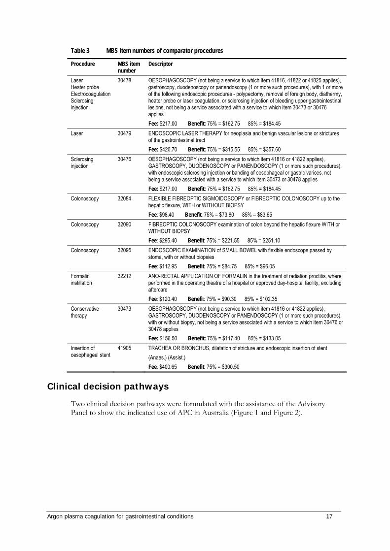
Argon plasma coagulation for gastrointestinal conditions 17
Table 3 MBS item numbers of comparator procedures
Procedure MBS item number
Descriptor
Laser Heater probe Electrocoagulation Sclerosing injection
30478 OESOPHAGOSCOPY (not being a service to which item 41816, 41822 or 41825 applies), gastroscopy, duodenoscopy or panendoscopy (1 or more such procedures), with 1 or more of the following endoscopic procedures - polypectomy, removal of foreign body, diathermy, heater probe or laser coagulation, or sclerosing injection of bleeding upper gastrointestinal lesions, not being a service associated with a service to which item 30473 or 30476 applies Fee: $217.00 Benefit: 75% = $162.75 85% = $184.45
Laser 30479 ENDOSCOPIC LASER THERAPY for neoplasia and benign vascular lesions or strictures of the gastrointestinal tract Fee: $420.70 Benefit: 75% = $315.55 85% = $357.60
Sclerosing injection
30476 OESOPHAGOSCOPY (not being a service to which item 41816 or 41822 applies), GASTROSCOPY, DUODENOSCOPY or PANENDOSCOPY (1 or more such procedures), with endoscopic sclerosing injection or banding of oesophageal or gastric varices, not being a service associated with a service to which item 30473 or 30478 applies Fee: $217.00 Benefit: 75% = $162.75 85% = $184.45
Colonoscopy 32084 FLEXIBLE FIBREOPTIC SIGMOIDOSCOPY or FIBREOPTIC COLONOSCOPY up to the hepatic flexure, WITH or WITHOUT BIOPSY Fee: $98.40 Benefit: 75% = $73.80 85% = $83.65
Colonoscopy 32090 FIBREOPTIC COLONOSCOPY examination of colon beyond the hepatic flexure WITH or WITHOUT BIOPSY Fee: $295.40 Benefit: 75% = $221.55 85% = $251.10
Colonoscopy 32095 ENDOSCOPIC EXAMINATION of SMALL BOWEL with flexible endoscope passed by stoma, with or without biopsies Fee: $112.95 Benefit: 75% = $84.75 85% = $96.05
Formalin instillation
32212 ANO-RECTAL APPLICATION OF FORMALIN in the treatment of radiation proctitis, where performed in the operating theatre of a hospital or approved day-hospital facility, excluding aftercare Fee: $120.40 Benefit: 75% = $90.30 85% = $102.35
Conservative therapy
30473 OESOPHAGOSCOPY (not being a service to which item 41816 or 41822 applies), GASTROSCOPY, DUODENOSCOPY or PANENDOSCOPY (1 or more such procedures), with or without biopsy, not being a service associated with a service to which item 30476 or 30478 applies Fee: $156.50 Benefit: 75% = $117.40 85% = $133.05
Insertion of oesophageal stent
41905 TRACHEA OR BRONCHUS, dilatation of stricture and endoscopic insertion of stent (Anaes.) (Assist.) Fee: $400.65 Benefit: 75% = $300.50
Clinical decision pathways
Two clinical decision pathways were formulated with the assistance of the Advisory Panel to show the indicated use of APC in Australia (Figure 1 and Figure 2).
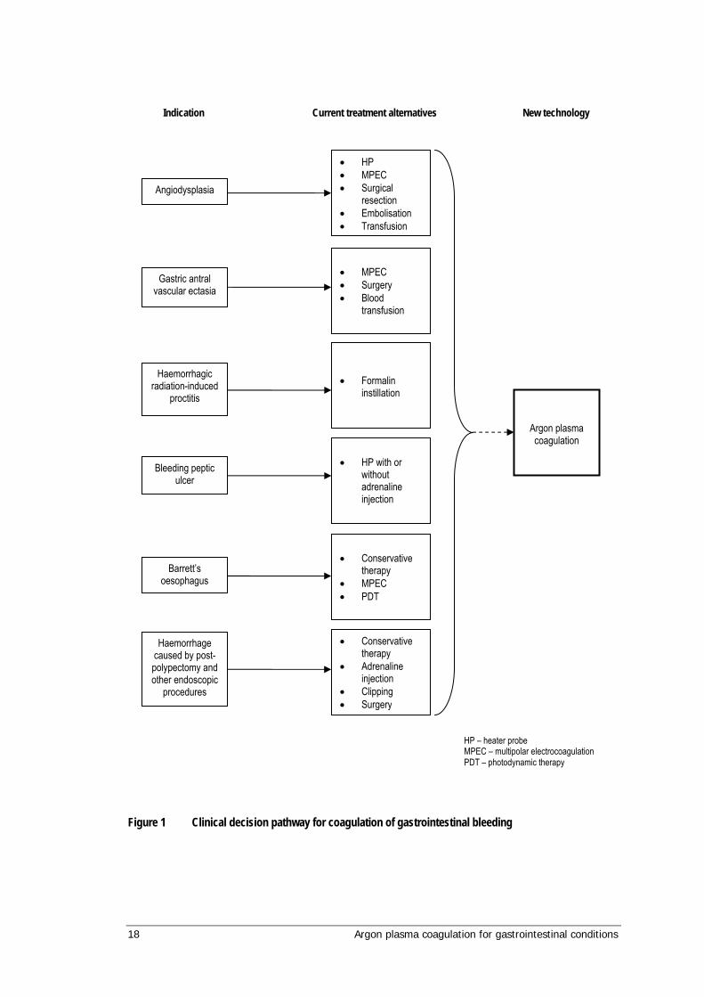
18 Argon plasma coagulation for gastrointestinal conditions
Figure 1 Clinical decision pathway for coagulation of gastrointestinal bleeding
Gastric antral vascular ectasia
Angiodysplasia
Haemorrhagic radiation-induced
proctitis
Bleeding peptic ulcer
Barrett’s oesophagus
Haemorrhage caused by post-polypectomy and other endoscopic
procedures
• HP • MPEC • Surgical
resection • Embolisation • Transfusion
• MPEC • Surgery • Blood
transfusion
• Formalin instillation
• HP with or
without adrenaline injection
• Conservative
therapy • MPEC • PDT
• Conservative therapy
• Adrenaline injection
• Clipping • Surgery
Argon plasma coagulation
HP – heater probe MPEC – multipolar electrocoagulation PDT – photodynamic therapy
Indication Current treatment alternatives New technology
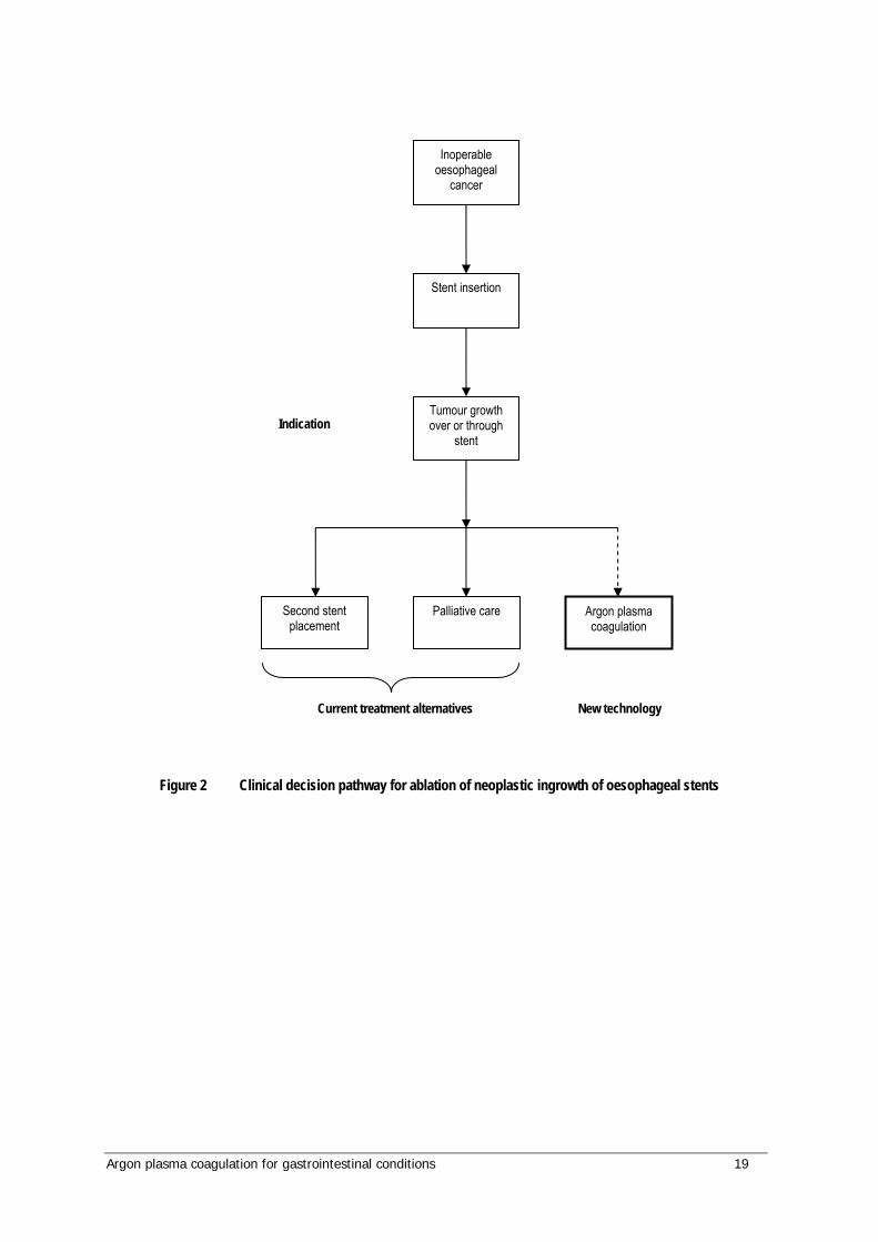
Argon plasma coagulation for gastrointestinal conditions 19
Figure 2 Clinical decision pathway for ablation of neoplastic ingrowth of oesophageal stents
Inoperable oesophageal
cancer
Stent insertion
Tumour growth over or through
stent
Second stent placement
Palliative care Argon plasma coagulation
Indication
Current treatment alternatives New technology

20 Argon plasma coagulation for gastrointestinal conditions
Marketing status of the device
Items relating to APC which are currently registered with the Therapeutic Goods Administration (TGA) in Australia are shown in Table 4.
Table 4 Therapeutic Goods Administration status of items relating to APC
Product ID Description Sponsor ARTG number 210621 ERBE Elektromedizin endotherapy
device, active, reusable Rymed Pty Ltd 126677
210622 ERBE Elektromedizin electrosurgical unit, gas delivery, argon
Rymed Pty Ltd 126678
210623 ERBE Elektromedizin electrosurgical unit, general purpose
Rymed Pty Ltd 126679
210625 ERBE Elektromedizin cable/lead, electrosurgical unit
Rymed Pty Ltd 126681
76710 Beacon argon beam coagulation system models Gas delivery units, argon-enhanced coagulation
ConMed Linvatec Australia Pty Ltd 33616
147210 Arco argon plasma surgery/coagulator system Electrosurgical units, monopolar, argon-enhanced coagulation
EMT Healthcare Pty Ltd 78128
198759 Force argon system, electrosurgical unit, argon-enhanced
Valleylab (a division of Tyco Healthcare Pty Ltd)
118234
214633 Electrosurgical units, argon-enhanced
ConMed Linvatec Australia Pty Ltd 129881
Current reimbursement arrangement
There is currently no item number for APC in the Medical Benefits Schedule (MBS). Item numbers for the comparator procedures are listed in Table 3, and include the procedures of diathermy, heater probe coagulation, laser coagulation, sclerotherapy injection, insertion of oesophageal stents and formalin instillation.
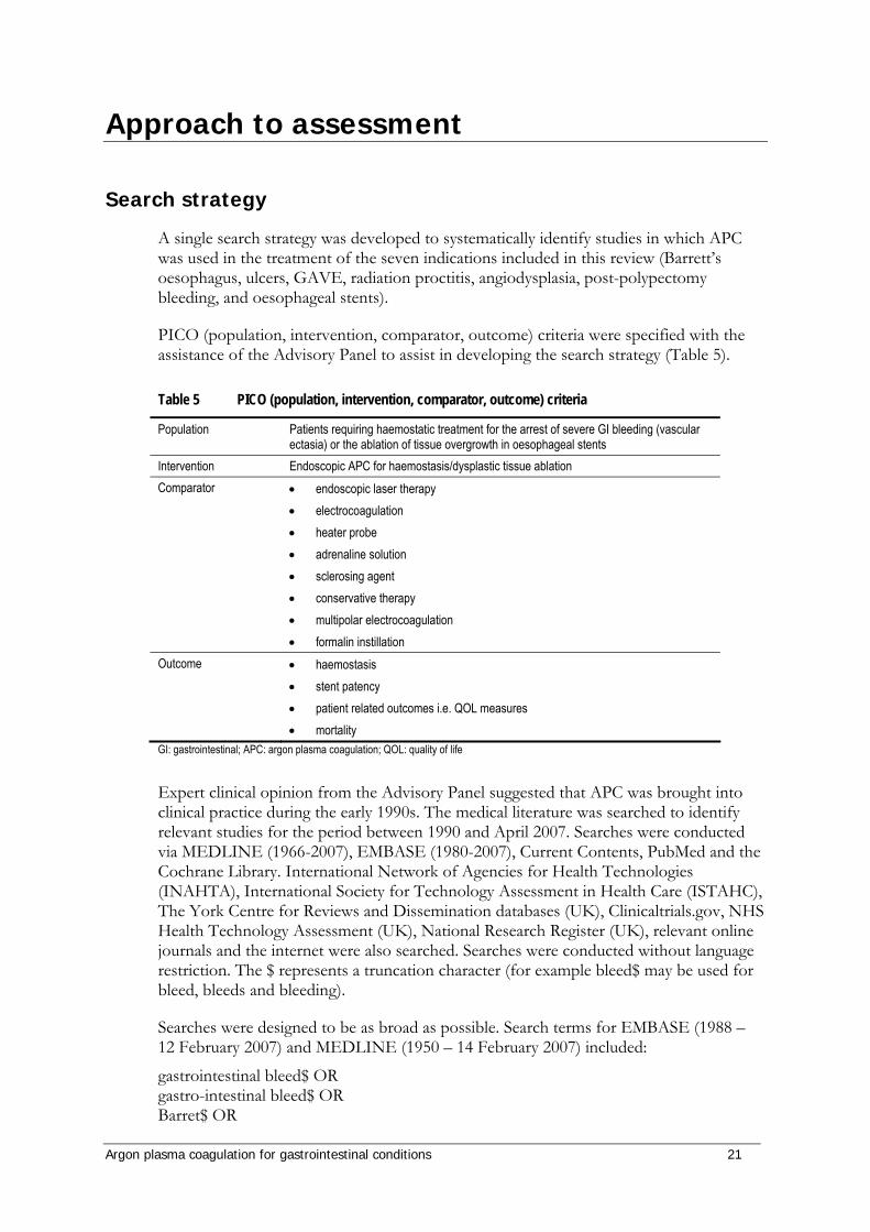
Argon plasma coagulation for gastrointestinal conditions 21
Approach to assessment
Search strategy
A single search strategy was developed to systematically identify studies in which APC was used in the treatment of the seven indications included in this review (Barrett’s oesophagus, ulcers, GAVE, radiation proctitis, angiodysplasia, post-polypectomy bleeding, and oesophageal stents).
PICO (population, intervention, comparator, outcome) criteria were specified with the assistance of the Advisory Panel to assist in developing the search strategy (Table 5).
Table 5 PICO (population, intervention, comparator, outcome) criteria
Population Patients requiring haemostatic treatment for the arrest of severe GI bleeding (vascular ectasia) or the ablation of tissue overgrowth in oesophageal stents
Intervention Endoscopic APC for haemostasis/dysplastic tissue ablation Comparator • endoscopic laser therapy
• electrocoagulation • heater probe • adrenaline solution • sclerosing agent • conservative therapy • multipolar electrocoagulation • formalin instillation
Outcome • haemostasis • stent patency • patient related outcomes i.e. QOL measures • mortality
GI: gastrointestinal; APC: argon plasma coagulation; QOL: quality of life
Expert clinical opinion from the Advisory Panel suggested that APC was brought into clinical practice during the early 1990s. The medical literature was searched to identify relevant studies for the period between 1990 and April 2007. Searches were conducted via MEDLINE (1966-2007), EMBASE (1980-2007), Current Contents, PubMed and the Cochrane Library. International Network of Agencies for Health Technologies (INAHTA), International Society for Technology Assessment in Health Care (ISTAHC), The York Centre for Reviews and Dissemination databases (UK), Clinicaltrials.gov, NHS Health Technology Assessment (UK), National Research Register (UK), relevant online journals and the internet were also searched. Searches were conducted without language restriction. The $ represents a truncation character (for example bleed$ may be used for bleed, bleeds and bleeding).
Searches were designed to be as broad as possible. Search terms for EMBASE (1988 – 12 February 2007) and MEDLINE (1950 – 14 February 2007) included:
gastrointestinal bleed$ OR gastro-intestinal bleed$ OR Barret$ OR

22 Argon plasma coagulation for gastrointestinal conditions
Gastric Antral Vascular Ectasia OR GAVE OR Watermelon stomach OR Angiodysplasia OR Arterioven$ malformation OR Polyp$ OR Stomal OR Ulcer$ OR Vascular malformation OR Blood vessel malformation OR Blood vessel bleed$ OR Non specific proctitis OR Endoscopic polypectomy OR $sophag$ OR Stent$ AND Argon plasma coagulat$ OR APC.
All terms were mapped to MeSH (Medical SubHeading) terms where appropriate, and keyword headings.
Search terms for EntrezPubMed (February 20 2007) were as follows:
Limit: Date 01/01/1980 – 22/01/2007 Limit: Humans Search terms: Argon Plasma Coagulation AND Upper Gastrointestinal Bleed$ OR Lower Gastrointestinal Bleed$ OR $sophageal stent$ OR Barrett$ $sophagus.
Search terms for Current Contents Connect (1998 – February 21 2007) were as follows:
Argon Plasma Coagulator OR Argon Plasma Coagulation AND Upper Gastrointestinal Bleed$ OR Lower Gastrointestinal Bleed$ OR Gastrointestinal Bleed$ OR Barrett$ $sophagus OR $sophageal stent$.
Search terms for the Cochrane Library (undertaken on April 18 2007) were:
Argon plasma coagulation Gastrointestinal bleeding Barrett’s oesophagus Oesophageal stent.

Argon plasma coagulation for gastrointestinal conditions 23
Inclusion criteria
Inclusion and exclusion criteria were identified as shown in Table 6 and Table 7. Advisory Panel opinion was that in the presence of high level evidence, lower level evidence (case reports) would not be included.
Table 6 Inclusion/exclusion criteria for identification of relevant studies for APC as a treatment for gastro-intestinal conditions: safety
Characteristic Criteria Publication type Clinical studies, systematic reviews, randomised comparative studies, non-randomised
comparative studies, case series and case reports included. Non-systematic reviews, letters, editorials, animal, in-vitro and laboratory studies were excluded. Case series and case reports were included for safety outcomes alone.
Patient Children and adults suffering any other condition that results in uncontrolled GI bleeding evidenced by expelled blood from the oesophagus or melaena, including GAVE, angiodysplasia, haemorrhagic radiation proctitis, Barrett’s oesophagus, peptic ulcers and other endoscopic procedure haemorrhage and post-polypectomy haemorrhage. **Studies were restricted to upper/lower GI tract excluding malignancies except Barrett’s oesophagus and stent overgrowth.
Intervention/test Endoscopic argon plasma coagulation. Comparator Endoscopic laser ablation therapy, electrocoagulation, heater probe, epinephrine solution,
photodynamic therapy, conservative therapy, and multipolar electrocoagulation. Outcome Any clinically-related outcomes including but not limited to short- and long-term safety (such as
haemorrhage, infection, mortality etc.). Language Non-English language articles were excluded unless they appeared to provide a higher level of
evidence than English language articles. GI: gastrointestinal; GAVE: gastric antral vascular ectasia
Table 7 Inclusion/exclusion criteria for identification of relevant studies for APC as a treatment for gastro-intestinal conditions: effectiveness
Characteristic Criteria Publication type Clinical studies, systematic reviews, randomised comparative studies, non-randomised
comparative studies will be included. Case series, case reports, non-systematic reviews, letters, editorials, animal, in-vitro and laboratory studies were excluded.
Patient Children and adults suffering any other condition that results in uncontrolled GI bleeding evidenced by expelled blood from the esophagus or melaena; including GAVE, angiodysplasia and haemorrhagic radiation proctitis, Barrett’s oesophagus, peptic ulcers and other endoscopic procedure haemorrhage and post-polypectomy haemorrhage. **Studies were restricted to upper/lower GI tract excluding malignancies except Barrett’s oesophagus and stent overgrowth.
Intervention/test Endoscopic argon plasma coagulation Comparator Endoscopic laser ablation therapy, electrocoagulation, heater probe, epinephrine solution,
photodynamic therapy, conservative therapy, and multipolar electrocoagulation. Outcome Any clinically-related outcomes including but not limited to long-term effectiveness (such as
haemostasis, patient-related quality of life etc.). Language Non-English language articles were excluded unless they appeared to provide a higher level of
evidence than English language articles. GI: gastrointestinal; GAVE: gastric antral vascular ectasia
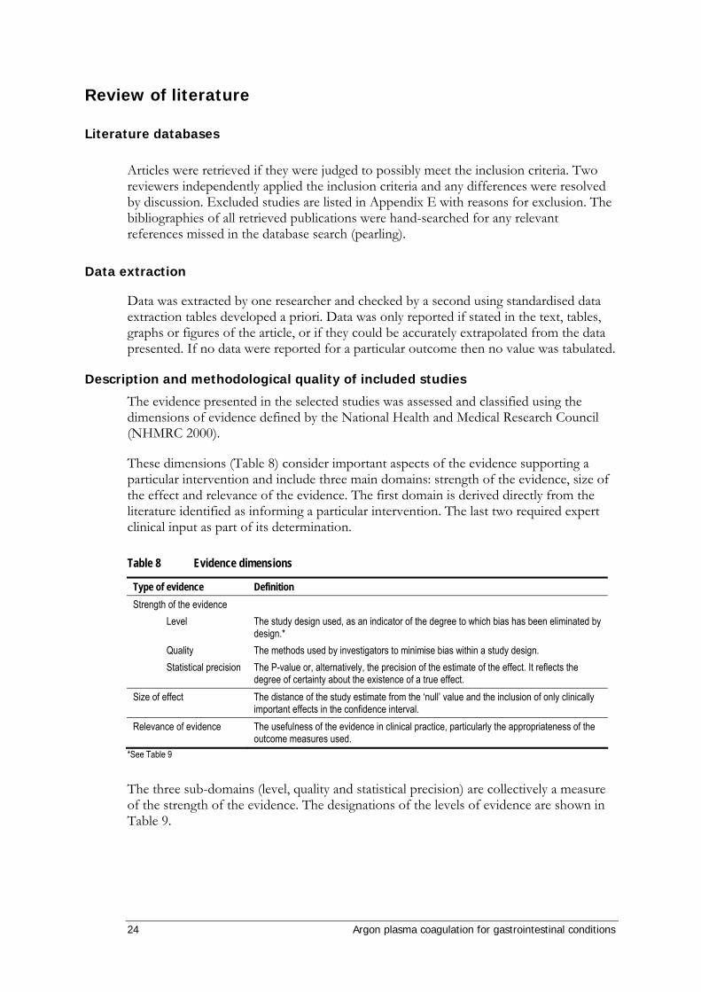
24 Argon plasma coagulation for gastrointestinal conditions
Review of literature
Literature databases
Articles were retrieved if they were judged to possibly meet the inclusion criteria. Two reviewers independently applied the inclusion criteria and any differences were resolved by discussion. Excluded studies are listed in Appendix E with reasons for exclusion. The bibliographies of all retrieved publications were hand-searched for any relevant references missed in the database search (pearling).
Data extraction
Data was extracted by one researcher and checked by a second using standardised data extraction tables developed a priori. Data was only reported if stated in the text, tables, graphs or figures of the article, or if they could be accurately extrapolated from the data presented. If no data were reported for a particular outcome then no value was tabulated.
Description and methodological quality of included studies
The evidence presented in the selected studies was assessed and classified using the dimensions of evidence defined by the National Health and Medical Research Council (NHMRC 2000).
These dimensions (Table 8) consider important aspects of the evidence supporting a particular intervention and include three main domains: strength of the evidence, size of the effect and relevance of the evidence. The first domain is derived directly from the literature identified as informing a particular intervention. The last two required expert clinical input as part of its determination.
Table 8 Evidence dimensions
Type of evidence Definition Strength of the evidence Level Quality Statistical precision
The study design used, as an indicator of the degree to which bias has been eliminated by design.* The methods used by investigators to minimise bias within a study design. The P-value or, alternatively, the precision of the estimate of the effect. It reflects the degree of certainty about the existence of a true effect.
Size of effect The distance of the study estimate from the ‘null’ value and the inclusion of only clinically important effects in the confidence interval.
Relevance of evidence The usefulness of the evidence in clinical practice, particularly the appropriateness of the outcome measures used.
*See Table 9
The three sub-domains (level, quality and statistical precision) are collectively a measure of the strength of the evidence. The designations of the levels of evidence are shown in Table 9.

Argon plasma coagulation for gastrointestinal conditions 25
Table 9 Designations of levels of evidence*
Level of evidence Study design I II III-1 III-2 III-3 IV
Evidence obtained from a systematic review of all relevant randomised controlled trials Evidence obtained from at least one properly-designed randomised controlled trial Evidence obtained from well-designed pseudorandomised controlled trials (alternate allocation or some other method) Evidence obtained from comparative studies (including systematic reviews of such studies) with concurrent controls and allocation not randomised, cohort studies, case-control studies, or interrupted time series with a control group Evidence obtained from comparative studies with historical control, two or more single arm studies, or interrupted time series without a parallel control group Evidence obtained from case series, either post-test or pre-test/post-test
*Modified from NHMRC 1999.
Included studies were critically appraised for study quality according to the guidelines in Chapter 6 of the Cochrane Reviewers’ Handbook (Higgins & Green 2005). Included randomised controlled trials (RCTs) were examined with respect to the adequacy of allocation concealment and blinding (if possible), handling of losses to follow-up, and any other aspect of the study design or execution that may have introduced bias, with reference to the CONSORT Statement (Altman et al 2001). Two reviewers critically appraised each of the included studies, and any differences in interpretation were resolved through discussion. A quality score was not assigned, instead the quality of the included studies was described in a narrative fashion, and any important quality issues were highlighted in the discussion of outcomes.
Data analysis
Meta-analysis
Where outcomes of RCTs could be sensibly combined (outcomes measured in comparable ways and no apparent heterogeneity), relative risks or weighted mean differences with 95 per cent confidence intervals (CI) were calculated using RevMan 4.2 (Update Software). Relative risks or weighted mean differences were also calculated for some outcomes of individual RCTs as an aid in the interpretation of results. The confidence intervals represent a range within which the ‘true’ value of an effect size is expected to lie, with a given degree of certainty e.g. 95 per cent CI.
Subgroup analyses were carried out for certain variables where possible.
Handling of non-randomised data
Where statistical pooling was not possible, medians of rates (for dichotomous outcomes) or medians of means (for continuous outcomes) for all studies reporting the outcome were calculated.
Included studies
The studies identified as fulfilling the inclusion criteria for the review are listed in Appendix D. The studies which were excluded from the review are listed in Appendix E, together with the reason for exclusion.

26 Argon plasma coagulation for gastrointestinal conditions
Current trials
Websites of clinical trials agencies were searched to identify all relevant ongoing or unpublished clinical trials related to the topics of this review. These included the Australian Clinical Trials Registry, ClinicalTrials.gov, the National Research Register (UK) and Controlled-Trials.com. As of 15 May 2006 only one trial investigating the use of APC in the treatment of GI pathologies was identified:
Principal investigator: Dr JG Freeman, at Derby City General Hospital, UK.
‘A randomised trial comparing argon plasma coagulation (APC) and self-expanding metal stents (SEMS) in oesophageal cancer’
The anticipated trial end date was December 2005, and it was registered as completed (10 May 2007); however, in this review the use of APC is not indicated for the direct ablation of oesophageal cancer.
Recent health technology assessments and systematic reviews on the use of APC for GI conditions
A list of electronic databases and websites of international HTA agencies can be found in Appendix G. The following seven studies and reviews were identified through searches of these databases, together with the main search strategy of this review.
Denton, A, Forbes, A, Andreyev, J, & Maher, EJ, 2002. ‘Non surgical interventions for late radiation proctitis in patients who have received radical radiotherapy to the pelvis’, Cochrane Database of Systematic Reviews no. 1.
Farrell, J.J & Friedman, LS, 2005. ‘Review article: the management of lower gastrointestinal bleeding’, Alimentary Pharmacology & Therapeutics, 21(11): 1281-98.
Faybush, EM & Sampliner, RE, 2005. ‘Randomized trials in the treatment of Barrett's esophagus’, Diseases of the Esophagus, 18(5): 291-7.
Havanond, C & Havanond, P, 2005. ‘Argon plasma coagulation therapy for acute non-variceal upper gastrointestinal bleeding - art. no. CD003791.pub2’, Cochrane Database of Systematic Reviews 2, UB2.
Pichon Riviere, A, Augustovski, F, Ferrante, F, Garcia Marti, S, Glujovsky, D, Lopes, A, & Regueiro, A, 2005. ‘Argon plasma usefulness for the treatment of gastrointestinal lesions’, Ciudad de Buenos Aires: Institute for Clinical Effectiveness and Health Policy (IECS), www.iecs.org.ar/
Tagkalidis, PP & Tjandra, JJ, 2001. ‘Chronic radiation proctitis’, ANZ Journal of Surgery, 71(4): 230-7.
Vargo, JJ, 2004. ‘Clinical applications of the argon plasma coagulator’, Gastrointestinal Endoscopy, 59(1): 81-8.
These reviews are discussed in detail in the results.

Argon plasma coagulation for gastrointestinal conditions 27
Expert advice
An Advisory Panel with expertise in GI conditions was established to evaluate the evidence and provide advice to MSAC from a clinical perspective. In selecting members for advisory panels, the practice of MSAC is to approach the appropriate medical colleges, specialist societies and associations and consumer bodies for nominees. Membership of the Advisory Panel is provided at Appendix B.
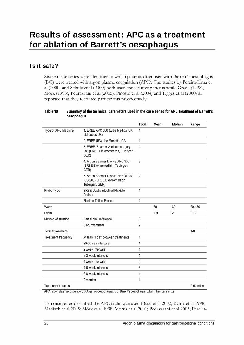
28 Argon plasma coagulation for gastrointestinal conditions
Results of assessment: APC as a treatment for ablation of Barrett’s oesophagus
Is it safe?
Sixteen case series were identified in which patients diagnosed with Barrett’s oesophagus (BO) were treated with argon plasma coagulation (APC). The studies by Pereira-Lima et al (2000) and Schulz et al (2000) both used consecutive patients while Grade (1998), Mörk (1998), Pedrazzani et al (2005), Pinotto et al (2004) and Tigges et al (2000) all reported that they recruited participants prospectively.
Table 10 Summary of the technical parameters used in the case series for APC treatment of Barrett’s oesophagus
Total Mean Median Range Type of APC Machine 1. ERBE APC 300 (Erbe Medical UK
Ltd Leeds UK) 1
2. ERBE USA, Inc Marietta, GA 1 3. ERBE ‘Beamer 2’ electrosurgury
unit (ERBE Elektromedizin, Tubingen, GER)
4
4. Argon Beamer Device APC 300 (ERBE Elektromedizin, Tubingen, GER)
8
5. Argon Beamer Device ERBOTOM ICC 200 (ERBE Elektromedizin, Tubingen, GER)
2
Probe Type ERBE Gastrointestinal Flexible Probes
1
Flexible Teflon Probe 1 Watts 68 60 30-150 L/Min 1.9 2 0.1-2 Method of ablation Partial circumference 8 Circumferential 2 Total # treatments 1-8 Treatment frequency At least 1 day between treatments 1 20-30 day intervals 1 2 week intervals 1 2-3 week intervals 1 4 week intervals 4 4-6 week intervals 3 6-8 week intervals 1 2 months 1 Treatment duration 2-50 mins APC: argon plasma coagulation; GO: gastro-oesophageal; BO: Barrett’s oesophagus; L/Min: litres per minute
Ten case series described the APC technique used (Basu et al 2002; Byrne et al 1998; Madisch et al 2005; Mörk et al 1998; Morris et al 2001; Pedrazzani et al 2005; Pereira-

Argon plasma coagulation for gastrointestinal conditions 29
Lima et al 2000; Tigges et al 2001; Van Laethem et al 1998). Each technique could be categorized into one of two methods (Table 10). Where reported, most studies used the partial circumference method of ablation in which longitudinal strips of Barrett’s mucosa were ablated usually to a maximum of fifty per cent of the circumference per treatment. This technique is aimed at reducing stricture formation. As a consequence, patients treated with APC for Barrett’s oesophagus often have multiple procedures.
Concurrent pharmaceutical regimes, when reported, were very similar between studies (Table 11). In most cases, proton pump inhibitors (PPIs: omaprazole, pantoprazole or lansoprazole) were provided to patients before, during and after APC treatment. Omeprazole was the most common PPI used. General anaesthesia was used in only one study (Tigges et al 2001). All other studies where reported used sedation during the APC procedure.
Table 11 Concurrent medical treatments used in the case series for Barrett’s oesophagus
Medical treatment # of studies
Amount used in treatment (range)
Pre- APC treatment (PPI) 8 Omeprazole 5 20-80mg
Pantoprazole 1 80mg Lansoprazole 2 30-70mg
During APC treatment (PPI) 12 Omeprazole 9 20-120mg Pantoprazole 1 80mg Lansoprazole 2 60-90 Post APC treatment (PPI) 11 Omeprazole 8 10-80mg Pantoprazole 1 80mg Lansoprazole 0 Pre-APC treatment (sedatives) 2 Midazolam 2 N/R Pethidine 2 N/R Glucogon 2 N/R During APC treatment (sedatives) 10 General anaesthesia 1 N/R Midazolam 6 2-20 Diazepam 1 N/R Pethidine 2 N/R Post APC treatment (sedatives) 3 Analgesia 3 N/R Other concurrent treatment Antacids 1 N/R Antibiotics 1 N/R PPI= proton pump inhibitor; N/R = not reported
Table 12 summarises the data collected from 652 patients across the included case series. Of the total patient population, 284 reportedly presented with no dysplasia, 44 presented with low-grade dysplasia and 13 presented with high-grade dysplasia; however, six of the case series did not report the dysplasia characteristics of participants (Basu et al 2002;

30 Argon plasma coagulation for gastrointestinal conditions
Madisch et al 2005; Morino et al 2003; Pagani et al 2003; Pinotti et al 2004; Tigges et al 2001).
Table 12 Summary of adverse events reported by case series for APC treatment of Barrett’s oesophagus
Total Mean Median Range Number of studies 16 Number of patients 652 Complete BO ablation 486 Male 329 Female 143 Age (years) 57.24 55.2 17-84 BO Length 4.08 3.6 0.5-19cm No dysplasia 284 LGD 44 HGD 13
Adverse Events Chest discomfort 4 Transient burning sensation 6 Chest pain 40 Retrosternal
discomfort/odynophagia/Pyrosis 104
Severe diffuse oesophagitis 1 Fever 13 Pleural effusion 5 Stricture 9 Oesophageal stenosis 5
Dysphagia 14 Ulcer formation 2 Bleeding 2 Pneumomediastinum/ subcutaneous
emphysema 1
Mediastinal emphysema 2 Scarring of oesophagus 1 Partial gastrectomy 1 Fundoplication 1 Perforation 5 Mortality 2
TOTAL REPORTED ADVERSE EVENTS
218
BO: Barrett’s oesophagus; LGD: low grade dysplasia; HGD: high grade dysplasia
There was some disparity in average Barrett’s oesophagus lengths between the studies, with reported average lengths ranging from 0.5cm (Pereira-Lima et al 2000) to 19cm (Basu et al 2002).
For convenience, the total adverse events reported in the case series were summarised into 19 categories, varying in severity from chest discomfort to oesophageal perforation

Argon plasma coagulation for gastrointestinal conditions 31
and death (Table 12). The most common adverse events pertained mainly to patient pain or discomfort, and included chest discomfort, transient burning sensation, chest pain, retrosternal discomfort, odynophagia, pyrosis, oesophagitis and fever. These ‘mild events’ accounted for 168 (77%) of all adverse events. All were reversible, with no long-lasting effects.
The next most common adverse events included pleural effusion, stricture, oesophageal stenosis and dysphagia. This mild to moderate group of events accounted for 33 (15%) of total adverse events and, while requiring a follow-up procedure, all were reversible.
Ten (5%) cases of adverse events fell into the category of moderate to severe. These events, such as ulcer formation, bleeding and emphysema, were rare, usually only occurring once or twice and in no more than one study. All were reversible after a follow-up procedure.
The two main adverse events that could be classed as ‘severe’ were perforation and death. Three of the non-comparative case series reported perforation events and two reported mortality outcomes. Given their serious nature, these events are examined in greater detail below.
Perforation
There were five reported cases of perforation (2%) across three studies (Table 13) (Byrne et al 1998; Manner et al 2006; Morris et al 2001); however, no clear pattern of patient characteristics or APC methodology emerged during analysis of the data. The earliest study, by Byrne (1998), reported a BO range of 3-17 cm but failed to supply details as to the power or rate of gas flow used. Morris (2001) did not report BO length range, power or gas flow, while the recent study by Manner (2006) reported a BO range of 1-8 cm, power of 90 watts and a 2 L/min gas flow. Byrne (1998) and Morris (2001) used an ERBE ‘Beamer 2’ unit and a similar ablation technique, while Manner (2006) used an ERBE APC 300 unit but did not describe the ablation technique used.
Table 13 Summary of perforations and mortalities
Study (date) Byrne (1998) Morris (2001) Manner (2006) Perforation (n:N) 2:30 2:55 1:60 Mortalities (n:N) 1:30 1:55 0:60 Mean age (range) NR (28-70) NR (NR) 57 (27-77) BO cm (range) NR (3-17) 6.06 (NR) 3.6 (1-8) LGD: HGD 4:3 9:9 NR:NR # complete BO ablation 16 NR 37 APC Machine 3 3 4 Watts / gas flow (L/min) NR / NR NR / NR 90 / 2 Method E E NR Mean # treatments (range) NR NR 2.7 (1-8) Treatment frequency 4-6 week intervals NR At least 1 day between
treatments LGD: low grade dysplasia; HGD: high grade dysplasia; NR: data not reported
Perforation was attributed to different characteristics by the researchers: Byrne (1998) reported that the patient suffering perforation had high-grade dysplasia, while Manner
32 Argon plasma coagulation for gastrointestinal conditions
(2006) excluded all patients suffering dysplasia and suggested that the patient’s age may have contributed to the perforation. The participant, at 61 years old, was slightly older than both the mean and median ages of these case series. Morris (2001) did not report on the patient’s characteristics and made no comment on a possible cause of perforation.
Three of the five cases of perforation were reversible (Manner 2006; Morris 2001; Byrne 1998), while two were irreversible and resulted in death.
Mortality
Two deaths (1%) were reported as an adverse event across two studies; both Byrne (1998) and Morris (2001) reported death to be a result of respiratory failure due to a perforation of the oesophagus (Table 13). It was difficult to undertake a reasonable comparison of the two cases, since both studies failed to adequately report important information such as the age and BO length of the deceased patient and study population as a whole, power and gas flow used, or the duration of the treatment undertaken.
With respect to safety, while these case series reported 218 adverse events associated with the use of APC, 214 of these events were reversible and 168 did not require a follow-up procedure. Interpretation of these case series is inhibited by a number of studies failing to report important information such as power, argon gas flow and treatment technique and duration. However, it is important to note that although 613 patients participated in these studies, the number of APC treatment sessions per patient ranged from 1 to 8, meaning the actual number of APC treatments may potentially number in the thousands. That only five perforations, of which only two led to mortality, were reported amongst so many applications suggests that APC may be a relatively safe procedure.

Argon plasma coagulation for gastrointestinal conditions 33
Is it effective?
Descriptive characteristics of comparative studies for Barrett’s oesophagus
Six comparative RCTs that compare the use of APC for the ablation of Barrett’s oesophagus to an alternative technique were identified through the systematic literature search (Ackroyd et al 2004; Dulai et al 2005; Hage et al 2004; Kelty et al 2004; Ragunath et al 2005; Sharma et al 2006). Of the six studies, one was performed in Australia, two in the United States, two in the United Kingdom and one in the Netherlands. Three different comparators were examined in the six RCTs; one study compared APC to conservative surveillance (Ackroyd et al 2004), two used multipolar electrocoagulation (Dulai et al 2005; Sharma et al 2006) and three used photodynamic therapy (Hage et al 2004; Kelty et al 2004; Ragunath et al 2005). It is important to note, however, that for the purposes of this review MSAC can only accept procedures currently listed on the MBS schedule as comparators. Photodynamic therapy is not currently MBS-listed and thus cannot be used as a comparator. The three RCTs that use photodynamic therapy cannot be used to provide evidence of the relative effectiveness of APC. These studies are retained only to assess safety outcomes for APC alone. Full descriptive characteristics of the six comparative studies are listed in Table 14.

34 Argon plasma coagulation for gastrointestinal conditions
Table 14 Descriptive characteristics of comparative studies
Study Study design (NHMRC level of evidence)
Study period Follow-up Inclusion criteria Exclusion criteria
Ackroyd et al (2004) Adelaide, AUSTRALIA
Comparative RCT, APC v CS (Level II)
NR 1 year Patients aged from 18 to 75 with Barrett’s oesophagus, with or without low-grade dysplasia, confirmed through biopsy or histology
High-grade dysplasia, ulcerative oesophagitis
Dulai et al (2005) Los Angeles, USA
Comparative RCT, APC v MPEC (Level II)
December 1999 – January 2003
Immediate follow-up
Patients at least 18 years of age, with prior histopathologic diagnosis of Barrett’s oesophagus (≥2 but ≤7cm) and life expectancy of 36 months, ability to comprehend English and answer questionnaires
Inability to cooperate, provide consent or return for follow-up, severe active co-morbidities that increase risk of endoscopy and cannot be controlled by medical therapy, histopathologic evidence of high-grade dysplasia or adenocarcinoma, prior anti-reflux surgery, inability to discontinue NSAID therapy for duration of study, pregnancy, lactation or non-use of contraception, allergy to PPI, uncontrolled coagulopathy
Sharma et al (2006) Kansas City, USA
Comparative RCT, APC v MPEC (Level II)
February 2000 – December 2004
2 years Patients with histological confirmation of Barrett’s oesophagus (≥2 but ≤6cm)
Aged 18 years or younger, history of oesophageal surgery, high-grade dysplasia or cancer, oesophageal stricture, oesophageal or gastric varices, allergy to PPI, uncontrolled coagulopathy, uncontrolled significant co-morbidities
Studies with non-MBS-listed comparators Hage et al (2004) Rotterdam, NETHERLANDS
Comparative RCT, APC v photodynamic therapy (Level II)
January 2001 – July 2004
18 months Patients at least 18 years of age with specialised intestinal metaplasia and no worse than low-grade dysplasia on histological examination
Intolerance to repeated endoscopy, pregnancy, acute porphyria, intercurrent diseases precluding survival during study period
Kelty et al (2004) Sheffield, UNITED KINGDOM
Comparative RCT, APC v photodynamic therapy (Level II)
NR 2 years Patients with biopsy-proven Barrett’s oesophagus
NR
Ragunath et al (2005) Liverpool, UNITED KINGDOM
Comparative RCT, APC v photodynamic therapy (Level II)
NR 1 year Barrett’s oesophagus > 3cm in length confirmed by biopsy no more than 3 months before study entry, dysplasia (low- and high-grade)
Oesophageal malignancy, previous oesophageal resection, previous mucosal ablation/resection, ‘tongues’ rather than circumferential Barrett’s oesophagus, porphyria, pregnancy, non-use of contraception, intolerance to endoscopy
APC: argon plasma coagulation; CS: conservative surveillance; MPEC: multipolar electrocoagulation; RCT: randomised control trial; NSAID: non-steroidal anti-inflammatory drug; PPI: proton pump inhibitor; NR: data not reported

Argon plasma coagulation for gastrointestinal conditions 35
Critical appraisal of comparative studies
Inclusion and exclusion criteria
Inclusion and exclusion criteria for the recruitment of patients in each of the studies are displayed in Table 14. There were some differences in the included patients across studies: three studies imposed age restrictions on patients (Ackroyd et al 2004; Dulai et al 2005; Hage et al 2004), and three studies set restrictions on the length of Barrett’s oesophagus to be included (Dulai et al 2005; Hage et al 2004; Sharma et al 2006). Five of the six studies provided explicit exclusion criteria; Kelty et al (2004) did not report any explicit exclusion criteria. Exclusion criteria primarily involved severe comorbidities, intolerance to the procedure, pregnancy, malignancies and high-grade dysplasia (although Ragunath et al (2005) did include patients with high-grade dysplasia).
Validity characteristics of comparative studies
Table 15 and Table 16 provide a summary of the quality of the three studies used in this review comparing APC for the ablation of Barrett’s oesophagus to an MBS-listed comparator. The criteria used were based on the CONSORT statement of Altman et al (2001). All three of the studies were randomised using sealed opaque envelopes (Ackroyd et al 2004; Dulai et al 2005; Sharma et al 2006), and Sharma et al stratified patients by length of Barrett’s oesophagus. Ackroyd et al (2004) and Dulai et al (2005) masked the method of treatment from investigators, and Dulai et al (2005) also blinded patients and the physician providing post-operative care to the intervention performed. All three studies provided well-defined eligibility criteria for patients, descriptions of outcomes and interventions, and used appropriate statistical methods. However, all three studies reported adverse events poorly, often not suitably quantifying or stratifying patient morbidities and mortalities. It is important to note that in the study by Dulai et al (2005), patients in the two treatment groups differed significantly in length of Barrett’s oesophagus at baseline (which will be discussed later in more detail); patients in the other two studies matched well at baseline.
Follow-up and losses to follow-up
Maximum follow-up amongst the three studies with MBS-listed comparators ranged from the surveillance endoscopy immediately after treatment success or failure (Dulai et al 2005) to 2 years (Sharma et al 2006), as can be seen in Table 14. Ackroyd et al (2004) lost one patient from the APC treatment group who died in the follow-up period, and Dulai et al (2005) lost two patients from each treatment group to follow-up (none of which were directly related to treatment) but did not report the cause of dropout (Table 16). Sharma et al (2006) had no losses to follow-up.
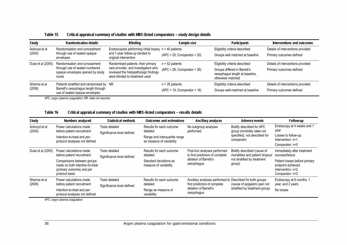
36 Argon plasma coagulation for gastrointestinal conditions
Table 15 Critical appraisal summary of studies with MBS-listed comparators – study design details
Study Randomisation details Blinding Sample size Participants Interventions and outcomes Ackroyd et al (2004)
Randomisation and concealment through use of sealed opaque envelopes
Endoscopists performing initial biopsy and 1-year follow-up blinded to original intervention
n = 40 patients (APC = 20; Comparator = 20)
Eligibility criteria described Groups well matched at baseline
Details of interventions provided Primary outcomes defined
Dulai et al (2005) Randomisation and concealment through use of sealed numbered opaque envelopes opened by study nurse
Randomised patients, their primary care provider, and investigators who reviewed the histopathologic findings were blinded to treatment used
n = 52 patients (APC = 26; Comparator = 26)
Eligibility criteria described Groups differed in Barrett’s oesophagus length at baseline, otherwise matched
Details of interventions provided Primary outcomes defined
Sharma et al (2006)
Patients stratified and randomised by Barrett’s oesophagus length through use of sealed opaque envelopes
NR n = 35 patients (APC = 19; Comparator = 16)
Eligibility criteria described Groups well-matched at baseline
Details of interventions provided Primary outcomes defined
APC: argon plasma coagulation; NR: data not reported
Table 16 Critical appraisal summary of studies with MBS-listed comparators – results details
Study Numbers analysed Statistical methods Outcomes and estimations Ancillary analyses Adverse events Follow-up Ackroyd et al (2004)
Power calculations made before patient recruitment Intention-to-treat and per-protocol analyses not defined
Tests detailed Significance level defined
Results for each outcome detailed Range and interquartile range as measure of variability
No subgroup analyses performed
Briefly described for APC group (morbidity rates not specified), not described for comparator
Endoscopy at 4 weeks and 1 year Losses to follow-up: Intervention: n=1 Comparator: n=0
Dulai et al (2005) Power calculations made before patient recruitment Comparisons between groups made on both intention-to-treat (primary outcome) and per protocol basis
Tests detailed Significance level defined
Results for each outcome detailed Standard deviations as measure of variability
Post-hoc analyses performed to find predictors of complete ablation of Barrett’s oesophagus
Briefly described (cause of mortalities and patient dropout not stratified by treatment group)
Immediately after treatment success/failure Patient losses before primary endpoint achieved: Intervention: n=2 Comparator: n=2
Sharma et al (2006)
Power calculations made before patient recruitment Intention-to-treat and per-protocol analyses not defined
Tests detailed Significance level defined
Results for each outcome detailed Range as measure of variability
Ancillary analyses performed to find predictors of complete ablation of Barrett’s oesophagus
Described for both groups (cause of epigastric pain not stratified by treatment group)
Endoscopy at 6 months, 1 year, and 2 years No losses
APC: argon plasma coagulation
Argon plasma coagulation for gastrointestinal conditions 37
Patient characteristics of comparative studies
Table 17 summarises the patient population characteristics in all six comparative studies. Patient treatment group characteristics were generally comparable within each of the studies; however, Dulai et al (2005) reported patients in the APC group had significantly longer Barrett’s oesophagus at baseline than patients in the comparator group (4.0 cm versus 3.1 cm, P=0.03). The same study found a significant positive correlation between initial Barrett’s oesophagus length and number of endoscopic treatments required for ablation (r=0.55, P=0.002), and thus were obliged to allow for this finding in their comparisons between treatment groups.
Baseline patient characteristics appeared comparable across studies. Study populations were predominantly male, and the mean age of participants was similar across studies. Median Barrett’s oesophagus lengths were relatively similar across studies (approximately 3-4 cm); however, Ragunath et al (2005) reported a slightly longer median Barrett’s oesophagus length (6 cm). Standard dosages of PPI medication were used in all the studies according to expert clinical opinion of the Advisory Panel.
The comparative evidence base for the use of APC in treating Barrett’s oesophagus relies primarily on studies which have included patients with non-dysplastic Barrett’s oesophagus (Table 18). However, currently in Australia the preferred treatment for non-dysplastic Barrett’s oesophagus is acid suppression therapy with proton-pump inhibitor treatment. Although this will not reverse Barrett’s oesophagus it may prevent the disease from becoming dysplastic. It is a low-risk treatment for a low-risk disease, which may also be combined with endoscopic surveillance to monitor the development of the condition. The majority of patients diagnosed with Barrett’s oesophagus will not have dysplasia and would therefore not be suitable candidates for ablative therapy (such as laser, heater probe, MPEC or APC).
For patients with dysplastic Barrett’s oesophagus there is a greater risk of malignant transformation, especially for those with high-grade dysplasia. In these cases, ablative therapy is an option to remove the dysplastic epithelium to reduce the risk of cancer while maintaining the integrity of the submucosa. The oesophagus is then allowed to heal in conjunction with aggressive acid suppression therapy, promoting restoration of the normal squamous mucosa. Ablation is a less severe alternative to oesophagectomy. In cases of severe high-grade dysplasia, or when adenocarcinoma has been confirmed, oesophagectomy may be required.
As there are so few patients with dysplasia in any one centre, a comparative trial of dysplasia alone would be very difficult to arrange. In the case of this review, expert clinical opinion suggested that the comparative evidence for the safety and effectiveness of APC in the treatment of non-dysplastic Barrett’s oesophagus would be of relevance to its use in the treatment of dysplastic Barrett’s oesophagus. However, there is currently limited evidence that ablative techniques including APC are beneficial in the treatment of non-dysplastic Barrett’s oesophagus. Patients should remain on acid suppression therapy.
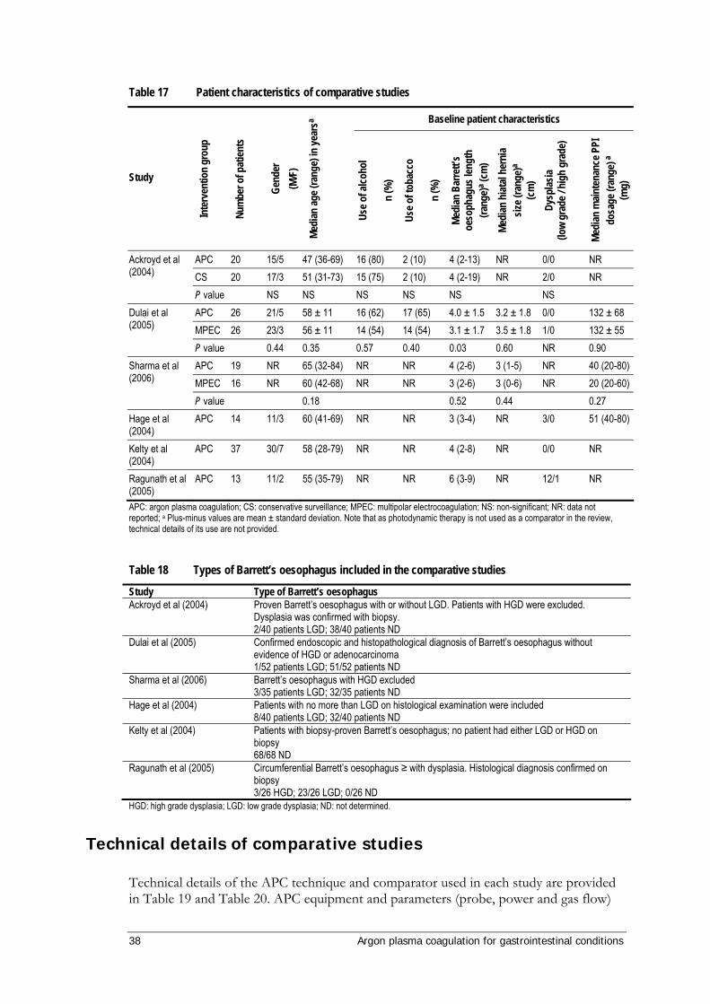
38 Argon plasma coagulation for gastrointestinal conditions
Table 17 Patient characteristics of comparative studies
Baseline patient characteristics
Study
Inte
rven
tion
grou
p
Num
ber o
f pat
ients
Gend
er
(M/F
)
Medi
an ag
e (ra
nge)
in ye
arsa
Use o
f alco
hol
n
(%)
Use o
f tob
acco
n (%
)
Medi
an B
arre
tt’s
oeso
phag
us le
ngth
(ra
nge)
a (cm
) Me
dian
hiat
al he
rnia
size (
rang
e)a
(cm
) Dy
splas
ia (lo
w gr
ade /
hig
h gr
ade)
Medi
an m
ainte
nanc
e PPI
do
sage
(ran
ge) a
(mg)
APC 20 15/5 47 (36-69) 16 (80) 2 (10) 4 (2-13) NR 0/0 NR CS 20 17/3 51 (31-73) 15 (75) 2 (10) 4 (2-19) NR 2/0 NR
Ackroyd et al (2004)
P value NS NS NS NS NS NS APC 26 21/5 58 ± 11 16 (62) 17 (65) 4.0 ± 1.5 3.2 ± 1.8 0/0 132 ± 68 MPEC 26 23/3 56 ± 11 14 (54) 14 (54) 3.1 ± 1.7 3.5 ± 1.8 1/0 132 ± 55
Dulai et al (2005)
P value 0.44 0.35 0.57 0.40 0.03 0.60 NR 0.90 APC 19 NR 65 (32-84) NR NR 4 (2-6) 3 (1-5) NR 40 (20-80) MPEC 16 NR 60 (42-68) NR NR 3 (2-6) 3 (0-6) NR 20 (20-60)
Sharma et al (2006)
P value 0.18 0.52 0.44 0.27 Hage et al (2004)
APC 14 11/3 60 (41-69) NR NR 3 (3-4) NR 3/0 51 (40-80)
Kelty et al (2004)
APC 37 30/7 58 (28-79) NR NR 4 (2-8) NR 0/0 NR
Ragunath et al (2005)
APC 13 11/2 55 (35-79) NR NR 6 (3-9) NR 12/1 NR
APC: argon plasma coagulation; CS: conservative surveillance; MPEC: multipolar electrocoagulation; NS: non-significant; NR: data not reported; a Plus-minus values are mean ± standard deviation. Note that as photodynamic therapy is not used as a comparator in the review, technical details of its use are not provided.
Table 18 Types of Barrett’s oesophagus included in the comparative studies
Study Type of Barrett’s oesophagus Ackroyd et al (2004) Proven Barrett’s oesophagus with or without LGD. Patients with HGD were excluded.
Dysplasia was confirmed with biopsy. 2/40 patients LGD; 38/40 patients ND
Dulai et al (2005) Confirmed endoscopic and histopathological diagnosis of Barrett’s oesophagus without evidence of HGD or adenocarcinoma 1/52 patients LGD; 51/52 patients ND
Sharma et al (2006) Barrett’s oesophagus with HGD excluded 3/35 patients LGD; 32/35 patients ND
Hage et al (2004) Patients with no more than LGD on histological examination were included 8/40 patients LGD; 32/40 patients ND
Kelty et al (2004) Patients with biopsy-proven Barrett’s oesophagus; no patient had either LGD or HGD on biopsy 68/68 ND
Ragunath et al (2005) Circumferential Barrett’s oesophagus ≥ with dysplasia. Histological diagnosis confirmed on biopsy 3/26 HGD; 23/26 LGD; 0/26 ND
HGD: high grade dysplasia; LGD: low grade dysplasia; ND: not determined.
Technical details of comparative studies
Technical details of the APC technique and comparator used in each study are provided in Table 19 and Table 20. APC equipment and parameters (probe, power and gas flow)
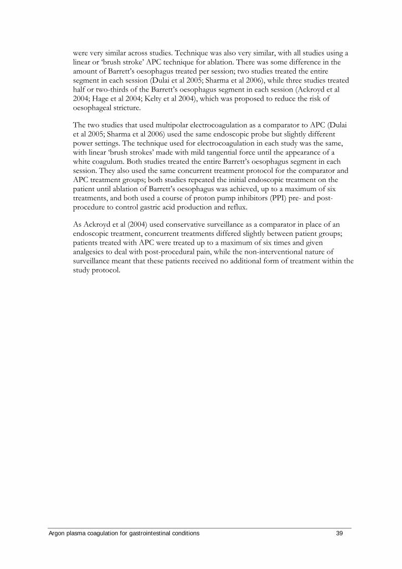
Argon plasma coagulation for gastrointestinal conditions 39
were very similar across studies. Technique was also very similar, with all studies using a linear or ‘brush stroke’ APC technique for ablation. There was some difference in the amount of Barrett’s oesophagus treated per session; two studies treated the entire segment in each session (Dulai et al 2005; Sharma et al 2006), while three studies treated half or two-thirds of the Barrett’s oesophagus segment in each session (Ackroyd et al 2004; Hage et al 2004; Kelty et al 2004), which was proposed to reduce the risk of oesophageal stricture.
The two studies that used multipolar electrocoagulation as a comparator to APC (Dulai et al 2005; Sharma et al 2006) used the same endoscopic probe but slightly different power settings. The technique used for electrocoagulation in each study was the same, with linear ‘brush strokes’ made with mild tangential force until the appearance of a white coagulum. Both studies treated the entire Barrett’s oesophagus segment in each session. They also used the same concurrent treatment protocol for the comparator and APC treatment groups; both studies repeated the initial endoscopic treatment on the patient until ablation of Barrett’s oesophagus was achieved, up to a maximum of six treatments, and both used a course of proton pump inhibitors (PPI) pre- and post-procedure to control gastric acid production and reflux.
As Ackroyd et al (2004) used conservative surveillance as a comparator in place of an endoscopic treatment, concurrent treatments differed slightly between patient groups; patients treated with APC were treated up to a maximum of six times and given analgesics to deal with post-procedural pain, while the non-interventional nature of surveillance meant that these patients received no additional form of treatment within the study protocol.
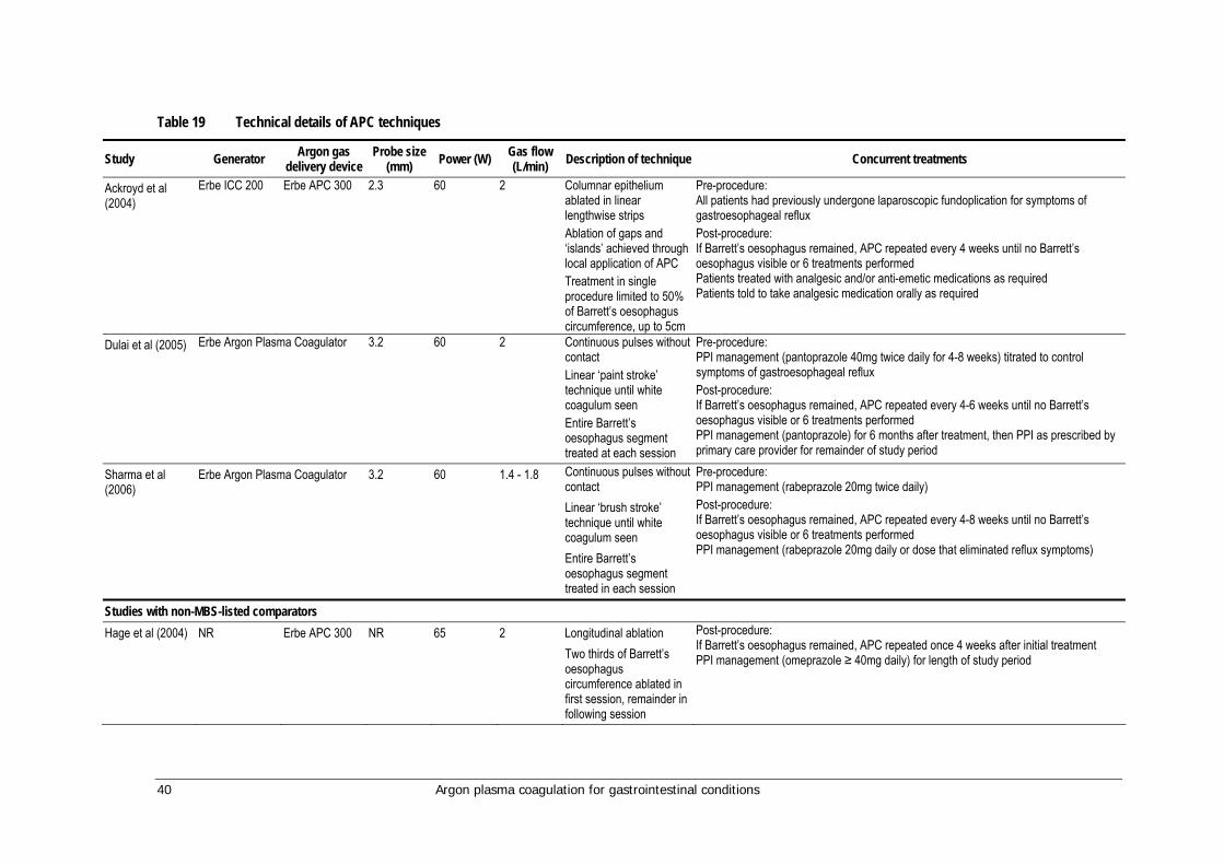
40 Argon plasma coagulation for gastrointestinal conditions
Table 19 Technical details of APC techniques
Study Generator Argon gas delivery device
Probe size (mm) Power (W) Gas flow
(L/min) Description of technique Concurrent treatments
Ackroyd et al (2004)
Erbe ICC 200 Erbe APC 300 2.3 60 2 Columnar epithelium ablated in linear lengthwise strips Ablation of gaps and ‘islands’ achieved through local application of APC Treatment in single procedure limited to 50% of Barrett’s oesophagus circumference, up to 5cm
Pre-procedure: All patients had previously undergone laparoscopic fundoplication for symptoms of gastroesophageal reflux Post-procedure: If Barrett’s oesophagus remained, APC repeated every 4 weeks until no Barrett’s oesophagus visible or 6 treatments performed Patients treated with analgesic and/or anti-emetic medications as required Patients told to take analgesic medication orally as required
Dulai et al (2005) Erbe Argon Plasma Coagulator 3.2 60 2 Continuous pulses without contact Linear ‘paint stroke’ technique until white coagulum seen Entire Barrett’s oesophagus segment treated at each session
Pre-procedure: PPI management (pantoprazole 40mg twice daily for 4-8 weeks) titrated to control symptoms of gastroesophageal reflux Post-procedure: If Barrett’s oesophagus remained, APC repeated every 4-6 weeks until no Barrett’s oesophagus visible or 6 treatments performed PPI management (pantoprazole) for 6 months after treatment, then PPI as prescribed by primary care provider for remainder of study period
Sharma et al (2006)
Erbe Argon Plasma Coagulator 3.2 60 1.4 - 1.8 Continuous pulses without contact Linear ‘brush stroke’ technique until white coagulum seen Entire Barrett’s oesophagus segment treated in each session
Pre-procedure: PPI management (rabeprazole 20mg twice daily) Post-procedure: If Barrett’s oesophagus remained, APC repeated every 4-8 weeks until no Barrett’s oesophagus visible or 6 treatments performed PPI management (rabeprazole 20mg daily or dose that eliminated reflux symptoms)
Studies with non-MBS-listed comparators Hage et al (2004) NR Erbe APC 300 NR 65 2 Longitudinal ablation
Two thirds of Barrett’s oesophagus circumference ablated in first session, remainder in following session
Post-procedure: If Barrett’s oesophagus remained, APC repeated once 4 weeks after initial treatment PPI management (omeprazole ≥ 40mg daily) for length of study period

Argon plasma coagulation for gastrointestinal conditions 41
Study Generator Argon gas delivery device
Probe size (mm) Power (W) Gas flow
(L/min) Description of technique Concurrent treatments
Kelty et al (2004) NR Erbe APC 300 NR 65 2 Linear ablation in strips approx. 2mm wide One half of Barrett’s oesophagus circumference treated in each session
Post-procedure: If Barrett’s oesophagus remained, APC repeated until no Barrett’s oesophagus visible, up to a maximum of 5 treatments Patients discharged with oral analgesia to take as required PPI management (esomeprazole 40mg daily) throughout study period
Ragunath et al (2005)
Erbe ICC 200 NR NR 65 1.8 Linear ‘brush stroke’ technique
During treatment period: PPI management (lansoprazole 60mg/daily) Post-procedure: If Barrett’s oesophagus remained, APC repeated every 2-4 weeks until no Barrett’s oesophagus visible or 6 treatments performed PPI management (lansoprazole 30mg/daily) Co-codamol (codeine phosphate 8mg/paracetamol 500mg) 2 tablets orally every 6 hours for pain relief for 24 to 48 hours after treatment ‘A few patients received sucralfate’ 1g every 6 hours to relieve dysphagia
APC: argon plasma coagulation; PPI: proton pump inhibitor
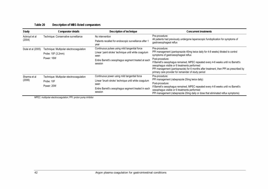
42 Argon plasma coagulation for gastrointestinal conditions
Table 20 Description of MBS-listed comparators
Study Comparator details Description of technique Concurrent treatments Ackroyd et al (2004)
Technique: Conservative surveillance
No intervention Patients recalled for endoscopic surveillance after 1 year
Pre-procedure: All patients had previously undergone laparoscopic fundoplication for symptoms of gastroesophageal reflux
Dulai et al (2005) Technique: Multipolar electrocoagulation Probe: 10F (3.2mm) Power: 16W
Continuous pulses using mild tangential force Linear ‘paint stroke’ technique until white coagulum seen Entire Barrett’s oesophagus segment treated at each session
Pre-procedure: PPI management (pantoprazole 40mg twice daily for 4-8 weeks) titrated to control symptoms of gastroesophageal reflux Post-procedure: If Barrett’s oesophagus remained, MPEC repeated every 4-6 weeks until no Barrett’s oesophagus visible or 6 treatments performed PPI management (pantoprazole) for 6 months after treatment, then PPI as prescribed by primary care provider for remainder of study period
Sharma et al (2006)
Technique: Multipolar electrocoagulation Probe: 10F Power: 20W
Continuous power using mild tangential force Linear ‘brush stroke’ technique until white coagulum seen Entire Barrett’s oesophagus segment treated in each session
Pre-procedure: PPI management (rabeprazole 20mg twice daily) Post-procedure: If Barrett’s oesophagus remained, MPEC repeated every 4-8 weeks until no Barrett’s oesophagus visible or 6 treatments performed PPI management (rabeprazole 20mg daily or dose that eliminated reflux symptoms)
MPEC: multipolar electrocoagulation; PPI: proton pump inhibitor
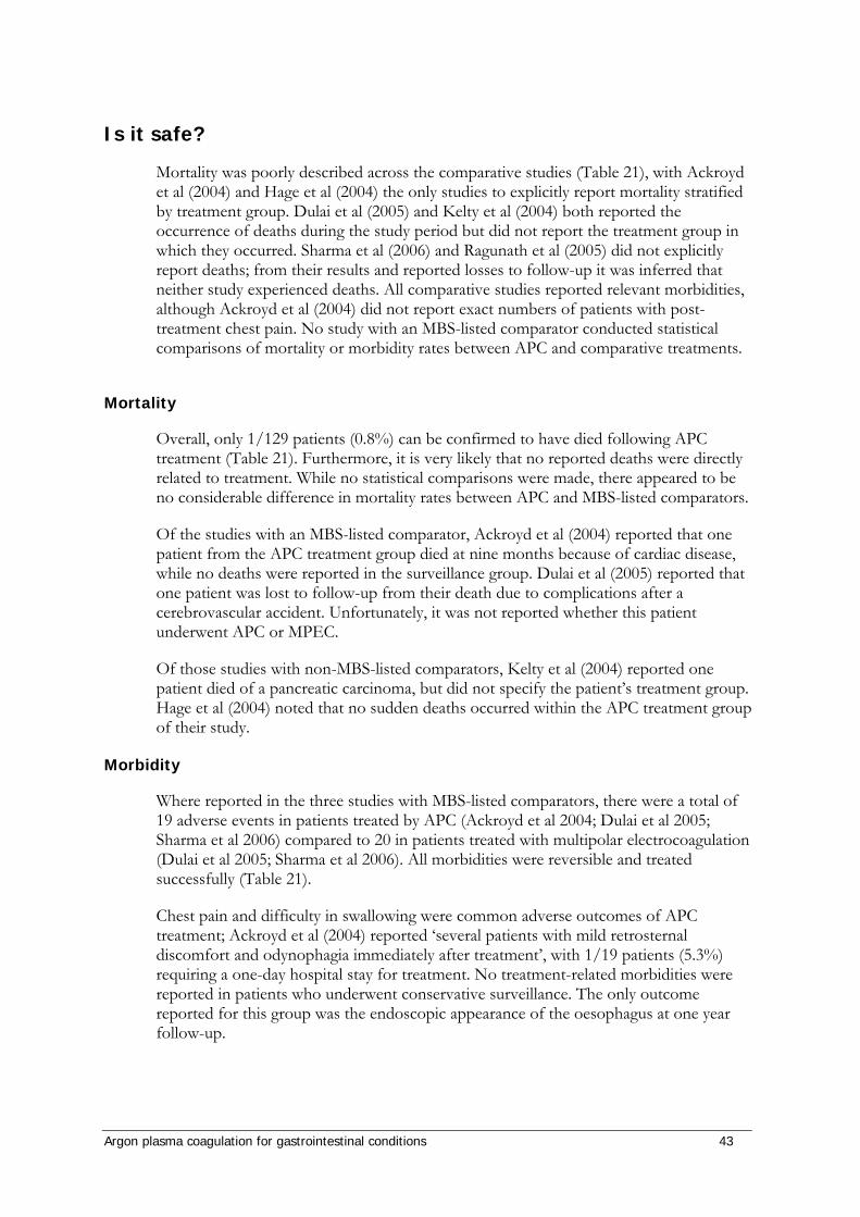
Argon plasma coagulation for gastrointestinal conditions 43
Is it safe?
Mortality was poorly described across the comparative studies (Table 21), with Ackroyd et al (2004) and Hage et al (2004) the only studies to explicitly report mortality stratified by treatment group. Dulai et al (2005) and Kelty et al (2004) both reported the occurrence of deaths during the study period but did not report the treatment group in which they occurred. Sharma et al (2006) and Ragunath et al (2005) did not explicitly report deaths; from their results and reported losses to follow-up it was inferred that neither study experienced deaths. All comparative studies reported relevant morbidities, although Ackroyd et al (2004) did not report exact numbers of patients with post-treatment chest pain. No study with an MBS-listed comparator conducted statistical comparisons of mortality or morbidity rates between APC and comparative treatments.
Mortality
Overall, only 1/129 patients (0.8%) can be confirmed to have died following APC treatment (Table 21). Furthermore, it is very likely that no reported deaths were directly related to treatment. While no statistical comparisons were made, there appeared to be no considerable difference in mortality rates between APC and MBS-listed comparators.
Of the studies with an MBS-listed comparator, Ackroyd et al (2004) reported that one patient from the APC treatment group died at nine months because of cardiac disease, while no deaths were reported in the surveillance group. Dulai et al (2005) reported that one patient was lost to follow-up from their death due to complications after a cerebrovascular accident. Unfortunately, it was not reported whether this patient underwent APC or MPEC.
Of those studies with non-MBS-listed comparators, Kelty et al (2004) reported one patient died of a pancreatic carcinoma, but did not specify the patient’s treatment group. Hage et al (2004) noted that no sudden deaths occurred within the APC treatment group of their study.
Morbidity
Where reported in the three studies with MBS-listed comparators, there were a total of 19 adverse events in patients treated by APC (Ackroyd et al 2004; Dulai et al 2005; Sharma et al 2006) compared to 20 in patients treated with multipolar electrocoagulation (Dulai et al 2005; Sharma et al 2006). All morbidities were reversible and treated successfully (Table 21).
Chest pain and difficulty in swallowing were common adverse outcomes of APC treatment; Ackroyd et al (2004) reported ‘several patients with mild retrosternal discomfort and odynophagia immediately after treatment’, with 1/19 patients (5.3%) requiring a one-day hospital stay for treatment. No treatment-related morbidities were reported in patients who underwent conservative surveillance. The only outcome reported for this group was the endoscopic appearance of the oesophagus at one year follow-up.

44 Argon plasma coagulation for gastrointestinal conditions
Table 21 Safety results of comparative studies
Study Level of Evidence
Length of follow-up
Intervention group Adverse events (n) Patient outcome
APC (n=20)
Morbidity: ‘Several’ patients reported mild retrosternal discomfort and odynophagia immediately after treatment (requiring 1-day hospital stay after first treatment, n=1) Mortality: Cardiac disease 9 months after treatment (n=1)
Reversible: Retrosternal discomfort and odynophagia resolved in up to 3 days Irreversible: All other adverse events
Ackroyd et al (2004)
II 1 year
CS (n=20)
Morbidity: None reported Mortality: None reported
APC (n=26)
Morbidity: Severe substernal chest pains (n=1) Mortality: Cannot be determineda
Reversible: Chest pain resolved with oral narcotic
Dulai et al (2005)
II Immediate follow-up
MPEC (n=26)
Morbidity: None reported Mortality: Cannot be determineda
APC (n=19)
Morbidity: Sore throat (n=9), difficulty swallowing (n=2), chest pains (n=4), fever (n=1), oesophageal stricture (n=1) Mortality: None reported
Reversible: All adverse events (stricture treated successfully with single dilation)
Sharma et al (2006)
II 2 years
MPEC (n=16)
Morbidity: Sore throat (n=9), difficulty swallowing (n=5), chest pain (n=6) Mortality: None reported
Reversible: All adverse events
Studies with non-MBS-listed comparators Hage et al (2004)
II 18 months APC (n=14)
Morbidity: Pain during treatment (n=5), odynophagia (n= 12), fever (n=2), oesophageal stricture (n=1) Mortality: None reported
Reversible: All adverse events (stricture treated successfully with single dilation)
Kelty et al (2004)
II 2 years APC (n=37)
Morbidity: Discomfort during treatment (n= 37), transient dysphagia and odynophagia (n= 32), oesophageal stricture (n=1) Mortality: Cannot be determinedb
Reversible: All adverse events (stricture treated with 4 dilations, dysphagia and odynophagia resolved within 3 days with oral analgesics)
Ragunath et al (2005)
II 1 year APC (n=13)
Morbidity: Oesophageal stricture (n= 2), severe chest pain (n=1), fever (n=1), odynophagia (n=1) Mortality: None reported
Reversible: All adverse events (stricture treated successfully with dilation, chest pains treated with analgesics and intravenous fluids)
APC: argon plasma coagulation; CS: conservative surveillance; MBS: Medicare Benefits Schedule MPEC: multipolar electrocoagulation ; a One patient died of complications after a cerebrovascular accident, but treatment group not noted; b One patient died of a pancreatic carcinoma, but treatment group not noted

Argon plasma coagulation for gastrointestinal conditions 45
Amongst the studies that compared APC to MPEC, Dulai et al (2005) reported severe substernal chest pain in 1/26 patients (3.8%) who underwent APC, with no morbidities reported in the MPEC treatment group. Sharma et al (2006) reported more morbidities overall than Dulai et al. However, incidence rates were comparable between treatment groups: 9/19 APC patients (47.4%) and 9/16 MPEC patients (56.3%) complained of a sore throat; 2/19 APC patients (10.5%) and 5/16 MPEC patients (31.3%) had difficulty swallowing; and 4/19 APC patients (21.1%) and 6/16 MPEC patients (37.5%) suffered chest pains. In addition, 1/19 APC patients (5.3%) experienced low-grade fever post-procedure, and 1/19 APC patients (5.3%) suffered an oesophageal stricture.
Incidence of adverse events in patients who underwent APC in the three studies with non-MBS-listed comparators were generally consistent with those reported in the aforementioned studies. All morbidities were reversible and treated successfully (Table 21). Oesophageal stricture was reported in 1/14 patients (7.1%) by Hage et al, 1/37 patients (2.7%) by Kelty et al (2004), and 2/13 patients (15.4%) by Ragunath et al (2005); fever was reported in 2/14 patients (14.3%) by Hage et al (2004) and in 1/13 patients (7.7%) by Ragunath et al; Ragunath et al also reported that 1/13 patients (7.7%) suffered severe chest pain and odynophagia. Incidence of odynophagia and dysphagia were higher in the studies by Hage et al, who reported 12/14 patients (85.7%) suffered odynophagia, and Kelty et al, who reported 32/37 patients (86.5%) experienced both. An adverse event not reported in the three studies with MBS-listed comparators was pain and discomfort during treatment; Hage et al reported 5/14 patients (35.8%) experienced pain during treatment, while Kelty et al reported ‘discomfort during treatment was experienced by all patients undergoing APC’.
Is it effective?
While the primary effectiveness outcomes reported by the three studies with MBS-listed comparators were essentially the same (ablation and reversal of Barrett’s oesophagus) there was considerable variation in the time period after which these outcomes were reported, ranging from immediately after the final ablation treatment to two-year follow-up. Unfortunately, no two studies reported the same clinical outcome at the same time point. Therefore, meta-analysis of the study outcomes was not appropriate. The full range of reported effectiveness outcomes are presented in Table 23
Clinical outcomes
Regarding the reversal of Barrett’s oesophagus, Ackroyd et al (2004) found APC to be significantly more successful than conservative surveillance; reversal was found in 60 per cent of patients treated with APC one month after treatment, and although this dropped slightly to 58 per cent at one-year follow up, it was found to be significantly higher than the reversal in 15 per cent of patients in the surveillance group (P<0.001). Patients treated with APC were also found to have significantly shorter Barrett’s oesophagus length at one-year follow-up (median length: 0 cm versus 2 cm, P<0.001). No other significant differences were found between the treatments.
No significant differences in reversal of Barrett’s oesophagus were found in the two studies comparing APC to MPEC. Dulai et al (2005) found 88 per cent of APC patients to have successful reversal immediately after their final treatment session, compared to 96 per cent of MPEC patients (P=0.64), while Sharma et al (2006) reported successful

46 Argon plasma coagulation for gastrointestinal conditions
reversal in 63.1 per cent of APC patients compared to 75.0 per cent of MPEC patients after a minimum two-year follow-up (p=0.49).
Procedural outcomes
The number of endoscopic procedures required for ablation was comparable across all three studies. Comparing APC to MPEC, Sharma et al (2006) reported no significant difference between the treatment groups, while Dulai et al (2005) reported a difference approaching significance that favoured MPEC over APC (3.0 versus 3.9 sessions, P=0.05) using a single statistical test; however, this difference in treatments was non-significant after correction for the difference in patient group baseline Barrett’s oesophagus length (3.6 versus 3.17 sessions, P=0.249).
When analysed using a single statistical test, Dulai et al (2005) also reported that treatment time was significantly faster for MPEC than for APC in the first (6 versus 10 minutes, P=0.01) and second (4 versus 7 minutes, P=0.01) sessions; however, significance was lost after correction for the multiple testing of data arising from individual patients.
Meta-analysis of outcomes
The clinical outcome of the effectiveness of APC compared with MPEC in the reversal of Barrett’s oesophagus for Dulai et al (2005) and Sharma et al (2006) underwent meta-analysis (Table 22 and Figure 3).
Table 22 Clinical outcomes of Barrett’s oesophagus reversal
Study Comparator Reversal at Definition of ‘reversal’ Ackroyd et al 2004 Conservative surveillance 12 months No visible Barrett’s oesophagus at
endoscopy and biopsy 4 weeks after final treatment session. Endoscopy at 1 year
Dulai et al 2005 Multipolar electrocoagulation 6 months No endoscopic evidence of Barrett’s oesophagus; using Lugol’s iodine at discretion of investigator
Sharma et al 2006 Multipolar electrocoagulation 24 months Endoscopic visualisation of Barrett’s oesophagus absence, confirmed using negative Lugol’s iodine staining
Figure 3 Meta-analysis of the clinical outcome for Barrett’s oesophagus
The results of the meta-analysis show a relative risk of 0.89 in favour of MPEC (P=0.22) (Figure 3).
Argon plasma coagulation for gastrointestinal conditions 47
Table 23 Effectiveness results of studies with MBS-listed comparators
Clinical outcome Procedural outcome
Study Level of evidence
Length of follow-up
Intervention group
Barre
tt’s o
esop
hagu
s re
vers
al (Im
med
iate)
n (%
) Ba
rrett’
s oes
opha
gus
reve
rsal
(1 m
onth
)
n (%
) Ba
rrett’
s oes
opha
gus
reve
rsal
(12 m
onth
s)
n
(%)
Barre
tt’s o
esop
hagu
s re
vers
al (2
4 mon
ths)
n (%
) Me
dian
leng
th o
f Bar
rett’
s oe
soph
agus
(cm
) (ra
nge)
Post
-trea
tmen
t dys
plas
ia
n (%
) Po
st-tr
eatm
ent b
uried
Ba
rrett’
s glan
ds
n
(%)
Medi
an n
umbe
r of
proc
edur
es re
quire
d
(rang
e)a
Tim
e per
1st se
ssio
n (m
inut
es)
Me
an (S
D)
Tim
e per
2nd se
ssio
n (m
inut
es)
Me
an (S
D)
APC (n=19)
NR 12 (60) 11 (58) NR 0 (0-3) 0 (0) 1 (5) 3 (2-6) NR NR
CS (n=20)
NR NR 3 (15) NR 2 (1-3) 0 (0) 0 (0) NR NR NR
Ackroyd et al (2004)
II 1 year
p value < 0.01 <0.001 NS NS APC (n=24)
21 (88) NR NR NR NR 0 (0) NR 3.9 ± 1.6 10 (7) 7 (3)
MPEC (n=24)
23 (96) NR NR NR NR 0 (0) NR 3.0 ± 1.5 6 (4) 4 (3)
Dulai et al (2005) II Immediate follow-up
p valueb 0.64 NR 0.05c 0.01 0.01
APC (n=19)
NR NR NR 12 (63.1) NR 0 (0) NR 3 (2-6) NR NR
MPEC (n=16)
NR NR NR 12 (75.0) NR 0 (0) NR 4 (2-6) NR NR
Sharma et al (2006)
II 2 years
p value 0.49 NR 0.76 APC: Argon plasma coagulation; CS: conservative surveillance; MPEC: multipolar electrocoagulation; NR: data not reported a Plus-minus values are mean ± standard deviation b P values are for between group comparisons without adjustment for baseline differences or multiple comparisons; by Bonferroni method, P < 0.00625 for significance at α=0.05 level c After adjustment for difference in Barrett’s oesophagus length, mean number of treatment sessions required was 3.6 for APC versus 3.17 for MPEC (P=0.249)

48 Argon plasma coagulation for gastrointestinal conditions
Discussion
A total of six randomised control trials compared APC to an alternative treatment for the ablation and reversal of Barrett’s oesophagus. Three of these studies used an MBS-listed treatment as a comparator; Ackroyd et al (2004) used conservative surveillance without endoscopic intervention, while Dulai et al (2005) and Sharma et al (2006) both used multipolar electrocoagulation. The three remaining studies (Hage et al 2004; Kelty et al 2004; Ragunath et al 2005) used photodynamic therapy as a comparator; as photodynamic therapy is not an MBS-listed procedure, these three RCTs were excluded from analysis of effectiveness outcomes, and informed safety outcomes for APC treatment only.
The three studies with MBS-listed comparators appeared to sufficiently meet validity criteria outlined in the CONSORT statement of Altman et al (2001). Two studies incorporated relevant blinding measures, and all studies provided well-defined eligibility criteria for patients, descriptions of outcomes and interventions, and used appropriate statistical methods. Follow-up length and drop-out rates were also reported. Patients in the two treatment groups of the study by Dulai et al differed significantly by length of Barrett’s oesophagus at baseline; however, appropriate corrections were made for this within study analyses. Patients in the other two studies matched well at baseline.
While no statistical comparisons were made, there appeared to be no considerable difference in mortality rates between APC and the MBS-listed comparators of conservative surveillance and multipolar electrocoagulation. Only one of 129 patients was confirmed to have died, from cardiac disease nine months after undergoing APC. It is likely that this was not directly related to treatment (Ackroyd et al 2004).
All six comparative studies reported on relative morbidities; however, they did not all suitably quantify or stratify patient morbidities by treatment group, and none made inter-treatment statistical comparisons of prevalence rates. These issues have made accurate comparison of morbidity rates of APC and conservative surveillance unfeasible. While incidence of odynophagia and dysphagia in patients who underwent APC were somewhat elevated in the studies by Hage et al (2004) and Kelty et al (2004), there appeared to be little difference in morbidity rates of APC and multipolar electrocoagulation in the studies by Dulai et al (2005) and Sharma et al (2006). All morbidities were reversible and successfully treated in all patients. Overall, APC appears to be as safe as the MBS-listed comparator procedures of conservative surveillance and multipolar electrocoagulation for the ablation and reversal of Barrett’s oesophagus.
Safety was also investgated in the 16 included case series. There were 218 reported adverse events from a total treatment of 652 patients, each of which had received between one and eight sessions of APC. The great majority of complications (214, 98%) were reversible and 168 (77%) did not require re-treatment. There were five cases of perforation and two deaths resulting from APC treatment. Both deaths were as result of oesophageal perforation. These outcomes were poorly reported in terms of patient characteristics and treatment settings; therefore, no overall conclusion could be reached regarding the causes of these events.
In the two RCTs used to assess the effectiveness of APC in the treatment of Barrett’s oesophagus the majority of patients (89/92) had the non-dysplastic form of the disease. It is important to note that in Australia, non-dysplastic Barrett’s mucosa would be

Argon plasma coagulation for gastrointestinal conditions 49
controlled through acid suppression therapy rather than with the use of ablation. It is unlikely that enough patients with dysplastic Barrett’s oesophagus could be enrolled into a comparative trial as only a minority of patients have the more severe type of the disease. Therefore, evidence concerning the use of APC in the treatment of non-dysplastic Barrett’s oesophagus was used to assess the effectiveness of the procedure.
Few differences in effectiveness outcomes were found between APC and comparators in the three studies with MBS-listed comparators. Ackroyd et al (2004) reported that APC provided significant improvement in the reduction in length and reversal of Barrett’s oesophagus over conservative surveillance at one-year follow-up. Dulai et al (2005) and Sharma et al (2006) showed no significant differences between APC and multipolar electrocoagulation in clinical outcomes. While Dulai et al reported tentative evidence that multipolar electrocoagulation may provide some procedural benefits over APC, this finding was mitigated by differences in patient characteristics at baseline. In summary, APC appears to be as effective as multipolar electrocoagulation, and more effective than conservative surveillance, in the ablation of Barrett’s oesophagus.
Summary
These results suggest that APC is at least as safe and effective as the comparative techniques of multipolar electrocoagulation, and as safe as but more effective than conservative surveillance for the ablation of Barrett’s oesophagus, with few differences reported between the techniques. It must be noted that these comparative results were obtained from three RCTs incorporating two comparators that met inclusion criteria. More definitive conclusions regarding the safety and effectiveness of the APC procedure for this indication could have been given if there were more RCTs available using suitable comparator procedures. In addition, the RCTs included mainly patients with non-dysplastic Barrett’s oesophagus in their studies, even though in Australia these patients would not be considered for ablative treatment. In the absence of comparative studies with dysplastic Barrett’s oesophagus alone these studies were used to provide evidence for this report. The assumption was made with expert clinical advice that the data on APC in treating non-dysplastic Barrett’s mucosa was of relevance to the treatment of dysplastic Barrett’s oesophagus.
50 Argon plasma coagulation for gastrointestinal conditions
Results of assessment: APC as a treatment for bleeding peptic ulcers
Level 1 evidence
A Cochrane review was published in 2005 which investigated APC as a treatment for non-variceal upper GI bleeding (Havanond & Havanond 2005). Two RCTs were included in this review: one compared APC to heater probe and the other compared APC to injection sclerotherapy (Cipolletta et al 1998; Skok et al 2001). Both these studies are discussed in the ulcers indication section of this review. The authors concluded that there was very little published research in this field, and that further randomised controlled trials are required.
A recent meta-analysis was identified which reported on dual therapy versus mono-therapy in the endoscopic treatment of high-risk bleeding ulcers (Marmo et al 2007), that is, adrenaline injection with or without a second methodology such as thermal, injection or mechanical therapy. APC was not discussed independently from other types of thermal treatment. The authors concluded that there was no benefit to using adrenaline injection therapy in addition to any type of thermal therapy, and that dual therapy should not be recommended as it was associated with an increased risk of perforation.
Descriptive characteristics of comparative studies
Four comparative studies that compared the use of APC for the treatment of bleeding in gastric ulcers to an alternative technique were identified through the search strategy (Chau et al 2003; Cipolletta et al 1998; Skok et al 2001; Occhigrossi et al 2002). Of the three RCTs identified, one was conducted in Hong Kong, one in Italy and one in Slovenia; the one internal comparative (historical control) study was conducted in Italy. Three different comparators were examined in the three RCTs: one study used a heater probe (Cipolletta et al), one used adrenaline injection sclerotherapy (Skok et al), and one used a combination of heater probe and adrenaline injection (Chau et al). Full descriptive characteristics of the four comparative studies are listed in Table 24.
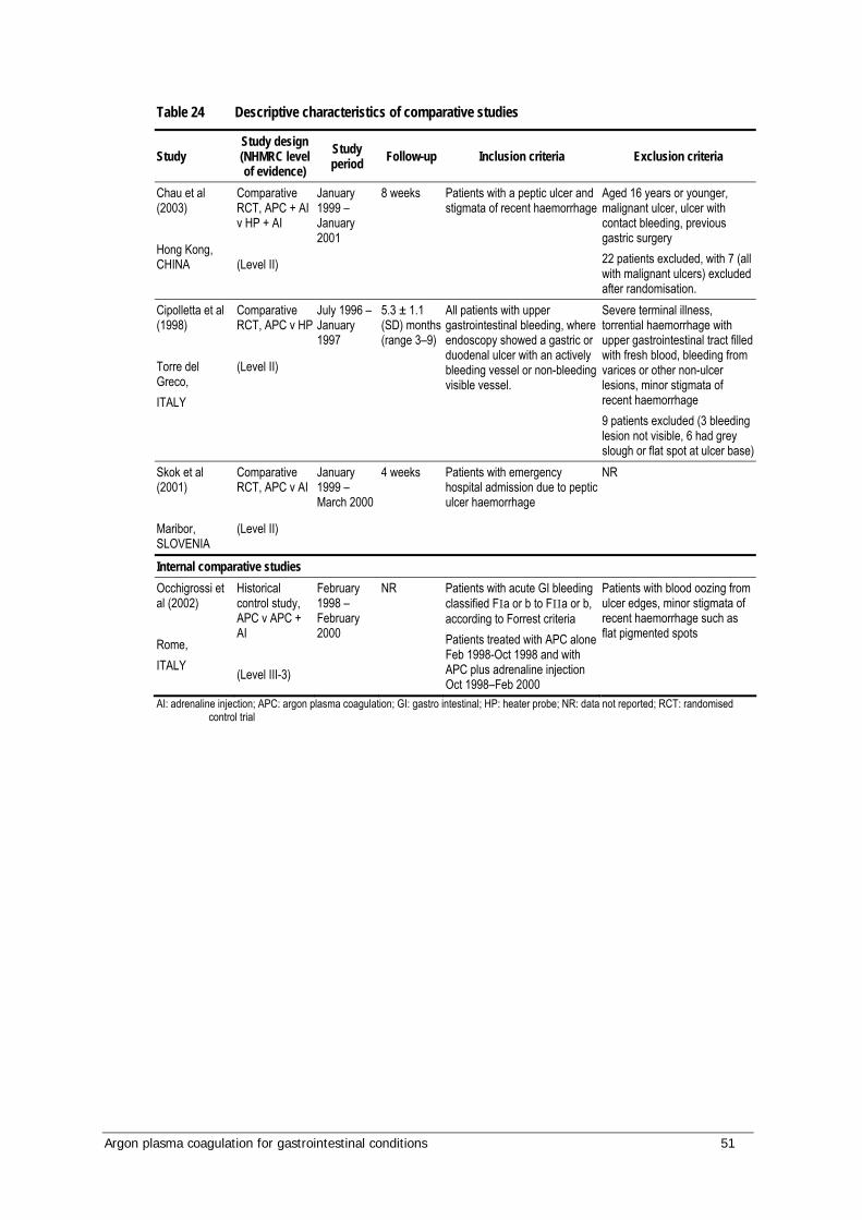
Argon plasma coagulation for gastrointestinal conditions 51
Table 24 Descriptive characteristics of comparative studies
Study Study design (NHMRC level of evidence)
Study period Follow-up Inclusion criteria Exclusion criteria
Chau et al (2003) Hong Kong, CHINA
Comparative RCT, APC + AI v HP + AI (Level II)
January 1999 – January 2001
8 weeks Patients with a peptic ulcer and stigmata of recent haemorrhage
Aged 16 years or younger, malignant ulcer, ulcer with contact bleeding, previous gastric surgery 22 patients excluded, with 7 (all with malignant ulcers) excluded after randomisation.
Cipolletta et al (1998) Torre del Greco, ITALY
Comparative RCT, APC v HP (Level II)
July 1996 – January 1997
5.3 ± 1.1 (SD) months (range 3–9)
All patients with upper gastrointestinal bleeding, where endoscopy showed a gastric or duodenal ulcer with an actively bleeding vessel or non-bleeding visible vessel.
Severe terminal illness, torrential haemorrhage with upper gastrointestinal tract filled with fresh blood, bleeding from varices or other non-ulcer lesions, minor stigmata of recent haemorrhage 9 patients excluded (3 bleeding lesion not visible, 6 had grey slough or flat spot at ulcer base)
Skok et al (2001) Maribor, SLOVENIA
Comparative RCT, APC v AI (Level II)
January 1999 – March 2000
4 weeks Patients with emergency hospital admission due to peptic ulcer haemorrhage
NR
Internal comparative studies Occhigrossi et al (2002) Rome, ITALY
Historical control study, APC v APC + AI (Level III-3)
February 1998 – February 2000
NR Patients with acute GI bleeding classified FIa or b to FIIa or b, according to Forrest criteria Patients treated with APC alone Feb 1998-Oct 1998 and with APC plus adrenaline injection Oct 1998–Feb 2000
Patients with blood oozing from ulcer edges, minor stigmata of recent haemorrhage such as flat pigmented spots
AI: adrenaline injection; APC: argon plasma coagulation; GI: gastro intestinal; HP: heater probe; NR: data not reported; RCT: randomised control trial

52 Argon plasma coagulation for gastrointestinal conditions
Critical appraisal of comparative studies
Inclusion and exclusion criteria
Inclusion and exclusion criteria for the recruitment of patients in each of the studies are displayed in Table 24. All studies provided similar descriptions of enrolled patients, which all involved gastrointestinal bleeding or haemorrhage due to ulcers. Occhigrossi et al (2002) included specific Forrest-criteria grades of bleeding. Three of the four studies provided explicit exclusion criteria, primarily severe comorbidities or minor stigmata of recent haemorrhage; Skok et al (2001) did not report any explicit exclusion criteria. Cipolletta et al (1998) excluded nine patients, while Chau et al (2003) excluded 22 patients, including seven patients (all with malignant ulcers) after randomisation had taken place; however, this did not create a substantial difference between treatment group sizes.
Validity characteristics of comparative studies
Table 25 and Table 26 provide a summary of the quality of the four comparative studies examining the use of APC for bleeding gastric ulcers used in this review. The criteria used were based on the CONSORT statement of Altman et al (2001). Three of the studies were randomised (Chau et al 2003; Cipolletta et al 1998; Skok et al 2001), with Chau et al using a computer-generated randomisation scheme. Patients were not masked to the method of treatment in any study, and only Cipolletta et al masked the physician providing post-operative care to the treatment used. Whilst being a historical internal comparison, twice as many patients in the study by Occhigrossi et al (2002) were treated with APC plus adrenaline injection than APC alone (n=53 versus n=27). The study by Skok et al (2001) appears the weakest of the four in describing study methodology, with patient eligibility criteria, primary outcomes and adverse events reported poorly. All studies, with the exception of Skok et al (2001), provided well-defined eligibility criteria for patients, and treatment groups in all studies were well matched at baseline.
Follow-up and losses to follow-up
Maximum follow-up amongst the RCTs ranged from 4 weeks (Skok et al 2001) to 9 months (Cipolletta et al 1998). Cipolletta et al had no losses to follow-up, Chau et al (2003) lost 41% (36/87) of patients from the APC patient group and 38% (37/97) from the comparator group at 8-week follow-up endoscopy but did not report reasons for these losses, and Skok et al did not report losses to follow up. While the study by Occhigrossi et al (2002) took place over a two-year period, length of follow-up was not reported.
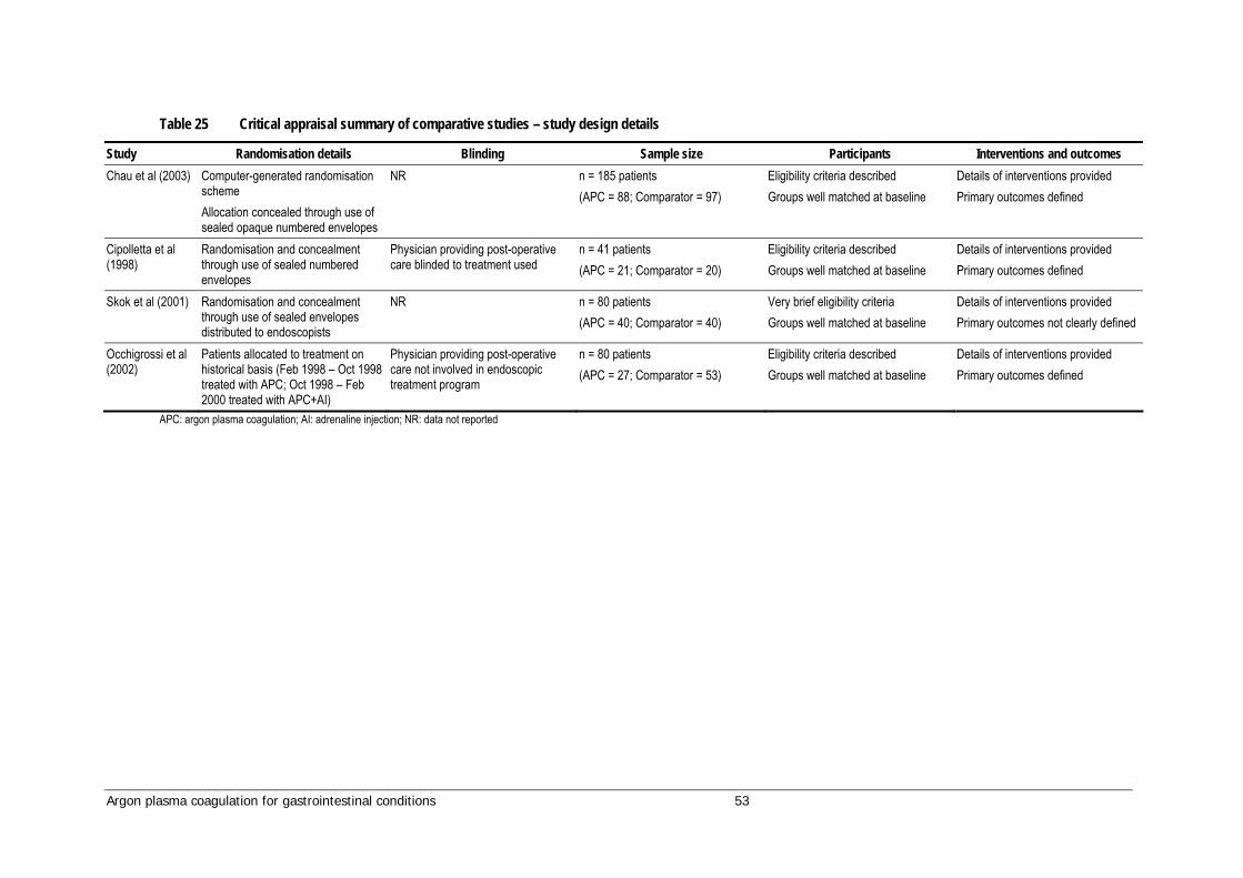
Argon plasma coagulation for gastrointestinal conditions 53
Table 25 Critical appraisal summary of comparative studies – study design details
Study Randomisation details Blinding Sample size Participants Interventions and outcomes Chau et al (2003) Computer-generated randomisation
scheme Allocation concealed through use of sealed opaque numbered envelopes
NR n = 185 patients (APC = 88; Comparator = 97)
Eligibility criteria described Groups well matched at baseline
Details of interventions provided Primary outcomes defined
Cipolletta et al (1998)
Randomisation and concealment through use of sealed numbered envelopes
Physician providing post-operative care blinded to treatment used
n = 41 patients (APC = 21; Comparator = 20)
Eligibility criteria described Groups well matched at baseline
Details of interventions provided Primary outcomes defined
Skok et al (2001) Randomisation and concealment through use of sealed envelopes distributed to endoscopists
NR n = 80 patients (APC = 40; Comparator = 40)
Very brief eligibility criteria Groups well matched at baseline
Details of interventions provided Primary outcomes not clearly defined
Occhigrossi et al (2002)
Patients allocated to treatment on historical basis (Feb 1998 – Oct 1998 treated with APC; Oct 1998 – Feb 2000 treated with APC+AI)
Physician providing post-operative care not involved in endoscopic treatment program
n = 80 patients (APC = 27; Comparator = 53)
Eligibility criteria described Groups well matched at baseline
Details of interventions provided Primary outcomes defined
APC: argon plasma coagulation; AI: adrenaline injection; NR: data not reported

54 Argon plasma coagulation for gastrointestinal conditions
Table 26 Critical appraisal summary of comparative studies – results details
Study Numbers analysed Statistical methods Outcomes and estimations Ancillary analyses Adverse events Follow-up Chau et al (2003) Comparisons between groups
made on an intention-to-treat basis
Tests detailed Significance level defined
Outcome results detailed Standard deviations as measure of variability
Subgroup analyses performed, stratified by Forrest bleeding classification
Briefly described Reasons for losses to follow-up not detailed
Endoscopy at 8 weeks Losses to follow-up: Intervention: n=36 Comparator: n=37
Cipolletta et al (1998)
Intention-to-treat and per-protocol analyses not defined
Tests detailed Significance level defined
Outcome results detailed Standard deviations as measure of variability
No subgroup analyses performed
Described for both groups 5.3 ± 1.1 months (range 3–9) No losses
Skok et al (2001) Intention-to-treat and per-protocol analyses not defined
Tests detailed Significance level defined
Outcome results detailed Range and standard deviations as measure of variability
Subgroup analyses performed, stratified by Forrest bleeding classification
Briefly described (cause of mortalities not stratified by treatment group) Losses to follow-up not detailed
Endoscopy at 4 days and examination at 4 weeks
Occhigrossi et al (2002)
Intention-to-treat and per-protocol analyses not defined
Tests detailed Significance level defined
Outcome results detailed Range and standard deviations as measure of variability
Subgroup analyses performed, stratified by Forrest bleeding classification
Described for both groups NR
NR: data not reported

Argon plasma coagulation for gastrointestinal conditions 55
Patient characteristics of comparative studies
Table 27 summarises the patient population characteristics in each comparative study. Patient treatment group characteristics were comparable within each of the three RCTs, with no study finding any statistically significant differences between APC and comparator patient groups in baseline characteristics. Study populations were predominantly male, and the mean age of participants was similar across studies. The patient population of Chau et al (2003) appeared to be notably lower in urease and Helicobacter pylori than that of Cipolletta et al (1998) and Skok et al (2001), while the patient group of Occhigrossi et al (2002) appeared to have slightly higher use of non-steroidal anti-inflammatory drugs (NSAIDs) or aspirin than the three RCTs. Otherwise, patient baseline characteristics appeared to be comparable across all four studies.
Table 27 Patient characteristics of comparative studies
Baseline patient characteristics
Study
Inte
rven
tion
grou
p
Num
ber o
f pat
ients
Gend
er
(M/F
)
Medi
an ag
e (ra
nge)
in ye
arsa
NSAI
Ds / a
spiri
n us
age
n
(%)
Posit
ive fo
r rap
id
urea
se te
st
n (%
) Po
sitive
for h
elico
bact
er
pylo
ri in
fect
ion
n (%
) Ha
emog
lobi
n lev
els
(g / d
L)
Mean
(SD)
Si
te o
f ulce
r (g
astri
c / d
uode
nal /
othe
r)
Mean
size
of u
lcer (
mm
)
Mean
(SD)
Bl
eedi
ng ve
ssels
(a
ctive
/ non
-act
ive)
APC+AI 88 62/26 62.8 ± 16.5 24 (27.3) 19 (21.6) 39 (44.3) 10.1(2.4) 26/62/0 9.5 (5.6) 47/41 HP+AI 97 63/34 62.6 ± 16.5 21 (21.6) 24 (24.7) 43 (44.3) 9.7 (2.5) 37/60/0 9.9 (6.6) 55/42
Chau et al (2003)
P value NS NS NS NR NR NS NR NR NR APC 21 12/9 57 ± 18 7 (33.3) 18 (85.7) NR NR 6/15/0 12 (3) 9/12 HP 20 11/9 59 ± 16 6 (30) 17 (85) NR NR 5/15/0 11 (4) 8/12
Cipolletta et al (1998)
P value NR NR NR NR NR NR NR APC 40 25/15 56.9 ± 14.5 13 (32.5) NR 29 (72.5) 10.2(1.8) 20/20/0 13 (3) 19/21 AI 40 25/15 57.9 ± 14.0 12 (30) NR 28 (70) 10.4(1.6) 20/20/0 12 (4) 19/21
Skok et al (2001)
P value NS NS NS NS NS NS NS NS APC 27 21/6 62 (18-87) 16 (59) NR NR 8.5 (2.0) 7/19/1 NR 13/14 APC+AI 53 34/19 67 (22-93) 35 (66) NR NR 8.2 (2.6) 17/30/6 NR 23/30
Occhigrossi et al (2002)
P value NS NS NS NS NS NS APC: argon plasma coagulation; HP: heater probe; AI: adrenaline injection; NSAID: non-steroidal anti-inflammatory drug; NS: non-significant; NR: data not reported a Plus-minus values are mean ± standard deviation
Technical details of comparative studies
Technical details of the APC technique and comparator used in each comparative study are provided in Table 28 and Table 29. Skok et al (2001) used an ARCO electro-surgery unit for APC instead of the Erbe unit used in the other two RCTs. APC equipment and parameters (probe, power and gas flow) were similar across studies; however, for at least some of their patients Chau et al (2003) and Cipolletta et al (1998) did use a lower power setting (40 W) than Skok et al (2001) and Occhigrossi et al (2002). Clinical advice from the Advisory Panel suggests that a minimum power is required to establish the argon
56 Argon plasma coagulation for gastrointestinal conditions
plasma. This may vary between machines, with certain new models having automatically variable settings which are independent of the operator. A higher power setting may increase the risk of perforation.
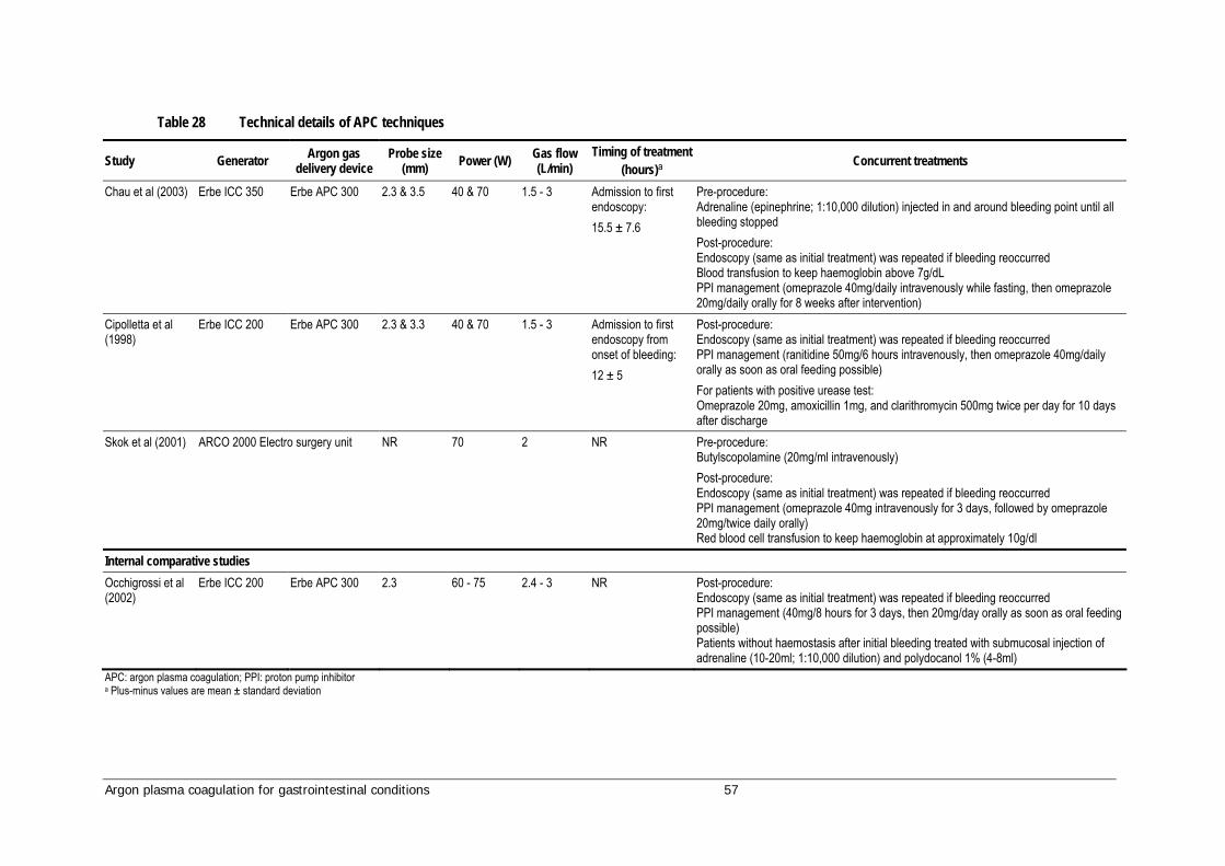
Argon plasma coagulation for gastrointestinal conditions 57
Table 28 Technical details of APC techniques
Study Generator Argon gas delivery device
Probe size (mm) Power (W) Gas flow
(L/min) Timing of treatment
(hours)a Concurrent treatments
Chau et al (2003) Erbe ICC 350 Erbe APC 300 2.3 & 3.5 40 & 70 1.5 - 3 Admission to first endoscopy: 15.5 ± 7.6
Pre-procedure: Adrenaline (epinephrine; 1:10,000 dilution) injected in and around bleeding point until all bleeding stopped Post-procedure: Endoscopy (same as initial treatment) was repeated if bleeding reoccurred Blood transfusion to keep haemoglobin above 7g/dL PPI management (omeprazole 40mg/daily intravenously while fasting, then omeprazole 20mg/daily orally for 8 weeks after intervention)
Cipolletta et al (1998)
Erbe ICC 200 Erbe APC 300 2.3 & 3.3 40 & 70 1.5 - 3 Admission to first endoscopy from onset of bleeding: 12 ± 5
Post-procedure: Endoscopy (same as initial treatment) was repeated if bleeding reoccurred PPI management (ranitidine 50mg/6 hours intravenously, then omeprazole 40mg/daily orally as soon as oral feeding possible) For patients with positive urease test: Omeprazole 20mg, amoxicillin 1mg, and clarithromycin 500mg twice per day for 10 days after discharge
Skok et al (2001) ARCO 2000 Electro surgery unit NR 70 2 NR Pre-procedure: Butylscopolamine (20mg/ml intravenously) Post-procedure: Endoscopy (same as initial treatment) was repeated if bleeding reoccurred PPI management (omeprazole 40mg intravenously for 3 days, followed by omeprazole 20mg/twice daily orally) Red blood cell transfusion to keep haemoglobin at approximately 10g/dl
Internal comparative studies Occhigrossi et al (2002)
Erbe ICC 200 Erbe APC 300 2.3 60 - 75 2.4 - 3 NR Post-procedure: Endoscopy (same as initial treatment) was repeated if bleeding reoccurred PPI management (40mg/8 hours for 3 days, then 20mg/day orally as soon as oral feeding possible) Patients without haemostasis after initial bleeding treated with submucosal injection of adrenaline (10-20ml; 1:10,000 dilution) and polydocanol 1% (4-8ml)
APC: argon plasma coagulation; PPI: proton pump inhibitor a Plus-minus values are mean ± standard deviation

58 Argon plasma coagulation for gastrointestinal conditions
Table 29 Description of comparators
Study Comparator details Timing of treatment (hours)a Concurrent treatments
Chau et al (2003) Technique: Heater probe plus adrenaline injection Instrument used: Olympus CD-20Z & CD-1OZ Probe: 2.7mm and 3.3mm (3.3mm used on 13.4% of patients during initial endoscopy, 11.8% during second) Power used: 25 - 30J Mean (SD) number of pulses used:
First endoscopy: 5.1 (1.3) Second endoscopy: 3.2 (1.0)
Admission to first endoscopy: 16.4 ± 7.5
Pre-procedure: Adrenaline (epinephrine; 1:10,000 dilution) injected in and around bleeding point until all bleeding stopped Post-procedure: Endoscopy (same as initial treatment) was repeated if bleeding reoccurred Blood transfusion to keep haemoglobin above 7g/dL PPI management (omeprazole 40mg/daily intravenously while fasting, then omeprazole 20mg/daily orally for 8 weeks after intervention)
Cipolletta et al (1998)
Technique: Heater probe Instrument used: Olympus GIF1T130 Probe: 10F Power used: 25 - 30J Number of pulses used: 4 - 8
Admission to first endoscopy from onset of bleeding: 14 ± 6
Post-procedure: Endoscopy (same as initial treatment) was repeated if bleeding reoccurred PPI management (ranitidine 50mg/6 hours intravenously, then omeprazole 40mg/daily orally as soon as oral feeding possible For patients with positive urease test: omeprazole 20mg, amoxicillin 1mg; clarithromycin 500mg twice per day for 10 days after discharge
Skok et al (2001) Technique: Adrenaline injection sclerotherapy Instrument used: Adrenaline sclerosant (1:10,000 dilution) plus 1% polidocanol Adrenaline administered in aquilots of 1ml close to the bleeding site (up to 6ml) until bleeding stopped; subsequently polidocanol injected closely around bleeding lesion (up to 3ml)
NR Pre-procedure: Butylscopolamine (20mg/ml intravenously) Post-procedure: Endoscopy (same as initial treatment) was repeated if bleeding reoccurred PPI management (omeprazole 40mg intravenously for 3 days, followed by omeprazole 20mg/twice daily orally) Red blood cell transfusion to keep haemoglobin at approximately 10g/dl
Internal comparative studies Technique: APC plus adrenaline injection Pre-treatment: Adrenaline (1:10,000 dilution) injected:
Around edge of small ulcers (1-2ml) Around bleeding site of large ulcers (10-20ml)
Generator Argon gas delivery device
Probe size (mm) Power (W) Gas flow
(L/min)
Occhigrossi et al (2002)
Erbe ICC 200 Erbe APC 3000 2.3 60 - 75 2.4 - 3
NR Post-procedure: Endoscopy (same as initial treatment) was repeated if bleeding reoccurred PPI management (omeprazole 40mg/8 hours for 3 days, then 20mg/day orally as soon as oral feeding possible) Patients without haemostasis after initial bleeding were treated with submucosal injection of adrenaline (10-20ml; 1:10,000 dilution) and polydocanol 1% (4-8ml)
PPI: proton pump inhibitor; NR: data not reported a Plus-minus values are mean ± standard deviation
Argon plasma coagulation for gastrointestinal conditions 59
All studies used the same concurrent treatment protocol for the comparator and APC. If there was recurrence of bleeding post-treatment, all three RCTs repeated the initial endoscopic treatment on the patient. All used a course of proton pump inhibitors (PPI) post-procedure, primarily omeprazole, to control gastric acid production and reflux. Chau et al (2003) and Skok et al (2001) used post-treatment blood transfusions as necessary. It is important to note when comparing study findings that Chau et al combined APC and heater probe treatment with the injection of adrenaline into the bleeding site, and therefore haemostasis was achieved before APC or heater probe treatments were performed. Occhigrossi et al (2002) used the injection of adrenaline into the bleeding site followed by APC as a comparator to APC alone.
Regarding safety and effectiveness outcomes, the study by Occhigrossi et al (2002) should be discussed separately from the three RCTs; whilst a comparative study, its aim was to evaluate whether a combination of adrenaline injection before APC treatment was more effective than APC alone for the haemostasis of bleeding ulcers, and it did not evaluate APC relative to an external comparator. However, this internal comparison may help provide context and determine the comparability of the results of the three RCTs; as previously noted Chau et al (2003) used a comparable injection of adrenaline to achieve haemostasis before APC and the heater probe comparator, while Skok et al (2001) used adrenaline injection alone as a comparator to APC.
Is it safe?
Mortality was described in the three RCTs, with studies reporting overall mortality rates, causes by treatment group, and statistical differences between APC and comparators (refer to Table 30 for details). Skok et al (2001) described causes of mortality but did not report according to treatment group. Only one RCT (Cipolletta et al 1998) reported morbidities, while the internal comparative study by Occhigrossi et al (2002) reported relevant mortalities and morbidities but were somewhat vague in their reporting of rates of post-treatment abdominal pain. No study statistically compared morbidity rates between APC and comparative treatments.
For all methods of thermal ablation, patients may receive multiple treatment sessions to achieve haemostasis of a bleeding ulcer (Table 31). This would lead to an increased risk of procedure-related complications.
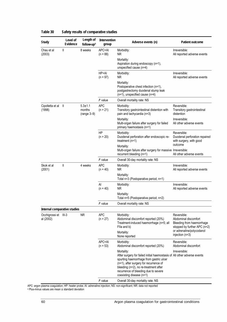
60 Argon plasma coagulation for gastrointestinal conditions
Table 30 Safety results of comparative studies
Study Level of Evidence
Length of follow-upa
Intervention group Adverse events (n) Patient outcome
APC+AI (n = 88)
Morbidity: NR Mortality: Aspiration during endoscopy (n=1), unspecified cause (n=4)
Irreversible: All reported adverse events
HP+AI (n = 97)
Morbidity: NR Mortality: Postoperative chest infection (n=1), postgastrectomy duodenal stump leak (n=1), unspecified cause (n=4)
Irreversible: All reported adverse events
Chau et al (2003)
II 8 weeks
P value Overall mortality rate: NS APC (n = 21)
Morbidity: Transitory gastrointestinal distention with pain and tachycardia (n=3) Mortality: Multi-organ failure after surgery for failed primary haemostasis (n=1)
Reversible: Transitory gastrointestinal distention Irreversible: All other adverse events
HP (n = 20)
Morbidity: Duodenal perforation after endoscopic re-treatment (n=1) Mortality: Multi-organ failure after surgery for massive recurrent bleeding (n=1)
Reversible: Duodenal perforation repaired with surgery, with good outcome Irreversible: All other adverse events
Cipolletta et al (1998)
II 5.3±1.1 months (range 3–9)
P value Overall 30-day mortality rate: NS APC (n = 40)
Morbidity: NR Mortality: Total n=3 (Postoperative period, n=1)
Irreversible: All reported adverse events
AI (n = 40)
Morbidity: NR Mortality: Total n=5 (Postoperative period, n=2)
Irreversible: All reported adverse events
Skok et al (2001)
II 4 weeks
P value Overall mortality rate: NS Internal comparative studies
APC (n = 27)
Morbidity: Abdominal discomfort reported (20%) Treatment-induced haemorrhage (n=5; all FIIa and b) Mortality: None reported
Reversible: Abdominal discomfort Bleeding from haemorrhage stopped by further APC (n=2) or adrenaline/polycodanol injection (n=3)
APC+AI (n = 53)
Morbidity: Abdominal discomfort reported (20%) Mortality: After surgery for failed initial haemostasis of spurting haemorrhage from gastric ulcer (n=1), after surgery for recurrence of bleeding (n=2), no re-treatment after recurrence of bleeding due to severe coexisting disease (n=1)
Reversible: Abdominal discomfort Irreversible: All other adverse events
Occhigrossi et al (2002)
III-3 NR
P value Overall 30-day mortality rate: NS APC: argon plasma coagulation; HP: heater probe; AI: adrenaline injection; NS: non-significant; NR: data not reported a Plus-minus values are mean ± standard deviation
Argon plasma coagulation for gastrointestinal conditions 61
Mortality
Examining the RCTs, Chau et al (2003) reported 6 per cent mortality within both the APC plus adrenaline injection (5/88) and heater probe plus adrenaline injection (6/91) patient groups; no significant statistical difference was found between the overall mortality of the treatment groups. Treatment-related mortality rates were 1 per cent (1/88) amongst APC plus adrenaline injection patients and 2 per cent (2/97) in heater probe plus adrenaline injection patients.
Cipolletta et al (1998) reported 5 per cent treatment-related mortality in both APC (1/21) and heater probe (1/20) patient groups, with no significant difference found.
Skok et al (2001) reported 8 per cent (3/40) mortality within the APC group and 13 per cent (5/40) mortality in the heater probe patient group; no statistical difference was found in the overall 30-day mortality rates of the treatment groups. Mortality rates in the post-operative period were 3 per cent (1/40) in APC patients and 5 per cent (2/40) amongst injection sclerotherapy patients.
The internal comparative study (Occhigrossi et al 2002), reported no deaths in the APC-only patient group after a 30-day period, while an 8 per cent (4/53) mortality rate occurred in the APC plus adrenaline injection group over the same time period; this was not found to be a significant difference.
Overall, there was no statistically significant difference in mortality rates between patients treated with APC or a comparator procedure in any of the three RCTs.
Morbidity
Only one RCT reported morbidities (Cipolletta et al 1998); transitional gastrointestinal distention with pain and tachycardia was reported in 3/21 patients (14.3%) who underwent APC, while perforation of the duodenum was recorded in 1/20 patients (5%) who underwent the comparative heater probe treatment. All morbidities were reversible and treated successfully.
In the internal comparative study (Occhigrossi et al 2002), abdominal pain was reported by 20 per cent of patients and treatment-induced haemorrhage occurred in 5/21 patients (23.8%) who underwent APC alone. In comparison, 20 per cent of patients undergoing APC plus adrenaline injection also reported abdominal pain; however, no cases of treatment-induced haemorrhage were recorded. All morbidities were reversible and treated successfully.
62 Argon plasma coagulation for gastrointestinal conditions
Is it effective?
Effectiveness outcomes were reported to varying levels of detail in the three RCTs; Chau et al (2003) and Cipolletta et al (1998) reported a broader and more thorough range of outcomes relating to effectiveness than Skok et al (2001), who did not report procedural outcomes. The full range of effectiveness outcomes are presented in Table 31. Chau et al was the only study to present exact P-values when comparing effectiveness outcomes between treatment groups; the remaining RCTs provided exact P-values for significant differences only. Like Skok et al, the internal comparative study by Occhigrossi et al (2002) did not report procedural outcomes or exact P-values for all comparisons, including significant findings.
Clinical outcomes
When analysed using a single statistical test, Chau et al (2003) reported significantly higher rates of permanent haemostasis (signified by a healing or healed ulcer at 8-week follow-up) in patients treated with APC plus adrenaline injection than those treated with heater probe plus adrenaline injection (94.2% versus 78.3%, P=0.03); however, significance was lost after correction for the multiple testing of data arising from individual patients. It is worth noting here that the rate of permanent haemostasis found in the heater probe plus adrenaline patient group was notably lower than was found by Cipolletta et al (1998) using heater probe alone. No other significant clinical differences were found in the study.
Cipolletta et al (1998) reported on the same clinical outcomes as Chau et al (2003) but found no significant differences between APC and heater probe treatment.
Skok et al (2001) reported no significant differences between APC and adrenaline injection sclerotherapy in regard to clinical outcomes, with results appearing close to identical on most measures.
Occhigrossi et al (2002) found patients treated with APC plus adrenaline injection to have significantly higher rates of initial haemostasis (98.1% versus 77.7%, P=0.0052) and permanent haemostasis (92.5% versus 77.7%, P<0.05) than patients treated with APC alone. While rates of haemostasis achieved using APC plus adrenaline are consistent with the results of Chau et al (2003), a finding of note is the relatively low rate of haemostasis amongst patients treated with APC alone, as both Cipolletta et al (1998) and Skok et al (2001) achieved initial and permanent haemostasis rates of greater than 90 per cent using APC alone.
Procedural outcomes
Procedural outcomes were reported in only two RCTs; Chau et al (2003) found no significant differences between APC plus adrenaline injection and heater probe plus adrenaline injection, while Cipolletta et al (1998) reported that haemostasis was achieved in a mean of 60 seconds with APC, significantly faster than the mean of 115 seconds with a heater probe (p=0.0246). For both procedures patients may have repeat treatment sessions until haemostasis is achieved. Where reported, patients receive similar number of sessions for both APC and HP (2.2 versus 2.1) (Table 31).

Argon plasma coagulation for gastrointestinal conditions 63
Table 31 Effectiveness results of comparative studies
Clinical outcomes Procedural outcomes
Study Level of evidence
Length of follow-up
Intervention group
Initi
al ha
emos
tasis
n (%
)
Recu
rrenc
e of
blee
ding
n (%
)
Perm
anen
t ha
emos
tasis
n (%
)
Emer
genc
y su
rger
y req
uire
d
n (%
)
Bloo
d tra
nsfu
sion
requ
irem
ents
(uni
t)
Mean
(SD)
Leng
th o
f hos
pita
l st
ay (d
ays)
Mean
(SD)
Tim
e to
haem
osta
sis (s
ec)
Me
an (S
D)
Dura
tion
of
proc
edur
e (m
in)
Me
an (S
D)
No. o
f pro
cedu
res
requ
ired
Me
an (S
D)
APC+AI (n = 88)
86 (97.7) 15 (17.0) 49/52 (94.2) 4 (4.5) 1.7 (2.8) 7.0 (8.4) NR 13.8 (5.7) 2.2 (0.6)
HP+AI (n = 97)
93 (95.9) 21 (21.6) 47/60 (78.3) 9 (9.3) 2.4 (3.3) 8.2 (9.7) NR 14.9 (6.8) 2.1 (0.5)
Chau et al (2003) II 8 weeks
P value 0.68 0.43 0.03a 0.11 0.09 0.38 0.42 0.43 APC (n = 21)
20 (95.2) 3 (15) 19 (90.5) 2 (9.5) 1.9 (0.9) 8 (2) 60 (19) 11.1 (4.6) NR
HP (n = 20)
19 (95) 4 (21) 17 (85) 3 (15) 2.1 (0.4) 9 (3) 115 (28) 13.6 (3.8) NR
Cipolletta et al (1998)
II 5.3 ± 1.1 months (range 3–9)
P value NS NS NS NS NS NS 0.0246 NS APC (n = 40)
39 (97.5) 7 (17.5) 37 (92.5) 3 (7.5) NR NR NR NR NR
AI (n = 40)
38 (95) 9 (22.5) 36 (90) 3 (7.5)b
NR NR NR NR NR
Skok et al (2001) II 4 weeks
P value NS NS NS NS Internal comparative studies
APC (n = 27)
21 (77.7) 2 (9.5) 21 (77.7) 1 (3.7) NR NR NR NR NR
APC+AI (n = 53)
52 (98.1) 5 (9.6) 49 (92.5) 3 (5.6) NR NR NR NR NR
Occhigrossi et al (2002)
III-3 NR
P value 0.0052 NS < 0.05 NS APC: argon plasma coagulation; HP: heater probe; AI: adrenaline injection; NS: non-significant; NR: data not reported; SD: standard deviation a Significant value is lost after correcting for multiple testing of data from individual patients; corrected P value by Bonferroni method is 0.36. b One patient refused surgical treatment.
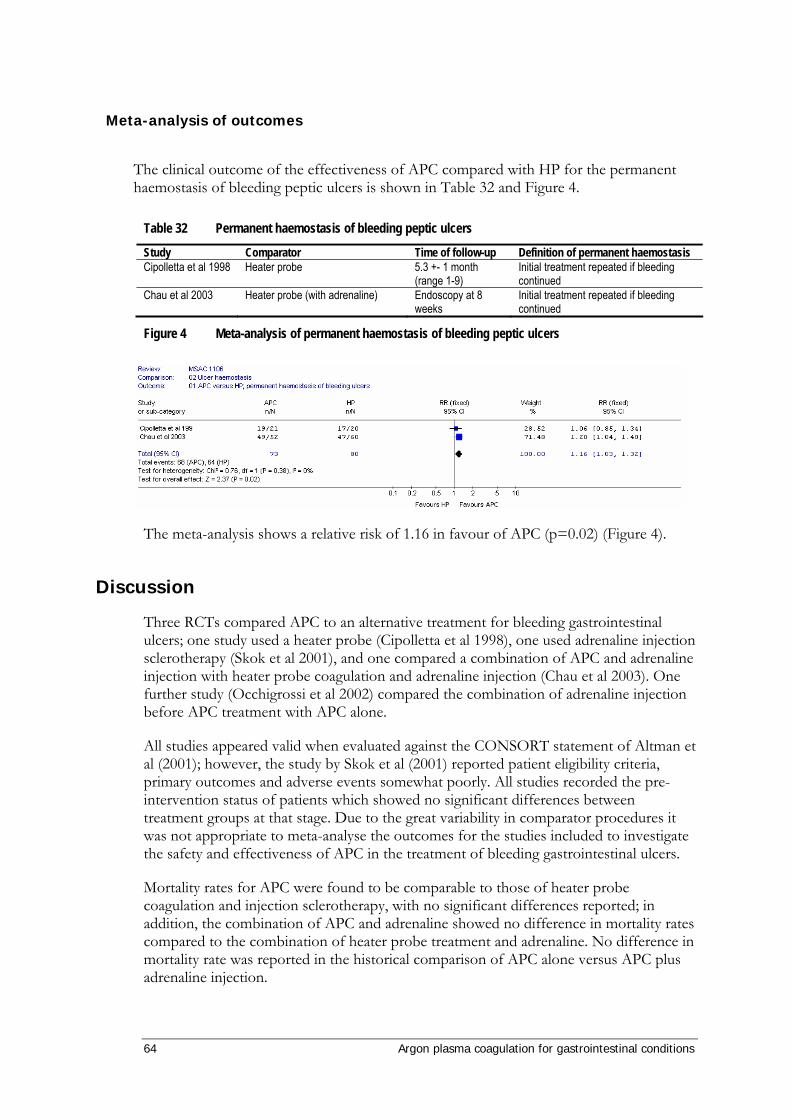
64 Argon plasma coagulation for gastrointestinal conditions
Meta-analysis of outcomes
The clinical outcome of the effectiveness of APC compared with HP for the permanent haemostasis of bleeding peptic ulcers is shown in Table 32 and Figure 4.
Table 32 Permanent haemostasis of bleeding peptic ulcers
Study Comparator Time of follow-up Definition of permanent haemostasis Cipolletta et al 1998 Heater probe 5.3 +- 1 month
(range 1-9) Initial treatment repeated if bleeding continued
Chau et al 2003 Heater probe (with adrenaline) Endoscopy at 8 weeks
Initial treatment repeated if bleeding continued
Figure 4 Meta-analysis of permanent haemostasis of bleeding peptic ulcers
The meta-analysis shows a relative risk of 1.16 in favour of APC (p=0.02) (Figure 4).
Discussion
Three RCTs compared APC to an alternative treatment for bleeding gastrointestinal ulcers; one study used a heater probe (Cipolletta et al 1998), one used adrenaline injection sclerotherapy (Skok et al 2001), and one compared a combination of APC and adrenaline injection with heater probe coagulation and adrenaline injection (Chau et al 2003). One further study (Occhigrossi et al 2002) compared the combination of adrenaline injection before APC treatment with APC alone.
All studies appeared valid when evaluated against the CONSORT statement of Altman et al (2001); however, the study by Skok et al (2001) reported patient eligibility criteria, primary outcomes and adverse events somewhat poorly. All studies recorded the pre-intervention status of patients which showed no significant differences between treatment groups at that stage. Due to the great variability in comparator procedures it was not appropriate to meta-analyse the outcomes for the studies included to investigate the safety and effectiveness of APC in the treatment of bleeding gastrointestinal ulcers.
Mortality rates for APC were found to be comparable to those of heater probe coagulation and injection sclerotherapy, with no significant differences reported; in addition, the combination of APC and adrenaline showed no difference in mortality rates compared to the combination of heater probe treatment and adrenaline. No difference in mortality rate was reported in the historical comparison of APC alone versus APC plus adrenaline injection.

Argon plasma coagulation for gastrointestinal conditions 65
Two studies reported morbidities, but neither made a statistical comparison of the incidence of complications. Cipolletta et al (1998) reported gastrointestinal distension with pain in three APC patients and perforation of the duodenum in one heater probe patient. Occhigrossi et al (2002) found abdominal pain to occur equally in both APC and APC plus adrenaline injection patients, while treatment-induced haemorrhage occurred in five patients (18.5%) who underwent APC alone. All morbidities were reversible and successfully treated in all patients. Overall, APC appears to be as safe as the included comparator procedures for the treatment of bleeding gastrointestinal ulcers. No case series reporting the use of APC for the treatment of bleeding ulcers was identified.
Few differences in clinical or procedural outcomes were found between APC and comparators in the three RCTs. Chau et al (2003) reported tentative evidence that APC plus adrenaline may be more successful in providing long-term haemostasis than heater probe treatment plus adrenaline, and Cipolletta et al (1998) found APC to achieve haemostasis significantly quicker than heater probe treatment. No significant differences were found between APC and adrenaline injection sclerotherapy. In summary, APC appears to be as effective as the available comparative procedures in the treatment of bleeding gastrointestinal ulcers.
Regarding the finding of Chau et al (2003), it is important to note that pre-procedural injection of adrenaline into the bleeding site was found by Occhigrossi et al (2002) to significantly improve rates of haemostasis for APC. This was a historical control study where APC alone was used initially for an eight-month period before a switch to APC plus adrenaline injection. This suggests the difference in haemostasis rates may to some extent be due to a practice effect, with haemostasis success improving as experience with APC treatment was gained over time. Nonetheless, it is reasonable to suggest the finding by Chau et al (2003) may be due in part to the addition of adrenaline injection to the APC procedure. The relatively low rate of haemostasis achieved with the combination of heater probe coagulation and adrenaline injection also suggests that addition of adrenaline may be more beneficial to APC than heater probe treatment. Without adrenaline, the results of Chau et al (2003) may have more closely reflected those of Cipolleta et al (1998) who, using APC and heater probe coagulation alone, found no significant clinical differences between the procedures.
Summary
These results suggest that APC is at least as safe and effective as the comparative techniques of heater probe coagulation with or without adrenaline injection for the treatment of bleeding gastric ulcers, with almost no differences reported between the techniques in key outcomes. Some evidence was found favouring the effectiveness of APC over heater probe treatment; however, this finding was confounded by the study’s methodology of using adrenaline injection to achieve haemostasis prior to thermal treatment. Evidence also suggested that this concurrent use of adrenaline pre-injection enhances the effectiveness of the APC procedure.
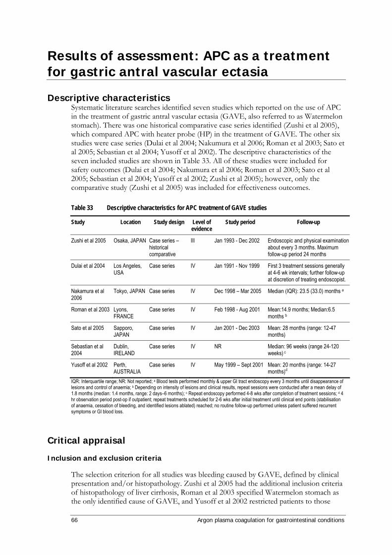
66 Argon plasma coagulation for gastrointestinal conditions
Results of assessment: APC as a treatment for gastric antral vascular ectasia
Descriptive characteristics Systematic literature searches identified seven studies which reported on the use of APC in the treatment of gastric antral vascular ectasia (GAVE, also referred to as Watermelon stomach). There was one historical comparative case series identified (Zushi et al 2005), which compared APC with heater probe (HP) in the treatment of GAVE. The other six studies were case series (Dulai et al 2004; Nakumura et al 2006; Roman et al 2003; Sato et al 2005; Sebastian et al 2004; Yusoff et al 2002). The descriptive characteristics of the seven included studies are shown in Table 33. All of these studies were included for safety outcomes (Dulai et al 2004; Nakumura et al 2006; Roman et al 2003; Sato et al 2005; Sebastian et al 2004; Yusoff et al 2002; Zushi et al 2005); however, only the comparative study (Zushi et al 2005) was included for effectiveness outcomes.
Table 33 Descriptive characteristics for APC treatment of GAVE studies
Study Location Study design Level of evidence
Study period Follow-up
Zushi et al 2005 Osaka, JAPAN Case series – historical comparative
III Jan 1993 - Dec 2002 Endoscopic and physical examination about every 3 months. Maximum follow-up period 24 months
Dulai et al 2004 Los Angeles, USA
Case series IV Jan 1991 - Nov 1999 First 3 treatment sessions generally at 4-6 wk intervals; further follow-up at discretion of treating endoscopist.
Nakamura et al 2006
Tokyo, JAPAN Case series IV Dec 1998 – Mar 2005 Median (IQR): 23.5 (33.0) months a
Roman et al 2003 Lyons, FRANCE
Case series IV Feb 1998 - Aug 2001 Mean:14.9 months; Median:6.5 months b
Sato et al 2005 Sapporo, JAPAN
Case series IV Jan 2001 - Dec 2003 Mean: 28 months (range: 12-47 months)
Sebastian et al 2004
Dublin, IRELAND
Case series IV NR Median: 96 weeks (range 24-120 weeks) c
Yusoff et al 2002 Perth, AUSTRALIA
Case series IV May 1999 – Sept 2001 Mean: 20 months (range: 14-27 months)d
IQR: Interquartile range; NR: Not reported; a Blood tests performed monthly & upper GI tract endoscopy every 3 months until disappearance of lesions and control of anaemia; b Depending on intensity of lesions and clinical results, repeat sessions were conducted after a mean delay of 1.8 months (median: 1.4 months, range: 2 days–6 months); c Repeat endoscopy performed 4-8 wks after completion of treatment sessions; d 4 hr observation period post-op if outpatient; repeat treatments scheduled for 2-6 wks after initial treatment until clinical end points (stabilisation of anaemia, cessation of bleeding, and identified lesions ablated) reached; no routine follow-up performed unless patient suffered recurrent symptoms or GI blood loss.
Critical appraisal
Inclusion and exclusion criteria
The selection criterion for all studies was bleeding caused by GAVE, defined by clinical presentation and/or histopathology. Zushi et al 2005 had the additional inclusion criteria of histopathology of liver cirrhosis, Roman et al 2003 specified Watermelon stomach as the only identified cause of GAVE, and Yusoff et al 2002 restricted patients to those
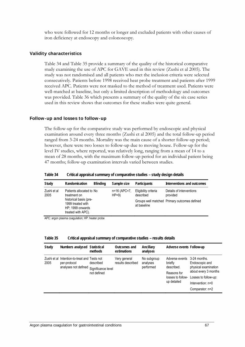
Argon plasma coagulation for gastrointestinal conditions 67
who were followed for 12 months or longer and excluded patients with other causes of iron deficiency at endoscopy and colonoscopy.
Validity characteristics
Table 34 and Table 35 provide a summary of the quality of the historical comparative study examining the use of APC for GAVE used in this review (Zushi et al 2005). The study was not randomised and all patients who met the inclusion criteria were selected consecutively. Patients before 1998 received heat probe treatment and patients after 1999 received APC. Patients were not masked to the method of treatment used. Patients were well-matched at baseline, but only a limited description of methodology and outcomes was provided. Table 36 which presents a summary of the quality of the six case series used in this review shows that outcomes for these studies were quite general.
Follow-up and losses to follow-up
The follow-up for the comparative study was performed by endoscopic and physical examination around every three months (Zushi et al 2005) and the total follow-up period ranged from 3-24 months. Mortality was the main cause of a shorter follow-up period; however, there were two losses to follow-up due to moving house. Follow-up for the level IV studies, where reported, was relatively long, ranging from a mean of 14 to a mean of 28 months, with the maximum follow-up period for an individual patient being 47 months; follow-up examination intervals varied between studies.
Table 34 Critical appraisal summary of comparative studies – study design details
Study Randomisation Blinding Sample size Participants Interventions and outcomes
Zushi et al 2005
Patients allocated to treatment on historical basis (pre-1999 treated with HP; 1999 onwards treated with APC).
No n=16 (APC=7, HP=9)
Eligibility criteria described Groups well matched at baseline
Details of interventions provided Primary outcomes defined
APC: argon plasma coagulation; HP: heater probe
Table 35 Critical appraisal summary of comparative studies – results details
Study Numbers analysed Statistical methods
Outcomes and estimations
Ancillary analyses
Adverse events Follow-up
Zushi et al 2005
Intention-to-treat and per-protocol analyses not defined
Tests not described Significance level not defined
Very general results described
No subgroup analyses performed
Adverse events briefly described. Reasons for losses to follow-up detailed
3-24 months. Endoscopic and physical examination about every 3 months Losses to follow-up: Intervention: n=0 Comparator: n=2

68 Argon plasma coagulation for gastrointestinal conditions
Table 36 Critical appraisal summary of GAVE case studies – results details
Study Sample size Statistical methods Outcomes and estimations
Follow-up
Dulai et al 2004 6 Tests described Significance level defined
Very general results described
First 3 treatment sessions generally at 4-6 wk intervals; further follow-up at discretion of treating endoscopist. Losses to follow-up: NR
Nakamura et al 2006
22 Tests described Significance level defined
Very general results described
Median: 23.5 months; IQR: 33.0 months c
Losses to follow-up: NR
Roman et al 2003 21a
consecutive Tests not described Significance level not defined
Very general results described
Mean:14.9 months; Median:6.5 months d
Losses to follow-up: 2 (reasons not detailed)
Sato et al 2005 8 consecutive
Tests not described Significance level not defined
Very general results described
Mean: 28 months; range: 12-47 months Losses to follow-up: NR
Sebastian et al 2004
12 Tests described Significance level defined
Very general results described
Median: 96 weeks; range 24-120 weeks e
Losses to follow-up: NR
Yusoff et al 2002 5b
consecutive Tests not described Significance level not defined
Very general results described
Mean: 20 months; range: 14-27 months f
Losses to follow-up: NR IQR: interquartile range; NR: Not reported; a 59 treatments; b 13 treatments; c Blood tests performed monthly; Upper GI tract endoscopy every 3 months until disappearance of lesions and control of anaemia; d Depending on intensity of lesions and clinical results, repeat sessions were conducted after a mean delay of 1.8 months (median 1.4 months, range 2 days – 6 months); e Repeat endoscopy performed 4-8 wks after completion of treatment sessions; f 4 hr observation period post-op if outpatient; repeat treatments scheduled for 2-6 wks after initial treatment until clinical end points (stabilisation of anaemia, cessation of bleeding, and identified lesions ablated) reached; no routine follow-up performed unless patient suffered recurrent symptoms or GI blood loss.
Patient characteristics
Comparative Studies
Table 37 summarises the patient population for the comparative study. Patients were well-matched for gender mix and age and all patients had liver cirrhosis (Zushi et al 2005). No information was reported regarding other co-morbidities or concurrent treatments.
Table 37 Study population of comparative studies
Pre-intervention patient status (liver disease)
Study
Inte
rven
tion
grou
p
Num
ber o
f pat
ients
Gend
er
(M/F
)
Mean
age (
rang
e) in
year
s
Cirrh
osis
Alco
hol
Prim
ary b
iliary
cirrh
osis
Unkn
own
APC 7 5/2 67.7(58-75) 4 1 1 1
HP 9 5/4 69.6(61-84) 7 1 1 0
Zushi et al 2005
P value NR NR NR NR NR NR NR APC: Argon plasma coagulation; HP: heater probe; NR: Not reported

Argon plasma coagulation for gastrointestinal conditions 69
Case series
Table 38 summarises the patient population for the case series. There were 74 patients included in these six studies and the age range was fairly similar. Two of these studies reported the number of patients with either ‘Watermelon’ or ‘diffuse’ type of GAVE (Nakumura et al 2006; Sebastian et al 2004). ‘Watermelon’ type describes the classic stripes of bleeding seen on the stomach walls on endoscopy while ‘diffuse’ type describes a more diffuse, honeycomb pattern of bleeding on the stomach walls. Fifteen of the patients in these two studies had ‘Watermelon’ type GAVE and the other 19 patients had the ‘diffuse’ type. One study reported that 14 patients had ‘typical’ (no other lesions) manifestations of GAVE and that the other 7 patients had ‘atypical’ (associated oesophageal varices) (Roman et al 2003).
Table 38 Study population of GAVE case series
Study
# pat
ients
Gend
er (M
/F)
Age i
n ye
ars
(rang
e; m
ean)
Wat
erm
elon/
di
ffuse
type
Typi
cal d /
atyp
ical e
man
ifest
atio
ns
Tran
sfus
ion
depe
nden
t
Dulai et al 2004 6 NR NR NR NR
Nakamura et al 2006
22 10/12 65.8; [7.7c] 3/19 NR
Roman et al 2003
21a 9/12 51-83; 71.5 NR 14/7 21
Sato et al 2005 8 3/5 57-78; 67.9 NR NR Sebastian et al 2004
12 5/7 46-88; 72 12/0 NR 7
Yusoff et al 2002 5b 1/4 58-83; 71 NR NR 4f Total 74 28/40 46-88; 69 15/19 14/7 32 NR: Not reported; a 59 treatments; b 13 treatments; c SD; d no other lesions; e Oesophageal varices in association with watermelon stomach; f Mean packed cell requirements per month was 1.2 U/month
Table 39 and Table 40 provide details of the co-morbidity burden of patients in the case series. There was considerable co-morbidity associated with GAVE. Issues relating to blood loss such as history of GI bleeding and melaena, anaemia, transfusion dependence and haemoglobin levels were commonly reported. Liver disease is a common cause of GAVE and was often present in patients in the included studies. It is important to note that patients in the included studies had a high degree of morbidity at baseline, and although this is a common feature between all studies, the high level of comorbidities could potentially have a significant impact on outcomes.
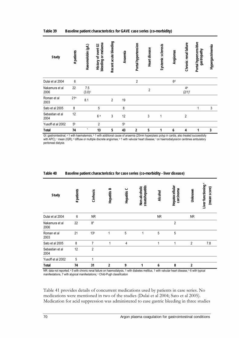
70 Argon plasma coagulation for gastrointestinal conditions
Table 39 Baseline patient characteristics for GAVE case series (co-morbidity)
Study
# pat
ients
Haeo
mol
obin
(g/L
)
Hist
ory o
f ove
rt GI
bl
eedi
ng o
r mela
ena
Rece
nt ac
ute b
leedi
ng
Anae
mia
Porta
l hyp
erte
nsio
n
Hear
t dise
ase
Syst
emic
scler
osis
Angi
omas
Chro
nic r
enal
failu
re
Porta
l hyp
erse
nsiti
ve
gast
ropa
thy
Hype
rgas
trine
mia
Dulai et al 2004 6 2 6d
Nakamura et al 2006
22 7.5 (3.0)c 2 4e
(2/1)f
Roman et al 2003
21a 8.1 2 19
Sato et al 2005 8 5 8 1 3 Sebastian et al 2004
12 6 a 3 12 3 1 2
Yusoff et al 2002 5b 2 5b Total 74 ` 13 5 43 2 5 1 6 4 1 3 GI: gastrointestinal; a 1 with haematemsis; b 1 with additional cause of anaemia (20mm hyperplasic polyp in cardia, also treated successfully with APC); c mean (IQR); d diffuse or multiple discrete angiomas; e 1 with valvular heart disease; f on haemodialysis/on centimes ambulatory peritoneal dialysis
Table 40 Baseline patient characteristics for case series (co-morbidity - liver disease)
Study
# pat
ients
Cirrh
osis
Hepa
titis
B
Hepa
titis
C
Non-
alcoh
olic
stea
tohe
patit
is
Alco
hol
Hepa
toce
llular
ca
rcin
oma
Unkn
own
Live
r fun
ctio
ning
c (m
ean
scor
e)
Dulai et al 2004 6 NR NR NR
Nakamura et al 2006
22 8a 2
Roman et al 2003
21 13b 1 5 1 5 5
Sato et al 2005 8 7 1 4 1 1 2 7.8 Sebastian et al 2004
12 2
Yusoff et al 2002 5 1 Total 74 31 2 9 1 6 8 2 NR: data not reported; a 5 with chronic renal failure on haemodialysis, 1 with diabetes mellitus, 1 with valvular heart disease; b 6 with typical manifestations, 7 with atypical manifestations; c Child-Pugh classification
Table 41 provides details of concurrent medications used by patients in case series. No medications were mentioned in two of the studies (Dulai et al 2004; Sato et al 2005). Medication for acid suppression was administered to ease gastric bleeding in three studies

Argon plasma coagulation for gastrointestinal conditions 71
(Nakamura et al 2006; Roman et al 2003; Sebastian et al 2004). One patient was receiving anticoagulants (Yusoff et al 2002), so the dose of anticoagulant was delayed four hours on APC treatment days to prevent excessive bleeding caused by APC.
Table 41 Concurrent medications used by patients being treated for GAVE with APC in case series
Study # p
atien
ts
Acid
supp
ress
ion
(PPI
s/H2
anta
goni
sts)
Sucr
alfat
e
Oral
ster
oids
Nitra
tes
Non-
selec
tive b
eta
bloc
kers
Octre
otid
e
Antic
oagu
lants
Coag
ulan
ts
Dulai et al 2004 6
Nakamura et al 2006
22 22 22
Roman et al 2003
21 15 15 3 6 1
Sato et al 2005 8 Sebastian et al 2004
12 12 3
Yusoff et al 2002 5 1a Total 74 49 15 3 1 6 1 1 22 a Dose delayed 4 hrs on treatment days
Technical details
Technical details of the APC technique and comparator used in the comparative study are provided in Table 42 and details of APC technique used in the case series are provided in Table 43. Patients received between one and nine APC treatment sessions each. The mean number of treatments per patients in the comparative study was 1.45 ± 0.69 for APC and 3.09 ± 1.58 for HP. For the case series, the mean number of APC treatments was 2.4 sessions. In all of the studies, an Erbe APC machine was used to administer APC treatments. In cases where reported, the APC power output and APC apparatus used were similar. Power settings ranged from 40-80 W, with a mean of 53 W. The flow rate ranged from 0.5-2.0 L/min and the mean flow rate was 1.5 L/min. The duration of APC treatment sessions was not reported for any of the studies. Where reported APC was carried out under conscious sedation (Nakamura et al 2006; Sebastian et al 2004; Yusoff et al 2002), apart from one case series which used general anaesthesia (Roman et al 2003).

72 Argon plasma coagulation for gastrointestinal conditions
Table 42 Description of APC technique – comparative studies
Stud
y ID
Coag
ulat
ion
syst
em
Powe
r
Gas f
low
(L/m
in)
Anae
sthe
sia/se
datio
n
Meth
od
# tre
atm
ents
Trea
tmen
t in
terv
al (w
eeks
)
Erbe APC-300 with ICC-350a
60 W 2.0 L/min
NR Goal: to coagulate as much lesion as possible until apparent red spot disappeared
1-4 (mean: 1.45 ± 0.69)
1c
Zushi et al 2005 Olympus Heater Probe Unit b
20 joules
NA NR Goal: to coagulate as much lesion as possible until apparent red spot disappeared
1-7 (3.09 ± 1.58)
1d
NA: Not applicable; NR: Not reported; a Erbe, Tubingen, Germany; b HPU, Olympus; c Used since 1999 until red spot not apparent on endoscopy; d Used before 1998 until red spot not apparent on endoscopy
Table 43 Description of APC technique for treatment of GAVE in case series
Stud
y ID
APC
inst
rum
ent
Powe
r (W
)
Gas f
low
(L/m
in)
Anae
sthe
sia/se
datio
n
Meth
od
# tre
atm
ents
Trea
tmen
t in
terv
al (w
eeks
)
Dulai et al 2004 Erbe APC a 50-60 0.5-1.6 NR Preference for lowest settings necessary to achieve satisfactory coagulation
2-8 (median: 4)
4-6 (First 2 treatments sessions)
Nakamura et al 2006
Argon Beamer Two Order APC-300 with
Erbotom ICC-200 & flexible probe b
50-60 1.5-2 Local anaesthetic (lidocaine spray). IV sedation as required.
All patients drank 60 ml solution containing antifoaming & mucolytic agents to clear mucus from stomach
2-5 (median:4; IQR:3)
1 until screen g.
Roman et al 2003 Erbe APC c 50-80 0.8 General anaesthesia.
Trawl back 'Paintbrush' technique used to electrocoagulate all visible angiectasis until lesions became bleached d.
1-5 (median:3; mean: 2.8)
2 days- 6 months h (median: 6; mean: 7.7)
Sato et al 2005 Erbe APC-300 with ICC-350 & 2.3 mm flexible probe b
60 2 NR Combination of spot touch ‘woodpecker’ & trawl back 'paintbrush' techniques. Treatment commenced at pylorus, moving proximally. Goal: formation of pale yellow coagulum over vascular lesions.
1-3 (mean: 1.8)
NR
Sebastian et al 2004
Erbe APC with 3.2 mm probe b
40 1.5 Conscious sedation (midazolam)
Ablation of all visible lesions attempted until no longer tolerated by patient e.
1-5 (median: 2)
3-15 (median: 4)
Yusoff et al 2002 Erbe APC-300
with Erbotom ICC-200 & 2.3 mm flexible probe b
40-50 2.0 Conscious sedation (midazolam & fentanyl/ pethidine)
Combination of spot touch ‘woodpecker’ & trawl back 'paintbrush' techniques. Treatment commenced at pylorus, moving proximally. Goal: formation of pale yellow coagulum over vascular lesions f.
1-4 (mean: 2.6)
2-6
APC: argon plasma coagulation; IQR: Interquartile range; IV: intravenous; NR: Not reported; PPI: proton pump inhibitor; a Erbe Inc., Marietta, GA, USA; b Erbe Electromedezin, Tuebingen, GER; c Erbe, Lyons, FR; d 3 patients underwent endoscopic ligation before APC treatment; e Hyoscine butyl bromide administered before APC to reduce peristaltic movements; f prophylactic antibiotics (amoxicillin/ gentamicin) given to patients at risk of infection before APC; g All GAVE lesions eradicated & HP stabilised without transfusion; h depending on intensity of lesions & clinicobiological results

Argon plasma coagulation for gastrointestinal conditions 73
Safety
Comparative studies
The comparative study that reported treatment of GAVE with APC and HP reported limited safety outcomes, which are presented in Table 44. There were a large number of deaths reported for patients treated with either method; however, no cases of mortality were directly attributable to either treatment modality.
Table 44 Adverse events reported by comparative studies for APC treatment of GAVE
Number of adverse events (month) Mortality
Study
L of
E
Leng
th fo
llow-
up
Trea
tmen
t
Num
ber o
f pat
ients
Lost
to fo
llow-
up
Recu
rrent
blee
ding
Hepa
tic fa
ilure
Hepa
toce
llular
ca
rcin
oma
Pneu
mon
ia
Oeso
phag
eal
varix
rupt
ure
Tota
l mor
talit
y
APC 7 0 2
(8; 12)b 1
(8) 2
(3; 3) 1
(21) 0 4
Zushi et al 2005 III 3
months HP 9 2
2 (11; 11)
2 (4; 20)
3 (2; 5;18)
1 (2)
1 (24)
7 a
APC: argon plasma coagulation; HP: heater probe; a 100% of patients left in follow-up died; b numbers in parenthesis represent the time, in months, from the treatment when the adverse event occurred
Case series
Safety outcomes were reported for APC treatment of GAVE in the six case series included in this review. Follow-up in these studies ranged from 14.9 to 28 months (Table 33). Adverse events are summarised in Table 45 and details are given in Table 46. Although 18 deaths were reported in the follow-up period, none of these were related to the APC treatment, and only one was due to a bleeding-related cause (uncontrolled haemorrhage). Other adverse events, which included minor bleeding controlled by applying more APC, minor ulcers and haematemesis, were considered minor and temporary. No perforations were reported. Thus, no significant adverse events could be attributed to the use of APC in the treatment of GAVE. No other safety data, such as length of hospital stay, was reported in any of the studies.
Expert opinion of the Advisory Panel suggests that the primary endpoint of thermal coagulation of GAVE is the formation of superficial ulceration (Jensen et al 2004). Common adverse events associated with the use of APC in this manner include overdistension of the stomach (with argon gas), smoke and dyspepsia (Jensen et al 2004). These may all be easily managed with medical therapy.
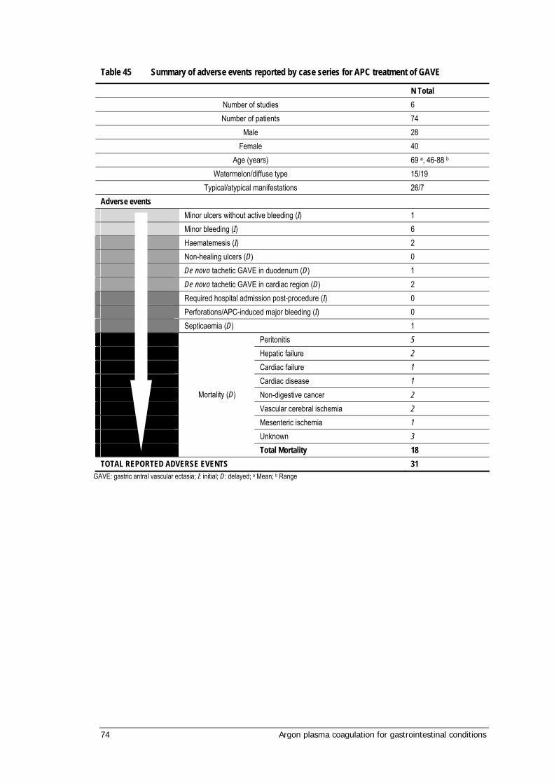
74 Argon plasma coagulation for gastrointestinal conditions
Table 45 Summary of adverse events reported by case series for APC treatment of GAVE
N Total Number of studies 6 Number of patients 74
Male 28 Female 40
Age (years) 69 a, 46-88 b Watermelon/diffuse type 15/19
Typical/atypical manifestations 26/7 Adverse events Minor ulcers without active bleeding (I) 1 Minor bleeding (I) 6 Haematemesis (I) 2 Non-healing ulcers (D) 0 De novo tachetic GAVE in duodenum (D) 1
De novo tachetic GAVE in cardiac region (D) 2 Required hospital admission post-procedure (I) 0 Perforations/APC-induced major bleeding (I) 0 Septicaemia (D) 1 Peritonitis 5 Hepatic failure 2 Cardiac failure 1 Cardiac disease 1 Non-digestive cancer 2 Vascular cerebral ischemia 2 Mesenteric ischemia 1 Unknown 3
Mortality (D)
Total Mortality 18 TOTAL REPORTED ADVERSE EVENTS 31
GAVE: gastric antral vascular ectasia; I: initial; D: delayed; a Mean; b Range

Argon plasma coagulation for gastrointestinal conditions 75
Table 46 Details of adverse events reported by case series for APC treatment of GAVE
Adverse event Dulai et al 2004
Nakamura et al 2006
Roman et al 2003
Sato et al 2005
Sebastian et al 2004
Yusoff et al 2002
INITIAL COMPLICATIONS Minor ulcers without active bleeding NR 1 8 Minor bleeding a 0 1 5 Haematemesis NR 2 Perforations/APC-induced major bleeding
0
Required hospital admission post-procedure
0 0 0 0 0 0
Mortality 0 0 0 0 0 0 DELAYED COMPLICATIONS Non-healing ulcers 0 Recurrent bleeding 12 3 2 De novo tachetic GAVE in duodenum De novo tachetic GAVE in cardia 2 Septicaemia 1 b
Peritonitis 1 2 Uncontrolled haemorrhage 1 Hepatic failure 2 Cardiac failure 1 Cardiac disease 1 Non-digestive cancer 2 Vascular cerebral ischemia 2 Mesenteric ischemia 1
Morta
lity
Unknown 3 Total mortality NR 5 11 0 0 1 TOTAL ADVERSE EVENTS 0 8 8 10 1 6
APC: argon plasma coagulation; GAVE: gastric antral vascular ectasia; NR: Not reported; a Controlled by applying more APC; b Patient with cirrhosis
Effectiveness
Comparative studies
Effectiveness outcomes from the comparative study (Zushi et al 2005) are included in this review (Table 47). One to four treatment sessions (mean: 1.45 ± 0.69) were required to treat GAVE with APC and one to seven (mean: 3.09 ± 1.58) sessions were required when using HP. Recurrence of bleeding was reported in two patients each for APC and HP treatment, but this was successfully treated with further treatment. Efficacy outcomes reported in this study were very limited. It was stated that both treatments were ‘equally effective’ in diminishing GAVE lesions; however, APC was stated as being technically easier, and on average required fewer number of treatments per patient (1.5 compared to 3.1 for HP).

76 Argon plasma coagulation for gastrointestinal conditions
Table 47 Effectiveness results reported by comparative studies for APC treatment of GAVE.
Outcome Study
L of
E
Leng
th fo
llow-
up
Trea
tmen
t
N
Lost
to fo
llow-
up
# tre
atm
ents
re
quire
d pe
r pat
ient
Recu
rrenc
e of
blee
ding
N (%
)
Prog
ress
ive
anae
mia
Effe
ctive
ness
in
dim
inish
ing
GAVE
les
ion
Ease
of d
imin
ishin
g GA
VE le
sion
APC 7 0 1-4 (mean: 1.45 ± 0.69) 2 a (28.6%) b Zushi et al 2005 III 3 months
HP 9 2 1-7 (mean: 3.09 ± 1.58) 2 a (22.2%) c ‘Improved in all patients’
‘Equally effective’
APC ‘technically easier’
APC: argon plasma coagulation; HP: heater probe; a Re-treatment successful in all cases; b Re-bleeding at 8 and 12 months; c Re-bleeding at 11 months for both
As transfusion dependency and iron-deficiency anaemia are important aspects of GAVE pathogenesis, the Advisory Panel were interested in identifying studies which reported these outcomes. Of the included studies, three (all case series) reported anaemia-related outcomes before and after treatment with APC (Table 48). APC treatment provided an improvement in these outcomes. Where reported, these differences were statistically significant.
Table 48 Transfusion-dependency or haemoglobin levels of patients before and after treatment with APC
Transfusion-dependency Mean haemoglobin levels Study
Number of patients Before APC After APC p-value Before APC After APC p-value
Roman et al 2003
21 NR NR NR 80.9 g/l SD 14.8 g/l
103.2 g/l SD 16.2 g/l
NR
Sebastian et al 2004
12 Mean number of units 11.3 (SD 5.68)
Mean number of units 1.1 (SD 0.57)
0.018 8.13 g/l SD 0.7 g/dl
12.2 g/l SD 0.3 g/dl
0.008
Yusoff et al 2002
5 4/5 transfusion-dependent
0/5 transfusion-dependent
NR 7.2g/dl 12.7g/dl NR
NR: not reported; APC: argon plasma coagulation; SD: standard deviation
Discussion Outcomes from the included studies for the safety and efficacy of APC for the treatment of GAVE were limited due to the low level of evidence of the available studies. Of the seven included studies, there was only one comparative study. This was a historical case comparative study (level of evidence III-3). Furthermore, the outcomes in this study were only described in brief and very generally. As the comparative treatment was administered to patients at an earlier timepoint to the APC treatment, it is likely that other factors may have affected outcomes, such as improved technique, surgeon experience and improvement of general patient healthcare. The other six included studies were all of level IV evidence, and thus could not be used for effectiveness outcomes.
Argon plasma coagulation for gastrointestinal conditions 77
Additionally, the high level of co-morbidities which led to mortality made it difficult to obtain long term results for GAVE treatments.
Safety
Although a high number of adverse events were reported in patients who underwent APC treatment for GAVE, the majority of these events were not attributable to this treatment. The high mortality rate could be attributed to the age and high level of serious co-morbidity of patients in these studies. This was further demonstrated by the equally high level of adverse events observed in patients treated with HP in the comparative study. The only adverse events found to be attributable to APC use were minor bleeding (controlled by applying more APC) and minor, temporary ulceration. The Advisory Panel noted that minor ulceration is an end-point of thermal coagulation techniques. Thus, the use of APC for the treatment of GAVE appears to be safe for use in a clinical setting and is at least as safe as HP treatment.
Effectiveness
The effectiveness outcomes reported narratively in the one comparative study suggest that APC is at least as effective as HP in diminishing GAVE lesions and is technically easier to use. The mean number of treatment sessions required was lower for APC treated patients than for HP treated patients, so APC may in fact be more effective; however, further information is required to support this. Recurrence of bleeding was similar in both groups and successfully managed by further APC or HP treatment. However, the Advisory Panel suggested that any endoscopic GAVE therapy only provides treatment for the symptoms of bleeding and is not a permanent cure for the disease. It is possible that recurrence is not related to the treatment method used but the nature of the disease. From the evidence evaluated, APC appears to be at least as effective as HP treatment for GAVE, requiring fewer treatment sessions, and is easier to use.

78 Argon plasma coagulation for gastrointestinal conditions
Results of assessment: APC as a treatment for radiation proctitis
Systematic review evidence
Two systematic reviews were identified on the topic of radiation proctitis (Forbes & Maher 2002; Tagkalidis & Tjandra 2001). The Cochrane report investigated a variety of treatment modalities, but did not investigate APC (Forbes & Maher 2002). A meta-analysis by Tagkalidis and Tjandra (2001) reported on five case series, together with unpublished data from their own experience. As no comparative evidence was available no formal conclusion could be reached regarding the safety and effectiveness of APC in the treatment of radiation proctitis.
Is it safe?
The clinical experts of the Advisory Panel suggested that radiation proctitis has significant morbidity, and many of the adverse events observed during treatment are as a result of the disease itself, and not due to the treatment. Radiation proctitis involves significant submucosal injury, including bleeding, fibrosis, ischaemia and ulceration to the wall of the rectum (Forbes & Maher 2002). Clinical presentation of radiation proctitis may in addition include tenesmus, abdominal or rectal pain, urgency, diarrhoea or constipation, and anal sphincter dysfunction (Forbes & Maher 2002; Ramage & Gostout 2003; Silva 1999). Patients also present with bloody rectal discharge and anaemia and are frequently highly transfusion-dependent. At present the only treatment options for this condition in Australia are formalin insertion and thermal coagulation.
As a result of the systematic literature searches, 18 case series were identified (Ben-Soussan et al 2004; Canard et al 2003; Dees et al 2006; de la Serna Higuera et al 2004; Fantin et al 1999; Kaassis et al 2000; Panos 1999; Ravizza et al 2003; Rotondano et al 2003; Sebastian et al 2004; Silva et al 1999; Smith et al 2001; Taieb et al 2001; Tjandra et al 2001; Tam et al 2000; Villavicencio et al 2002; Venkatesh et al 2002; Zinicola et al 2003). No studies which compared APC to another treatment modality (such as formalin instillation) were identified. The descriptive, technical and patient characteristics of the 18 case series are summarised in Table 49. More detailed information on each study can be found in Appendix H.
The study population varied in size from 5 (Panos 1999) to 50 (Dees et al 2006) participants, and the length of follow-up of participants ranged from 1 to 60 months (Table 49). In the 15 studies where it was reported, the majority of participants in all but three studies (de la Serna Higuera et al 2004; Rotondano et al 2003; Silva et al 1999) were male. The mean age of the participants included in the studies was similar between studies, where reported.
Participants received between one and eight APC treatment sessions each (Table 49). Where reported, patients were usually treated under local anaesthesia or sedation. General anaesthesia was required by a small percentage of patients in two studies. The APC power setting ranged from 30 to 80 W, while the APC gas flow used ranged from 0.8 to 3.0 L/min (Table 49). Expert opinion of the Advisory Panel indicated that the
Argon plasma coagulation for gastrointestinal conditions 79
power can vary between APC machines, and needs to be sufficient to establish the argon plasma.
In total, 369 participants were treated with APC for radiation proctitis (Table 50). A total of 79 complications were reported in the 18 included case series; however, as some participants experienced more than one complication, it was not possible to determine the proportion of the participants that suffered from adverse events. The complications reported varied, although the majority were transient and did not require re-treatment.
In six cases superficial ulcers were described as short-term complications; however, 14 participants in a single study developed rectal ulcers as long-term complications, although all were asymptomatic (Ravizza et al 2003). In this study patients were treated with APC settings of 3 L/min and 60 W or 2 L/min and 40 W. The ulcers were as common in both treatment modalities. Expert clinical advice from the Advisory Panel suggests that in the treatment of radiation proctitis with thermal coagulation techniques, the intention of the treatment is to cause minor ulceration of the site. In 10 cases, rectal pain was a transient complication which resolved within a few days; however, in two cases, this pain was a longer-term complication, which took between 1 and 10 months to resolve. There were 10 reported cases of bloating or cramping caused by gastrointestinal tract distension from insufflation of the argon gas; however, this number is an underestimation, as three of the six studies that reported on this stated only that a small number or minority of patients suffered from this complication.
Three cases of bleeding or haemorrhage were reported (Canard et al 2003; Dees et al 2006; Venkatesh et al 2002). One patient had been prescribed aspirin for a transient ischaemic attack two years after the initial therapy and was successfully treated with a haemoclip, and another bled as a result of treatment failure and was successfully treated with a haemoclip. One case of perforation was reported when a power setting of 80 W was used, the highest power utilised in this patient cohort and in all the included studies (Canard et al 2003). No information was provided regarding the treatment of the perforation.
A total of 15 deaths were reported in the 18 included case series; however, none of these deaths were related to APC treatment (data not shown). Thirteen patients died as a result of the recurrence of underlying malignancies or disease, one patient died from cardiac failure and another died following surgical treatment of radiation-induced stricture of the small intestine (data not shown).
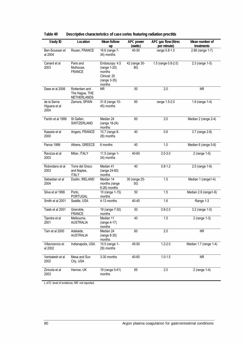
80 Argon plasma coagulation for gastrointestinal conditions
Table 49 Descriptive characteristics of case series featuring radiation proctitis
Study ID Location Mean follow-up
APC power (watts)
APC gas flow (litres per minute)
Mean number of treatments
Ben-Soussan et al 2004
Rouen, FRANCE 16.6 (range 7-34) months
40-50 range 0.8-1.0 2.66 (range 1-7)
Canard et al 2003
Paris and Mulhouse, FRANCE
Endoscopy: 4.5 (range 1-20) months Clinical: 20 (range 3-35) months
42 (range 30-80)
1.5 (range 0.8-2.0) 2.3 (range 1-5)
Dees et al 2006
Rotterdam and The Hague, THE NETHERLANDS
NR 50 2.0 NR
de la Serna Higuera et al 2004
Zamora, SPAIN 31.8 (range 10-45) months
60 range 1.5-2.0 1.9 (range 1-4)
Fantin et al 1999
St Gallen, SWITZERLAND
Median 24 (range 18-24) months
60 3.0 Median 2 (range 2-4)
Kaassis et al 2000
Angers, FRANCE 10.7 (range 8-28) months
40 0.6 3.7 (range 2-8)
Panos 1999
Athens, GREECE 6 months 40 1.0 Median 6 (range 5-8)
Ravizza et al 2003
Milan, ITALY 11.5 (range 1-24) months
40-60 2.0-3.0 2 (range 1-5)
Rotondano et al 2003
Torre del Greco and Naples, ITALY
Median 41 (range 24-60) months
40 0.8-1.2 2.5 (range 1-6)
Sebastian et al 2004
Dublin, IRELAND Median 14 months (range 6-26) months
30 (range 25-50)
1.5 Median 1 (range1-4)
Silva et al 1999
Porto, PORTUGAL
10 (range 1-15) months
50 1.5 Median 2.9 (range1-8)
Smith et al 2001
Seattle, USA 4-13 months 40-45 1.6 Range 1-3
Taieb et al 2001
Grenoble, FRANCE
19 (range 7-30) months
50 0.8-2.0 3.2 (range 1-5)
Tjandra et al 2001
Melbourne, AUSTRALIA
Median 11 (range 4-17) months
40 1.5 2 (range 1-3)
Tam et al 2000
Adelaide, AUSTRALIA
Median 24 (range 8-35) months
60 2.0 NR
Villavicencio et al 2002
Indianapolis, USA 10.5 (range 1-29) months
45-50 1.2-2.0 Median 1.7 (range 1-4)
Venkatesh et al 2002
Mesa and Sun City, USA
3-30 months 40-60 1.0-1.5 NR
Zinicola et al 2003
Harrow, UK 19 (range 5-41) months
65 2.0 2 (range 1-4)
L of E: level of evidence; NR: not reported.

Argon plasma coagulation for gastrointestinal conditions 81
Table 50 Adverse events of APC treatment for radiation proctitis
Total number of studies 18 Total number of patients 369 Male (a) 215 Female 91 Age (years) median [mean range] 72 [65-76] (b) Adverse events Transient anal or rectal pain 15 Gas distension 10 (c) Minor inflammation 3 Superficial ulcers 6 Tenesmus 2 short-term, 2 long-term Diarrhoea 5 Urinary retention 1 Fever 3 Fistula 1 Stricture / stenosis 8 Extensive necrosis 1 Long-term pain 2 Long-term asymptomatic ulcers 14 Colonic explosion 2 Haemorrhage 3 Perforation 1 Mortality 0 Total complications 79
a three studies did not report the number of males and females; b three studies did not report the mean age of participants; c three studies reported that some patients suffered from gas distension that resolved, but did not state a number
Is it effective?
As bleeding and transfusion dependency are important aspects of radiation proctitis, the Advisory Panel were interested in identifying studies which reported these outcomes. Of the included case series, 12 studies reported bleeding-related outcomes before and after treatment with APC (Table 51). There was a reduction in all bleeding measures and mean haemoglobin concentrations after treatment with APC. These differences were statistically significant, where reported.

82 Argon plasma coagulation for gastrointestinal conditions
Table 51 Bleeding-related outcomes for patients before and after APC treatment
Transfusion-dependency or bleeding (mean)
Mean haemoglobin levels Study
Patient number Before APC After APC p-value Before APC After APC p-value
Ben-Soussan et al 2004
27 8 patients TD
1 patient TD p<0.05
Chutkan bleeding score 3.03
Chutkan bleeding score 0.42
p<0.001
Canard et al 2003
30 Chutkan bleeding score 2.67
Chutkan bleeding score 0.77
p<0.001
Kaassis et al 2000
15 Bleeding score 2.4
Bleeding score 0.6
3/15 TD 0/15 TD Panos 1999 5 NR Ravizza et al 2003
27 Bleeding score 2.8
Bleeding score 0.5
p<0.001 8.0g/dl 11.2g/dl NR
Rotonando et al 2003
24 Bleeding score 2.9
Bleeding score 0.8
p<0.01 9.2g/dl 10.6g/dl p<0.05
Sebastian et al 2004
25 Median bleeding score 3
Median bleeding score 0
p<0.0005 10.1g/dl 12.4g/dl p<0.002
Silva et al 1999
28 2.96 0.68 NR NR Reduction of 1.2g/dl
NR
Taieb et al 2001
11 7/11 TD 0/11 TD NR 7.7g/dl 11.5g/dl p=0.003
Tjandra and Segupta 2001
12 4/12 TD 0/12 TD p<0.05 11.2g/dl 12.3g/dl p<0.001
Tam et al 2000
15 Median bleeding score 3
Median bleeding score 1
p<0.001 108g/l 133g/l p<0.05
3/15 TD 0/15 TD Zinicola et al 2003
14 Bleeding scale 2.6
Bleeding scale 0.9
p<0.0001
NR: not reported; TD: transfusion-dependent
Unpublished comparative study
No RCTs comparing the use of APC for the treatment of radiation proctitis were uncovered in the published literature. The Advisory Panel highlighted one unpublished RCT manuscript which compared APC with formalin instillation for the treatment of radiation proctitis (Hayes et al). As there were no published RCTs available, the Panel’s decision was to include this study in the review as it would add significantly to the information from the included studies. The study is included in full in Appendix I.

Argon plasma coagulation for gastrointestinal conditions 83
Table 52 Descriptive characteristics of comparative study
Study Study design (NHMRC level of evidence)
Study period Follow-up Inclusion criteria Exclusion criteria
Hayes et al (unpublished) Adelaide, AUSTRALIA
Comparative RCT (Level II)
NR 48 weeks Patients with overt rectal bleeding due to radiation proctitis, proven on colonoscopy (with or without biopsy)
• Previous treatment for bleeding
• Other sources of bleeding from the colon
NR: not reported
Hayes et al was performed in Australia and compared treatment of radiation proctitis with APC with topical application of 4 per cent formalin. Inclusion and exclusion criteria for the recruitment of patients in each of the studies are displayed in Table 52. Nineteen patients were randomised to either APC (n=10) or formalin (n=9) treatment (Table 53). The method of randomisation was not reported and patients were not blinded to treatment. Eligibility criteria were well defined. There were differences in age distribution and haemoglobin levels between groups; however, these differences were not statistically significant.
Study endpoints were mostly measured by a visual analogue scale (VAS) completed by the patient. Data was analysed using Students t-tests and statistical significance was taken as p<0.05.
There was some variability between follow-up periods: the mean follow up period for the APC treated patient group was 60 weeks (range: 22-114) and the mean follow-up for the formalin treated group was 37 weeks (range: 9-100 weeks). There were no reported losses to follow-up.
Patient characteristics
Table 53 summarises the patient population characteristics.
Table 53 Patient characteristics of comparative studies
Study
Inte
rven
tion
grou
p
Num
ber o
f pat
ients
Gend
er
(M/F
)
Mean
age (
rang
e) in
year
sa
Mean
pre
-trea
tmen
t ha
emog
lobi
n g/
L (ra
nge)
No tr
ansf
usio
n de
pend
ent
patie
nts
APC 10 NR 75 (60-83) 118 (71-159) 3 Formalin 9 NR 63 (38-79) 141 (82-164) 1 Total 19 17/2a 4
Hayes et al (unpublished)
p value NS NS NS 0.063 NS APC: argon plasma coagulation; NR: data not reported; a all male patients had been treated for prostate cancer and both female patients had
radiotherapy for cervical cancer; NS: not significant.
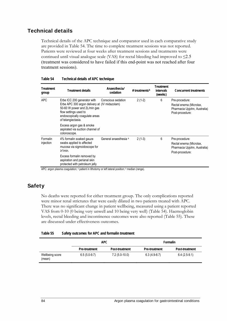
84 Argon plasma coagulation for gastrointestinal conditions
Technical details
Technical details of the APC technique and comparator used in each comparative study are provided in Table 54. The time to complete treatment sessions was not reported. Patients were reviewed at four weeks after treatment sessions and treatments were continued until visual analogue scale (VAS) for rectal bleeding had improved to ≤2.5 (treatment was considered to have failed if this end-point was not reached after four treatment sessions).
Table 54 Technical details of APC technique
Treatment group Treatment details Anaesthesia/
sedation # treatmentsb Treatment intervals (weeks)
Concurrent treatments
APC Erbe ICC 200 generator with Erbe APC 300 argon delivery at 50-60 W power and 2L/min gas flow settings used to endoscopically coagulate areas of telangiectasia. Excess argon gas & smoke aspirated via suction channel of colonoscope.
Conscious sedation (IV midazolam)
2 (1-2) 6 Pre-procedure: Rectal enema (Microlax, Pharmacia Upjohn, Australia) Post-procedure:
Formalin injection
4% formalin soaked gauze swabs applied to affected mucosa via sigmoidoscope for ≥1min. Excess formalin removed by aspiration and perianal skin protected with petroleum jelly
General anaesthesia a 2 (1-3) 6 Pre-procedure: Rectal enema (Microlax, Pharmacia Upjohn, Australia) Post-procedure:
APC: argon plasma coagulation; a patient in lithotomy or left lateral position; b median (range).
Safety
No deaths were reported for either treatment group. The only complications reported were minor renal strictures that were easily dilated in two patients treated with APC. There was no significant change in patient wellbeing, measured using a patient reported VAS from 0-10 (0 being very unwell and 10 being very well) (Table 54). Haemoglobin levels, rectal bleeding and incontinence outcomes were also reported (Table 55). These are discussed under effectiveness outcomes.
Table 55 Safety outcomes for APC and formalin treatment
APC Formalin
Pre-treatment Post-treatment Pre-treatment Post-treatment Wellbeing score (mean)
6.5 (5.0-9.7) 7.2 (5.0-10.0) 6.3 (4.9-8.7) 6.4 (2.5-9.1)

Argon plasma coagulation for gastrointestinal conditions 85
Effectiveness
APC and formalin treatment had a similar effect on rectal bleeding score and both groups required a mean of two treatment sessions per patient; however, the number of sessions ranged from 1 to 2 for APC and from 1 to 3 for formalin. The number of treatment sessions required was determined by patient reported rectal bleeding score using a VAS from 0 to 10 (0 being no bleeding and 10 being heavy bleeding), which are shown in Table 56.
None of the four transfusion dependant patients required transfusion post-treatment and the mean haemoglobin levels before and after treatment improved in both groups, but was more improved in the APC treated group, although this improvement was not statistically significant (possibly due to differences in baseline characteristics).
There was no significant difference in anorectal dysfunction, measured using the Cleveland Clinic-Florida Incontinence Score with faecal urgency added. Frequency of incontinence to solid stool, liquid stool or gas, requirement for pads for soiling, degree of lifestyle alteration and faecal urgency were complied to give a total score ranging from 0 to 24 (0 being normal continence and 24 being completely incontinent with severe urgency).
Table 56 Effectiveness outcomes for APC and formalin treatment
APC Formalin
Pre- Post- Pre- Post- Rectal bleeding score (mean) 5.9 (4.0-7.6) 1.5 (0-2.5) * 6.1 (4.6-10.0) 1.6 (0-2.5) *
Haemoglobin (mean, g/L) 118 (71-159) 137 (117-162) † 141 (82-164) 145 (116-167) Anorectal dysfunction (mean score) 9.0 (3 -18) 9.3 (3-15) 6.6 (1-13) 8.0 (1 -17) APC: argon plasma coagulation; * P < 0.0001; † P = 0.047
Conclusion
In summary, the evidence base for APC in the treatment of radiation proctitis is relatively small, with no comparative studies published to date. Eighteen case series were identified with a total of 369 participants. From these data, APC appeared to be relatively safe, although there were a variety of short-term complications including rectal pain, rectal ulcers, bloating and cramping, tenesmus, diarrhoea, fever and rectal stenosis. The majority of complications were transient and resolved without the need for re-treatment. Many of the adverse events which could also be directly related to the presentation of radiation proctitis (such as bleeding) would commonly be expected as part of a thermal modality treatment (ulceration), or were short-term problems associated with the use of APC in a confined space (bloating). There were no deaths as a result of APC treatment.
One unpublished RCT was identified which compared APC with formalin instillation in the treatment of radiation proctitis. Both techniques appeared similar in effectiveness, with regard to rectal bleeding, haemoglobin concentration and rectal dysfunction; however, more RCT evidence is required to clearly assess the safety and effectiveness of APC for this indication.
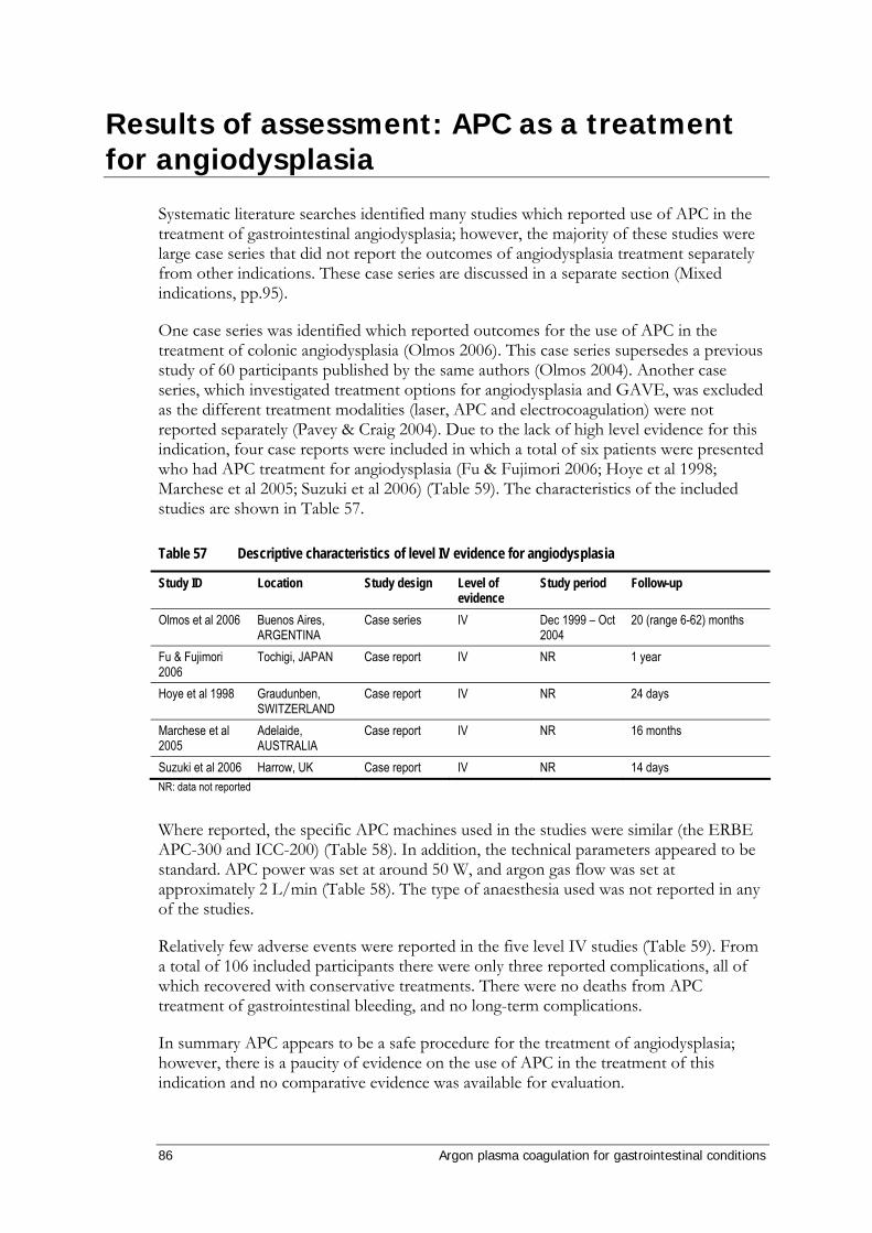
86 Argon plasma coagulation for gastrointestinal conditions
Results of assessment: APC as a treatment for angiodysplasia
Systematic literature searches identified many studies which reported use of APC in the treatment of gastrointestinal angiodysplasia; however, the majority of these studies were large case series that did not report the outcomes of angiodysplasia treatment separately from other indications. These case series are discussed in a separate section (Mixed indications, pp.95).
One case series was identified which reported outcomes for the use of APC in the treatment of colonic angiodysplasia (Olmos 2006). This case series supersedes a previous study of 60 participants published by the same authors (Olmos 2004). Another case series, which investigated treatment options for angiodysplasia and GAVE, was excluded as the different treatment modalities (laser, APC and electrocoagulation) were not reported separately (Pavey & Craig 2004). Due to the lack of high level evidence for this indication, four case reports were included in which a total of six patients were presented who had APC treatment for angiodysplasia (Fu & Fujimori 2006; Hoye et al 1998; Marchese et al 2005; Suzuki et al 2006) (Table 59). The characteristics of the included studies are shown in Table 57.
Table 57 Descriptive characteristics of level IV evidence for angiodysplasia
Study ID Location Study design Level of evidence
Study period Follow-up
Olmos et al 2006 Buenos Aires, ARGENTINA
Case series IV Dec 1999 – Oct 2004
20 (range 6-62) months
Fu & Fujimori 2006
Tochigi, JAPAN Case report IV NR 1 year
Hoye et al 1998 Graudunben, SWITZERLAND
Case report IV NR 24 days
Marchese et al 2005
Adelaide, AUSTRALIA
Case report IV NR 16 months
Suzuki et al 2006 Harrow, UK Case report IV NR 14 days NR: data not reported
Where reported, the specific APC machines used in the studies were similar (the ERBE APC-300 and ICC-200) (Table 58). In addition, the technical parameters appeared to be standard. APC power was set at around 50 W, and argon gas flow was set at approximately 2 L/min (Table 58). The type of anaesthesia used was not reported in any of the studies.
Relatively few adverse events were reported in the five level IV studies (Table 59). From a total of 106 included participants there were only three reported complications, all of which recovered with conservative treatments. There were no deaths from APC treatment of gastrointestinal bleeding, and no long-term complications.
In summary APC appears to be a safe procedure for the treatment of angiodysplasia; however, there is a paucity of evidence on the use of APC in the treatment of this indication and no comparative evidence was available for evaluation.

Argon plasma coagulation for gastrointestinal conditions 87
Table 58 Technical characteristics of case series featuring angiodysplasia
Study ID APC instrument used
Technique APC power (watts)
APC gas flow (litres per minute)
Mean number of treatments
Olmos et al 2006 APC-300, ICC-200, ERBE
0.5-2 sec duration impact
60 (rectum and left colon), 40 (right colon and caecum
1.5-2.5 Median 1 (range 1-3)
Fu & Fujimori 2006
NR Submucosal saline injection to minimise thermal injury and prevent perforation
NR NR 1
Hoye et al 1998 NR NR 50 2.0 1 Marchese et al 2005
APC-300, ERBE Adrenaline (1:10,000) injected prior to APC
50 1.0 2
Suzuki at al 2006 ICC-200, ERBE Adrenaline 1:200,000 (2-3ml) injected prior to APC. Pulses of 1 sec duration.
50 2 NR
APC: argon plasma coagulation; NR: data not reported
Table 59 Patient characteristics and outcomes of case series featuring angiodysplasia
Study ID Patient number
M/F Mean age (years)
Indications Adverse events
Olmos et al 2006
100 51/49 72 (range 20-87)
Colonic angiodysplasia (387 in total)
2 complications in 118 procedures (1.7%): Fever immediately after procedure, 1 Pneumoperitoneum without laparoscopic evidence of perforation, 1 Both recovered with conservative treatment. 6 deaths during follow-up from unrelated causes.
Fu & Fujimori 2006
1 0/1 86 Bleeding angiodysplasia in duodenum
None reported
Hoye et al 1998
1 0/1 86 Angiodysplasia Pneumoperitoneum due to occult perforation was treated with prophylactic antibiotics. Patient recovered fully.
Marchese et al 2005
1 1/0 65 Angiodysplasia (Dieulafoy’s lesion), with co-morbidity of type 2a Von Willebrand’s disease
None reported
Suzuki et al 2006
3 3/0 70 (range 62-79)
Colonic angiodysplasia (10 in total)
None reported
TOTAL 106 3 complications
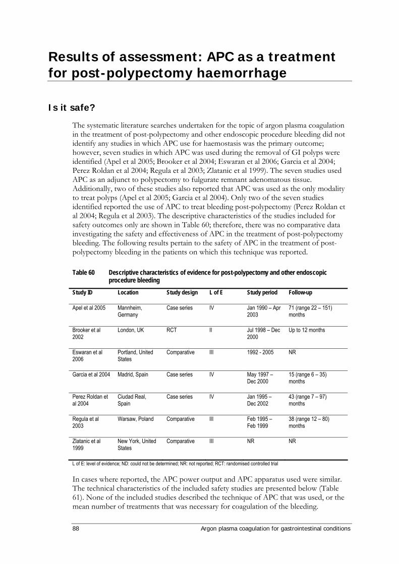
88 Argon plasma coagulation for gastrointestinal conditions
Results of assessment: APC as a treatment for post-polypectomy haemorrhage
Is it safe?
The systematic literature searches undertaken for the topic of argon plasma coagulation in the treatment of post-polypectomy and other endoscopic procedure bleeding did not identify any studies in which APC use for haemostasis was the primary outcome; however, seven studies in which APC was used during the removal of GI polyps were identified (Apel et al 2005; Brooker et al 2004; Eswaran et al 2006; Garcia et al 2004; Perez Roldan et al 2004; Regula et al 2003; Zlatanic et al 1999). The seven studies used APC as an adjunct to polypectomy to fulgurate remnant adenomatous tissue. Additionally, two of these studies also reported that APC was used as the only modality to treat polyps (Apel et al 2005; Garcia et al 2004). Only two of the seven studies identified reported the use of APC to treat bleeding post-polypectomy (Perez Roldan et al 2004; Regula et al 2003). The descriptive characteristics of the studies included for safety outcomes only are shown in Table 60; therefore, there was no comparative data investigating the safety and effectiveness of APC in the treatment of post-polypectomy bleeding. The following results pertain to the safety of APC in the treatment of post-polypectomy bleeding in the patients on which this technique was reported.
Table 60 Descriptive characteristics of evidence for post-polypectomy and other endoscopic procedure bleeding
Study ID Location Study design L of E Study period Follow-up
Apel et al 2005 Mannheim, Germany
Case series IV Jan 1990 – Apr 2003
71 (range 22 – 151) months
Brooker et al 2002
London, UK RCT II Jul 1998 – Dec 2000
Up to 12 months
Eswaran et al 2006
Portland, United States
Comparative III 1992 - 2005 NR
Garcia et al 2004 Madrid, Spain Case series IV May 1997 – Dec 2000
15 (range 6 – 35) months
Perez Roldan et al 2004
Ciudad Real, Spain
Case series IV Jan 1995 – Dec 2002
43 (range 7 – 97) months
Regula et al 2003
Warsaw, Poland Comparative III Feb 1995 – Feb 1999
38 (range 12 – 80) months
Zlatanic et al 1999
New York, United States
Comparative III NR NR
L of E: level of evidence; ND: could not be determined; NR: not reported; RCT: randomised controlled trial
In cases where reported, the APC power output and APC apparatus used were similar. The technical characteristics of the included safety studies are presented below (Table 61). None of the included studies described the technique of APC that was used, or the mean number of treatments that was necessary for coagulation of the bleeding.

Argon plasma coagulation for gastrointestinal conditions 89
Table 61 Technical characteristics of studies featuring post-polypectomy and other endoscopic procedure bleeding
Study ID APC instrument used
APC power (watts)
APC gas flow (litres per minute)
Anaesthesia Mean number of treatments
Apel et al 2005 Erbe Elektromedizin
50 W NR NR NR
Brooker et al 2002
Erbe Argon Plasma Coagulator
45 – 55 W (right colon)
65 W (left colon)
2.0 L/min NR NR
Eswaran et al 2006
NR 60 W 1.0 L/min Patients under propofol anaesthesia
NR
Garcia et al 2004 Erbe APC300 40 – 80 W 2.5 L/min Conscious sedation with intravenous propofol when unsedated colonoscopy not tolerated.
NR
Perez Roldan et al 2004
Erbe HF-100 Olympus Europe model knife and Soring Arco 2000 model (Soring Medizintechnik)
NR NR NR NR
Regula et al 2003
Erbe beamer 2 with ICCC200 unit or an APC 300 unit with ICC 350 unit
60 W ‘large flow’ (Erbe beamer 2) or 2 L/min (APC 300)
Polypectomy procedure usually performed without sedation
NR
Zlatanic et al 1999
Erbe ICC 350 NR 0.8 L/min NR NR
APC: argon plasma coagulation; NR: not reported
The seven studies which reported the use of APC as an adjunct to polypectomy demonstrated that APC used in this manner was generally safe. Two studies reported no complications directly attributed to APC in ablation of remnant adenomatous tissue following endoscopic removal of colonic, duodenal and ampullary polyps (Eswaran et al 2006; Brooker et al 2002). Zlatanic et al 1999 reported that in patients who received APC treatment, inadvertent tissue contact with the colon wall with the flexible tip of the APC apparatus occasionally led to the submucosa becoming alarmingly inflated (due to argon gas flowing into tissue space via the mucosa burned by the coagulator). However, this was the only report of this event occurring in all identified studies. A further two studies reported the incidence of adverse events in patients who received polypectomy plus APC, although it was unclear from both studies whether the events were a result of APC application or the polypectomy technique itself. Two cases of bleeding and one case of ulceration were reported in a case series of 18 patients in which six received APC ablation following polypectomy of sessile duodenal adenomas (Apel et al 2005). One
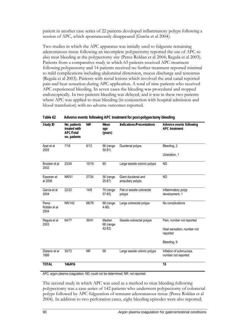
90 Argon plasma coagulation for gastrointestinal conditions
patient in another case series of 22 patients developed inflammatory polyps following a session of APC, which spontaneously disappeared (Garcia et al 2004).
Two studies in which the APC apparatus was initially used to fulgurate remaining adenomatous tissue following an incomplete polypectomy reported the use of APC to also treat bleeding at the polypectomy site (Perez Roldan et al 2004; Regula et al 2003). Patients from a comparative study in which 63 patients received APC treatment following polypectomy and 14 patients received no further treatment reported minimal to mild complications including abdominal distension, mucus discharge and tenesmus (Regula et al 2003). Patients with rectal lesions which involved the anal canal reported pain and heat sensation during APC application. A total of nine patients who received APC experienced bleeding. In seven cases the bleeding was procedural and stopped endoscopically. In two patients bleeding was delayed, and it was in these two patients where APC was applied to treat bleeding (in conjunction with hospital admission and blood transfusion) with no adverse outcomes reported.
Table 62 Adverse events following APC treatment for post-polypectomy bleeding
Study ID No. patients treated with APC/Total no. patients
M/F Mean age (years)
Indications/Presentations Adverse events following APC treatment
Apel et al 2005
7/18 6/12 66 (range 50-81)
Duodenal polyps Bleeding, 2
Ulceration, 1
Brooker et al 2002
23/34 15/19 65 Large sessile colonic polyps ND
Eswaran et al 2006
NR/51 27/24 56 (range 25-87)
Giant duodenal and ampullary polyps
ND
Garcia et al 2004
22/22 14/8 70 (range 57-83)
Flat or sessile colorectal polyps
Inflammatory polyp development, 1
Perez Roldan et al 2004
NR/142 68/79 68 (range 4-90)
Large colorectal polyps No complications
Regula et al 2003
64/77 36/41 Median 68 (range 42-83)
Sessile colorectal polyps Pain, number not reported
Heat sensation, number not reported
Bleeding, 9
Zlatanic et al 1999
30/72 NR 68 Large sessile colonic polyps Inflation of submucosa, number not reported
TOTAL 146/416 13
APC: argon plasma coagulation; ND: could not be determined; NR: not reported
The second study in which APC was used as a method to treat bleeding following polypectomy was a case series of 142 patients who underwent polypectomy of colorectal polyps followed by APC fulguration of remnant adenomatous tissue (Perez Roldan et al 2004). In addition to two perforation cases, eight bleeding episodes were also reported;
Argon plasma coagulation for gastrointestinal conditions 91
however, these were not attributed to the use of APC to fulgurate any remaining adenomatous tissue. The APC apparatus was used to treat two bleeding episodes. No complications regarding the use of APC to treat bleeding were reported.
In summary, there is a lack of higher levels of evidence reporting the use of APC in the treatment of post-polypectomy bleeding. Given the available evidence, APC appears to be a safe treatment option for this indication.

92 Argon plasma coagulation for gastrointestinal conditions
Results of assessment: APC as a treatment for tumour ingrowth in oesophageal stents
Oesophageal malignancies
Expert advice from the advisory panel suggested that APC is not a cure for GI malignancies, and that the primary treatment for symptomatic oesophageal tumours in Australia would be stent placement. Very few studies were identified through the systematic literature search which used APC for the ablation of tumourous outgrowth through an oesophageal stent (e.g. self-expanding metallic stent (SEMS)) as indicated by this review. There were many case reports and case series in which APC was used to ablate oesophageal tumours in the absence of stent placement. In addition, one completed but unpublished trial was identified in which APC was compared to SEMS in the treatment of oesophageal cancer (principal investigator Dr JG Freeman, Derby, UK). There were no published comparative trials in which APC was used for the ablation of oesophageal tumours either with or without previous stent placement.
Two case series (Akhtar et al 2000; Crosta et al 2001) and one case report (Rajendran et al 2000) were identified in which APC was used to ablate oesophageal tumour ingrowth through SEMS (Table 63). In Crosta (2001), the majority of patients (46/47) received palliative APC treatment for a malignant oesophageal obstruction, in the absence of stents. One patient received APC treatment for the removal of tumour ingrowth through a silicon stent.
Table 63 Descriptive characteristics of level IV evidence for stents
Study ID Location Study design L of E Study period Follow-up (months) Akhtar et al 2000 Salford, UK Case series IV Jan 1996 – Oct
1999 Range 3-12
Crosta et al 2001 Milan, ITALY Case series IV Jan 1998 – Dec 1999
NR
Rajendran et al 2000
Liverpool, UK Case report IV NR 3
NR: not reported; L of E: level of evidence
Where reported, APC treatment was carried out under conscious sedation (Table 64). A low number of repeat treatments were required (a median of 1 or 2 treatments). The power of the diathermy unit ranged between 40 and 70 W, with a wide variety of gas flow settings (0.3-2.0 L/min).
All patients presented with either dysphagia or bleeding (Table 65). One of the patients suffered from oesophageal Crohn’s disease (Rajendran et al 2000). There were no reported complications of APC treatment in any of the included studies (Table 65). Therefore APC appears to be a safe procedure for the treatment of oesophageal malignancies. However, as reported by Akhtar and colleagues (2000), the long-term prognosis from advanced oesophageal cancer is poor, with a median survival of four months even with a high rate of successful treatment and with no complications or adverse events.
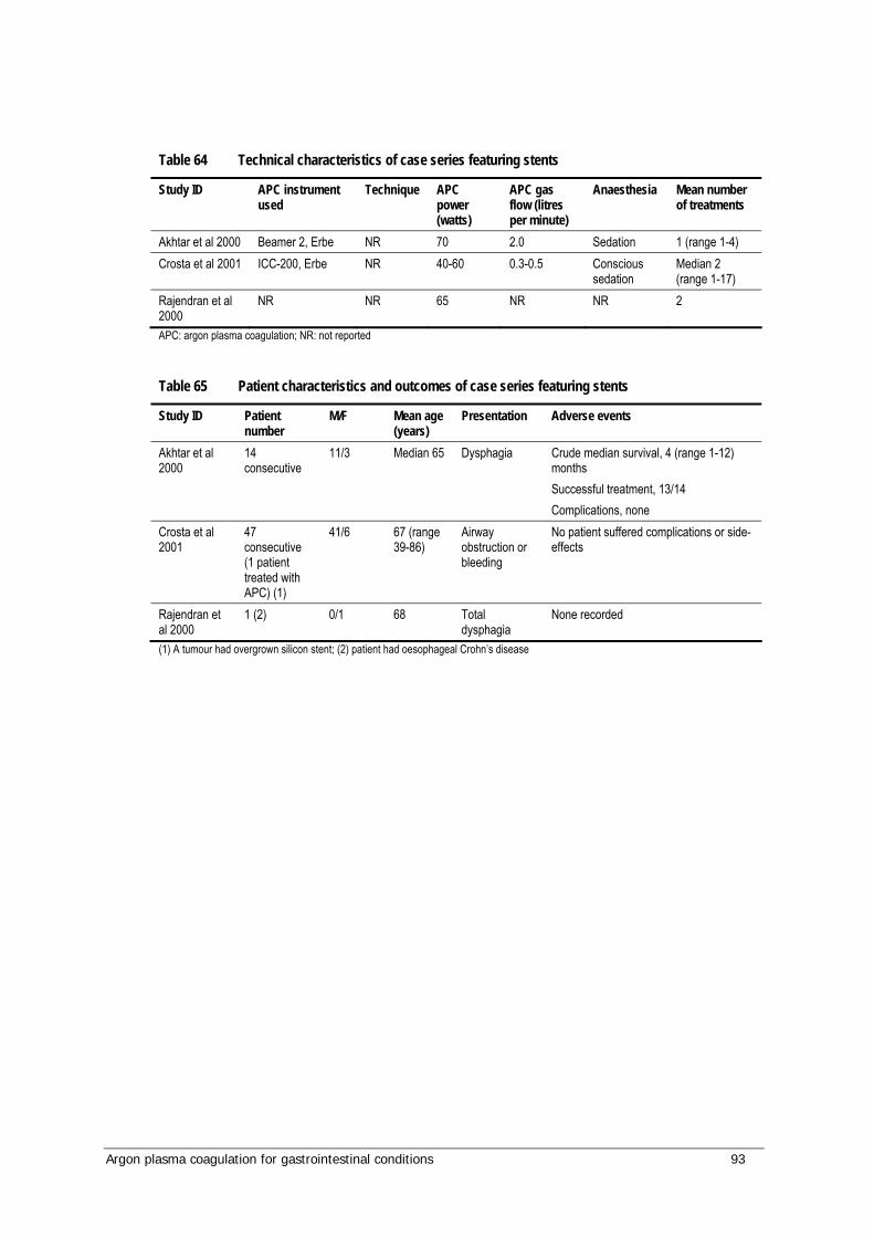
Argon plasma coagulation for gastrointestinal conditions 93
Table 64 Technical characteristics of case series featuring stents
Study ID APC instrument used
Technique APC power (watts)
APC gas flow (litres per minute)
Anaesthesia Mean number of treatments
Akhtar et al 2000 Beamer 2, Erbe NR 70 2.0 Sedation 1 (range 1-4) Crosta et al 2001 ICC-200, Erbe NR 40-60 0.3-0.5 Conscious
sedation Median 2 (range 1-17)
Rajendran et al 2000
NR NR 65 NR NR 2
APC: argon plasma coagulation; NR: not reported
Table 65 Patient characteristics and outcomes of case series featuring stents
Study ID Patient number
M/F Mean age (years)
Presentation Adverse events
Akhtar et al 2000
14 consecutive
11/3 Median 65 Dysphagia Crude median survival, 4 (range 1-12) months Successful treatment, 13/14 Complications, none
Crosta et al 2001
47 consecutive (1 patient treated with APC) (1)
41/6 67 (range 39-86)
Airway obstruction or bleeding
No patient suffered complications or side-effects
Rajendran et al 2000
1 (2) 0/1 68 Total dysphagia
None recorded
(1) A tumour had overgrown silicon stent; (2) patient had oesophageal Crohn’s disease

94 Argon plasma coagulation for gastrointestinal conditions
Results of assessment: APC as a treatment used in studies of mixed indications
Systematic reviews and health technology assessments on the use of APC in gastrointestinal bleeding
Four systematic reviews were identified which investigated the use of argon plasma coagulation (APC) in the treatment of the indications discussed in this review (Pichon Riviere et al 2005; Havanond & Havanond 2005; Vargo 2004, Farrell & Friedman 2005). None of the reviews provided a conclusion regarding the safety and effectiveness of APC in the treatment of GI indications, and all suggested that more high quality, comparative evidence is required.
The review by Pichon Riviere and colleagues (2005) investigated the use of APC for gastrointestinal tract pathologies. Twelve studies (mainly case series) were included for the review, which concluded that the evidence base was poor and provided no mid- or long-term data on safety and effectiveness. The GI lesions investigated were polyps, radiation proctitis and ulcers.
Vargo (2004) reported a systematic review on the use of APC in the treatment of GI conditions (polypectomy, radiation proctitis, vascular lesions, ablation of Barrett’s oesophagus, bleeding ulcers and GI malignancies). There was no overall conclusion regarding the safety and effectiveness of APC for any of these conditions as the evidence was mainly from case series. The authors suggested that APC should be considered experimental until larger trials are published to validate its use.
A Cochrane review was published in 2005 which investigated APC as a treatment for non-variceal upper GI bleeding (Havanond & Havanond 2005). Two RCTs were included in this review; one compared APC to heater probe, and the other compared APC to injection sclerotherapy (Cipolletta et al 1998; Skok et al 2001). Both of these studies are discussed in the ulcers indication section of this review. The authors concluded that there was very little published research in this field, and that further randomised controlled trials are required.
Farrell and Friedman (2005) conducted a systematic review on the broad subject of lower gastrointestinal bleeding. Although procedures such as formalin instillation, injection therapy with sclerosing agents such as ethanolamine, adrenaline injection, thermal coagulation and laser are discussed in detail for a wide variety of indications, APC was mentioned only briefly in terms of treatment of angiodysplasia and radiation proctitis. No overall conclusion regarding the safety and effectiveness of APC in the treatment of GI bleeding was provided.
Mixed gastrointestinal indications for the use of APC
During the systematic literature searches undertaken for the topic of argon plasma coagulation in the treatment of various gastrointestinal pathologies, ten case series were identified which reported consecutive patients suffering from various conditions (Abou-
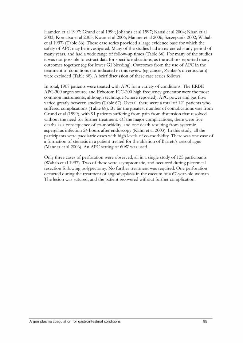
Argon plasma coagulation for gastrointestinal conditions 95
Hamden et al 1997; Grund et al 1999; Johanns et al 1997; Kanai et al 2004; Khan et al 2003; Komatsu et al 2005; Kwan et al 2006; Manner et al 2006; Szczepanik 2002; Wahab et al 1997) (Table 66). These case series provided a large evidence base for which the safety of APC may be investigated. Many of the studies had an extended study period of many years, and had a wide range of follow-up times (Table 66). For many of the studies it was not possible to extract data for specific indications, as the authors reported many outcomes together (eg for lower GI bleeding). Outcomes from the use of APC in the treatment of conditions not indicated in this review (eg cancer, Zenker’s diverticulum) were excluded (Table 68). A brief discussion of these case series follows.
In total, 1907 patients were treated with APC for a variety of conditions. The ERBE APC-300 argon source and Erbotom ICC-200 high frequency generator were the most common instruments, although technique (where reported), APC power and gas flow varied greatly between studies (Table 67). Overall there were a total of 121 patients who suffered complications (Table 68). By far the greatest number of complications was from Grund et al (1999), with 91 patients suffering from pain from distension that resolved without the need for further treatment. Of the major complications, there were five deaths as a consequence of co-morbidity, and one death resulting from systemic aspergillus infection 24 hours after endoscopy (Kahn et al 2003). In this study, all the participants were paediatric cases with high levels of co-morbidity. There was one case of a formation of stenosis in a patient treated for the ablation of Barrett’s oesophagus (Manner et al 2006). An APC setting of 60W was used.
Only three cases of perforation were observed, all in a single study of 125 participants (Wahab et al 1997). Two of these were asymptomatic, and occurred during piecemeal resection following polypectomy. No further treatment was required. One perforation occurred during the treatment of angiodysplasia in the caecum of a 67-year-old woman. The lesion was sutured, and the patient recovered without further complication.
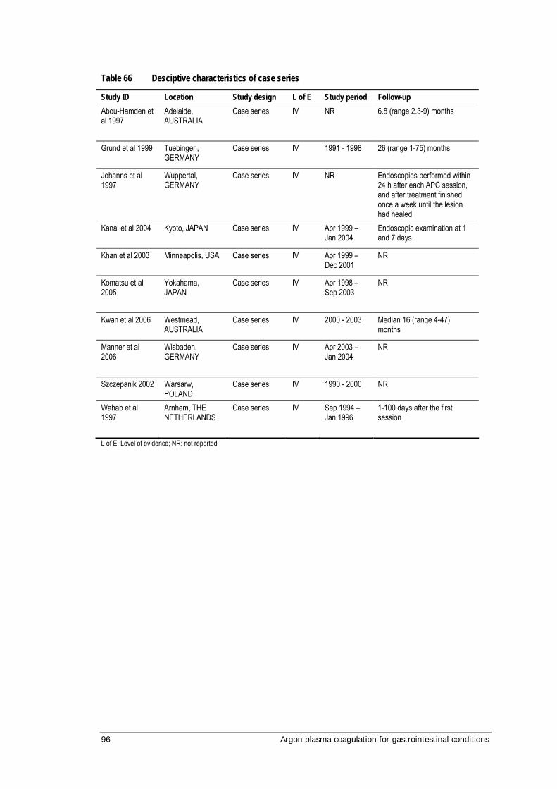
96 Argon plasma coagulation for gastrointestinal conditions
Table 66 Desciptive characteristics of case series
Study ID Location Study design L of E Study period Follow-up Abou-Hamden et al 1997
Adelaide, AUSTRALIA
Case series IV NR 6.8 (range 2.3-9) months
Grund et al 1999
Tuebingen, GERMANY
Case series IV 1991 - 1998 26 (range 1-75) months
Johanns et al 1997
Wuppertal, GERMANY
Case series IV NR Endoscopies performed within 24 h after each APC session, and after treatment finished once a week until the lesion had healed
Kanai et al 2004
Kyoto, JAPAN Case series IV Apr 1999 – Jan 2004
Endoscopic examination at 1 and 7 days.
Khan et al 2003
Minneapolis, USA Case series IV Apr 1999 – Dec 2001
NR
Komatsu et al 2005
Yokahama, JAPAN
Case series IV Apr 1998 – Sep 2003
NR
Kwan et al 2006
Westmead, AUSTRALIA
Case series IV 2000 - 2003 Median 16 (range 4-47) months
Manner et al 2006
Wisbaden, GERMANY
Case series IV Apr 2003 – Jan 2004
NR
Szczepanik 2002 Warsarw, POLAND
Case series IV 1990 - 2000 NR
Wahab et al 1997
Arnhem, THE NETHERLANDS
Case series IV Sep 1994 – Jan 1996
1-100 days after the first session
L of E: Level of evidence; NR: not reported

Argon plasma coagulation for gastrointestinal conditions 97
Table 67 Technical characteristics of case series featuring mixed indications
Study ID Instrument used Technique APC power (watts)
APC gas flow (litres per minute)
Anaesthesia Mean number of treatments
Abou-Hamden et al 1997
NR NR NR NR NR Median 2
Grund et al 1999
APC 300 ERBE; Erbotom ICC 350 or 200, ERBE
Brush-like strokes
Mean 79 (range 10-100)
Mean 1.3 (range 0.2-18)
NR 2 (1-18)
Johanns et al 1997
NR NR 40 or 75 2 or 3 Sedation and analgesia
2 (range 1-8)
Kanai et al 2004
APC-300 and Erbotom ICC-200, ERBE
0.5-2 sec coagulation
45 1.0 NR NR
Khan et al 2003
APC-300 and ICC-200, ERBE
Short bursts (1-2 sec); probe was removed after 2-3 bursts to allow gas removal
55 0.9 NR Median 1 (range 1-5)
Komatsu et al 2005
ARCO-3000 NR 35 or 50 2.0 or 2.5 NR NR
Kwan et al 2006
ARCO-3000 Focal pulse and spray painting technique
20-40 1.0 Conscious sedation
NR
Manner et al 2006
ERBE APC2, VIO-300D
Pulsed APC used to completely ablate dynamically in strips
15-120 1.0 Conscious sedation
ND
Szczepanik 2002 NR NR NR NR NR NR Wahab et al 1997
ERBE ICC-200 NR 40 (caecum), 100 (rectum)
2.5 Intravenous sedation
2.6
APC: argon plasma coagulation; NR: not reported; ND: could not be determined
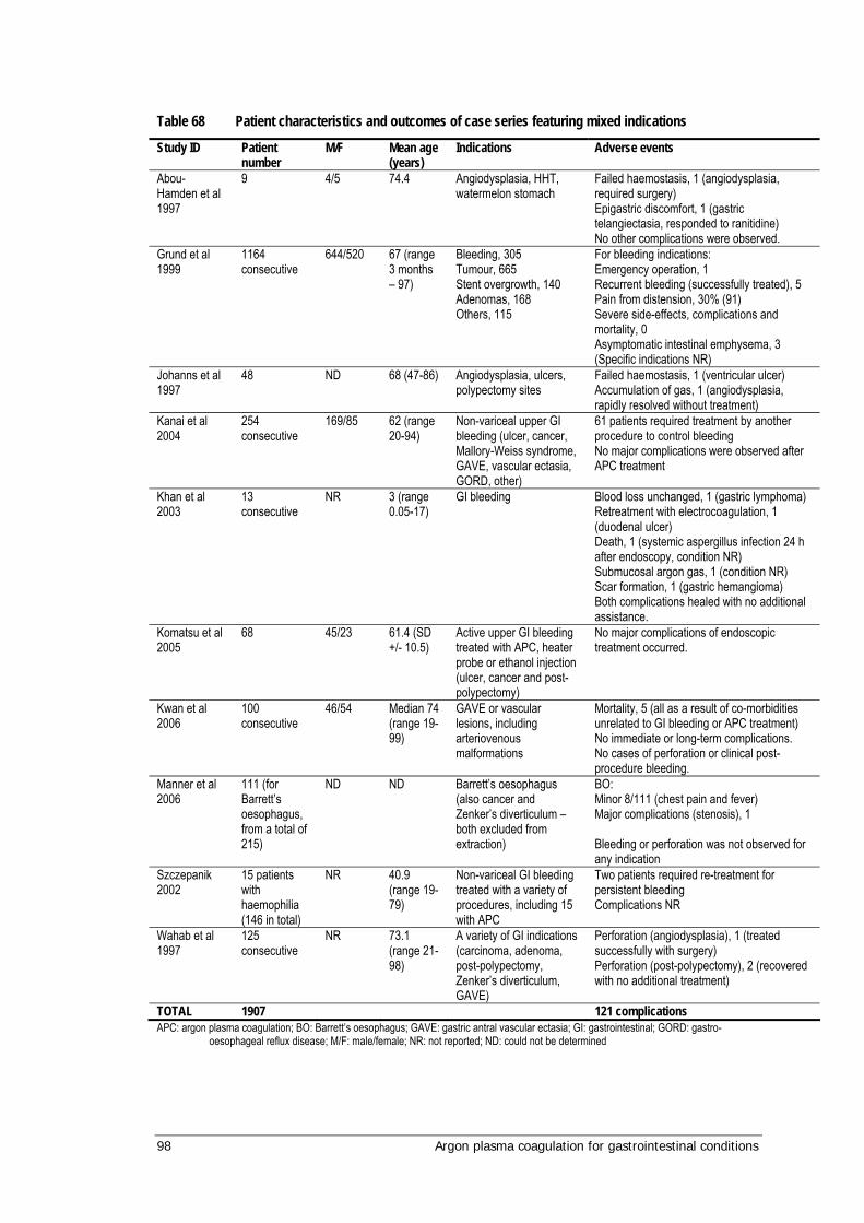
98 Argon plasma coagulation for gastrointestinal conditions
Table 68 Patient characteristics and outcomes of case series featuring mixed indications
Study ID Patient number
M/F Mean age (years)
Indications Adverse events
Abou-Hamden et al 1997
9 4/5 74.4 Angiodysplasia, HHT, watermelon stomach
Failed haemostasis, 1 (angiodysplasia, required surgery) Epigastric discomfort, 1 (gastric telangiectasia, responded to ranitidine) No other complications were observed.
Grund et al 1999
1164 consecutive
644/520 67 (range 3 months – 97)
Bleeding, 305 Tumour, 665 Stent overgrowth, 140 Adenomas, 168 Others, 115
For bleeding indications: Emergency operation, 1 Recurrent bleeding (successfully treated), 5 Pain from distension, 30% (91) Severe side-effects, complications and mortality, 0 Asymptomatic intestinal emphysema, 3 (Specific indications NR)
Johanns et al 1997
48 ND 68 (47-86) Angiodysplasia, ulcers, polypectomy sites
Failed haemostasis, 1 (ventricular ulcer) Accumulation of gas, 1 (angiodysplasia, rapidly resolved without treatment)
Kanai et al 2004
254 consecutive
169/85 62 (range 20-94)
Non-variceal upper GI bleeding (ulcer, cancer, Mallory-Weiss syndrome, GAVE, vascular ectasia, GORD, other)
61 patients required treatment by another procedure to control bleeding No major complications were observed after APC treatment
Khan et al 2003
13 consecutive
NR 3 (range 0.05-17)
GI bleeding Blood loss unchanged, 1 (gastric lymphoma) Retreatment with electrocoagulation, 1 (duodenal ulcer) Death, 1 (systemic aspergillus infection 24 h after endoscopy, condition NR) Submucosal argon gas, 1 (condition NR) Scar formation, 1 (gastric hemangioma) Both complications healed with no additional assistance.
Komatsu et al 2005
68 45/23 61.4 (SD +/- 10.5)
Active upper GI bleeding treated with APC, heater probe or ethanol injection (ulcer, cancer and post-polypectomy)
No major complications of endoscopic treatment occurred.
Kwan et al 2006
100 consecutive
46/54 Median 74 (range 19-99)
GAVE or vascular lesions, including arteriovenous malformations
Mortality, 5 (all as a result of co-morbidities unrelated to GI bleeding or APC treatment) No immediate or long-term complications. No cases of perforation or clinical post-procedure bleeding.
Manner et al 2006
111 (for Barrett’s oesophagus, from a total of 215)
ND ND Barrett’s oesophagus (also cancer and Zenker’s diverticulum – both excluded from extraction)
BO: Minor 8/111 (chest pain and fever) Major complications (stenosis), 1 Bleeding or perforation was not observed for any indication
Szczepanik 2002
15 patients with haemophilia (146 in total)
NR 40.9 (range 19-79)
Non-variceal GI bleeding treated with a variety of procedures, including 15 with APC
Two patients required re-treatment for persistent bleeding Complications NR
Wahab et al 1997
125 consecutive
NR 73.1 (range 21-98)
A variety of GI indications (carcinoma, adenoma, post-polypectomy, Zenker’s diverticulum, GAVE)
Perforation (angiodysplasia), 1 (treated successfully with surgery) Perforation (post-polypectomy), 2 (recovered with no additional treatment)
TOTAL 1907 121 complications APC: argon plasma coagulation; BO: Barrett’s oesophagus; GAVE: gastric antral vascular ectasia; GI: gastrointestinal; GORD: gastro-
oesophageal reflux disease; M/F: male/female; NR: not reported; ND: could not be determined
Argon plasma coagulation for gastrointestinal conditions 99
What are the economic considerations?
Economic evaluation of new health care technologies is important when determining whether the new initiative offered additional benefits and at what cost. Economic evaluations are able to determine whether the new initiative is dominated by (or dominates) the existing technology, such that the costs are higher (lower) and the effectiveness is less (greater). Economic evaluation is particularly important where the new initiative offers health benefits at additional costs. Within a constrained health care budget, the additional cost that would be paid for a given health gain should be determined in order to ascertain whether such incremental costs represent value for money.
The usual process for an economic evaluation is first to determine the incremental effectiveness, which is the additional benefits associated with the new technology relative to current practice. Secondly, to determine the incremental costs, this is the difference in costs between the new initiative and current practice. Finally the incremental cost-effectiveness ratio (ICER) can be calculated using the following ratio:
Restrictions of the economic evaluation
It was decided by the Advisory Panel that the economic evaluation of APC would be limited to indications with the strongest available evidence, in terms of effectiveness, as measured by randomised controlled trials. For this reason the economic evaluation of argon plasma coagulation (APC) is restricted to its use in the treatment of bleeding peptic ulcers and Barrett’s oesophagus.
Search strategies
As described in the ‘approach to assessment’, a search strategy was developed to systematically identify studies in which APC was used in the treatment of Barrett’s oesophagus and bleeding peptic ulcers.
Databases of peer-reviewed literature including Medline, PubMed, CINAHL and Cochrane have been searched. The bibliographies of all retrieved publications were hand-searched for any relevant references missing in the database search. Web-based searches included the Internet engines ‘Google’ and ‘Google scholar’.
In addition to the search terms described in the ‘approach to assessment’ section, Cost$ or Econ$ were added. This was to identify any published cost-effectiveness analysis. The inclusion and exclusion criteria remained the same.
Cost New – Cost Comparator Effectiveness New – Effectiveness Comparator
ICER =
100 Argon plasma coagulation for gastrointestinal conditions
Barrett’s oesophagus
Background
Three comparative RCTs comparing APC for the ablation of Barrett’s oesophagus to an alternative technique were identified through the systematic literature review. Three other comparative studies were rejected because the comparator, photodynamic therapy, is not an MBS-listed item. None of the studies include a cost-effectiveness analysis. Therefore, for the purpose of this cost analysis, the effectiveness data is extracted from the aforementioned studies and cost parameters are estimated using current Australian data. Of the three RCTs reviewed in the earlier section, two studies compared APC with multipolar electrocoagulation (MPEC) (Dulai et al 2005, Sharma et al 2006), whilst the other study compared APC with conservative surveillance (Ackroyd et al 2004). The effectiveness results have been previously summarised in Table 23. APC demonstrated improved effectiveness when compared to conservative surveillance and APC appears to be as effective as MPEC for ablation of Barrett’s oesophagus.
Rationale for the cost analysis
The Advisory Panel decided that Barrett’s oesophagus reversal was the primary clinical endpoint, and that MPEC would be the comparator for the cost analysis.
As previously discussed, no significant differences in the Barrett’s oesophagus reversal were demonstrated between the two treatment options. Consequently, until more data are published supporting the superior effectiveness of either APC or MPEC for the ablation of Barrett’s oesophagus, a cost-effectiveness analysis is not warranted. Therefore, the aim of the present economic evaluation will be to review the costs of APC ablation compared to MPEC for the treatment of patients with Barrett’s oesophagus when these interventions are provided under Australian conditions, and to provide an indication of the extent of uncertainty.
Assumptions
• Reversal of Barrett’s oesophagus with low-grade dysplasia is used as the primary outcome.
• Effectiveness data is obtained from two studies comparing APC with multipolar electrocoagulation (MPEC) (Dulai et al 2005; Sharma et al 2006).
• The majority of patients in both studies had non-dysplastic Barrett’s oesophagus, which in Australia would be treated using acid suppression therapy. Clinical experts advised that it would be appropriate to assume that the safety and effectiveness from these studies would be similar to the use of APC in the treatment of low-grade dysplastic Barrett’s oesophagus.
• A conservative estimate of the total number of patients who would be treated with APC has been used, based on the total number of patients diagnosed with Barrett’s oesophagus. Only a small proportion of these patients would have low-grade dysplasia and therefore would be considered for ablative treatment such as APC or MPEC. An exact estimate of this number was unavailable.

Argon plasma coagulation for gastrointestinal conditions 101
• It is assumed that the intermediate reversal of Barrett’s oesophagus reported by Dulai et al (2005) and the 24 month reversal of Barrett’s oesophagus reported by Sharma et al (2006) are comparable.
• The perspective of the cost analysis is limited to the costs faced by the health care system.
• A discount rate of 5 per cent per annum was applied to all costs
Estimate of effectiveness
No significant differences in reversal of Barrett’s oesophagus were found in the two studies comparing APC to MPEC. Dulai et al (2005) and Sharma et al (2006) found 88 (63 %) of APC patients to have successful reversal immediately after their final treatment session, compared to 96 (75 %) of MPEC patients, P=0.64 (p=0.49). The meta-analysis conducted noted that there was no statistical difference in the ablation of Barrett’s oesophagus with MPEC or APC, and relative risk of 0.89 in favour of MPEC (P=0.22) was reported.
The number of endoscopic procedures required for ablation was comparable across both studies. Sharma et al (2006) reported no significant difference between the treatment groups (APC=3 versus MPEC=4, P=0.76). The Dulai et al (2005) study suggested a difference favouring MPEC over APC (3.0 versus 3.9 sessions, p=0.05); however, this was non-significant after correction for baseline Barrett’s oesophagus length (3.6 versus 3.17 sessions, P=0.249). It is worth noting that patients in Dulai et al (2006) differed significantly by length of Barrett’s oesophagus at baseline. Those in the MPEC group had a mean Barrett’s oesophagus segment of 3.1 cm (SD 1.7) compared to those in the APC group who had a mean of 4.0 cm (SD 1.5).
Because of the lack of evidence supporting improved effectiveness (clinical or procedural) of APC or MPEC, for the purpose of this cost-analysis the assumption is that clinical outcomes are identical.
Estimate of costs
Average capital costs per procedure are calculated in the next section (bleeding ulcers, Table 72). For the basis of the analysis, average capital costs for APC are estimated based upon the average number of procedures (175 per annum) over the estimated lifetime of the machine (A$68 per procedure). For the sensitivity analysis, the lower estimate is A$52 per procedure based upon 200 procedures per annum over 10 years and the upper estimate is A$98 per procedure based upon 150 procedures per annum over 6 years. As discussed in the economic evaluation of bleeding ulcers, the number of patients per year is dependent on the number of indications (if any) that APC is funded for. Consequently, the average capital cost could be higher or lower than these estimates.
There are no capital costs associated with MPEC, since the Gold probe 7Fr can be adapted to any endoscope (advice from Boston Scientific). Table 69 presents the Medicare Benefits Schedule (MBS) item numbers associated with APC, MPEC and endoscopy for patients with Barrett’s oesophagus.
The direct treatment costs per procedure are estimated in Table 70. It is assumed that all patients undergo one upper GI endoscopy to determine whether the treatment is

102 Argon plasma coagulation for gastrointestinal conditions
successful. Since all patients undergo this procedure the incremental cost is zero. The APC (MPEC) procedure included the proposed fee as estimated by the applicant (MBS 30478 for MPEC), anaesthetic fee (MBS 20740 and 23023) and additional resources (day theatre, pharmaceutical and histology). Estimated cost of the disposable APC probe was provided by the applicant.
Table 69 Medicare benefit schedules for endoscopic associated items
MBS Item No.
Schedule fee (100%) (A$)
Definition
20740 87.50 INITIATION OF MANAGEMENT OF ANAESTHESIA for upper gastrointestinal endoscopic procedures
23023 35.00 Time unit cost for anaesthesia for a 26 to 30 MINUTES
30473 156.50 OESOPHAGOSCOPY (not being a service to which item 41816 or 41822 applies), GASTROSCOPY, DUODENOSCOPY or PANENDOSCOPY (1 or more such procedures), with or without biopsy, not being a service associated with a service to which item 30476 or 30478 applies
30478 217.00 OESOPHAGOSCOPY (not being a service to which item 41816, 41822 or 41825 applies), gastroscopy, duodenoscopy or panendoscopy (1 or more such procedures), with 1 or more of the following endoscopic procedures - polypectomy, removal of foreign body, diathermy, heater probe or laser coagulation, or sclerosing injection of bleeding upper gastrointestinal lesions, not being a service associated with a service to which item 30473 or 30476 applies
MBS: Medical benefits schedule.
Table 70 Average incremental costs per patient of disposables in performing ablation of Barrett’s oesophagus with MPEC or APC – (Base case)
MPEC APC
Consumables Units/patient Cost A$ Units/patient Cost A$
Incremental cost of APC patient
Upper GI endoscopy
Anaesthetic (MBS 20740 and 23023) 3.4 $122.50 3.4 $122.50
Additional resource (Weller et al) c 3.4 $701.10 3.4 $701.10
Total cost per patient $823.60 $823.60 0
Probes
Gold probe™ a 3.4 $380.00 3.4
Disposable APC b 3.4 $300.00 $-272.00
MBS fee
MBS 30478 3.4 $217.00
Proposed fee b 3.4 $312.30 $324.02
Capital cost (including opportunity cost) 0 0 3.4 $68.00 $231.20
Incremental cost APC per patient $283.22 aGold probe™ Electrohaemostasis catheters (for single use only) Boston Scientific bProvided by the applicant. c Additional resources include,
day theatre, pharmaceuticals and histology (adapted from MSAC 1057). APC: argon plasma coagulation; MPEC: multipolar electrocoagulation; GI: gastrointestinal; MBS: medical benefits schedule.

Argon plasma coagulation for gastrointestinal conditions 103
Cost-analysis
The cost analysis of the base case scenario is based upon the weighted average effectiveness data obtained from the two RCTs comparing APC with MPEC. This demonstrates that the incremental cost per patient of receiving APC rather than MPEC for the ablation of Barrett’s oesophagus is $283. The bulk of this additional cost is associated with the higher procedural fee ($312 versus $217) and the additional capital cost of buying the APC equipment. These costs are partially offset by a saving in the costs of the relevant probes ($300 versus $380). The estimated average number of procedures also influences the results, in effect multiplying the incremental cost difference of the procedure to give the cost per patient.
To estimate the cost per annum to the health care system of replacing MPEC with APC, the number of patients with Barrett’s oesophagus was estimated from the AIHW National Hospital Morbidity Database (http://www.aihw.gov.au). In 2005, the number of patients treated for gastro-oesophageal reflux disease (K21) was 57,923. It is estimated that 10 per cent (5,792) of these patients would have been treated for Barrett’s oesophagus (expert opinion). These data refer to public hospitals; it is assumed that a similar number of patients are treated in a private hospital per year. Based on the base case model, the total additional cost of treating Barrett’s oesophagus patients with APC is $1,633,000 per annum. This figure is estimated from the total number of patients who might be diagnosed with Barrett’s oesophagus in Australia. However, as mentioned previously, only a small proportion of these patients would be considered for ablative treatment, therefore the actual cost to the healthcare system is likely to be much lower.
However, it should be noted that the majority of patients diagnosed with Barrett’s oesophagus are non-dysplastic and as such would not be considered for ablative procedures. These patients would be treated with acid suppression medication. In addition, patients with the more severe highly dysplastic form of the disease would be treated with more aggressive therapies such as endoscopic mucosal resection or oesophagectomy. Therefore, only a small portion of the patients diagnosed with Barrett’s oesophagus would have low-grade dysplasia and would be considered for ablative therapy (such as MPEC or APC) (Bergman and Fockens 2006). The exact proportion of patients who would be indicated for ablative therapy was unavailable, and therefore the higher, more conservative figure has been used.
Sensitivity analysis
The incremental cost per additional patient with reversal in Barrett’s oesophagus is highly sensitive to the number of procedures per patient, the estimated procedural fee, the cost of the disposable probes and to a lesser extent the inclusion of capital costs. There is more certainty regarding the cost of the APC probe, capital costs and procedural fee, all of which were provided by the applicant.
In the base case model we assumed (because of non-statistical significance) that there is no difference in the effectiveness in terms of primary outcome or no difference in the number of procedures per patient required to achieve this outcome. The results are highly sensitive to differences in the number of procedures. For example in the base case model, the estimated average number of procedures per patient was 3.4 independent of whether the procedure was performed using APC or MPEC. If we relax this assumption, small differences in the average number of procedures between APC and MPEC will alter the incremental cost per patient noticeably. For example, if the average number of procedures required to achieve significant ablation is 3.3 for APC and 3.5 MPEC (-0.2
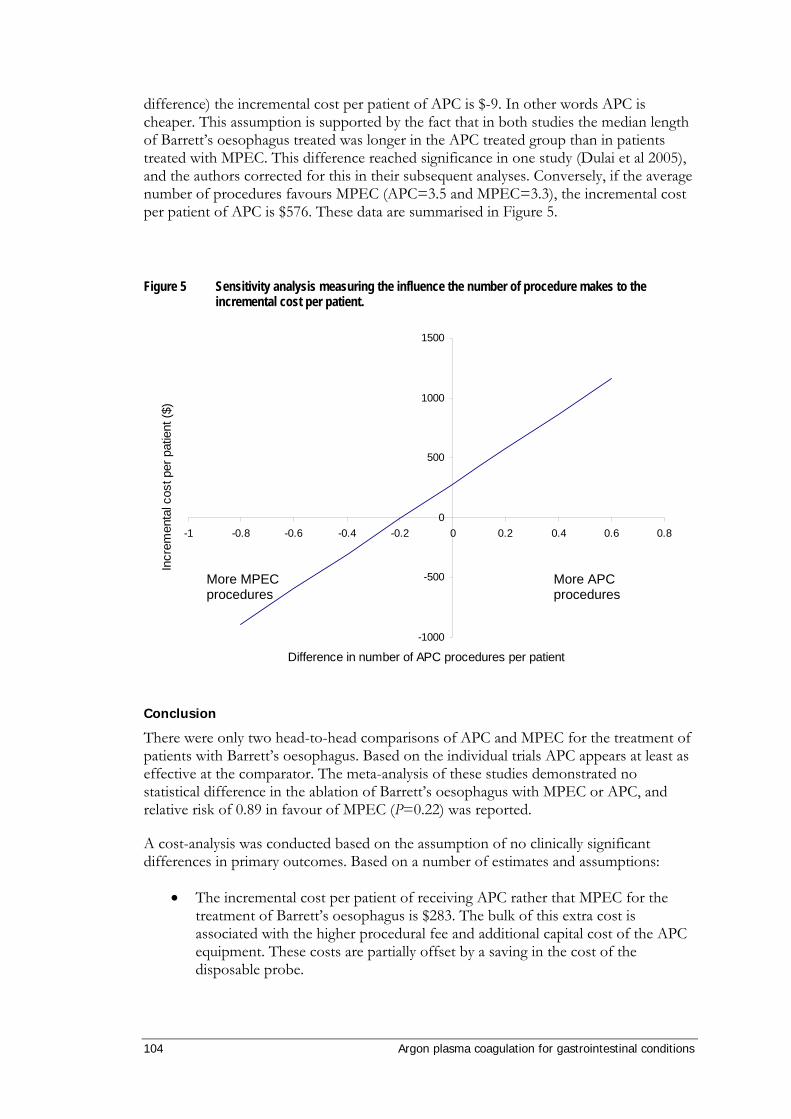
104 Argon plasma coagulation for gastrointestinal conditions
difference) the incremental cost per patient of APC is $-9. In other words APC is cheaper. This assumption is supported by the fact that in both studies the median length of Barrett’s oesophagus treated was longer in the APC treated group than in patients treated with MPEC. This difference reached significance in one study (Dulai et al 2005), and the authors corrected for this in their subsequent analyses. Conversely, if the average number of procedures favours MPEC (APC=3.5 and MPEC=3.3), the incremental cost per patient of APC is $576. These data are summarised in Figure 5.
Figure 5 Sensitivity analysis measuring the influence the number of procedure makes to the incremental cost per patient.
-1000
-500
0
500
1000
1500
-1 -0.8 -0.6 -0.4 -0.2 0 0.2 0.4 0.6 0.8
Difference in number of APC procedures per patient
Incr
emen
tal c
ost p
er p
atie
nt ($
)
Conclusion
There were only two head-to-head comparisons of APC and MPEC for the treatment of patients with Barrett’s oesophagus. Based on the individual trials APC appears at least as effective at the comparator. The meta-analysis of these studies demonstrated no statistical difference in the ablation of Barrett’s oesophagus with MPEC or APC, and relative risk of 0.89 in favour of MPEC (P=0.22) was reported.
A cost-analysis was conducted based on the assumption of no clinically significant differences in primary outcomes. Based on a number of estimates and assumptions:
• The incremental cost per patient of receiving APC rather that MPEC for the treatment of Barrett’s oesophagus is $283. The bulk of this extra cost is associated with the higher procedural fee and additional capital cost of the APC equipment. These costs are partially offset by a saving in the cost of the disposable probe.
More APC procedures
More MPEC procedures
Argon plasma coagulation for gastrointestinal conditions 105
• A conservative estimate of the total number of patients who would be treated with APC has been used, based on the total number of patients diagnosed with Barrett’s oesophagus. Only a small proportion of these patients would have low-grade dysplasia and therefore would be considered for ablative treatment such as APC or MPEC.
• Based on these estimates, the total additional cost to the health care system of treating Barrett’s oesophagus patients with APC is $1,633,000 per annum. This figure is estimated from the total number of patients who might be diagnosed with Barrett’s oesophagus in Australia. However, as mentioned previously, only a small proportion of these patients would be considered for ablative treatment, therefore the actual cost to the healthcare system is likely to be much lower.
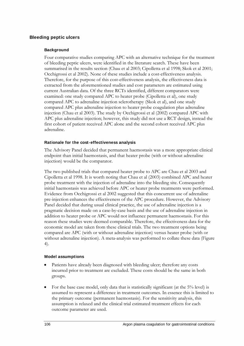
106 Argon plasma coagulation for gastrointestinal conditions
Bleeding peptic ulcers
Background
Four comparative studies comparing APC with an alternative technique for the treatment of bleeding peptic ulcers, were identified in the literature search. These have been summarised in the results section (Chau et al 2003; Cipolletta et al 1998; Skok et al 2001; Occhigrossi et al 2002). None of these studies include a cost-effectiveness analysis. Therefore, for the purpose of this cost-effectiveness analysis, the effectiveness data is extracted from the aforementioned studies and cost parameters are estimated using current Australian data. Of the three RCTs identified, different comparators were examined: one study compared APC to heater probe (Cipolletta et al), one study compared APC to adrenaline injection sclerotherapy (Skok et al), and one study compared APC plus adrenaline injection to heater probe coagulation plus adrenaline injection (Chau et al 2003). The study by Occhigrossi et al (2002) compared APC with APC plus adrenaline injection; however, this study did not use a RCT design, instead the first cohort of patient received APC alone and the second cohort received APC plus adrenaline.
Rationale for the cost-effectiveness analysis
The Advisory Panel decided that permanent haemostasis was a more appropriate clinical endpoint than initial haemostasis, and that heater probe (with or without adrenaline injection) would be the comparator.
The two published trials that compared heater probe to APC are Chau et al 2003 and Cipolletta et al 1998. It is worth noting that Chau et al (2003) combined APC and heater probe treatment with the injection of adrenaline into the bleeding site. Consequently initial haemostasis was achieved before APC or heater probe treatments were performed. Evidence from Occhigrossi et al 2002 suggested that this concurrent use of adrenaline pre-injection enhances the effectiveness of the APC procedure. However, the Advisory Panel decided that during usual clinical practice, the use of adrenaline injection is a pragmatic decision made on a case-by-case basis and the use of adrenaline injection in addition to heater probe or APC would not influence permanent haemostasis. For this reason these studies were deemed comparable. Therefore, the effectiveness data for the economic model are taken from these clinical trials. The two treatment options being compared are APC (with or without adrenaline injection) versus heater probe (with or without adrenaline injection). A meta-analysis was performed to collate these data (Figure 4).
Model assumptions
• Patients have already been diagnosed with bleeding ulcer; therefore any costs incurred prior to treatment are excluded. These costs should be the same in both groups.
• For the base case model, only data that is statistically significant (at the 5% level) is assumed to represent a difference in treatment outcomes. In essence this is limited to the primary outcome (permanent haemostasis). For the sensitivity analysis, this assumption is relaxed and the clinical trial estimated treatment effects for each outcome parameter are used.

Argon plasma coagulation for gastrointestinal conditions 107
• Adrenaline injection does not influence the primary outcome.
• The clinical trials comparing heater probe to APC (Chau et al 2003; Cipolletta et al 1998) are assumed to be comparable (expert opinion). Consequently a meta-analysis was conducted collating these data.
• Permanent haemostasis equates to haemostasis at the end of the trial follow-up period (8 weeks for Chau et al 2003 and 5 months for Cipolletta et al 1998). No inference is made for haemostasis beyond this period.
• If there was a recurrence of bleeding post-treatment, the same endoscopic treatment (APC or heater probe) was repeated.
• The perspective of the cost-effectiveness analysis is limited to the costs faced by the health care system.
• A discount rate of 5 per cent per annum was applied to all costs.
Estimates of effectiveness
Chau et al (2003) reported significantly higher rates of permanent haemostasis (at 8-week follow-up) in patients treated with APC plus adrenaline injection than those treated with heater probe plus adrenaline injection (94.2% versus 78.3%, P=0.03). However, this evidence should be treated with caution since significance was lost after correction for the multiple testing of data arising from individual patients. Cipolletta et al (1998) reported on the same clinical outcomes as Chau et al (2003) but found no significant differences between APC and heater probe treatment (90.5% versus 85.0%, P=ns).
Few differences in clinical or procedural outcomes were found between APC and heater probe in the two RCTs. Cipolletta et al (1998) found APC to achieve haemostasis significantly quicker than heater probe treatment. However, for the purpose of the cost-effectiveness analysis this difference is inconsequential.
Maximum follow-up amongst the RCTs ranged from 8 weeks (Chau et al 2003) to 9 months (Cipolletta et al 1998). Cipolletta et al (1998) had no losses to follow-up; Chau et al (2003) lost 41 per cent (36/87) of patients from the APC patient group and 38 per cent (37/97) from the comparator group at 8-week follow-up endoscopy, but did not report reasons for these losses.
As reported earlier, there were no significant differences in the recorded treatment-related mortality. Chau et al (2003) reported 1 per cent (1/88) amongst APC plus adrenaline injection patients and 2 per cent (2/97) in heater probe plus adrenaline injection patients and Cipolletta et al (1998) reported 5 per cent treatment-related mortality in both APC (1/21) and heater probe (1/20) patient groups.
The results of the meta-analysis are reported in Table 71 and details of the analysis can be found in Appendix J. The rate of permanent haemostasis in the APC patient groups is significantly higher than in the heater probe patient group (93.2% versus 80.0%, P=0.02). This converts into a relative risk of 1.16 in favour of APC. No differences were noted in the rates of initial haemostasis (95.7% versus 97.2%, P=0.52) or the mean number of procedures required per patient (1.2 versus 1.1, P=0.43). APC appeared to demonstrate beneficial procedural outcomes in terms of the mean number of blood transfusions

108 Argon plasma coagulation for gastrointestinal conditions
required (1.7 versus 2.3, p=0.13), mean number of nights in hospital (7.2 versus 8.3, p=0.12) and rate of emergency surgery (8.3% versus 10.3%, p=0.19). However, none of these outcomes achieved statistical significance at the 5 per cent level. Therefore, in the base case model only permanent haemostasis was assumed to differ between treatment options. All other outcomes were included as weighted averages. For the sensitivity analysis, this assumption was relaxed and the outcomes in Table 71 were used.
Table 71 Summary of effectiveness data – based on meta-analysis
Heater probe % (mean)
APC % (mean)
RR (95% CI) WMD* (95% CI)
P value
Primary outcome
Permanent haemostasis 80.0% 93.2% 1.16 (1.03.1.32) 0.02
Other outcomes
Initial haemostasis 97.2% 95.7% 1.02 (0.97, 1.07) 0.52
Number of endoscopies (1) (1) - -
Number of procedures (1.1) (1.2) - 0.43
Blood transfusions (2.3) (1.7) -0.29* (-0.68, 0.09) 0.13
Number of nights in hospital (8.3) (7.2) -1.05* (-2.40, 0.29) 0.12
Emergency surgery 10.3% 8.3% 1.02 (0.97, 1.07) 0.19 APC: argon plasma coagulation; CI: confidence interval; RR: relative risk; WMD: weighted mean difference
Estimates of costs
Average capital costs per procedure
Average capital costs per procedure are based on estimates of the purchase price of equipment (argon gas source, high frequency electrosurgical unit and foot switches), life of equipment, maintenance and number of procedures performed per annum. These estimates were provided by the applicant (Table 72). Comparable capital cost estimates for heater probe are given in Table 73. The opportunity cost of capital was included with the forgone capital return calculated using a 5 per cent discount rate. The values are sensitive to the number of procedures per annum. APC is applicable to a number of different indications; therefore, the number of procedures per annum may be higher than those suggested. Conversely, if APC is restricted to a limited number of indications, the number of procedures per annum is likely to be an over-estimate1.
For the basis of the analysis, average capital costs for APC are estimated based upon the average number of procedures (175 per annum) over the estimate lifetime of the machine (A$68 per procedure). For the sensitivity analysis, the lower estimate is A$52 per procedure based upon 200 procedures per annum over 10 years and the upper estimate is A$98 per procedure based upon 150 procedures per annum over 6 years.
1 For example, based on 25 patients per year, the average capital cost of APC per procedure would be $479 (over 8 years). Conversly, based on 300 patients per year the average capital cost would be $40 per procedure.

Argon plasma coagulation for gastrointestinal conditions 109
Average capital costs for heater probe are estimated based upon the average number of procedures (175 per annum) over the estimate life-time of the machine (A$15 per procedure). For the sensitivity analysis, the lower estimate is A$12 per procedure based upon 200 procedures per annum over 10 years and the upper estimate is A$22 per procedure based upon 150 procedures per annum over 6 years.
Table 72 Calculation of average capital costs per procedure for APC
Item Cost $A (range)
Cost $A +GST (range)
Life (range) Annual cost $A /machine (range)
Purchase price of APC* 60,000 66,000 8 (6-10 years)
$8,250 (6,600 – 11,000)
Foregone capital return 5% of $66,000 Annual $3,300
Maintenance 3 years warranty, thereafter $850 per year*** $417 (350-446)
Total opportunity cost of capital $11,967 (10,346 – 14,650)
Average cost based on estimated procedures/machine/year**
150 175 200
$80 (69 – 98) $68 (59 – 84) $60 (52 – 73)
* Cost of major capital equipment provided by applicant, ** estimated procedures per year provided by applicant. *** maintenance cost provided by applicant, three years warranty thereafter discounted at 5%; APC: argon plasma coagulation; GST: goods and services tax.
Table 73 Calculation of average capital costs per procedure for heater probe
Item Cost $A (range)
Cost $A +GST (range)
Life (range) Annual cost $A /machine (range)
Purchase price of HeatProbe unit (HPU-20)*
12,700 13,970 8 (6-10 years)
$1,746 (1,397 – 2,328)
Foregone capital return 5% of $14,472 Annual $699
Maintenance 3 years warranty, thereafter $500 per year $245 (206-293)
Total opportunity cost of capital $2690 (2,389 – 3,233)
Average cost based on estimated procedures/machine/year**
150 175 200
$18 (16 – 22) $15 (14 – 18) $13 (12 – 16)
* Cost of major capital equipment provided by Olympus Australia, ** estimated procedures per year provided by applicant. *** maintenance costs are estimated, three years warranty thereafter discounted at 5%; GST: goods and services tax.

110 Argon plasma coagulation for gastrointestinal conditions
Direct treatment costs per procedure
The direct treatment costs per procedure are estimated in Table 74. It is assumed that all patients undergo one upper GI endoscopy to determine whether the treatment is successful. Since all patients undergo this procedure the incremental cost is zero. The APC (heater probe) procedure included the proposed fee as estimated by the applicant (MBS 30478 for heater probe), anaesthetic fee (MBS 20745 and 23023) and additional resources (day theatre, pharmaceutical and histology). Estimated cost of the disposable APC probe was provided by the applicant. The costs of a reuseable probe, and the associated cleaning costs, were used for assessing the costs of the heater probe. A disposable probe (Gold Probe, A$300, Boston Scientific) could be used as an alternative. The cost of a blood transfusion was taken from MSAC report 1057 ‘M2A® capsule endoscopy for the evaluation of obscure gastrointestinal bleeding in adult patients’ and inflated to current prices. Emergency surgery costs were derived from AR-DRG (G62Z) and the cost per hospital days was derived from AR-DRG (G62Z) and adjusted to reflect the higher cost weighting during the first couple of days in hospital.
Cost-effectiveness
The cost-effectiveness analysis of the base case scenario is based upon the meta-analysed effectiveness data. This demonstrates that the incremental cost per patient of receiving APC rather than heater probe treatment for bleeding peptic ulcer is $343. The bulk of this additional cost is associated with the disposable APC probe and estimated higher procedural fee. Based on an estimated 13.2 per cent improvement in effectiveness (permanent haemostasis), the incremental cost-effectiveness per additional patient with permanent haemostasis is $2606.
Given the choice, patients may prefer the use of a disposable probe rather than a re-useable probe which may have been used on more than one previous occasion. If a disposable probe is used for heater probe, this would reduce the incremental cost of APC per patient in the base case (Table 72). For example, the incremental cost per patient receiving APC instead of heater probe under these conditions is $-61. This means the cost of APC is lower than the cost of heater probe. Under these assumptions APC is less costly and more effective than heater probe, therefore, APC dominates heater probe. Based on an estimated 13.2 per cent improvement in effectiveness (permanent haemostasis), the incremental cost-effectiveness per additional patient with permanent haemostasis is $-466.
To estimate the cost per annum to the health care system of replacing heater probe with APC, the number of patients with peptic and gastric ulcers were estimated from the AIHW National Hospital Morbidity Database (http://www.aihw.gov.au). In 2005, the number of patients treated for haemorrhagic, not perforated, gastric or peptic ulcers (K25.0, K25.4, K27.0, and K27.4) was 2560. The number of patients treated for all bleeding ulcers (not perforated) was 4241. These data refer to public hospitals; it is assumed that a similar number of patients are treated in a private hospital per year. Based on the base case model, the total additional cost of treating all haemorrhagic gastric and peptic ulcers (all bleeding ulcers) with APC is $877,286 ($1,453,348). This would yield 337 (597) more patients with permanent haemostasis.

Argon plasma coagulation for gastrointestinal conditions 111
Table 74 Average incremental costs per patient of performing peptic ulcer repair (base case)
Heater probe (HP) APC
Consumables Units/patient Cost A$ Units/patient Cost A$
Incremental cost of APC patient
Upper GI endoscopy
Procedure (MBS 30473) 1 $156.50 1 $156.50
Anaesthetic (MBS 20745 and 23010)
1 $122.50 1 $122.50
Additional resource a (Weller et al)
1 $624.35 1 $624.35
Total cost per patient 1 $980.10 1 $980.10 $0
Procedure (APC or HP)
MBS 30478 1.15 $217.00
Proposed fee 1.15 $312.30 b $109.60
Anaesthetic (MBS 20745 and 23023)
1.15 $140.00 1.15 $140.00 $0
Additional resources a (Weller et al)
1.15 $624.35 1.15 $624.35 $0
Probes
Re-usable (HeatProbe) c 1.15 $100.32
Cleaning re-usable probe 1.15 $50.00
Disposable APC 1.15 $300.00 d $172.13
Capital cost (including opportunity cost)
1.15 $16.00 1.15 $68.00 $60.96
Other expenditures
Blood transfusions 2.05 $73.18 2.05 $73.18 $0
Days in hospital e 7.78 $517.50 7.78 $517.50 $0
Emergency surgery e 0.09 $8524.41 0.09 $8524.41 $0
Incremental cost APC per patient $342.69
Primary Outcome
Permanent Haemostasis 80.0% 93.15% -13.15%
Cost-effectiveness ($ per additional patient with permanent haemostasis) $2605.88 a Additional resources include, day theatre, pharmaceuticals and histology (adapted from MSAC 1057); b Provided by Applicant; c Cost of re-
useable probe based on 5 uses, cleaning costs estimated by expert advice; d Provided by Olympus Australia; e Derived from AR-DRG (G62Z); APC: argon plasma coagulation; HP: heater probe; GI: gastrointestinal; MBS: Medicare benefits schedule.
Sensitivity analysis
The incremental cost-effectiveness per additional patient with permanent haemostasis is highly sensitive to the cost of the disposable APC probe, estimated procedural fee and estimated improved effectiveness. There is more certainty regarding the cost of the APC probe and procedural fee, both of which were provided by the applicant. The results are robust to the higher and lower estimates for the capital cost of equipment.
Taking into account the uncertainty surrounding the effectiveness of APC was achieved by one-way sensitivity analysis. The base case assumed a 13.2 per cent improvement in permanent haemostasis. This gave an incremental cost-effectiveness per additional patient with permanent haemostasis of $2606. Using the data from the individual clinical
112 Argon plasma coagulation for gastrointestinal conditions
trials, the more pessimistic outcome was estimated by Cipolletta et al (1998). In this study APC yielded an estimated improvement in permanent haemostasis of 5.5 per cent. When this lower value is used, the incremental cost-effectiveness per additional patient with permanent haemostasis is $6231.
Figure 6 presents the incremental cost-effectiveness per additional patient with permanent haemostasis against different values of improvement in APC effectiveness. The graph demonstrates that if the estimated improvement in effectiveness of APC is less than 1per cent, the corresponding incremental cost-effectiveness per additional patient with permanent haemostasis will be over $40,000.
Figure 6 The incremental cost-effectiveness per additional patient with permanent haemostasis against the effectiveness of APC
Cost-effectiveness per additional patient with permanent haemostasis
0
20000
40000
60000
80000
100000
120000
0 2 4 6 8 10 12 14 16
Improved effectiveness of APC relative to heater probe (% improvement)
ICER
($)
APC: argonplasma coagulation; ICER: incremental cost-effectiveness ratio
The sensitivity analysis was extended to model other differences in effectiveness between the two procedures. For example, the number of emergency surgery procedures required was higher when using heater probe than APC (10.3% versus 8.3%). These variables are presented in Table 71. Using these values, the incremental cost per patient receiving APC instead of heater probe was A$-335. This means the cost of APC is lower than the cost of heater probe coagulation. Under these assumptions APC is less costly and more effective than heater probe; therefore, APC dominates heater probe (Table 75).
These estimates are influenced by the costs and frequency of blood transfusions, length of hospital stay and emergency surgery. On average the APC treatment groups require 0.6 less transfusions, 1.2 less hospital days and 2 per cent less emergency surgery procedures. It is worth noting that these differences are not significant; therefore, further work is required to determine whether these trends are indeed potential cost savings.

Argon plasma coagulation for gastrointestinal conditions 113
Table 75 Average incremental costs per patient of performing peptic ulcer repair (including non-significant data)
Heater probe (HP) APC
Consumables Units/patient Cost A$ Units/patient Cost A$
Incremental cost of APC patient
Upper GI endoscopy
Procedure (MBS 30473) 1 $156.50 1 $156.50
Anaesthetic (MBS 20745 and 23010)
1 $122.50 1 $122.50
Additional resource a (Weller et al)
1 $624.35 1 $624.35
Total cost per patient 1 $980.10 1 $980.10 $0
Procedure (APC or HP)
MBS 30478 1.1 $217.00
Proposed fee 1.2 $312.30 b $136.06
Anaesthetic (MBS 20745 and 23023)
1.1 $140.00 1.2 $140.00 $14.00
Additional resources a (Weller et al)
1.1 $624.35 1.2 $624.35 $62.43
Probes
Re-usable (HeatProbe) c 1.1 $100.32
Cleaning re-useble probe 1.1 $50
Disposable APC 1.2 $300.00 d $194.65
Capital cost (including opportunity cost)
1.1 $15.00 1.2 $68.00 $65.15
Other expenditures
Blood transfusions 2.35 $73.18 1.74 $73.18 $-44.65
Days in hospital e 8.34 $517.50 7.19 $517.50 $-592.06
Emergency surgery e 0.103 $8524.41 0.083 $8524.41 $-170.65
Incremental cost APC per patient $-335.07 a Additional resources include, day theatre, pharmaceuticals and histology (adapted from MSAC 1057); b Provided by Applicant; c Cost of re-
useable probe based on 5 uses, cleaning costs estimated by expert advice; d Provided by Olympus Australia; e Derived from AR-DRG (G62Z); APC: argon plasma coagulation; HP: heater probe; GI: gastrointestinal; MBS: Medicare benefits schedule.
Limitations
The estimates of clinical effectiveness are based upon the assumption that the two clinical trails are comparable. As previously noted the validity of the meta-analysis was confounded by one study using adrenaline injection to achieve haemostasis before APC or heater probe treatment. Evidence also suggested that this concurrent use of adrenaline pre-injection enhances the effectiveness of the APC procedure (Occhigrossi et al 2002). Without adrenaline, the results of Chau et al (2003) may have more closely reflected those of Cipolleta et al (1998) who, using APC and heater probe alone, demonstrated a 5.5 per cent benefit; however, this was not significant.
The second caveat is the length of follow-up. In the context of this evaluation, permanent haemostasis means haemostasis at the end of the follow-up period (8 week to 9 months). The inference is made that there is no difference between the recurrences of bleeding for patient treated with heater probe or APC after this period.

114 Argon plasma coagulation for gastrointestinal conditions
The third caveat is the number of patients lost to follow-up in the Chau study (Chau et al 2003). We assume that those patients followed-up and those lost to follow-up are homogenous. Any deviation from this assumption will introduce bias.
Conclusion
There were only two reliable head-to-head comparisons of APC and heater probe for the treatment of patients with bleeding peptic ulcer. Based on the individual trials APC appears at least as effective at the comparator, although both studies demonstrated a tendency favouring APC. Based on the combined meta-analysed data, APC demonstrated greater effectiveness in terms of the primary outcome, permanent haemostasis (APC=93.2% and HP=80.0%). This difference was statistically significant. However, the validity of the meta-analysis was confounded by one study using adrenaline injection to achieve haemostasis before APC and heater probe treatment.
A modelled cost-effectiveness analysis was conducted based on the improved effectiveness of APC as determined by the meta-analysis. Based on a number of estimates and assumptions the cost-effectiveness of APC would be as follows:
• The incremental cost per patient of receiving APC rather that heater probe treatment for bleeding peptic ulcer is $343. The bulk of this extra cost is associated with the APC probe, which is disposable, and estimated higher procedural fee.
• Based on an estimated 13.2 per cent improvement in effectiveness (permanent haemostasis), the incremental cost-effectiveness per additional patient with permanent haemostasis is $2606 (or $6231 for a 5.5% improvement in effectiveness).

Argon plasma coagulation for gastrointestinal conditions 115
Conclusions
The evidence was presented for the use of argon plasma coagulation (APC) in the treatment of seven clinically distinct indications related to the gastrointestinal tract:
• ablation of dysplastic Barrett’s oesophagus
• haemostasis of bleeding ulcers
• haemostasis of gastric antral vascular ectasia (GAVE)
• haemostasis of radiation proctitis
• haemostasis of bleeding angiodysplasia
• coagulation of post-polypectomy bleeding
• ablation of tumourous growth through or over oesophageal metal stents.
In addition to the primary clinical studies included in the review, seven systematic reviews were identified as part of the literature search. Four of the reviews were published in 2005; the earliest was published in 2001. None of the reviews were able to provide a formal conclusion for the safety and effectiveness of the use of APC due to the lack of comparative data. Provided below is a summary of the conclusions of the current systematic review of APC.
Safety
Barrett’s oesophagus
A total of six RCTs were identified which investigated APC for this condition. Of these, three had an MBS-listed procedure as a comparator (two studies used multipolar electrocoagulation (MPEC) and one used conservative therapy). The three remaining RCTs used photodynamic therapy as the comparator, a procedure which is not indicated for this use by Medicare. However, safety outcomes from the APC arm of these studies was used. In addition to the RCTs, 16 case series were identified in which APC had been used to ablate Barrett’s oesophagus. The results suggested that APC was as least as safe as MPEC, and as safe as conservative surveillance. In absolute terms, APC appears to be a relatively safe treatment for Barrett’s oesophagus. The majority of complications were transient and resolved without additional procedures. There were five cases of perforation, which led to deaths of two patients. There did not appear to be a common factor in any of the reported adverse events. However, it must be noted that although 613 patients participated in these studies, patients received multiple treatments (an average of between one and eight); therefore, these complications are as a result of some thousands of uses of APC.

116 Argon plasma coagulation for gastrointestinal conditions
Haemostasis of bleeding ulcers
Four RCTs were identified in which the effectiveness of APC was investigated in the treatment of bleeding ulcers, involving 386 participants. Two RCTs compared APC with heater probe coagulation: one was an internal comparison, and one compared APC with adrenaline. The Advisory Panel suggested that adrenaline is used in Australia for the short-term haemostasis of non-variceal bleeding and is commonly used as part of a bi-modal technique prior to thermal coagulation. The results suggest that APC is at least as safe as a heater probe in the thermal coagulation of peptic ulcers. The majority of adverse events in both groups involved transient discomfort which resolved spontaneously. Only one case of perforation was observed in a patient who was treated with heater probe. There were nine deaths in the APC group and 17 in the comparator group. Where reported, most deaths occurred during or following surgery as a result of massive haemorrhage. Follow-up ranged from four days to a mean of five months.
No case series investigating the effectiveness of APC for this indication were identified.
Gastric antral vascular ectasia
Six case series and one small historical comparative study were included in which APC was used in the ablation of GAVE. A total study population was 90 patients. From the results, APC appears to be at least as safe as heater probe coagulation in the treatment of this indication. Most of the adverse events reported were directly attributable to the high level of morbidity of the participants.
Radiation proctitis
Eighteen case series with a total of 369 participants were identified in which APC was used in the treatment of radiation proctitis. Overall, APC appeared to be relatively safe treatment modality for this indication. The majority of complications were transient, and many could be related to the morbidity of the disease itself, rather than as a complication of the treatment. There were no treatment-related deaths, and one perforation.
In addition to the case series evidence for radiation proctitis, one unpublished RCT was identified through the Advisory Panel in which APC was compared to formalin instillation. Nineteen patients were randomised. This manuscript is reproduced in full in Appendix I. APC appears to be as safe as formalin instillation, with no significant complications reported in either arm of the study.
Angiodysplasia
One case series and four case reports were included for the indication of bleeding angiodysplasia. The total number of participants was 106, each of which received a median of one treatment session with APC. In total, three complications were reported, all of which resolved with conservative treatment. Although six deaths were observed, all were unrelated to treatment with APC. Therefore, APC appears to be a relatively safe procedure for the treatment of bleeding angiodysplasia. However, more research (both RCT and prospective case series) is needed to fully assess this indication.
Argon plasma coagulation for gastrointestinal conditions 117
Post-polypectomy bleeding
There were few reported cases of the use of APC in the treatment of post-polypectomy bleeding. Seven studies were identified in which APC had been used in some, but not all, of the participants to aid in coagulation following polyp removal. No comparative data was available. No serious complications were reported. All adverse events resolved without additional treatment. Therefore from the available data APC appears to be a safe treatment for post-polypectomy bleeding; however, more research is required.
Ablation of tumour ingrowth through oesophageal stents
Many studies were identified in which APC was used to ablate oesophageal tumours in the absence of stent placement. However, Advisory Panel advice was that thermal ablation is not a cure for malignancies, and that the primary treatment for symptomatic oesophageal tumours in Australia would be the use of a stent. Two case series and one case report were identified in which APC was used to ablate tumour growth through oesophageal stents. No comparative evidence was available. All procedures were undertaken under conscious sedation, where reported, and there were no reported complications or adverse events. Therefore, given the limited evidence base, APC appears to be a safe treatment for the ablation of tumours through oesophageal stents.
Mixed indications
In addition to the evidence for the use of APC for specific indications, 10 large case series involving a total of 1907 participants were identified in which APC was used in the treatment of mixed indications in the gastrointestinal tract. Study size ranged from nine to 1164 patients, and the outcomes related to all seven of the conditions indicated for this review. Most of the indications were for non-variceal and upper GI bleeding. The majority of the complications were minor and temporary. A total of six deaths were reported. Five of these were as a result of co-morbidities, and one was as a result of aspergillus infection in a paediatric patient with a high level of co-morbidity. Three perforations were observed; two were asymptomatic and required no further treatment and one perforation required suturing. All patients recovered fully.
Effectiveness
There was no comparative evidence available for the use of APC in the treatment of bleeding angiodysplasia, post-polypectomy bleeding and for the ablation of tumour ingrowth through stents. Therefore no estimation of its effectiveness could be made for these three indications.
Barrett’s oesophagus
The majority of patients in the comparative studies had non-dysplastic Barrett’s oesophagus. In Australia, these patients would not be treated with any kind of ablative therapy but would be medicated with acid suppression therapy. Only patients suffering from low-grade dysplasia would be considered for ablative therapy. However, this condition is less common and it is unlikely that any comparative evidence for the use of
118 Argon plasma coagulation for gastrointestinal conditions
APC in this group of patients will be available in the future. Therefore, the use of APC in the treatment of non-dysplastic Barrett’s oesophagus has been used to provide evidence for the dysplastic form of the disease. Expert opinion suggests that overall safety and effectiveness will be similar in both groups. The two RCTs randomised a total of 87 participants to receive either APC or multipolar electrocoagulation (MPEC). Both studies were of similar quality and methodology. The overall clinical outcome was Barrett’s oesophagus reversal at the end of the study (6 months for one study and 24 months at another). Meta-analysis of the results showed a relative risk of 0.89 in favour of MPEC (p=0.22), suggesting that APC and MPEC are equally effective in the ablation of Barrett’s oesophagus. The median number of treatments required was similar (3.5) for APC and MPEC. An increased number of high quality RCTs are required to assess whether this small variance is a real or significant clinical difference.
Haemostasis of bleeding ulcers
Of the four included RCTs which investigated APC for the treatment of bleeding peptic ulcers, two studies with 226 participants compared APC to heater probe coagulation. One study used adrenaline injection in addition to the thermal modality. Expert clinical advice from the Advisory Panel suggested that adrenaline was frequently used to assist in short-term coagulation of non-variceal bleeding in Australia, but that an additional modality was required for the long-term haemostasis. Therefore, the effectiveness outcomes from these two studies underwent meta-analysis. The main effectiveness outcome (permanent haemostasis) was defined in a similar manner in both studies and was recorded at follow-up endoscopy (at 2 months or 3-9 months following the procedure). From the available data, APC was significantly more effective than heater probe in the coagulation of bleeding ulcers. The relative risk was 1.16 in favour of APC (p=0.02). Where reported, the mean number of procedures required was the same (2) for APC and heater probe coagulation. An increased number of RCTs are needed to confidently assess whether this is a real difference.
Gastric antral vascular ectasia
One comparative study was identified. This was a historical comparative study which investigated APC and heater probe coagulation for haemostasis of GAVE with a total of 16 participants. Both treatment modalities appeared equally effective in treating GAVE; however, higher quality RCT evidence is required to assess the effectiveness of APC for GAVE.
Radiation proctitis
Although no published comparative studies were identified from the formal literature search, the Advisory Panel was able to provide a single unpublished RCT manuscript in which APC was compared to formalin instillation in the treatment of radiation proctitis. Nineteen patients were randomised. From these data, APC appeared to be as effective as formalin instillation. An increased number of high quality RCT evidence is required to fully assess the effectiveness of APC in the treatment of radiation proctitis.

Argon plasma coagulation for gastrointestinal conditions 119
Summary
In summary, APC appears to be at least as safe and at least as effective as any comparative procedure in the treatment of the seven gastrointestinal conditions indicated in this review. For most of the indications it is common to require more than one endoscopic procedure; therefore, the relative number of adverse events per APC procedure is less than the rate of events for each patient.
However, further research and RCT evidence is required to fully assess the safety outcomes for some of the included indications.
Cost-effectiveness
Bleeding peptic ulcers
There were only two reliable head-to-head comparisons of APC and heater probe for the treatment of patients with bleeding peptic ulcer. Based on the individual trials APC appears at least as effective at the comparator, although both studies demonstrated a tendency favouring APC. Based on the combined meta-analysed data, APC demonstrated greater effectiveness in terms of the primary outcome, permanent haemostasis (APC=93.2% and HP=80.0%). This difference was statistically significant. However, the validity of the meta-analysis was confounded by one study using adrenaline injection to achieve haemostasis before APC and heater probe treatment.
A modelled cost-effectiveness analysis was conducted based on the improved effectiveness of APC as determined by the meta-analysis. Based on a number of estimates and assumptions the cost-effectiveness of APC would be as follows:
• The incremental cost per patient of receiving APC rather than heater probe treatment for bleeding peptic ulcer is $343. The bulk of this extra cost is associated with the APC probe, which is disposable, and estimated higher procedural fee.
• Based on an estimated 13.2 per cent improvement in effectiveness (permanent haemostasis), the incremental cost-effectiveness per additional patient with permanent haemostasis is $2606 (or $6231 for a 5.5% improvement in effectiveness).
Barrett’s oesophagus
There were only two head-to-head comparisons of APC and MPEC for the treatment of patients with Barrett’s oesophagus. Based on the individual trials APC appears at least as effective at the comparator. The meta-analysis of these studies demonstrated no statistical difference in the ablation of Barrett’s oesophagus with MPEC or APC, and relative risk of 0.89 in favour of MPEC (p=0.22) was reported.
A cost-analysis was conducted based on the assumption of no clinically significant differences in primary outcomes. Based on a number of estimates and assumptions:
• The majority of patients in both studies had non-dysplastic Barrett’s oesophagus, which in Australia would be treated using acid suppression therapy. Clinical
120 Argon plasma coagulation for gastrointestinal conditions
experts advised that it would be appropriate to assume that the safety and effectiveness from these studies would be similar to the use of APC in the treatment of low-grade dysplastic Barrett’s oesophagus.
• A conservative estimate of the total number of patients who would be treated with APC has been used, based on the total number of patients diagnosed with Barrett’s oesophagus. Only a small proportion of these patients would have low-grade dysplasia and therefore would be considered for ablative treatment such as APC or MPEC. An exact estimate of this number was unavailable.
• The incremental cost per patient of receiving APC rather that MPEC for the treatment of Barrett’s oesophagus is $283. The bulk of this extra cost is associated with the higher procedural fee and additional capital cost of the APC equipment. These costs are partially offset by a saving in the cost of the disposable probe.
• Based on these estimates, the total additional cost to the health care system of treating Barrett’s oesophagus patients with APC is $1,633,000 per annum. This figure is estimated from the total number of patients who might be diagnosed with Barrett’s oesophagus in Australia. However, as mentioned previously, only a small proportion of these patients would be considered for ablative treatment; therefore, the actual cost to the healthcare system is likely to be much lower.

Argon plasma coagulation for gastrointestinal conditions 121
Recommendation
MSAC recommended that on the strength of evidence pertaining to argon plasma coagulation for gastrointestinal bleeding public funding should be supported for this procedure.
MSAC has considered the safety, effectiveness and cost-effectiveness of endoscopic argon plasma coagulation compared with alternative modalities used to secure gastrointestinal haemostasis under certain circumstances and for the ablation of tumorous growth through or over oesophageal stents. MSAC finds that argon plasma coagulation is as safe as other forms of heat coagulation or local vasoconstrictor therapy in peptic ulcer disease. Although data for the other conditions with low incidence is very limited, argon plasma coagulation is considered by inference to be similar in safety profile for haemostasis of radiation proctitis, haemostasis of bleeding angiodysplasia, coagulation of post-polypectomy bleeding, other allied conditions of low incidence (haemostasis of gastric antral vascular ectasia (GAVE), and ablation of tumorous growth through or over oesophageal stents). MSAC considers that argon plasma coagulation is at least as effective and as cost-effective as other local methods of treatment of bleeding in peptic ulcer disease. There are insufficient data to demonstrate effectiveness and cost-effectiveness for haemostasis of radiation proctitis, haemostasis of bleeding angiodysplasia, coagulation of post-polypectomy bleeding, other allied conditions of low incidence (haemostasis of gastric antral vascular ectasia (GAVE), and ablation of tumorous growth through or over oesophageal stents). MSAC considers that the incidence of these conditions is insufficient to allow the collection of these data. MSAC recommends that public funding is supported for endoscopic argon plasma coagulation as an option for the treatment of peptic ulcer disease and other less common causes of gastro-intestinal bleeding including radiation proctitis, bleeding angiodysplasia, post-polypectomy bleeding, gastric antral vascular ectasia (GAVE), and for ablation of tumorous growth through or over oesophageal stents.
- The Minister for Health and Ageing endorsed this recommendation on 20 May 2008 -

122 Argon plasma coagulation for gastrointestinal conditions
Appendix A MSAC terms of reference and membership
MSAC's terms of reference are to:
• advise the Minister for Health and Ageing on the strength of evidence pertaining to new and emerging medical technologies and procedures in relation to their safety, effectiveness and cost-effectiveness and under what circumstances public funding should be supported;
• advise the Minister for Health and Ageing on which new medical technologies and procedures should be funded on an interim basis to allow data to be assembled to determine their safety, effectiveness and cost-effectiveness;
• advise the Minister for Health and Ageing on references related either to new and/or existing medical technologies and procedures; and
• undertake health technology assessment work referred by the Australian Health Ministers’ Advisory Council (AHMAC) and report its findings to AHMAC.
The membership of MSAC comprises a mix of clinical expertise covering pathology, nuclear medicine, surgery, specialist medicine and general practice, plus clinical epidemiology and clinical trials, health economics, consumers, and health administration and planning:
Member Expertise or Affiliation
Dr Stephen BLAMEY (Chair) General Surgery
Associate Professor John ATHERTON Cardiology
Associate Professor Michael CLEARY Emergency Medicine
Associate Professor Paul CRAFT Clinical Epidemiology and Oncology
Professor Geoff FARRELL Gastroenterology
Dr Kwun FONG Thoracic Medicine
Professor Richard FOX Medical Oncology
Dr David GILLESPIE Gastroenterology
Professor Jane HALL Health Economics
Professor John HORVATH Chief Medical Officer, Department of Health and Ageing
Associate Professor Terri JACKSON Health Economics
Professor Brendon KEARNEY Health Administration and Planning
Associate Professor Frederick KHAFAGI Nuclear Medicine
Argon plasma coagulation for gastrointestinal conditions 123
Dr Bill GLASSON Ophthalmologist
Dr Ray KIRK Health Research
Dr Ewa PIEJKO General Practice
Dr Ian Prosser Haematology
Ms Sheila RIMMER Consumer Health Issues
Dr Judy SOPER Radiology
Professor Ken THOMSON Radiology
Dr Mary TURNER Australian Health Ministers' Advisory Council Representative
Dr David WOOD Orthopaedics
Mr Peter WOODLEY Assistant Secretary, Medical Benefits Schedule (MBS) Policy Development Branch, Department of Health and Ageing

124 Argon plasma coagulation for gastrointestinal conditions
Appendix B Advisory Panel
Advisory panel Application1106 Argon plasma coagulator for gastrointestinal bleeding and tumour ingrowth of oesophageal stents
Professor Ken Thomson (Chair) Radiology
Member of MSAC
Dr David Gillespie Gastroenterology
Member of MSAC
Mr Mark Schoeman, Gastroenterology
Gastroenterological Society of Australia nominee
Mr Ian Hayes, Colorectal surgery
Royal Australasian College of Surgeons Colorectal Surgical Society of Australia nominee
Associate Professor Jonathon Gani, Colorectal surgery
Royal Australasian College of Surgeons Colorectal Surgical Society of Australia nominee
Ms Judi Fisher Consumer Representative
Consumers’ Health Forum of Australia nominee

Argon plasma coagulation for gastrointestinal conditions 125
Appendix C AIHW Tables
Table 76 The number of separations related to ulcers in 2004-2005 as primary diagnosis
Item number Description 2004 - 05 K25 Gastric ulcer 6,977 K26 Duodenal ulcer 4,418 K27 Peptic ulcer, site unspecified 1,054 K28 Gastrojejunal ulcer 106 K22.1 Ulcer of oesophagus 3,850 K62.6 Ulcer of anus and rectum 402 K63.3 Ulcer of intestine 389
Table 77 The number of separations related to polyps in 2004-2005 as primary diagnosis
Item number Description 2004 - 05 K37.1 Polyp of stomach and duodenum 4,814 K62.0 Anal polyp 694 K62.1 Rectal polyp 9,015 K63.5 Polyp of colon 24,244
Table 78 The number of separations related to various other gastrointestinalconditions in 2004-2005
as primary diagnosis
Item number Description 2004 - 05 K21 Gastro-oesophageal reflux disease 57,923 K22.8 Other specified disease of oesophagus 8,785 K22.9 Disease of oesophagus, unspecified 195 K29.0 Acute haemorrhagic gastritis 607 K55.2 Angiodysplasia of colon 731 K62.5 Haemorrhage of anus and rectum 2,295 K62.7 Radiation proctitis 2,042
Table 79 The number of separations related to gastric ulcers in 2004-2005 as primary diagnosis
Item number Description 2004 - 05 K25.0 Gastric ulcer, acute with haemorrhage 414 K25.1 Gastric ulcer, acute with perforation 39 K25.2 Gastric ulcer, acute with both haemorrhage and perforation 14 K25.3 Gastric ulcer, acute without haemorrhage or perforation 429 K25.4 Gastric ulcer chronic or unspecified with haemorrhage 1,926 K25.5 Gastric ulcer chronic or unspecified with perforation 165 K25.6 Gastric ulcer chronic or unspecified with both haemorrhage
and perforation 30
K25.7 Gastric ulcer chronic or unspecified without haemorrhage or perforation
943
K25.9 Gastric ulcer, unspecified as acute or chronic, without haemorrhage or perforation
3,017
TOTAL HAEMORRHAGE, WITHOUT PERFORATION 2,340

126 Argon plasma coagulation for gastrointestinal conditions
Table 80 The number of separations related to duodenal ulcers in 2004-2005 as primary diagnosis
Item number Description 2004 - 05 K26.0 Duodenal ulcer, acute with haemorrhage 270 K26.1 Duodenal ulcer, acute with perforation 93 K26.2 Duodenal ulcer, acute with both haemorrhage and perforation 15 K26.3 Duodenal ulcer, acute without haemorrhage or perforation 132 K26.4 Duodenal ulcer chronic or unspecified with haemorrhage 1,382 K26.5 Duodenal ulcer chronic or unspecified with perforation 425 K26.6 Duodenal ulcer chronic or unspecified with both haemorrhage
and perforation 60
K26.7 Duodenal ulcer chronic or unspecified without haemorrhage or perforation
658
K26.9 Duodenal ulcer, unspecified as acute or chronic, without haemorrhage or perforation
1,383
TOTAL HAEMORRHAGE, WITHOUT PERFORATION 1,652
Table 81 The number of separations related to peptic ulcers in 2004-2005 as primary diagnosis
Item number Description 2004 - 05 K27.0 Peptic ulcer, acute with haemorrhage 28 K27.1 Peptic ulcer, acute with perforation 12 K27.2 Peptic ulcer, acute with both haemorrhage and perforation 6 K27.3 Peptic ulcer, acute without haemorrhage or perforation 35 K27.4 Peptic ulcer chronic or unspecified with haemorrhage 192 K27.5 Peptic ulcer chronic or unspecified with perforation 53 K27.6 Peptic ulcer chronic or unspecified with both haemorrhage
and perforation 11
K27.7 Peptic ulcer chronic or unspecified without haemorrhage or perforation
36
K27.9 Peptic ulcer, unspecified as acute or chronic, without haemorrhage or perforation
681
TOTAL HAEMORRHAGE, WITHOUT PERFORATION 220
Table 82 The number of separations related to gastrojejunal ulcers in 2004-2005 as primary diagnosis
Item number Description 2004 - 05 K28.0 Gastrojejunal ulcer, acute with haemorrhage 2 K28.1 Gastrojejunal ulcer, acute with perforation 0 K28.3 Gastrojejunal ulcer, acute without haemorrhage or perforation 3 K28.4 Gastrojejunal ulcer chronic or unspecified with haemorrhage 27 K28.5 Gastrojejunal ulcer chronic or unspecified with perforation 1 K28.6 Gastrojejunal ulcer chronic or unspecified with both
haemorrhage and perforation 1
K28.7 Gastrojejunal ulcer chronic without haemorrhage or perforation
5
K28.9 Gastrojejunal ulcer, unspecified as acute or chronic, without haemorrhage or perforation
67
TOTAL HAEMORRHAGE, WITHOUT PERFORATION 29
Table 83 Number of procedures undertaken in the oesophagus according to AIHW data cubes
Subset Code Description Count of procedures, Australia 2004-2005
30478-06 Endoscopic administration of agent into bleeding lesion of oesophagus 224 851 30478-09 Endoscopic administration of agent into bleeding lesion of
oesophagogastric junction 94
30479-00 Endoscopic laser therapy to oesophagus 135 856 30478-19 Oesophagoscopy with other coagulation 107 30473-04 Oesophagoscopy with biopsy 644 30478-13 Oesophagoscopy with excision of lesion 79 861 41822-00 Rigid oesophagoscopy with biopsy 229

Argon plasma coagulation for gastrointestinal conditions 127
Table 84 Number of procedures undertaken in the stomach according to AIHW data cubes
Subset Code Description Count of procedures, Australia 2004-2005
870 30478-07 Endoscopic administration of agent into lesion of stomach or duodenum 1875 30476-03 Endoscopic banding of gastric varices 266 874 30505-00 Control of bleeding peptic ulcers 161 30075-12 Biopsy of stomach 57 880 30520-00 Local excision of lesion of stomach 177
887 30375-10 Suture of perforated ulcer 818
Table 85 Number of procedures undertaken in the small intestine according to AIHW data cubes
Subset Code Description Count of procedures, Australia 2004-2005
891 32095-00 Endoscopic examination of small intestine via artificial stoma 143 893 30568-00 Endoscopic examination of small intestine via intraoperative enterotomy 42 894 30569-00 Endoscopic examination of small intestine via laparotomy 23
30075-13 Biopsy of small intestine 95 30375-09 Excision of Merckel’s diverticulum 244 896 30583-00 Excision of lesion of duodenum 86 30375-19 Other repair of small intestine 591 30375-24 Suture of small intestine 779 30382-00 Radical repair of enterocutaneous fistula of small intestine 80 30382-01 Percutaneous repair of enterocutaneous fistula of small intestine 41 30564-00 Strictureplasty of small intestine 119
901
90340-00 Closure of fistula of small intestine 95
Table 86 Number of procedures undertaken in the large intestine according to AIHW data cubes
Subset Code Description Count of procedures, Australia 2004-2005
904 32075-00 Rigid sigmoidoscopy 3936 905 Fibreoptic colonoscopy 247,588 907 30375-23 Endoscopic examination of large intestine via laparotomy 136
30479-02 Endoscopic laser therapy to large intestine 48 908 90308-00 Endoscopic destruction of lesion of large intestine 1461 909 30075-14 Biopsy of large intestine 109
32075-01 Rigid sigmoidoscopy with biopsy 287 910 32078-00 Rigid sigmoidoscopy with polypectomy (removal ≤ 9 polyps) 338 32084-01 Fibreoptic colonoscopy to hepatic flexure with biopsy 5720 32087-00 Fibreoptic colonoscopy to hepatic flexure with polypectomy 3304 32090-01 Fibreoptic colonoscopy to caecum with biopsy 73,314 911
32093-00 Fibreoptic colonoscopy to caecum with polypectomy 122,122
Table 87 Number of procedures undertaken in the rectum and anus according to AIHW data cubes
Subset Code Description Count of procedures, Australia 2004-2005
929 32212-00 Application of formalin to anorectal region 180 30479-01 Endoscopic laser therapy to rectum 67 90312-00 Electrocoagulation of tissue of rectum 146 931 90345-00 Control of haemorrhage of rectum or anus 111 30071-01 Rectal suction biopsy 167 30075-34 Biopsy of anus 275 932 32096-00 Full thickness biopsy of rectum 384 32142-01 Excision of anal polyp 717 933 90315-00 Endoscopic excision of lesion or tissue of anus 263

128 Argon plasma coagulation for gastrointestinal conditions
Appendix D Studies included in the review
Full details of the included studies for each indication are provided in the main text of the review (see Results section, pp. 29).
Systematic reviews:
Farrell, JJ & Friedman, LS, 2005. ‘Review article: the management of lower gastrointestinal bleeding’, Alimentary Pharmacology & Therapeutics, 21(11), 1281-98.
Havanond, C & Havanond, P, 2005. ‘Argon plasma coagulation therapy for acute non-variceal upper gastrointestinal bleeding - art. no. CD003791.pub2’, Cochrane Database of Systematic Reviews, no. 2, UB2.
Pichon Riviere, A, Augustovski, F, Ferrante, F, Garcia Marti, S, Glujovsky, D, Lopes, A & Regueiro, A. 2005. ‘Argon plasma usefulness for the treatment of gastrointestinal lesions’, Ciudad de Buenos Aires: Institute for Clinical Effectiveness and Health Policy (IECS). www.iecs.org.ar/
Vargo, JJ, 2004. ‘Clinical applications of the argon plasma coagulator’, Gastrointestinal Endoscopy, 59(1), 81-8.
Denton, A, Forbes, A, Andreyev, J & Maher, EJ, 2002. ‘Non surgical interventions for late radiation proctitis in patients who have received radical radiotherapy to the pelvis’, Cochrane Database of Systematic Reviews, no. 1.
Tagkalidis, PP & Tjandra, JJ, 2001. ‘Chronic radiation proctitis’, ANZ Journal of Surgery, 71(4), 230-7.
Faybush, EM & Sampliner, RE, 2005. ‘Randomized trials in the treatment of Barrett's esophagus’, Diseases of the Esophagus, 18(5), 291-7.
Barrett’s oesophagus
RCTs:
Ackroyd et al 2004; Dulai et al 2005; Hage et al 2004; Kelty et al 2004; Ragunath et al 2005; Sharma et al 2006
Ackroyd, R, Tam, W, Schoeman, M, Devitt, PG & Watson, DI, 2004. ‘Prospective randomized controlled trial of argon plasma coagulation ablation vs. endoscopic surveillance of patients with Barrett's esophagus after antireflux surgery’, Gastrointestinal Endoscopy, 59(1), 1-7.
Dulai, GS, Jensen, DM, Cortina, G, Fontana, L & Ippoliti, A, 2005. ‘Randomized trial of argon plasma coagulation vs. multipolar electrocoagulation for ablation of Barrett's esophagus’, Gastrointestinal Endoscopy, 61(2), 232-40.
Hage, M, Siersema, PD, Van Dekken, H, Steyerberg, EW, Haringsma, J, Van, DV, Grool, TE, Van Veen, RLP Sterenborg, HJCM & Kuipers, EJ, 2004. ‘5-Aminolevulinic acid photodynamic therapy versus argon plasma coagulation for ablation of Barrett's oesophagus: A randomised trial’, Gut, 53(6), 785-90.

Argon plasma coagulation for gastrointestinal conditions 129
Kelty, CJ, Ackroyd, R, Brown, NJ, Stephenson, TJ, Stoddard, CJ & Reed, MWR, 2004. ‘Endoscopic ablation of Barrett's oesophagus: A randomized-controlled trial of photodynamic therapy vs. argon plasma coagulation’, Alimentary Pharmacology & Therapeutics., 20(11-12), 1289-1296.
Ragunath, K, Krasner, N, Raman, VS, Haqqani, MT, Phillips, CJ & Cheung, I, 2005. ‘Endoscopic ablation of dysplastic Barrett's oesophagus comparing argon plasma coagulation and photodynamic therapy: a randomized prospective trial assessing efficacy and cost-effectiveness’, Scandinavian Journal of Gastroenterology, 40(7), 750-8.
Sharma, P, Wani, S, Weston, AP, Bansal, A, Hall, M, Mathur, S, Prasad, A & Sampliner, RE, 2006. ‘A randomised controlled trial of ablation of Barrett's oesophagus with multipolar electrocoagulation versus argon plasma coagulation in combination with acid suppression: Long term results’, Gut, 55(9), 1233-9.
Case series:
Basu, KK, Pick, B, Bale, R, West, KP & De Caestecker, JS, 2002. ‘Efficacy and one year follow up of argon plasma coagulation therapy for ablation of Barrett's oesophagus: Factors determining persistence and recurrence of Barrett's epithelium’, Gut, 51(6), 776-80.
Byrne, JP, Armstrong, GR & Attwood, SEA, 1998. ‘Restoration of the normal squamous lining in Barrett's esophagus by argon beam plasma coagulation’, American Journal of Gastroenterology, 93(10), 1810-15.
Grade, AJ, Shah, IA, Medlin, SM & Ramirez, FC, 1999. ‘The efficacy and safety of argon plasma coagulation therapy in Barrett's esophagus’, Gastrointestinal Endoscopy, 50(1), 18-22.
Kahaleh, M, Van Laethem, JL, Nagy, N, Cremer, M & Deviere, J, 2002. ‘Long-term follow-up and factors predictive of recurrence in Barrett's esophagus treated by argon plasma coagulation and acid suppression’, Endoscopy, 34(12), 950-5.
Madisch, A, Miehlke, S, Bayerdoerffer, E, Wiedemann, B, Antos, D, Sievert, A, Vieth, M, Stolte, M & Schulz, H, 2005. ‘Long-term follow-up after complete ablation of Barrett's esophagus with argon plasma coagulation’, World Journal of Gastroenterology, 11(8), 1182-6.
Manner, H, May, A, Miehlke, S, Dertinger, S, Wigginghaus, B, Schimming, W, Kramer, W, Niemann, G, Stolte, M & Ell, C, 2006. ‘Ablation of nonneoplastic Barrett's mucosa using argon plasma coagulation with concomitant esomeprazole therapy (APBANEX): a prospective multicenter evaluation’, American Journal of Gastroenterology, 101(8), 1762-9.
Morino, M, Rebecchi, F, Giaccone, C, Taraglio, S, Sidoli, L & Ferraris, R, 2003. ‘Endoscopic ablation of Barrett's esophagus using argon plasma coagulation (APC) following surgical laparoscopic fundoplication’, Surgical Endoscopy, 17(4), 539-42.
Mork, H, Barth, T, Kreipe, HH, Kraus, M, Al Taie, O, Jakob, F & Scheurlen, M, 1998. ‘Reconstitution of squamous epithelium in Barrett's oesophagus with endoscopic argon plasma coagulation: a prospective study’, Scandinavian Journal of Gastroenterology, 33(11), 1130-4.

130 Argon plasma coagulation for gastrointestinal conditions
Morris, CD, Byrne, JP, Armstrong, GRA & Attwood, SEA, 2001. ‘Prevention of the neoplastic progression of Barrett's oesophagus by endoscopic argon beam plasma ablation’, British Journal of Surgery, 88(10), 1357-62.
Pagani, M, Granelli, P, Chella, B, Antoniazzi, L, Bonavina, L & Peracchia, A, 2003. ‘Barrett's esophagus: combined treatment using argon plasma coagulation and laparoscopic antireflux surgery’, Diseases of the Esophagus, 16(4), 279-83.
Pedrazzani, C, Catalano, F, Festini, M, Zerman, G, Tomezzoli, A, Ruzzenente, A, Guglielmi, A & de Manzoni, G, 2005. ‘Endoscopic ablation of Barrett's esophagus using high power setting argon plasma coagulation: a prospective study’, World Journal of Gastroenterology, 11(12), 1872-75.
Pereira-Lima, JC, Busnello, JV, Saul, C, Toneloto, EB, Lopes, CV, Rynkowski, CB & Blaya, C, 2000. ‘High power setting argon plasma coagulation for the eradication of Barrett's esophagus’, American Journal of Gastroenterology, 95(7), 1661-8.
Pinotti, AC, Cecconello, I, Filho, FM, Sakai, P, Gama-Rodrigues, JJ & Pinotti, HW, 2004. ‘Endoscopic ablation of Barrett's esophagus using argon plasma coagulation: a prospective study after fundoplication’, Diseases of the Esophagus, vol. 17, no. 3, 243-246.
Schulz, H, Miehlke, S, Antos, D, Schentke, K-U, Vieth, M, Stolte, M & Bayerdorffer, E, 2000. ‘Ablation of Barrett's epithelium by endoscopic argon plasma coagulation in combination with high-dose omeprazole’, Gastrointestinal Endoscopy, 51(6), 659-63.
Tigges, H, Fuchs, KH, Maroske, J, Fein, M, Freys, SM, Muller, J & Thiede, A, 2001. ‘Combination of endoscopic argon plasma coagulation and antireflux surgery for treatment of Barrett's esophagus’, Journal of Gastrointestinal Surgery, 5(3), 251-9.
Van Laethem, J-L, Cremer, M, Peny, MO, Delhaye, M & Deviere, J, 1998. ‘Eradication of Barrett's mucosa with argon plasma coagulation and acid suppression: Immediate and mid term results’, Gut, 43(6), 747-51.
Ulcers
Comparative studies:
Chau et al 2003; Cipolletta et al 1998; Skok et al 2001; Occhigrossi et al 2002
Chau, CH, Siu, WT, Law, BK, Tang, CN, Kwok, SY, Luk, YW, Lao, WC & Li, MK, 2003. ‘Randomized controlled trial comparing epinephrine injection plus heat probe coagulation versus epinephrine injection plus argon plasma coagulation for bleeding peptic ulcers’, Gastrointest.Endosc, 57(4), 455-61.
Cipolletta, L, Bianco, MA, Rotondano, G, Piscopo, R, Prisco, A & Garofano, ML, 1998. ‘Prospective comparison of argon plasma coagulator and heater probe in the endoscopic treatment of major peptic ulcer bleeding’, Gastrointestinal Endoscopy, 48(2), 191-5.
Occhigrossi, G, Scamporrino, A, Pica, R, Paoluzi, OA & Paoluzi, P, 2002. ‘Efficacy of endoscopic adrenaline injection followed by Argon plasma coagulation for bleeding peptic ulcers: Comparison with APC alone’, Gastroenterology International., 15(1-2), 12-7.
Argon plasma coagulation for gastrointestinal conditions 131
Skok, P, Ceranic, D, Sinkovic, A & Pocajt, M, 2001. ‘Peptic ulcer hemorrhage: Argon plasma coagulation versus injection sclerotherapy: A prospective, randomised, controlled study’, Verdauungskrankheiten, 19(3), 107-13.
GAVE
Comparative studies:
Zushi 2005
Zushi, S, Imai, Y, Fukuda, K, Yabuta, T, Tsujino, S, Yamada, T & Kurokawa, M, 2005. ‘Endoscopic coagulation therapy is useful for improving encephalopathy in cirrhotic patients with gastric antral vascular ectasia’, Digestive Endoscopy, 17(1), 32-5.
Case series:
Dulai 2004; Nakumura 2006; Roman 2003; Sato 2005; Sebastian 2004; Yusoff 2002
Dulai, GS, Jensen, DM, Kovacs, TOG, Gralnek, IM & Jutabha, R, 2004. ‘Endoscopic treatment outcomes in watermelon stomach patients with and without portal hypertension’, Endoscopy, 36(1), 68-72.
Nakamura, S, Mitsunaga, A, Konishi, H, Oi, I, Shiratori, K & Suzuki, S, 2006. ‘Long-term follow up of gastric antral vascular ectasia treated by argon plasma coagulation’, Digestive Endoscopy, 18(2), 128-33.
Roman, S, Saurin, JC, Dumortier, J, Perreira, A, Bernard, G & Ponchon, T, 2003. ‘Tolerance and efficacy of argon plasma coagulation for controlling bleeding in patients with typical and atypical manifestations of watermelon stomach’, Endoscopy, 35(12), 1024-28.
Sato, T, Yamazaki, K, Toyota, J, Karino, Y, Ohmura, T, Akaike, J, Kuwata, Y & Suga, T, 2005. ‘Efficacy of argon plasma coagulation for gastric antral vascular ectasia associated with chronic liver disease’, Hepatology Research, 32(2), 121-6.
Sebastian, S, McLoughlin, R, Qasim, A, O'Morain, CA & Buckley, MJ, 2004. ‘Endoscopic argon plasma coagulation for the treatment of gastric antral vascular ectasia (watermelon stomach): long-term results’, Digestive and Liver Disease, 36(3), 212-7.
Yusoff, I, Brennan, F, Ormonde, D & Laurence, B, 2002. ‘Argon plasma coagulation for treatment of watermelon stomach’, Endoscopy, 34(5), 407-10.
Radiation proctitis
Comparative studies:
No comparative studies were identified for the use of APC in this indication.
Case series
Ben Soussan, E, Antonietti, M, Savoye, G, Herve, S, Ducrotte, P & Lerebours, E, 2004. ‘Argon plasma coagulation in the treatment of hemorrhagic radiation proctitis is efficient

132 Argon plasma coagulation for gastrointestinal conditions
but requires a perfect colonic cleansing to be safe’, Eur J Gastroenterol Hepatol, 16(12), 1315-18.
Canard, J-M, Vedrenne, B, Bors, G, Claude, P, Bader, R & Sondag, D, 2003. ‘Treatment of radiation proctitis by argon plasma coagulation: Long term results’, Gastroenterologie Clinique et Biologique, 27(5), 455-9.
de la Serna, HC, Martin, AM, Rodriguez, GS, Perez, VA, Martinez, MJ & Betancourt, GA, 2004. ‘Efficacy and safety of argon plasma coagulation for the treatment of hemorrhagic radiation proctitis’, Revista Espanola de Enfermedades Digestivas, 96(11), 758-64.
Dees, J, Meijssen, M & Kuipers, E, 2006. ‘Argon plasma coagulation for radiation proctitis’, Scandinavian Journal of Gastroenterology, 41(SUPPL. 243), 175-8.
Fantin, AC, Binek, J, Suter, WR & Meyenberger, C, 1999. ‘Argon beam coagulation for treatment of symptomatic radiation-induced proctitis’, Gastrointestinal Endoscopy, 49(4 I), 515-8.
Kaassis, M, Oberti, E, Burtin, P & Boyer, J, 2000. ‘Argon plasma coagulation for the treatment of hemorrhagic radiation proctitis’, Endoscopy, 32(9), 673-6.
Panos, MZ, 1999. ‘Use of argon plasma coagulator for bleeding due to radiation-induced proctitis’, Hellenic Journal of Gastroenterology, 12(2), 101-3.
Ravizza, D, Fiori, G, Trovato, C & Crosta, C, 2003. ‘Frequency and outcomes of rectal ulcers during argon plasma coagulation for chronic radiation-induced proctopathy’, Gastrointestinal Endoscopy, 57(4), 519-25.
Rotondano, G, Bianco, MA, Marmo, R, Piscopo, R & Cipolletta, L, 2003. ‘Long-term outcome of argon plasma coagulation therapy for bleeding caused by chronic radiation proctopathy’, Digestive & Liver Disease, 35(11), 806-10.
Sebastian, S, O'Connor, H, O'Morain, C & Buckley, M, 2004. ‘Argon plasma coagulation as first-line treatment for chronic radiation proctopathy’, J Gastroenterol Hepatol, 19(10), 1169-73.
Silva, RA, Correia, AJ, Dias, LM, Viana, HL & Viana, RL, 1999. ‘Argon plasma coagulation therapy for hemorrhagic radiation proctosigmoiditis’, Gastrointestinal Endoscopy, 50(2), 221-4.
Smith, S, Wallner, K, Dominitz, JA, Han, B, True, L, Sutlief, S & Billingsley, K, 2001. ‘Argon plasma coagulation for rectal bleeding after prostate brachytherapy’, International Journal of Radiation Oncology, Biology, Physics, 51(3), 636-42.
Taieb, S, Rolachon, A, Cenni, JC, Nancey, S, Bonvoisin, S, Descos, L, Fournet, J, Gerard, JP & Flourie, B, 2001. ‘Effective use of argon plasma coagulation in the treatment of severe radiation proctitis’, Diseases of the Colon & Rectum, 44(12), 1766-71.
Tam, W, Moore, J & Schoeman, M, 2000. ‘Treatment of radiation proctitis with argon plasma coagulation’, Endoscopy, 32(9), 667-72.
Tjandra, JJ & Sengupta, S, 2001. ‘Argon plasma coagulation is an effective treatment for refractory hemorrhagic radiation proctitis’, Diseases of the Colon & Rectum, 44(12), 1759-65.
Argon plasma coagulation for gastrointestinal conditions 133
Venkatesh, KS & Ramanujam, P, 2002. ‘Endoscopic therapy for radiation proctitis-induced hemorrhage in patients with prostatic carcinoma using Argon Plasma Coagulator application’, Surgical Endoscopy, 16(4), 707-10.
Villavicencio, RT, Rex, DK & Rahmani, E, 2002. ‘Efficacy and complications of argon plasma coagulation for hematochezia related to radiation proctopathy’, Gastrointestinal Endoscopy, 55(1), 70-4.
Zinicola, R, Rutter, MD, Falasco, G, Brooker, JC, Cennamo, V, Contini, S & Saunders, BP, 2003. ‘Haemorrhagic radiation proctitis: endoscopic severity may be useful to guide therapy.[erratum appears in Int J Colorectal Dis. 2004 May;19(3):294]’, International Journal of Colorectal Disease, 18(5), 439-44.
Angiodysplasia
Comparative studies:
No comparative studies were identified for the use of APC in this indication.
Level IV studies:
Olmos 2006
Fu & Fujimori 2006; Hoye et al 1998; Marchese et al 2005; Suzuki et al 2006
Fu, K-I & Fujimori, T, 2006. ‘Bleeding angiodysplasia in the duodenum’, New England Journal of Medicine, 354(3), 283.
Hoyer, N, Thouet, R & Zellweger, U, 1998. ‘Massive pneumoperitoneum after endoscopic argon plasma coagulation’, Endoscopy, 30(3), S44-S45.
Marchese, M, De Cristofaro, R, Federici, AB, Biondi, A, Petruzziello, L, Tringali, A, Spada, C, Mutignani, M, Ronconi, P & Costamagna, G, 2005. ‘Duodenal and gastric Dieulafoy's lesions in a patient with type 2A von Willebrand's disease’, Gastrointestinal Endoscopy, 61(2), 322-5.
Olmos, JA, Marcolongo, M, Pogorelsky, V, Herrera, L, Tobal, F & Davolos, JR, 2006. ‘Long-term outcome of argon plasma ablation therapy for bleeding in 100 consecutive patients with colonic angiodysplasia’, Diseases of the Colon & Rectum, 49(10), 1507-16.
Suzuki, N, Arebi, N & Saunders, BP, 2006. ‘A novel method of treating colonic angiodysplasia’, Gastrointestinal Endoscopy, 64(3), 424-7.
Bleeding post-polypectomy
Comparative studies:
No comparative studies were identified for the use of APC in this indication.
Level IV studies:
Apel et al 2005; Brooker et al 2004; Eswaran et al 2006; Garcia et al 2004; Perez Roldan et al 2004; Regula et al 2003; Zlatanic et al 1999

134 Argon plasma coagulation for gastrointestinal conditions
Apel, D, Jakobs, R, Spiethoff, A & Riemann, JF, 2005. ‘Follow-up after endoscopic snare resection of duodenal adenomas’, Endoscopy, 37(5), 444-8.
Brooker, JC, Saunders, BP, Shah, SG, Thapar, CJ, Suzuki, N & Williams, CB, 2002. ‘Treatment with argon plasma coagulation reduces recurrence after piecemeal resection of large sessile colonic polyps: a randomized trial and recommendations’, Gastrointestinal Endoscopy, 55(3), 371-5.
Eswaran, SL, Sanders, M, Bernadino, KP, Ansari, A, Lawrence, C, Stefan, A, Mattia, A & Howell, DA, 2006. ‘Success and complications of endoscopic removal of giant duodenal and ampullary polyps: a comparative series’, Gastrointestinal Endoscopy, 64(6), 925-32.
Garcia, A, Nunez, O, Gonzalez-Asanza, C, Parera, A, Menchen, L, Ripoll, C, Senent, C, Cos, E & Menchen, P, 2004. ‘Safety and efficacy of argon plasma coagulator ablation therapy for flat colorectal adenomas’, Revista Espanola de Enfermedades Digestivas, 96(5), 315-21.
Perez, RF, Gonzalez, CP, Legaz Huidobro, ML, Villafanez Garcia, MC, Soto, FS, de Pedro, EA, Roncero Garcia-Escribano, O & Ruiz, CF, 2004. ‘Endoscopic resection of large colorectal polyps’, Revista Espanola de Enfermedades Digestivas, 96(1), 36-47.
Regula, J, Wronska, E, Polkowski, M, Nasierowska-Guttmejer, A, Pachlewski, J, Rupinski, M & Butruk, E, 2003. ‘Argon plasma coagulation after piecemeal polypectomy of sessile colorectal adenomas: long-term follow-up study’, Endoscopy, 35(3), 212-8.
Zlatanic, J, Waye, JD, Kim, PS, Baiocco, PJ & Gleim, GW, 1999. ‘Large sessile colonic adenomas: use of argon plasma coagulator to supplement piecemeal snare polypectomy’, Gastrointestinal Endoscopy, 49(6), 731-5.
Oesophageal stents
Comparative studies:
No comparative studies were identified for the use of APC in this indication.
Level IV studies:
Akhtar et al 2000; Crosta et al 2001; Rajendran et al 2000
Akhtar, K, Byrne, JP, Bancewicz, J & Attwood, SE 2000, ‘Argon beam plasma coagulation in the management of cancers of the esophagus and stomach’, Surg Endosc, 14(12), 1127-30.
Crosta, C, Spaggiari, L, Stefano, A D, Fiori, G, Ravizza, D & Pastorino, U 2001, ‘Endoscopic argon plasma coagulation for palliative treatment of malignant airway obstructions: Early results in 47 cases’, Lung Cancer, 33(1), 75-80.
Rajendran, N, Haqqani, MT, Crumplin, MK & Krasner, N, 2000. ‘Management of stent overgrowth in a patient with Crohn's oesophagitis by argon plasma coagulation’, Endoscopy, 32(7), S44.
Argon plasma coagulation for gastrointestinal conditions 135
Mixed indications
Case series:
Abou-Hamden et al 1997; Grund et al 1999; Johanns et al 1997; Kanai et al 2004; Khan et al 2003; Komatsu et al 2005; Kwan et al 2006; Manner et al 2006; Szczepanik 2002; Wahab et al 1997
Abou-Hamden, A, Craig, A & Schouman, M, 1997. ‘Endoscopic argon plasma coagulation therapy for vascular ectasias of the gastrointestinal tract’, Journal of Gastroenterology & Hepatology, 12, A51.
Grund, KE, Straub, T & Farin, G, 1999. ‘New haemostatic techniques: argon plasma coagulation’, Best Practice & Research in Clinical Gastroenterology, 13(1), 67-84.
Johanns, W, Luis, W, Janssen, J, Kahl, S & Greiner, L, 1997. ‘Argon plasma coagulation (APC) in gastroenterology: experimental and clinical experiences’, European Journal of Gastroenterology & Hepatology, 9(6), 581-7.
Kanai, M, Hamada, A, Endo, Y, Takeda, Y, Yamakawa, M, Nishikawa, H & Torii, A, 2004. ‘Efficacy of argon plasma coagulation in nonvariceal upper gastrointestinal bleeding’, Endoscopy, 36(12), 1085-8.
Khan, K, Schwarzenberg, SJ, Sharp, H & Weisdorf-Schindele, S, 2003. ‘Argon plasma coagulation: Clinical experience in pediatric patients’, Gastrointestinal Endoscopy, 57(1), 110-2.
Komatsu, H, Hara, Y, Naito, Y, Hosaka, Y, Yamanaka, S, Masuda, H & Imamura, K, 2005. ‘Effect of argon plasma coagulation in patients with upper gastrointestinal active hemorrhage’, Digestive Endoscopy, 17(1), 13-6.
Kwan, V, Bourke, MJ, Williams, SJ, Gillespie, PE, Murray, MA, Kaffes, AJ, Henriquez, MS & Chan, RO, 2006. ‘Argon plasma coagulation in the management of symptomatic gastrointestinal vascular lesions: experience in 100 consecutive patients with long-term follow-up’, American Journal of Gastroenterology, 101(1), 58-63.
Manner, H, May, A, Faerber, M, Rabenstein, T & Ell, C, 2006. ‘Safety and efficacy of a new high power argon plasma coagulation system (hp-APC) in lesions of the upper gastrointestinal tract’, Digestive & Liver Disease, 38(7), 471-8.
Szczepanik, AB, 2002. ‘Treatment of upper gastrointestinal bleeding in patients with hemophilia’, Polski Przeglad Chirurgiczny, 74(11), 1003-14.
Wahab, PJ, Mulder, CJ, den Hartog, G & Thies, JE, 1997. ‘Argon plasma coagulation in flexible gastrointestinal endoscopy: pilot experiences’, Endoscopy, 29(3), 176-81.
136 Argon plasma coagulation for gastrointestinal conditions
Appendix E Studies excluded from the review
Study Reason for exclusion BARRETT’S OESOPHAGUS Attwood, SEA, Lewis, CJ, Caplin, S, Hemming, K. & Armstrong, G, 2003. ‘Argon beam plasma coagulation as therapy for high-grade dysplasia in Barrett's esophagus’, Clinical Gastroenterology & Hepatology., 1(4), 258-63.
High grade dysplasia
Orth, K, Stanescu, A, Ruck, A, Russ, D & Beger, HG, 1999. ‘Photodynamic ablation and argon-plasma coagulation of premalignant and early-stage malignant lesions of the oesophagus--an alternative to surgery?]. [German]’, Chirurg, 70(4), 431-8.
High grade dysplasia
Sauve, G, Croue, A, Denez, B & Boyer, J, 2001. ‘High-grade dysplasia in heterotopic gastric mucosa in the upper esophagus after radiotherapy: Successful eradication 2 years after endoscopic treatment by argon plasma coagulation’, Endoscopy, 33(8), 732.
High grade dysplasia
Van Laethem, J-L, Jagodzinski, R, Peny, MO, Cremer, M & Deviere, J, 2001. ‘Argon plasma coagulation in the treatment of Barrett's high-grade dysplasia and in situ adenocarcinoma’, Endoscopy, 33(3), 257-61.
High grade dysplasia
ULCERS Asakura, Y, Imai, Y, Arai, S, Kinoshita, M, Kakinuma, T, Kakoi, K, Rai, F, Eguchi, Y, Fujiwara, K & Ota, S, 2002. ‘Efficacy of argon plasma coagulation for bleeding gastroduodenal ulcers’, Digestive Endoscopy, 14(3), 99-102.
Full text unavailable online or through library order
GAVE Chen, SC, Liangpunsakul, S & Rex, DK, 2005. ‘Watermelon colon treated by argon plasma coagulation’, Gastrointestinal Endoscopy, 61(4), 631-3.
Case report – did not add to the higher level evidence
Izquierdo, S, Rey, E, Gutierrez, d. O, Almansa, C, Andres Ramirez, AJ & Diaz-Rubio, M, 2005. ‘Polyp as a complication of argon plasma coagulation in watermelon stomach’, Endoscopy, 37(9), 921.
Case report – did not add to the higher level evidence
Shaffer, RA. & Scobey, MW, 2003. ‘Ring around the cardia: A watermelon stomach variant’, Gastrointestinal Endoscopy, 57( 2), 280-2.
Case report – did not add to the higher level evidence
Shudo, R, Yazaki, Y, Sakurai, S, Uenishi, H, Yamada, H & Sugawara, K, 2001. ‘Diffuse antral vascular ectasia: EUS after argon plasma coagulation’, Gastrointestinal Endoscopy, 54( 5), 623.
Case report – did not add to the higher level evidence
Stotzer, P-O, Willen, R & Kilander, AF, 2002. ‘Watermelon stomach: not only an antral disease’, Gastrointestinal Endoscopy, 55(7), 897-900.
APC and heater probe outcomes not reported separately
RADIATION PROCTITIS Ben Soussan, E, Mathieu, N, Roque, I & Antonietti, M, 2003. ‘Bowel explosion with colonic perforation during argon plasma coagulation for hemorrhagic radiation-induced proctitis’, Gastrointestinal Endoscopy, 57(3), 412-3.
Case report – did not add to the higher level evidence
Buyukberber, M, Savas, MC, Gulsen, MT, Koruk, M & Kadayifci, A, 2005. ‘Argon plasma coagulation in the
Case report – did not add to the higher level evidence
Argon plasma coagulation for gastrointestinal conditions 137
treatment of hemorrhagic radiation proctitis’, Turkish Journal of Gastroenterology, 16(4), 232-5. Chino, A, Uragami, N, Hosaka, H, Ishiyama, A, Tatewaki, M, Yamamoto, Y, Tsuchida, T, Fujisaki, J, Hoshino, E, Takahashi, H, Fujita, R. & Koizumi, K, 2005. ‘Therapeutic strategy for hemorrhagic radiation proctitis - The optimum condition of argon plasma coagulation (APC). [Japanese]’, Japanese Journal of Gastroenterology, 102(11), 1405-11.
Non English-language – did not add significantly to the English language evidence
Chutkin, R, Lipp, A & Waye, J, 1997. ‘The Argon Plasma Coagulator: a new and effective modality for treatment of radiation proctitis’, Gastrointestinal Endoscopy, 45(4), AB27.
Abstract only
Morrow, JB, Dumot, JA & Vargo, JJ, 2000. ‘Radiation-induced hemorrhagic carditis treated with argon plasma coagulator’, Gastrointestinal Endoscopy, 51(4):Pt 1, t-9.
Case report – did not add to the higher level evidence
Toyoda, H, Jaramillo, E, Mukai, K, Saito, T, Imai, N, Naota, H, Sase, T, Mizuno, Shiku, H, Imoto, I & Adachi, Y, 2004. ‘Treatment of radiation-induced hemorrhagic duodenitis with argon plasma coagulation’, Endoscopy, 36(2), 192.
Case report – did not add to the higher level evidence
Wada, S, Tamada, K, Tomiyama, T, Yamamoto, H, Nakazawa, K & Sugano, K, 2003. ‘Endoscopic hemostasis for radiation-induced gastritis using argon plasma coagulation’, Journal of Gastroenterology & Hepatology, 18(10), 1215-8.
Not radiation-induced proctitis
POST-POLYPECTOMY BLEEDING Adedeji, OA, Allison, SI & Varma, JS, 2002. ‘Outcome of argon beam and LASER ablation of large rectal adenomas’, Colorectal Disease, 4(2), 107-10.
APC or laser used in the complete removal of polyps
OESOPHAGEAL STENTS Miyazawa, T, Yamakido, M, Ikeda, S, Furukawa, K, Takiguchi, Y, Tada, H & Shirakusa, T, 2000. ‘Implantation of Ultraflex nitinol stents in malignant tracheobronchial stenoses’, Chest, 118(4), 959-65.
Stent outcomes only – no APC
Schubert, D, Kuhn, R, Lippert, H & Pross, M, 2003. ‘Endoscopic treatment of benign gastrointestinal anastomotic strictures using argon plasma coagulation in combination with diathermy’, Surgical Endoscopy, 17(10), 1579-82.
No stents used
ANGIODYSPLASIA No studies were excluded MIXED INDICATIONS Jakobs, R, Hartmann, D, Benz, C, Schilling, D, Weickert, U, Eickhoff, A, Schoenleben, K & Riemann, JF, 2006. ‘Diagnosis of obscure gastrointestinal bleeding by intra-operative enteroscopy in 81 consecutive patients’, World Journal of Gastroenterology, 12(2), 313-6.
Patient presentation and outcomes are not reported according to treatment
Rockall, TA, Logan, RFA, Devlin, HB & Northfield, TC, 1997. ‘Influencing the practice and outcome in acute upper gastrointestinal haemorrhage’, Gut, 41(5), 606-11.
Diagnosis only – no treatment
Pavey, DA & Craig, PI, 2004. ‘Endoscopic therapy for upper-GI vascular ectasias’, Gastrointestinal Endoscopy, 59(2), 233-8.
Patient presentation and outcomes are not reported according to treatment
138 Argon plasma coagulation for gastrointestinal conditions
Appendix F HTA websites searched in this review
AUSTRALIA Australian Safety and Efficacy Register of New Interventional Procedures – Surgical (ASERNIP-S) http://www.surgeons.org/open/asernip-s.htm Centre for Clinical Effectiveness, Monash University http://www.med.monash.edu.au/healthservices/cce/evidence/ Health Economics Unit, Monash University http://chpe.buseco.monash.edu.au AUSTRIA Institute of Technology Assessment / HTA unit http://www.oeaw.ac.at/ita/e1-3.htm CANADA Agence d’Evaluation des Technologies et des Modes d’Intervention en Santé (AETMIS) http://www.aetmis.gouv.qc.ca/en/ Alberta Heritage Foundation for Medical Research (AHFMR) http://www.ahfmr.ab.ca/publications.html Canadian Coordinating Office for Health Technology Assessment (CCHOTA) http://www.ccohta.ca/entry_e.html Canadian Health Economics Research Association (CHERA/ACRES) – Cabot database http://www.mycabot.ca Centre for Health Economics and Policy Analysis (CHEPA), McMaster University http://www.chepa.org Centre for Health Services and Policy Research (CHSPR), University of British Columbia http://www.chspr.ubc.ca Health Utilities Index (HUI) http://www.fhs.mcmaster.ca/hug/index.htm Institute for Clinical and Evaluative Studies (ICES) http://www.ices.on.ca DENMARK Danish Institute for Health Technology Assessment (DIHTA) http://www.dihta.dk/publikationer/index_uk.asp Danish Institute for Health Services Research (DSI) http://www.dsi.dk/engelsk.html FINLAND FINOHTA http://www.stakes.fi/finohta/e/ FRANCE L’Agence Nationale d’Accréditation et d’Evaluation en Santé (ANAES) http://www.anaes.fr/ GERMANY German Institute for Medical Documentation and Information (DIMDI) / HTA http://www.dimdi.de/en/hta/index.html THE NETHERLANDS Health Council of the Netherlands Gezondheidsraad http://www.gr.nl/adviezen.php NEW ZEALAND New Zealand Health Technology Assessment (NZHTA) http://nzhta.chmeds.ac.nz/ NORWAY Norwegian Centre for Health Technology Assessment (SMM) http://www.oslo.sintef.no/smm/Publications/Engsmdrag/FramesetPublications.htm SPAIN Agencia de Evaluación de Tecnologias Sanitarias, Instituto de Salud “Carlos III”I/Health Technology Assessment Agency (AETS) http://www.isciii.es/aets/ Catalan Agency for Health Technology Assessment (CAHTA) http://www.aatm.es/cgi-bin/frame.pl/ang/pu.html SWEDEN Swedish Council on Technology Assessment in Health Care (SBU) http://www.sbu.se/admin/index.asp Center for Medical Health Technology Assessment http://www.cmt.liu.se/English/Engstartsida.html SWITZERLAND Swiss Network on Health Technology Assessment (SNHTA) http://www.snhta.ch/ UNITED KINGDOM Health Technology Board for Scotland http://www.htbs.org.uk/

Argon plasma coagulation for gastrointestinal conditions 139
National Health Service Health Technology Assessment (UK) / National Coordinating Centre for Health Technology Assessment (NCCHTA) http://www.hta.nhsweb.nhs.uk/ University of York NHS Centre for Reviews and Dissemination (NHS CRD) http://www.york.ac.uk/inst/crd/ National Institute for Clinical Excellence (NICE) http://www.nice.org.uk/index.htm UNITED STATES Agency for Healthcare Research and Quality (AHRQ) http://www.ahrq.gov/clinic/techix.htm Harvard School of Public Health – Cost-Utility Analysis Registry http://www.tufts-nemc.org/cearegistry/index.html U.S. Blue Cross/ Blue Shield Association Technology Evaluation Center (TEC) http://www.bcbs.com/consumertec/index.html

140 Argon plasma coagulation for gastrointestinal conditions
Appendix G Electronic databases
Organisation Database/website NHS Centre for reviews and Dissemination databases/ International Network of Agencies for Health Technology Assessment (INAHTA) Economic evaluation database (EED) Database of abstracts of reviews of effectiveness (DARE) Heath Technology Assessment (HTA)
www.york.ac.uk/inst/crd/
Cochrane Database of Systematic Reviews and Cochrane Controlled Trials Register www.cochrane.org
British Columbia Office of Health Technology Assessment (Canada) www.chspr.ubc.ca Swedish Council on Technology Assessment in Healthcare (Sweden) www.sbu.se
Oregon Health Resources Commission (US) http://egov.oregon.gov/DAS/OHPPR/HRC/about_us.shtml
Agency for Healthcare Research and Quality - US Department of Health and Human Services www.ahrq.gov/clinic/techix.htm
Minnesota Department of Health (US) www.health.state.mn.us/htac/index.htm Blue Cross Blue Shield Association – Technology Evaluation Center (US) www.bcbs.com/tec/index.html Canadian Coordinating Office for Health Technology Assessment (Canada) www.ccohta.ca Alberta Heritage Foundation for Medical Research (Canada) www.ahfmr.ca Veteran’s Affairs Research and Development Technology Assessment Program (US) www.va.gov/resdev
National Library of Medicine Health Service/Technology Assessment text (US) www.hstat.nlm.nih.gov Office of Health Technology Assessment Archive (US) www.wws.princeton.edu/~ota Institute for Clinical Evaluative Science (Canada) www.ices.on.ca/webpage.cfm Agence d’evaluation des technologies et des modes d’intervention en sante (Quebec, Canada) www.aetmis.gouv.qc.ca/site/index.php?accueil
DIMDI - Institute for Medical Documentation and Information (Germany) www.dimdi.de National Information Centre of Health Services Research and Health Care Technology (US) www.nlm.nih.gov/hsrph.html
Health Services/Technology Assessment Text (HSTAT) – National Library of Medicine (US) www.ncbi.nlm.nih.gov/books/bv.fcgi?rid=hstat
Finnish Office for Health Technology Assessment (FinOHTA) (Finland) www.stakes.fi/finohta/linkit/ Institute for Medical Technology Assessment (Netherlands) www.imta.nl/
Danish Centre for Evaluation and Health Technology Assessment (DACEHTA) www.sst.dk/Planlaegning_og_behandling/Medicinsk_teknologivurdering.aspx?lang=en
Andalusian Agency for Health Technology Assessment (Spain) www.juntadeandalucia.es/salud/orgdep/AETSA/default.asp?V=EN
Agence Nationale d’Accreditation et d’Evaluation en Sante (France) www.anaes.fr NHS Quality Improvement Scotland www.nhshealthquality.org/ National Coordinating Centre for HTA (NCCHTA) (UK) www.hta.nhsweb.nhs.uk The European Information Network on New and Changing Health Technologies www.euroscan.bham.ac.uk/ Saskatchewan Health Quality Council (Canada) www.hqc.sk.ca Institute for Clinical Systems Improvement (ICSI) www.icsi.org Centre for Health Economics (Australia) www.buseco.monash.edu.au/centres/che/
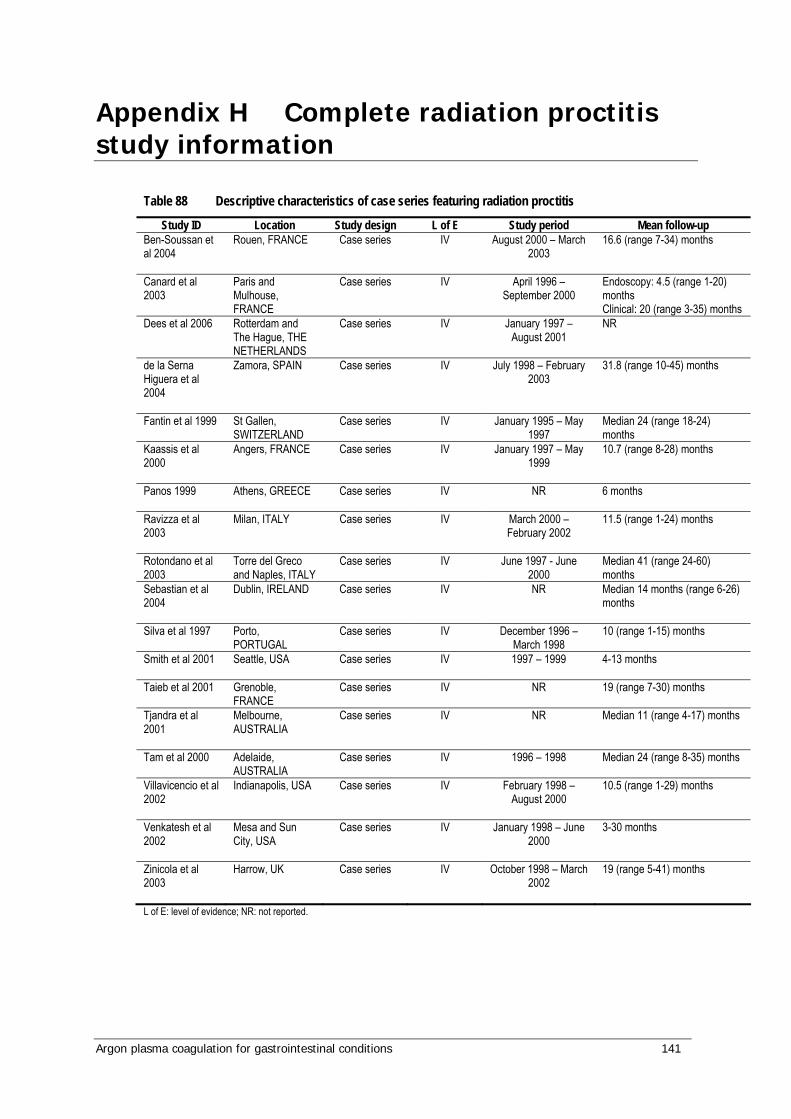
Argon plasma coagulation for gastrointestinal conditions 141
Appendix H Complete radiation proctitis study information
Table 88 Descriptive characteristics of case series featuring radiation proctitis
Study ID Location Study design L of E Study period Mean follow-up Ben-Soussan et al 2004
Rouen, FRANCE Case series IV August 2000 – March 2003
16.6 (range 7-34) months
Canard et al 2003
Paris and Mulhouse, FRANCE
Case series IV April 1996 – September 2000
Endoscopy: 4.5 (range 1-20) months Clinical: 20 (range 3-35) months
Dees et al 2006
Rotterdam and The Hague, THE NETHERLANDS
Case series IV January 1997 – August 2001
NR
de la Serna Higuera et al 2004
Zamora, SPAIN Case series IV July 1998 – February 2003
31.8 (range 10-45) months
Fantin et al 1999
St Gallen, SWITZERLAND
Case series IV January 1995 – May 1997
Median 24 (range 18-24) months
Kaassis et al 2000
Angers, FRANCE Case series IV January 1997 – May 1999
10.7 (range 8-28) months
Panos 1999
Athens, GREECE Case series IV NR 6 months
Ravizza et al 2003
Milan, ITALY Case series IV March 2000 – February 2002
11.5 (range 1-24) months
Rotondano et al 2003
Torre del Greco and Naples, ITALY
Case series IV June 1997 - June 2000
Median 41 (range 24-60) months
Sebastian et al 2004
Dublin, IRELAND Case series IV NR Median 14 months (range 6-26) months
Silva et al 1997
Porto, PORTUGAL
Case series IV December 1996 – March 1998
10 (range 1-15) months
Smith et al 2001
Seattle, USA Case series IV 1997 – 1999 4-13 months
Taieb et al 2001
Grenoble, FRANCE
Case series IV NR 19 (range 7-30) months
Tjandra et al 2001
Melbourne, AUSTRALIA
Case series IV NR Median 11 (range 4-17) months
Tam et al 2000
Adelaide, AUSTRALIA
Case series IV 1996 – 1998 Median 24 (range 8-35) months
Villavicencio et al 2002
Indianapolis, USA Case series IV February 1998 – August 2000
10.5 (range 1-29) months
Venkatesh et al 2002
Mesa and Sun City, USA
Case series IV January 1998 – June 2000
3-30 months
Zinicola et al 2003
Harrow, UK Case series IV October 1998 – March 2002
19 (range 5-41) months
L of E: level of evidence; NR: not reported.

142 Argon plasma coagulation for gastrointestinal conditions
Table 89 Technical characteristics of case series featuring radiation proctitis
Study ID Instrument used Technique APC power (watts)
APC gas flow (litres per minute)
Anaesthesia Mean number of treatments
Ben-Soussan et al 2004
APC 300 ERBE; ICC 200
Single-shot, non-contact procedure
40-50 range 0.8-1.0 No sedation or analgesia in 25/27 patients, but 2/27 experienced pain in first session and required general anaesthesia for further sessions
2.66 (range 1-7)
Canard et al 2003
APC-300 and Erbotom ICC-200, ERBE
Depending on the endoscopic aspect, shots were delivered in spots or scanned over the surface
42 (range 30-80)
1.5 (range 0.8-2.0)
NR 2.3 (range 1-5)
Dees et al 2006
NR No-touch spotting technique. Pulse duration limited to 0.5 sec
50 2.0 NR NR
de la Serna Higuera et al 2004
ERBE ICC 200 Single point targeting with application time per point less than one second (shots)
60 range 1.5-2.0 No sedation 1.9 (range 1-4)
Fantin et al 1999
ERBE Beamer Two instrument system
NR 60 3.0 No sedation or analgesia required
Median 2 (range 2-4)
Kaassis et al 2000
ERBE APC Probe Spotting technique
40 0.6 No premedication was given
3.7 (range 2-8)
Panos 1999
NR Duration of bursts 0.2-0.5 sec
40 1.0 NR Median 6 (range 5-8)
Ravizza et al 2003
APC 2200 and ERBE ICC-200
Single-shot (<2 secs) and/or a trawl-back technique
40-60 2.0-3.0 Intravenous sedation 2 (range 1-5)
Rotondano et al 2003
ERBE APC 300 and ICC 200
Single-shot and/or a trawl-back manoeuvres
40 0.8-1.2 Conscious sedation when needed
2.5 (range 1-6)
Sebastian et al 2004
ERBE APC system
Single pulses <2 secs
30 (range 25-50)
1.5 Conscious intravenous sedation
Median 1 (range1-4)
Silva et al 1997
ARCO-MC Single-shot (<2 secs)
50 1.5 No sedation or analgesia
Median 2.9 (range1-8)
Smith et al 2001 APC 300 ERBE NR 40-45 1.6 NR Range 1-3 Taieb et al 2001
APC 300 ERBE NR 50 0.8-2.0 2 patients had lesions situated adjacent to the dentate line and required general anaesthesia, but the rest did not require sedation.
3.2 (range 1-5)
Tjandra et al 2001
Conmed Argon Beam Coagulator
NR 40 1.5 Sedation 2 (range 1-3)

Argon plasma coagulation for gastrointestinal conditions 143
Tam et al 2000
ERBE APC 300 and ICC 200
Single point targeting(<0.5 to 3 secs) and trawl-back manoeuvres
60 2.0 Sedation NR
Villavicencio et al 2002
ERBE ICC 200 Pulse duration <1 sec unless telangiectasias were virtually confluent, in which case longer pulse durations were used to allow painting of small areas.
45-50 1.2-2.0 16/21 patients chose to undergo APC under conscious sedation
Median 1.7 (range 1-4)
Venkatesh et al 2002
ERBE APC NR 40-60 1.0-1.5 NR NR
Zinicola et al 2003
ERBE APC 300 and ICC 200
NR 65 2.0 Intravenous sedation 2 (range 1-4)
APC: argon plasma coagulation; L of E: level of evidence; NR: not reported.
Table 90 Patient characteristics and outcomes of case series featuring radiation proctitis
Study ID Patient number
M/F Mean age (years) Adverse events
Ben-Soussan et al 2004
27 19/8 73.1 (range 53-86) • Transient anal or rectal pain (3) • Vagal symptoms due to colorectal overdistension by argon flow
(2) • Colonic explosions (2), which led to an immediate perforation
in one patienta Canard et al 2003
30 23/7 70.7 (range 58-85) • Pain (6) • Extensive necrosis(1) • Haemorrhage (1) • Perforation (1) • Stricture (2) • Microrectitis (3)
Dees et al 2006
50 consecutive
46/4 73.6 (range 59-89) No adverse effects after initial APC treatment. One patient suffered major rectal bleeding after APC retreatment two years after initial treatment. This patient had been prescribed aspirin for a TIA.
de la Serna Higuera et al 2004
10 3/7 67.8 (range 58-76) No complications, other than one patient who developed tenesmus after APC treatment, which resolved spontaneously after 3 days.
Fantin et al 1999
7 6/1 76.3 (range 69-89) All interventions were tolerated well by patients, and no complications occurred.
Kaassis et al 2000
16 NR 73.5 (range 62-80) Treatment was well tolerated, but four patients noted a transitory and minimal dysenteric syndrome. No delayed complications such as fistulas, ulcers or strictures were observed.
Panos 1999 5 consecutive
3/2 range 55-75 In two patients, superficial ulcers developed after the second and third treatment, which healed spontaneously after 2 and 4 weeks observation respectively.
Ravizza et al 2003
27 consecutive
23/4 72 (range 62-83) • The procedure was well tolerated by all patients; a minority experienced mild bloating caused by GI tract distension from insufflation of the argon gas. • 2 patients developed short-term complications: transient anal
pain (1) and fever 7 hours after APC session (1). Both resolved spontaneously by the following day. • 14 patients developed rectal ulcers as long-term complications.

144 Argon plasma coagulation for gastrointestinal conditions
Rotondano et al 2003
24 5/19 69.2 (range 22-81) • 5 patients suffered from mild bloating and cramping caused by distension of the GI tract. • 1 patient had transient anal pain 24 h after treatment, and
subsequently developed a non-symptomatic rectal stenosis 11 months after first APC session. • 1 patient discontinued treatment when a recto-vaginal fistula
developed. Sebastian et al 2004
25 consecutive
24/1 69 (range 53-77) All patients tolerated APC well with no immediate complications. Increased rectal pain after treatment was reported by 1 patient. No long-term complications were noted.
Silva et al 1997
28 4/24 65 (range 42-77) • No serious complications or side-effects relayed to the technique itself were observed.
• Some patients experienced mild bloating and cramping caused by distension of the GI tract from argon insufflation.
• 3 patients reported transient anal pain within 24 hours of treatment
• APC therapy was discontinued in 1 patient when a rectovaginal fistula developed; however, analysis of videotapes confirmed that ulceration of the anterior rectal wall was already present at the first examination and APC application was avoided in that area.
Smith et al 2001
7 NR NR No patient developed clinically evident progressive rectal wall abnormalities after APC.
Taieb et al 2001
11 10/1 73 (range 54-86) • No major complication directly linked to the technique was reported.
• Some patients felt bloated after the sessions due to distension of the intestine by the insufflation of argon gas.
• 4 patients had asymptomatic superficial ulceration, observed during endoscopic checks 1 month after the first of second APC session.
• Late complications included: -1 patient who had painful rectal sensation 3 months after 2 APC sessions, which healed 4 months later without stricture formation. -2 patients with rectal stenosis 7 and 11 months respectively after the first APC session.
Tjandra et al 2001
12 10/2 71.3 (range 62-78) • The procedures were well tolerated by all patients. • 2 patients reported some abdominal bloating, related to gas
insufflation. • None of the patients developed rectal ulceration or impairment
of faecal continence. Tam et al 2000
15 consecutive
14/1 74.4 (range 62-89) • 2 patients early in the study developed a high fever due to myelodysplastic syndromes. This was settled promptly with antibiotics.
• 2 patients developed asymptomatic rectal strictures which were successfully dilated using Savary dilators.
• There were no instances of bowel perforation or other serious complications attributable to APC.
Villavicencio et al 2002
21 consecutive
15/6 72.7 (range 58-86) • The following short-term side-effects of APC resolved within 24 hours: -1 patient developed rectal pain immediately after 2 separate APC sessions. -I patient reported excessive bloating, secondary to luminal distension with argon gas. -1 patient had severe tenesmus after APC.
• Long-term complications included: -1 patient that had rectal pain and tenesmus that persisted for 3 months after APC. -1 patient reported dark stools and rectal pain that persisted for 10 months. -1 patient had diarrhoea and tenesmus that lasted over 1 month. -1 patient had tenesmus for 2 months.
Venkatesh et 40 NR Range 64-83 • 2 patients had a low-grade fever and required antibiotics.
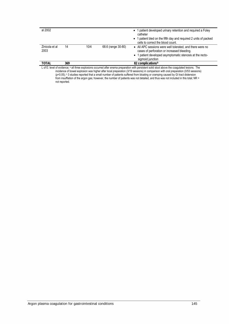
Argon plasma coagulation for gastrointestinal conditions 145
al 2002
• 1 patient developed urinary retention and required a Foley catheter
• 1 patient bled on the fifth day and required 2 units of packed cells to correct the blood count.
Zinicola et al 2003
14 10/4 68.6 (range 30-80) • All APC sessions were well tolerated, and there were no cases of perforation or increased bleeding.
• 1 patient developed asymptomatic stenosis at the recto-sigmoid junction
TOTAL 369 82 complicationsb L of E: level of evidence; a all three explosions occurred after enema preparation with persistent solid stool above the coagulated lesions. The
incidence of bowel explosion was higher after local preparation (3/19 sessions) in comparison with oral preparation (0/53 sessions) (p<0.05); b 3 studies reported that a small number of patients suffered from bloating or cramping caused by GI tract distension from insufflation of the argon gas; however, the number of patients was not detailed, and thus was not included in this total; NR = not reported.

146 Argon plasma coagulation for gastrointestinal conditions
Appendix I Unpublished RCT for radiation proctitis
A PROSPECTIVE RANDOMISED TRIAL OF ARGON PLASMA COAGULATION
VERSUS TOPICAL FORMALIN FOR THE TREATMENT OF RADIATION
PROCTITIS
Hayes J 1, Tam W 2, Moore J 1, 3, Schoeman M 2.
1 Colorectal Surgical Unit and 2 Dept. of Gastroenterology, Royal Adelaide Hospital, 2 Dept. of Surgery,
University of Adelaide, Adelaide, South Australia.
Correspondence to: Mr J Moore
Colorectal Surgical Unit
Royal Adelaide Hospital
North Terrace
Adelaide 5000
Australia
Email: [email protected]
Phone:
Fax:
Abstract presented at the Tripartite Colorectal meeting, October 2002, Melbourne, Australia
Key words: Radiation proctitis, bleeding, treatment
Financial support: None

Argon plasma coagulation for gastrointestinal conditions 147
Aim
To compare the effectiveness of Argon plasma coagulation (APC) with 4% topical formalin in the
treatment of rectal bleeding due to radiation proctitis, in a prospective randomised trial.
Methods
Nineteen patients (median age 74 years) with proven radiation proctitis and significant daily rectal
bleeding were randomised to receive day-case APC or 4% topical formalin. Rectal bleeding and well-
being were assessed using visual analogue scales (VAS 0-10). Haemoglobin (Hb) and transfusion
requirements were recorded. Anorectal function was assessed with the modified Cleveland Clinic-Florida
Incontinence Score. Treatment was given at six-weekly intervals until bleeding had improved to VAS ≤
2.5.
Results
Patients were followed for a mean of 48 weeks. APC and topical formalin treatment had a similar effect,
with significant improvement in rectal bleeding after a median of two treatment sessions. Hb also
increased in patients treated with APC. Four patients were transfusion-dependent before treatment, and
did not require transfusions after treatment. Patient well-being and continence score did not change
significantly. Two patients treated with APC developed minor rectal strictures, which were easily dilated.
Conclusion
APC and topical formalin are similarly effective in treating rectal bleeding from radiation proctitis. APC
treatment may have a higher risk of rectal stricture.
148 Argon plasma coagulation for gastrointestinal conditions
Introduction:
Chronic radiation proctitis is a well-described and troublesome complication of pelvic radiotherapy for
malignancy, affecting some 3-8% of patients treated for carcinoma of the prostate, cervix or bladderi. Symptoms
present a median of 8 to 13 months after radiation treatment in most reported series 3 but may first occur as
long as 30 years after treatment 4. Chronic radiation proctitis frequently presents with rectal bleeding, but faecal
urgency and incontinence may also be problems experienced by many patients, to the detriment of their quality
of life.
The prevalence of 'chronic radiation-induced rectal bleeding' (CRRB) is difficult to determine, but
retrospective data from a single Australian centre suggests that some 5-7% of patients have serious rectal
bleeding ii iii to the extent that it interferes with their lives, and requires treatment. The natural history of
chronic radiation proctitis is not well defined; a number of studies have shown spontaneous remission rates from
bleeding of 70% at 2 years 2.
The origin of the bleeding in chronic radiation proctitis is widespread mucosal telangiectasia iv and this can be
difficult to manage. Medical treatment has included the use of steroids or sucralfate, either orally or as enemas 1
v . Aminosalycylic acid derivatives and short chain fatty acids have also been tried with varying levels of
success 1 vi vii. All of these are generally unhelpful in the long term management of bleeding from chronic
radiation proctitis 2. Hyperbaric oxygen treatment (HBOT) has also been used viii ix , but presents logistical
difficulties and is not widely available. Surgery is considered a last resort for resistant rectal bleeding 2 and
postoperative outcomes are generally poor. A defunctioning stoma is ineffective in controlling bleeding x.
Topical formalin has been described for the management of recurrent rectal bleeding due to radiation injury.
Most studies have used 4% formalin applied directly to the rectal mucosa. Mathai and Seow-Choen treated
twenty-nine patients with one or two applications of four percent formalin, with 22 of the 29 patients having no
more bleeding at a median of twelve months follow-up 12.
2. Tagkalidis PP, Tjandra JJ. Chronic radiation proctitis. ANZ J Surg 2001; 71: 230-237.
3. Lucarotti ME, Mountford RA, Bartolo DC. Surgical management of intestinal radiation injury. Dis
Colon Rectum 1991; 34: 865-869.
Argon plasma coagulation for gastrointestinal conditions 149
More recently argon plasma coagulation (APC) has shown promise 6. APC is a non-contact technique that uses
argon gas to carry an electrical current to superficially coagulate the telangiectatic areas of rectal mucosa to a
controlled depth of 2 mm. With this technique large areas of mucosa can be treated with relative safety 6.
There are no published randomised trials comparing APC to topical formalin. The aim of this study was to
compare the effectiveness of APC with topical formalin in the treatment of rectal bleeding due to radiation
proctitis. Measured outcomes included control of rectal bleeding, blood transfusion requirements, impact on
anorectal function and impact on general well being.
Methods:
Patients with overt rectal bleeding due to radiation proctitis, proven on colonoscopy with or without biopsy,
were invited to participate in the study. No patients had previously undergone treatment for their bleeding. The
study was approved by the Ethics Committee of the Royal Adelaide Hospital. Written informed consent was
obtained from patients before randomisation. Patients were randomised to either APC during flexible
sigmoidoscopy under intravenous sedation or to topical application of 4% formalin as a day case under general
anaesthetic. A pre-treatment colonoscopy was performed to exclude other sources of bleeding from the colon.
Patients were given a rectal enema before APC or topical formalin treatment (Microlax, Pharmacia Upjohn,
Australia). The technique for argon plasma coagulation has been previously described 6. Briefly, areas of
telangiectasia were treated endoscopically using a flexible APC catheter attached to an ERBE ICC 200
electrosurgical generator and an ERBE APC 300 argon delivery unit ( MedTech Systems, Adelaide, South
Australia). Argon gas flow was set at 2 litres per minute and electrical power at 50-60 Watts. Excess argon gas
and the smoke plume from APC was aspirated via the suction channel of the colonoscope. Patients usually
required intravenous sedation with midazolam for this procedure to minimize discomfort.
Topical formalin treatment was administered using a general anaesthetic and with the patient in lithotomy or left
lateral position. Four percent formalin soaked gauze swabs were applied to the affected mucosa via the
operating sigmoidoscope for at least one minute, or as long as it took to control bleeding 12. Excess formalin was
removed by aspiration and the perianal skin was protected with petroleum jelly to prevent damage.

150 Argon plasma coagulation for gastrointestinal conditions
The primary study endpoint was control of rectal bleeding, measured by a Visual Analogue Scale (VAS)
completed by the patient. VAS for rectal bleeding was rated from 0-10, with 0 being no bleeding and 10 being
heavy bleeding. Patients’ blood transfusion requirements were also recorded.
Secondary endpoints were anorectal dysfunction and general wellbeing. Anorectal dysfunction before and after
treatment was measured by the modified Cleveland Clinic-Florida Incontinence score xi (Table 1). This
measures the frequency of incontinence to solid stool, liquid stool or gas, as well as the requirement for pads for
soiling, and degree of lifestyle alteration. Faecal urgency was added, again on a 0-4 scale. The summed result
was a score from 0-24, with 0 being normal continence and 24 being complete incontinence with severe
urgency. General wellbeing was assessed with a visual analogue scale. Patients were asked to score themselves
from 0-10 with 0 being very unwell and 10 being very well. Patients were provided with a diary to record these
and their rectal bleeding scores.
Patients were reviewed 4 weeks after each treatment session. Treatment was continued at 6-weekly intervals
until the VAS for rectal bleeding had improved to < 2.5. Treatment was considered to have failed if this end-
point was not reached after 4 courses of treatment.
Data was analysed using GraphPad Prism [GraphPad Software, 1995]. Mean bleeding, incontinence and
wellbeing scores, and mean haemoglobin levels pre-treatment and post-treatment were compared using Students
t-tests. Statistical significance was taken as P < 0.05.
Results:
Nineteen patients who had received external-beam radiotherapy (ERBT) and had subsequently developed
endoscopically proven proctitis with overt rectal bleeding, were recruited for the study. There were seventeen
men (aged 58 to 83 years), and two women (aged 38 and 45 years). All seventeen men had been treated for
prostate cancer, both women had radiotherapy for cervical cancer. There were no patients who had radiotherapy
for bladder or rectal cancer.
Ten patients were randomised to have treatment with APC, nine had topical formalin treatment.
Argon plasma coagulation for gastrointestinal conditions 151
The mean age of the APC-treated group was 75 years (range 60-83 years) while that of the formalin-treated
group was 63 years (range 38-79 years). The mean pre-treatment haemoglobin in the APC group was 118 g/L,
with a mean of 141 g/L in the formalin arm. This difference was not statistically significant. Three patients in
the APC group were blood transfusion dependent pre-treatment, compared to one in the formalin-treatment
group (Table 2).
Both groups required a median of two treatment sessions (APC range 1-2; formalin range 1-3). Patients were
followed for a mean of 48 weeks overall.
Both APC and topical formalin had a similar effect on rectal bleeding score, with significant improvement after
a median of two treatment sessions (Table 3). One patient having formalin treatment required three treatment
sessions. No patient in either group was considered to have failed their allocated treatment modality.
Haemoglobin improved in both groups, though the change in the formalin group was less, and not statistically
significant, most likely because of a higher pre-treatment baseline Hb. None of the four patients that were
transfusion dependent before treatment required blood transfusion after treatment with APC or formalin. There
was no significant change in either incontinence score or wellbeing score with APC or formalin treatment
(Table 4).
Two patients treated with APC developed minor rectal strictures. Both were easily dilated with Savary-Gillard
dilators without complication.
Discussion:
Topical formalin was first described for the treatment of radiation-induced cystitis xii. More recently it has been
used for chronic radiation proctitis 12. Four percent formalin is applied directly to the rectal mucosa, either by
instillation xiii or by formalin-soaked gauze pads 12. The effect of topical formalin is likely to be due to a local
chemical cauterization of telangiectatic rectal mucosal vessels 2. Care must be taken to prevent excessive
exposure to formalin by aspirating the rectum and protecting the anal verge and perianal skin 4. Complications
of formalin treatment include perianal or rectal ulceration, or rectal stricture xiv.
152 Argon plasma coagulation for gastrointestinal conditions
Argon plasma coagulation involves the delivery of diathermy current to the rectal mucosa, using inert argon gas
passed through a catheter in the operating port of the colonoscope. The diathermy current is broken once the
target tissue is dessicated, resulting in a more predictable and controlled depth of burn. Excessive use of APC,
"painting" the rectal mucosa, may result in mucosal ulceration xv and subsequent stricture formation.
Both APC and topical formalin have become established treatments for bleeding from chronic radiation
proctitis, however, to our knowledge, they have not previously been compared in a randomised trial for
treatment naïve patients. This study suggests that APC and topical formalin are similarly effective in the
management of bleeding from chronic radiation proctitis. Control of rectal bleeding (as measured by rectal
bleeding score), pre- and post-treatment anorectal function and wellbeing scores were similar in both groups.
This is a small study and it is possible that a difference in outcomes between the two treatments may have been
missed. It may also be that 10% formalin is more effective but this study was designed using the known safety
of 4% formalin. The authors were concerned that the use of 10% formalin may lead to major rectal injury and
stricture formation. Interestingly however, the only rectal strictures were after APC, as has previously been
noted. Though the APC treatment group had a significant increase in haemoglobin compared to the formalin
group, this difference is most likely due to the lower pre-treatment haemoglobin in the APC group.
The telangiectasias associated with bleeding from chronic radiation proctitis often lie very close to the dentate
line and formalin treatment may be preferable for these lesions 4. APC via a retroflexed colonoscope is an
alternative17, however lesions in the upper anal canal or anorectal junction are easily accessed with a handheld
APC applicator via an anoscope.
In conclusion, we have shown that argon plasma coagulation and topical formalin are similarly effective in the
management of bleeding from chronic radiation proctitis. There may be an increased risk of rectal stricture with
APC. The choice between APC and topical formalin will largely depend on local availability and patient or
clinician preference.

Argon plasma coagulation for gastrointestinal conditions 153
Table 1: Modified Cleveland Clinic Florida Incontinence Score.
Incontinence to Never < 2 per month < 1 per week < 1 per day Daily
Solid stool 0 1 2 3 4
Liquid stool 0 1 2 3 4
Gas 0 1 2 3 4
Never Rarely Occasionally Often Always
Wears pads (soiling) 0 1 2 3 4
Lifestyle altered 0 1 2 3 4
Urgency 0 1 2 3 4
Table 2: Patient characteristics
APC Formalin
No. patients 10 9
Age, mean (range) 75 (60-83) 63 (38-79) P = 0.041
No. transfusion dependent
patients
3 1
Mean pre-treatment
Haemoglobin g/L (range)
118 (71-159) 141 (82-164) P = 0.063
No. treatment sessions (range) 2 (1-2) 2 (1-3)
Mean followup weeks (range) 60 (22-114) 37 (9-100) P = 0.165
Table 3: Contro l of rectal bleeding
Pre-APC Post-APC Pre-formalin Post-formalin
Rectal bleeding
score (mean)
5.9 (4.0-7.6) 1.5 (0-2.5) * 6.1 (4.6-10.0) 1.6 (0-2.5) *
Haemoglobin
(mean, g/L)
118 (71-159) 137 (117-162) † 141 (82-164) 145 (116-167)
* P < 0.0001; † P = 0.047

154 Argon plasma coagulation for gastrointestinal conditions
Table 4: Anorectal function and well-being
Pre-APC Post-APC Pre-formalin Post-formalin
Incontinence
score (mean)
9.0 (3 -18) 9.3 (3-15) 6.6 (1-13) 8.0 (1 -17)
Wellbeing score
(mean)
6.5 (5.0-9.7) 7.2 (5.0-10.0) 6.3 (4.9-8.7) 6.4 (2.5-9.1)
References
1. Hayne D, Vaizey CJ, Boulos PB. Anorectal injury following pelvic radiotherapy. Br J Surg 2001; 88: 1037-
1048.
2. Tagkalidis PP, Tjandra JJ. Chronic radiation proctitis. ANZ J Surg 2001; 71: 230-237.
3. Lucarotti ME, Mountford RA, Bartolo DC. Surgical management of intestinal radiation injury. Dis Colon
Rectum 1991; 34: 865-869.
4. Chapuis PH. Editorial. ANZ J Surg 2001; 71: 200-201.
5. Mameghan H, Fisher R, Mameghan J, Brook S. Results of external beam radiotherapy in 448 patients with
clinically localized adenocarcinoma of the prostate. ANZ J Surg 1994; 64: 384-394.
6. Tam W, Moore J, Schoeman, M. Treatment of radiation proctitis with argon plasma coagulation. Endoscopy
2000; 32: 667-672.
7. Kochhar R, Patel F, Dhar A, Sharma SC, Ayyagari S et al. Radiation-induced proctosigmoiditis. Prospective,
randomized, double-blind controlled trial of oral sulphasalazine plus rectal steroids versus rectal sucralfate. Dig
Dis Sci 1991; 36: 103-107.
8. Baum CA, Biddle WL, Miner PB Jr. Failure of 5 –aminosalicylic acid enemas to improve chronic radiation
proctitis. Dig Dis Sci 1989; 34: 758-760.
9. Talley NA, Chen F, King D, Jones M. Short chain fatty acids in the treatment of radiation proctitis: a
randomized, double-blind placebo-controlled, cross-over pilot trial. Dis Colon Rectum 1997; 40: 1046-1050.
10. Kitta T, Shinohara N, Shirato H, Otsuka H, Koyanagi T. The treatment of chronic radiation proctitis with
hyperbaric oxygen in patients with prostate cancer. Br J Urol 2000; 85: 372-374.

Argon plasma coagulation for gastrointestinal conditions 155
11. Warren DC, Feehan P, Slade JB, Cianci PE. Chronic radiation proctitis treated with hyperbaric oxygen.
Undersea Hyperb Med 1997; 24: 181-184.
12. Mathai V, Seow-Choen F. Endoluminal formalin therapy for haemorrhagic radiation proctitis. Br J Surg
1995; 82: 190.
13. Jorge JMN, Wexner SD. Etiology and management of fecal incontinence. Dis Colon Rectum 1993; 36: 77-
97.
14. Brown RB. A method for the management of inoperable carcinoma of the bladder. Med J Aust 1969; 1: 23-
24
15. Saclarides TJ, King, DG, Franklin JL, Doolas A. Formalin instillation for refractory radiation-induced
haemorrhagic proctitis. Report of 16 patients. Dis Colon Rectum 1996; 39: 196-199.
16. Tjandra JJ, Sengupta S. Argon plasma coagulation is an effective treatment for refractory haemorrhagic
radiation proctitis. Dis Colon Rectum 2001; 44: 1759-1765.
17. Villavicencio RT, Rex DK, Rahmani E. Efficacy and complications of argon plasma coagulation for
hematochezia related to radiation proctopathy. Gastrointest Endosc 2002; 55: 70-74.

156 Argon plasma coagulation for gastrointestinal conditions
Appendix J Economic evaluation
Results of the meta-analysis (Chau et al 2003; Cipolletta et al 1998)
Rate of permanent haemostasis Rev iew: MSAC 1106 - APC ulcersComparison: 01 Permanent haemostasis Outcome: 01 Permanent haemostasis
Study APC HP RR (f ixed) Weight RR (f ixed)or sub-category n/N n/N 95% CI % 95% CI Quality
Chau 2003 49/52 47/60 71.48 1.20 [1.04, 1.40] D Cipolletta 1998 19/21 17/20 28.52 1.06 [0.85, 1.34] D
Total (95% CI) 73 80 100.00 1.16 [1.03, 1.32]Total events: 68 (APC), 64 (HP)Test f or heterogeneity : Chi² = 0.76, df = 1 (P = 0.38), I² = 0%Test f or ov erall ef f ect: Z = 2.37 (P = 0.02
0.5 0.7 1 1.5 2
Favours HP Favours APC
Rate of initial haemostasis Rev iew: MSAC 1106 - APC ulcersComparison: 06 Initial haemostasis Outcome: 01 Initial haemostasis
Study APC HP RR (f ixed) Weight RR (f ixed)or sub-category n/N n/N 95% CI % 95% CI Quality
Chau 2003 86/88 93/97 81.97 1.02 [0.97, 1.07] D Cipolletta 1998 20/21 19/20 18.03 1.00 [0.87, 1.15] D
Total (95% CI) 109 117 100.00 1.02 [0.97, 1.07]Total events: 106 (APC), 112 (HP)Test f or heterogeneity : Chi² = 0.05, df = 1 (P = 0.82), I² = 0%Test f or ov erall ef f ect: Z = 0.64 (P = 0.52
0.5 0.7 1 1.5 2
Favours HP Favours APC
Probability of emergency surgery Rev iew: MSAC 1106 - APC ulcersComparison: 02 Emergency surgery required Outcome: 01 Emergency surgery required
Study APC HP RR (f ixed) Weight RR (f ixed)or sub-category n/N n/N 95% CI % 95% CI Quality
Chau 2003 4/88 9/97 73.59 0.49 [0.16, 1.53] D Cipolletta 1998 2/21 3/20 26.41 0.63 [0.12, 3.41] D
Total (95% CI) 109 117 100.00 0.53 [0.21, 1.36]Total events: 6 (APC), 12 (HPTest f or heterogeneity : Chi² = 0.06, df = 1 (P = 0.80), I² = 0%Test f or ov erall ef f ect: Z = 1.32 (P = 0.19
0.1 0.2 0.5 1 2 5 10
Favours APC Favours HP
Number of blood transfusions Rev iew: MSAC 1106 - APC ulcersComparison: 03 Blood transf usion required Outcome: 01 Blood transf usion required
Study APC HP WMD (f ixed) Weight WMD (f ixed)or sub-category N Mean (SD) N Mean (SD) 95% CI % 95% CI Quality
Chau 2003 88 1.70(2.80) 97 2.40(3.30) 18.78 -0.70 [-1.58, 0.18] DCipolletta 1998 21 1.90(0.90) 20 2.10(0.40) 81.22 -0.20 [-0.62, 0.22] D
Total (95% CI) 109 117 100.00 -0.29 [-0.68, 0.09]Test f or heterogeneity : Chi² = 1.01, df = 1 (P = 0.32), I² = 0.8%Test f or overall ef f ect: Z = 1.51 (P = 0.13
-4 -2 0 2 4
Fav ours APC Fav ours HP
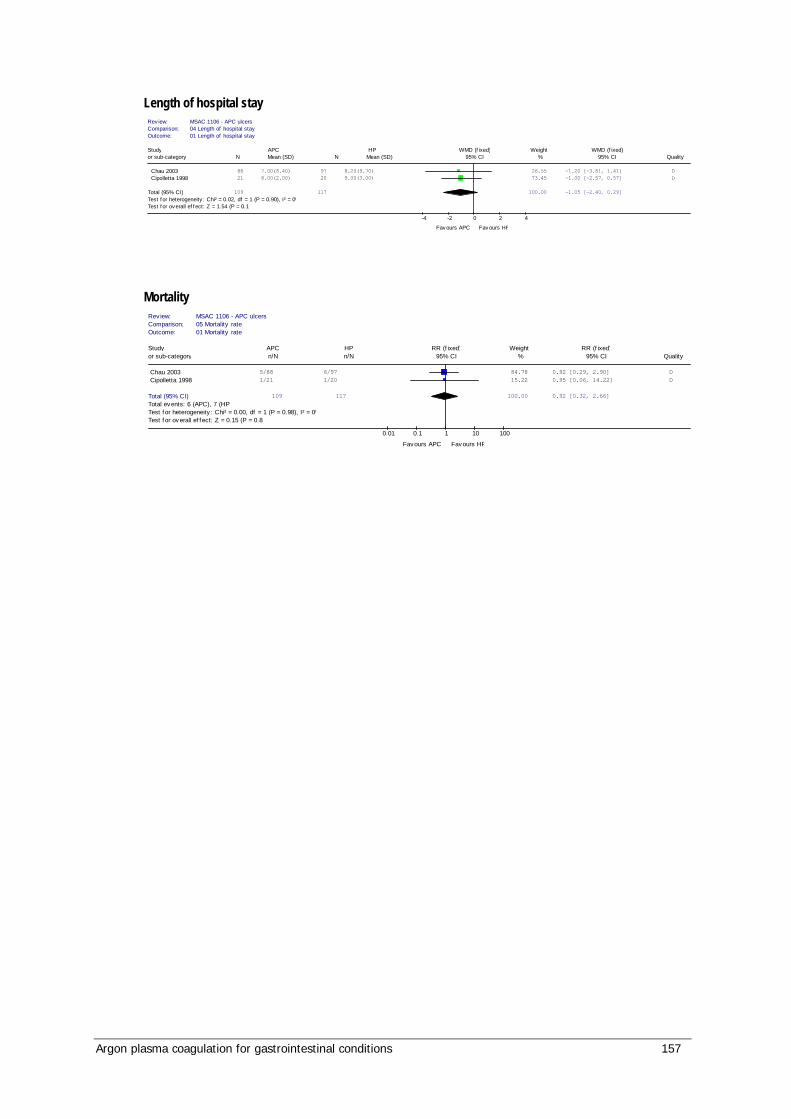
Argon plasma coagulation for gastrointestinal conditions 157
Length of hospital stay Rev iew: MSAC 1106 - APC ulcersComparison: 04 Length of hospital stay Outcome: 01 Length of hospital stay
Study APC HP WMD (f ixed) Weight WMD (f ixed)or sub-category N Mean (SD) N Mean (SD) 95% CI % 95% CI Quality
Chau 2003 88 7.00(8.40) 97 8.20(9.70) 26.55 -1.20 [-3.81, 1.41] DCipolletta 1998 21 8.00(2.00) 20 9.00(3.00) 73.45 -1.00 [-2.57, 0.57] D
Total (95% CI) 109 117 100.00 -1.05 [-2.40, 0.29]Test f or heterogeneity : Chi² = 0.02, df = 1 (P = 0.90), I² = 0%Test f or overall ef f ect: Z = 1.54 (P = 0.12
-4 -2 0 2 4
Fav ours APC Fav ours HP
Mortality Rev iew: MSAC 1106 - APC ulcersComparison: 05 Mortality rate Outcome: 01 Mortality rate
Study APC HP RR (f ixed) Weight RR (f ixed)or sub-category n/N n/N 95% CI % 95% CI Quality
Chau 2003 5/88 6/97 84.78 0.92 [0.29, 2.90] D Cipolletta 1998 1/21 1/20 15.22 0.95 [0.06, 14.22] D
Total (95% CI) 109 117 100.00 0.92 [0.32, 2.66]Total events: 6 (APC), 7 (HPTest f or heterogeneity : Chi² = 0.00, df = 1 (P = 0.98), I² = 0%Test f or ov erall ef f ect: Z = 0.15 (P = 0.88
0.01 0.1 1 10 100
Favours APC Favours HP

158 Argon plasma coagulation for gastrointestinal conditions
Abbreviations
AIHW Australian Institute of Health and Welfare
ALA 5-aminolevulinic acid
AMV arteriovenous malformation
APC argon plasma coagulation
AR-DRG Australian refined diagnosis related groups
AVM arteriovenous malformations
BO Barrett’s oesophagus
CI confidence interval
cm centimetre
CS conservative surveillance
FAP familial adenomatous polyposis
Fr French
GAVE gastric antral vascular ectasia
GI gastrointestinal
GORD gastro-oesophageal reflux disease
HGD high-grade dysplasia
HTA Health Technology Assessment
HP heater probe
H. Pylori Helicobactor pylori
l litres
L/min litres per minute
LGD low-grade dysplasia
MBS Medicare Benefits Schedule
ml millilitre
mm millimetre
MPEC multipolar electrocoagulation
Argon plasma coagulation for gastrointestinal conditions 159
MSAC Medical Services Advisory Committee
N cohort number
n patient number
ND not determined
Nd:YAG neodymium:yttrium-aluminum-garnet
NHMRC National Health and Medical Research Council
nm nanometre
NR not reported
NSAID non-steroidal anti-inflammatory drug
PDT photodynamic therapy
PICO population, intervention, comparator, outcome
PPI proton pump inhibitor
RCT randomised control trial
RevMan Reference Manager
SD standard deviation
SEMS self-expanding metallic stents
TGA Therapeutic Goods Administration
UK United Kingdom
USA United States of America
V volts
W watts
160 Argon plasma coagulation for gastrointestinal conditions
References
Abou-Hamden, A, Craig, A & Schouman, M, 1997. ‘Endoscopic argon plasma coagulation therapy for vascular ectasias of the gastrointestinal tract’, Journal of Gastroenterology & Hepatology, 12, A51.
Ackroyd, R, Tam, W, Schoeman, M, Devitt, PG & Watson, DI, 2004. ‘Prospective randomized controlled trial of argon plasma coagulation ablation vs. endoscopic surveillance of patients with Barrett's esophagus after antireflux surgery’, Gastrointestinal Endoscopy, 59(1), 1-7.
Akhtar, K, Byrne, JP, Bancewicz, J & Attwood, SE, 2000. ‘Argon beam plasma coagulation in the management of cancers of the esophagus and stomach’, Surg Endosc, 14(12), 1127-30.
Apel, D, Jakobs, R, Spiethoff, A & Riemann, JF, 2005. ‘Follow-up after endoscopic snare resection of duodenal adenomas’, Endoscopy, 37(5), 444-8.
Altman DG, Schulz KF, Moher D, Egger M, Davidoff F, Elbourne D, Gøtzsche PC, and Lang T, 2001. For the CONSORT Group ‘The Revised CONSORT Statement for Reporting Randomized Trials: Explanation and Elaboration’ Ann Intern Med, 134, 663-94.
Basu, KK, Pick, B, Bale, R, West, KP & De Caestecker, JS, 2002. ‘Efficacy and one year follow up of argon plasma coagulation therapy for ablation of Barrett's oesophagus: Factors determining persistence and recurrence of Barrett's epithelium’, Gut, 51(6), 776-80.
Ben Soussan, E, Antonietti, M, Savoye, G, Herve, S, Ducrotte, P & Lerebours, E, 2004. ‘Argon plasma coagulation in the treatment of hemorrhagic radiation proctitis is efficient but requires a perfect colonic cleansing to be safe’, Eur J Gastroenterol Hepatol, 16(12), 1315-18.
Bergman, JJGHM, Fockens, P, 2006. ‘Ablating Barrett’s metaplastic epithelium: are the techniques ready for clinical use?’ Gut, 55, 1222-3.
Bond, JH, 2000. ‘Polyp guideline: diagnosis, treatement, and surveillance for patients with colorectal polyps’, American Journal of Gastroenterology, 95(11), 3053-63.
Bounds, BC & Friedman, LS, 2003. ‘Lower gastrointestinal bleeding’, Gastroenterology Clinics of North America, 32(4), 1107-+.
Brooker, JC, Saunders, BP, Shah, SG, Thapar, CJ, Suzuki, N & Williams, CB, 2002. ‘Treatment with argon plasma coagulation reduces recurrence after piecemeal resection of large sessile colonic polyps: a randomized trial and recommendations’, Gastrointestinal Endoscopy, 55(3), 371-5.
Byrne, JP, Armstrong, GR & Attwood, SEA, 1998. ‘Restoration of the normal squamous lining in barrett's esophagus by argon beam plasma coagulation’, American Journal of Gastroenterology, 93(10), 1810-15.
Argon plasma coagulation for gastrointestinal conditions 161
Canard, J-M, Vedrenne, B, Bors, G, Claude, P, Bader, R & Sondag, D, 2003. ‘Treatment of radiation proctitis by argon plasma coagulation: Long term results. [French]’, Gastroenterologie Clinique et Biologique, 27(5), 455-9.
Chau, CH, Siu, WT, Law, BK, Tang, CN, Kwok, SY, Luk, YW, Lao, WC & Li, MK, 2003. ‘Randomized controlled trial comparing epinephrine injection plus heat probe coagulation versus epinephrine injection plus argon plasma coagulation for bleeding peptic ulcers’, Gastrointest.Endosc, 57(4), 455-61.
Cipolletta, L, Bianco, MA, Rotondano, G, Piscopo, R, Prisco, A & Garofano, ML, 1998. ‘Prospective comparison of argon plasma coagulator and heater probe in the endoscopic treatment of major peptic ulcer bleeding’, Gastrointestinal Endoscopy, 48(2), 191-5.
Conio, M, Lapertosa, G, Blanchi, S & Filiberti, R, 2003. ‘Barrett's esophagus: an update’, Critical Reviews in Oncology-Hematology, 46(2), 187-206.
Cotti, G, Seid, V, Araujo, S, Souza, AH, Jr., Kiss, DR & Habr-Gama, A, 2003. ‘Conservative therapies for hemorrhagic radiation proctitis: a review’, Revista do Hospital das Clinicas; Faculdade de Medicina Da Universidade de Sao Paulo, 58(5), 284-92.
Crosta, C, Spaggiari, L, Stefano, AD, Fiori, G, Ravizza, D & Pastorino, U, 2001. ‘Endoscopic argon plasma coagulation for palliative treatment of malignant airway obstructions: Early results in 47 cases’, Lung Cancer, 33(1), 75-80.
de la Serna, HC, Martin, AM, Rodriguez, GS, Perez, VA, Martinez, MJ & Betancourt, GA, 2004. ‘Efficacy and safety of argon plasma coagulation for the treatment of hemorrhagic radiation proctitis’, Revista Espanola de Enfermedades Digestivas, 96(11), 758-64.
Dees, J, Meijssen, M & Kuipers, E, 2006. ‘Argon plasma coagulation for radiation proctitis’, Scandinavian Journal of Gastroenterology, 41(SUPPL. 243), 175-8.
Denton, A, Forbes, A, Andreyev, J & Maher, EJ, 2002. ‘Non surgical interventions for late radiation proctitis in patients who have received radical radiotherapy to the pelvis’, Cochrane Database of Systematic Reviews, no. 1.
Dulai, GS, Jensen, DM, Kovacs, TOG, Gralnek, IM & Jutabha, R, 2004. ‘Endoscopic treatment outcomes in watermelon stomach patients with and without portal hypertension’, Endoscopy, 36(1), 68-72.
Dulai, GS, Jensen, DM, Cortina, G, Fontana, L & Ippoliti, A, 2005. ‘Randomized trial of argon plasma coagulation vs. multipolar electrocoagulation for ablation of Barrett's esophagus’, Gastrointestinal Endoscopy, 61(2), 232-40.
Dulai, GS & Jensen, DM, 2006. ‘Treatment of watermelon stomach’, Current Treatment Options in Gastroenterology, 9(2), 175-80.
Eswaran, SL, Sanders, M, Bernadino, KP, Ansari, A, Lawrence, C, Stefan, A, Mattia, A & Howell, DA, 2006. ‘Success and complications of endoscopic removal of giant duodenal and ampullary polyps: a comparative series’, Gastrointestinal Endoscopy, 64(6), 925-32.
Fantin, AC, Binek, J, Suter, WR & Meyenberger, C, 1999. ‘Argon beam coagulation for treatment of symptomatic radiation-induced proctitis’, Gastrointestinal Endoscopy, 49(4) I, 515-8.
162 Argon plasma coagulation for gastrointestinal conditions
Farrell, JJ & Friedman, LS, 2005. ‘Review article: the management of lower gastrointestinal bleeding’, Alimentary Pharmacology & Therapeutics, 21(11), 1281-98.
Faybush, EM & Sampliner, RE, 2005. ‘Randomized trials in the treatment of Barrett's esophagus’, Diseases of the Esophagus, 18(5), 291-7.
Ford, AC, Delaney, BC, Forman, D & Moayyedi, P, 2006. ‘Eradication therapy for peptic ulcer disease in Helicobacter pylori positive patients’, Cochrane Database of Systematic Reviews, no. 2.
Fu, K-I & Fujimori, T, 2006. ‘Bleeding angiodysplasia in the duodenum’, New England Journal of Medicine, 354(3), 283.
Garcia, A, Nunez, O, Gonzalez-Asanza, C, Parera, A, Menchen, L, Ripoll, C, Senent, C, Cos, E & Menchen, P, 2004. ‘Safety and efficacy of argon plasma coagulator ablation therapy for flat colorectal adenomas’, Revista Espanola de Enfermedades Digestivas, 96(5), 315-21.
Ginsberg, GG, Barkun, AN, Bosco, JJ, Burdick, JS, Isenberg, GA, Nakao, NL, Petersen, BT, Silverman, WB, Slivka, A, Kelsey, PB & American Society for Gastrointestinal Endoscopy, 2002. ‘The argon plasma coagulator: February 2002’, Gastrointestinal Endoscopy, 55(7), 807-10.
Grund, KE, Straub, T & Farin, G, 1999. ‘New haemostatic techniques: argon plasma coagulation’, Best Practice & Research in Clinical Gastroenterology, 13(1), 67-84.
Haag, S, Nandurkar, S & Talley, NJ, 1999. ‘Regression of Barrett's esophagus: The role of acid suppression, surgery, and ablative methods’, Gastrointestinal Endoscopy, 50(2), 229-40.
Hage, M, Siersema, PD, Van Dekken, H, Steyerberg, EW, Haringsma, J, Van, DV, Grool, TE, Van Veen, RLP, Sterenborg, HJCM & Kuipers, EJ, 2004. ‘5-Aminolevulinic acid photodynamic therapy versus argon plasma coagulation for ablation of Barrett's oesophagus: A randomised trial’, Gut, 53(6), 785-90.
Havanond, C & Havanond, P 2005, ‘Argon plasma coagulation therapy for acute non-variceal upper gastrointestinal bleeding - art. no. CD003791.pub2’, Cochrane Database of Systematic Reviews 2, p. UB2.
Higgins JPT, Green S, editors. Cochrane Handbook for Systematic Reviews of Interventions 4.2.6 [updated September 2006]. In: The Cochrane Library, Issue 4, 2006. Chichester, UK: John Wiley & Sons, Ltd.
Hong, JJ, Park, W & Ehrenpreis, ED, 2001. ‘Review article: current therapeutic options for radiation proctopathy’, Alimentary Pharmacology & Therapeutics, 15, 1253-62.
Hoyer, N, Thouet, R & Zellweger, U, 1998. ‘Massive pneumoperitoneum after endoscopic argon plasma coagulation’, Endoscopy, 30(3), S44-S45.
Hui, AJ & Sung, JJY, 2005. ‘Endoscopic treatment of upper gastrointestinal bleeding’, Current Treatment Options in Gastroenterology, 8(2), 153-62.
Jarvinen, HJ, 1991. ‘Other gastrointestinal polyps’, World Journal of Surgery, 15(1), 50-6.

Argon plasma coagulation for gastrointestinal conditions 163
Jensen, DM, Chaves, DM & Grund, KE, 2004. ‘Endoscopic diagnosis and treatment of watermelon stomach’, Endoscopy, 36(7), 640-7.
Johanns, W, Luis, W, Janssen, J, Kahl, S & Greiner, L, 1997. ‘Argon plasma coagulation (APC) in gastroenterology: experimental and clinical experiences’, European Journal of Gastroenterology & Hepatology, 9(6), 581-7.
Kaassis, M, Oberti, E, Burtin, P & Boyer, J, 2000. ‘Argon plasma coagulation for the treatment of hemorrhagic radiation proctitis’, Endoscopy, 32(9), 673-6.
Kanai, M, Hamada, A, Endo, Y, Takeda, Y, Yamakawa, M, Nishikawa, H & Torii, A, 2004. ‘Efficacy of argon plasma coagulation in nonvariceal upper gastrointestinal bleeding’, Endoscopy, 36(12), 1085-8.
Kelty, CJ, Ackroyd, R, Brown, NJ, Stephenson, TJ, Stoddard, CJ & Reed, MWR, 2004. ‘Endoscopic ablation of Barrett's oesophagus: A randomized-controlled trial of photodynamic therapy vs. argon plasma coagulation’, Alimentary Pharmacology & Therapeutics, 20(11-12), 1289-96.
Ker, TS, Wasserberg, N & Beart Jr, RW, 2004. ‘Colonoscopic perforation and bleeding of the colon can be treated safely without surgery’, The American Surgeon, 70(10), 922-4.
Khan, K, Schwarzenberg, SJ, Sharp, H & Weisdorf-Schindele, S, 2003. ‘Argon plasma coagulation: Clinical experience in pediatric patients’, Gastrointestinal Endoscopy, 57(1), 110-2.
Komatsu, H, Hara, Y, Naito, Y, Hosaka, Y, Yamanaka, S, Masuda, H & Imamura, K, 2005. ‘Effect of argon plasma coagulation in patients with upper gastrointestinal active hemorrhage’, Digestive Endoscopy, 17(1), 13-6.
Kwan, V, Bourke, MJ, Williams, SJ, Gillespie, PE, Murray, MA, Kaffes, AJ, Henriquez, MS & Chan, RO, 2006. ‘Argon plasma coagulation in the management of symptomatic gastrointestinal vascular lesions: experience in 100 consecutive patients with long-term follow-up’, American Journal of Gastroenterology, 101(1), 58-63.
Lai, HL & Sung, JJY, 2007. ‘Helicobacter pylori and benign upper digestive disease’, Best Practice & Research in Clinical Gastroenterology, 21(2), 261-76.
Leontiadis, G, Sharma, VK & Howden, CW, 2005. ‘Systematic review and meta-analysis of proton pump inhibitor therapy in peptic ulcer bleeding’, BMJ, 330, 568-75.
Madisch, A, Miehlke, S, Bayerdoerffer, E, Wiedemann, B, Antos, D, Sievert, A, Vieth, M, Stolte, M & Schulz, H, 2005. ‘Long-term follow-up after complete ablation of Barrett's esophagus with argon plasma coagulation’, World Journal of Gastroenterology, 11(8), 1182-6.
Manner, H, May, A, Faerber, M, Rabenstein, T & Ell, C, 2006. ‘Safety and efficacy of a new high power argon plasma coagulation system (hp-APC) in lesions of the upper gastrointestinal tract’, Digestive & Liver Disease, 38(7), 471-8.
Marchese, M, De Cristofaro, R, Federici, AB, Biondi, A, Petruzziello, L, Tringali, A, Spada, C, Mutignani, M, Ronconi, P & Costamagna, G, 2005. ‘Duodenal and gastric Dieulafoy's lesions in a patient with type 2A von Willebrand's disease’, Gastrointestinal Endoscopy, 61(2), 322-5.
164 Argon plasma coagulation for gastrointestinal conditions
Marmo, R, Rotondamo, G, Piscopo, R, Bianco, MA, D'Angella, R & Cipolletta, L, 2007. ‘Dual therapy versusmonotherapy in the endoscopic treatment of high-risk bleeding ulcers: a meta-analysis of controlled trials’, American Journal of Gastroenterology, 102, 279-89.
Mason, R, 2001. ‘Palliation of oesophageal cancer’, Surgical Oncology, 10(3), 123-6.
Morino, M, Rebecchi, F, Giaccone, C, Taraglio, S, Sidoli, L & Ferraris, R, 2003. ‘Endoscopic ablation of Barrett's esophagus using argon plasma coagulation (APC) following surgical laparoscopic fundoplication’, Surgical Endoscopy, 17(4), 539-42.
Mork, H, Barth, T, Kreipe, HH, Kraus, M, Al Taie, O, Jakob, F & Scheurlen, M, 1998. ‘Reconstitution of squamous epithelium in Barrett's oesophagus with endoscopic argon plasma coagulation: a prospective study’, Scandinavian Journal of Gastroenterology, 33(11), 1130-4.
Morris, CD, Byrne, JP, Armstrong, GRA & Attwood, SEA, 2001. ‘Prevention of the neoplastic progression of Barrett's oesophagus by endoscopic argon beam plasma ablation’, British Journal of Surgery, 88(10), 1357-62.
MSAC report 1057 ‘M2A® capsule endoscopy for the evaluation of obscure gastrointestinal bleeding in adult patients’
Nakamura, S, Mitsunaga, A, Konishi, H, Oi, I, Shiratori, K & Suzuki, S, 2006. ‘Long-term follow up of gastric antral vascular ectasia treated by argon plasma coagulation’, Digestive Endoscopy, 18(2), 128-33.
NHMRC (1999). A guide to the development, implementation and evaluation of clinical practice guidelines, National Health and Medical Research Council, Canberra.
NHMRC (2000). How to use the evidence: assessment and application of scientific evidence, National Health and Medical Research Council, Canberra.
Nivatvongs, S, 1983. ‘Complications in colonic polypectomy. An experience with 1555 polypectomies’, Diseases of the Colon & Rectum, 29(12), 825-30.
Novitsky, YW, Kercher, KW, Czerniach, DR & Litwin, DEM, 2003. Watermelon stomach: Pathophysiology, diagnosis, and management, Journal of Gastrointestinal Surgery, 7(5), 652-61.
Occhigrossi, G, Scamporrino, A, Pica, R, Paoluzi, OA & Paoluzi, P, 2002. ‘Efficacy of endoscopic adrenaline injection followed by Argon plasma coagulation for bleeding peptic ulcers: Comparison with APC alone’, Gastroenterology International, 15(1-2), 12-7.
Olmos, JA, Marcolongo, M, Pogorelsky, V, Varela, E & Davolos, JR, 2004. ‘Argon plasma coagulation for prevention of recurrent bleeding from GI angiodysplasias’, Gastrointestinal Endoscopy, 60(6), 881-6.
Olmos, JA, Marcolongo, M, Pogorelsky, V, Herrera, L, Tobal, F & Davolos, JR, 2006. ‘Long-term outcome of argon plasma ablation therapy for bleeding in 100 consecutive patients with colonic angiodysplasia’, Diseases of the Colon & Rectum, 49(10), 1507-16.
Argon plasma coagulation for gastrointestinal conditions 165
Pagani, M, Granelli, P, Chella, B, Antoniazzi, L, Bonavina, L & Peracchia, A, 2003. ‘Barrett's esophagus: combined treatment using argon plasma coagulation and laparoscopic antireflux surgery’, Diseases of the Esophagus, 16(4), 279-83.
Palmer, K, 2004. ‘Management of haematemesis and melaena’, Postgraduate Medical Journal, 80(945), 399-404.
Panos, MZ, 1999. ‘Use of argon plasma coagulator for bleeding due to radiation-induced proctitis’, Hellenic Journal of Gastroenterology, 12(2), 101-3.
Parfitt, JR & Driman, DK, 2007. ‘Pathological effects of drugs on the gastrointestinal tract: a review’, Human Pathology, 38, 527-36.
Pavey, DA & Craig, PI, 2004. ‘Endoscopic therapy for upper-GI vascular ectasias’, Gastrointestinal Endoscopy, 59(2), 233-8.
Pereira-Lima, JC, Busnello, JV, Saul, C, Toneloto, EB, Lopes, CV, Rynkowski, CB & Blaya, C, 2000. ‘High power setting argon plasma coagulation for the eradication of Barrett's esophagus’, American Journal of Gastroenterology, 95(7), 1661-8.
Perez, RF, Gonzalez, CP, Legaz Huidobro, ML, Villafanez Garcia, MC, Soto, FS, de Pedro, EA, Roncero Garcia-Escribano, O & Ruiz, CF, 2004. ‘Endoscopic resection of large colorectal polyps’, Revista Espanola de Enfermedades Digestivas, 96(1), 36-47.
Pichon Riviere, A, Augustovski, F, Ferrante, F, Garcia Marti, S, Glujovsky, D, Lopes, A, & Regueiro, A, 2005. ‘Argon plasma usefulness for the treatment of gastrointestinal lesions’, Ciudad de Buenos Aires: Institute for Clinical Effectiveness and Health Policy (IECS).
Ragunath, K, Krasner, N, Raman, VS, Haqqani, MT, Phillips, CJ & Cheung, I, 2005. ‘Endoscopic ablation of dysplastic Barrett's oesophagus comparing argon plasma coagulation and photodynamic therapy: a randomized prospective trial assessing efficacy and cost-effectiveness’, Scandinavian Journal of Gastroenterology, 40(7), 750-8.
Rajendran, N, Haqqani, MT, Crumplin, MK & Krasner, N, 2000. ‘Management of stent overgrowth in a patient with Crohn's oesophagitis by argon plasma coagulation’, Endoscopy, 32(7), S44.
Ramage Jr, JI & Gostout, CJ, 2003. ‘Endoscopic treatment of chronic radiation proctopathy’, Techniques in Gastrointestinal Endoscopy, 5(4), 155-9.
Ravizza, D, Fiori, G, Trovato, C & Crosta, C, 2003. ‘Frequency and outcomes of rectal ulcers during argon plasma coagulation for chronic radiation-induced proctopathy’, Gastrointestinal Endoscopy, 57(4), 519-25.
Regula, J, Wronska, E, Polkowski, M, Nasierowska-Guttmejer, A, Pachlewski, J, Rupinski, M & Butruk, E, 2003. ‘Argon plasma coagulation after piecemeal polypectomy of sessile colorectal adenomas: long-term follow-up study’, Endoscopy, 35(3), 212-8.
Repici, A & Tricerri, R, 2004. ‘Endoscopic polypectomy: techniques, complications and follow-up’, Techniques in Coloproctology, 8, S283-S290.

166 Argon plasma coagulation for gastrointestinal conditions
Roman, S, Saurin, JC, Dumortier, J, Perreira, A, Bernard, G & Ponchon, T, 2003. ‘Tolerance and efficacy of argon plasma coagulation for controlling bleeding in patients with typical and atypical manifestations of watermelon stomach’, Endoscopy, 35(12), 1024-8.
Rosen, L, Bub, DS, Reed, JF & Nastasee, SA, 1993. ‘Hemorrhage following colonoscopic polypectomy’, Diseases of the Colon & Rectum, 36(12), 1126-31.
Rotondano, G, Bianco, MA, Marmo, R, Piscopo, R & Cipolletta, L, 2003. ‘Long-term outcome of argon plasma coagulation therapy for bleeding caused by chronic radiation proctopathy’, Digestive & Liver Disease, 35(11), 806-10.
Sato, T, Yamazaki, K, Toyota, J, Karino, Y, Ohmura, T, Akaike, J, Kuwata, Y & Suga, T, 2005. ‘Efficacy of argon plasma coagulation for gastric antral vascular ectasia associated with chronic liver disease’, Hepatology Research, 32(2), 121-6.
Schulz, H, Miehlke, S, Antos, D, Schentke, K-U, Vieth, M, Stolte, M & Bayerdorffer, E, 2000. ‘Ablation of Barrett's epithelium by endoscopic argon plasma coagulation in combination with high-dose omeprazole’, Gastrointestinal Endoscopy, 51(6), 659-63.
Sebastian, S, O'Morain, CA & Buckley, MJ, 2003. ‘Review article: current therapeutic options for gastric antral vascular ectasia’, Alimentary Pharmacology & Therapeutics, 18(2), 157-65.
Sebastian, S, McLoughlin, R, Qasim, A, O'Morain, CA & Buckley, MJ, 2004. ‘Endoscopic argon plasma coagulation for the treatment of gastric antral vascular ectasia (watermelon stomach): long-term results’, Digestive and Liver Disease, 36(3), 212-7.
Sebastian, S, O'Connor, H, O'Morain, C & Buckley, M, 2004. ‘Argon plasma coagulation as first-line treatment for chronic radiation proctopathy’, J Gastroenterol Hepatol, 19(10), 1169-73.
Seitz, U, Seewald, S, Bohnacker, S & Soehendra, N, 2003. ‘Advances in interventional gastrointestinal endoscopy in colon and rectum’, International Journal of Colorectal Disease , 18(1), 12-18.
Sharma, P, Wani, S, Weston, AP, Bansal, A, Hall, M, Mathur, S, Prasad, A & Sampliner, RE, 2006. ‘A randomised controlled trial of ablation of Barrett's oesophagus with multipolar electrocoagulation versus argon plasma coagulation in combination with acid suppression: Long term results’, Gut, 55(9), 1233-9.
Silva, RA, Correia, AJ, Dias, LM, Viana, HL & Viana, RL, 1999. ‘Argon plasma coagulation therapy for hemorrhagic radiation proctosigmoiditis’, Gastrointestinal Endoscopy, 50(2), 221-4.
Skok, P, Ceranic, D, Sinkovic, A & Pocajt, M, 2001. ‘Peptic ulcer hemorrhage: Argon plasma coagulation versus injection sclerotherapy: A prospective, randomised, controlled study’, Verdauungskrankheiten, 19(3), 107-13.
Smith, S, Wallner, K, Dominitz, JA, Han, B, True, L, Sutlief, S & Billingsley, K, 2001. ‘Argon plasma coagulation for rectal bleeding after prostate brachytherapy’, International Journal of Radiation Oncology, Biology, Physics, 51(3), 636-42.

Argon plasma coagulation for gastrointestinal conditions 167
Suzuki, N, Arebi, N & Saunders, BP, 2006. ‘A novel method of treating colonic angiodysplasia’, Gastrointestinal Endoscopy, 64(3), 424-7.
Szczepanik, AB, 2002. ‘Treatment of upper gastrointestinal bleeding in patients with hemophilia. [Polish, English]’, Polski Przeglad Chirurgiczny, 74(11), 1003-14.
Tagkalidis, PP & Tjandra, JJ, 2001. ‘Chronic radiation proctitis’, ANZ Journal of Surgery, 71(4), 230-7.
Taieb, S, Rolachon, A, Cenni, JC, Nancey, S, Bonvoisin, S, Descos, L, Fournet, J, Gerard, JP & Flourie, B, 2001. ‘Effective use of argon plasma coagulation in the treatment of severe radiation proctitis’, Diseases of the Colon & Rectum, 44(12), 1766-71.
Tam, W, Moore, J & Schoeman, M, 2000. ‘Treatment of radiation proctitis with argon plasma coagulation.’, Endoscopy, 32(9), 667-72.
Tigges, H, Fuchs, KH, Maroske, J, Fein, M, Freys, SM, Muller, J & Thiede, A, 2001. ‘Combination of endoscopic argon plasma coagulation and antireflux surgery for treatment of Barrett's esophagus’, Journal of Gastrointestinal Surgery, 5(3), 251-9.
Tjandra, JJ & Sengupta, S 2001, ‘Argon plasma coagulation is an effective treatment for refractory hemorrhagic radiation proctitis’, Diseases of the Colon & Rectum, 44(12), pp. 1759-65.
Van Laethem, J-L, Cremer, M, Peny, MO, Delhaye, M & Deviere, J, 1998. ‘Eradication of Barrett's mucosa with argon plasma coagulation and acid suppression: Immediate and mid term results’, Gut, 43(6), 747-51.
Vargo, JJ, 2004. ‘Clinical applications of the argon plasma coagulator’, Gastrointestinal Endoscopy, 59(1), 81-8.
Villavicencio, RT, Rex, DK & Rahmani, E, 2002. ‘Efficacy and complications of argon plasma coagulation for hematochezia related to radiation proctopathy’, Gastrointestinal Endoscopy, 55(1), 70-4.
Wahab, PJ, Mulder, CJ. den Hartog, G & Thies, JE, 1997. ‘Argon plasma coagulation in flexible gastrointestinal endoscopy: pilot experiences’, Endoscopy, 29(3), 176-81.
Waye, JD, 2005. ‘Advanced polypectomy’, Gastrointestinal Endoscopy Clinics of North America, 15(4), 733-56.
Winawer, SJ, O'Brien, MJ & Waye, JD, 1990. ‘Risk and surveillance of individuals with colorectal polyps: WHO Collaborating Centre for the Prevention of Colorectal Cancer’, Bulleting of the World Health Organisation, 68(6), 789-95.
Winawer, SJ, Zauber, AG & Ho, MN, 1993. ‘Prevention of colorectal cancer by colonoscopic polypectomy. The National Polyp Study Workgroup’, New England Journal of Medicine, 329(27), 1977-81.
Yusoff, I, Brennan, F, Ormonde, D & Laurence, B, 2002. ‘Argon plasma coagulation for treatment of watermelon stomach’, Endoscopy, 34(5), 407-10.

168 Argon plasma coagulation for gastrointestinal conditions
Zinicola, R, Rutter, MD, Falasco, G, Brooker, JC, Cennamo, V, Contini, S & Saunders, BP, 2003. ‘Haemorrhagic radiation proctitis: endoscopic severity may be useful to guide therapy.[erratum appears in Int J Colorectal Dis. 2004 May;19(3):294]’, International Journal of Colorectal Disease, 18(5), 439-44.
Zlatanic, J, Waye, JD, Kim, PS, Baiocco, PJ & Gleim, GW, 1999. ‘Large sessile colonic adenomas: use of argon plasma coagulator to supplement piecemeal snare polypectomy’, Gastrointestinal Endoscopy, 49(6), 731-5.
Zushi, S, Imai, Y, Fukuda, K, Yabuta, T, Tsujino, S, Yamada, T & Kurokawa, M, 2005. ‘Endoscopic coagulation therapy is useful for improving encephalopathy in cirrhotic patients with gastric antral vascular ectasia’, Digestive Endoscopy, 17(1), 32-5.
4. Chapuis PH. Editorial. ANZ J Surg 2001; 71: 200-201. 5. Mameghan H, Fisher R, Mameghan J, Brook S. Results of external beam radiotherapy in 448 patients with clinically localized adenocarcinoma of the prostate. ANZ J Surg 1994; 64: 384-394.
6. Tam W, Moore J, Schoeman, M. Treatment of radiation proctitis with argon plasma coagulation. Endoscopy 2000; 32: 667-672.
7. Kochhar R, Patel F, Dhar A, Sharma SC, Ayyagari S et al. Radiation-induced proctosigmoiditis. Prospective, randomized, double-blind controlled trial of oral sulphasalazine plus rectal steroids versus rectal sucralfate. Dig Dis Sci 1991; 36: 103-107.
8. Baum CA, Biddle WL, Miner PB Jr. Failure of 5 –aminosalicylic acid enemas to improve chronic radiation proctitis. Dig Dis Sci 1989; 34: 758-760.
9. Talley NA, Chen F, King D, Jones M. Short chain fatty acids in the treatment of radiation proctitis: a randomized, double-blind placebo-controlled, cross-over pilot trial. Dis Colon Rectum 1997; 40: 1046-1050.
10. Kitta T, Shinohara N, Shirato H, Otsuka H, Koyanagi T. The treatment of chronic radiation proctitis with hyperbaric oxygen in patients with prostate cancer. Br J Urol 2000; 85: 372-374.
11. Warren DC, Feehan P, Slade JB, Cianci PE. Chronic radiation proctitis treated with hyperbaric oxygen. Undersea Hyperb Med 1997; 24: 181-184.
12. Mathai V, Seow-Choen F. Endoluminal formalin therapy for haemorrhagic radiation proctitis. Br J Surg 1995; 82: 190.
13. Jorge JMN, Wexner SD. Etiology and management of fecal incontinence. Dis Colon Rectum 1993; 36: 77-97.
14. Brown RB. A method for the management of inoperable carcinoma of the bladder. Med J Aust 1969; 1: 23-24
15. Saclarides TJ, King, DG, Franklin JL, Doolas A. Formalin instillation for refractory radiation-induced haemorrhagic proctitis. Report of 16 patients. Dis Colon Rectum 1996; 39: 196-199.
16. Tjandra JJ, Sengupta S. Argon plasma coagulation is an effective treatment for refractory haemorrhagic radiation proctitis. Dis Colon Rectum 2001; 44: 1759-1765.
17. Villavicencio RT, Rex DK, Rahmani E. Efficacy and complications of argon plasma coagulation for hematochezia related to radiation proctopathy. Gastrointest Endosc 2002; 55: 70-74.