Embed Size (px)
Citation preview
Endo- and exoglucanase activities in bacteria from mangrove sediment
Fábio Lino Soares Júnior1,3, Armando Cavalcante Franco Dias1,3,
Cristiane Cipola Fasanella2, Rodrigo Gouvêa Taketani1,
André Oliveira de Souza Lima4, Itamar Soares Melo1, Fernando Dini Andreote2
1Laboratório de Microbiologia Ambiental, Embrapa, Jaguariúna, SP, Brazil.2Departamento de Ciência do Solo, Escola Superior de Agricultura “Luiz de Queiroz”,
Universidade de São Paulo, Piracicaba, SP, Brazil.3Centro de Energia Nuclear na Agricultura, Universidade de São Paulo, Piracicaba, SP, Brazil.
4Laboratório de Microbiologia Aplicada, Universidade do Vale do Itajaí, Itajaí, SC, Brazil.
Submitted: December 13, 2011; Approved: November 13, 2012.
Abstract
The mangrove ecosystem is an unexplored source for biotechnological applications. In this unique
environment, endemic bacteria have the ability to thrive in the harsh environmental conditions (salin-
ity and anaerobiosis), and act in the degradation of organic matter, promoting nutrient cycles. Thus,
this study aimed to assess the cellulolytic activities of bacterial groups present in the sediment from a
mangrove located in Ilha do Cardoso (SP, Brazil). To optimize the isolation of cellulolytic bacteria,
enrichments in two types of culture media (tryptone broth and minimum salt medium), both supple-
mented with 5% NaCl and 1% of cellulose, were performed. Tests conducted with the obtained colo-
nies showed a higher occurrence of endoglycolytic activity (33 isolates) than exoglycolytic (19
isolates), and the degradation activity was shown to be modulated by the presence of NaCl. The iso-
lated bacteria were clustered by BOX-PCR and further classified on the basis of partial 16S rRNA
sequences as Alphaproteobacteria, Gammaproteobacteria, Actinobacteria, Firmicutes or Bacte-
roidetes. Therefore, this study highlights the importance of studies focusing on the endemic species
found in mangroves to exploit them as novel biotechnological tools for the degradation of cellulose.
Key words: cellulose, endo-1,4-�-D-glucanase, exo-1,4-�-D-glucanase, salinity.
Introduction
Mangrove ecosystems are geographically widely dis-
tributed, covering approximately 60 to 75% of the shoreline
in tropical and sub-tropical rainforests. These unique eco-
systems are most abundant in Brazil, Indonesia and Austra-
lia, and in Latin America, mangroves cover approximately
400,000 hectares (Hoguin et al., 2001). On the southeastern
Brazilian shore, the state of São Paulo has an area of 125
km2 covered by mangroves, which are distributed from
Ubatuba (north coast) to the city of Cananéia (south coast)
(Said and Pietro, 2004).
These ecosystems reveal significant biological diver-
sity of fish, crustaceans, mollusks, birds, reptiles and mam-
mals (Lankau and Strauss, 2007). This diversity requires a
high availability of nutrients at the beginning of the food
web (Dias et al., 2009), what is provided by microbes that
are adapted to the variability of salinity and the low avail-
ability of oxygen within the mangrove sediment (Lane et
al., 1985; Taketani et al., 2010). Such microorganisms con-
stitute the major route for nutrient cycling by aiding in the
decomposition of organic matter and performing mineral
transformations (Flores-Mireles et al., 2007). Thus, one
can conclude that the microbial communities form the nu-
tritional basis of this ecosystem (Hoguin et al., 2001).
In addition to the processes of photosynthesis per-
formed by mangrove plants, another source of carbon in
this environment is the degradation of organic matter,
mainly achieved by the senescence of plants and the re-
leases of plant roots. Cellulolytic microorganisms can per-
Brazilian Journal of Microbiology 44, 3, 969-976 (2013) Copyright © 2013, Sociedade Brasileira de Microbiologia
ISSN 1678-4405 www.sbmicrobiologia.org.br
Send correspondence to F.D. Andreote. Laboratory of Soil Microbiology, Department of Soil Science, “Luiz de Queiroz” College of Agriculture, Univer-
sity of São Paulo, Piracicaba, SP, Brazil. E-mail: [email protected].
Research Paper
form the degradation of cellulose-based compounds, result-
ing in the generation of simple-sugar derivatives in these
sediments (Hanson and Hanson, 1996; Tengerdy and Sza-
kacs, 2003). From a biotechnological perspective, the
unique environmental conditions present in mangroves may
serve as a selective pressure for the generation of unique
cellulolytic organisms that are able to degrade cellulose in
harsh environments (Wilson, 2009). The literature alludes
to the usefulness of mangrove species, like bacteria, to pro-
duce esterase, lipases and other enzymes (Dias et al., 2009).
The cellulases are the second largest group of carbo-
hydrases that have been commercially exploited (Angelo,
2004), mainly due to the high specificity and efficiency of
degradation. The conversion of cellulose into simple forms
of carbon may be useful for the production of bioenergy
(Bisaria and Ghose, 1981; Schallmey et al., 2004). In gen-
eral, the degradation of cellulose occurs by the induction of
three types of enzymes: endo-1,4-�-D-glucanase (endocel-
lulase), exo-1,4-�-D-glucanase (exocellulase) and �-gluco-
sidase (cellubiose hydrolase) (Eveleigh, 1981; Hamada et
al., 1999). Enzymes called endoglucanases or endocel-
lulases represent a group of cellulases active in the cleavage
of random regions within cellulose, producing oligosaccha-
rides with variable sizes. The exocellulases or exoglu-
canases are also known as cellodextrinases and act on the
terminals of oligosaccharide chains generated by endo-
cellulases, releasing glucose or cellobiose. Thus, cellulases
are capable of breaking the glycosidic bonds of cellulose
microfibrils, resulting in the release of oligosaccharides
and improving the digestibility of cellulose (Dillon, 2004).
Thus, the discovery of bacteria that produce enzymes
with cellulolytic activity in environments with unique fea-
tures, such as mangroves, is of great interest to broaden the
genetic base on which biotechnological processes can be
explored (Akhtar et al., 2008; Thauer and Shima, 2006).
The present work aimed to screen the cellulolytic activity
of endo- and exoglucanases in bacteria isolated from man-
grove sediment by the enrichment of samples collected in a
preserved area at the Ilha do Cardoso (Cananéia, Brazil).
Materials and Methods
Sample analysis
The samples used in this study were derived from
sediments collected in a mangrove located at the Ilha do
Cardoso in the city of Cananéia (SP, Brazil). The samples
were collected from the superficial sediment (0-10 cm),
aseptically stored in sterilized plastic bags and transported
to the laboratory at 4 °C. A total of five sub-samples were
collected in the environment. These samples were later ho-
mogenized, resulting in one composite sample.
Enrichment methodology
Enrichments were performed in triplicate using
aliquots of five grams of sediment. Flasks containing
50 mL of two types of culture media were used for sediment
enrichment: Trypic Soy Broth (TSB) at 5% of the recom-
mended concentration (1.5 g.L-1) and MM (6.0 g.L-1
NaNO3, 1.5 g.L-1 KH2PO4, 0.5 g.L-1 KCl, 0.5 g.L-1 MgSO4,
0.01 g.L-1 FeSO4 and 0.01 g.L-1 ZnSO4). These flasks were
also supplemented with NaCl (5%) and cellulose (1%)
(Cellulose Powder, Sigma) or glucose (1% in control tubes)
as the carbon sources. In total, 12 flasks were used for en-
richments (2 medium x 2 treatments x 3 replicates), which
were incubated at 28°C with shaking at 150 rpm. The sam-
ples were then transferred to new culture medium (10%
v/v) at periods of 3, 6, 12 and 24 days after the initiation of
the enrichment and incubated at the same conditions. This
methodology is based on the enrichment of bacterial popu-
lations that are able to utilize cellulose as a carbon source.
Isolation of bacteria from the enrichments
During the transfer of enrichments at days 12 and 24
after setup, aliquots were used for bacterial isolation. Serial
dilutions (10-1 to 10-5) from the aliquots were used to inocu-
late Petri dishes containing the same media used in the en-
richment but supplemented with 1.5% (w/v) of agar. The
plates were incubated at the same temperature at which the
enrichments were performed. After 7 days of incubation,
the development of bacterial colonies was recorded. Addi-
tionally, colonies from each treatment were randomly se-
lected, comprising a total of 132 isolates. The number of 33
isolates from each enrichment was selected to allow the
BOX-PCR analysis in one gel lane, and also to represent
roughly half of the colonies obtained from each enrichment
plating. These isolates were grown in liquid media and pre-
served in a 20% glycerol solution at -80 °C.
Evaluation of cellulolytic activities of isolates
Determination of the capacity of the isolates in de-
grading cellulose was indirectly inferred by the use of two
marker molecules: the degradation of carboxymethyl cellu-
lose (CMC), indicating the endoglycolytic activity, and the
degradation of Avicel, providing evidence regarding the
exoglycolytic activity. To determine the production of
endoglucanases, the isolates were grown on medium con-
taining CMC (0.5 g.L-1 NaNO3, 1.0 g.L-1 K2HPO4, 0.5 g.L-1
MgSO4.7H2O, 0.01 g.L-1 FeSO4, 1.0 g.L-1 yeast extract,
10.0 g.L-1 CMC, and 15.0 g.L-1 agar), and incubated at
28 °C for 72 h. After bacterial growth was detected, 5 mL of
1% iodine solution was added to each plate, and the pres-
ence of a stainless halo around the colony indicated produc-
tion of the enzyme (Kasana et al., 2008). A semi-quan-
titative approach was taken using the values obtained by the
ratio of the halo size (enzyme activity) and diameter of the
bacterial colony, resulting in what is named here as the en-
zymatic index (Lima et al., 2005).
Similarly, the exoglycolytic activity was estimated by
the cultivation of isolates in plates containing MM medium
supplemented with microcrystalline cellulose-avicel
970 Soares Júnior et al.
(6.0 g.L-1 NaNO3, 1.5 g.L-1 KH2PO4, 0.5 g.L-1 KCl,
0.5 g.L-1 MgSO4.7H2O, traces of FeSO4, ZnSO4, 5.0 g.L-1
Avicel and 15.0 g.L-1 agar). In this case, the observation of
halos was not possible, and the presence of the exoglu-
canases was shown by the development of bacterial colo-
nies. To rule out the possibility that the bacteria were
growing by using a residual carbon source from the previ-
ous culture media, isolates were subjected to three replica-
tions to guarantee the use of avicel as a carbon source.
Additionally, a test was performed to evaluate the re-
sponse of the isolates to salinity shifting. The test was based
on the endoglycolytic activity (CMC) using the same tech-
nique described above except that media lacking or con-
taining 5% NaCl (w/v) was also compared. The induction
or repression of endoglycolytic activity was considered to
have occurred when the enzymatic index was doubled or
diminished by half of that observed in the medium free of
NaCl, respectively.
Identification of isolates
Identification of the isolates was based on partial se-
quencing of the 16S rDNA gene. DNA was extracted by the
commonly used bead beating methodology and purified
with phenol:chloroform. Briefly, cells were suspended in
500 �L of TE buffer, and lysis was promoted by adding
0.1 g of glass beads (0.1 mm) and 5 �L of 10% SDS. This
mixture was agitated by a bead beater for 30 seconds. After
lysis, DNA purification was performed by extractions in
phenol and chloroform. The DNA obtained was precipi-
tated with isopropanol, dried and re-suspended in deionized
water. After extraction, the integrity and quality of the
DNA obtained was verified by agarose gel electrophoresis
(1% w/v) followed by ethidium bromide staining and visu-
alization using UV light.
Prior to sequencing, the isolates were clustered into
groups using the whole genome profiling technique, BOX-
PCR (Soto-Ramírez et al., 2010). BOX-PCR was per-
formed using approximately 50 ng of genomic DNA added
to 25 �L PCR reactions containing 1 mM dNTPs, 3 mM
MgCl2, 1x Taq buffer, 0.4 mM primer BOX-1AR (5’- CTA
CGG CAA GGC GAC GCT GAC G -3’), 10% DMSO
(2.5 �L), and 2 U of Taq DNA polymerase. The amplifica-
tion began with one cycle of denaturation at 95 °C for 2 min
that was followed by 35 cycles of 94 °C for 2 s, 92 °C for
30 s, 50 °C for 1 min and 65 °C for 8 min. After all of the cy-
cles were complete, a final extension step was performed at
65 °C for 10 min.
The resulting amplicons were separated on a 2%
agarose gel for 2 hours and 40 min, stained with ethidium
bromide and visualized under UV light. Clustering of the
isolates was performed based on the BOX-PCR patterns
observed after scanning the agarose gel images at Bionu-
merics (Applied Maths, Belgium), where the band profiles
were compared and clustered by UPGMA based on a
Pearson correlation analysis (Tacão et al., 2005).
Bacterial isolates representing the clusters defined by
BOX-PCR analysis were used for identification based on
the sequencing of approximately 500 bp covering the vari-
able regions V6-V8 of the 16S rDNA gene. PCR products
generated with primers P027 (5’- GAG AGT TTG ATC
CTG GCT CAG -3’) and R1387 (5’- CGG TGT GTA CAA
GGC CCG GGA ACG -3’) (Kathiresan and Bingham,
2001) were purified using the PowerCleanTM DNA
Clean-Up Kit (MoBio Laboratories, USA). The resulting
DNA fragments were sequenced using an automated se-
quencer with the reverse primer (R1387) as the basis for the
sequencing reaction (ABI Prism377, PE Applied Bio-
systems, Foster City, CA, USA). The obtained sequences
were assessed for quality and subjected to similarity analy-
sis using RDPQuery and BlastN analysis by GenBank
(nr/nt). The best matches from the comparisons were re-
trieved from the databases to compose the dataset used in
the phylogenetic approach. The sequences of the isolates
were deposited in GenBank under the accession numbers
EF488520 to EF488531.
For the phylogenetic analysis, the sequences were
aligned and further clustered by a neighbor-joining analysis
based on the Kimura-2 parameters from a study conducted
using MEGA version 4.0 (Tamura et al., 2003), which
aided in the determination of the preferred phylogenetic
tree. The analysis was supported by bootstrap values and
was based on an analysis of 1,000 subsamples.
Results and Discussion
In 1981, Bisaria and Ghose (Bisaria and Ghose, 1981)
proposed that cellulose was the only renewable energy
source capable of meeting the long-term demands of modern
society and in a sustainable manner. However, cellulose is a
complex source of organic carbon and often has a high mo-
lecular weight. For this reason, the use of cellulose requires
the action of enzymes that assist in its degradation and that
can function under varying conditions of temperature, pres-
sure and salinity (Angelo, 2004; Taketani et al., 2010).
The combination of new methodologies and the ex-
ploitation of unexplored environments are the most impor-
tant factors that may lead to an increase in the efficiency in
the degradation of cellulose by enzymatic activities.
Mesbah and Wiegel (2008) performed studies in saline
lakes throughout the world using culture-independent tech-
nologies to identify cellulolytic genes in environmental
DNA samples, which improved the description of cellula-
se-codifier genes. In this study, we analyzed the cellulose
degradation capability of a collection of isolates retrieved
from mangroves and obtained by enrichments with cellu-
lose powder in an attempt to discover their biotechnologi-
cal potential.
Enrichment and isolation of bacteria
The enrichment was successfully carried out in accor-
dance with the methodology described in the materials and
Endo- and exoglucanase activities 971
methods section. It was possible to observe the develop-
ment of microorganisms in all culture media under the dis-
tinct levels of salinity imposed. Some important visual
observations could be made in bottles where the enrich-
ment was carried out in which flasks with 5% NaCl re-
vealed the formation of biofilms on the glass walls. This
finding corroborates previous observations made by Sá and
Melo (2008), who described biofilm formation by cellu-
lolytic bacteria in roots of Rhizophora mangle (a typical
plant found in mangroves), and identified biofilm forma-
tion as an adaptation strategy to the changing salinity con-
ditions in this niche. Concerning the isolation of bacteria,
after 12 and 24 days of enrichment, the density of the bacte-
rial communities was between 18 and 30 colonies per plate
in the 10-4 dilution. Colonies were selected from these
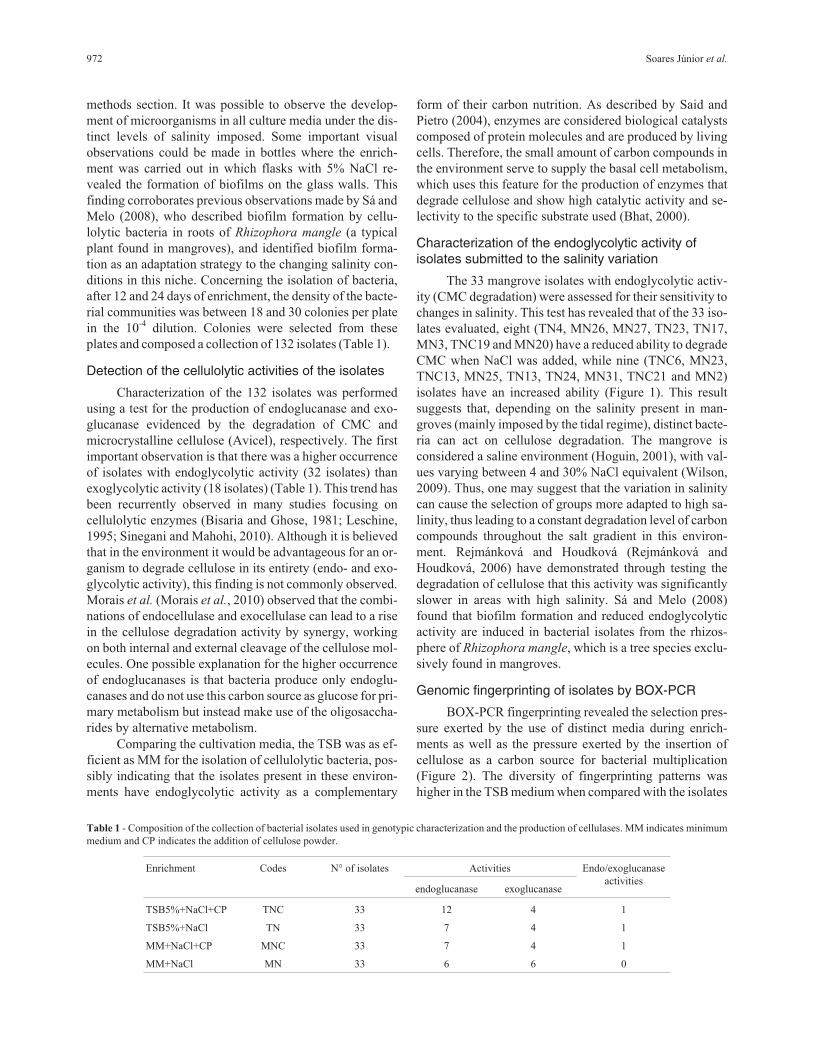
plates and composed a collection of 132 isolates (Table 1).
Detection of the cellulolytic activities of the isolates
Characterization of the 132 isolates was performed
using a test for the production of endoglucanase and exo-
glucanase evidenced by the degradation of CMC and
microcrystalline cellulose (Avicel), respectively. The first
important observation is that there was a higher occurrence
of isolates with endoglycolytic activity (32 isolates) than
exoglycolytic activity (18 isolates) (Table 1). This trend has
been recurrently observed in many studies focusing on
cellulolytic enzymes (Bisaria and Ghose, 1981; Leschine,
1995; Sinegani and Mahohi, 2010). Although it is believed
that in the environment it would be advantageous for an or-
ganism to degrade cellulose in its entirety (endo- and exo-
glycolytic activity), this finding is not commonly observed.
Morais et al. (Morais et al., 2010) observed that the combi-
nations of endocellulase and exocellulase can lead to a rise
in the cellulose degradation activity by synergy, working
on both internal and external cleavage of the cellulose mol-
ecules. One possible explanation for the higher occurrence
of endoglucanases is that bacteria produce only endoglu-
canases and do not use this carbon source as glucose for pri-
mary metabolism but instead make use of the oligosaccha-
rides by alternative metabolism.
Comparing the cultivation media, the TSB was as ef-
ficient as MM for the isolation of cellulolytic bacteria, pos-
sibly indicating that the isolates present in these environ-
ments have endoglycolytic activity as a complementary
form of their carbon nutrition. As described by Said and
Pietro (2004), enzymes are considered biological catalysts
composed of protein molecules and are produced by living
cells. Therefore, the small amount of carbon compounds in
the environment serve to supply the basal cell metabolism,
which uses this feature for the production of enzymes that
degrade cellulose and show high catalytic activity and se-
lectivity to the specific substrate used (Bhat, 2000).
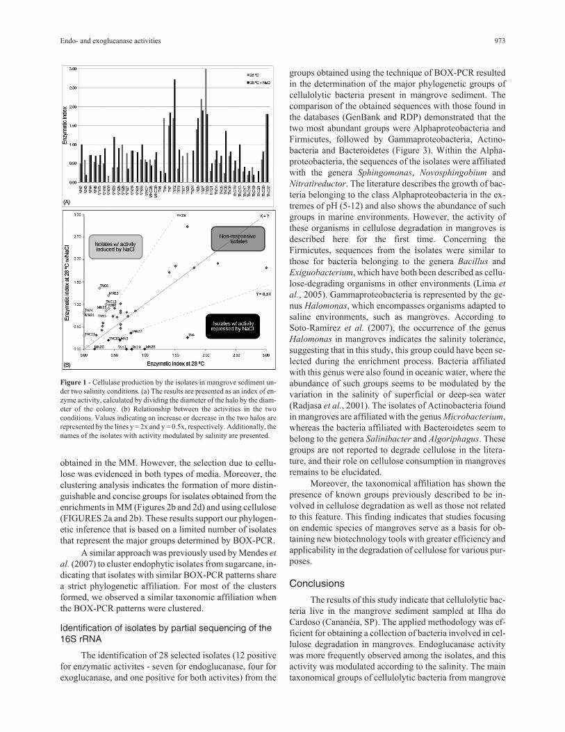
Characterization of the endoglycolytic activity ofisolates submitted to the salinity variation
The 33 mangrove isolates with endoglycolytic activ-
ity (CMC degradation) were assessed for their sensitivity to
changes in salinity. This test has revealed that of the 33 iso-
lates evaluated, eight (TN4, MN26, MN27, TN23, TN17,
MN3, TNC19 and MN20) have a reduced ability to degrade
CMC when NaCl was added, while nine (TNC6, MN23,
TNC13, MN25, TN13, TN24, MN31, TNC21 and MN2)
isolates have an increased ability (Figure 1). This result
suggests that, depending on the salinity present in man-
groves (mainly imposed by the tidal regime), distinct bacte-
ria can act on cellulose degradation. The mangrove is
considered a saline environment (Hoguin, 2001), with val-
ues varying between 4 and 30% NaCl equivalent (Wilson,
2009). Thus, one may suggest that the variation in salinity
can cause the selection of groups more adapted to high sa-
linity, thus leading to a constant degradation level of carbon
compounds throughout the salt gradient in this environ-
ment. Rejmánková and Houdková (Rejmánková and
Houdková, 2006) have demonstrated through testing the
degradation of cellulose that this activity was significantly
slower in areas with high salinity. Sá and Melo (2008)
found that biofilm formation and reduced endoglycolytic
activity are induced in bacterial isolates from the rhizos-
phere of Rhizophora mangle, which is a tree species exclu-
sively found in mangroves.
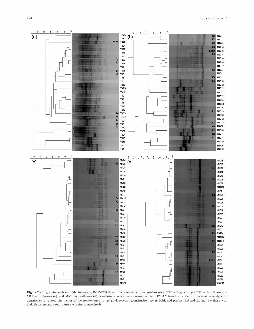
Genomic fingerprinting of isolates by BOX-PCR
BOX-PCR fingerprinting revealed the selection pres-
sure exerted by the use of distinct media during enrich-
ments as well as the pressure exerted by the insertion of
cellulose as a carbon source for bacterial multiplication
(Figure 2). The diversity of fingerprinting patterns was
higher in the TSB medium when compared with the isolates
972 Soares Júnior et al.
Table 1 - Composition of the collection of bacterial isolates used in genotypic characterization and the production of cellulases. MM indicates minimum
medium and CP indicates the addition of cellulose powder.
Enrichment Codes N° of isolates Activities Endo/exoglucanase
activitiesendoglucanase exoglucanase
TSB5%+NaCl+CP TNC 33 12 4 1
TSB5%+NaCl TN 33 7 4 1
MM+NaCl+CP MNC 33 7 4 1
MM+NaCl MN 33 6 6 0
obtained in the MM. However, the selection due to cellu-
lose was evidenced in both types of media. Moreover, the
clustering analysis indicates the formation of more distin-
guishable and concise groups for isolates obtained from the
enrichments in MM (Figures 2b and 2d) and using cellulose
(FIGURES 2a and 2b). These results support our phylogen-
etic inference that is based on a limited number of isolates
that represent the major groups determined by BOX-PCR.
A similar approach was previously used by Mendes et
al. (2007) to cluster endophytic isolates from sugarcane, in-
dicating that isolates with similar BOX-PCR patterns share
a strict phylogenetic affiliation. For most of the clusters
formed, we observed a similar taxonomic affiliation when
the BOX-PCR patterns were clustered.
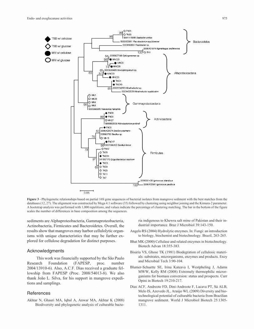
Identification of isolates by partial sequencing of the16S rRNA
The identification of 28 selected isolates (12 positive
for enzymatic activites - seven for endoglucanase, four for
exoglucanase, and one positive for both activites) from the
groups obtained using the technique of BOX-PCR resulted
in the determination of the major phylogenetic groups of
cellulolytic bacteria present in mangrove sediment. The
comparison of the obtained sequences with those found in
the databases (GenBank and RDP) demonstrated that the
two most abundant groups were Alphaproteobacteria and
Firmicutes, followed by Gammaproteobacteria, Actino-
bacteria and Bacteroidetes (Figure 3). Within the Alpha-
proteobacteria, the sequences of the isolates were affiliated
with the genera Sphingomonas, Novosphingobium and
Nitratireductor. The literature describes the growth of bac-
teria belonging to the class Alphaproteobacteria in the ex-
tremes of pH (5-12) and also shows the abundance of such
groups in marine environments. However, the activity of
these organisms in cellulose degradation in mangroves is
described here for the first time. Concerning the
Firmicutes, sequences from the isolates were similar to
those for bacteria belonging to the genera Bacillus and
Exiguobacterium, which have both been described as cellu-
lose-degrading organisms in other environments (Lima et
al., 2005). Gammaproteobacteria is represented by the ge-
nus Halomonas, which encompasses organisms adapted to
saline environments, such as mangroves. According to
Soto-Ramírez et al. (2007), the occurrence of the genus
Halomonas in mangroves indicates the salinity tolerance,
suggesting that in this study, this group could have been se-
lected during the enrichment process. Bacteria affiliated
with this genus were also found in oceanic water, where the
abundance of such groups seems to be modulated by the
variation in the salinity of superficial or deep-sea water
(Radjasa et al., 2001). The isolates of Actinobacteria found
in mangroves are affiliated with the genus Microbacterium,
whereas the bacteria affiliated with Bacteroidetes seem to
belong to the genera Salinibacter and Algoriphagus. These
groups are not reported to degrade cellulose in the litera-
ture, and their role on cellulose consumption in mangroves
remains to be elucidated.
Moreover, the taxonomical affiliation has shown the
presence of known groups previously described to be in-
volved in cellulose degradation as well as those not related
to this feature. This finding indicates that studies focusing
on endemic species of mangroves serve as a basis for ob-
taining new biotechnology tools with greater efficiency and
applicability in the degradation of cellulose for various pur-
poses.
Conclusions
The results of this study indicate that cellulolytic bac-
teria live in the mangrove sediment sampled at Ilha do
Cardoso (Cananéia, SP). The applied methodology was ef-
ficient for obtaining a collection of bacteria involved in cel-
lulose degradation in mangroves. Endoglucanase activity
was more frequently observed among the isolates, and this
activity was modulated according to the salinity. The main
taxonomical groups of cellulolytic bacteria from mangrove
Endo- and exoglucanase activities 973
Figure 1 - Cellulase production by the isolates in mangrove sediment un-
der two salinity conditions. (a) The results are presented as an index of en-
zyme activity, calculated by dividing the diameter of the halo by the diam-
eter of the colony. (b) Relationship between the activities in the two
conditions. Values indicating an increase or decrease in the two halos are
represented by the lines y = 2x and y = 0.5x, respectively. Additionally, the
names of the isolates with activity modulated by salinity are presented.
974 Soares Júnior et al.
Figure 2 - Fingerprint analyses of the isolates by BOX-PCR from isolates obtained from enrichments in TSB with glucose (a), TSB with cellulose (b),
MM with glucose (c), and MM with cellulose (d). Similarity clusters were determined by UPGMA based on a Pearson correlation analysis of
densitometric curves. The names of the isolates used in the phylogenetic reconstruction are in bold, and prefixes Ed and Ex indicate those with
endoglucanase and exoglucanase activities, respectively.
sediments are Alphaproteobacteria, Gammaproteobacteria,
Actinobacteria, Firmicutes and Bacteroidetes. Overall, the
results show that mangroves may harbor cellulolytic organ-
isms with unique characteristics that may be further ex-
plored for cellulose degradation for distinct purposes.
Acknowledgments
This work was financially supported by the São Paulo
Research Foundation (FAPESP, proc. number
2004/13910-6). Also, A.C.F. Dias received a graduate fel-
lowship from FAPESP (Proc. 2008/54013-8). We also
thank João L. Silva, for his support in mangrove expedi-
tions and samplings.
References
Akhtar N, Ghauri MA, Iqbal A, Anwar MA, Akhtar K (2008)
Biodiversity and phylogenetic analysis of culturable bacte-
ria indigenous to Khewra salt mine of Pakistan and their in-
dustrial importance. Braz J Microbiol 39:143-150.
Angelo RS (2004) Hydrolytic enzymes. In: Fungi: an introduction
to biology, biochemist and biotechnology. Brazil, 263-265.
Bhat MK (2000) Cellulase and related enzymes in biotechnology.
Biotech Advan 18:355-383.
Bisaria VS, Ghose TK (1981) Biodegration of cellulosic materi-
als: substrates, microrganisms, enzymes and products. Enzy
and Microbial Tech 3:90-104.
Blumer-Schuette SE, Irina Kateava I, Westpheling J, Adams
MWW, Kelly RM (2008) Extremely thermophilic micror-
ganisms for biomass conversion: status and prospects. Curr
Opini in Biotech 19:210-217.
Dias ACF, Andreote FD, Dini-Andreote F, Lacava PT, Sá ALB,
Melo IS, Azevedo JL, Araújo WL (2009) Diversity and bio-
technological potential of culturable bacteria from Brazilian
mangrove sediment. World J Microbiol Biotech 25:1305-
1311.
Endo- and exoglucanase activities 975
Figure 3 - Phylogenetic relationships based on partial 16S gene sequences of bacterial isolates from mangrove sediment with the best matches from the
databases (12, 27). The alignment was constructed by Mega 4.1 software (35) followed by clustering using neighbor joining and the Kimura-2 parameter.
A bootstrap analysis was performed with 1,000 repetitions, and values indicate the percentage of clustering matching. The bar in the bottom of the figure
scales the number of differences in base composition among the sequences.
Dias ACF, Andreote FD, Rigonato J, Fiore MF, Melo IS, Araújo
WL (2010) The bacterial diversity in Brazilian non-dis-
turbed mangrove sediment. Anto van Leeuwe 98:541-551.
Dillon A (2004) Cellulases. In: Enzymes as agents Biotechnol-
ogy, 243-270.
Eveleigh DE (1981) The microbial production of industrial chem-
icals. Sci American 245: 155-178.
Fengel D, Wegner G (1989) Wood chemistry, ultrastructure, reac-
tions. NCSU. Available at http://cnr.ncsu.edu/fb/
extensionoutreach/techservices/woodchem.html
Flores-Mireles AL, Winans SC, Holguin G (2007) Molecular
characterization of diazotrophic and denitrifying bacteria as-
sociated with mangrove roots. Appl Environ Microbiol
11:7308-7321.
Hamada N, Fuse N, Kodaira R, Kanda J (1999) Cloning and char-
acterization of a new exo-cellulase gene, cel3, in L. lactus.
FEMS Microbiology Letters 172:231-237.
Hanson RS, Hanson TE (1996) Methanotrofic bacteria. Microbiol
Reviews 60:439-471.
Holguin G, Vazquez P, Bashan Y (2001) The role of sediment
microrganisms in the productivity, conservation, and reha-
bilation of mangrove ecosystems: an overview. Biol and
Fert of Soils 33:265-278.
Kasana RC, Salwan R, Dhar H, Dutt S, Gulati A (2008) Arapid
and easy method for the detection of microbial cellulases on
agar plates using gram’s iodine. Curr Microbiol 57:503-507.
Kathiresan K, Bingham BL (2001) Biology of mangrove and
mangrove ecosystems. Adv Mar Biol 40:81-251.
Lane DJ, Pace B, Olsen GJ, Sthal DA, Sogin ML, Pace NR (1985)
Rapid determination of 16S ribosomal RNA sequences for
phylogenetic analyses. Procee of the Natio Acade of Scien
of the Unit Sta of America 82:6955-6959.
Lankau RA, Strauss SY (2007) Mutual feedbacks maintain both
genetic and species diversity in a plant community. Science
317:1561-1563.
Leschine S (1995) Cellulose degradation in anaerobic environ-
ments. Annual Review of Microbiol 49:399-426.
Lima AOS, Quenice MC, Fungaro MHP, Andreote FD, Mache-
roni JrW, Araújo WL, Silva Filho MC, Pizzirani-Kleiner
AA, Azevedo JL (2005) Molecular characterization of a
�-1,4-endoglucanase from a endophytic Bacillus pumilus
strain. Applied Microbiology and Biotechnology 68:57-65.
Mendes R, Pizzirani-Kleiner AA, Araujo WL, Raaijmakers JM
(2007) Diversity of cultivated endophytic bacteria from sug-
arcane: genetic and biochemical characterization of
Burkholderia cepacia complex isolates. Appl Environ
Microbiol 73:7259-7267.
Mesbah NM, Wiegel J (2008) The anaerobic halophilic alka-
lithermophiles. New York Acad of Scienc 1125:44-57.
Morais S, Heyman A, Barak Y, Caspi J, Wilson DB, Lamed R,
Shoseyov O, Bayer EA (2010) Enhanced cellulose degrada-
tion by nano-complexed enzymes: Synergism between a
scaffold-linked exoglucanase and a free endoglucanase. J of
Biotechnol 147:205-211.
Radjasa OK, Urakawa H, Kita-Tsukamoto K, Ohwada K (2001)
Characterization of psychrotrophic bacteria in the surface
and dee-sea waters from the Northwestern Pacific Ocean
based on 16s ribossomal DNA analysis. Marine Biotech
3:454-462.
Rejmánková E, Houdková K (2006) Wetland plant decomposition
under different nutrient conditions: what is more important,
litter quality or site quality? Biogeochemistry 80:245-262.
Said S, Pietro R (2004) Generalities on industrial enzyme applica-
tion. In: Enzymes as Biotechnological Agents Brazil, pp 1-7.
Sá ALB, Melo IS (2008) Diversidade de rizobactérias endo-
glicoliticas isoladas de mangue vermelho (Rhizophora man-
gle). São Paulo, Brazil, 61 pp (M.Sc. Dissertation. Instituto
de Biotecnologia. USP).
Schallmey M, Singh A, Ward OP (2004) Developments in the use
of Bacillus species for industrial production. Canad J Micro-
biol 50:1-17.
Sinegani AAS, Mahohi A (2010) Soil water potential effects on
the cellulase activities of soil treated with sewage sludge.
Plant Soil Environme 56:333-339.
Soto-Ramírez N, Sánchez-Porro C, Rosas S, González W, Quiño-
nes M, Ventosa A, Montalvo-Rodríguez R (2007) Halo-
monas Avicenniae sp. nov., isolated from the salty leaves of
the black mangrove Avicennia germinans in Puerto Rico. Int
J Syst Evol Microbiol 57:900-905.
Tacão M, Alves A, Saavedra MJ, Correia A (2005) BOX-PCR is
an adequate tool for typing Aeromonas spp. Antonie van
Leeuwenhoek 88:173-179.
Taketani RG, Yoshiura CA, Dias ACF, Andreote FD, Tsai SM
(2010) Diversity and identification of methanogenic archaea
and sulphate-reducing bacteria in sediments from a pristine
tropical mangrove. Anto van Leeuwe 97:401-411.
Tamura K, Dudley J, Nei M, Kumar S (2007) MEGA4: Molecular
Evolutionary Genetics Analysis (MEGA) software versión
4.0. Mol Biolo and Evolu 24:1596-1599.
Tengerdy RP, Szakacs G (2003) Bioconversion of lignocellulose
in solid substrate fermentation. Bioche l Enginee J 13:169-
179.
Thauer R, Shima S (2006) Biogeochemistry - methane and mi-
crobes. Nature 440:878-879.
Wilson DB (2009) Cellulases and biofuels. Curr Opi in Biotech
20:295-299.
Zahran HH (1997) Diversity, adaptation and activity of the bacte-
rial flora in saline environments. Biol and Fert of Soils
25:211-223.
All the content of the journal, except where otherwise noted, is licensed under a
Creative Commons License CC BY-NC.
976 Soares Júnior et al.