Embed Size (px)
Citation preview
Fusion Proteins as Research ToolsJosephine C Dorsman, University of Leiden, Leiden, The Netherlands
Fusion proteins encompass a protein or small peptide moiety which is used as a tag, fused
to another protein of interest. Fusion proteins are used in biochemical and in genetic
(high-throughput) applications and are especially suited to study protein–protein
interactions, even on a genome-wide scale, and for the generation of antibodies.
Introduction
In today’s protein research, engineered fusion proteinsencompassing a protein or parts of a protein of interestcoupled to a tag provide invaluable research tools. Thecoupling of proteins to specific tags allows an easydetection and/or easy purification of the protein ofinterest. Purified fusion proteins can be used inbiochemical studies and for the generation of anti-bodies. In addition, fusion proteins provide excellenttools to identify protein–protein interactions, even ona genome-wide scale. Although these approaches havebeen pioneered in academic settings, many of thesestrategies have now been successfully commercialized.
Purification of Fusion Proteins in aSingle Step
A new stage for protein purification was set by thedevelopment of procedures in which fusion proteinsencompassing a tag could be purified in a single stepvia affinity chromatography, and over the yearsvarious prokaryotic- and eukaryotic-based systemshave been developed. Sizes of the tag range from verysmall peptides, which are mainly used for immunolo-gical detection, to full-length proteins from whichspecial properties are exploited. As of 2002, theutilization of the glutathione-S-transferase (GST)system for prokaryotic expression is, nevertheless,unsurpassed in its proven capabilities in numerousapplications (Smith and Johnson, 1988). The combi-nation of efficient and mild purification conditionsmakes the system amenable to the expression andisolation of the majority of proteins in their nativeform. Other systems that have proved themselves arethe His-tag system, in which proteins are fused to asmall stretch of histidines (see Hoffmann and Roeder,1991) and the MBP system, in which proteins are fusedto the maltose-binding protein (see Maina et al., 1988).The GST system will be used as an example to explainthe principles of the methodologies and theirlimitations.
In the original GST system, parts of the gene ofinterest are cloned in the multicloning site of the so-called pGEX vectors, as in-frame fusions with theSchistosoma japonicum GST gene (Smith and Johnson,1988). The pGEX vectors allow a chemically inducibleand high-level expression of proteins encompassingthe 26-kDa GST protein in Escherichia coli. Theproduced proteins can be purified from the extractsin one step by exploiting the affinity of the GSTenzyme for its substrate, glutathione, using glu-tathione beads (see Figure 1). The purified fusionproteins can be used in biochemical studies or for thegeneration of antibodies. Another possibility is to usethe purified fusion protein bound to the beads forprotein–protein interaction studies.
Various new and versatile derivatives of the initialpGEX vectors have been developed which displayfeatures that increase the utility of this system. Vectorsare available, for example, in which a short sequence ispresent between the GST sequence and the polylinkersequence encoding a recognition site for a commer-cially available protease. This feature allows cleavageof the protein of interest from the GST moiety. Thisproperty is extremely useful, especially when thepurified proteins are subjected to structural studiessuch as nuclear magnetic resonance (NMR), since thetag may interfere with the proper folding of the proteinof interest.
When prokaryotic-based systems are used for theexpression of eukaryotic proteins, some problems andinherent limitations are, nevertheless, encountered,some of which can be addressed relatively easily.Firstly, the relative abundance of transfer ribonucleicacids (tRNAs) for certain codons differs significantlybetween prokaryotes and eukaryotes, resulting in the
Advanced article
Article contents
� Introduction
� Purification of Fusion Proteins in a Single Step
� Utilization of Purified Fusion Proteins for theGeneration of Antibodies
� Purified Fusion Proteins as Tools for the Study ofProtein–Protein Interactions
� Genetic Approaches to Identify Protein–ProteinInteractions
� High-throughput Applications
� Conclusion
doi: 10.1038/npg.els.0005685
Fusion Proteins as Research Tools
ENCYCLOPEDIA OF LIFE SCIENCES & 2005, John Wiley & Sons, Ltd. www.els.net 1
poor expression of certain eukaryotic proteins inE. coli. This problem can be addressed via variousapproaches, including site-directed mutagenesis totriplets that are more favorable for expression inE. coli or the use of specially engineered E. coli strainsthat express specific eukaryotic tRNAs (see Dieci et al.,2000). Another, more difficult problem is that proteinsproduced in E. coli lack the appropriate eukaryoticprotein modifications such as phosphorylation. Suc-cessful attempts have been described to produce fusionproteins in eukaryotic expression systems (see Braunand Suske, 1999), such as the insect-based Baculovirussystems. Although the latter systems are more expen-sive than the prokaryotic systems and in addition mayrequire, for example, facilities for dealing with viruses,it is clear that for some purposes they are preferred.
Utilization of Purified Fusion Proteinsfor the Generation of Antibodies
Purified GST- and other fusion proteins can be usedas antigens for immunization of animals. For thispurpose, it is less important that the purified proteinsare in their native state than for the protein–proteininteraction studies. Proteins that are at least partlydenatured can be even more immunogenic than thenative proteins. When fusion proteins are used toinject animals, for example rabbits, a mixture ofantibodies will be produced which react with eitherthe tag or the protein of interest. The antibodies thatreact with the protein of interest can subsequently be
purified using small-scale (e.g. on membrane strips) orlarge-scale (affinity chromatography) approaches(Harlow and Lane, 1988). In addition, the quality ofphage-display antibody libraries, which in principleprovide an unlimited source of monoclonal antibodieswithout the intervention of animals, was increasingrapidly in the year 2000 (see Holt et al., 2000). In thephage-display systems, fusion proteins have alreadybeen successfully used to retrieve relevant antibodiesuseful in biomedical research. In the special situationwhere one is interested in the generation of antibodiesthat specifically react with modified, for exampleacetylated or phosphorylated, forms of a peptide, itis better to opt for peptide-based strategies. Fusionproteins nevertheless provide, in many cases, avaluable and inexpensive source for the generation ofpotent and specific antibodies.
Purified Fusion Proteins as Tools forthe Study of Protein–ProteinInteractions
One of the main reasons for the success of the GSTsystem is its excellent suitability for the study ofprotein–protein interactions in pull-down assays andalso, to a lesser extent, far-Western experiments. Thepull-down procedure is suitable to detect relativelystrong protein–protein interactions, and both directand indirect interactions via intermediate proteins canbe identified. The procedure can be summarized asfollows: GST-fusion proteins bound to glutathione
Purification of GST (-fusion) proteins in a single step
Elution of purified GST (-fusion) proteins from beads by glutathione
(a)
(b)
Glutathione beads
Glutathione beadsGlutathionein solution
Glutathione beads bound toGST (-fusion) proteins
GST (-fusion) proteinsin solution
Glutathione beads bound toGST (-fusion) proteins
GST (-fusion) proteinsin cell extract
+
+ +
Washing andcentrifugation
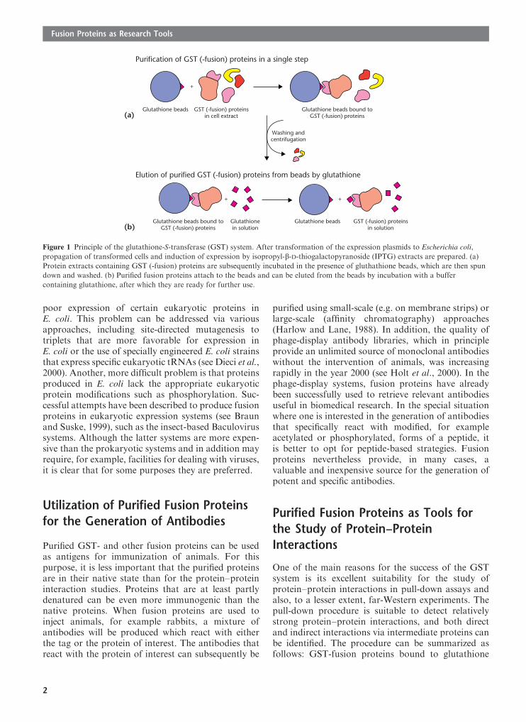
Figure 1 Principle of the glutathione-S-transferase (GST) system. After transformation of the expression plasmids to Escherichia coli,
propagation of transformed cells and induction of expression by isopropyl-b-D-thiogalactopyranoside (IPTG) extracts are prepared. (a)
Protein extracts containing GST (-fusion) proteins are subsequently incubated in the presence of gluthathione beads, which are then spun
down and washed. (b) Purified fusion proteins attach to the beads and can be eluted from the beads by incubation with a buffer
containing glutathione, after which they are ready for further use.
Fusion Proteins as Research Tools
2

beads are incubated with a source for the proteins to betested, for example, cell extracts or in vitro translatedproteins. After incubation, the GST-fusion proteinstogether with the associating proteins are isolated viacentrifugation, and the potential binding of proteinscan be analyzed subsequently via standard sodiumdodecyl sulfate–polyacrylamide gel electrophoresis(SDS-PAGE) and detection techniques (see Figure 2).When combined with strategies that allow in vitrotranscription–translation performed on polymerasechain reaction (PCR) fragments, it is possible to testvirtually any desired protein fragment for bindingrapidly (see Keblusek et al., 1999). This method isoften used to confirm interactions identified with other
approaches, such as in yeast two-hybrid screens or forthe delineation of regions of a protein involved inbinding.
In far-Western analysis, a purified and labeledfusion protein is used as a probe on protein blots orto screen protein-expression libraries. Some of the newgeneration of pGEX vectors, for example pGEX-2TK,are valuable tools in this respect, because they enablethe efficient radiolabeling of GST-fusion proteins bythe commercially available heart muscle kinase(HMK). A possible procedure for a far-Westernanalysis is depicted in Figure 3. In brief, proteins areloaded on an SDS-PAGE gel; after electrophoresis theproteins are blotted to a membrane, followed by
35S
35S
Glutathione beads withGST (-fusion) proteins
In vitro translated and35S-methionine-labeled
proteins
+
35S
35S
1 2 3 4 1 2 3 4 1 2 3 4
Boiling of samples indenaturing buffer and
electrophoresis of samples
‘Pull-down’ of GST (-fusion) proteinswith 35S-methionine-labeled
proteins
Detection Drying of gels
Figure 2 GST pull-down. GST (-fusion) proteins are incubated with in vitro translated proteins labeled with, for example, [35S]methionine.
After isolation of the fusion proteins plus associating proteins by centrifugation, the isolated proteins are subsequently detached from the
beads by boiling in a denaturing buffer, followed by SDS-PAGE and detection. Lane 1, molecular size marker. Lane 2, input of labeled
in vitro translated protein. Lane 3, results of a control pull-down experiment of GST proteins and the in vitro translated proteins. Lane 4, pull-
down of GST-fusion proteins with the in vitro translated proteins. The translated protein binds to the GST-fusion protein (lane 4),
but not to the GST control (lane 3).
1 2 3 41 2 3 41 2 3 4
SDS-PAGE of proteins(extracts)
Blotting of proteinsto membrane
Addition of protein probelabeled with 32P
Detection
32P
+
Figure 3 Far-Western analysis. Protein extracts or (partially) purified proteins are loaded on an SDS-PAGE gel. The proteins are
subsequently blotted to a membrane. Optionally, the proteins on the filter can be completely denatured, followed by renaturation. The
blot is subsequently incubated with a purified and labeled protein probe, followed by washing of the membrane and detection. Lane 1,
molecular size marker; lane 2, whole-cell extract (WCE) containing protein X; lane 3, protein fraction lacking proteins X; lane 4, purified
protein X. The protein probe can bind directly to protein X. In the WCE, one smaller protein is present which also can directly bind to
the protein probe.
Fusion Proteins as Research Tools
3

incubation with the labeled protein probe and sub-sequent detection of the bound fusion protein.Optionally, the proteins on the filter membrane canbe completely denatured, for example with guanidi-nium chloride, followed by renaturation to increasethe chance that proteins will be folded in their nativeconformation (Hager and Burgess (1980) pioneeredthese denaturation–renaturation protocols). Far-Western analysis has been used primarily to determinewhether proteins bind directly to each other, butsuccessful attempts at screening of expression librarieshave also been reported (see Shvarts et al., 1996).
Genetic Approaches to IdentifyProtein–Protein Interactions
A complementary approach to identifying protein–protein interactions is the so-called yeast two-hybridsystem. In contrast to the biochemical methods, weakand/or even transient interactions can be identified. Inthe most popular version of this system, which waspioneered by Song and Fields, the modular structureof transcription factors, consisting of a DNA-bindingdomain and a transcriptional activation domain, isexploited: a protein–protein interaction is identified bymeansof reconstitution of the activity of a transcriptionfactor in the yeast Saccharomyces cerevisiae(Fields and Song, 1989). One plasmid encodes theDNA-binding domain of a transcription factor,usually of Gal4 or LexA, fused to the protein ofinterest (bait), whereas the other plasmid encodes atranscriptional activation domain, usually of Gal4 orVP16, fused to a protein (prey). Efficient transcriptioncan occur when the two hybrid proteins interact (seeFigure 4). The most important requirement of thesystem is that the bait itself does not result intranscriptional activation itself (Figure 4). Manylibraries are now available which have been cloned inactivation-domain plasmids and which can be used forthe rapid identification of complementary deoxyribo-nucleic acids (cDNAs) for associating proteins. Sincethe original description of the system, many adapta-tions and variations have been added. One importantvariation of the nuclear system allows the use of baitsthat already give rise to transcriptional activationthemselves. In this system the bait is present in theactivation-domain plasmid, and the prey (or library) ispresent in theDNA-binding domain plasmid (Du et al.,1996). This system is, nevertheless, more laboriousthan the original system. Other systems have beendescribed which can detect protein–protein interac-tions in the cytoplasm (Broder et al., 1998). As of 2002,the improved versions of the nuclear two-hybrid
GAL4-D
B
(a)
(b)
(c)
(d)
(e)
P
B
P
B
P
GAL4-UAS
LacZ
LacZ
LacZ
LacZ
LacZ
GAL4-A
GAL4-UAS
GAL4-A
GAL4-D
GAL4-UAS
GAL4-UAS
GAL4-UAS
GAL4-D
GAL4-A
GAL4-D
GAL4-A
Figure 4 The classic yeast two-hybrid assay exploits the modular
structure of transcriptional activators: a protein–protein interaction
is identified by reconstitution of the activities of a transcriptional
activator. (a) One yeast expression plasmid encodes the DNA
binding domain of the transcription factor GAL4 (GAL4-D) fused
to the protein of interest (B, ‘bait’). This fusion protein binds to the
promoter region, but, in the absence of a transcriptional activation
domain, this protein will not give rise to significant transcriptional
activation of the b-galactosidase reporter (LacZ) gene. (b) The otheryeast expression plasmid encodes the transcriptional activation
domain of GAL4 fused to the protein of interest or proteins encoded
by a cDNA library (P, ‘prey’). In the absence of a DNA-binding
domain or promoter-targeting domain, this fusion protein will not
give rise to transcriptional activation. (c) Two-hybrid experiment.
The bait plasmid and the prey plasmid are cotransformed. The
binding of the prey to the bait gives rise to the presence of a strong
transcriptional activation domain in the promoter region, resulting
in (strongly) increased transcriptional activation. (d, e) Two
additional control experiments are necessary to verify that the
interaction does not involve binding to GAL4-A or GAL4-D.
(d) The bait together with the GAL activation domain alone should
not give rise to transcriptional activation. (e) The GAL4 DNA-
binding domain alone with the prey should not result in
transcriptional activation.
Fusion Proteins as Research Tools
4

system remain the most powerful. (See Yeast Two-hybrid System and Related Methodology.)
High-throughput Applications
The applications described previously have been usedmainly for small-scale purposes. However, as of 2002,there is a glimpse of the exciting possibilities offered bythe versatile single-step protein purification and yeasttwo-hybrid systems for high-throughput purposes.Several biochemical methods for purification of fusionproteins and study of protein function on a large scalehave been described (see e.g. Martzen et al., 1999).Systems with fluorescent proteins as tags, for examplegreen fluorescent protein (GFP), can be used ascomplementary analyses. The latter systems have anadded value, because they allow the follow-up ofprocesses in the living cell (see Brachat et al., 2000). Togenerate protein–protein interaction maps, yeast two-hybrid analyses have been used very successfully(Walhout et al., 2000). These genetic approaches arecomplemented by biochemical approaches that com-bine powerful techniques for the purification ofprotein complexes containing a tagged protein, withrapidly evolving protein-identification techniques suchas mass spectroscopy (Rigaut et al., 1999).
Conclusion
GST-fusion proteins and other fusion proteins thatcan be purified in a single step will remain valuableresearch tools in small-scale protein–protein interac-tion studies and for the generation of antibodies. Inaddition, their use will increase owing to their extremeusefulness in high-throughput functional genomicsand proteomics applications. The biochemical systemsare complemented by the genetic systems, which alsoprovide excellent tools for the identification ofprotein–protein interactions.
See alsoProtein–Protein Interaction MapsYeast Two-hybrid System and Related Methodology
References
Brachat A, Liebundguth N, Rebischung C, et al. (2000) Analysis ofdeletion phenotypes and GFP fusions of 21 novel Saccharomycescerevisiae open reading frames. Yeast 16: 241–253.
Braun H and Suske G (1999) Vectors for inducible expression ofdual epitope-tagged proteins in insect cells. Biotechniques 6:1038–1042.
Broder YC, Katz S and Aronheim A (1998) The Ras recruitmentsystem: a novel approach to the study of protein–proteininteractions. Current Biology 8: 1121–1124.
Dieci G, Bottarelli L, Ballabeni A and Ottonello S (2000) tRNA-assisted overproduction of eukaryotic ribosomal proteins.Protein Expression and Purification 18: 346–354.
Du W, Vidal M, Xie JE and Dyson N (1996) RBF, a novel RB-related gene that regulates E2F activity and interacts with cyclinE in Drosophila. Genes and Development 10: 1206–1218.
Fields S and Song O (1989) A novel genetic system to detect protein–protein interactions. Nature 340: 245–246.
Hager DA and Burgess RR (1980) Elution of proteins from sodiumdodecyl sulfate–polyacrylamide gels, removal of sodium dodecylsulfate, and renaturation of enzymatic activity: results with sigmasubunit of Escherichia coli RNA polymerase, wheat germ DNAtopoisomerase, and other enzymes. Analytical Biochemistry 109:76–86.
Harlow E and Lane D (1988) Antibodies: A Laboratory Manual. ColdSpring Harbor, NY: Cold Spring Harbor Laboratory Press.
Hoffmann A and Roeder RG (1991) Purification of his-taggedproteins in non-denaturing conditions suggests a convenientmethod for protein interaction studies. Nucleic Acids Research 19:6337–6338.
Holt LJ, Enever C, DeWildt RM and Tomlinson IM (2000) The useof recombinant antibodies in proteomics. Current Opinion in
Biotechnology 11: 445–449.Keblusek P, Dorsman JC, Teunisse AF, et al. (1999) The adenoviral
E1A oncoproteins interfere with the growth-regulatory inhibitingeffect of the cdk-inhibitor p21 CIP1/WAF1. Journal of GeneralVirology 80: 381–390.
Maina CV, Riggs PD, Grandea AG, et al. (1988) An Escherichia coli
vector to express and purify foreign proteins by fusion to andseparation from maltose-binding protein. Gene 74: 365–373.
Martzen MR, et al. (1999) A biochemical genomics approach foridentifying genes by the activity of their products. Science 286:1153–1155.
Rigaut G, Shevchenko A, Rutz B, et al. (1999) A generic proteinpurification method for protein complex characterization andproteome exploration. Nature Biotechnology 17: 1030–1032.
Shvarts A, Steegenga WT, Riteco N, et al. (1996) MDMX: a novelp53-binding protein with some functional properties of MDM2.The EMBO Journal 15: 5349–5357.
Smith DB and Johnson KS (1988) Single-step purification ofpolypeptides expressed in Escherichia coli as fusions withglutathione-S-transferase. Gene 67: 31–40.
Walhout AJ, Sordells R, Lu X, et al. (2000) Protein interactionmapping in C. elegans using proteins involved in vulvaldevelopment. Science 287: 116–122.
Further Reading
Becker-Hapak M, McAllister SS and Dowdy SF (2001) Tat-mediated protein transduction into mammalian cells. Methods
24: 247–256.Carlson M (2000) The awesome power of yeast biochemical
genomics. Trends in Genetics 16: 49–51.Clackson T, Yang W, Rozamus LW, et al. (1998) Redesigning an
FKBP-ligand interface to generate chemical dimerizers withnovel specificity. Proceedings of the National Academy of Sciencesof the United States of America 95: 10 437–10 442.
Golemis EA (ed.) (2002) Protein–Protein Interactions: A Molecular
Cloning Manual. Cold Spring Harbor, NY: Cold Spring HarborLaboratory Press.
Gonzalez C and Bejarano LA (2000) Protein traps: usingintracellular localization for cloning. Trends in Cell Biology 10:162–165.
Kemmeren P, van Berkum NL, Vilo J, et al. (2002) Proteininteraction verification and functional annotation by integratedanalysis of genome-scale data. Molecular Cell 9: 1133–1143.
Fusion Proteins as Research Tools
5

MacDonald P (ed.) (2001) Two-hybrid Systems: Methods and
Protocols. Totowa, NJ: Humana Press.Phizicky EM,MartzenMR,McCraith SM, et al. (2002) Biochemical
genomics approach to map activities to genes. Methods in
Enzymology 350: 546–559.Rooney I, Butrovich K and Ware CF (2000) Expression of
lymphotoxins and their receptor-Fc fusion proteins by baculo-virus. Methods in Enzymology 322: 345–363.
Smith DB (2000) Generating fusions to glutathione-S-transferase forprotein studies. Methods in Enzymology 326: 254–270.
Vidal M and Legrain P (1999) Yeast forward and reverse ‘n’-hybridsystems. Nucleic Acids Research 27: 919–929.
Fusion Proteins as Research Tools
6