Embed Size (px)
Citation preview
Chapter 3Roles of SNARE Proteins in Synaptic Vesicle Fusion
Mark T. Palfreyman and Erik M. Jorgensen
Abstract Neurotransmitters are stored in small membrane-bound vesicles at synapses. Neurotransmitter release is initiated by depolarization of the neuron, which in turn activates voltage-gated calcium channels. Calcium influx then trig-gers the fusion of the synaptic vesicles with the plasma membrane. Fusion of the vesicular and plasma membranes is mediated by SNARE (soluble N-ethylmaleimide–sensitive factor attachment receptor) proteins. The SNAREs are now known to be used in all trafficking steps of the secretory pathway, including neurotransmission. This chapter describes the discovery of the SNAREs, their relevant structural features, models for their function, the specificity of interactions, and their interactions with the calcium-sensing machinery.
Keywords SNARE, syntaxin, SNAP-25, synaptobrevin, membrane fusion
Z.-W. Wang (ed.) Molecular Mechanisms of Neurotransmitter Release, 35© Humana Press 2008
Erik M. JorgensenDepartment of Biology and Howard Hughes Medical Institute, University of Utah, Salt Lake City, UT 84112e-mail: [email protected]
Contents
SNARE Discovery: A Convergence of Genetics and Biochemistry .......................................... 36Defi nitions: The World Turned Upside Down and Given a Good Shake .................................. 37Molecular Characteristics of the SNAREs ................................................................................. 38Assembly and Disassembly Cycles in SNARE Function .......................................................... 40A Model for Membrane Fusion ................................................................................................. 42The Reliable Opposition: Protein Models for the Fusion Pore .................................................. 46SNAREs Encode Specifi city ...................................................................................................... 47SNARE Regulation .................................................................................................................... 49Conclusion ................................................................................................................................. 50References .................................................................................................................................. 51
Wang_Ch03.indd 35Wang_Ch03.indd 35 5/15/2008 5:27:11 PM5/15/2008 5:27:11 PM
36 M.T. Palfreyman, E.M. Jorgensen
SNARE Discovery: A Convergence of Genetics and Biochemistry
To understand the mechanisms of synaptic vesicle fusion, it is useful to think about the evolution of neurotransmission. Eukaryotic cells separate cellular functions into membrane-bound organelles. The content of these organelles are moved between compartments and the extracellular environment by transport vesicles. Cellular com-partments must be kept distinct, but membrane-impermeable cargo must be trans-ferred to the target organelle. To transfer cargo the lipid bilayers of the vesicle and the target must merge so that their luminal contents can intermingle. In some cases, cargo must be secreted into the extracellular space via exocytosis. It was perhaps a small step for the cell to develop a mechanism for calcium-dependent regulation of exocytosis, but it was a giant leap for evolution. The nervous system is arguably the universe’s greatest invention.
A convergence of independent tracks led to the identification of SNAREs as the central players in membrane fusion. In the late 1980s SNARE proteins were identified in the brain as components of the synapse. Specifically, synaptobrevin (also called vesicle-associated membrane protein [VAMP]) was purified from syn-aptic vesicles (1). Subsequently, two additional SNAREs, syntaxin and SNAP-25 (synaptosome-associated protein of 25 kDa), were found localized to the plasma membrane of neurons (2–4). The identification of homologues among the yeast secgenes linked the mechanisms of synaptic function to vesicular trafficking (5,6) and hinted at the universality of membrane fusion. Although the SNARE proteins were well placed to mediate synaptic vesicle fusion and were related to proteins required for trafficking, there was at this point no evidence that these proteins functioned in calcium-dependent exocytosis of synaptic vesicles.
The groups of Heiner Niemann, Reinhard Jahn, and Cesare Montecucco were looking for the targets of the clostridial toxins. The clostridial toxins from the anaerobic bacteria Clostridium botulinum and Clostridium tetani can potently inhibit neurotransmission (7). Thus, it was reasoned that their targets would iden-tify essential proteins in synaptic transmission. Botulinum and tetanus toxins cleave the SNARE proteins, demonstrating the central role of the SNAREs in synaptic vesicle release (8–11). These were the first functional data that the SNAREs were involved in neurotransmission (12,13). The central role of the SNAREs in neuro-transmission would later be confirmed from electrophysiologic studies on null mutants in the SNARE proteins in Drosophila, mice, and Caenorhabditis elegans(14–19). Thus, the functional data identified the SNAREs as perpetrators but their association had not been described.
The discovery that these proteins formed a complex was demonstrated soon after. Jim Rothman’s group was taking a biochemical approach to understand traf-ficking in the Golgi apparatus. The toxin N-ethylmaleimide (NEM) potently inhib-its Golgi trafficking (20). Wilson et al (21) found that the target of NEM was the mammalian homologue of a previously cloned yeast gene SEC18 (22). Rothman’s group named this new protein the NEM-sensitive factor (NSF) (23), and NSF was
Wang_Ch03.indd 36Wang_Ch03.indd 36 5/15/2008 5:27:11 PM5/15/2008 5:27:11 PM
3 Roles of SNARE Proteins in Synaptic Vesicle Fusion 37
found to bind, via the action of the soluble NSF adaptors (SNAPs) (24), to a set of proteins from brain detergent extracts that came to be collectively known as the soluble N-ethylmaleimide–sensitive factor attachment receptor proteins (SNAREs). The evidence for SNARE involvement in synaptic vesicle exocytosis was now overwhelming, but a list of names in a complex did not constitute a model.
The first coherent model, called the SNARE hypothesis, would arise from the melding of the genetic and biochemical observations described above. Although wrong in detail, it would catalyze a number of hypothesis-driven experiments that would lead to more accurate models. Based on the finding that unique SNAREs are found at each of the trafficking steps (25,26), Thomas Söllner and Jim Rothman proposed that SNARE interactions provided the specificity for vesicular trafficking by tethering the vesicle to its target membrane (27,28). The SNAREs would then be acted on by the adenosine triphosphatase (ATPase) NSF which, by disassem-bling the SNAREs, would drive fusion (27,29).
Further experiments from Bill Wickner’s lab, using a purified vacuole fusion assay, demonstrated that NSF acted not at the final step of fusion, but rather to recover monomeric SNAREs for use in further rounds of fusion (28,29,30,31). NSF was acting as a chaperone to separate the embracing SNAREs on the plasma mem-brane to reactivate the system for further fusion (32,33). Thus assembly of the SNAREs, not disassembly, catalyzes fusion.
Finally, Rothman’s group demonstrated that the SNAREs alone could fuse mem-branes. The SNAREs were incorporated into vesicles composed of artificial lipid bilayers. Donor vesicles containing synaptobrevin were capable of fusion with acceptor vesicles containing syntaxin and SNAP-25 (34). This experiment was extended to native membranes by engineering SNAREs to face out of the cell; in this configuration the SNAREs could induce fusion of whole cells (35). Thus, the current thinking is that the SNAREs function in the final steps in fusion and repre-sent the minimal fusion machinery.
In the following sections we briefly define the steps leading to fusion, introduce the structure of the SNARE proteins, present a model for fusion, discuss SNARE specificity, and finally touch on the regulation of the SNARE complex by other proteins.
Definitions: The World Turned Upside Down and Given a Good Shake
In the past, synaptic vesicles were thought to dock with the plasma membrane, and then undergo a maturation step in which they became release ready. Depolarization activated a calcium sensor that then allowed the vesicle to fuse with the plasma membrane. Only a subset of docked vesicles were considered to be in the readily releasable pool (36). Thus, the life of a vesicle could be divided into four steps: docking, maturing to release-ready, calcium sensing, and fusing. The definition of these stages in vesicle fusion relied on morphologic and electrophysiologic criteria.
Wang_Ch03.indd 37Wang_Ch03.indd 37 5/15/2008 5:27:11 PM5/15/2008 5:27:11 PM
38 M.T. Palfreyman, E.M. Jorgensen
Current studies have sought to associate these pools with particular molecular inter-actions and thereby more precisely define these states.
Paradoxically, recent studies have tended to confuse rather than clarify the states of a vesicle. Although some have argued that very few docked vesicles are in the readily releasable pool (36,37), others suggest that docked vesicles are equivalent to the readily releasable pool (38–41). Studies of SNARE proteins have also mud-died our previously clean definitions of these pools. The assembly of SNARE pro-teins between synaptic vesicle and plasma membrane is defined as vesicle “priming.” Initial studies suggested that priming occurred after docking (15). However, recent studies suggest that the primed state may correspond to “docked” vesicles as observed in electron micrographs (14). Thus, the morphologic, electro-physiologic, and molecular definitions have seemingly converged on a single state. It is hoped that as the actions of various proteins are more precisely understood, we will once again refine synaptic vesicle fusion into discrete steps.
There is one last sorry note concerning our attempts to define steps in vesicle fusion: the terminology used for synaptic vesicle fusion is at odds with the terminol-ogy used in yeast. In yeast, priming” refers to the generation of free SNAREs rather than the formation of the SNARE complex, tethering rather than docking describes the initial membrane association, and docking includes SNARE engagement. Only the word fusion seems to mean the same thing in these different languages.
Molecular Characteristics of the SNAREs
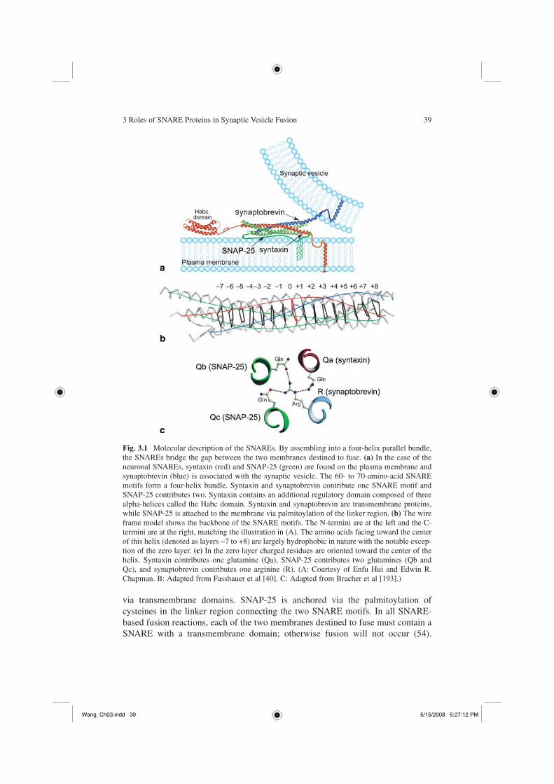
The SNARE proteins are characterized by a conserved 60- to 70-amino-acid SNARE motif. Phylogenetic analysis indicates that SNARE proteins can be divided into four families (25,42,43). The individual SNARE motifs are largely unstructured in solution, but when all four family members are mixed, the SNARE motifs come together to form a four-helix parallel bundle known as the core complex (Fig. 3.1A,B) (44). The SNARE complex is remarkably stable and can only be separated by boiling in the presence of sodium dedocyl sulfonate (SDS) (45,46). The hydrophobic residues of the alpha-helical SNARE motifs are oriented inward to form layers like those in the coiled coil domains of classical leucine zippers. However, the layer in the middle of the complex, called the “0” layer, is formed by ionic interactions between an arginine (R-SNARE) and three glutamines (Qa, Qb, and Qc SNAREs) (Fig. 3.1B,C). The role for these conserved resi-dues buried in the hydrophobic core is briefly discussed in the next section. At each fusion site a unique SNARE complex consisting of all four flavors is formed. While other complexes have been observed in vitro, the only complexes that have been shown to efficiently support fusion are QabcR complexes (47–52).
The SNAREs that are used for synaptic vesicle exocytosis are synaptobrevin (R-SNARE, also called VAMP2), syntaxin 1a (Qa SNARE), and SNAP-25 (con-tains both the Qb and Qc SNARE motifs) (Fig. 3.1) (1–4,53).
In addition to the SNARE motifs, all three SNAREs contain sequences that anchor them to the membrane (Fig. 3.1A). Syntaxin and synaptobrevin are anchored
Wang_Ch03.indd 38Wang_Ch03.indd 38 5/15/2008 5:27:12 PM5/15/2008 5:27:12 PM
3 Roles of SNARE Proteins in Synaptic Vesicle Fusion 39
Fig. 3.1 Molecular description of the SNAREs. By assembling into a four-helix parallel bundle, the SNAREs bridge the gap between the two membranes destined to fuse. (a) In the case of the neuronal SNAREs, syntaxin (red) and SNAP-25 (green) are found on the plasma membrane and synaptobrevin (blue) is associated with the synaptic vesicle. The 60- to 70-amino-acid SNARE motifs form a four-helix bundle. Syntaxin and synaptobrevin contribute one SNARE motif and SNAP-25 contributes two. Syntaxin contains an additional regulatory domain composed of three alpha-helices called the Habc domain. Syntaxin and synaptobrevin are transmembrane proteins, while SNAP-25 is attached to the membrane via palmitoylation of the linker region. (b) The wire frame model shows the backbone of the SNARE motifs. The N-termini are at the left and the C-termini are at the right, matching the illustration in (A). The amino acids facing toward the center of this helix (denoted as layers −7 to +8) are largely hydrophobic in nature with the notable excep-tion of the zero layer. (c) In the zero layer charged residues are oriented toward the center of the helix. Syntaxin contributes one glutamine (Qa), SNAP-25 contributes two glutamines (Qb and Qc), and synaptobrevin contributes one arginine (R). (A: Courtesy of Enfu Hui and Edwin R. Chapman. B: Adapted from Fasshauer et al [40]. C: Adapted from Bracher et al [193].)
via transmembrane domains. SNAP-25 is anchored via the palmitoylation of cysteines in the linker region connecting the two SNARE motifs. In all SNARE-based fusion reactions, each of the two membranes destined to fuse must contain a SNARE with a transmembrane domain; otherwise fusion will not occur (54).
Wang_Ch03.indd 39Wang_Ch03.indd 39 5/15/2008 5:27:12 PM5/15/2008 5:27:12 PM
40 M.T. Palfreyman, E.M. Jorgensen
Synaptobrevin is located on synaptic vesicles, while syntaxin and SNAP-25 are on the plasma membrane. The assembly of synaptobrevin, syntaxin, and SNAP-25 into the SNARE complex would thus bridge the vesicle and plasma membrane, forming what is known as a trans SNARE complex (Fig. 3.1A).
Assembly and Disassembly Cycles in SNARE Function
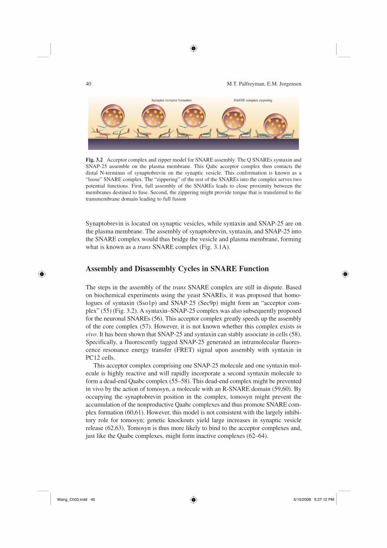
The steps in the assembly of the trans SNARE complex are still in dispute. Based on biochemical experiments using the yeast SNAREs, it was proposed that homo-logues of syntaxin (Sso1p) and SNAP-25 (Sec9p) might form an “acceptor com-plex” (55) (Fig. 3.2). A syntaxin–SNAP-25 complex was also subsequently proposed for the neuronal SNAREs (56). This acceptor complex greatly speeds up the assembly of the core complex (57). However, it is not known whether this complex exists invivo. It has been shown that SNAP-25 and syntaxin can stably associate in cells (58). Specifically, a fluorescently tagged SNAP-25 generated an intramolecular fluores-cence resonance energy transfer (FRET) signal upon assembly with syntaxin in PC12 cells.
This acceptor complex comprising one SNAP-25 molecule and one syntaxin mol-ecule is highly reactive and will rapidly incorporate a second syntaxin molecule to form a dead-end Qaabc complex (55–58). This dead-end complex might be prevented in vivo by the action of tomosyn, a molecule with an R-SNARE domain (59,60). By occupying the synaptobrevin position in the complex, tomosyn might prevent the accumulation of the nonproductive Qaabc complexes and thus promote SNARE com-plex formation (60,61). However, this model is not consistent with the largely inhibi-tory role for tomosyn; genetic knockouts yield large increases in synaptic vesicle release (62,63). Tomosyn is thus more likely to bind to the acceptor complexes and, just like the Qaabc complexes, might form inactive complexes (62–64).
Fig. 3.2 Acceptor complex and zipper model for SNARE assembly. The Q SNAREs syntaxin and SNAP-25 assemble on the plasma membrane. This Qabc acceptor complex then contacts the distal N-terminus of synaptobrevin on the synaptic vesicle. This conformation is known as a “loose” SNARE complex. The “zippering” of the rest of the SNAREs into the complex serves two potential functions. First, full assembly of the SNAREs leads to close proximity between the membranes destined to fuse. Second, the zippering might provide torque that is transferred to the transmembrane domain leading to full fusion
Wang_Ch03.indd 40Wang_Ch03.indd 40 5/15/2008 5:27:12 PM5/15/2008 5:27:12 PM
3 Roles of SNARE Proteins in Synaptic Vesicle Fusion 41
A second protein family that might serve to stabilize the acceptor complex is the SM (Sec1/Munc-18) family. At the synapse these proteins are called Unc18 proteins (UNC-18 in C. elegans, Munc18 in mammals, and ROP in Drosophila). It was originally thought that Unc18 exclusively bound to syntaxin monomers (65–69). However, more recent experiments have suggested alternative modes of binding (70–73). When reconstituted into lawns of plasma membrane, Unc18 was displaced from syntaxin by synaptobrevin but only when SNAP-25 was also present (70). Unc18 might therefore stabilize a syntaxin/SNAP-25 acceptor com-plex awaiting synaptobrevin (70). Nonetheless, it is still at present unclear how acceptor complexes are maintained or even whether they are true intermediates in core complex assembly. Indeed, synaptobrevin and syntaxin have been shown to assemble in vitro in the absence of SNAP-25 (74–76), suggesting that SNAP-25 might join the complex last. It has even been suggested that syntaxin might be the last molecule to enter the core complex in vivo (77). Until SNARE assembly can be monitored in vivo, we are forced to rely on these studies of in vitro SNARE interactions.
Once synaptobrevin enters the complex it is proposed to make contact at the N-terminal portion of the SNARE domain distal from the membrane. This conformation of the SNAREs is termed a loose configuration and is then thought to zipper down to a tight conformation (Fig. 3.2). Synaptic vesicles are held in a release-ready state in which the trans SNARE complex is likely to be arrested in a partially zippered state. Calcium binding to synaptotagmin would release arrest so that the SNARE complex could fully zipper to the tight conformation. This transition to the tight conformation would pull the transmembrane domains of the SNAREs, and hence the membranes, into close proximity and induce fusion (78,79). Models for the action of SNAREs in membrane fusion are described below.
Once the two membranes have merged, the core complex is now located in a single membrane and is referred to as a cis SNARE complex. To undergo further rounds of fusion, this cis complex must be disassembled and the SNAREs repar-titioned to their appropriate compartments. Disassembly is mediated by the action of NSF and the SNAPs. Together NSF and the SNAPs are able to disas-semble all SNARE complexes thus far tested (80). The ATPase NSF itself does not directly bind SNAREs; instead, it binds SNAREs through the action of the SNAPs. The SNAPs bind to the surface of the cis SNAREs around the central zero layer, which contains the conserved Q and R residues (81). The disassembly of the mammalian core complexes in PC12 cells is inhibited by mutation in these conserved residues (82). However, the disassembly of the C. elegans core com-plex is not affected by the same mutations (83). An alternative model for the function of these conserved residues is that they have a role before fusion in get-ting the four helixes to align in register to ensure that their transmembrane domains are directly opposed at their C-terminal ends (42,78). It has also been proposed that they might function in the prevention of full SNARE zippering (77). The next section explores how the formation of these SNARE complexes might catalyze fusion.
Wang_Ch03.indd 41Wang_Ch03.indd 41 5/15/2008 5:27:13 PM5/15/2008 5:27:13 PM
42 M.T. Palfreyman, E.M. Jorgensen
A Model for Membrane Fusion
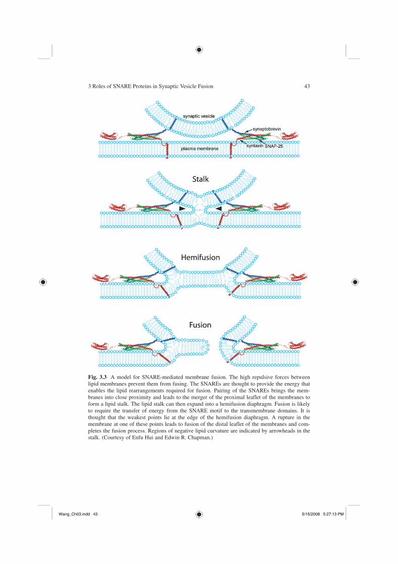
Membranes do not spontaneously fuse, because of the high repulsive forces between two phospholipid bilayers 1 to 2 nm apart. How might the SNAREs fuse mem-branes? Three characteristics of the SNAREs are central to the current models for their function in fusing membranes. First, the assembled SNARE complex is remarkably stable. The formation of the SNARE complex is therefore an energy source that can be used to overcome barriers to fusion. Second, the SNARE complex must consist of at least two SNARE molecules with transmembrane domains (84). The transmembrane domains must be inserted into both of the membranes destined to fuse (54). Third, the SNAREs assemble in a parallel orientation (44,78,79,85). Due to the parallel orientation of the SNARE motifs, SNARE assembly leads to the close apposition of the transmembrane domains and hence the membranes them-selves. This section describes how the assembly of the SNARE complexes might lead the membranes through the sequential intermediates of a lipid stalk, a hemifu-sion diaphragm, an initial fusion pore, and finally full fusion (Fig. 3.3).
The stability of the SNARE complexes combined with their parallel orientation led to the idea that their formation might provide the driving force for fusion. By first assembling at their N-terminals and subsequently “zippering” down to their membrane proximal C-terminals, the assembly of the SNAREs would bring the transmembrane domains of synaptobrevin and syntaxin into close proximity (77–79, 86–88) (Fig. 3.2). Evidence for zippering comes from two complementary experiments. First, biochemical and structural studies have shown that the membrane proximal domain of syntaxin becomes sequentially more ordered upon binding synaptobrevin in a directed N- to C-terminal fashion (55,57,87,89). The temperatures for assembly and disassembly of SNARE complex differ by as much as 10°C. Thus, assembly and dissociation follow different reaction pathways. This hysteresis suggests a kinetic barrier between folded and unfolded states (45). Mutations in the N-terminal hydro-phobic core of the SNARE complex selectively slowed SNARE assembly, while those in the C-terminal did not slow assembly (56,87), suggesting that the N-terminal nucleates SNARE assembly. The kinetic barrier to assembly also suggests that loose SNARE complexes could be an intermediate.
The second line of evidence for zippering comes from in vivo disruption studies using clostridial toxins, antibodies directed toward the SNARE motifs, and muta-tions in the hydrophobic core of the SNARE complex (77,86–88,90). The toxin and antibody disruption studies demonstrated that the N-termini of SNAREs become resistant to cleavage or antibody block at early stages, while C-termini are only resistant to disruptions at late stages. As a specific example, Hua et al injected either botulinum toxin D, which cleaves free synaptobrevin at the N-terminal side of the SNARE motif, or botulinum toxin B, which cleaves synaptobrevin toward the C-terminal side of the SNARE motif (88). SNAREs cannot be cleaved once they have assembled into the four helix SNARE complex (46). Exocytosis from the crayfish neuromus-cular junction was not sensitive to cleavage at the N-terminus of the SNARE motif, suggesting that this region was protected, presumably by the SNARE complex. By contrast, neurotransmitter release was blocked by cleavage at the C-terminus of the
Wang_Ch03.indd 42Wang_Ch03.indd 42 5/15/2008 5:27:13 PM5/15/2008 5:27:13 PM
3 Roles of SNARE Proteins in Synaptic Vesicle Fusion 43
Fig. 3.3 A model for SNARE-mediated membrane fusion. The high repulsive forces between lipid membranes prevent them from fusing. The SNAREs are thought to provide the energy that enables the lipid rearrangements required for fusion. Pairing of the SNAREs brings the mem-branes into close proximity and leads to the merger of the proximal leaflet of the membranes to form a lipid stalk. The lipid stalk can then expand into a hemifusion diaphragm. Fusion is likely to require the transfer of energy from the SNARE motif to the transmembrane domains. It is thought that the weakest points lie at the edge of the hemifusion diaphragm. A rupture in the membrane at one of these points leads to fusion of the distal leaflet of the membranes and com-pletes the fusion process. Regions of negative lipid curvature are indicated by arrowheads in the stalk. (Courtesy of Enfu Hui and Edwin R. Chapman.)
Wang_Ch03.indd 43Wang_Ch03.indd 43 5/15/2008 5:27:13 PM5/15/2008 5:27:13 PM
44 M.T. Palfreyman, E.M. Jorgensen
SNARE motif (86). Importantly, once the neuromuscular junction was electrically stimulated, botulinum toxin B was able to block exocytosis, demonstrating that the crayfish synaptobrevin monomers were indeed targets for the toxin. Thus, these data suggested that the N-terminus, but not the C-terminus, of synaptobrevin, is zippered into a SNARE complex in primed vesicles; presumably, calcium influx stimulates full zippering and membrane fusion.
As a second example, the mutations in the hydrophobic core of the SNARE complex have been expressed in neurosecretory chromaffin cells (87,90). Mutations in the C-terminal hydrophobic core incrementally reduced the kinetics of the rapid component of secretion, while those in the N-terminal reduced the sustained com-ponent of release, which is thought to correspond to engagement of new SNARE complexes (87). Importantly, the N-terminal mutants did not change the kinetics of the fast or slow components of release, only the amplitude of the response. Thus, it was interpreted that the C-terminal mutations were slowing “zippering” while those in the N-terminal were disrupting nucleation of the SNARE complexes (87). By contrast, when SNAREs bearing mutations in the hydrophobic core were intro-duced into the neurosecretory PC12 cells, there was no gradient in the efficacy of mutations in the kinetics of exocytosis (90).
The zippering of the membrane proximal portion of the SNARE complex likely serves two functions. First, the SNAREs are thought to catalyze the formation of a “hemifusion” transition state in which the proximal membrane leaflets have merged. This state can be achieved with comparatively low-energy requirements (91–94) and might simply need the SNAREs to bring the membranes into close proximity (95). Second, the SNAREs have been proposed to open up a fusion pore. This step requires the transmembrane domains of the SNAREs and likely involves the transfer of energy from the zippering of the SNARE cytoplasmic domains being passed to the transmembrane domain in order to locally disrupt lipid membranes (96).
Inspired by experiments in viral fusion and modeling of lipid bilayers, it is pro-posed that the initial steps of membrane merger result in a lipid stalk (97,98). The stalk corresponds to an hourglass-like structure that may contain as few as a dozen lipid molecules (98–100). The expansion of the stalk then results in a hemifusion diaphragm (91,101). These steps are not as highly energetically unfavorable as later steps and can be experimentally observed by dehydration of planar lipid bilayers, even in the absence of SNAREs (92,93,100,102). Direct evidence for lipid stalks has come from x-ray–scattering experiments that have given us a structure of this intermediate (100). The hemifusion state has been shown to be a metastable inter-mediate in vivo and can be observed for extensive periods of time in certain fusion reactions (103). Importantly, in vitro liposome fusion experiments have shown that hemifusion is an intermediate in the fusion pathway mediated by the synaptic vesi-cle SNAREs (104–106). Hemifusion intermediates have also been seen at central synapses using conical electron tomography; hemifused vesicles corresponded to those vesicles that were docked at the active zone (107).
Aside from the tomography and x-ray–scattering experiments, the evidence for stalk intermediates and hemifusion diaphragms comes from two observations:
Wang_Ch03.indd 44Wang_Ch03.indd 44 5/15/2008 5:27:14 PM5/15/2008 5:27:14 PM
3 Roles of SNARE Proteins in Synaptic Vesicle Fusion 45
the sensitivity of the fusion reaction to lipids of different intrinsic curvature (108), and the exchange of lipid membrane without luminal content mixing (103,109–111). The intrinsic curvature of lipids is determined by the ratio of the size of the lipid head group to their acyl chain tails. For example, a lipid with a single acyl chain would promote a positive intrinsic curvature (convex). At stalk structures and hemifusion diaphragms the outer, nonfused monolayer must adopt a negative curvature (concave) (arrowheads in Fig. 3.3) compared to the fused proximal monolayer, which adopts a net positive curvature (Fig. 3.3). This model predicts that, when added at the final steps of fusion, lipids with negative curva-ture would stimulate fusion while those with positive curvature would hinder fusion. Indeed, for all fusion reactions thus far tested, this prediction has been borne out (112). The application of lipids with altered curvature has been particu-larly useful in determining at which step fusion is arrested in various experimen-tal manipulations (91,95,112).
When the SNARE transmembrane domain is replaced by artificial lipid anchors or when the transmembrane domain is truncated, fusion no longer proceeds (95,96,113). However, these perturbations do lead to a state in which lipids can exchange—a hallmark of hemifusion (91). Interestingly, replacement of the mem-brane anchor of the influenza hemagglutinin with an artificial membrane anchor, a glycosylphosphatidylinositol (GPI) tail, traps influenza viral fusion at a hemifusion stage (111). This observation demonstrates that membrane fusion events as varied as synaptic vesicle exocytosis and viral fusion might use a common mechanism to catalyze fusion. Importantly, the fusion arrest that results from the replacement of the transmembrane domain in both SNARE-based fusion and viral fusion can be bypassed by the addition of lipids with intrinsic negative curvature to the outer membrane or lipids that induce positive curvature to the inner membrane (95,114,115). This demonstrates that the proximity resulting from the SNARE pair-ing might be enough to achieve a hemifusion state, but that full fusion requires the transmembrane domains of the respective fusion proteins (95,111,114).
The dependence on the transmembrane domains for full fusion also suggests that the zippering of the SNAREs might result in the transduction of force to the transmembrane domain. The domain linking the SNARE motif to the membrane may be rather rigid; when synaptobrevin and syntaxin are placed in planar bilay-ers, they stand straight up from the membrane (116,117). Disrupting this rigidity by the addition of flexible linkers of incremental lengths, between the SNARE motif and the transmembrane domain, incrementally reduces fusion to complete elimination (50,96,113,118). In addition, mutations in the linker domain do not disrupt liposome fusion, while those in the SNARE motif have dramatic effects (119). This experiment favors the model of the linker as largely a force transducer (119). By contrast, mutations in the linker domain of yeast syntaxin (Sso1p) do cause dramatic decreases in fusion (120). Nonetheless, these results suggest that the winding of the SNARE proteins during core complex assembly transduces force to the transmembrane domains (96,116). Torque on the transmembrane domains might force dimples in the lipid bilayer at regions of trans SNARE com-plex formation (84) (Fig. 3.3).
Wang_Ch03.indd 45Wang_Ch03.indd 45 5/15/2008 5:27:14 PM5/15/2008 5:27:14 PM
46 M.T. Palfreyman, E.M. Jorgensen
It is likely that more than one core complex is required to catalyze fusion. Like viral fusion proteins, the SNAREs used in exocytosis also seem to work as higher order multimers (121). Thus, a ring of SNAREs could induce a controlled local disruption of lipids. One possibility is that the hemifusion diaphragm would be delineated by a ring of SNARE transmembrane domains (84,121). Alternatively, it has been suggested that the transmembrane domains of the SNAREs might serve as a proteinaceous pore (122). Though the interactions are quite weak (123), it has been shown that both syntaxin and synaptobrevin form higher order multimers via conserved regions located in their transmembrane domains (124–127). Electron microscopy has provided images of these multimers and shows that they form star-shaped structures with the transmembrane domains at the vertex (128). In vivo evi-dence for the existence of such multimers comes from the cooperative action of the SNAREs and dose dependency of inhibition by botulinum neurotoxins and SNARE peptide blockers (121,129–132). Together, the evidence has suggested multimers containing from between three and 15 complexes (121). Nonetheless, working models for multimerization are currently quite preliminary; it will remain to be seen how these multimers might aid in catalyzing fusion.
The Reliable Opposition: Protein Models for the Fusion Pore
Despite the appeal and considerable evidence for a lipidic fusion pore, there remain data suggesting that the fusion pore could be proteinaceous (133). First, it has been proposed that the SNAREs are the fusogen but that the pore is lined by the trans-membrane of the five to eight syntaxin molecules rather than by lipids (122). This model derived from the observation that the replacement of residues in the trans-membrane domain of syntaxin with bulky amino acids slowed the conductance of the initial fusion pore. Second, some data indicate that SNAREs were not involved in the fusion step. NSF disassembles SNARE complexes, yet in yeast overexpression of NSF (Sec18p) did not block vacuole fusion (134). Third, techniques that can detect early stages of pore formation, amperometry, and capacitance measurements indicate that the fusion pore in chromaffin cells might be formed by a protein. In these experiments the initial fusion pore was found to have a pore size equivalent to a large ion channel (approximately 1 to 2 nm in diameter) (135). In addition, these initial fusion pores “flickered” like ion channel fusion pores (132,135,136). Fourth, it has been proposed that the V
o sector of the vacuolar ATPase could act as a protein-
aceous fusion pore (137,138). In yeast, calcium and calmodulin might be required in a step after SNARE complex formation in the process of fusion (139). The target of calcium-calmodulin in this late step in fusion was identified as the V
o sector of the
vacuolar ATPase (137). Furthermore, analysis of Drosophila mutants indicated that the vacuolar ATPase was important for fusion of synaptic vesicles (140).
Nonetheless, several points are difficult to reconcile with a protein pore–based model for fusion. First, trans SNARE complexes are resistant to the action of NSF, suggesting that functional SNAREs were still present in yeast experiments (141).
Wang_Ch03.indd 46Wang_Ch03.indd 46 5/15/2008 5:27:14 PM5/15/2008 5:27:14 PM
3 Roles of SNARE Proteins in Synaptic Vesicle Fusion 47
Second, fusion pore sizes have been found to vary considerably in different fusion reactions, an observation more consistent with a lipid-based pore (142). Third, null mutants in many of the SNAREs proteins have been shown to have a stronger phe-notype—often they are completely inviable—than respective mutants in the vacu-olar ATPase (143). Fourth, lysophosphatidyl choline, a lipid that induces positive membrane curvature, is able to block all fusion reactions so far tested (112). Finally, the observed fusion pore flickering has been seen in pure lipid bilayers induced to fuse by polyethylene glycol (PEG) (144). PEG dehydrates the spaces between lipid bilayers and drives lipid mixing. Flickering is therefore not a hall-mark solely of proteinaceous fusion pores.
Other observations that are apparently inconsistent with the lipid-based model for fusion have arisen from liposome fusion assays. For example, NSF and other pro-teins can catalyze the fusion of liposomes (145). However, the liposome fusion assay can be problematic (146). First, the lipid composition is critical in these assays and can produce misleading results; NSF could no longer fuse membranes when more physiologic lipid mixes were used (147). Second, many liposome fusion assays have used excessive and nonphysiologic concentrations of the SNARE molecules. Third, in most instances the speed of neurotransmitter release has not been replicated in this assay. Thus, results from liposome fusion assays must be interpreted cautiously and be supported by in vivo or genetic experiments.
SNAREs Encode Specificity
The original SNARE hypothesis proposed that compartmental specificity of fusion was encoded by SNARE proteins. Each intracellular fusion would be mediated by a specific set of SNARE proteins and thereby provide an addressing system for vesicle trafficking (27,28). This model makes several predictions. First, SNAREs should only bind their cognate SNARE partners. Second, SNAREs should only catalyze fusion when mixed with their SNARE partners. Third, SNAREs should be required for docking of vesicles to the correct target membrane. Fourth, the removal of a SNARE should selectively and completely eliminate fusion in one and only one fusion reaction. All of these hypotheses have been tested.
In vitro, the binding between cytoplasmic SNARE motifs is surprisingly promis-cuous (148–150). However, these same SNAREs exhibited specificity in catalyzing fusion reactions when inserted into artificial lipid bilayers (151–152). Specifically, only cognate SNARE complexes could catalyze fusion reaction. To date, out of the 275 pairwise combinations of yeast SNAREs tried, only nine are functional in the liposome fusion assay. Eight of these nine SNARE combinations represented inter-actions that occur in vivo, thus the specificity of fusion is greater than 99% (274/275) accurate (151). This specificity is preserved among the neuronal SNAREs; after cleavage of SNAP-25 in PC12 cells, secretion could only be rescued by SNAP-25 itself and not other SNAP-25 homologues (152). Thus, the SNAREs can encode the specificity of fusion.
Wang_Ch03.indd 47Wang_Ch03.indd 47 5/15/2008 5:27:14 PM5/15/2008 5:27:14 PM
48 M.T. Palfreyman, E.M. Jorgensen
Morphologic docking of synaptic vesicles long appeared to be independent of SNAREs. Genetic or pharmacologic disruption of SNAREs did not perturb synaptic vesicle docking (12,13,15,154). However, more recent experiments indicate that docking of synaptic vesicles (14) and dense core vesicles requires syntaxin (155–157). Importantly, if syntaxin is required for docking, experiments claiming roles for syntaxin in fusion must be interpreted with caution since fusion is downstream of docking. Docking defects will lead by necessity to defects in fusion. The discrepancy for syntaxin’s role in docking could be due to different morphologic definitions of docking, which has been defined as everything from direct contact with the plasma membrane to vesicles 50 nm from the plasma membrane. Alternatively, additional docking factors might be present in some cell types to ensure the specificity of fusion (155). For example, syntaxin is required for docking in neurosecretory cells but not neurons in mice (155,157). Perhaps tethering factors also contribute to docking of synaptic vesicles at the active zone (158–162). Overlapping roles for SNAREs and docking factors have been observed in yeast (163,164). Specifically, sec35 encodes a tethering protein for Golgi trafficking in yeast; sec35 mutants can be partially bypassed by overexpression of the relevant SNARE proteins (165). Similarly, over-expression of SNAREs can bypass mutations in the tethering complex for plasma membrane fusion (166,167). It is likely that these overlapping redundant functions are necessary to achieve the high level of fidelity seen in membrane trafficking.
Thus far in vivo perturbations of the SNAREs have mostly been shown to selec-tively eliminate single trafficking steps. However, in all cases fusion was not completely eliminated. There are two possible explanations. First, it is possible that the SNAREs are not executing fusion—an unlikely interpretation given the wealth of data described above. Second, the SNAREs might be partially redundant. Evidence so far points to the latter interpretation. Knockout mice in synaptobrevin II were found to retain some synaptic activity in hippocampal neurons (16). In chromaffin cells, this remnant activity could be attributed to the synaptobrevin par-alog cellubrevin (168). Redundancy can also explain the remaining fusion events in synaptobrevin null Drosophila mutants. Syb, the Drosophila equivalent of cellubre-vin, can functionally substitute for n-Syb, the Drosophila equivalent of synaptobre-vin, when overexpressed in neurons (169). Redundancy is also seen in the Q SNAREs. SNAP-23, SNAP-47, and SNAP-24 can provide partial function when SNAP-25 is absent (19,170,171). Finally, redundancy might also explain the almost complete lack of phenotype in syntaxin 1a knockout mice (172), where it is likely that syntaxin 1b is sufficient to almost entirely replace syntaxin 1a action. These observations are supported by experiments in yeast where redundancy between SNAREs has also been conclusively demonstrated in numerous trafficking reac-tions (173–175). By contrast, loss of syntaxin (unc-64) in C. elegans neurons results in a 500-fold reduction in neurotransmitter release with no apparent devel-opmental defects (14); UNC-64 is committed to synaptic vesicle fusion and is unlikely to have a redundant syntaxin, like in mice; nor is it involved in other cellu-lar functions, like in flies (176). In summary, the SNAREs do encode specificity; nonetheless, in some instances it is likely that other factors can provide overlapping functions to ensure that fusion happens with the appropriate target membrane.
Wang_Ch03.indd 48Wang_Ch03.indd 48 5/15/2008 5:27:15 PM5/15/2008 5:27:15 PM
3 Roles of SNARE Proteins in Synaptic Vesicle Fusion 49
SNARE Regulation
We will only touch on SNARE regulation briefly in this chapter, since other chap-ters will cover this topic in greater depth. SNARE regulation can roughly be divided into two forms: before and after initiation of complex formation. Before core com-plex formation, regulation involves occlusion of the SNARE motif of syntaxin to prevent the assembly of SNARE core complexes. After the initiation of SNARE assembly regulation likely takes place at the level of complex zippering. The cal-cium-sensing machinery works at these later steps.
Syntaxin itself has its own regulatory domain; the N-terminal Habc domain can fold over and occlude the SNARE motif (Fig. 3.1). Syntaxin can adopt two confor-mations: a closed form, in which the SNARE motif is occluded, and an open form, in which the SNARE motif is available to interact with SNAP-25 and synaptobre-vin. At least two synaptic proteins, Unc13 and Unc18 proteins, have been proposed to act directly on this N-terminal extension of syntaxin (65,177). In C. elegans, unc-13 mutants can be partially bypassed by an open form of syntaxin, demonstrat-ing a direct or indirect role of UNC-13 in the conversion of syntaxin from a closed to an open form (14,62,178). Several additional proteins may regulate SNARE complex assembly by directly occluding the SNARE motif of syntaxin. These molecules includetomosyn, amisyn, and syntaphilin (59,62–64,179–181).
At steps after core complex assembly, regulation might take place at the level of preventing full zippering of the SNARE proteins. Three proteins—Unc18, com-plexin, and synaptotagmin—may act at this late stage. The precise function of the SM superfamily of proteins, which include the Unc18 synaptic proteins, is not yet known (see Chapter 7), but Unc18 proteins might function in these later stages (70–73,182–184). Sec1p, the yeast SM homologue that acts at the plasma mem-brane, binds to the SNARE complex rather than syntaxin monomers (185). Recent data suggest that Unc18 also uses this mode of interaction (70–73).
Complexin and synaptotagmin serve as part of the calcium-sensing machinery. The coupling of fusion to calcium influx is the key evolutionary modifications of SNARE function to adapt it for neurotransmission. At synapses, the time delay between the elevation in calcium concentration and the postsynaptic response can be as little as 60 to 200 μs (186). Though calcium is needed for fusion in other membrane trafficking steps, it usually serves as a facilitator of fusion rather than directly functioning as a signal in triggering fusion (187,188). The addition of com-plexin and synaptotagmin appear to impart the calcium trigger to SNARE-mediated fusion (189, 190). Complexin appears to act as a fusion clamp—a brake preventing constitutive fusion from occurring (191–194).
Interestingly, recent experiments have shown that the complexin clamp holds the SNAREs in a state where the membranes are hemifused (193). This obser-vation demonstrates that the transition from hemifusion to full fusion can be regulated at the cytoplasmic SNARE motifs. Complexin sits in a groove between syntaxin and synaptobrevin, potentially preventing the full zippering of the core SNARE complex (195,196). The calcium sensor is synaptotagmin (197–201). Synaptotagmin binds to lipids and to syntaxin and SNAP-25 in a
Wang_Ch03.indd 49Wang_Ch03.indd 49 5/15/2008 5:27:15 PM5/15/2008 5:27:15 PM
50 M.T. Palfreyman, E.M. Jorgensen
calcium-dependent manner (200–204). Importantly, synaptotagmin appears to compete with complexin for SNARE complex binding and relieves the clamp when calcium is present (reviewed in ref. 194). One possibility is that calcium binding allows synaptotagmin to actively displace complexin from the SNARE complex, which is then free to fully wind and to break the membrane of the hemifused intermediate. In this model the SNAREs could function like a wheel, with complexin the stick in the spokes preventing the wheel from turning. Calcium binding to synaptotagmin would pull the stick from the spokes and allow the wheel to turn and drive fusion. This model, however, remains specula-tive, and several pieces of data are currently incompatible with the above model. First, complexin knockout in mice do not have elevated synaptic vesicle fusion, as would be predicted (205). In addition, synaptotagmin when reconstituted with the neuronal SNAREs in the liposome fusion assay, can act alone as both a fusion clamp in the absence of calcium as well as an accelerator of fusion in the presence of calcium (206). However, a second group did not observe calcium sensitivity in SNARE-mediated liposome fusion assays by the addition of syn-aptotagmin; instead, synaptotagmin simply accelerated the rate of liposome fusion independent of calcium (207). Since subsequent chapters will delve fur-ther into the murky depths of calcium regulation, here we will suffice to stay in the shallow end of the pool.
Conclusion
Rounds of SNARE assembly and disassembly lie at the center of all vesicular traf-ficking. Assembly of the SNAREs into a four-helix bundle drives fusion of synaptic vesicles with the plasma membrane and thereby mediates the release of neurotrans-mitter. The entwined SNAREs are then pulled apart by the ATPase NSF, which reenergizes the system for further rounds of fusion. This model is widely accepted, yet its details are in considerable dispute. So far, reconstitution experiments have examined interactions between only a very few of the proteins involved in what is undoubtedly a complex and highly regulated fusion machine. As such, they have given us largely static images of the complex. Thus, the overarching challenge in the coming years will be to understand the regulation of the SNAREs and how the assembly of SNAREs catalyzes fusion.
Several questions must be resolved. First, is a preassembled Q-SNARE acceptor complex present on the plasma membrane in vivo, and if so how is it stabilized? Second, how is assembly of the SNAREs regulated? SNARE regulators, including MUN domain proteins such as Unc13, SM proteins, and Tomosyn, have been iden-tified, yet their mechanism of action is unclear. Third, are SNAREs fully zippered prior to or during fusion? Fourth, is SNARE complex zippering arrested in the read-ily releasable pool of synaptic vesicles? Fifth, does formation of the SNARE com-plex generate a hemifusion intermediate? And finally, what rearrangements occur in the SNARE complex when synaptotagmin binds calcium and phospholipids?
Wang_Ch03.indd 50Wang_Ch03.indd 50 5/15/2008 5:27:15 PM5/15/2008 5:27:15 PM
3 Roles of SNARE Proteins in Synaptic Vesicle Fusion 51
Acknowledgments We thank Enfu Hui and Edwin R. Chapman for providing versions of Figures 3.1 and 3.3. Thanks also to Winfried Weissenhorn, Dirk Fasshauer, and Reinhard Jahn for allowing us to use and modify their images for Figure 3.1. Michael Ailion, Eric Bend, M. Wayne Davis, and Robert Hobson were instrumental in reading early versions of the manuscript.
References
1. Trimble WS, Cowan DM, Scheller RH. VAMP-1: a synaptic vesicle-associated integral mem-brane protein. Proc Natl Acad Sci U S A 1988;85(12):4538–4542.
2. Inoue A, Obata K, Akagawa K. Cloning and sequence analysis of cDNA for a neuronal cell membrane antigen, HPC-1. J Biol Chem 1992;267(15):10613–10619.
3. Bennett MK, Calakos N, Scheller RH. Syntaxin: a synaptic protein implicated in docking of synaptic vesicles at presynaptic active zones. Science 1992;257(5067):255–259.
4. Oyler GA, Higgins GA, Hart RA, et al. The identification of a novel synaptosomal-associated protein, SNAP-25, differentially expressed by neuronal subpopulations. J Cell Biol 1989;109(6 pt 1):3039–3052.
5. Brennwald P, Kearns B, Champion K, Keranen S, Bankaitis V, Novick P. Sec9 is a SNAP-25–like component of a yeast SNARE complex that may be the effector of Sec4 function in exo-cytosis. Cell 1994;79(2):245–258.
6. Novick P, Field C, Schekman R. Identification of 23 complementation groups required for post-translational events in the yeast secretory pathway. Cell 1980;21(1):205–215.
7. Burgen AS, Dickens F, Zatman LJ. The action of botulinum toxin on the neuro-muscular junc-tion. J Physiol 1949;109(1–2):10–24.
8. Blasi J, Chapman ER, Link E, et al. Botulinum neurotoxin A selectively cleaves the synaptic protein SNAP-25. Nature 1993;365(6442):160–163.
9. Blasi J, Chapman ER, Yamasaki S, Binz T, Niemann H, Jahn R. Botulinum neurotoxin C1 blocks neurotransmitter release by means of cleaving HPC-1/syntaxin. EMBO J 1993;12(12):4821–4828.
10. Link E, Edelmann L, Chou JH, et al. Tetanus toxin action: inhibition of neurotransmitter release linked to synaptobrevin proteolysis. Biochem Biophys Res Commun 1992;189(2):1017–1023.
11. Schiavo G, Benfenati F, Poulain B, et al. Tetanus and botulinum-B neurotoxins block neuro-transmitter release by proteolytic cleavage of synaptobrevin. Nature 1992;359(6398):832–835.
12. Marsal J, Ruiz-Montasell B, Blasi J, et al. Block of transmitter release by botulinum C1 action on syntaxin at the squid giant synapse. Proc Natl Acad Sci U S A 1997;94(26):14871–14876.
13. O’Connor V, Heuss C, De Bello WM, et al. Disruption of syntaxin-mediated protein interactions blocks neurotransmitter secretion. Proc Natl Acad Sci U S A 1997;94(22):12186–12191.
14. Hammarlund M, Palfreyman MT, Watanabe S, Olsen S, Jorgensen EM. Open syntaxin docks synaptic vesicles. PLoS Biol 2007;5(8):e198.
15. Broadie K, Prokop A, Bellen HJ, O’Kane CJ, Schulze KL, Sweeney ST. Syntaxin and synapto-brevin function downstream of vesicle docking in Drosophila. Neuron 1995;15(3):663–673.
16. Schoch S, Deak F, Konigstorfer A, et al. SNARE function analyzed in synaptobrevin/VAMP knockout mice. Science 2001;294(5544):1117–1122.
17. Washbourne P, Thompson PM, Carta M, et al. Genetic ablation of the t-SNARE SNAP-25 distinguishes mechanisms of neuroexocytosis. Nat Neurosci 2002;5(1):19–26.
18. Deitcher DL, Ueda A, Stewart BA, Burgess RW, Kidokoro Y, Schwarz TL. Distinct require-ments for evoked and spontaneous release of neurotransmitter are revealed by mutations in the Drosophila gene neuronal-synaptobrevin. J Neurosci 1998;18(6):2028–2039.
19. Vilinsky I, Stewart BA, Drummond JA, Robinson IM, Deitcher DL. A Drosophila SNAP-25 null mutant reveals context-dependent redundancy with SNAP-24 in neurotransmission. Genetics 2002;162(1):259–271.
Wang_Ch03.indd 51Wang_Ch03.indd 51 5/15/2008 5:27:15 PM5/15/2008 5:27:15 PM
52 M.T. Palfreyman, E.M. Jorgensen
20. Balch WE, Glick BS, Rothman JE. Sequential intermediates in the pathway of intercompart-mental transport in a cell-free system. Cell 1984;39(3 Pt 2):525–536.
21. Wilson DW, Wilcox CA, Flynn GC, et al. A fusion protein required for vesicle-mediated transport in both mammalian cells and yeast. Nature 1989;339(6223):355–359.
22. Eakle KA, Bernstein M, Emr SD. Characterization of a component of the yeast secretion machinery: identification of the SEC18 gene product. Mol Cell Biol 1988;8(10):4098–4109.
23. Block MR, Glick BS, Wilcox CA, Wieland FT, Rothman JE. Purification of an N-ethylmaleimide-sensitive protein catalyzing vesicular transport. Proc Natl Acad Sci U S A 1988;85(21):7852–7856.
24. Clary DO, Griff IC, Rothman JE. SNAPs, a family of NSF attachment proteins involved in intracellular membrane fusion in animals and yeast. Cell 1990;61(4):709–721.
25. Bock JB, Matern HT, Peden AA, Scheller RH. A genomic perspective on membrane compart-ment organization. Nature 2001;409(6822):839–841.
26. Jahn R, Lang T, Südhof TC. Membrane fusion. Cell 2003;112(4):519–533.27. Rothman JE. Intracellular membrane fusion. Adv Second Messenger Phosphoprotein Res
1994;29:81–96.28. Mayer A, Wickner W, Haas A. Sec18p (NSF)-driven release of Sec17p (alpha-SNAP) can
precede docking and fusion of yeast vacuoles. Cell 1996;85(1):83–94.29. Nichols BJ, Ungermann C, Pelham HR, Wickner WT, Haas A. Homotypic vacuolar fusion
mediated by t- and v-SNAREs. Nature 1997;387(6629):199–202.30. Littleton JT, Chapman ER, Kreber R, Garment MB, Carlson SD, Ganetzky B. Temperature-
sensitive paralytic mutations demonstrate that synaptic exocytosis requires SNARE complex assembly and disassembly. Neuron 1998;21(2):401–413.
31. Grote E, Carr CM, Novick PJ. Ordering the final events in yeast exocytosis. J Cell Biol 2000;151(2):439–452.
32. Weber T, Zemelman BV, McNew JA, et al. SNAREpins: minimal machinery for membrane fusion. Cell 1998;92(6):759–772.
33. Hu C, Ahmed M, Melia TJ, Söllner TH, Mayer T, Rothman JE. Fusion of cells by flipped SNAREs. Science 2003;300(5626):1745–1749.
34. Wickelgren WO, Leonard JP, Grimes MJ, Clark RD. Ultrastructural correlates of transmitter release in presynaptic areas of lamprey reticulospinal axons. J Neurosci 1985;5(5):1188–1201.
35. Xu-Friedman MA, Harris KM, Regehr WG. Three-dimensional comparison of ultrastructural characteristics at depressing and facilitating synapses onto cerebellar Purkinje cells. J Neurosci 2001;21(17):6666–6672.
36. Satzler K, Sohl LF, Bollmann JH, et al. Three-dimensional reconstruction of a calyx of Held and its postsynaptic principal neuron in the medial nucleus of the trapezoid body. J Neurosci 2002;22(24):10567–10579.
37. Schneggenburger R, Meyer AC, Neher E. Released fraction and total size of a pool of imme-diately available transmitter quanta at a calyx synapse. Neuron 1999;23(2):399–409.
38. Söllner T, Whiteheart SW, Brunner M, Erdjument-Bromage H, Geromanos S, Tempst P, Rothman JE. SNAP receptors implicated in vesicle targeting and fusion. Nature 1993;362(6418):318–324.
39. Söllner T, Bennett MK, Whiteheart SW, Scheller RH, Rothman JE. A protein assembly-disquard assembly pathway in vitro may correspond to sequential steps of synaptic vesicle docking, activation, and fusion. Cell 1993;75(3):409–418.
40. Stevens CF, Tsujimoto T. Estimates for the pool size of releasable quanta at a single central syn-apse and for the time required to refill the pool. Proc Natl Acad Sci U S A 1995;92(3):846–849.
41. Rosenmund C, Stevens CF. Definition of the readily releasable pool of vesicles at hippocam-pal synapses. Neuron 1996;16(6):1197–1207.
42. Fasshauer D, Sutton RB, Brunger AT, Jahn R. Conserved structural features of the synaptic fusion complex: SNARE proteins reclassified as Q- and R-SNAREs. Proc Natl Acad Sci U S A 1998;95(26):15781–15786.
43. Kloepper TH, Nickias Kienle C, Fasshauer D. An elaborate classification of SNARE proteins sheds light on the conservation of the eukaryotic endomembrane system. Mol Biol Cell 2007;18(9):3463–3471.
Wang_Ch03.indd 52Wang_Ch03.indd 52 5/15/2008 5:27:15 PM5/15/2008 5:27:15 PM
3 Roles of SNARE Proteins in Synaptic Vesicle Fusion 53
44. Sutton RB, Fasshauer D, Jahn R, Brunger AT. Crystal structure of a SNARE complex involved in synaptic exocytosis at 2.4 A resolution. Nature 1998;395(6700):347–353.
45. Fasshauer D, Antonin W, Subramaniam V, Jahn R. SNARE assembly and disassembly exhibit a pronounced hysteresis. Nat Struct Biol 2002;9(2):144–151.
46. Hayashi T, McMahon H, Yamasaki S, et al. Synaptic vesicle membrane fusion complex: action of clostridial neurotoxins on assembly. EMBO J 1994;13(21):5051–5061.
47. Fratti RA, Collins KM, Hickey CM, Wickner W. Stringent 3Q.1R composition of the SNARE 0–layer can be bypassed for fusion by compensatory SNARE mutation or by lipid bilayer modification. J Biol Chem 2007;282(20):14861–14867.
48. Ossig R, Schmitt HD, de Groot B, et al. Exocytosis requires asymmetry in the central layer of the SNARE complex. EMBO 2000;19(22):6000–6010.
49. Wei S, Xu T, Ashery U, et al. Exocytotic mechanism studied by truncated and zero layer mutants of the C-terminus of SNAP-25. EMBO J 2000;19(6):1279–1289.
50. Wang Y, Dulubova I, Rizo J, Südhof TC. Functional analysis of conserved structural elements in yeast syntaxin Vam3p. J Biol Chem 2001;276(30):28598–28605.
51. Dilcher M, Kohler B, von Mollard GF. Genetic interactions with the yeast q-SNARE vti1 reveal novel functions for the r-snare ykt6. J Biol Chem 2001;276(37):34537–34544.
52. Gil A, Gutierrez LM, Carrasco-Serrano C, Alonso MT, Viniegra S, Criado M. Modifications in the C terminus of the synaptosome-associated protein of 25 kDa (SNAP-25) and in the complementary region of synaptobrevin affect the final steps of exocytosis. J Biol Chem 2002;277(12):9904–9910.
53. Baumert M, Maycox PR, Navone F, De Camilli P, Jahn R. Synaptobrevin: an integral mem-brane protein of 18,000 daltons present in small synaptic vesicles of rat brain. EMBO J 1989;8(2):379–384.
54. Parlati F, McNew JA, Fukuda R, Miller R, Söllner TH, Rothman JE. Topological restriction of SNARE-dependent membrane fusion. Nature 2000;407(6801):194–198.
55. Fiebig KM, Rice LM, Pollock E, Brunger AT. Folding intermediates of SNARE complex assembly. Nat Struct Biol 1999;6(2):117–123.
56. Fasshauer D, Margittai M. A transient N-terminal interaction of SNAP-25 and syntaxin nucle-ates SNARE assembly. J Biol Chem 2004;279(9):7613–7621.
57. Pobbati AV, Stein A, Fasshauer D. N- to C-terminal SNARE complex assembly promotes rapid membrane fusion. Science 2006;313(5787):673–676.
58. An SJ, Almers W. Tracking SNARE complex formation in live endocrine cells. Science 2004;306(5698):1042–1046.
59. Fujita Y, Shirataki H, Sakisaka T, et al. Tomosyn: a syntaxin-1–binding protein that forms a novel complex in the neurotransmitter release process. Neuron 1998;20(5):905–915.
60. Widberg CH, Bryant NJ, Girotti M, Rea S, James DE. Tomosyn interacts with the t-SNAREs syntaxin4 and SNAP23 and plays a role in insulin-stimulated GLUT4 translocation. J Biol Chem 2003;278(37):35093–35101.
61. Hatsuzawa K, Lang T, Fasshauer D, Bruns D, Jahn R. The R-SNARE motif of tomosyn forms SNARE core complexes with syntaxin 1 and SNAP-25 and down-regulates exocytosis. J Biol Chem 2003;273(33):31159–31166.
62. McEwen JM, Madison JM, Dybbs M, Kaplan JM. Antagonistic Regulation of Synaptic Vesicle Priming by Tomosyn and UNC-13. Neuron 2006;51(3):303–315.
63. Gracheva EO, Burdina AO, Holgado AM, et al. Tomosyn inhibits synaptic vesicle priming in Caenorhabditis elegans. PLoS Biol 2006;4(8):e261
64. Pobbati AV, Razeto A, Boddener M, Becker S, Fasshauer D. Structural basis for the inhibitory role of tomosyn in exocytosis. J Biol Chem 2004;279(45):47192–47200.
65. Hata Y, Slaughter CA, Südhof TC. Synaptic vesicle fusion complex contains une-18 homo-logue bound to syntaxin. Nature 1993;366(6453):347–351.
66. Garcia EP, Gatti E, Butler M, Burton J, De Camilli P. A rat brain Sec1 homologue related to Rop and UNC18 interacts with syntaxin. Proc Natl Acad Sci U S A 1994;91(6):2003–2007.
67. Pevsner J, Hsu SC, Scheller RH. n-Sec1: a neural-specific syntaxin-binding protein. Proc Natl Acad Sci U S A 1994;91(4):1445–1449.
Wang_Ch03.indd 53Wang_Ch03.indd 53 5/15/2008 5:27:15 PM5/15/2008 5:27:15 PM
54 M.T. Palfreyman, E.M. Jorgensen
68. Misura KM, Scheller RH, Weis WI. Three-dimensional structure of the neuronal-Sec1–syn-taxin 1a complex. Nature 2000;404(6776):355–362.
69. Gallwitz D, Jahn R. The riddle of the Sec1/Munc-18 proteins—new twists added to their interactions with SNAREs. Trends Biochem Sci 2003;28(3):113–116.
70. Zilly FE, Sorensen JB, Jahn R, Lang T. Munc18–bound syntaxin readily forms SNARE com-plexes with synaptobrevin in native plasma membranes. PLoS Biol 2006;4(10):e330.
71. Shen J, Tareste DC, Paumet F, Rothman JE, Melia TJ. Selective activation of cognate SNAREpins by Sec1/Munc18 proteins. Cell 2007;128(1):183–195.
72. Rickman C, Medine CN, Bergmann A, Duncan RR. Functionally and spatially distinct modes of MUNC18–syntaxin 1 interaction. J Biol Chem 2007;282(16):12097–12103.
73. Dulubova I, Khvotchev M, Liu S, Huryeva I, Südhof TC, Rizo J. Munc18–1 binds directly to the neuronal SNARE complex. Proc Natl Acad Sci U S A 2007;104(8):2697–2702.
74. Liu T, Tucker WC, Bhalla A, Chapman ER, Weisshaar JC. SNARE-driven, 25-millisecond vesicle fusion in vitro. Biophys J 2005;89(4):2458–2472.
75. Bowen ME, Weninger K, Brunger AT, Chu S. Single molecule observation of liposome-bilayer fusion thermally induced by soluble N-ethyl maleimide sensitive-factor attachment protein receptors (SNAREs). Biophys J 2004;87(5):3569–3584.
76. Fix M, Melia TJ, Jaiswal JK, et al. Imaging single membrane fusion events mediated by SNARE proteins. Proc Natl Acad Sci U S A 2004;101(19):7311–7316.
77. Chen YA, Scales SJ, Scheller RH. Sequential SNARE assembly underlies priming and trig-gering of exocytosis. Neuron 2001;30:161–170.
78. Hanson PI, Roth R, Morisaki H, Jahn R, Heuser JE. Structure and conformational changes in NSF and its membrane receptor complexes visualized by quick-freeze/deep-etch electron microscopy. Cell 1997;90(3):523–535.
79. Lin RC, Scheller RH. Structural organization of the synaptic exocytosis core complex. Neuron 1997;19(5):1087–1094.
80. Zhao C, Slevin JT, Whiteheart SW. Cellular functions of NSF: not just SNAPs and SNAREs. FEBS Lett 2007;581(11):2140–2149.
81. Marz KE, Lauer JM, Hanson PI. Defining the SNARE complex binding surface of α-SNAP: implications for SNARE complex disassembly. J Biol Chem 2003;278(29):27000–27008.
82. Scales SJ, Yoo BY, Scheller RH. The ionic layer is required for efficient dissociation of the SNARE complex by α-SNAP and NSF. Proc Natl Acad Sci U S A 2001;98(25):14262–14267.
83. Lauer JM, Dalal S, Marz KE, Nonet ML, Hanson PI. SNARE complex zero layer residues are not critical for N-ethylmaleimide-sensitive factor-mediated disassembly. J Biol Chem 2006;281(21):14823–14832.
84. Langosch D, Hofmann M, Ungermann C. The role of transmembrane domains in membrane fusion. Cell Mol Life Sci 2007;64(7–8):850–864.
85. Poirier MA, Xiao W, Macosko JC, Chan C, Shin YK, Bennett MK. The synaptic SNARE complex is a parallel four-stranded helical bundle. Nat Struct Biol 1998;5(9):765–769.
86. Hua SY, Charlton MP. Activity-dependent changes in partial VAMP complexes during neuro-transmitter release. Nat Neurosci 1999;2(12):1078–1083.
87. Sorensen JB, Wiederhold K, Muller EM, et al. Sequential N- to C-terminal SNARE complex assembly drives priming and fusion of secretory vesicles. EMBO J 2006;25(5):955–966.
88. Xu T, Rammner B, Margittai M, Artalejo AR, Neher E, Jahn R. Inhibition of SNARE complex assembly differentially affects kinetic components of exocytosis. Cell 1999;99(7):713–722.
89. Melia TJ, Weber T, McNew JA, et al. Regulation of membrane fusion by the membrane-proxi-mal coil of the t-SNARE during zippering of SNAREpins. J Cell Biol 2002;158(5):929–940.
90. Han X, Jackson MB. Structural transitions in the synaptic SNARE complex during Ca2+-trig-gered exocytosis. J Cell Biol 2006;172(2):281–293.
91. Chernomordik LV, Kozlov MM. Membrane hemifusion: crossing a chasm in two leaps. Cell 2005;123(3):375–382.
92. Kasson PM, Kelley NW, Singhal N, Vrljic M, Brunger AT, Pande VS. Ensemble molecular dynamics yields submillisecond kinetics and intermediates of membrane fusion. Proc Natl Acad Sci U S A 2006;103(32):11916–11921.
Wang_Ch03.indd 54Wang_Ch03.indd 54 5/15/2008 5:27:15 PM5/15/2008 5:27:15 PM
3 Roles of SNARE Proteins in Synaptic Vesicle Fusion 55
93. Cohen FS, Melikyan GB. The energetics of membrane fusion from binding, through hemi-fusion, pore formation, and pore enlargement. J Membr Biol 2004;199(1):1–14.
94. Kuzmin PI, Zimmerberg J, Chizmadzhev YA, Cohen FS. A quantitative model for mem-brane fusion based on low-energy intermediates. Proc Natl Acad Sci U S A 2001;98(13):7235–7240.
95. Grote E, Baba M, Ohsumi Y, Novick PJ. Geranylgeranylated SNAREs are dominant inhibi-tors of membrane fusion. J Cell Biol 2000;151(2):453–466.
96. McNew JA, Weber T, Parlati F, et al. Close is not enough: SNARE-dependent membrane fusion requires an active mechanism that transduces force to membrane anchors. J Cell Biol 2000;150(1):105–117.
97. Earp LJ, Delos SE, Park HE, White JM. The many mechanisms of viral membrane fusion proteins. Curr Top Microbiol Immunol 2005;285:25–66.
98. Kozlov MM, Markin VS. [Possible mechanism of membrane fusion]. Biofizika 1983;28(2):242–247.
99. Knecht V, Mark AE, Marrink SJ. Phase behavior of a phospholipid/fatty acid/water mixture studied in atomic detail. J Am Chem Soc 2006;128(6):2030–2034.
100. Yang L, Huang HW. Observation of a membrane fusion intermediate structure. Science 2002;297(5588):1877–1879.
101. Jahn R, Südhof TC. Membrane fusion and exocytosis. Annu Rev Biochem 1999;68:863–911.
102. Efrat A, Chernomordik LV, Kozlov MM. Point-like protrusion as a prestalk intermediate in membrane fusion pathway. Biophys J 2007;92(8):L61–63.
103. Wong JL, Koppel DE, Cowan AE, Wessel GM. Membrane hemifusion is a stable intermedi-ate of exocytosis. Dev Cell 2007;12(4):653–659.
104. Xu Y, Zhang F, Su Z, McNew JA, Shin YK. Hemifusion in SNARE-mediated membrane fusion. Nat Struct Mol Biol 2005;12(5):417–422.
105. Lu X, Zhang F, McNew JA, Shin YK. Membrane fusion induced by neuronal SNAREs transits through hemifusion. J Biol Chem 2005;280(34):30538–30541.
106. Yoon TY, Okumus B, Zhang F, Shin YK, Ha T. Multiple intermediates in SNARE-induced membrane fusion. Proc Natl Acad Sci U S A 2006;103(52):19731–19736.
107. Zampighi GA, Zampighi LM, Fain N, Lanzavecchia S, Simon SA, Wright EM. Conical electron tomography of a chemical synapse: vesicles docked to the active zone are hemi-fused. Biophys J 2006;91(8):2910–2918.
108. Melia TJ, You D, Tareste DC, Rothman JE. Lipidic antagonists to SNARE-mediated fusion. J Biol Chem 2006;281(40):29597–29605.
109. Reese C, Heise F, Mayer A. Trans-SNARE pairing can precede a hemifusion intermediate in intracellular membrane fusion. Nature 2005;436(7049):410–414.
110. Jun Y, Wickner W. Assays of vacuole fusion resolve the stages of docking, lipid mixing, and content mixing. Proc Natl Acad Sci U S A 2007;104(32):13010–13015.
111. Kemble GW, Danieli T, White JM. Lipid-anchored influenza hemagglutinin promotes hemi-fusion, not complete fusion. Cell 1994;76(2):383–391.
112. Chernomordik LV, Zimmerberg J, Kozlov MM. Membranes of the world unite! J Cell Biol 2006;175(2):201–207.
113. McNew JA, Weber T, Engelman DM, Söllner TH, Rothman JE. The length of the flexible SNAREpin juxtamembrane region is a critical determinant of SNARE-dependent fusion. Mol Cell 1999;4(3):415–421.
114. Melikyan GB, Brener SA, Ok DC, Cohen FS. Inner but not outer membrane leaflets control the transition from glycosylphosphatidylinositol-anchored influenza hemagglutinin-induced hemifusion to full fusion. J Cell Biol 1997;136(5):995–1005.
115. Razinkov VI, Melikyan GB, Epand RM, Epand RF, Cohen FS. Effects of spontaneous bilayer curvature on influenza virus-mediated fusion pores. J Gen Physiol 1998;112(4):409–422.
116. Knecht V, Grubmüller H. Mechanical coupling via the membrane fusion SNARE protein syntaxin 1A: a molecular dynamics study. Biophys J 2003;84(3):1527–1547.
Wang_Ch03.indd 55Wang_Ch03.indd 55 5/15/2008 5:27:15 PM5/15/2008 5:27:15 PM
56 M.T. Palfreyman, E.M. Jorgensen
117. Kiessling V, Tamm LK. Measuring distances in supported bilayers by fluorescence interfer-ence-contrast microscopy: polymer supports and SNARE proteins. Biophys J 2003;84(1):408–418.
118. Deak F, Shin OH, Kavalali ET, Südhof TC. Structural determinants of synaptobrevin 2 func-tion in synaptic vesicle fusion. J Neurosci 2006;26(25):6668–6676.
119. Siddiqui TJ, Vites O, Stein A, Heintzmann R, Jahn R, Fasshauer D. Determinants of synap-tobrevin regulation in membranes. Mol Biol Cell 2007;18(6):2037–2046.
120. Van Komen JS, Bai X, Rodkey TL, Schaub J, McNew JA. The polybasic juxtamembrane region of Sso1p is required for SNARE function in vivo. Eukaryot Cell 2005;4(12):2017–2028.
121. Montecucco C, Schiavo G, Pantano S. SNARE complexes and neuroexocytosis: how many, how close? Trends Biochem Sci 2005;30(7):367–372.
122. Han X, Wang CT, Bai J, Chapman ER, Jackson MB. Transmembrane segments of syntaxin line the fusion pore of Ca2+-triggered exocytosis. Science 2004;304(5668):289–292.
123. Bowen ME, Engelman DM, Brunger AT. Mutational analysis of synaptobrevin transmem-brane domain oligomerization. Biochemistry 2002;41(52):15861–15866.
124. Roy R, Laage R, Langosch D. Synaptobrevin transmembrane domain dimerization-revis-ited. Biochemistry 2004;43(17):4964–4970.
125. Roy R, Peplowska K, Rohde J, Ungermann C, Langosch D. Role of the Vam3p transmem-brane segment in homodimerization and SNARE complex formation. Biochemistry 2006;45(24):7654–7660.
126. Laage R, Rohde J, Brosig B, Langosch D. A conserved membrane-spanning amino acid motif drives homomeric and supports heteromeric assembly of presynaptic SNARE pro-teins. J Biol Chem 2000;275(23):17481–17487.
127. Margittai M, Otto H, Jahn R. A stable interaction between syntaxin 1a and synaptobrevin 2 mediated by their transmembrane domains. FEBS Lett 1999;446(1):40–44.
128. Rickman C, Hu K, Carroll J, Davletov B. Self-assembly of SNARE fusion proteins into star-shaped oligomers. Biochem J 2005;388(1):75–79.
129. Raciborska DA, Trimble WS, Charlton MP. Presynaptic protein interactions in vivo: evi-dence from botulinum A, C, D and E action at frog neuromuscular junction. Eur J Neurosci 1998;10(8):2617–2628.
130. Stewart BA, Mohtashami M, Trimble WS, Boulianne GL. SNARE proteins contribute to calcium cooperativity of synaptic transmission. Proc Natl Acad Sci U S A 2000;97(25): 13955–13960.
131. Hua Y, Scheller RH. Three SNARE complexes cooperate to mediate membrane fusion. Proc Natl Acad Sci U S A 2001;98(14):8065–8070.
132. Keller JE, Cai F, Neale EA. Uptake of botulinum neurotoxin into cultured neurons. Biochemistry 2004;43(2):526–532.
133. Almers W, Tse FW. Transmitter release from synapses: does a preassembled fusion pore initiate exocytosis? Neuron 1990;4(6):813–818.
134. Ungermann C, Sato K, Wickner W. Defining the functions of trans-SNARE pairs. Nature 1998;396(6711):543–548.
135. Spruce AE, Breckenridge LJ, Lee AK, Almers W. Properties of the fusion pore that forms during exocytosis of a mast cell secretory vesicle. Neuron 1990;4(5):643–654.
136. Monck JR, Fernandez JM. The fusion pore and mechanisms of biological membrane fusion. Curr Opin Cell Biol 1996;8(4):524–533.
137. Peters C, Bayer MJ, Buhler S, Andersen JS, Mann M, Mayer A. Trans-complex formation by proteolipid channels in the terminal phase of membrane fusion. Nature 2001;409 (6820):581–588.
138. Israel M, Morel N, Lesbats B, Birman S, Manaranche R. Purification of a presynaptic mem-brane protein that mediates a calcium-dependent translocation of acetylcholine. Proc Natl Acad Sci U S A 1986;83(23):9226–9230.
139. Peters C, Mayer A. Ca2+/calmodulin signals the completion of docking and triggers a late step of vacuole fusion. Nature 1998;396(6711):575–580.
Wang_Ch03.indd 56Wang_Ch03.indd 56 5/15/2008 5:27:16 PM5/15/2008 5:27:16 PM
3 Roles of SNARE Proteins in Synaptic Vesicle Fusion 57
140. Hiesinger PR, Fayyazuddin A, Mehta SQ, et al. The v-ATPase V0 subunit a1 is required for a late step in synaptic vesicle exocytosis in Drosophila. Cell 2005;121(4):607–620.
141. Weber T, Parlati F, McNew JA, et al. SNAREpins are functionally resistant to disruption by NSF and alphaSNAP. J Cell Biol 2000;49(5):1063–1072.
142. Klyachko VA, Jackson MB. Capacitance steps and fusion pores of small and large-dense-core vesicles in nerve terminals. Nature 2002;418(6893):89–92.
143. Giaever G, Chu AM, Ni L, et al. Functional profiling of the Saccharomyces cerevisiae genome. Nature 2002;418(6896):387–391.
144. Chanturiya A, Chernomordik LV, Zimmerberg J. Flickering fusion pores comparable with initial exocytotic pores occur in protein-free phospholipid bilayers. Proc Natl Acad Sci U S A 1997;94(26):14423–14428
145. Otter-Nilsson M, Hendriks R, Pecheur-Huet E, Hoekstra D, Nilsson T. Cytosolic ATPases, p97 and NSF, are sufficient to mediate rapid membrane fusion. EMBO J 1999;18(8): 2074–2083.
146. Rizo J, Chen X, Araç D. Unraveling the mechanisms of synaptotagmin and SNARE func-tion in neurotransmitter release. Trends Cell Biol 2006;16(7):339–350.
147. Brugger B, Nickel W, Weber T, et al. Putative fusogenic activity of NSF is restricted to a lipid mixture whose coalescence is also triggered by other factors. EMBO J 2000;19(6):1272–1278.
148. Yang B, Gonzalez LJ, Prekeris R, Steegmaier M, Advani RJ, Scheller RH. SNARE interac-tions are not selective. Implications for membrane fusion specificity. J Biol Chem 1999;274(9):5649–5653.
149. Tsui MM, Banfield DK. Yeast Golgi SNARE interactions are promiscuous. J Cell Sci 2000;113(1):145–152.
150. Fasshauer D, Antonin W, Margittai M, Pabst S, Jahn R. Mixed and non-cognate SNARE complexes. Characterization of assembly and biophysical properties. J Biol Chem 1999;274(22):15440–15446.
151. Parlati F, Varlamov O, Paz K, et al. Distinct SNARE complexes mediating membrane fusion in Golgi transport based on combinatorial specificity. Proc Natl Acad Sci U S A 2002;99(8): 5424–5429.
152. McNew JA, Parlati F, Fukuda R, et al. Compartmental specificity of cellular membrane fusion encoded in SNARE proteins. Nature 2000;407(6801):153–159.
153. Scales SJ, Chen YA, Yoo BY, Patel SM, Doung YC, Scheller RH. SNAREs contribute to the specificity of membrane fusion. Neuron 2000;26(2):457–464.
154. Hunt JM, Bommert K, Charlton MP, et al. A post-docking role for synaptobrevin in synaptic vesicle fusion. Neuron 1994;12(6):1269–1279.
155. de Wit H, Cornelisse LN, Toonen RF, Verhage M. Docking of secretory vesicles is syntaxin dependent. PLoS ONE 2006;1(1):e126.
156. Toonen RF, Kochubey O, de Wit H, et al. Dissecting docking and tethering of secretory vesicles at the target membrane. EMBO J 2006;25(16):3725–3737.
157. Ohara-Imaizumi M, Fujiwara T, Nakamichi Y, et al. Imaging analysis reveals mechanistic dif-ferences between first- and second-phase insulin exocytosis. J Cell Biol 2007;177(4):695–705.
158. Sztul E, Lupashin V. Role of tethering factors in secretory membrane traffic. Am J Physiol Cell Physiol 2006;290(1):C11–26.
159. Whyte JR, Munro S. Vesicle tethering complexes in membrane traffic. J Cell Sci 2002;115(13):2627–2637.
160. Zerial M, McBride H. Rab proteins as membrane organizers. Nat Rev Mol Cell Biol 2001;2(2):107–117.
161. Waters MG, Pfeffer SR. Membrane tethering in intracellular transport. Curr Opin Cell Biol 1999;11(4):453–459.
162. Guo W, Sacher M, Barrowman J, Ferro-Novick S, Novick P. Protein complexes in transport vesicle targeting. Trends Cell Biol 2000;10(6):251–255.
163. Wiederkehr A, Du Y, Pypaert M, Ferro-Novick S, Novick P. Sec3p is needed for the spatial regulation of secretion and for the inheritance of the cortical endoplasmic reticulum. Mol Biol Cell 2003;14(12):4770–4782.
Wang_Ch03.indd 57Wang_Ch03.indd 57 5/15/2008 5:27:16 PM5/15/2008 5:27:16 PM
58 M.T. Palfreyman, E.M. Jorgensen
164. Finger FP, Hughes TE, Novick P. Sec3p is a spatial landmark for polarized secretion in budding yeast. Cell 1998;92(4):559–571.
165. VanRheenen SM, Cao X, Lupashin VV, Barlowe C, Waters MG. Sec35p, a novel peripheral membrane protein, is required for ER to Golgi vesicle docking. J Cell Biol 1998;141(5): 1107–1119.
166. Wiederkehr A, De Craene JO, Ferro-Novick S, Novick P. Functional specialization within a vesicle tethering complex: bypass of a subset of exocyst deletion mutants by Sec1p or Sec4p. J Cell Biol 2004;167(5):875–887.
167. Novick P, Medkova M, Dong G, Hutagalung A, Reinisch K, Grosshans B. Interactions between Rabs, tethers, SNAREs and their regulators in exocytosis. Biochem Soc Trans 2006;34(5):683–686.
168. Borisovska M, Zhao Y, Tsytsyura Y, et al. v-SNAREs control exocytosis of vesicles from priming to fusion. EMBO J 2005;24(12):2114–2126.
169. Bhattacharya S, Stewart BA, Niemeyer BA, et al. Members of the synaptobrevin/vesicle-associated membrane protein (VAMP) family in Drosophila are functionally interchangea-ble in vivo for neurotransmitter release and cell viability. Proc Natl Acad Sci U S A 2002;99(21):13867–13872.
170. Sorensen JB, Nagy G, Varoqueaux F, et al. Differential control of the releasable vesicle pools by SNAP-25 splice variants and SNAP-23. Cell 2003;114(1):75–86.
171. Holt M, Varoqueaux F, Wiederhold K, et al. Identification of SNAP-47, a novel Qbc-SNARE with ubiquitous expression. J Biol Chem 2006;281(25):17076–17083.
172. Fujiwara T, Mishima T, Kofuji T, et al. Analysis of knock-out mice to determine the role of HPC-1/syntaxin 1A in expressing synaptic plasticity. J Neurosci 2006;26(21):5767–5776.
173. Liu Y, Barlowe C. Analysis of Sec22p in endoplasmic reticulum/Golgi transport reveals cellular redundancy in SNARE protein function. Mol Biol Cell 2002;13(9):3314–3324.
174. Fischer von Mollard G, Stevens TH. The Saccharomyces cerevisiae v-SNARE Vti1p is required for multiple membrane transport pathways to the vacuole. Mol Biol Cell 1999;10 (6):1719–1732.
175. Darsow T, Rieder SE, Emr SD. A multispecificity syntaxin homologue, Vam3p, essential for autophagic and biosynthetic protein transport to the vacuole. J Cell Biol 1997;138b (11):517–529.
176. Burgess RW, Deitcher DL, Schwarz TL. The synaptic protein syntaxin1 is required for cel-lularization of Drosophila embryos. J Cell Biol 1997;138(4):861–875.
177. Betz A, Okamoto M, Benseler F, Brose N. Direct interaction of the rat unc-13 homologue Munc13–1 with the N terminus of syntaxin. J Biol Chem 1997;272(4):2520–2526.
178. Richmond JE, Weimer RM, Jorgensen EM. An open form of syntaxin bypasses the require-ment for UNC-13 in vesicle priming. Nature 2001;412(6844):338–341.
179. Scales SJ, Hesser BA, Masuda ES, Scheller RH. Amisyn, a novel syntaxin-binding protein that may regulate SNARE complex assembly. J Biol Chem 2002;277(31):28271–28279.
180. Masuda ES, Huang BC, Fisher JM, Luo Y, Scheller RH. Tomosyn binds t-SNARE proteins via a VAMP-like coiled coil. Neuron 1998;21(3):479–480.
181. Lao G, Scheuss V, Gerwin CM, et al. Syntaphilin: a syntaxin-1 clamp that controls SNARE assembly. Neuron 2000;25(1):191–201.
182. Fisher RJ, Pevsner J, Burgoyne RD. Control of fusion pore dynamics during exocytosis by Munc18. Science 2001;291(5505):875–878.
183. Latham CF, Lopez JA, Hu SH, et al. Molecular dissection of the munc18c/syntaxin4 interac-tion: implications for regulation of membrane trafficking. Traffic 2006;7(10):1408–1419.
184. Hu SH, Latham CF, Gee CL, James DE, Martin JL. Structure of the Munc18c/Syntaxin4 N-peptide complex defines universal features of the N-peptide binding mode of Sec1/Munc18 proteins. Proc Natl Acad Sci U S A 2007;104(21):8773–8778.
185. Togneri J, Cheng YS, Munson M, Hughson FM, Carr CM. Specific SNARE complex bind-ing mode of the Sec1/Munc-18 protein, Sec1p. Proc Natl Acad Sci U S A 2006;103 (47):17730–17735.
Wang_Ch03.indd 58Wang_Ch03.indd 58 5/15/2008 5:27:16 PM5/15/2008 5:27:16 PM
3 Roles of SNARE Proteins in Synaptic Vesicle Fusion 59
186. Sabatini BL, Regehr WG. Timing of neurotransmission at fast synapses in the mammalian brain. Nature 1996;384(6605):170–172.
187. Flanagan JJ, Barlowe C. Cysteine-disulfide cross-linking to monitor SNARE complex assembly during endoplasmic reticulum-Golgi transport. J Biol Chem 2006;281 (4):2281–2288.
188. Starai VJ, Thorngren N, Fratti RA, Wickner W. Ion regulation of homotypic vacuole fusion in Saccharomyces cerevisiae. J Biol Chem 2005;280(17):16754–16762.
189. McMahon HT, Missler M, Li C, Südhof TC. Complexins: cytosolic proteins that regulate SNAP receptor function. Cell 1995;83(1):111–119.
190. Perin MS, Fried VA, Mignery GA, Jahn R, Südhof TC. Phospholipid binding by a synaptic vesicle protein homologous to the regulatory region of protein kinase C. Nature 1990;345(6272):260–263.
191. Giraudo CG, Eng WS, Melia TJ, Rothman JE. A clamping mechanism involved in SNARE-dependent exocytosis. Science 2006;313(5787):676–680.
192. Tang J, Maximov A, Shin OH, Dai H, Rizo J, Südhof TC. A complexin/synaptotagmin 1 switch controls fast synaptic vesicle exocytosis. Cell 2006;126(6):1175–1187.
193. Schaub JR, Lu X, Doneske B, Shin YK, McNew JA. Hemifusion arrest by complexin is relieved by Ca2+-synaptotagmin I. Nat Struct Mol Biol 2006;13(8):748–750.
194. Melia TJ. Putting the clamps on membrane fusion: How complexin sets the stage for cal-cium-mediated exocytosis. FEBS Lett 2007;581(11):2131–2139.
195. Bracher A, Kadlec J, Betz H, Weissenhorn W. X-ray structure of a neuronal complexin-SNARE complex from squid. J Biol Chem 2002;277(29):26517–26523.
196. Chen X, Tomchick DR, Kovrigin E, et al. Three-Dimensional Structure of the Complexin/SNARE Complex. Neuron 2002;33(3):397–409.
197. Bai J, Wang CT, Richards DA, Jackson MB, Chapman ER. Fusion Pore Dynamics Are Regulated by Synaptotagmin t-SNARE Interactions. Neuron 2004;41(6):929–942.
198. Fernandez-Chacon R, Konigstorfer A, Gerber SH, et al. Synaptotagmin I functions as a cal-cium regulator of release probability. Nature 2001;410(6824):41–49.
199. Chapman ER. Synaptotagmin: A Ca(2+) sensor that triggers exocytosis? Nat Rev Mol Cell Biol 2002;3(7):498–508.
200. Davis AF, Bai J, Fasshauer D, Wolowick MJ, Lewis JL, Chapman ER. Kinetics of synapto-tagmin responses to Ca2+ and assembly with the core SNARE complex onto membranes. Neuron 1999;24(4):363–376.
201. Brose N, Petrenko AG, Südhof TC, Jahn R. Synaptotagmin: a calcium sensor on the synap-tic vesicle surface. Science 1992;256(5059):1021–1025.
202. Bennett MK, Calakos N, Kreiner T, Scheller RH. Synaptic vesicle membrane proteins inter-act to form a multimeric complex. J Cell Biol 1992;116(3):761–775.
203. Chapman ER, Hanson PI, An S, Jahn R. Ca2+ regulates the interaction between synapto-tagmin and syntaxin 1. J Biol Chem 1995;270(40):23667–23671.
204. Schiavo G, Stenbeck G, Rothman JE, Söllner TH. Binding of the synaptic vesicle v-SNARE, synaptotagmin, to the plasma membrane t-SNARE, SNAP-25, can explain docked vesicles at neurotoxin-treated synapses. Proc Natl Acad Sci U S A 1997;94(3):997–1001.
205. Reim K, Mansour M, Varoqueaux F, et al. Complexins regulate a late step in Ca2+-dependent neurotransmitter release. Cell 2001;104(1):71–81.
206. Tucker WC, Weber T, Chapman ER. Reconstitution of Ca2+-regulated membrane fusion by synaptotagmin and SNAREs. Science 2004;304(5669):435–438.
207. Mahal LK, Sequeira SM, Gureasko JM, Söllner TH. Calcium-independent stimulation of membrane fusion and SNAREpin formation by synaptotagmin I. J Cell Biol 2002;158 (2):273–283.
Wang_Ch03.indd 59Wang_Ch03.indd 59 5/15/2008 5:27:16 PM5/15/2008 5:27:16 PM