Embed Size (px)
Citation preview
Site- and strand-specific nicking of DNA byfusion proteins derived from MutH and I-SceIor TALE repeatsLilia Gabsalilow, Benno Schierling, Peter Friedhoff, Alfred Pingoud and
Wolfgang Wende*
Institute for Biochemistry, Justus-Liebig-University Giessen, Heinrich-Buff-Ring 58, D-35392 Giessen, Germany
Received October 10, 2012; Revised January 14, 2013; Accepted January 22, 2013
ABSTRACT
Targeted genome engineering requires nucleasesthat introduce a highly specific double-strandbreak in the genome that is either processed byhomology-directed repair in the presence of a hom-ologous repair template or by non-homologousend-joining (NHEJ) that usually results in insertionsor deletions. The error-prone NHEJ can be effi-ciently suppressed by ‘nickases’ that produce asingle-strand break rather than a double-strandbreak. Highly specific nickases have beenproduced by engineering of homing endonucleasesand more recently by modifying zinc finger nucle-ases (ZFNs) composed of a zinc finger array andthe catalytic domain of the restriction endonucleaseFokI. These ZF-nickases work as heterodimers inwhich one subunit has a catalytically inactive FokIdomain. We present two different approaches toengineer highly specific nickases; both rely on thesequence-specific nicking activity of the DNAmismatch repair endonuclease MutH which wefused to a DNA-binding module, either a catalyticallyinactive variant of the homing endonuclease I-SceIor the DNA-binding domain of the TALE proteinAvrBs4. The fusion proteins nick strand specificallya bipartite recognition sequence consisting of theMutH and the I-SceI or TALE recognition sequences,respectively, with a more than 1000-fold preferenceover a stand-alone MutH site. TALE–MutH is a pro-grammable nickase.
INTRODUCTION
Precise gene targeting requires custom-designed highlyspecific nucleases. Two basically different approaches arebeing pursued for this purpose, (i) protein engineering of
homing endonucleases, which results in ‘meganucleases’of predefined specificity (1,2) and (ii) the fusion of a pro-grammable DNA-binding module and a DNA-cleavagemodule, as exemplified by the zinc finger nucleases[ZFNs, (3–6)] and the TALE nucleases [TALENs,(7–10)]. ZFNs and TALENs use the non-specificcleavage domain of FokI as cleavage module and anarray of zinc fingers or the DNA-binding domain ofTAL effector proteins as DNA-binding module, respect-ively. Recently, also fusion proteins with a catalyticallyinactive I-SceI or a ZF array as DNA-binding modulesand the restriction endonuclease PvuII as sequence-specific DNA-cleavage module were described (11,12), aswell as fusion proteins with a catalytically inactive I-OnuIor a ZF array as DNA-binding modules and the I-TevInuclease domain as DNA-cleavage module (13).All nucleases mentioned above introduce DNA
double-strand breaks at a specific target site that can berepaired by two different pathways: homology-directedrepair (HDR) which requires a donor DNA template, orerror-prone non-homologous end-joining (NHEJ), result-ing in insertions, deletions or translocations. For thepurpose of genome engineering by introducing sequencealterations or insertions at or near the site of the double-strand break, NHEJ is an unwanted and possibly geno-toxic side reaction. It can be largely circumvented by usinga DNA-nicking endonuclease [vulgo ‘nickase’, (14,15)]which had been shown to stimulate HDR (16).Nickases occur naturally or can be obtained by engin-
eering restriction or homing endonucleases [reviewed in(17,18)]. Naturally occurring nickases are for examplethe restriction endonuclease Nt.CviPII (19,20) orsubunits of heterodimeric restriction endonucleases, e.g.Nt.BstD6I (21,22), Nb.BsrDI and Nb.BtsI (23). I-HmuIand I-BasI represent naturally occurring nickases in thehoming endonuclease family (24–26). Restriction andhoming endonucleases that usually recognize palindromicor quasi-palindromic DNA sequences and catalyzedouble-strand cuts making use of two catalytic centres
*To whom correspondence should be addressed. Tel: +49 641 35407; Fax: +49 641 35409; Email: [email protected]
Published online 13 February 2013 Nucleic Acids Research, 2013, Vol. 41, No. 7 e83doi:10.1093/nar/gkt080
� The Author(s) 2013. Published by Oxford University Press.This is an Open Access article distributed under the terms of the Creative Commons Attribution Non-Commercial License (http://creativecommons.org/licenses/by-nc/3.0/), which permits unrestricted non-commercial use, distribution, and reproduction in any medium, provided the original work is properly cited.
Downloaded from https://academic.oup.com/nar/article-abstract/41/7/e83/1069392by gueston 18 March 2018

can be engineered to become nickases by inactivating onecatalytic centre, as had been shown for EcoRV (27,28),FokI (29), I-SceI (30) and I-AniI (31).ZFNs that use the non-specific DNA-cleavage domain
of FokI to introduce a programmable DNA double-strandcut can be converted to a ZF-nickase by inactivating thecatalytic centre of one FokI monomer in a ZFNheterodimer. This has been shown in a few studies pub-lished in 2012 (32–34), which demonstrated that theZF-nickases allow site-specific genome modifications atthe predetermined target site, while reducing unwantedmutagenesis caused by error-prone NHEJ.It had been argued by Halford (35) that ‘the reaction
mechanism of FokI excludes the possibility of targetingZFNs to unique DNA sites’. Although this risk has beenminimized by using obligate heterodimers (36,37), it wasshown in two recent studies that ZFNs that form obligateheterodimers caused residual off-target cleavage (38,39).What is a problem with ZFNs might be also one withZF-nickases, when the same FokI cleavage module isused. The question arises, whether one can substitute thecatalytic domain of FokI in ZF-nickases with a differentDNA-cleavage module, as we have done in three differentfusion proteins, PvuII–I-SceI (12), ZF-PvuII (11) andTALE–PvuII (M. Yanik, unpublished data) that intro-duce double-strand cuts into DNA.In this study, we engineered and tested two highly
specific nickases, MutH–I-SceI and TALE–MutH. TheDNA mismatch repair protein MutH is a naturallyoccurring monomeric site-specific nickase that can nickthe DNA at GATC sites in un- or hemimethylated DNA(40,41). It is part of the MutHLS system from Escherichiacoli and a few other bacteria; it serves to repair DNAmismatches, e.g. caused by rare errors of the replicativepolymerases. The mismatch is recognized by MutS whichrecruits MutL to the site of error to form a ternary complex(42). This complex activates MutH to nick the erroneousdaughter strand at the 50-side of a hemimethylated GATCsite which can be >1000 bp upstream or downstream fromthe mismatch (43). The nick in the unmethylated strand isthe entry point for UvrD helicase to generatesingle-stranded DNA that is digested by exonucleasespast the original mismatch. The resulting gap is filled byDNA polymerase and the DNA is ligated (44). MutH byitself hardly shows any cleavage activity on unmethylatedDNA under physiological ionic strength (41,45). Onlywhen guided by the fused DNA-binding module doesMutH exhibit nicking activity at the targeted GATC site.We show here that MutH–I-SceI and TALE–MutH can beconsidered as site- and strand-specific nickases, andTALE–MutH, in addition, as a programmable nickase.
MATERIALS AND METHODS
Design and construction of the MutH–I-SceI andTALE–MutH fusion proteins
For the construction of the MutH–I-SceI fusion protein, acatalytically inactive variant of I-SceI (46), which hadbeen truncated at the C-terminus (�C9), was fused tothe C-terminal end of a cysteine-free variant of MutH
via a 10-amino-acid linker (ASENLYFQGG) harbouringa TEV protease recognition site (underlined), or forcontrol a linker without the TEV site (TKQLVKSE).The gene for MutH (C96S), which contains the codingsequence for an N-terminal His6-tag, and the gene forI-SceI (�C9 D44S D145A) were connected by thecoding sequence of the linker and inserted into the expres-sion vector pASK-IBA63b-plus (IBA, Gottingen,Germany) coding for a C-terminal Strep-tag. Thus, theexpected fusion protein would be: His6-MutH–I-SceI-Strep.
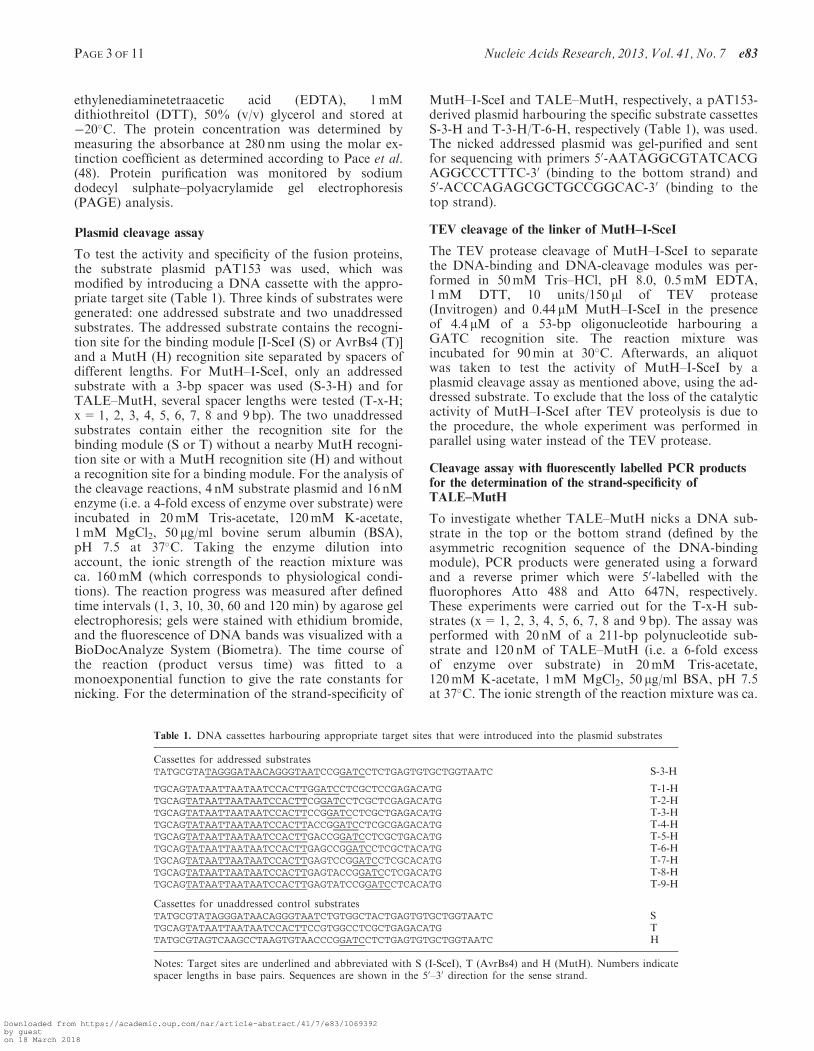
For the TALE–MutH fusion protein, a truncatedvariant of the TALE protein AvrBs4, corresponding tothe previously described AvrBs3 DNA-binding module(9), was fused directly to the N-terminal end of MutH.The gene for the TALE–MutH fusion contains twoparts: (i) the gene of the truncated TALE variant,missing the coding sequence of the first 152 amino acidsat the N-terminal end and the last 250 amino acids at theC-terminal end [28 amino acids remain after the lasthalf-repeat (M. Yanik, unpublished data)]. (ii) The geneof MutH, which contains the coding sequence for aC-terminal His6-tag. The two parts were connected andintroduced into the expression vector pQE30 (Qiagen),coding for an N-terminal Strep-tag. Thus, the expectedfusion protein would be: Strep-TALE–MutH-His6. Thesequence of both fusion constructs was confirmed byDNA sequencing of the entire coding region. We hadvaried the linker length between AvrBs3 and PvuIIwhich is structurally very similar to MutH (47) andfound the 28-amino-acid linker superior to a 63- and 16-amino-acid linker (M. Yanik, unpublished data). There-fore, we have chosen the 28-amino-acid linker for theTALE–MutH fusion protein.
Protein expression and purification
The expression vectors for the recombinant fusionproteins were introduced into the E. coli strainXL1-Blue (Stratagene). The cells were grown at 37�C toOD600 nm ca. 0.7 in LB-medium containing 75 mg/ml ampi-cillin. Protein expression was induced by adding 200mg/lanhydrotetracycline or 1mM isopropyl-b-D-thiogalacto-pyranoside for MutH–I-SceI or TALE–MutH, respect-ively, followed by further growth at 20�C overnight. Thecells were harvested by centrifugation and resuspended in20mM Tris–HCl, pH 7.9, 1 M NaCl, 20mM imidazole,1mM phenylmethanesulfonyl fluoride (PMSF), lysed bysonification and centrifuged for 30min (>17 000g) at 4�Cto remove cell debris. The His6- and Strep-tagged proteinswere first purified via Ni2+-NTA agarose (Qiagen) by a 1 hincubation of Ni2+-NTA agarose with the supernatant at4�C and washing with resuspension buffer; the protein waseluted with 20mM Tris–HCl, pH 7.9, 1 M NaCl and200mM imidazole. The eluted fractions were thentransferred to a column with Strep-Tactin Sepharose(IBA) for further purification. After washing with100mM Tris–HCl, pH 7.9, 1M NaCl, the protein waseluted with 100mM Tris–HCl, pH 7.9, 500mM NaCl,2.5mM desthiobiotin, dialyzed overnight at 4�C against10mM HEPES-KOH, pH 7.9, 500mM KCl, 1mM
e83 Nucleic Acids Research, 2013, Vol. 41, No. 7 PAGE 2 OF 11
Downloaded from https://academic.oup.com/nar/article-abstract/41/7/e83/1069392by gueston 18 March 2018
ethylenediaminetetraacetic acid (EDTA), 1mMdithiothreitol (DTT), 50% (v/v) glycerol and stored at�20�C. The protein concentration was determined bymeasuring the absorbance at 280 nm using the molar ex-tinction coefficient as determined according to Pace et al.(48). Protein purification was monitored by sodiumdodecyl sulphate–polyacrylamide gel electrophoresis(PAGE) analysis.
Plasmid cleavage assay
To test the activity and specificity of the fusion proteins,the substrate plasmid pAT153 was used, which wasmodified by introducing a DNA cassette with the appro-priate target site (Table 1). Three kinds of substrates weregenerated: one addressed substrate and two unaddressedsubstrates. The addressed substrate contains the recogni-tion site for the binding module [I-SceI (S) or AvrBs4 (T)]and a MutH (H) recognition site separated by spacers ofdifferent lengths. For MutH–I-SceI, only an addressedsubstrate with a 3-bp spacer was used (S-3-H) and forTALE–MutH, several spacer lengths were tested (T-x-H;x=1, 2, 3, 4, 5, 6, 7, 8 and 9 bp). The two unaddressedsubstrates contain either the recognition site for thebinding module (S or T) without a nearby MutH recogni-tion site or with a MutH recognition site (H) and withouta recognition site for a binding module. For the analysis ofthe cleavage reactions, 4 nM substrate plasmid and 16 nMenzyme (i.e. a 4-fold excess of enzyme over substrate) wereincubated in 20mM Tris-acetate, 120mM K-acetate,1mM MgCl2, 50 mg/ml bovine serum albumin (BSA),pH 7.5 at 37�C. Taking the enzyme dilution intoaccount, the ionic strength of the reaction mixture wasca. 160mM (which corresponds to physiological condi-tions). The reaction progress was measured after definedtime intervals (1, 3, 10, 30, 60 and 120 min) by agarose gelelectrophoresis; gels were stained with ethidium bromide,and the fluorescence of DNA bands was visualized with aBioDocAnalyze System (Biometra). The time course ofthe reaction (product versus time) was fitted to amonoexponential function to give the rate constants fornicking. For the determination of the strand-specificity of
MutH–I-SceI and TALE–MutH, respectively, a pAT153-derived plasmid harbouring the specific substrate cassettesS-3-H and T-3-H/T-6-H, respectively (Table 1), was used.The nicked addressed plasmid was gel-purified and sentfor sequencing with primers 50-AATAGGCGTATCACGAGGCCCTTTC-30 (binding to the bottom strand) and50-ACCCAGAGCGCTGCCGGCAC-30 (binding to thetop strand).
TEV cleavage of the linker of MutH–I-SceI
The TEV protease cleavage of MutH–I-SceI to separatethe DNA-binding and DNA-cleavage modules was per-formed in 50mM Tris–HCl, pH 8.0, 0.5mM EDTA,1mM DTT, 10 units/150 ml of TEV protease(Invitrogen) and 0.44 mM MutH–I-SceI in the presenceof 4.4 mM of a 53-bp oligonucleotide harbouring aGATC recognition site. The reaction mixture wasincubated for 90min at 30�C. Afterwards, an aliquotwas taken to test the activity of MutH–I-SceI by aplasmid cleavage assay as mentioned above, using the ad-dressed substrate. To exclude that the loss of the catalyticactivity of MutH–I-SceI after TEV proteolysis is due tothe procedure, the whole experiment was performed inparallel using water instead of the TEV protease.
Cleavage assay with fluorescently labelled PCR productsfor the determination of the strand-specificity ofTALE–MutH
To investigate whether TALE–MutH nicks a DNA sub-strate in the top or the bottom strand (defined by theasymmetric recognition sequence of the DNA-bindingmodule), PCR products were generated using a forwardand a reverse primer which were 50-labelled with thefluorophores Atto 488 and Atto 647N, respectively.These experiments were carried out for the T-x-H sub-strates (x=1, 2, 3, 4, 5, 6, 7, 8 and 9 bp). The assay wasperformed with 20 nM of a 211-bp polynucleotide sub-strate and 120 nM of TALE–MutH (i.e. a 6-fold excessof enzyme over substrate) in 20mM Tris-acetate,120mM K-acetate, 1mM MgCl2, 50 mg/ml BSA, pH 7.5at 37�C. The ionic strength of the reaction mixture was ca.
Table 1. DNA cassettes harbouring appropriate target sites that were introduced into the plasmid substrates
Cassettes for addressed substratesTATGCGTATAGGGATAACAGGGTAATCCGGATCCTCTGAGTGTGCTGGTAATC S-3-H
TGCAGTATAATTAATAATCCACTTGGATCCTCGCTCCGAGACATG T-1-HTGCAGTATAATTAATAATCCACTTCGGATCCTCGCTCGAGACATG T-2-HTGCAGTATAATTAATAATCCACTTCCGGATCCTCGCTGAGACATG T-3-HTGCAGTATAATTAATAATCCACTTACCGGATCCTCGCGAGACATG T-4-HTGCAGTATAATTAATAATCCACTTGACCGGATCCTCGCTGACATG T-5-HTGCAGTATAATTAATAATCCACTTGAGCCGGATCCTCGCTACATG T-6-HTGCAGTATAATTAATAATCCACTTGAGTCCGGATCCTCGCACATG T-7-HTGCAGTATAATTAATAATCCACTTGAGTACCGGATCCTCGACATG T-8-HTGCAGTATAATTAATAATCCACTTGAGTATCCGGATCCTCACATG T-9-H
Cassettes for unaddressed control substratesTATGCGTATAGGGATAACAGGGTAATCTGTGGCTACTGAGTGTGCTGGTAATC STGCAGTATAATTAATAATCCACTTCCGTGGCCTCGCTGAGACATG TTATGCGTAGTCAAGCCTAAGTGTAACCCGGATCCTCTGAGTGTGCTGGTAATC H
Notes: Target sites are underlined and abbreviated with S (I-SceI), T (AvrBs4) and H (MutH). Numbers indicatespacer lengths in base pairs. Sequences are shown in the 50–30 direction for the sense strand.
PAGE 3 OF 11 Nucleic Acids Research, 2013, Vol. 41, No. 7 e83
Downloaded from https://academic.oup.com/nar/article-abstract/41/7/e83/1069392by gueston 18 March 2018
160mM (which corresponds to physiological conditions).The reaction progress was analysed after defined timeintervals (3, 10, 30, 60, 120 and 180 min) by denaturingPAGE; the fluorescence of DNA bands was visualizedwith the VersaDoc Imaging System (Bio Rad).
RESULTS
Design of the fusion constructs
Two principally different types of approaches had beenrecently used by us to produce highly specific nucleasesfor the purpose of genome engineering: (i) fusion proteinswith a specificity defined by a catalytically inactive homingendonuclease [I-SceI, (12)] and (ii) fusion proteins with aprogrammable specificity defined by a ZF array (11) or aTALEprotein (M.Yanik, unpublished data). In both cases,the Type II restriction endonuclease PvuII was the DNA-cleavage module. We have now used these two differentapproaches in generating highly specific nickases,MutH–I-SceI and TALE–MutH. In analogy to ourPvuII–I-SceI construct (12), we have produced aMutH–I-SceI fusion protein with an N-terminal His6-tagand a C-terminal Strep-tag. Likewise, in analogy to ourZF- (11) and TALE-PvuII constructs (M. Yanik, unpub-lished data), a TALE–MutH fusion protein with anN-terminal Strep-tag and a C-terminal His6-tag wasproduced (Figure 1).
Determination of the cleavage preference of MutH–I-SceIfor the addressed substrate
The rates of DNA nicking were determined withunmethylated plasmid substrates with three different rec-ognition sites: (i) the addressed bipartite recognition sitecomposed of an I-SceI recognition site next to a MutHrecognition site (GATC), (ii) a stand-alone I-SceI
recognition site and (iii) a stand-alone MutH recognitionsite. All three plasmid substrates have 19 additionalGATC sites. As shown in Figure 2, only the plasmid sub-strate with the addressed bipartite site is nicked. Even at a4-fold excess of enzyme over unaddressed substrate and atprolonged incubation time (2 h), no non-specific nickingor cleavage is observed. Given the sensitivity of the assay,these results show that the plasmid substrate with the ad-dressed site is nicked by a factor of 1000 faster than theother plasmid substrates. Actually, the preference for theaddressed site over an unaddressed site might exceed thefactor of 1000, because (i) the plasmids used in the assaycontain 19 unaddressed sites, i.e. stand-alone GATC sitesand only one addressed site and (ii) in the determinationof the rate of nicking of the undressed substrate, the lowerlimit of detection had been reached.
Inactivation of the specific nicking activity ofMutH–I-SceI by proteolytic separation of thebinding and the cleavage module
The activity of the MutH–I-SceI fusion construct can besuppressed by separating the DNA-binding and DNA-cleavage modules by pre-incubating MutH–I-SceI withthe TEV protease which cleaves the linker peptide thatconnects the two modules. Figure 3 shows that thecovalent linkage of I-SceI and MutH is necessary fornicking of the addressed site. There is no nicking orcleavage observed when I-SceI and MutH are present inthe reaction mixture but not covalently linked to eachother. If the MutH–I-SceI fusion construct, in which thelinker does not contain a TEV protease cleavage site,is pre-incubated with TEV protease, the addressed sub-strate is nicked, demonstrating that TEV protease doesnot cleave MutH or I-SceI at a cryptic TEV proteaserecognition site.
Figure 1. (A) Scheme of the fusion constructs MutH–I-SceI and TALE-MutH binding to their respective recognition sites (50!30). The highlyspecific interaction is mediated mainly by the binding module, I-SceI or the DNA-binding domain of the TALE protein AvrBs4, respectively. Themonomeric cleavage module MutH is recruited by the binding module to nick the GATC site in close proximity to the I-SceI or TALE site. MutH isfused to I-SceI via a 10-amino-acid linker (ASENLYFQGG, shown as an orange line; the star indicates a TEV protease recognition site). In the caseof the TALE–MutH construct, no additional linker was introduced; the fusion was done 28 amino acids (shown as red line) after the last half-repeat.(B) Model of the designed fusion constructs bound to specific DNA, but without the oligopeptide linking the DNA-binding and DNA-cleavagemodule. The colour code can be deduced from the scheme, I-SceI in green, the TALE protein in red (only the 23 repeats of the 3UGM structure areshown) and MutH in blue. Modelling was done using the crystal structures 1R7M (I-SceI), 3UGM (TALE) and 2AOQ (MutH). The model wasgenerated with PyMOL and 3D-DART (56).
e83 Nucleic Acids Research, 2013, Vol. 41, No. 7 PAGE 4 OF 11
Downloaded from https://academic.oup.com/nar/article-abstract/41/7/e83/1069392by gueston 18 March 2018

Determination of the strand specificity of MutH–I-SceI
Since in the fusion proteinMutH–I-SceI, the DNA-bindingand the cleavage module were fused with a flexible linker,the question arose, whether both strands can be nickedby MutH or whether one strand is preferred. Guidedby the molecular model shown in Figure 1, we expectedthe bottom strand to be cleaved preferentially if notexclusively. The strand specificity of MutH–I-SceI wasdetermined by sequencing the nicked product. As shownin Figure 4, the top strand remains intact and the bottomstrand is cleaved 50 of the GATC site.
Determination of the cleavage preference of TALE–MutHfor the addressed substrate
Similar experiments as carried out to determine the pref-erence of the MutH–I-SceI fusion protein for addressedover unaddressed substrate cleavage were carried out forthe TALE–MutH fusion proteins. As shown in Figure 5,the TALE–MutH fusion constructs are specific for theaddressed bipartite recognition site, consisting of theTALE target site (here we used the AvrBs4 target site)in a defined distance next to a MutH recognition site.We tested spacer lengths of 1, 2, 3, 4, 5, 6, 7, 8 and 9 bpbetween the two sites on the addressed substrate andfound that a distance of 3 bp (T-3-H) is optimal(Figure 5C) for nicking. No double-strand breaks weredetectable.
Investigation of the strand specificity of TALE–MutH
To determine the strand specificity of TALE–MutH, wehave used a 211-bp PCR product that carried differentfluorophores on the 50-ends of the top and bottomstrand, respectively. Depending on which strand is prefer-entially nicked, the electrophoretic analysis of the nickingreaction would yield a different characteristic fluorescenceimage. As shown in Figure 6, TALE–MutH nicks thebottom strand of the substrate T-3-H with more thantwo orders of magnitude higher preference over the topstrand. Intriguingly, the substrate T-6-H is nicked prefer-entially in the top strand, albeit by a factor of ca. 3 moreslowly than the bottom strand of T-3-H. Similar experi-ments were performed also for the other substrates: T-1-Hto T-4-H are nicked preferentially in the bottom strand,whereas T-5-H to T-9-H are nicked preferentially in thetop strand. This change in preference for bottom strandnicking to top strand nicking could be correlated with thefinding shown in Figure 5 that there is a decrease in therate of nicking between T-3-H and T-6-H. Similarly, as forMutH–I-SceI (Figure 4), determination of strand specifi-city was also determined for the substrates T-3-H andT-6-H by sequencing. The results show that the bottomstrand (T-3-H) and the top strand (T-6-H), respectively,are nicked only at the #GATC site.
Figure 2. Kinetics of DNA nicking by the MutH–I-SceI fusion con-struct of addressed and unaddressed substrates analysed with plasmidcleavage assays using a 4-fold excess of enzyme over substrate (16 nMenzyme, 4 nM substrate). The addressed substrate plasmid contains anI-SceI and a MutH recognition site separated by 3 bp (S-3-H). Theunaddressed substrate plasmids (controls) contain either an I-SceIrecognition site without a nearby MutH recognition site (S) or noI-SceI recognition site (H). Each of the substrates has 19 additionalGATC sites distributed over the plasmid. The position of the bandsrepresenting supercoiled (sc), open circular (oc) and linear (lin) forms ofthe plasmid is indicated. The first lane shows the reaction withoutMgCl2 (‘�’). ‘Nick’ and ‘lin’ represent the controls for nicking andcleavage activity, respectively, and correspond to the open circularand linear forms of the plasmid, respectively. The electrophoreticanalysis shows that the addressed substrate plasmid is nicked by thefusion construct MutH–I-SceI, whereas the unaddressed substrate
Figure 2. Continuedplasmids are not nicked or cleaved. The calculated apparent cleavagerate constant for the addressed plasmid substrate is 4.0� 10�2min�1,whereas the apparent cleavage rate constants for both unaddressedsubstrates are below the detection limit of approximately4� 10�5min�1.
PAGE 5 OF 11 Nucleic Acids Research, 2013, Vol. 41, No. 7 e83
Downloaded from https://academic.oup.com/nar/article-abstract/41/7/e83/1069392by gueston 18 March 2018

DISCUSSION
The efficiency of homologous recombination can beincreased by several orders of magnitude by a specificdouble-strand break at the locus of interest (49). Thetargeted insertion of DNA into a pre-defined locus by hom-ologous recombination requires highly specific nucleases(50,51). Such nucleases became available with the fusionof zinc fingers to the FokI cleavage domain (52). It wasdemonstrated, however, that engineered double-strand-spe-cific nucleases could introduce mutations when thedouble-strand break is repaired by the error-prone NHEJpathway (53) rather than by HDR and that nickingenzymes suppress NHEJ (14,15), which means that DNAnicks can initiate efficient gene correction, with lessgenomic instability than a targeted DNA double-strandbreak. Ramirez et al. (33), Wang et al. (34) and Kimet al. (32) introduced Zinc finger nickases (ZFNs) fortargeted gene insertion and showed that they induceHDR with reduced mutagenic effects. As there are otherarchitectures for highly specific double-strand-specific nu-cleases [e.g. (11,12), M. Yanik, unpublished data], it shouldbe possible to generate highly site- and strand-specificnickases other than the ZF-based nickases. In this article,we have shown that specificity-determining DNA-bindingmodules (catalytically inactive I-SceI and the DNA-binding
Figure 3. Effect of separating the I-SceI and the MutH module on addressed substrate nicking. The kinetics of DNA-nicking by the MutH–I-SceIfusion construct of the addressed substrate was analysed with a plasmid cleavage assay (16 nM enzyme, 4 nM substrate). The addressed substrateplasmid contains an I-SceI and a MutH recognition site separated by 3 bp (S-3-H). The substrate has 19 additional GATC sites distributed over theplasmid. (A) Assay performed with MutH–I-SceI that had not been pre-incubated with TEV protease. (B) Assay performed with MutH–I-SceI thathad been pre-incubated with TEV protease. (C) For control, we have also incubated the fusion protein, in which the linker did not contain a TEVprotease cleavage site, with the TEV protease. The position of the bands representing supercoiled (sc), open circular (oc) and linear (lin) forms of theplasmid is indicated. The first lane shows the reaction without MgCl2 (‘�’). ‘Nick’ and ‘lin’ are the controls for cleavage and nicking activity andcorrespond to the open circular and linear forms of the plasmid, respectively. Whereas in (A) and (C), nicking is observed, in (B), it is not.
Figure 4. Determination of the strand specificity of MutH–I-SceI bysequence analysis of the nicked addressed plasmid after cleavage withthe fusion construct MutH–I-SceI. The cleavage product wasgel-purified and sequenced (top strand and bottom strand) over thespecific site (S-3-H). For the purpose of illustration, the sequence isshown in the reverse and complementary orientation. The recognitionsites of I-SceI and MutH are highlighted by green and blue bars,respectively. The sequencing results indicate that the bottom strand iscleaved 50 of the addressed GATC site. *Taq polymerase artefact.
e83 Nucleic Acids Research, 2013, Vol. 41, No. 7 PAGE 6 OF 11
Downloaded from https://academic.oup.com/nar/article-abstract/41/7/e83/1069392by gueston 18 March 2018

domain of a TALE protein, respectively) can be fused to aspecific nicking enzyme (MutH) to produce a highlysequence- and strand-specific nickase. The fusion proteinsthat we obtained, MutH–I-SceI and TALE–MutH, recog-nize their respective bipartite recognition sequence,
consisting of the recognition site of the DNA-bindingmodule and the MutH recognition site (GATC). Theyonly nick one strand 50 of the GATC site and do notnick stand-alone GATC sites or any other sites, makingthem potentially useful tools for site-specific nicking of
Figure 5. Kinetics of DNA-cleavage by TALE–MutH fusion constructs of addressed and unaddressed substrates analysed with plasmid cleavageassays using a 4-fold excess of enzyme over substrate (16 nM enzyme, 4 nM substrate). (A) Catalytic activity of TALE–MutH with a plasmidsubstrate containing an AvrBs4 and a MutH recognition site separated by� base pairs (T-x-H; x=1–9 bp). (B) Catalytic activity on the unaddressedsubstrate plasmids (controls) containing either an AvrBs4 recognition site without a nearby MutH recognition site (T) or no AvrBs4 recognition site(H). Each of the substrates has 18 additional GATC sites distributed over the plasmid. The position of the bands representing supercoiled (sc), opencircular (oc) and linear (lin) forms of the plasmid is indicated. The first lane shows the reaction without MgCl2 (‘�’). ‘Nick’ and ‘lin’ representcontrols for nicking and cleavage activity, respectively, and correspond to the open circular and linear forms of the plasmid, respectively.(C) Calculated rate constants for TALE–MutH nicking the above-mentioned substrates. The electrophoretic analysis shows that the addressedsubstrate plasmid is nicked by the fusion construct TALE–MutH, whereas the unaddressed substrate plasmids remain uncleaved. The graphshows the quantitative evaluation of the experiments shown in (A) and (B). Note that the enzyme exhibits the best activity on the addressedsubstrate with the 3 bp spacer and no activity with the stand-alone TALE (T) or MutH (H) recognition sites. The quantitative analyses reveal thefollowing apparent cleavage rate constants: 1.3� 10�4min�1 (T-1-H), 1.2� 10�2min�1 (T-2-H), 3.6� 10�2min�1 (T-3-H), 1.1� 10�2min�1 (T-4-H),0.3� 10�2min�1 (T-5-H), 1.8� 10�2min�1 (T-6-H), 0.6� 10�2min�1 (T-7-H), 0.8� 10�2min�1 (T-8-H) and 8.9� 10�4min�1 (T-9-H); the nickingrate for T and H was below the detection limit of approximately 4� 10�5min�1.
PAGE 7 OF 11 Nucleic Acids Research, 2013, Vol. 41, No. 7 e83
Downloaded from https://academic.oup.com/nar/article-abstract/41/7/e83/1069392by gueston 18 March 2018
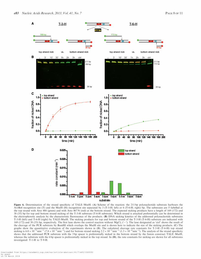
Figure 6. Determination of the strand specificity of TALE–MutH. (A) Scheme of the reaction: the 211 bp polynucleotide substrate harbours theAvrBs4 recognition site (T) and the MutH (H) recognition site separated by 3 (T-3-H; left) or 6 (T-6-H; right) bp. The substrates are 50-labelled atthe top strand with Atto 488 (green) and with Atto 467N (red) at the bottom strand. The expected nicking products have a length of 169 (172) and38 (35) bp for top and bottom strand nicking of the T-3-H substrate (T-6-H substrate). Which strand is attacked preferentially can be determined inthe electrophoretic analysis by the characteristic fluorescence of the products. (B) DNA nicking kinetics of the addressed polynucleotide substratesT-3-H (left) and T-6-H (right) by TALE-MutH. The nicking products for top and bottom strand of the T-3-H (T-6-H) substrate are indicated with169 (172) and 38 (35) bp, respectively. The first lane shows the control reaction without MgCl2 (‘�’). The lane designated as ‘ctrl’ shows the result ofthe cleavage of the PCR substrate by BamHI which overlaps the MutH site and is shown here to indicate the size of the nicking products. (C) Thegraphs show the quantitative evaluation of the experiments shown in (B). The calculated cleavage rate constants for T-3-H (T-6-H) top strandnicking is 6.0� 10�5min�1 (7.8� 10�3min�1) and for bottom strand nicking 2.2� 10�2min�1 (1.3� 10�4min�1). The analysis of the strand specificityshows that the addressed PCR substrate with the 3 bp spacer is preferentially nicked in the bottom strand by the fusion construct TALE–MutH,whereas the substrate with the 6 bp spacer is preferentially nicked in the top strand. In (D), the rate constants for nicking are shown for all substratesinvestigated: T-1-H to T-9-H.
e83 Nucleic Acids Research, 2013, Vol. 41, No. 7 PAGE 8 OF 11
Downloaded from https://academic.oup.com/nar/article-abstract/41/7/e83/1069392by gueston 18 March 2018

DNA in general and precision genome engineering inparticular.
MutH is a site-specific DNA nicking enzyme which inits natural function requires complex formation withactivated MutL to be directed to its target site, ahemimethylated GATC site that is nicked in theunmethylated strand. By itself and at physiological ionicstrength and Mg2+ concentration (which we had deliber-ately chosen to be prepared for applications in vivo),MutH does not attack unmethylated GATC sites, but isstrictly dependent on a covalent coupling to a DNA-binding module, as was shown here for MutH–I-SceI.Proteolytic separation of the DNA-binding and DNA-cleavage module prevents DNA cleavage. This findingsuggests that MutH cannot bind in a productive mannerto the bipartite recognition site, unless it is positionedproperly by the I-SceI (or TALE) module as part of thefusion protein.
We had shown before (12) that catalytically inactiveI-SceI can serve as a specific DNA-binding module forthe restriction endonuclease PvuII. Our results withMutH–I-SceI demonstrate that this is also possible withother nucleases, here a nicking enzyme that is structurallyrelated to PvuII (47). MutH–I-SceI recognizes a uniquesequence. If such a sequence is introduced into acomplex genome, this sequence could be used as a targetsite for genome engineering, as it was done with I-SceIsites for in planta gene targeting (54).
Of particular interest is the TALE-MutH fusionprotein, which in contrast to MutH–I-SceI is program-mable to recognize almost any DNA sequence. Similarto the MutH–I-SceI, the TALE–MutH fusion proteinrequires a GATC site next to the recognition sequenceof its DNA-binding module, the TALE recognitionsequence. This requirement should not limit the usefulnessof this programmable nickase, as GATC sites occur onaverage every 256 bp and are unmethylated in eukaryoticgenomes. In contrast to the ZF-nickases based on FokI(32–34), TALE–MutH only needs one TALE protein fortargeting to a specific site, which reduces the size of aMutH-based TALE-nickase by 50% compared with aFokI-based TALE-nickase, because MutH functions asa monomer whereas FokI is a functional dimer. As theDNA-binding module of TALE proteins can be used toprogram the restriction endonuclease PvuII to cleave abipartite recognition sequence consisting of the TALE rec-ognition sequence and the PvuII recognition sequence (M.Yanik, unpublished data), we believe that other single-strand-specific nucleases can function as highly specificnickases in fusion proteins consisting of a DNA-bindingmodule and a DNA-nicking module. Examples are restric-tion enzymes such as Nt.CviPII (19,20) or subunits ofheterodimeric restriction endonucleases, e.g. Nt.BstD6I(21,22), Nb.BsrDI and Nt.BtsI (23) (Nt or Nb indicatethe specificity for top or bottom strand nicking).Different from these naturally occurring nickingenzymes, the TALE–MutH fusion protein can nick theupper or lower strand, depending on the distancebetween the TALE recognition site and the GATCsequence. For example, with a spacer length of 2–4 bp(optimally T-3-H), the bottom strand is nicked with
several hundred-fold preference over the other strand.In contrast, with a spacer length of 5–8 bp (optimallyT-6-H), the rate of nicking decreases but now the topstrand is preferred. Substrates with a spacer length of1 and 9 bp are hardly attacked at all. This suggests thatthere is some flexibility in the junction of the DNA-binding module and the DNA-cleavage module whichallows MutH to reach the scissile phosphodiester bondeither in the bottom strand or in the top strand, dependingon the distance between the MutH recognition site relativeto the TALE recognition site. We believe that decreasingor increasing the length of the linker between TALE andMutH could have a similar effect, i.e. changing the pref-erence for bottom or top strand nicking. As we have notdetermined the optimal linker between AvrBs4 and MutHexperimentally, but rather extrapolated the linker length(28 aa) in our TALE–MutH fusion protein from a previ-ously constructed TALE–PvuII fusion construct, it couldbe that the specificity could be further increased by slightmodifications in the linker length. Likewise, we have notoptimized the DNA-binding module of TALE protein butrather used the natural AvrBs4 protein sequence; it couldwell be that exchanging some of the RVDs by ‘strong’RVDs (55) could even further increase specificity.
ACKNOWLEDGEMENTS
The authors thank Mert Yanik, Marika Midon, InesFonfara, Andreas Marx and Roger Heinze for fruitfuldiscussions and plasmids; Laura Waltl for assistance andAnja Drescher for critical reading of the article. L.G. isa member of the International Research Training Group‘Enzymes and Multienzyme Complexes acting on NucleicAcids’ funded by the Deutsche Forschungsgemeinschaft(DFG): B.S. has been a recipient of a grant (Just’us) bythe Justus-Liebig-University Giessen.
FUNDING
Deutsche Forschungsgemeinschaft [InternationalResearch Training Group GRK 1384, and ExcellenceCluster ECCPS] and the Justus-Liebig-UniversityGiessen [Just’us]. Funding for open access charge: DFGand the Justus-Liebig-University Giessen.
Conflict of interest statement. None declared.
REFERENCES
1. Marcaida,M.J., Munoz,I.G., Blanco,F.J., Prieto,J. andMontoya,G. (2010) Homing endonucleases: from basics totherapeutic applications. Cell. Mol. Life Sci., 67, 727–748.
2. Silva,G., Poirot,L., Galetto,R., Smith,J., Montoya,G.,Duchateau,P. and Paques,F. (2011) Meganucleases and othertools for targeted genome engineering: perspectives and challengesfor gene therapy. Curr. Gene Ther., 11, 11–27.
3. Urnov,F.D., Rebar,E.J., Holmes,M.C., Zhang,H.S. andGregory,P.D. (2010) Genome editing with engineered zinc fingernucleases. Nat. Rev. Genet., 11, 636–646.
4. Ramalingam,S., Kandavelou,K., Rajenderan,R. andChandrasegaran,S. (2011) Creating designed zinc-finger nucleaseswith minimal cytotoxicity. J. Mol. Biol., 405, 630–641.
PAGE 9 OF 11 Nucleic Acids Research, 2013, Vol. 41, No. 7 e83
Downloaded from https://academic.oup.com/nar/article-abstract/41/7/e83/1069392by gueston 18 March 2018

5. Rahman,S.H., Maeder,M.L., Joung,J.K. and Cathomen,T. (2011)Zinc-finger nucleases for somatic gene therapy: the next frontier.Hum. Gene. Ther., 22, 925–933.
6. Carroll,D. (2011) Genome engineering with zinc-finger nucleases.Genetics, 188, 773–782.
7. Christian,M., Cermak,T., Doyle,E.L., Schmidt,C., Zhang,F.,Hummel,A., Bogdanove,A.J. and Voytas,D.F. (2010) TALeffector nucleases create targeted DNA double-strand breaks.Genetics, 186, 757–761.
8. Li,T., Huang,S., Jiang,W.Z., Wright,D., Spalding,M.H.,Weeks,D.P. and Yang,B. (2010) TAL nucleases (TALNs): hybridproteins composed of TAL effectors and FokI DNA-cleavagedomain. Nucleic Acids Res., 39, 359–372.
9. Miller,J.C., Tan,S., Qiao,G., Barlow,K.A., Wang,J., Xia,D.F.,Meng,X., Paschon,D.E., Leung,E., Hinkley,S.J. et al. (2011)A TALE nuclease architecture for efficient genome editing.Nat. Biotechnol., 29, 143–148.
10. Mussolino,C., Morbitzer,R., Lutge,F., Dannemann,N., Lahaye,T.and Cathomen,T. (2011) A novel TALE nuclease scaffold enableshigh genome editing activity in combination with low toxicity.Nucleic Acids Res., 39, 9283–9293.
11. Schierling,B., Dannemann,N., Gabsalilow,L., Wende,W.,Cathomen,T. and Pingoud,A. (2012) A novel zinc-finger nucleaseplatform with a sequence-specific cleavage module. Nucleic AcidsRes., 40, 2623–2638.
12. Fonfara,I., Curth,U., Pingoud,A. and Wende,W. (2012) Creatinghighly specific nucleases by fusion of active restrictionendonucleases and catalytically inactive homing endonucleases.Nucleic Acids Res., 40, 847–860.
13. Kleinstiver,B.P., Wolfs,J.M., Kolaczyk,T., Roberts,A.K., Hu,S.X.and Edgell,D.R. (2012) Monomeric site-specific nucleases forgenome editing. Proc. Natl Acad. Sci. USA, 109, 8061–8066.
14. Metzger,M.J., McConnell-Smith,A., Stoddard,B.L. andMiller,A.D. (2011) Single-strand nicks induce homologousrecombination with less toxicity than double-strand breaks usingan AAV vector template. Nucleic Acids Res., 39, 926–935.
15. Davis,L. and Maizels,N. (2011) DNA nicks promote efficient andsafe targeted gene correction. PLoS One, 6, e23981.
16. van Nierop,G.P., de Vries,A.A., Holkers,M., Vrijsen,K.R. andGoncalves,M.A. (2009) Stimulation of homology-directed genetargeting at an endogenous human locus by a nickingendonuclease. Nucleic Acids Res., 37, 5725–5736.
17. Zheleznaya,L.A., Kachalova,G.S., Artyukh,R.I., Yunusova,A.K.,Perevyazova,T.A. and Matvienko,N.I. (2009) Nickingendonucleases. Biochemistry, 74, 1457–1466.
18. Chan,S.H., Stoddard,B.L. and Xu,S.Y. (2011) Naturaland engineered nicking endonucleases—from cleavagemechanism to engineering of strand-specificity. Nucleic Acids Res.,39, 1–18.
19. Xia,Y.N., Morgan,R., Schildkraut,I. and Van Etten,J.L. (1988)A site-specific single strand endonuclease activity induced byNYs-1 virus infection of a Chlorella-like green alga. Nucleic AcidsRes., 16, 9477–9487.
20. Chan,S.H., Zhu,Z., Van Etten,J.L. and Xu,S.Y. (2004) Cloning ofCviPII nicking and modification system from chlorella virusNYs-1 and application of Nt.CviPII in random DNAamplification. Nucleic Acids Res., 32, 6187–6199.
21. Zheleznaya,L.A., Perevyazova,T.A., Alzhanova,D.V. andMatvienko,N.I. (2001) Site-specific nickase from bacillus speciesstrain d6. Biochemistry, 66, 989–993.
22. Kachalova,G.S., Rogulin,E.A., Yunusova,A.K., Artyukh,R.I.,Perevyazova,T.A., Matvienko,N.I., Zheleznaya,L.A. andBartunik,H.D. (2008) Structural analysis of the heterodimeric typeIIS restriction endonuclease R.BspD6I acting as a complexbetween a monomeric site-specific nickase and a catalytic subunit.J. Mol. Biol., 384, 489–502.
23. Xu,S.Y., Zhu,Z., Zhang,P., Chan,S.H., Samuelson,J.C., Xiao,J.,Ingalls,D. and Wilson,G.G. (2007) Discovery of natural nickingendonucleases Nb.BsrDI and Nb.BtsI and engineering oftop-strand nicking variants from BsrDI and BtsI. Nucleic AcidsRes., 35, 4608–4618.
24. Shen,B.W., Landthaler,M., Shub,D.A. and Stoddard,B.L. (2004)DNA binding and cleavage by the HNH homing endonucleaseI-HmuI. J. Mol. Biol., 342, 43–56.
25. Landthaler,M., Shen,B.W., Stoddard,B.L. and Shub,D.A. (2006)I-BasI and I-HmuI: two phage intron-encoded endonucleases withhomologous DNA recognition sequences but distinct DNAspecificities. J. Mol. Biol., 358, 1137–1151.
26. Landthaler,M. and Shub,D.A. (2003) The nicking homingendonuclease I-BasI is encoded by a group I intron in the DNApolymerase gene of the Bacillus thuringiensis phage Bastille.Nucleic Acids Res., 31, 3071–3077.
27. Wende,W., Stahl,F. and Pingoud,A. (1996) The production andcharacterization of artificial heterodimers of the restrictionendonuclease EcoRV. Biol. Chem., 377, 625–632.
28. Stahl,F., Wende,W., Jeltsch,A. and Pingoud,A. (1996)Introduction of asymmetry in the naturally symmetric restrictionendonuclease EcoRV to investigate intersubunit communication inthe homodimeric protein. Proc. Natl Acad. Sci. USA, 93,6175–6180.
29. Sanders,K.L., Catto,L.E., Bellamy,S.R. and Halford,S.E. (2009)Targeting individual subunits of the FokI restriction endonucleaseto specific DNA strands. Nucleic Acids Res., 37, 2105–2115.
30. Niu,Y., Tenney,K., Li,H. and Gimble,F.S. (2008) Engineeringvariants of the I-SceI homing endonuclease with strand-specificand site-specific DNA-nicking activity. J. Mol. Biol., 382,188–202.
31. McConnell Smith,A., Takeuchi,R., Pellenz,S., Davis,L.,Maizels,N., Monnat,R.J. Jr and Stoddard,B.L. (2009) Generationof a nicking enzyme that stimulates site-specific gene conversionfrom the I-AniI LAGLIDADG homing endonuclease. Proc. NatlAcad. Sci. USA, 106, 5099–5104.
32. Kim,E., Kim,S., Kim,D.H., Choi,B.S., Choi,I.Y. and Kim,J.S.(2012) Precision genome engineering with programmableDNA-nicking enzymes. Genome Res., 22, 1327–1333.
33. Ramirez,C.L., Certo,M.T., Mussolino,C., Goodwin,M.J.,Cradick,T.J., McCaffrey,A.P., Cathomen,T., Scharenberg,A.M.and Joung,J.K. (2012) Engineered zinc finger nickases inducehomology-directed repair with reduced mutagenic effects. NucleicAcids Res., 40, 5560–5568.
34. Wang,J., Friedman,G., Doyon,Y., Wang,N.S., Li,C.J., Miller,J.C.,Hua,K.L., Yan,J.J., Babiarz,J.E., Gregory,P.D. et al. (2012)Targeted gene addition to a predetermined site in the humangenome using a ZFN-based nicking enzyme. Genome Res., 22,1316–1326.
35. Halford,S.E., Catto,L.E., Pernstich,C., Rusling,D.A. andSanders,K.L. (2011) The reaction mechanism of FokI excludesthe possibility of targeting zinc finger nucleases to unique DNAsites. Biochem. Soc. Trans., 39, 584–588.
36. Miller,J.C., Holmes,M.C., Wang,J., Guschin,D.Y., Lee,Y.L.,Rupniewski,I., Beausejour,C.M., Waite,A.J., Wang,N.S., Kim,K.A.et al. (2007) An improved zinc-finger nuclease architecture forhighly specific genome editing. Nat. Biotechnol., 25, 778–785.
37. Szczepek,M., Brondani,V., Buchel,J., Serrano,L., Segal,D.J. andCathomen,T. (2007) Structure-based redesign of the dimerizationinterface reduces the toxicity of zinc-finger nucleases. Nat.Biotechnol., 25, 786–793.
38. Gabriel,R., Lombardo,A., Arens,A., Miller,J.C., Genovese,P.,Kaeppel,C., Nowrouzi,A., Bartholomae,C.C., Wang,J.,Friedman,G. et al. (2011) An unbiased genome-wide analysis ofzinc-finger nuclease specificity. Nat. Biotechnol., 29, 816–823.
39. Pattanayak,V., Ramirez,C.L., Joung,J.K. and Liu,D.R. (2011)Revealing off-target cleavage specificities of zinc-finger nucleasesby in vitro selection. Nat. Methods, 8, 765–770.
40. Lee,J.Y., Chang,J., Joseph,N., Ghirlando,R., Rao,D.N. andYang,W. (2005) MutH complexed with hemi- and unmethylatedDNAs: coupling base recognition and DNA cleavage. Mol. Cell,20, 155–166.
41. Welsh,K.M., Lu,A.L., Clark,S. and Modrich,P. (1987) Isolationand characterization of the Escherichia coli mutH gene product.J. Biol. Chem., 262, 15624–15629.
42. Hall,M.C. and Matson,S.W. (1999) The Escherichia coli MutLprotein physically interacts with MutH and stimulates theMutH-associated endonuclease activity. J. Biol. Chem., 274,1306–1312.
43. Lahue,R.S., Su,S.S. and Modrich,P. (1987) Requirement ford(GATC) sequences in Escherichia coli mutHLS mismatchcorrection. Proc. Natl Acad. Sci. USA, 84, 1482–1486.
e83 Nucleic Acids Research, 2013, Vol. 41, No. 7 PAGE 10 OF 11
Downloaded from https://academic.oup.com/nar/article-abstract/41/7/e83/1069392by gueston 18 March 2018

44. Modrich,P. and Lahue,R. (1996) Mismatch repair in replicationfidelity, genetic recombination, and cancer biology. Annu. Rev.Biochem., 65, 101–133.
45. Thomas,E., Pingoud,A. and Friedhoff,P. (2002) An efficientmethod for the preparation of long heteroduplex DNA assubstrate for mismatch repair by the Escherichia coli MutHLSsystem. Biol. Chem., 383, 1459–1462.
46. Lippow,S.M., Aha,P.M., Parker,M.H., Blake,W.J., Baynes,B.M.and Lipovsek,D. (2009) Creation of a type IIS restrictionendonuclease with a long recognition sequence.Nucleic Acids Res., 37, 3061–3073.
47. Ban,C. and Yang,W. (1998) Structural basis for MutH activationin E. coli mismatch repair and relationship of MutH torestriction endonucleases. EMBO J., 17, 1526–1534.
48. Pace,C.N., Vajdos,F., Fee,L., Grimsley,G. and Gray,T. (1995)How to measure and predict the molar absorption coefficientof a protein. Protein Sci., 4, 2411–2423.
49. Jasin,M. (1996) Genetic manipulation of genomes withrare-cutting endonucleases. Trends Genet., 12, 224–228.
50. Porteus,M.H. and Baltimore,D. (2003) Chimeric nucleasesstimulate gene targeting in human cells. Science, 300, 763.
51. Bibikova,M., Carroll,D., Segal,D.J., Trautman,J.K., Smith,J.,Kim,Y.G. and Chandrasegaran,S. (2001) Stimulation ofhomologous recombination through targeted cleavage by chimericnucleases. Mol. Cell. Biol., 21, 289–297.
52. Kim,Y.G., Cha,J. and Chandrasegaran,S. (1996) Hybridrestriction enzymes: zinc finger fusions to Fok I cleavage domain.Proc. Natl Acad. Sci. USA, 93, 1156–1160.
53. Bibikova,M., Golic,M., Golic,K.G. and Carroll,D. (2002)Targeted chromosomal cleavage and mutagenesis in Drosophilausing zinc-finger nucleases. Genetics, 161, 1169–1175.
54. Fauser,F., Roth,N., Pacher,M., Ilg,G., Sanchez-Fernandez,R.,Biesgen,C. and Puchta,H. (2012) In planta gene targeting.Proc. Natl Acad. Sci. USA, 109, 7535–7540.
55. Streubel,J., Blucher,C., Landgraf,A. and Boch,J. (2012) TAL effectorRVD specificities and efficiencies. Nat. Biotechnol., 30, 593–595.
56. van Dijk,M. and Bonvin,A.M. (2009) 3D-DART: a DNAstructure modelling server. Nucleic Acids Res., 37, W235–W239.
PAGE 11 OF 11 Nucleic Acids Research, 2013, Vol. 41, No. 7 e83
Downloaded from https://academic.oup.com/nar/article-abstract/41/7/e83/1069392by gueston 18 March 2018


![Aspects of DNA Strand Exchange: Recombination Proteins and ...publications.lib.chalmers.se/records/fulltext/127152.pdf · different proteins are involved . One[7]approach to understanding](https://img.pdfslide.us/doc/110x75/5fc0998778b63308bc2217e6/aspects-of-dna-strand-exchange-recombination-proteins-and-different-proteins.jpg)