Embed Size (px)
Citation preview
Behavioral/Cognitive
Encoding of Touch Intensity But Not Pleasantness in HumanPrimary Somatosensory Cortex
X Laura K. Case,1 Claire M. Laubacher,1 Håkan Olausson,3 Binquan Wang,1 Primavera A. Spagnolo,2
and X M. Catherine Bushnell1
1National Center for Complementary and Integrative Health and 2National Institute on Alcohol Abuse and Alcoholism, National Institutes of Health,Bethesda, Maryland 20892, and 3Department of Clinical and Experimental Medicine, Linkoping University, 581 83 Linkoping, Sweden
Growing interest in affective touch has delineated a neural network that bypasses primary somatosensory cortex (S1). Several recentstudies, however, have cast doubt on the segregation of touch discrimination and affect, suggesting that S1 also encodes affectivequalities. We used functional magnetic resonance imaging (fMRI) and repetitive transcranial magnetic stimulation (rTMS) to examinethe role of S1 in processing touch intensity and pleasantness. Twenty-six healthy human adults rated brushing on the hand during fMRI.Intensity ratings significantly predicted activation in S1, whereas pleasantness ratings predicted activation only in the anterior cingulatecortex. Nineteen subjects also received inhibitory rTMS over right hemisphere S1 and the vertex (control). After S1 rTMS, but not aftervertex rTMS, sensory discrimination was reduced and subjects with reduced sensory discrimination rated touch as more intense. Incontrast, rTMS did not alter ratings of touch pleasantness. Our findings support divergent neural processing of touch intensity andpleasantness, with affective touch encoded outside of S1.
Key words: affect; c-tactile fibers; pleasantness; rTMS; somatosensory cortex; touch discrimination
IntroductionBoth the affective and discriminative aspects of touch are criti-cally important in everyday life. Discriminative aspects of touchsupport object recognition and motor activities and socially rel-evant tactile information may allow the detection of hedonic en-vironmental features that serve the well-being and homeostasis ofthe organism (Craig, 2013). Extensive research has identified therole of primary somatosensory cortex (S1) in discriminative as-
pects of somatosensation, including tactile detection thresholds(Cohen et al., 1991), temporal frequency discrimination (Knechtet al., 2003), two-point discrimination (2PD; Tegenthoff et al.,2005), and tactile direction discrimination (Lundblad et al.,2011). However, the role of S1 in the affective dimension of so-matosensation is under dispute.
A growing body of research on C-tactile (CT) afferents hascharacterized an affective touch network that appears to by-pass S1. CT afferents are a class of peripheral unmyelinatedC-fibers that respond preferentially to slow stroking of the hairyskin and in which firing rate correlates with subjective ratings oftouch pleasantness (Loken et al., 2009). Two rare patients selec-tively lacking A� afferents (with C fibers intact; Sterman et al.,1980) exhibited impoverished touch discrimination, yet reportedpleasure and showed a robust response in the insula, but not S1,when their hairy skin was stroked at CT-optimal speeds(Olausson et al., 2002, 2008). Although CT-optimized stimulialso activate S1 cortex in healthy subjects, correlations with rat-
Received March 18, 2015; revised April 19, 2016; accepted April 21, 2016.Author contributions: L.K.C., H.O., P.A.S., and M.C.B. designed research; L.K.C., C.M.L., and B.W. performed
research; L.K.C., C.M.L., and B.W. analyzed data; L.K.C., C.M.L., H.O., and M.C.B. wrote the paper.This work was supported by the Intramural Research program of the National Center for Complementary and
Integrative Health–National Institutes of Health. We thank John Gracely, Marta Ceko, and Mark Hallett for assistancewith the experimental design and data analysis.
Correspondence should be addressed to Dr. Laura Case, National Center for Complementary and IntegrativeHealth, National Institutes of Health, Building 10, CRC Room 4-1730, MSC 1302, Bethesda, MD 20892. E-mail:[email protected].
DOI:10.1523/JNEUROSCI.1130-15.2016Copyright © 2016 the authors 0270-6474/16/365850-11$15.00/0
Significance Statement
Growing interest in affective touch has identified a neural network that bypasses primary somatosensory cortex (S1). Severalrecent studies, however, cast doubt on the separation of touch discrimination and affect. We used functional magnetic resonanceimaging and repetitive transcranial magnetic stimulation to demonstrate the representation of touch discrimination and intensityin S1, but the representation of pleasantness in the anterior cingulate cortex, not S1. Our findings support divergent neuralprocessing of touch intensity and pleasantness, with affective touch encoded outside of S1. Our study contributes to growingdelineation of the affective touch system, a crucial step in understanding its dysregulation in numerous clinical conditions such asautism, eating disorders, depression, and chronic pain.
5850 • The Journal of Neuroscience, May 25, 2016 • 36(21):5850 –5860

ings of touch pleasantness have been reported only in other re-gions, including the orbitofrontal cortex (OFC; McCabe et al.,2008) and the insula (Kress et al., 2011).
Despite the evidence that touch pleasantness is processed out-side of S1, several recent studies challenge this model, showingcorrelations between ratings of touch pleasantness and activationof S1 (though in opposite directions; McCabe et al., 2008; Gaz-zola et al., 2012). These studies demonstrate possible modulationof S1 by social context, but contain confounds related to atten-tion and motivation and do not demonstrate the causal necessityof S1 in the perception of touch pleasantness. These studies alsodid not test bottom-up sensory contributions to touch pleasant-ness, such as the contribution of CT-optimal touch.
The current study focuses on affective qualities conveyed bythe physical touch stimulus, rather than through top-down mod-ulation of touch processing by social context. We used both func-tional magnetic resonance imaging (fMRI; n � 26) and repetitivetranscranial magnetic stimulation (rTMS; n � 19) to test theinvolvement of S1 in representing the pleasantness and intensityof touch. During the fMRI portion of our experiment, we admin-istered slow and fast brushing to hairy and glabrous skin of thehand to produce differential activation of CT fibers and collectedsubjects’ ratings of pleasantness. This variable CT activation af-forded manipulation of touch pleasantness without altering thetexture or physical intensity of the touch stimuli, allowing us tolook for stimulus-driven neural representation of touch pleasant-ness. However, intensity and pleasantness ratings vary by brush-ing speed even when administered by a robot exerting constantforce (Triscoli et al., 2013). We therefore also collected intensityratings to account for differences in intensity perception in ouranalyses.
We then used 1 Hz rTMS over right hemisphere S1 to causallytest the necessity of S1 activity for the perception of touch pleas-antness. Low-frequency rTMS of �0.2–1 Hz produces a period ofcortical inhibition (Chen et al., 1997; Maeda et al., 2000; Hallett,2007) and is often used to deactivate a brain region temporarily toexamine its direct causal contribution to a particular percept orbehavior (Hallett, 2007; Wassermann and Zimmermann, 2012).A number of studies performing rTMS directed at S1 show per-ceptual changes in tactile detection thresholds, as well as spatialand temporal discrimination of tactile stimuli (Knecht et al.,2003; Tegenthoff et al., 2005; Schneider et al., 2010; Vidoni et al.,2010). Subjects rated the intensity and pleasantness of gentletouch before and after rTMS to S1 or to the vertex (control).We also tested 2PD at multiple distances as a positive controlto confirm rTMS deactivation of S1. We hypothesized that S1activation during fMRI would correlate with ratings of stim-ulus intensity, but not pleasantness, and that a temporary re-duction of S1 activation would alter discriminative touch, butnot pleasantness.
Materials and MethodsParticipantsTwenty-six healthy right-handed adults (mean age � 24.8 � 7.0 years;range � 19 – 43; 11 male) participated in the MRI data analysis. Nineteenof the subjects (11 male) returned for TMS and successfully completedboth TMS sessions (one additional subject was excluded due to inatten-tion during sensory testing and one subject dropped out of the study dueto discomfort during the initial TMS pulses). Each subject provided in-formed consent in accordance with approval from the National Institutesof Health’s CNS Institutional Review Board. Participants were compen-sated monetarily for each study session.
Experimental designSubjects participated in four study sessions that took place on separatedays: screening, MRI, TMS session 1, and TMS session 2.
ScreeningSubjects were screened for psychological conditions by an experimenterusing the MINI (Sheehan et al., 1998). Screening for major medical con-ditions was conducted by a nurse practitioner who conducted a briefmedical examination and asked participants questions about their med-ical history. A urine drug test was conducted. Participants were excludedfor major medical and psychological conditions (past or present), sub-stance and alcohol dependence or abuse, chronic pain, current preg-nancy, non-right-handedness, dermatological abnormalities relevant tosomatosensation, and abnormal sensory acuity on the palm of the hand(2PD performance below chance levels). Participants were also screenedfor MRI safety and TMS safety (Rossi et al., 2009).
MRI sessionThe MRI session included a structural scan, a functional hand motorcortex localizer, and two functional scans collected during slow and fastbrushing on the palm and back of the left hand.
Motor localizer taskParticipants moved their left thumb during four 30 s movement blocksseparated by 30 s of rest in response to visual prompts.
Brushing blocksParticipants received somatosensory brushing stimulation across 6 cm ofthe palm or back of the hand. The order of brushing of these two handlocations was counterbalanced across participants. A trained experi-menter brushed each participant in the proximo-distal direction with a3-inch-diameter goat-hair watercolor brush. Audio cues allowed the ex-perimenter to brush at a constant velocity. Each scan was composed of 4randomly ordered blocks: 2 blocks of slow brushing (3 cm/s) and 2 blocksof fast brushing (30 cm/s). Each block was composed of 8 trials of 6 s ofbrushing, followed by a rest period jittered around 15 s (Fig. 1) to preventsignificant CT fiber fatigue (Vallbo et al., 1999). After each block, partic-ipants rated the brushing on two 100-point VAS scales for intensity (an-chored at 0 � no sensation and 100 � the most intense sensationimaginable) and pleasantness (0 � very unpleasant; 50 � neutral; 100 �very pleasant) using a button response box. Two buttons were used tomove the cursor left and right on the Visual Analogue Scale (VAS) scale,which was displayed to the subject on the projector screen. Participantswere trained on the rating scales and use of the response box beforeentering the scanner. The 100 point VAS scales are one of the mostcommon forms of rating scale used in experimental studies of pain.Studies have demonstrated that subjects can rate the intensity and pleas-antness/unpleasantness of touch stimuli independently of one another(Rainville et al., 1992) and these scales are sensitive to divergent effects ofmood induction on pain intensity and (un)pleasantness (Villemure et al.,2003; Loggia et al., 2008).
Imaging parametersImages were acquired on a Siemens Skyra 3T scanner. A whole-brainT1-weighted anatomical scan was acquired with an MPRAGE sequence(TR � 1900 ms; TE � 2.07 ms; FOV � 256 mm; image matrix � 256 �256; number of slices � 192; voxel size � 1 � 1 � 1 mm). Whole-brainfunctional images were acquired using a T2-weighted echoplanar imag-ing (EPI) sequence sensitive to BOLD contrast (TR � 2000 ms; TE � 29ms; flip angle � 70°; FOV � 224 mm; image matrix � 64 � 64; numberof slices � 38; voxel size � 3.5 � 3.5 � 3.5 mm; total volumes 395).
TMS sessionsTMS sessions (conducted on a later date than the MRI scan) targeted theright hemisphere S1 on one day and the vertex on the other day, in anorder counterbalanced across participants. The right hemisphere wasselected on the basis of previous TMS studies investigating social modu-lation of touch (Bolognini et al., 2013). TMS sessions were separated by atleast 24 h and were separated from any other TMS sessions in unrelated
Case et al. • S1 Codes Touch Intensity Not Affect J. Neurosci., May 25, 2016 • 36(21):5850 –5860 • 5851

studies by at least 1 week. The vertex was selected as the control conditionbecause TMS at this location produces sensation on the scalp, but shouldcause little to no brain activation due to the depth of the cortex beneaththe scalp at this location. The vertex is frequently used as a control con-dition in rTMS studies and has generally been found to not alter somato-sensory discriminative performance (Knecht et al., 2003; Duecker et al.,2013; Lockwood et al., 2013).
TMS was performed with a figure-eight coil (double 70 mm diameter)driven by a Super Rapid Transcranial Magnetic Stimulator (Magstim).The subject’s resting motor threshold (RMT) was determined by theminimum intensity of pulses over the right motor cortex required toelicit visible thumb twitches in the subject’s left hand on five of 10 trialsusing a standard localization procedure (Schutter and Van Honk, 2006).Then, 1 Hz rTMS was then administered at 110% RMT for 20 min overS1 or the vertex. The visual identification of RMT can lead to overesti-mation of RMT values compared with the common electromyographyRMT procedure (Westin et al., 2014), so it is possible that rTMS wasadministered at closer to 120% RMT. Although suprathreshold stimula-tion can cause stronger distal activation (Bestmann et al., 2005), this levelof intensity was chosen to be consistent with previous studies that haveexamined the role of S1 in social aspects of touch (Bolognini et al., 2011;Rossetti et al., 2012; Bolognini et al., 2013) in an attempt to obtain thebest chance of finding effects of TMS on our measures of touch percep-tion. The S1 target and the motor target where RMT determination wasinitiated were derived from each subject’s peak response to the brushingand motor fMRI tasks, respectively. Z-statistic maps of the brushingversus rest and movement versus rest contrasts, as well as each subject’sstructural MRI, were imported from FSL into Brainsight. The vertextarget was placed manually above the interhemispheric fissure in thesame coronal plane as the S1 target, falling approximately above themiddle of the precentral gyrus (due to curvature of the central sulcus).The vertex is typically localized at the midline above the precentral gyrus(Okamoto et al., 2004), so we believe our neuro-navigated placement wasconsistent with typical manual localization of the vertex point. No sub-jects reported sensation or movement in their feet. The average MNIcoordinate stimulated at the scalp was (46, �28, 72) for the S1 conditionand (0, �22, 92) for the vertex condition (Fig. 2). The coil was aligned inthe posterior-anterior direction at 0° for the vertex condition and �45°to the right of the midline for the S1 condition. The subject’s structuralMRI was coregistered to their head position, which was tracked in realtime using a Brainsight TMS Navigation system (Rogue Research) thatallowed us to navigate coil position relative to the target. During the 20min of rTMS, the participant watched a time-lapse movie of landscapeswith no audio. Participants wore earplugs during the rTMS stimulationand placed their left arm on the testing table to avoid additional somato-sensory stimulation.
Participants completed two sensory tasks both before and after rTMS:a brushing task and a 2PD task. The order of these tasks was counterbal-anced across subjects. These tasks were completed within �8 min of thetermination of the rTMS to remain within conservative estimates of the
duration of rTMS-induced cortical inhibition (Knecht et al., 2003). Thebrushing task was identical to the fMRI brushing task except that partic-ipants only experienced 1 trial (6 s) of each type of brushing and thenimmediately rated perceived intensity and pleasantness. The subject andthe experimenter who conducted sensory testing were blinded to theTMS location during each session so that sensory testing could not bebiased. The order of the four types of brushing was randomized. Fifteenof the 19 participants practiced the 2PD task once during the screeningsession.
In the 2PD task, participants were touched with one or two plastic tipson the thenar eminence of the palm of the left hand using an aesthesiom-eter (Lafayette Instrument). On each trial, subjects pressed a button on aresponse box to indicate whether they perceived one or two points. Twodescending series of blocks were administered with tip separations of 10,8, 6, and 4 mm (unless the subject performed during their first sessionwith �75% accuracy at 10 mm, in which case the distance was initiallyincreased by 2 mm, or at �75% accuracy at 4 mm, in which case testingcontinued to 2 mm). Each block contained 5 trials of 2-point stimulationand 5 trials of 1-point stimulation. Four subjects at the beginning of thestudy received a slightly different set of distances spanning a similarrange. For all subjects, all distances that were administered both beforeand after a given TMS session were analyzed so that all comparisons of
Figure 1. fMRI design. Brushing stimuli were delivered during two runs: one brushing the palm and one brushing the back of the hand. Each run contained 4 blocks: 2 fast brushing blocks(30 cm/s) and 2 slow brushing blocks (3 cm/s). Each block had 8 6 s periods of brushing with rest periods jittered around 15 s. Each block was followed by a 16 s rating period: 8 s for rating intensityand 8 s for rating pleasantness. The order of the two runs was counterbalanced across subjects and the order of the four blocks was randomized within each run.
Figure 2. rTMS target placement. Each point represents an individual subject’s right hemi-sphere primary somatosensory cortex S1 target (red) transformed into MNI space. The S1 targetwas individually selected based on the peak of the subject’s Z-statistic map of brushing � rest.The coil was aligned in the posterior-anterior direction at �45° to the right of midline in the S1condition. In the vertex condition, the coil was placed directly above the interhemisphericfissure in the same coronal plane as individual subject’s S1 target. The coil was aligned in theposterior-anterior direction at 0° to the midline. Each target is displayed at the section repre-senting the group average y-coordinate (coronal) in MNI space.
5852 • J. Neurosci., May 25, 2016 • 36(21):5850 –5860 Case et al. • S1 Codes Touch Intensity Not Affect
2PD accuracy within a TMS session arise from identical within-sessiontesting blocks.
Mood ratings were collected verbally at the beginning of each sensorytesting block (good mood, bad mood, anxiety, and calmness scales an-chored with 0 � neutral and 10 � extremely good/bad/calm/anxious).
Data analysisfMRI analysisPreprocessing. Data were preprocessed in FSL (Jenkinson et al., 2002;Smith et al., 2004; Woolrich et al., 2009; Jenkinson et al., 2012). Imageswere corrected for subject motion and aligned to the middle volume ofeach run (image registration: MCFLIRT; Jenkinson et al., 2002). Headmotion did not exceed the acquired voxel size. Five-millimeter Gaussiansmoothing was applied. The T1 brain volume was extracted from theskull using AFNI 3dSkullStrip and aligned to the mean functional EPIwith Boundary-Based Registration.
Mass univariate general linear model analysis. For each subject, a vox-elwise general linear model was conducted using FEAT (FMRI ExpertAnalysis Tool; part of FSL). Brushing, hand preparation/relaxation, andrating periods were modeled using a boxcar function convolved with thecanonical hemodynamic response function. Twelve additional parame-ters of no interest were included to model rigid body translation androtation during the alignment to standardized space (MNI 2 mm brain).BET (part of FSL) was used to mask activation outside the brain. A 100 shigh-pass filter was applied.
Group-level analysis. A group analysis was performed to identify BOLDactivity predicted by intensity and pleasantness ratings using FSL’s mixedeffects FLAME module (FMRIB’s local analysis of mixed effects; Smith etal., 2004). Regressors for intensity and pleasantness ratings were includedtogether in the analysis and contrasts were computed for intensity �pleasantness and pleasantness � intensity to identify brain areas withsignificant differences in intensity versus pleasantness representation, aswell as for each parameter individually to identify correlations with eitherparameter in regions with significant differences. Images were thresh-olded at a Z-score of 3 and corrected for whole-brain multiple compar-isons using Gaussian random field to select clusters significant at thep � 0.01 level.
ROI analysis. A region of interest (ROI) was drawn by hand in FSL forS1 guided by the Harvard–Oxford cortical atlas and the group BOLDresponse to hand brushing. In addition, to verify our ROI findings in anindependently defined ROI, a 5 mm spherical ROI was centered aroundthe peak S1 voxel responding to left palm brushing described in McCabeet al. (2008). Mean COPE (contrast of parameter estimates) values wereextracted from the ROIs for each subject for each brushing conditionusing Featquery (part of FSL). A general linear mixed-effects model wasrun in JMP version 11 (SAS Institute) with the following factors:brushing speed (fast or slow), brushing location (palm or back ofhand), speed � location, pleasantness ratings, intensity ratings, andsubjects as a random factor. Brushing order was also included as afirst-order covariate of noninterest. The variance inflation factor inthese analyses was 1.11, indicating that results were not particularlyinfluenced by multicollinearity.
Analysis of sensory testingBefore conducting analysis of the sensory testing, we checked for maineffects of TMS session order (S1 or vertex in session 1) and task order(2PD or brushing administered first) on changes in 2PD accuracy andbrushing ratings from before to after rTMS.
2PD accuracy. 2PD was scored for each distance as a percentage ofcorrect trials out of the total number of trials. Because subjects weretested on different distances depending on their performance, distanceswere recoded into 4 levels (1– 4) for each subject where 1 � the lowestdistance the subject was consistently tested on before and after both TMSsessions, 2 � the next lowest, and so on. Difference scores were calculatedfor each subject by subtracting 2PD accuracy before TMS from 2PDaccuracy after TMS. A general linear mixed-effects model was run in JMPwith the following factors: TMS session (S1 or vertex), distance level(1– 4), distance � session, and subjects as a random factor. Post tests
were conducted for each distance level (1– 4) and corrected for multiplecomparisons using a Bonferroni correction.
TMS brushing rating comparisons between TMS locations. Each analysiswas conducted separately for intensity ratings and for pleasantness rat-ings. A paired t test was conducted in JMP to test the effect of TMS sessionon average pre–post ratings. Differences in ratings at baseline andwhether brushing speed affected ratings (brushing speed and subjects asa random factor) were also determined.
TMS brushing rating comparisons within each TMS location. For S1 andthe vertex separately, one-sample t tests were conducted to determinewhether rating changes (before to after rTMS) differed significantly fromzero for each brushing type separately and for the average of all brushingtypes. This analysis was conducted in the subset of subjects who exhibitedreduced 2PD in the S1 condition to determine whether there were ratingchanges after S1 rTMS in those subjects assumed to have had successfulreduced activation of S1.
Finally, to determine whether 2PD change was associated with ratingchanges, a general linear mixed-effects model was run in JMP for inten-sity and pleasantness separately with the following factors: TMS session(S1 or vertex), 2PD change, TMS session � 2PD change, and subjects asa random effect (repeated measures). The effect of pleasantness ratingson intensity ratings was also tested in a linear mixed-effects model withpleasantness ratings and subjects as a random effect (repeated measures).This was tested for ratings collected before TMS, after TMS, and duringfMRI. In addition, the interaction of TMS session and pre–post pleasant-ness ratings in predicting pre–post intensity ratings (with subjects as arandom factor) were tested to determine whether TMS location causedany change in this relationship.
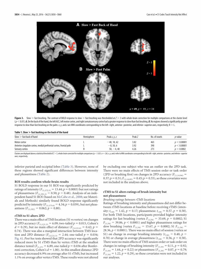
ResultsBrushing the hand activated somatosensory regions ofthe cortexAll speeds and locations of brushing activated the somatosensorybrain regions that we expected, including the primary and sec-ondary somatosensory cortices, anterior and posterior insula,thalamus, and cerebellum. For the contrast of slow � fast brush-ing on the back of the hand, significant clusters were found in theACC (Fig. 3, Table 1); no significant clusters were found for thesame contrast on the palm of the hand. Clusters were also foundin the sensory and motor cortices for the back of the hand slow �fast contrast, which could indicate slight differences in handmovement or tension during these brushing speeds.
S1 activity correlates with intensity; ACC activity correlateswith pleasantnessfMRI brushing ratings are displayed in Figure 4. Ratings of inten-sity and pleasantness were inversely related within subjects(F(1,205.8) � 44.0; p � 0.0001), so both were included in eachanalysis. In the whole-brain analysis, several brain regionsshowed significantly greater representation of intensity thanpleasantness and several areas showed greater representation ofpleasantness than intensity (Table 2). We then looked at eachpredictor separately and found that, within subjects, intensityratings but not pleasantness ratings correlated significantly withBOLD response in the right hemisphere (contralateral) S1, rightinsula, and bilateral S2 (statistical threshold Z � 3; whole-braincluster threshold p � 0.01; Fig. 5, Table 3), all regions with sig-nificantly greater intensity representation than pleasantness rep-resentation (Table 2). In contrast, the only region that showed asignificant positive correlation with pleasantness was a clusterwith a peak in the pregenual ACC (pgACC) that extended into thedorsal ACC (Table 3); again, a region with significantly greaterrepresentation of pleasantness than intensity (Table 2). For bothintensity and pleasantness ratings, the only regions showing sig-nificant negative correlations with BOLD response were in the
Case et al. • S1 Codes Touch Intensity Not Affect J. Neurosci., May 25, 2016 • 36(21):5850 –5860 • 5853
inferior parietal and occipital lobes (Table 3). However, none ofthese regions showed significant differences between intensityand pleasantness (Table 2).
ROI results confirm whole-brain resultsS1 BOLD response in our S1 ROI was significantly predicted byratings of intensity (F(1,172.5) � 13.44; p � 0.0003) but not ratingsof pleasantness (F(1,191.2) � 0.50; p � 0.48). Analysis of an inde-pendent hand S1 ROI (based on McCabe et al., 2008; see Materi-als and Methods) similarly found BOLD response significantlypredicted by intensity (F(1,173.6) � 4.34; p � 0.039), but not pleas-antness (F(1,192) � 0.062; p � 0.80).
rTMS to S1 alters 2PDThere was a main effect of TMS location (S1 vs vertex) on changesin 2PD accuracy (F(1,121.3) � 6.09; two-tailed p � 0.015, Cohen’sd � 0.29), but no main effect of distance (F(3,122.4) � 0.42; p �0.74). There was also a marginal interaction between TMS loca-tion and 2PD distance (F(3,121.3) � 2.44; one-tailed p � 0.034;Fig. 6). Post hoc tests showed that 2PD accuracy was significantlyreduced more by S1 rTMS than by vertex rTMS at the smallestdistance tested (F(1,17) � 6.89, one-tailed p � 0.036 after Bonfer-roni correction, Cohen’s d � 1.00). At this smallest distance 2PDaccuracy decreased 6.9% on average after S1 rTMS, but increased1.1% on average after vertex rTMS. These results were not altered
by excluding one subject who was an outlier on the 2PD task.There were no main effects of TMS session order or task order(2PD or brushing first) on changes in 2PD accuracy (F(1,14.94) �0.37; p � 0.51; F(1,14.85) � 0.45; p � 0.55), so these covariates werenot included in the analyses above.
rTMS to S1 alters ratings of brush intensity butnot pleasantnessBrushing ratings between rTMS locationsRatings of brushing intensity and pleasantness did not differ be-tween rTMS locations at baseline before receiving rTMS (inten-sity: t(18) � 1.42, p � 0.17; pleasantness: t(18) � 0.57, p � 0.58).For both TMS locations, participants provided higher intensityratings for fast brushing (vertex F(1,54) � 33.69, p � 0.0002; S1F(1,54) � 39.86, p � 0.0001) and higher pleasantness ratings forslow brushing (vertex F(1,54) � 15.67, p � 0.0002; S1 F(1,54) �28.36, p � 0.0001). There was no main effect of session (vertex orS1) on change in average brushing intensity (t(18) � 0.40, p �0.70) or change in average pleasantness (t(18) � 0.56, p � 0.58).There were no main effects of TMS session order or task order onchanges in ratings of brushing intensity (F(1,16) � 0.11, p � 0.92;F(1,16) � 1.64, p � 0.22) or pleasantness (F(1,16) � 0.07, p � 0.79;F(1,16) � 1.22, p � 0.29), so these covariates were not included inour analyses.
Figure 3. Slow � fast brushing. The contrast of BOLD response to slow � fast brushing was thresholded at Z � 3 with whole-brain correction for multiple comparisons at the cluster level( p � 0.01). A, On the back of the hand, the left ACC, left motor cortex, and right somatosensory cortex had a greater response to slow than fast brushing. B, No regions showed a significantly greaterresponse to slow than fast brushing on the palm. x, y, and z are MNI coordinates corresponding to the left–right, anterior–posterior, and inferior–superior axes, respectively; R � L.
Table 1. Slow > fast bushing on the back of the hand
Slow � fast back of hand Hemisphere Peak x, y, z Peak Z No. of voxels p-value
Motor cortex L �40, 18, 62 3.82 465 p � 0.00001Anterior cingulate cortex, medial prefrontal cortex, frontal pole L �8, 50, 6 3.92 390 p � 0.00001Sensory cortex R 50, �4, 40 4.26 273 p � 0.0002
Clusters are displayed above a statistical threshold of Z � 3, whole-brain corrected for multiple comparisons (p � 0.01; n � 26). x, y, and z refer to MNI coordinates corresponding to the left–right, anterior–posterior, and inferior–superioraxes, respectively.
5854 • J. Neurosci., May 25, 2016 • 36(21):5850 –5860 Case et al. • S1 Codes Touch Intensity Not Affect

Brushing ratings within each rTMS locationWhen we analyzed only subjects who showed decreased 2PD ac-curacy after S1 rTMS (n � 14), who were assumed to have S1deactivation, there was a significant increase in average brushingintensity ratings after S1 rTMS (t(13) � 3.38, p � 0.005, Cohen’sd � 0.93) and in the slow palm brushing condition in particular(t(13) � 3.5881, corrected p � 0.01), but no changes after vertexrTMS (t(13) � 0.35, p � 0.73; Fig. 7A). In these subjects withdecreased 2PD after S1 rTMS, there were no changes in ratings ofbrushing pleasantness for either rTMS location (S1 t(13) � 0.83,p � 0.42; vertex t(13) � 0.03, p � 0.98). A smaller number ofsubjects (n � 8) decreased in 2PD in the vertex condition. Theseindividuals did not show significant change in intensity percep-tion in the vertex condition (t(7) � 1.53, p � 0.17) or S1 condition(t(7) � 0.25, p � 0.81). For pleasantness ratings, we calculatedthat we had 80% power to observe an effect size of 6 points on the100 point VAS scale, given a 2-tailed � level of 0.05 and ourobserved SD of �10 points. The actual observed mean differencewas �1 point on a 100 point rating scale, an effect size of 0.1 thatwould require a sample size of 620 subjects to observe signifi-cance. In contrast, we were able to detect the marginal change inintensity ratings of 4 points (among 2PD responders) on the sameVAS scale, with similar SD in ratings to the pleasantness ratings.
As in the fMRI data, pleasantness and intensity ratings werecorrelated within-subjects both before TMS (F(1,148.4) � 27.0, p �0.0001) and after TMS (F(1,144.3) � 69.6, p � 0.0001). This rela-tionship was not affected by TMS (F(1,147.3) � 0.88, p � 0.35).
Changes in 2PD correlated with changes in intensity ratingsIn the full study sample, changes in brushing intensity (frombefore to after rTMS) were significantly predicted by changes in2PD accuracy (F(1,31.06) � 9.94; p � 0.004; Fig. 7B), but not byrTMS location (F(1,18.1) � 0.00; p � 0.96) or the interaction be-tween changes in 2PD accuracy and rTMS location (F(1,31.9) �0.06; p � 0.81). Average changes in pleasantness ratings were notpredicted by 2PD accuracy change (F(1,32.77) � 1.17, p � 0.29),rTMS location (F(1,17.48) � 0.10, p � 0.75), or the interaction ofthese factors (F(1,32.02) � 0.46, p � 0.50).
rTMS to S1 did not alter moodOn average, the 19 subjects reported high levels of good mood(7.06 � 1.85) and calm (7.43 � 1.82) and low levels of bad mood(0.75 � 0.95) and anxiety (0.68 � 1.16) during the TMS sessions.Subjects were slightly more happy and calm after rTMS thanbefore (t(18) � 2.84, p � 0.010; t(18) � 2.17, p � 0.043) and this
Figure 4. Brushing ratings. During the fMRI task, slow (3 cm/s) and fast brushing (30 cm/s) stimuli were delivered to the palm and back of the left hand. Each block of 8 stimuli was rated on a visualanalog scale for intensity (0 � “no sensation”; 100 � “the most intense sensation imaginable”) and for pleasantness (0 � “the most unpleasant imaginable”; 100 � “the most pleasantimaginable”). Error bars indicate � SEM.
Table 2. Whole-brain correlates of intensity versus pleasantness contrasts
Hemisphere Peak x, y, z Peak Z No. of voxels p-value
Intensity � pleasantnessPrimary somatosensory cortex R 28, �22, 62 5.78 1158 p � 0.00001Secondary somatosensory cortex/posterior insula R 50, 50, �24 4.99 379 p � 0.0001Secondary somatosensory cortex L �62, �18, 10 4.69 360 p � 0.0001
Pleasantness � intensityBilateral frontal cortex L �10, 38, 0 5.92 9347 p � 0.00001White matter L �36, �24, 44 4.41 594 p � 0.00001Cerebellum R 20, �46, �36 4.66 526 p � 0.00001White matter L �24, �30, �16 4.50 363 p � 0.0001Cerebellum L �12, �46, �34 4.59 272 p � 0.0003Superior frontal gyrus L �18, �6, 70 4.24 170 p � 0.006
Clusters above a threshold of Z � 3 are displayed with whole-brain correction for multiple comparisons (p � 0.01; n � 26). x, y, and z refer to the MNI coordinates corresponding to the left–right, anterior–posterior, and inferior–superioraxes, respectively; Z refers to the highest Z-score within a cluster. A positive Z-value indicates a positive correlation between the subject’s ratings and the BOLD response.
Case et al. • S1 Codes Touch Intensity Not Affect J. Neurosci., May 25, 2016 • 36(21):5850 –5860 • 5855

did not differ by rTMS location (F(1,16.8) � 0.24; p � 0.63;F(1,18.1) � 0.93; p � 0.35). There was not enough variability inmood ratings to use them as a covariate in our analyses.
DiscussionIn the current study, we used fMRI and rTMS to examine therole of S1 in representations of touch intensity and pleasant-ness. Using fMRI during fast and slow brushing on the palmand back of the hand, we found that the perceived intensity oftouch preferentially predicted activation of contralateral S1,posterior insula, and bilateral S2, whereas the perceived pleas-antness of touch preferentially predicted activation only in thepgACC. Pleasantness ratings showed no correlation with S1
response in ROI analyses. Further, 1 Hz inhibitory rTMS overS1 significantly reduced tactile 2PD, replicating Vidoni et al.(2010) and suggesting that we successfully reduced activationof S1, but this deactivation did not affect perception of touchpleasantness. Using decreased 2PD accuracy as an indicator ofsuccessful alteration of S1 activity, we observed increased rat-ings of brushing intensity after successful TMS to S1, althoughthe effect was weak. In contrast, perceived touch pleasantnesswas unaffected by rTMS. Given the robust dissociation be-tween the correlates of intensity and pleasantness ratings inour fMRI data and between ratings of fast and slow brushing,we believe that our scales were sensitive enough to meaningful
Figure 5. Whole-brain positive correlates of intensity and pleasantness. BOLD activation predicted by intensity and pleasantness ratings were thresholded at Z � 3 with whole-brain correctionfor multiple comparisons at the cluster level ( p � 0.01). A, Intensity ratings collected during the fMRI brushing task significantly predicted BOLD response in right S1, right posterior insula, andbilateral S2. B, Pleasantness ratings significantly predicted BOLD response in the ACC. x, y, and z refer to the MNI coordinates corresponding to the left–right, anterior–posterior, and inferior–superior axes, respectively; R � L.
Table 3. Whole-brain correlates of intensity and pleasantness
Hemisphere Peak x, y, z Peak Z Number of voxels p-value
Intensity: positive correlates (all regions significant in intensity � pleasantness; see Table 2)Primary somatosensory cortex R 32, �32, 72 5.22 1062 p � 0.00001Secondary somatosensory cortex/posterior insula R 46, �26, 20 6.52 745 p � 0.00001
L �50, �22, 16 5.54 460 p � 0.00001Intensity: negative correlates (not significant in contrast)
Angular gyrus R 26, �42, 40 �5.57 2474 p � 0.00001Supramarginal gyrus L �22, �58, 40 �4.44 277 p � 0.0002Occipital cortex L �24, �62, �6 �4.42 471 p � 0.00001
L �22, �74, 18 �4.25 212 p � 0.002L �48, �50, 22 �3.77 184 p � 0.004
Pleasantness: positive correlates (all regions significant in pleasantness � intensity; see Table 2)Anterior cingulate cortex L �8, 38, 4 4.46 196 p � 0.003
Pleasantness: negative correlates (not significant in contrast)Occipital cortex R 28, �86, 34 �5.09 300 p � 0.0001
Clusters above a threshold of Z � 3 are displayed with whole-brain correction for multiple comparisons (p � 0.01; n � 26). x, y, and z refer to the MNI coordinates corresponding to the left–right, anterior–posterior, and inferior–superioraxes, respectively; Z refers to the highest Z-score within a cluster. A positive Z-value indicates a positive correlation between the subject’s ratings and the BOLD response.
5856 • J. Neurosci., May 25, 2016 • 36(21):5850 –5860 Case et al. • S1 Codes Touch Intensity Not Affect

changes in CT stimulation. We also found an unexpected in-verse relationship between changes in tactile discriminationaccuracy and perceived touch intensity.
Together, these findings provide the most direct evidence todate that S1 is involved preferentially in touch intensity and dis-crimination and does not play a strong role in perception oftouch pleasantness.
Role of S1 cortex in discriminative but not affective touchOur data do not support the idea that S1 cortex is critically involvedin affective aspects of physical touch. In the fMRI portion of ourstudy, BOLD response positively correlated with subjects’ ratings of
touch pleasantness only in the dorsal/pgACC; no correlation wasfound in S1. S1 response did correlate, however, with the perceptionof touch intensity. In the TMS portion of our study, subjects whoshowed reduced discriminative ability after S1 deactivation (but notthose with reduced accuracy after vertex TMS) also had a significantalteration in intensity ratings, but no change in pleasantness ratings.
Nevertheless, some investigators have claimed that S1 may beimportant for affective aspects of touch. Gazzola et al. (2012)recently argued that S1 activation may play a role in the affectivesignificance of touch by showing that S1 response to sensual ca-ress is modified by the perceived sex of the caresser. However,
Figure 6. Effect of rTMS on 2PD accuracy. Participants were tested on 2PD before and after 20 min of inhibitory 1 Hz rTMS on 2 separate days. During the active session, rTMS wasdirected to S1 and, during the control session, rTMS was directed to the vertex. The 2PD task was administered as 2 descending series of blocks at tip distances of 2–10 mm (see Materialsand Methods for details). Each block contained 5 trials of 2-point stimulation and 5 trials of 1-point stimulation. 2PD accuracy was reduced significantly more after rTMS to S1 than afterrTMS to the vertex. In addition, there was a marginally significant interaction between rTMS session and distance. Post hoc tests showed that 2PD accuracy was significantly reduced afterrTMS to S1 at the smallest distance tested. S1 before, M � 58.1, SD � 11.1; S1 after, M � 50.8, SD � 9.7; vertex before, M � 56.3, SD � 9.4; vertex after, M � 58.6, SD � 9.2.*One-tailed p � 0.05. Error bars indicate � SEM.
Figure 7. Perception of intensity and pleasantness after S1 rTMS. A, Subjects rated the intensity and pleasantness of fast and slow brushing on the palm and back of the hand beforeand after 20 min of 1 Hz rTMS. The subjects who showed a drop in 2PD performance after rTMS to S1 (n � 14) also showed an increase in intensity ratings but not in pleasantness ratings(averaged across brushing types) after rTMS to S1. Five subjects who did not show a drop in 2PD accuracy are not included in graph. *Single-sample t test, p � 0.05. Error bars indicate �SEM. Intensity: S1 before, M � 41.9, SD � 26.0; S1 after, M � 46.1, SD � 27.5; vertex before, M � 45.6, SD � 23.2; vertex after, M � 43.4, SD � 24.7; pleasantness: S1 before, M �64.6, SD � 14.7; S1 after, M � 65.9, SD � 15.9; vertex before, M � 64.5, SD � 15.2; vertex after, M � 64.3, SD � 15.3. B, Changes in 2PD accuracy from before to after rTMSsignificantly predicted changes in ratings of brushing intensity.
Case et al. • S1 Codes Touch Intensity Not Affect J. Neurosci., May 25, 2016 • 36(21):5850 –5860 • 5857
regions found in numerous other studies to be involved in affec-tive touch, including insular and cingulate cortices, were notmodulated in this paradigm. In addition, the study containedconfounds related to attention and motivation that might ac-count for the S1 modulation; subjects were instructed to be look-ing for a date. Finally, the visual and posterior parietal areasfound in their connectivity analysis to modulate S1 are generallyimplicated in salience and cross-modal integration, not affect.McCabe et al. (2008) also reported a relationship between S1activity and the perceived pleasantness of rubbing with a creamlabeled as “rich moisturizing” versus “basic.” However, this cor-relation was opposite that found by Gazzola et al. (2012).
Several other studies suggest the involvement of S1 in affectivetouch, but contain confounds that complicate their interpreta-tion. Studies that have used visual stimuli to modulate touchaffect (Morrison et al., 2011; Bolognini et al., 2013) may havealtered S1 activity through the visual component of observedtouch, which has been shown to modulate S1 (Bolognini et al.,2011; Kuehn et al., 2013). Certain visual inputs may enhancesensorimotor-mirroring effects based on heightened attention oraffective engagement in other brain regions (Bufalari and Ionta,2013). Similarly, studies that have used physical stimuli to mod-ulate touch pleasantness have failed to control for differences intexture, physical intensity, and perceived intensity of their stimuli(Francis et al., 1999; Rolls et al., 2003; Gordon et al., 2013), whichstrongly modulate S1 (Lin et al., 2003; Arthurs et al., 2004).
In sum, studies showing modulation of S1 by pleasantnesscontain confounds that make it difficult to determine whether S1was modulated by affect, attention, motivation, or sensorimotormirroring. The current study overcomes the majority of theselimitations by brushing with speeds that produce varied CT/A�fiber stimulation and varied perceptual ratings of intensity andpleasantness without changing the texture or physical intensity ofthe touch stimulus itself. In addition, because fMRI can onlydemonstrate coactivation of S1 with pleasant touch, we con-ducted rTMS to test when S1 deactivation would affect touchpleasantness causally. Although manual administration addssome variability to the brushing, the experimenter brushing thesubjects was blinded to TMS location, so TMS-induced changesin perception cannot be attributed to variability in the brushingstimulus. With both correlational and experimental evidence, weprovide stronger evidence that S1 is not causally involved in per-ception of touch pleasantness.
Pleasantness representations only outside of S1The fMRI portion of our study found pleasantness correlationsthat also differed significantly from intensity correlations only inthe pgACC. The ACC also responded more strongly to slow thanfast brushing on the back of the hand but not the palm, consistentwith the greater pleasantness of CT-optimal touch. The pgACChas been reported previously to represent the pleasantness orvalue of a stimulus (Grabenhorst and Rolls, 2011). Although thedorsal ACC has consistently been implicated in the appraisal offrightening and painful stimuli, the mPFC/OFC and the ventralACC, including the pgACC, have been consistently associatedwith positive emotion and regulation of negative emotion (Etkinet al., 2011; Grabenhorst and Rolls, 2011; Ellingsen et al., 2013).pgACC activity has been correlated with the pleasantness, but notintensity, of taste, smell, and temperature stimuli (de Araujo etal., 2005; Grabenhorst and Rolls, 2008; Grabenhorst et al., 2008)during both physical and cognitive manipulations of pleasant-ness. The ACC was also the sole region identified as responding to
the pleasantness of touch massage by Lindgren et al. (2012), al-though this was a main effect of massage and not a correlationalanalysis. In addition, the unpleasantness of pain has been associ-ated with activation in areas outside of S1, including the ACC(Rainville et al., 1997; Hofbauer et al., 2001; Rolls et al., 2003;Schreckenberger et al., 2005; Villemure and Bushnell, 2009; Lutzet al., 2013).
Several other brain areas showed greater association withpleasantness than with intensity, but these areas were not corre-lated with pleasantness alone. These areas included a large clusterin the frontal cortex including the ACC, in addition to clusters inthe cerebellum and superior frontal gyrus. Representations oftouch pleasantness have also been reported in the OFC (McCabeet al. (2008)) and insula (Kress et al., 2011), areas not found in thecurrent study. Although there is evidence that CT input is pro-cessed in the insular cortex (Olausson et al., 2002) and the poste-rior insula often activates more strongly to CT targeted touch(Olausson et al., 2002; Bjornsdotter et al., 2009; Kress et al., 2011;Gordon et al., 2013), only Kress et al. (2011) have reported acorrelation between pleasantness ratings and insula response.
We also observed several brain areas that increased in activa-tion with lower intensity or pleasantness ratings, but these areasdid not show a significant difference between their representationof intensity and pleasantness. These regions may reflect defaultmode network engagement in the inferior parietal lobe (Buckneret al. (2008)), although the rest of the default mode network wasnot engaged. Occipital correlations might reflect wandering vi-sual attention or greater attention to the visual brushing prompton the screen when participants were less focused on the brushingsensation itself.
To demonstrate fully a double dissociation between represen-tation of intensity in S1 and representation of pleasantness in theACC, it would be ideal to attempt deactivation of the pgACC.However, selective stimulation of the pgACC is not yet possible.Our conclusions about pleasantness representation outside of S1thus rest on correlational brain-imaging data and the lack ofbehavioral double dissociation remains a significant limitation ofour study.
How are sensory discrimination and intensityperception related?We found a significant correlation between TMS-inducedchanges in 2PD performance and intensity perception. Oppositeto our original prediction, as discriminative ability decreased,perception of brushing intensity increased. Reducing activationof S1 could induce compensatory activity in other brain areas thatcontribute to intensity perception (O’Shea et al., 2007), such asS2. S2 was strongly correlated with intensity ratings in our fMRIdata and is causally implicated in judgments of pain intensity(Lockwood et al., 2013). In addition, TMS to S1 has been shownrecently to cause changes in functional connectivity to a numberof brain areas; theta-burst stimulation of S1 was found to alterfunctional connectivity with dorsal premotor cortex, cerebellum,basal ganglia, and anterior cingulate cortex (Valchev et al., 2015)and 1 Hz rTMS of left S1 was found to increase sensory responsein right S1 (Meehan et al., 2011).
The inverse correlation between changes in 2PD accuracy andintensity ratings, however, occurred for both the S1 and vertexrTMS locations. Although no mean change was found in thevertex condition (and vertex stimulation generally produces nulleffects; Duecker et al., 2013), individual shifts in attention mightshift the focus of sensory processing between sensory discrimina-tion and intensity perception. Alterations in 2PD performance
5858 • J. Neurosci., May 25, 2016 • 36(21):5850 –5860 Case et al. • S1 Codes Touch Intensity Not Affect

(strongly linked to S1) were correlated with changes in intensityperception, but not with changes in pleasantness perception, pro-viding further evidence of a link between intensity perceptionand S1, but no link between pleasantness perception and S1.
Summary and future directionsIn sum, our data support a causal role for right hemisphere S1 inperception of touch discrimination, and likely also intensity, butnot touch pleasantness. Our fMRI study found pleasantness rep-resentations in the ACC but not in S1. Subsequent (partial) de-activation of S1 decreased spatial discrimination and alteredintensity but not pleasantness perception. Within-subject de-creases in sensory discrimination were associated with increasesin perceived tactile intensity, potentially related to changes inrelative activation of S1 and other brain areas such as S2. Futurestudies might investigate more closely S2 and its coding of inten-sity when S1 activity is diminished. In addition, more work isneeded to clarify how S1 may be modulated indirectly by affectthrough changes in attention, expectation, motivation, or visualinput and whether our finding generalizes to left hemisphere S1.rTMS might also be applied to investigate the causal role of otherbrain areas in the perception of touch pleasantness and affectmore generally, although TMS is currently unable to targetdeeper cortical areas such as the pgACC selectively.
ReferencesArthurs OJ, Johansen-Berg H, Matthews PM, Boniface SJ (2004) Attention
differentially modulates the coupling of fMRI BOLD and evoked poten-tial signal amplitudes in the human somatosensory cortex. Exp Brain Res157:269 –274. Medline
Bestmann S, Baudewig J, Siebner HR, Rothwell JC, Frahm J (2005) BOLDMRI responses to repetitive TMS over human dorsal premotor cortex.Neuroimage 28:22–29. CrossRef Medline
Bjornsdotter M, Loken L, Olausson H, Vallbo A, Wessberg J (2009) Soma-totopic organization of gentle touch processing in the posterior insularcortex. J Neurosci 29:9314 –9320. CrossRef Medline
Bolognini N, Rossetti A, Maravita A, Miniussi C (2011) Seeing touch in thesomatosensory cortex: a TMS study of the visual perception of touch.Hum Brain Mapp 32:2104 –2114. CrossRef Medline
Bolognini N, Rossetti A, Convento S, Vallar G (2013) Understanding oth-ers’ feelings: the role of the right primary somatosensory cortex in encod-ing the affective valence of others’ touch. J Neurosci 33:4201– 4205.CrossRef Medline
Buckner RL, Andrews-Hanna JR, Schacter DL (2008) The brain’s defaultnetwork. Ann N Y Acad Sci 1124:1–38. CrossRef Medline
Bufalari I, Ionta S (2013) The social and personality neuroscience of empa-thy for pain and touch. Front Hum Neurosci 7:393. CrossRef Medline
Chen R, Classen J, Gerloff C, Celnik P, Wassermann EM, Hallett M, CohenLG (1997) Depression of motor cortex excitability by low-frequencytranscranial magnetic stimulation. Neurology 48:1398 –1403. CrossRefMedline
Cohen LG, Bandinelli S, Sato S, Kufta C, Hallett M (1991) Attenuation indetection of somatosensory stimuli by transcranial magnetic stimulation.Electroencephalogr Clin Neurophysiol 81:366 –376. CrossRef Medline
Craig A (2013) An interoceptive neuroanatomical perspective on feelings,energy, and effort. Behav Brain Sci 36:685– 686. CrossRef Medline
de Araujo IE, Rolls ET, Velazco MI, Margot C, Cayeux I (2005) Cognitivemodulation of olfactory processing. Neuron 46:671– 679. CrossRefMedline
Duecker F, de Graaf TA, Jacobs C, Sack AT (2013) Time-and task-dependent non-neural effects of real and sham TMS. PLoS One 8:e73813.CrossRef Medline
Ellingsen DM, Wessberg J, Eikemo M, Liljencrantz J, Endestad T, Olausson H,Leknes S (2013) Placebo improves pleasure and pain through oppositemodulation of sensory processing. Proc Natl Acad Sci U S A 110:17993–17998. CrossRef Medline
Etkin A, Egner T, Kalisch R (2011) Emotional processing in anterior cingu-late and medial prefrontal cortex. Trends Cogn Sci 15:85–93. CrossRefMedline
Francis S, Rolls ET, Bowtell R, McGlone F, O’Doherty J, Browning A, Clare S,Smith E (1999) The representation of pleasant touch in the brain and itsrelationship with taste and olfactory areas. Neuroreport 10:453– 459.CrossRef Medline
Gazzola V, Spezio ML, Etzel JA, Castelli F, Adolphs R, Keysers C (2012)Primary somatosensory cortex discriminates affective significance in so-cial touch. Proc Natl Acad Sci U S A 109:E1657–E1666. CrossRef Medline
Gordon I, Voos AC, Bennett RH, Bolling DZ, Pelphrey KA, Kaiser MD(2013) Brain mechanisms for processing affective touch. Hum BrainMapp 34:914 –922. CrossRef Medline
Grabenhorst F, Rolls ET (2008) Selective attention to affective value al-ters how the brain processes taste stimuli. Eur J Neurosci 27:723–729.CrossRef Medline
Grabenhorst F, Rolls ET (2011) Value, pleasure and choice in the ventralprefrontal cortex. Trends Cogn Sci 15:56 – 67. CrossRef Medline
Grabenhorst F, Rolls ET, Bilderbeck A (2008) How cognition modulatesaffective responses to taste and flavor: top-down influences on the orbito-frontal and pregenual cingulate cortices. Cereb Cortex 18:1549 –1559.CrossRef Medline
Hallett M (2007) Transcranial magnetic stimulation: a primer. Neuron 55:187–199. CrossRef Medline
Hofbauer RK, Rainville P, Duncan GH, Bushnell MC (2001) Cortical rep-resentation of the sensory dimension of pain. J Neurophysiol 86:402– 411.Medline
Jenkinson M, Bannister P, Brady M, Smith S (2002) Improved optimizationfor the robust and accurate linear registration and motion correction ofbrain images. Neuroimage 17:825– 841. CrossRef Medline
Jenkinson M, Beckmann CF, Behrens TE, Woolrich MW, Smith SM (2012)FSL. Neuroimage 62:782–790. CrossRef Medline
Knecht S, Ellger T, Breitenstein C, Bernd Ringelstein E, Henningsen H(2003) Changing cortical excitability with low-frequency transcranialmagnetic stimulation can induce sustained disruption of tactile percep-tion. Biol Psychiatry 53:175–179. CrossRef Medline
Kress IU, Minati L, Ferraro S, Critchley HD (2011) Direct skin-to-skin vsindirect touch modulates neural responses to stroking vs tapping. Neu-roreport 22:646 – 651. CrossRef Medline
Kuehn E, Trampel R, Mueller K, Turner R, Schutz-Bosbach S (2013) Judg-ing roughness by sight: a 7-tesla fMRI study on responsivity of the primarysomatosensory cortex during observed touch of self and others. HumBrain Mapp 34:1882–1895. CrossRef Medline
Lin YY, Shih YH, Chen JT, Hsieh JC, Yeh TC, Liao KK, Kao CD, Lin KP, WuZA, Ho LT (2003) Differential effects of stimulus intensity on peripheraland neuromagnetic cortical responses to median nerve stimulation. Neu-roimage 20:909 –917. CrossRef Medline
Lindgren L, Westling G, Brulin C, Lehtipalo S, Andersson M, Nyberg L(2012) Pleasant human touch is represented in pregenual anterior cin-gulate cortex. Neuroimage 59:3427–3432. CrossRef Medline
Lockwood PL, Iannetti GD, Haggard P (2013) Transcranial magnetic stim-ulation over human secondary somatosensory cortex disrupts perceptionof pain intensity. Cortex 49:2201–2209. CrossRef Medline
Loggia ML, Mogil JS, Bushnell MC (2008) Experimentally induced moodchanges preferentially affect pain unpleasantness. J Pain 9:784 –791.CrossRef Medline
Loken LS, Wessberg J, Morrison I, McGlone F, Olausson H (2009) Codingof pleasant touch by unmyelinated afferents in humans. Nat Neurosci12:547–548. CrossRef Medline
Lundblad LC, Olausson HW, Hermansson AK, Wasling HB (2011) Corticalprocessing of tactile direction discrimination based on spatiotemporalcues in man. Neurosci Lett 501:45– 49. CrossRef Medline
Lutz A, McFarlin DR, Perlman DM, Salomons TV, Davidson RJ (2013) Al-tered anterior insula activation during anticipation and experience ofpainful stimuli in expert meditators. Neuroimage 64:538 –546. CrossRefMedline
Maeda F, Keenan JP, Tormos JM, Topka H, Pascual-Leone A (2000) Mod-ulation of corticospinal excitability by repetitive transcranial magneticstimulation. Clin Neurophysiol 111:800 – 805. CrossRef Medline
McCabe C, Rolls ET, Bilderbeck A, McGlone F (2008) Cognitive influenceson the affective representation of touch and the sight of touch in thehuman brain. Soc Cogn Affect Neurosci 3:97–108. CrossRef Medline
Meehan SK, Linsdell MA, Handy TC, Boyd LA (2011) Interhemisphericenhancement of somatosensory cortical excitability through contralateral
Case et al. • S1 Codes Touch Intensity Not Affect J. Neurosci., May 25, 2016 • 36(21):5850 –5860 • 5859

repetitive transcranial magnetic stimulation. Clin Neurophysiol 122:1637–1644. CrossRef Medline
Morrison I, Bjornsdotter M, Olausson H (2011) Vicarious responses to so-cial touch in posterior insular cortex are tuned to pleasant caressingspeeds. J Neurosci 31:9554 –9562. CrossRef Medline
Okamoto M, Dan H, Sakamoto K, Takeo K, Shimizu K, Kohno S, Oda I, IsobeS, Suzuki T, Kohyama K, Dan I (2004) Three-dimensional probabilisticanatomical cranio-cerebral correlation via the international 10 –20 sys-tem oriented for transcranial functional brain mapping. Neuroimage 21:99 –111. CrossRef Medline
Olausson H, Lamarre Y, Backlund H, Morin C, Wallin BG, Starck G, EkholmS, Strigo I, Worsley K, Vallbo AB, Bushnell MC (2002) Unmyelinatedtactile afferents signal touch and project to insular cortex. Nat Neurosci5:900 –904. CrossRef Medline
Olausson H, Cole J, Rylander K, McGlone F, Lamarre Y, Wallin BG, KramerH, Wessberg J, Elam M, Bushnell MC, Vallbo A (2008) Functional roleof unmyelinated tactile afferents in human hairy skin: sympathetic re-sponse and perceptual localization. Exp Brain Res 184:135–140. Medline
O’Shea J, Johansen-Berg H, Trief D, Gobel S, Rushworth MF (2007) Func-tionally specific reorganization in human premotor cortex. Neuron 54:479 – 490. CrossRef Medline
Rainville P, Feine JS, Bushnell MC, Duncan GH (1992) A psychophysicalcomparison of sensory and affective responses to four modalities of ex-perimental pain. Somatosens Mot Res 9:265–277. CrossRef Medline
Rainville P, Duncan GH, Price DD, Carrier B, Bushnell MC (1997) Painaffect encoded in human anterior cingulate but not somatosensory cor-tex. Science 277:968 –971. CrossRef Medline
Rolls ET, O’Doherty J, Kringelbach ML, Francis S, Bowtell R, McGlone F(2003) Representations of pleasant and painful touch in the human or-bitofrontal and cingulate cortices. Cereb Cortex 13:308 –317. CrossRefMedline
Rossetti A, Miniussi C, Maravita A, Bolognini N (2012) Visual perception ofbodily interactions in the primary somatosensory cortex. Eur J Neurosci36:2317–2323. CrossRef Medline
Rossi S, Hallett M, Rossini PM, Pascual-Leone A; Safety of TMS ConsensusGroup (2009) Safety, ethical considerations, and application guidelinesfor the use of transcranial magnetic stimulation in clinical practice andresearch. Clin Neurophysiol 120:2008 –2039. CrossRef Medline
Schneider SA, Pleger B, Draganski B, Cordivari C, Rothwell JC, Bhatia KP,Dolan RJ (2010) Modulatory effects of 5Hz rTMS over the primary so-matosensory cortex in focal dystonia: an fMRI-TMS study. Mov Disord25:76 – 83. CrossRef Medline
Schreckenberger M, Siessmeier T, Viertmann A, Landvogt C, Buchholz HG,Rolke R, Treede RD, Bartenstein P, Birklein F (2005) The unpleasant-ness of tonic pain is encoded by the insular cortex. Neurology 64:1175–1183. CrossRef Medline
Schutter DJ, van Honk J (2006) A standardized motor threshold estimation
procedure for transcranial magnetic stimulation research. J ECT 22:176 –178. CrossRef Medline
Sheehan DV, Janavs J, Baker R, Harnett-Sheehan K, Knapp E, Sheehan M,Lecrubier Y, Weiller E, Hergueta T, Amorim P, Bonora LI, Lepine JP(1998) MINI-Mini International Neuropsychiatric Interview-EnglishVersion 5.0.0-DSM-IV. J Clin Psychiatry 59:34 –57.
Smith SM, Jenkinson M, Woolrich MW, Beckmann CF, Behrens TE,Johansen-Berg H, Bannister PR, De Luca M, Drobnjak I, Flitney DE,Niazy RK, Saunders J, Vickers J, Zhang Y, De Stefano N, Brady JM, Mat-thews PM (2004) Advances in functional and structural MR image anal-ysis and implementation as FSL. Neuroimage 23:S208 –S219. CrossRefMedline
Sterman AB, Schaumburg HH, Asbury AK (1980) The acute sensory neu-ronopathy syndrome: a distinct clinical entity. Ann Neurol 7:354 –358.CrossRef Medline
Tegenthoff M, Ragert P, Pleger B, Schwenkreis P, Forster AF, Nicolas V, DinseHR (2005) Improvement of tactile discrimination performance and en-largement of cortical somatosensory maps after 5 Hz rTMS. PLoS Biol3:e362. CrossRef Medline
Triscoli C, Olausson H, Sailer U, Ignell H, Croy I (2013) CT-optimized skinstroking delivered by hand or robot is comparable. Front Behav Neurosci7:208. CrossRef Medline
Valchev N, Curcic-Blake B, Renken RJ, Avenanti A, Keysers C, Gazzola V,Maurits NM (2015) cTBS delivered to the left somatosensory cortexchanges its functional connectivity during rest. Neuroimage 114:386 –397. CrossRef Medline
Vallbo AB, Olausson H, Wessberg J (1999) Unmyelinated afferents consti-tute a second system coding tactile stimuli of the human hairy skin. J Neu-rophysiol 81:2753–2763. Medline
Vidoni ED, Acerra NE, Dao E, Meehan SK, Boyd LA (2010) Role of theprimary somatosensory cortex in motor learning: an rTMS study. Neu-robiol Learn Mem 93:532–539. CrossRef Medline
Villemure C, Bushnell MC (2009) Mood influences supraspinal pain pro-cessing separately from attention. J Neurosci 29:705–715. CrossRefMedline
Villemure C, Slotnick BM, Bushnell MC (2003) Effects of odors on painperception: deciphering the roles of emotion and attention. Pain 106:101–108. CrossRef Medline
Wassermann EM, Zimmermann T (2012) Transcranial magnetic brainstimulation: therapeutic promises and scientific gaps. Pharmacol Ther133:98 –107. CrossRef Medline
Westin GG, Bassi BD, Lisanby SH, Luber B; New York State Psychiatric In-stitute, NY, USA (2014) Determination of motor threshold using visualobservation overestimates transcranial magnetic stimulation dosage:Safety implications. Clin Neurophysiol 125:142–147. CrossRef Medline
Woolrich MW, Jbabdi S, Patenaude B, Chappell M, Makni S, Behrens T,Beckmann C, Jenkinson M, Smith SM (2009) Bayesian analysis of neu-roimaging data in FSL. Neuroimage 45:S173–S186. CrossRef Medline
5860 • J. Neurosci., May 25, 2016 • 36(21):5850 –5860 Case et al. • S1 Codes Touch Intensity Not Affect


![View full document [PDF 2.07 MB]](https://img.pdfslide.us/doc/110x75/586939691a28ab4e0b8b4b03/view-full-document-pdf-207-mb.jpg)