Embed Size (px)
Citation preview

247
The principles as well as a few possibilities of application aredescribed in part I 1). This chapter concerns the transfer technique aswell as selected staining methods for blotting from SDS gradientpore gels according to method 7 2) and 8 3), and from IEF gels accord-ing to method 6 4). For the electrotransfer, the cooling plate is takenout of the chamber and the graphite plates are used instead (Fig. 1).
A few practical tips will be given first:
. The filter paper and the blotting membrane must be cut to thesize of the gel so that the current does not flow around thesides of the actual blot sandwich. Small blot sandwiches canbe placed beside one another.
. The filter paper does not have to be cut if a plastic frame ismade in which openings for the gels and blotting membraneare cut out. This frame is placed on the stack of anodic filterpaper before the blot is built up further (Fig. 2).
Method 9:
Semi-dry blotting of proteins
Fig. 1: Installing the graphite anode in placeof the cooling plate. Fig. 2: Mask
Electrophoresis in Practice, Fourth Edition. Reiner WestermeierCopyright � 2005 WILEY-VCH Verlag GmbH & Co. KGaA, WeinheimISBN: 3-527-31181-5
1 see pages 67 and following2 see pages 211 and following3 see pages 231 and following4 see pages 197 and following
The largest gel size is 20 � 27cm.When a gel of the size describedin method 7 should be blotted,it is easier to cut the gel intotwo halves before blotting.

Method 9: Semi-dry blotting of proteins
. Only gels of the same kind (IEF, SDS) can be blotted together,because the transfer time depends on the pore size and thestate of the proteins.
. The transfer buffer contains 20% methanol, so that the gels donot swell during the transfer and the binding capacity of thenitrocellulose is increased.
. When gels are bound to a plastic film, the gel and the supportfilm should first be separated with the Film Remover.
. When several gel-blot layers are blotted over one another, a di-alysis membrane (Cellophane) is placed between each layer sothat proteins which might migrate through, do not reach thenext transfer unit.
. For continuous buffer systems, the same buffer with a pH of9.5 is used for both the anodic and cathodic sides of the blots.To improve the transfer of hydrophobic proteins and to chargefocused proteins (proteins are not charged at their pI), thebuffer also contains SDS.
In discontinuous buffer systems the speed of migration of the pro-teins changes during the transfer because of the different ionicstrengths of both the anode buffers (0.3 mol/L and 0.025 mol/L).This means that fewer proteins are transferred. The slow terminatingion in the cathode buffer compensates for the differences in speed ofmigration of the leading ions (here the proteins): a more regulartransfer is obtained. In this case, SDS is only added in the cathodebuffer.
1Transfer buffers
Continuous buffer system:
39 mmol/L glycine 2.93 g,48 mmol/L Tris 5.81 g,0.0375% (w/v) SDS 0.375 g,20% (v/v) methanol 200 mL,make up to 1 L with distilled water.
248
Unfolded SDS-polypeptidemicelles behave differently fromfocused proteins in globularform.
If the biological activity ofenzymes must be preserved, themethanol should be omitted.
A thin wire is pulled betweenthe gel and support film.
Not recommeded, becausewhen several transfer units areblotted, transfer efficiency is lostin the direction of the cathode.
This method is the easiest. Butthe transfer is not as regular,and the bands not as sharp asin discontinuous buffer systems.
This method is especially valu-able for native blotting. Thesmall amounts of SDS(0.01%) in the cathode bufferdo not denature the proteinsduring the short contact time.
2 Technical procedure
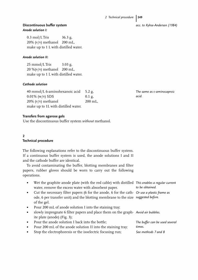
Discontinuous buffer systemAnode solution I:
0.3 mol/L Tris 36.3 g,20% (v/v) methanol 200 mL,make up to 1 L with distilled water.
Anode solution II:
25 mmol/L Tris 3.03 g,20 %(v/v) methanol 200 mL,make up to 1 L with distilled water.
Cathode solution
40 mmol/L 6-aminohexanoic acid 5.2 g,0.01% (w/v) SDS 0.1 g,20% (v/v) methanol 200 mL,make up to 1L with distilled water.
Transfers from agarose gelsUse the discontinuous buffer system without methanol.
2Technical procedure
The following explanations refer to the discontinuous buffer system.If a continuous buffer system is used, the anode solutions I and IIand the cathode buffer are identical.
To avoid contaminating the buffer, blotting membranes and filterpapers, rubber gloves should be worn to carry out the followingoperations.
. Wet the graphite anode plate (with the red cable) with distilledwater, remove the excess water with absorbent paper.
. Cut the necessary filter papers (6 for the anode, 6 for the cath-ode, 6 per transfer unit) and the blotting membrane to the sizeof the gel.
. Pour 200 mL of anode solution I into the staining tray;
. slowly impregnate 6 filter papers and place them on the graph-ite plate (anode) (Fig. 3);
. Pour the anode solution I back into the bottle;
. Pour 200 mL of the anode solution II into the staining tray;
. Stop the electrophoresis or the isoelectric focusing run;
249
acc. to Kyhse-Andersen (1984)
The same as e-aminocaproicacid.
This enables a regular currentto be obtained.
Or use a plastic frame assuggested before.
Avoid air bubbles.
The buffer can be used severaltimes.
See methods 7 and 8

Method 9: Semi-dry blotting of proteins
. wipe the kerosene off the bottom of the film;
. equilibrate the gels in the anode buffer I:SDS-PAGE: 5 minIEF: 2 min
Gels on support films are left to “swim” on the top of the bufferwith the gel surface on the bottom.
. remove the cooling plate from the electrophoresis chamber;
. place the anode plate in the electrophoresis chamber, plug inthe cables.
& Tip!The handling of a whole gel (25 � 10 cm) duringremoval is much easier if the gel is cut in two afterequilibration, and both halves are put togetheragain after pulling the FilmRemover wire through.A single blotting membrane can nevertheless beused for the whole gel.
. Place gels bound to support films with the film side down onthe Film Remover; so that a short side is in contact with boththe gel clamps;
. Press on the lever;
. Place the wire over the edge of the gel beside the clamps, hookit to the handle on the other side and push the lever down(Fig. 4);
250
Fig. 3: Assembling the blotting sandwich onthe graphite anode plate.
SDS gels are soaked inmethanol to prevent them fromswelling.
It is easier to remove IEF gelsfrom the blotting membraneafter blotting.
With the refrigeration tubingattached.
Folds often form when a largesupport film is removed and itis difficult to get rid of them.
The holding mechanism is nowin the elevated position.
Both teeth are now pressing onthe edge of the film and hold itin place.
The wire now has the mechan-ical tension necessary to sepa-rate the gel completely fromthe support film.

2 Technical procedure
. grasp the handles with both hands and smoothly pull the wiretowards you.
Film bound gels:
. Briefly soak the blotting membrane in the anode buffer II andplace it on the gel;
. slowly soak 3 filter papers in the buffer and place them on theprepared stack of filter papers;
. press on the top handle and lift the whole sandwich with thesupport film, turn in over and place it on the stack of filterpapers.
Gels without support film:
For gels which are not bound to support film, proceed in the oppositeway:
. slowly soak 3 filter papers in buffer and place them on the pre-pared stack of filter papers;
. briefly soak the blotting membrane in the anode buffer II andplace it on the stack of filter papers;
. place the gel on the blotting membrane;
. pour off the anode buffer II;
. rinse out the staining tray with distilled water and dry it withpaper;
. pour 200 mL of cathode solution in the staining tray;
. pull the film away slowly, starting at one corner;
. soak 9 filter papers in the cathode buffer and place them ontop.
251
Fig. 4: Removing the support film and transferringthe support film-gel-blotting membrane sandwichto the stack of anode paper.
While doing this, press the leverdown with a finger from theright hand so that it does notspring up.
The equilibration step is usuallynot carried out for gels whichare not bound to a supportfilm.
This buffer should only be usedonce (kerosene).
Reusable cathode buffer shouldnot be contaminated withtraces of anode buffer.
See the tip above.

Method 9: Semi-dry blotting of proteins
When building a blot sandwich as described here, it is difficult tocompletely avoid air bubbles. They must therefore be pressed outwith a roller (Fig. 5):
. Start in the centre and roll out in all four directions,
press in such a way that the buffer in the sandwich oozes outbut is not completely pressed out. When the roller is removed,the buffer should be “drawn” back in;
. wet the graphite cathode (black cable) with distilled water;remove the excess water with a paper towell;
. place the cathode plate on the stack, plug in the cable;
. place the safety lid on the electrophoresis chamber and con-nect the cable to the power supply;
. blot at a constant current: 0.8 mA/cm2
Transfer conditions:
0.5 mm thick gel (250 cm2) SDS pore gradient T = 8 to 20%:
I U P Room temperature
Set: 200 mA 10 V 5 W 20 �CRead: 200 mA 3 V 1 W
The blot does not warm up under these moderate conditions.
252
Fig. 5: Rolling out the air bubbles.
If not enough pressure isapplied, air bubbles will stay inthe sandwich and no transferwill take place at these points.On the other hand if too muchpressure is applied, the sand-wich will be too dry and irre-gular.
See anode plate.
Make sure that the polarity iscorrect.
Higher currents result in a riseof the temperature of the geland are not recommended.

3 Staining of blotting membranes
If thicker or more concentrated gels are blotted, the blotting timecan be increased up to two hours; it is then recommended to pressdown the cathode plate with a 1 to 2 kg weight so that no electrolyticgas pockets form.
Blotting native or focusing gels is quicker, because the proteins arein globular form and, for IEF, the gels have larger pores.
. Switch off the power supply, unplug the cable;
. remove the safety lid and the cathode plate;
. take the blot sandwich apart;
. stain the gel with Coomassie Blue to check.
. Before visualization, either dry the gel overnight or for 3 to 4 hat 60 �C in a heating cabinet.
& Caution:This treatment should not be used before detec-tion of biological activity (zymogram techniques)since most enzymes lose their activity.
3Staining of blotting membranes
Amido Black staining:
Dissolve 10.1 g of Amido Black in 100 mL of methanol-glacial aceticacid-water (40:10:50 v/v);
. stain for 3 to 4 min in 0.1% Amido Black solution;
. destain in methanol-acetic acid-water (25:10:65 v/v);
. air dry the nitrocellulose.
Reversible staining:If a specific immuno- or glycoprotein identification test is to be carriedout after the general detection, it is recommended that staining withPonceau S (Salinovich and Montelaro, 1986) or Fast Green FCF is used.
Fast Green staining
. dissolve 0.1% (w/v) of Fast Green in 1% acetic acid;
Mild staining for proteins!
. stain for 5 min;
. destain the background with distilled water for 5 min;
. complete destaining of the bands is achieved by incubatingthe film for 5 min in 0.2 mol/L NaOH.Blocking and immunological or lectin detection can now becarried out.
253
These gels have enoughmechanical stability that theyare not crushed.
guideline: 30 min
see methods 7 and 8.
The proteins bind more firmlyto the film during drying andare not washed out duringstaining and specific detection.
This only works for nitrocellu-lose, in addition alkaline treat-ment increases antigenreactivity (See page 75: Suther-land and Skerritt, 1986).
Method 9: Semi-dry blotting of proteins
Indian Ink staining (Hancock and Tsang, 1983)Unfortunately the original Indian Ink (“Fount India”) is no longerproduced. However, according to Dr. Christian Schmidt, Universityof Rostock, standard script-ink can be used alternatively, diluted withTBS-Tween instead of PBS-Tween.
. Soaking: 5 min in 0.2 mol/L NaOH;
. Washing: 4 � 10 min with PBS-Tween (or TBS-Tween)
. (250 mL per wash, agitate).
PBS-Tween: 48.8 g of NaCl + 14.5 g Na2HPO4 + 1.17 g NaH2PO4 +2.5 mL Tween 20, fill up to 5 L with distilled water.TBS-Tween: 2.92 g NaCl + 6.06 g Tris + 0.5 mL Tween 20, fill up to 5 Lwith distilled water.
. Staining: 2 h or overnight with 250 mL of PBS Tween (or TBSTween) + 2.5 mL of acetic acid + 250 lL of fountain pen ink;scan the image.
. Remove dye: 3 �10 wash with PBS (TBS) Tween.
. To conserve the image: Washing: 2 � 2 min with water.
. Drying: air-dry.
Plastic embedding of the blotting membraneNitrocellulose can be made totally and permanently transparent afterstaining or specific immunological detection methods, so that theresult looks like a stained electrophoresis gel.
254
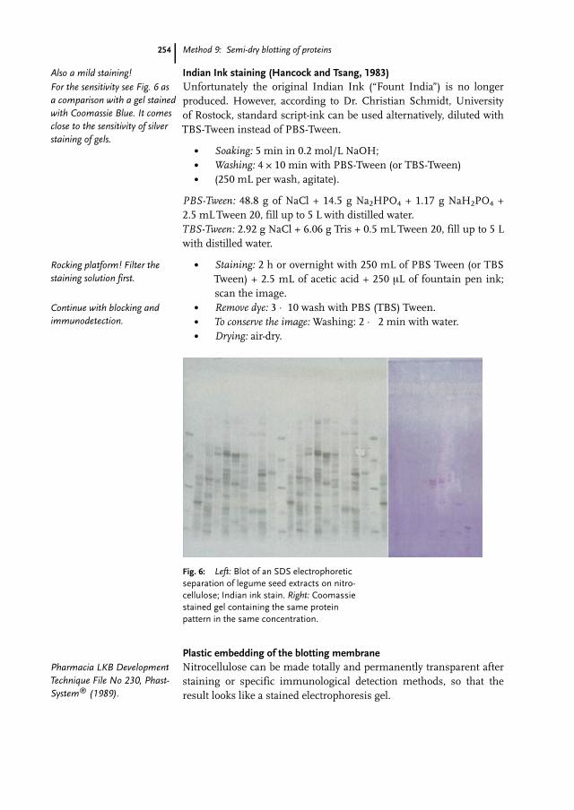
Fig. 6: Left: Blot of an SDS electrophoreticseparation of legume seed extracts on nitro-cellulose; Indian ink stain. Right: Coomassiestained gel containing the same proteinpattern in the same concentration.
Also a mild staining!For the sensitivity see Fig. 6 asa comparison with a gel stainedwith Coomassie Blue. It comesclose to the sensitivity of silverstaining of gels.
Rocking platform! Filter thestaining solution first.
Continue with blocking andimmunodetection.
Pharmacia LKB DevelopmentTechnique File No 230, Phast-System� (1989).

3 Staining of blotting membranes
The membrane is soaked in a monomer solution with the samerefractive index as polymerized nitrocellulose. For permanent trans-parency a photo-initiator, which starts polymerizing in the presenceof a UV light source, is added to the monomer solution.
Monomer solution: dissolve 0.5 g of benzoin methylether in 25 mLof TMPTMA. Store at room temperature in a brown flask or in thedark. The solution can be kept for about 2 weeks. For longer storage,keep it in the refrigerator at 4 �C to 8 �C.
. Dry the nitrocellulose completely;
. cut 2 sheets of PVC to a size larger than that of the blottingmembrane;
. pipette 0.3 mL of monomer solution on one of the plasticsheets;
. spread the solution so that it covers an area equal to the size ofthe membrane;
. stand the blotting membrane on its edge and slowly lower iton to the monomer solution so that the membrane is com-pletely impregnated;
. apply a few drops of solution on to the membrane and placethe second plastic sheet on top;
. carefully press out the air bubbles with a roller;
. irradiate both sides for about 15 s with a UV lamp;
. cut the sandwich to the size desired.
255
The monomer chosen for thispurpose is odorless and rela-tively safe.
Complete dissolution may takeseveral hours. Avoid skincontact: TMPTMA is an irritantfor the skin and mucousmembranes.
Sheets for the overheadprojector.
Or more, depending on the sizeof the blotting membrane.