Embed Size (px)
Citation preview
© 2
018
by A
cta
Neu
robi
olog
iae
Expe
rim
enta
lis
Effects of PINK1 mutation on synapses and behavior in the brain of Drosophila melanogaster
Bartosz Doktór, Milena Damulewicz, Wojciech Krzeptowski, Barbara Bednarczyk and Elżbieta Pyza*
Department of Cell Biology and Imaging, Institute of Zoology and Biomedical Research, Jagiellonian University, Kraków, Poland, * Email: [email protected]
Mutations in the PINK1 gene are responsible for typical symptoms of Parkinson’s disease. Using Drosophila melanogaster mutant PINK1B9 and after PINK1 silencing with RNAi using transgenic lines, we observed defects in synapses and behavior. The lack or reduced expression of PINK1 prolonged sleep during the day (nap) and decreased the total locomotor activity during 24 h, in addition to a decrease in climbing ability and a reduced lifespan. In the brain, PINK1 mutants had a lower level of Bruchpilot (BRP), a presynaptic scaffolding protein that is crucial for neurotransmission in all type of synapses in Drosophila. In addition, other proteins that are involved in synaptic transmission; Rab5, Syntaxin and Wishful Thinking were also decreased in abundance in mutants, except Synaptotagmin. Transmission electron microscopy (TEM) also confirmed less and abnormal synaptic vesicles at tetrad synapses in the visual system of PINK1 mutants. The lower level of BRP and longer day sleep observed was also detected in white mutants, which were examined to test the effect of the white background on the PINK1B9 strain. The reduced locomotor activity and longer day sleep in PINK1 mutants and after decreasing the PINK1 level in neurons seem to be correlated with a decrease in mitochondria number during the day, when they normally peak, and with impaired synaptic transmission.
Key words: Parkinson’s disease, Bruchpilot, Rab5, Syntaxin, Synaptotagmin, Wishful Thinking, white gene, motor activity, circadian rhythm, sleep, synaptic plasticity, mitochondria
INTRODUCTION
Parkinson’s disease (PD) is one of the most com‑mon neurodegenerative disorders characterized by de‑generation of dopaminergic neurons in the substantia nigra of the brain. PD, the familial form, is caused by mutations in ~150 genes while the sporadic form can be induced by exposure to toxins. One of the common mutations observed in the familiar form of PD is muta‑tion in the PINK1 (PARK6) gene, which encodes PTEN‑in‑duced putative kinase 1 (PINK1). In normal cells PINK1 accumulates on the surface of damaged mitochondria and together with the E3 ubiquitin ligase PARKIN, ini‑tiates mitophagy, a process of autophagy of damaged mitochondria (Matsuda et al. 2010). PINK1 contains an N‑terminal mitochondrial targeting sequence (MTS), a transmembrane sequence (TMS), and a Ser/Thr ki‑
nase domain located at the C‑terminus (Eiyama and Okamoto 2015). Under normal conditions PINK1 is translocated to the mitochondrial membrane, where MTS is cleaved by the mitochondrial processing pep‑tidase (MPP) (Greene et al. 2012), and the TMS by Pre‑senilin‑associated rhomboid‑like protein (PARL) (Deas et al. 2011). PINK1 without the MTS and TMS domains is transferred to the cytoplasm, where it is degraded mainly by the ubiquitin‑proteasome system (Yama‑no and Youle 2013). When mitochondria are damaged, they do not maintain the inner membrane potential, and in effect the MTS and TMS cannot be cleaved and PINK1 is stabilized on the outer mitochondrial mem‑brane (OMM) (Lazarou et al. 2012). After PINK1 attach‑ment to the translocase complex of the mitochondri‑al outer membrane (TOM), it phosphorylates PARKIN, which causes its activation and recruitment into the OMM (Chen and Dorn 2013). In the next step, PINK1, to‑
Received 20 April 2017, accepted 7 June 2018
RESEARCH PAPER
Acta Neurobiol Exp 2018, 78: 231–241DOI: 10.21307/ane‑2018‑021
232 B. Doktór et al. Acta Neurobiol Exp 2018, 78: 231–241
gether with PARKIN, targets mitofusins located on the mitochondrial surface for proteasomal degradation, leading to whole organellum degradation (Thomas et al. 2014). During PD, in the result of the PINK1 mutation, damaged and old mitochondria, which are the source of high amounts of free radicals, cannot be degraded and thus they cause dysfunction or death of neurons. The most sensitive structures to malfunctioning mitochon‑dria are synapses, because of the high energy require‑ment for synaptic transmission. Mitochondria provide ATP for the formation and transport of synaptic ves‑icles and for neurotransmitter exocytosis. Moreover, mitochondria are involved in uptake and release of calcium ions (Ly and Verstreken 2006), which regulate release of neurotransmitters from synaptic vesicles to the synaptic cleft. Disorders in calcium release from mitochondria and in ATP production affect motor and cognitive functions, similar to those observed in PD.
Various aspects of PD and other neurodegenerative diseases are often studied using the fruit fly, Drosoph‑ila melanogaster, as a model organism. Molecular and behavioral disorders in the Drosophila model of PD are similar to those observed in mammalian PD models (Feany and Bender 2000, Lu and Vogel 2009). In Dro‑sophila PINK1 is involved in the mitochondrial fission/fusion process and PINK1 mutants have already been described (Yang et al. 2006). Mutations in PINK1 cause inhibition of mitochondrial fission and in result the ap‑pearance of large and swollen mitochondria (Poole et al. 2008). One of the substrates of PINK1 is mitofusin (Mfn), a protein responsible for mitochondrial fusion. PINK1 with PARKIN directs Mfn to the degradation pathway and thus it contributes to the process of mi‑tochondrial fission (Ziviani et al. 2010). Mutation of PINK1 results in reduced production of ATP (Park et al. 2006), indirect flight muscle degradation (Yang et al. 2006) and disorders in the locomotor activity of flies. In addition, PINK1 mutation causes degeneration of do‑paminergic neurons (Wang et al. 2006), a marker of Par‑kinson’s disease, abnormal synaptic transmission and accumulation of synaptic vesicles (Morais et al. 2009).
One of the most important proteins involved in synaptic transmission in Drosophila is the presynaptic protein Bruchpilot (BRP) (Kittel et al. 2006). BRP is the human homolog of ELKS/CAST/ERC [CAST ‑ cytoskele‑tal matrix associated with the active zone (CAZ)‑associ‑ated structural protein, also called ERC (ELKS, Rab6‑in‑teracting protein 2, and CAST)] proteins and it is re‑sponsible for the accumulation of calcium channels in the active zone and release of neurotransmitter. BRP is expressed in all synapses as two subunits BRP190 and BRP170 (Wagh et al. 2006). It has been shown that a re‑duced level of BRP results in motor disorders (Wagh et al. 2006) similar to that present in flies with muta‑
tions causing PD symptoms. Besides BRP other proteins such as Rab5, Syntaxin, Synaptotagmin and Wishful Thinking (WIT) are crucial for synaptic transmission. Rab5 is a major protein that mediates membrane traf‑ficking with the specialized early endosome domain. Rab5 takes part in synaptic vesicle maturation during synaptic transmission (Hoop et al. 1994, Stenmark 2009, Wucherpfennig et al. 2003). Wucherpfennig et al. (2003) reported that lack of Rab5 causes locomotor de‑fects, abnormal morphology of synaptic terminals and a reduced size of synaptic vesicles. Syntaxin protein is involved in synaptic vesicle fusion in the presynaptic active zones and it mediates exocytosis (Sieber et al. 2006, Ullrich et al. 2015). In turn Synaptotagmin is an essential protein for the release of neurotransmitter into the synaptic cleft because it binds Ca2+ that trig‑gers vesicle fusion (Geppert et al. 1994, Shields et al. 2017). WIT regulates synaptic growth, the number of active zones in presynaptic elements and maintains the amplitude of excitatory junction potentials (Aberle et al. 2002).
In our present study, we found a correlation between the level of BRP protein and motor disorders caused by PINK1 mutation. We showed that PINK1 mutants have less BRP and all other proteins studied, except Synap‑totagmin. In addition, the PINK1 mutation affects sleep, increasing sleep during the day (nap), which leads to a decrease of total activity during 24 h.
METHODS
Animals
The following strains were used for the experi‑ments: Canton S, w1118 (null mutation of the gene white encoding the ABC transporter) (Krstic et al. 2013), PINK1B9 (point deletion of the gene encoding PINK1 ki‑nase) (Park et al. 2006), elav‑GAL4 (expressing the yeast transcription factor GAL4 under control of the elav promoter) (DiAntonio et al. 2001), 21D‑GAL4 (express‑ing the yeast transcription factor GAL4 in L2 neurons of the lamina, the first optic neuropil) (Weber et al. 2009), UAS‑Valium10 (expressing GFP and Valium under UAS control) (Ni et al. 2009), UAS‑PINK1RNAi (express‑ing interfering RNA for PINK1) (Yang et al. 2006) and UAS‑mitoGFP (expressing GFP with a mitochondrial im‑port signal) (Pilling et al. 2006).
Since the strain PINK1B9 used in our experiments has the white background, which may affect results, we used white mutants as a control in addition to wild type flies Canton S, and a strain with PINK1 RNAi expressed in neurons (elav‑GAL4>UAS‑PINK1RNAi) to decrease the level of PINK1 in neurons. The White gene encodes
PINK1 mutation affects synapses and behavior 233Acta Neurobiol Exp 2018, 78: 231–241
the ABC transporter that is one of the most important membrane transporters (Ewart et al. 1994) and is in‑volved in many physiological processes. As a control for the RNAi strain (elav‑GAL4>UAS‑PINK1RNAi) we used elav‑GAL4>UAS‑Valium10. The level of gene expres‑sion silencing in the elav‑GAL4>UAS‑PINK1RNAi strain was equal to 74%.
Transgenic strains were obtained from the Blooming‑ton Drosophila Stock Center. Flies were maintained on a standard yeast‑cornmeal‑agar medium at 25 ± 1°C, un‑der a day/night cycle (12 h of light and 12 h of darkness; LD 12:12). To downregulate PINK1 expression in neurons, elav‑GAL4 females were crossed to UAS‑PINK1RNAi males and elav‑GAL4 females were also crossed to UAS‑Valium10 males to express the VALIUM vector in neurons as the control in the RNAi experiments (Ni et al. 2009). To visu‑alize mitochondria in the L2 cells of the first optic neu‑ropil (lamina) of the optic lobe 21D‑GAL4 females were crossed to UAS‑mitoGFP males.
Locomotor activity and sleep analysis
Males, 1–2 days old (N=32), were transferred to small glass tubes containing the sugar‑agar food medium. Vi‑als were located in DAMS monitors (Drosophila Activity Monitoring System, TriKinetics) and placed in an incu‑bator (25°C). Monitors were equipped with infrared sen‑sors, which recorded the activity of the flies inside the vials every 5 min. For the first 5 days, monitors were held in LD 12:12 (12 h of light and 12 h of darkness) conditions and then for 6 days in constant darkness (DD). Results from the second day of recording were analyzed to esti‑mate the total activity and duration of sleep during the day and during the night [(Microsoft Excel plugin – Be‑Fly kindly donated by E. Green from Genetics, University of Leicester) (Rosato and Kyriacou 2006) and Python 22 (http://www.python.org/)]. Sleep in flies is defined as time in which they do not change their position for at least 5 min. The experiment was repeated three times. In LD 12:12 and DD the rhythm of locomotor activity was also examined, and the period of the circadian locomo‑tor activity rhythm was measured in DD.
Immunohistochemistry
Males, 7 and 35 days old, were decapitated at four times points: 1 h after lights‑on (ZT1), 4 h after lights‑on (ZT4), 1 h after lights‑off (ZT13) and 4 h after lights‑off (ZT16). Heads were fixed in 4% paraformal‑dehyde in phosphate buffer saline (PBS; pH 7.4) for 3 h at 4°C. Next, they were washed in PBS two times for 10 min and then cryoprotected by incubation in 12.5%
sucrose for 10 min and 25% sucrose overnight at 4°C. Heads were then embedded in Tissue‑Tek (Thermo Scientific, frozen medium), frozen in liquid nitrogen and 20 nm cryostat sections were cut. Sections were washed in PBS for 30 min, then washed two times in phosphate buffer with added 0.2% Triton X 100 (PBT) for 10 min, once in 2% PBT for 5 min and three times in 0.5% PBT for 5 min. Next, they were incubated in 5% Normal Goat Serum (NGS) in 0.5% Bovine Serum Albumin (BSA) for 30 min at room temperature. Sub‑sequently sections were incubated with primary anti‑bodies mouse nc82 against Bruchpilot protein, diluted 1:20 (Developmental Studies Hybridoma Bank) in 2% NGS in 0.5% PBT for 3 days at 4°C, or with rabbit an‑ti‑GFP antibodies (Novus Biologicals) diluted 1:1000 in 2% NGS in 0.5% PBT for 1 day at 4°C. Afterwards sec‑tions were washed six times in 0.2% BSA in 0.2% PBT for 5 min, blocked in 5% NGS in 0.2% BSA for 30 min and incubated overnight at 4°C with secondary antibodies [Cy3 conjugated goat anti‑mouse antibodies (Jackson Immuno Research) diluted 1:500 or Alexa488 conjugat‑ed goat anti‑rabbit antibodies (MolecularProbes) dilut‑ed 1:1000, respectively]. After the incubation, sections were washed twice in 0.2% BSA in 0.2% PBT for 10 min, six times in 0.2% PBT for 5 min and twice in PBS for 10 min. Finally, they were mounted in Vectashield me‑dium (Vector) and examined with a Zeiss Meta 510 La‑ser Scanning Microscope or Zeiss Axio Imager M2 fluo‑rescence microscope.
Quantification of Immunolabeling
To measure the fluorescence intensity of BRP in the first optic neuropil (lamina) of the Drosophila optic lobe, we used confocal images of the lamina cross sec‑tions. We used the lamina because in our earlier studies we found that in this optic neuropil, tetrad synapses formed between the eye photoreceptor terminals and lamina cells, oscillate during the day and night (Pyza and Meinertzhagen 1993, Woznicka et al. 2015) and this rhythm is correlated with the circadian changes of BRP in tetrad synapses (Górska‑Andrzejak et al. 2013). For the present study, we randomly selected 5–10 distal cartridges (the second and third row of cartridges from the lamina cortex) where BRP can be measured in tet‑rad synapses and measured the fluorescence intensity with ImageJ software (NIH, Bethesda). In the distal lam‑ina tetrad, synapses outnumber other synapse types in the lamina (Meinertzhagn and O’Neil 1999). The fluo‑rescence intensity of images was converted to gray val‑ues and the mean gray value (the sum of the gray values of all pixels in the area divided by the number of pixels within the selection) was calculated. Results from one
234 B. Doktór et al. Acta Neurobiol Exp 2018, 78: 231–241
head were averaged and a background signal was sub‑tracted. For each strain 15–25 measurements were col‑lected, and the experiment was repeated three times.
The fluorescence intensity of the GFP‑labeled mi‑tochondria was measured from images of longitudi‑nal sections of the lamina neuropil. The intensity of 2 random areas of the lamina neuropil was analyzed by ImageJ software as described above and results from one head were averaged. The experiment was repeated three times.
Western Blot
Males, 7 days old (N=30), were frozen in liquid ni‑trogen 1 h after lights‑on and decapitated. Heads were homogenized by sonication in 30 µl of Laemmli buffer with protease inhibitor (Boehringer, Mannheim). Ho‑mogenates of heads were incubated for 30 min at 4°C and frozen at ‑20°C until centrifugation. The homog‑enates were centrifuged at 13,200 rpm for 1 h at 4°C. Supernatants were collected and denatured at 85°C for 5 min. Total protein level was measured by Quant‑iT
Protein Assay Kit and Qubit fluorometer (Invitrogen). Afterwards, 20 µg of protein from each supernatant was subjected to electrophoresis (NuPAGE 4–12% bis‑Tris gels, Invitrogen) at 165 V for 40 min and then blotted by electrotransfer onto a PVDF membrane (Invitrogen) at 30 V for 60 min. The membrane was blocked in 5% non‑fat dry milk in PBS with 0.1% Tween 20 (TBS) for 1 h at 4°C and incubated with primary antibodies; an‑ti‑BRP (nc82, dilution 1:1000) and anti‑α tubulin (di‑lution 1:20000), anti‑WIT (23C7, dilution 1:1000), an‑ti‑Synaptotagmin (3H2 2D7, dilution 1:2), anti‑Syntaxin (8C3, dilution 1:1000) from the Developmental Studies Hybridoma Bank, and anti‑Rab5 (diluted 1:1000, Ab‑cam) in 1% BSA in 0.1% TBS overnight at 4°C. Next, the membrane was washed 5 times in 0.1% TBS for 10 min and incubated with the secondary antibody conjugated with HRP (dilution 1:10000, Abcam) in 1% BSA in 0.1% TBS for 1 h at room temperature. After this the incuba‑tion membrane was washed 5 times in 0.1% TBS and im‑munodetected with the ECL detection system (Perkin Elmer). Densitometric analysis of Western Blots was performed by ImageJ. The experiment was repeated three times.
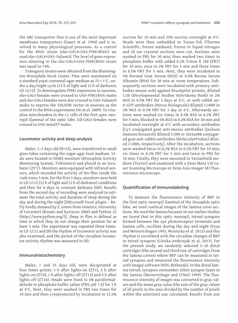
Fig. 1. PINK1 mutation and PINK1 RNAi in neurons cause locomotor activity impairment. (A) and (B) Total activity from the second day of locomotor activity recording in LD 12: 12 conditions (12 h of light and 12 h of darkness). Charts show time of total activity in minutes for each genotype. (A): the total activity time was the lowest in PINK1 mutants in comparing with white mutants and wild type Canton S. (B): flies with silenced PINK1 in neurons had also reduced activity when compared with the control (four stars represent p<0.01, one star represents p<0.05) (B). (C) and (D) Sleep duration in the day/light phase of LD 12: 12 conditions. PINK1B9 and w1118 flies had prolonged day sleep (nap) (one star represents p<0.05) in comparing with wild type strain (C). Flies with silenced PINK1 in neurons also exhibited longer sleep during the day than control flies (four stars represent p<0.01) (D). (E) and (F) Sleep duration in the night/dark phase of the second day of LD 12: 12 conditions. PINK1B9, w1118 and Canton S had the same sleep duration during the night (E). The duration of sleep at night did not change also in elav‑GAL4>UAS‑PINK1RNAi flies (F).
PINK1 mutation affects synapses and behavior 235Acta Neurobiol Exp 2018, 78: 231–241
Transmission Electron Microscopy (TEM)
Heads of 1‑week old males were dissected one hour after lights‑on and fixed in cacodyl‑buffered PFA (2.5%) and glutaraldehyde (2%) primary fixative for 2 h. They were post‑fixed in OsO4 (2%) in veronal acetate buffer for 1 h. Subsequently, the heads were dehydrated in a series of alcohols and propylene oxide and embedded in Poly/Bed 812 resin (Polysciences). Ultrathin sections (65 nm thick) of the lamina were cut and contrasted with uranyl acetate and lead citrate. Images of tetrad synapses in the lamina were taken using a Jeol JEM 2100 HT TEM. The experiment was repeated 3 times. 10 images were taken per 1 repetition.
Statistics
The statistical analyses were performed using Graph‑Pad Prism 6. Data were examined for distribution nor‑mality, and statistical tests were chosen accordingly. For lifespan results the Kaplan‑Meier test was used. The Wilcoxon–Mann–Whitney and Kruskal–Wallis tests were performed to assess differences in the fluorescence in‑tensity correlated with BRP protein levels from confo‑
cal images, GFP fluorescence intensity of mitochondria, sleep, total activity, period of the circadian rhythm of locomotor activity and for climbing assays. For Western Blot data the one‑way ANOVA and Tukey tests were used.
RESULTS
The effect of PINK1 on locomotor activity
Recordings of flies’ locomotor activity showed that the activity level during 24 h of PINK1B9 was lower when compared with w1118 and Canton S (Fig. 1A). The activity of PINK1 RNAi flies was also lower than the control Va‑lium10 (Fig. 1B). Sleep in both PINK1B9 and w1118 flies was increased but only during the day (Fig. 1C). Similar re‑sults were also obtained in flies with PINK1 RNAi, which exhibited longer sleep during the day (Fig. 1D), where‑as sleep during the night was unchanged (Fig. 1E, F).
The effect of PINK mutation on synapses
BRP level, measured as the fluorescence intensity after immunolabeling in the lamina at ZT1 (Fig. 3A‑C)
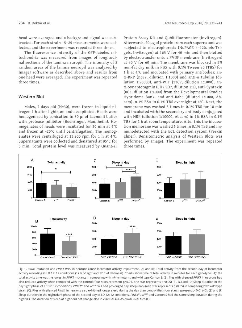
Fig. 2. PINK1 and white mutations are responsible for the reduced Bruchpilot (BRP) level in tetrad synapses in the lamina. (A‑E) Immunolabeling of BRP in tetrad synapses of the examined strains with nc82 antibodies. Reaction was carried out in the lamina sections of flies collected at ZT1 (one hour after lights‑on). Scale bar – 20 µm. (F) and (G) The fluorescence index of BRP. Charts show the fluorescent intensity correlated with BRP level. Statistically sig‑nificant differences (four stars and a,b,c represent p<0.05) are between all genotypes in both (F) and (G) charts. PINK1B9 and elav‑GAL4>UAS‑ PINK1RNAi had lower level of BRP in tetrad synapses in comparing with other strain studied.
236 B. Doktór et al. Acta Neurobiol Exp 2018, 78: 231–241
was the lowest in PINK1B9 flies (Fig. 2B), however, it was also lower in the white mutation in comparison with Canton S Flies and with silenced PINK1 in neurons there was also a reduced BRP level in the lamina (Fig. 2G). In contrast, the daily rhythm of the BRP level in tet‑rad synapses, with two peaks at ZT1 and ZT13, was not changed in both mutants; PINK1B9 and w1118, in compari‑son with Canton S flies.
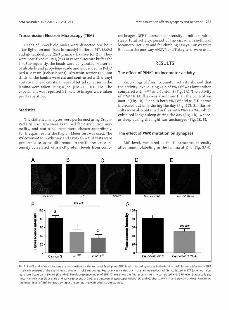
The reduced level of BRP was also detected in the whole brains of the studied flies. Western Blot analy‑sis showed a lower level of both BRP isoforms BRP170 and BRP190 in PINK1B9 and elav‑GAL4>UAS‑PINK1RNAi (Fig. 3A‑C) when compared with the controls Can‑ton S and elav‑GAL4>UAS‑Valium10, respectively. BRP level in w1118 was also lower than in Canton S but this reduction was not statistically significant. The daily rhythm in changes of the BRP level was not affected by aging. Moreover, the BRP level at ZT1 was similar in young (7 days old) and older (35 days old) flies of the Canton S and other strains studied: w1118, PINK1B9 and elav‑GAL4>UAS‑PINK1RNAi.
PINK1 mutants also exhibited reduced levels of other proteins involved in synaptic transmission. In these mutants, the levels of Syntaxin and Rab5 were lower when compared with the controls Canton S and w1118 (Fig. 4A‑B). The abundance of Wishful Thinking (WIT) was also lower compared with the control w1118
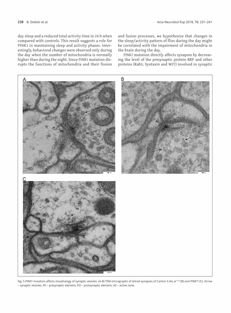
(Fig. 4C), in contrast to Synaptotagmin, where the lev‑el was similar in all genotypes studied (Fig. 4D). More‑over, morphology of the synaptic vesicles studied in tetrad synapses in the visual system in PINK1 mutants was changed when compared with white and Canton S controls (Fig. 5). The synaptic vesicles of PINK1 mutants had broken membranes and most of them were darker (higher electronic density) compared with the control strains and their number was reduced (Fig. 5).
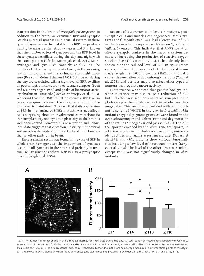
Daily oscillations of mitochondria number in the first optic neuropil
Since sleep in the mutant studied was affected only during the day we also measured daily changes in mito‑chondria number in neurons. We selected L2 interneu‑rons of the lamina, one of the four postsynaptic cells in tetrad synapses and analyzed the fluorescence intensi‑ty of GFP‑labeled mitochondria in L2 (Fig. 6A) at differ‑ent time points. The obtained results showed a signifi‑cantly higher signal during the day (ZT1 and ZT4) than during the night (ZT13 and ZT16) (Fig. 6B). This means that the number of mitochondria increases during the day, when insects are more active in locomotor activi‑ty, rather than during the night. Their number is not correlated with the two peaks, at ZT1 and ZT13, in the
Fig. 3. PINK1 and white mutations are responsible for the reduced Bruchpilot (BRP) level in the brain. (A) and (B) Densitometric analysis of BRP190 and BRP170 isoforms in Canton S, PINK1B9, w1118 (A), elav‑GAL4>UAS‑Valium and elav‑GAL4>UAS‑PINK1RNAi (B). BRP level was standardized to Canton S and elav‑GAL4>UAS‑Valium in (A) and (B), respectively. Statistically significant differences (four stars represent p<0.01, and one star represents p=0.05) are between PINK1B9 and Canton S and between elav‑GAL4>UAS‑PINK1RNAi and Valium controls. (C) Result of Western Blot of BRP in whole brain homoge‑nates of all genotype studied.

PINK1 mutation affects synapses and behavior 237Acta Neurobiol Exp 2018, 78: 231–241
number of tetrad synapses with postsynaptic elements that are also in L2 interneurons.
DISCUSSION
The malfunction of Mitochondrial Complex I, oxida‑tive stress and aggregation of abnormal/misfolded pro‑teins are typical molecular symptoms of Parkinson’s disease (PD) (Dawson and Dawson 2003). They lead to a decrease in mitochondria number, and the lack of en‑ergy may be responsible for subsequent neurodegener‑ation (Li et al. 2017), thus this may be correlated with the observed motor and non‑motor disorders in PD. It was previously shown that in PINK1 mutants the level of ATP is low, indicating dysfunction of mitochondria (Liu et al. 2011). In the present study, we found that PINK1 mutation causes not only motor disorders and reduced activity but also affect synapses and synaptic trans‑mission in the brain and neuromuscular junctions. Mo‑
tor disorders have already been reported in PD animal models (Feany and Bender 2000). Moreover, in PINK1 mutants of Drosophila morphological abnormalities of indirect flight muscles (Park et al. 2006) and apoptosis of muscle cells have been observed (Clark et al. 2006).
As reported by other authors both mutations also affect lifespan and the climbing ability of flies, which were both decreased (data not shown). In the present study we showed that in addition both PINK1 and white mutants have abnormal sleep and their total activity is decreased. The duration of sleep was lengthened during the day but not during the night in comparison with wild type Canton S flies, and in addition, total activity was decreased in PINK1 mutants. Longer sleep during the day shortens time for living functions and behavior in Drosophila, which are concentrated during the day and at the beginning of the night. The effect of PINK1 mutation on sleep and daily activity was also confirmed using a Drosophila strain in which PINK1 gene expression was reduced in neurons. These flies also showed longer
Fig. 4. PINK1 and white mutations reduce levels of other proteins involved in synaptic transmission in the Drosophila brain. (A‑D) Densitometric analysis of Syntaxin (A), Rab5 (B), Wishful Thinking (C) and Synaptotagmin (D) in Canton S, w1118 and PINK19 whole heads. Protein levels were standardized to Tubulin. Statistically significant differences are represented by three stars (p=0.01), two star (p<0.05) and one star (p=0.05).
238 B. Doktór et al. Acta Neurobiol Exp 2018, 78: 231–241
day sleep and a reduced total activity time in 24 h when compared with controls. This result suggests a role for PINK1 in maintaining sleep and activity phases. Inter‑estingly, behavioral changes were observed only during the day when the number of mitochondria is normally higher than during the night. Since PINK1 mutation dis‑rupts the functions of mitochondria and their fission
and fusion processes, we hypothesize that changes in the sleep/activity pattern of flies during the day might be correlated with the impairment of mitochondria in the brain during the day.
PINK1 mutation directly affects synapses by decreas‑ing the level of the presynaptic protein BRP and other proteins (Rab5, Syntaxin and WIT) involved in synaptic
Fig. 5 PINK1 mutation affects morphology of synaptic vesicles. (A‑B) TEM micrographs of tetrad synapses of Canton S (A), w1118 (B) and PINK19 (C). Arrow – synaptic vesicles, PE – presynaptic element, PO – postsynaptic element, AZ – active zone.
PINK1 mutation affects synapses and behavior 239Acta Neurobiol Exp 2018, 78: 231–241
transmission in the brain of Drosophila melanogaster. In addition to the brain, we examined BRP and synaptic vesicles in tetrad synapses in the visual system. In these types of synapses in the distal lamina BRP can predom‑inantly be measured in tetrad synapses and it is known that the number of tetrad synapses and the BRP level in these synapses oscillate during the day and night with the same pattern (Górska‑Andrzejak et al. 2013, Mein‑ertzhagen and Pyza 1999, Woźnicka et al. 2015). The number of tetrad synapses peaks twice, in the morning and in the evening and is also higher after light expo‑sure (Pyza and Meinertzhagen 1993). Both peaks during the day are correlated with a high level of BRP, swelling of postsynaptic interneurons of tetrad synapses (Pyza and Meinertzhagen 1999) and peaks of locomotor activ‑ity rhythm in Drosophila (Górska‑Andrzejak et al. 2013). We found that the PINK1 mutation reduces BRP level in tetrad synapses, however, the circadian rhythm in the BRP level is maintained. The fact that daily expression of BRP in the lamina of PINK1 mutants was not affect‑ed is surprising since an involvement of mitochondria in neuroplasticity and synaptic plasticity in the brain is well documented. However, this observation and behav‑ioral data suggests that circadian plasticity in the visual system is less dependent on the activity of mitochondria than in other parts of the brain.
Since a similar result was found in the case of BRP in whole brain homogenates, the impairment of synapses occurs in all synapses in the brain and probably in neu‑romuscular junctions where BRP is also a presynaptic protein (Wagh et al. 2006).
Because of low transmission levels in mutants, post‑synaptic cells and muscles can degenerate. PINK1 mu‑tants and flies with PINK1 RNAi had a lower level of BRP in the brain when compared with Canton S, w1118 and Valium10 controls. This indicates that PINK1 mutation affects synaptic contacts in the nervous system be‑cause of increasing the production of reactive oxygen species (ROS) (Chien et al. 2013). It has already been shown that the reduced level of BRP in brp mutants causes similar motor disorders to that observed in our study (Wagh et al. 2006). However, PINK1 mutation also causes degeneration of dopaminergic neurons (Yang et al. 2006), and perhaps may also affect other types of neurons that regulate motor activity.
Furthermore, we showed that genetic background, white mutation, may also cause a reduction of BRP but this effect was seen only in tetrad synapses in the photoreceptor terminals and not in whole head ho‑mogenates. This result is correlated with an import‑ant function of WHITE in the eye. In Drosophila white mutants atypical pigment granules were found in the eye (Schraermeyer and Dohms 1993) and degeneration of the retina (Ambegaokar and Jackson 2010). The ABC transporter encoded by the white gene transports, in addition to pigment in photoreceptors, ions, amino ac‑ids, peptides and sugars across membranes (Savary et al. 1996) and white mutants show various abnormali‑ties including a low level of neurotransmitters (Bory‑cz et al. 2008). The level of the other proteins studied, except Rab5, was not significantly changed in white mutants.
Fig. 6. The number of mitochondria in the lamina L2 interneurons oscillates during the day. (A) Localization of mitochondria labeled with GFP in L2 interneurons of the lamina of 21D‑GAL4>UAS‑mitoGFP. Re – retina, Ln – lamina neuropil, Arrow – cell bodies of L2 neurons, Frame – measurement area, Scale bar – 20µm. (B) The fluorescence index of GFP‑labeled mitochondria in the lamina neuropil measured in different time points of the day of 21D‑GAL4>UAS‑mtoGFP. Statistically significant differences (one star represents p=0.05) are between ZT1 and ZT13, ZT16; ZT4 and ZT13, ZT16.
240 B. Doktór et al. Acta Neurobiol Exp 2018, 78: 231–241
CONCLUSIONS
In the present study we proved that PINK1 is re‑quired for maintaining sleep during the day and synap‑tic transmission by the regulation of synaptic protein levels. The lack of PINK1 results in the reduction of syn‑aptic proteins responsible for exocytosis of neurotrans‑mitters, which in turn may cause previously described motor and sleep disorders. We also showed that using white background, to create transgenic strains, leads to motor and non‑motor disorders in those flies. We sug‑gest using w1118 as an additional control for strains with white background, because the comparison between Canton S and w1118 may give information on the effects of white mutation itself on the examined processes.
ACKNOWLEDGEMENTS
This study was supported by the Jagiellonian Univer‑sity grant K/ZDS/008070. In this study we used a Zeiss LSM 510 confocal microscope in the Laboratory of Mi‑croscopy, Department of Cell Biology and Imaging, In‑stitute of Zoology and Biomedical Sciences, Jagiellonian University.
REFERENCES
Aberle H, Haghighi PA, Fetter RD, McCabe BD, Magalhães TR, Goodman CS (2002) Wishful thinking encodes a BMP Type II receptor that regulates synaptic growth in Drosophila. Neuron 33: 545–558.
Ambegaokar SS and Jackson GR (2010) Interaction between eye pigment genes and tau‑induced neurodegeneration in Drosophila melanogaster. Genetics 186: 435–442.
Borycz J, Borycz JA, Kubów A, Lloyd V, Meinertzhagen IA (2008) Drosophila ABC transporter mutants white, brown and scarlet altered contents and distribution of biogenic amines in the brain. J Exp Biol 211: 3454–3466.
Chen Y and Dorn GW (2013) PINK1‑phosphorylated mitofusin 2 is a Parkin receptor for culling damaged mitochondria. Science 340: 471–475.
Chien WL, Lee TR, Hung SY, Kang KH, Wu RM, Lee MJ, Fu WM (2013) In‑crease of oxidative stress by a novel PINK1 mutation, P209A. Free Radic Biol Med 58: 160–169.
Clark IE, Dodson MW, Jiang C, Cao JH, Huh JR, Seol JH, Yoo SJ, Hay BA, Guo M (2006) Drosophila pink1 is required for mitochondrial function and inter‑acts genetically with parkin. Nature 441: 1162–1166.
Dawson TM and Dawson VL (2003) Molecular pathways of neurodegener‑ation in Parkinson’s disease. Science 302: 819–822.
Deas E, Plun‑Favreau H, Gandhi S, Desmond H, Kjaer S, Loh SH, Renton AE, Harvey RJ, Whitworth AJ, Martins LM, Abramov AY, Wood NW (2011) PINK1 cleavage at position A103 by the mitochondrial protease PARL. Hum Mol Genet 20: 867–879.
DiAntonio A, Haghighi AP, Portman SL, Lee JD, Amaranto AM, Goodman CS (2001) Ubiquitination‑dependent mechanisms regulate synaptic growth and function. Nature 412: 449–452.
Eiyama A and Okamoto K (2015) PINK1/Parkin‑mediated mitophagy in mammalian cells. Curr Opin Cell Biol 33: 95–101.
Ewart GD, Cannell D, Cox GB, Howells AJ (1994) Mutational analysis of the traffic ATPase (ABC) transporters involved in uptake of eye pigment pre‑
cursors in Drosophila melanogaster. Implications for structure‑function relationships. J Biol Chem 269: 10370–10377.
Feany MB and Bender WW (2000) A Drosphila model of Parkinson’s dis‑ease. Nature 404: 394–398.
Geppert M, Goda Y, Hammer RE, Li C, Rosahl TW, Stevens CF, Südhof TC (1994) Synaptotagmin I: a major Ca2+ sensor for transmitter release at a central synapse. Cell 79: 717–727.
Górska‑Andrzejak J, Makuch R, Stefan J, Görlich A, Semik D, Pyza E (2013) Circadian expression of the presynaptic active zone protein Bruchpilot in the lamina of Drosophila melanogaster. Dev Neurobiol 73: 14–26.
Greene AW, Grenier K, Aguileta MA, Muise S, Farazifard R, Haque ME, McBride HM, Park DS, Fon EA (2012) Mitochondrial processing pepti‑dase regulates PINK1 processing, import and Parkin recruitment. EMBO Rep 13: 378–385.
Hoop MJ, Huber LA, Stenmark H, Williamson E, Zerial M, Parton RG, Dotti CG (1994) The involvement of the small GTP‑binding protein Rab5a in neuronal endocytosis. Neuron 13: 11–22.
Kittel RJ, Wichmann C, Rasse TM, Fouquet W, Schmidt M, Schmid A, Wagh DA, Pawlu C, Kellner RR, illig KI, Hell SW, Buchner E, Heckmann M, Sigrist SJ (2006) Bruchpilot promotes active zone assembly, Ca2+ chan‑nel clustering, and vesicle release. Science 312: 1051–1054.
Krstic D, Boll W, Noll M (2013) Influence of the white locus on the courtship behavior of Drosophila males. PLoS One 8: e77904.
Lazarou M, Jin SM, Kane LA, Youle RJ (2012) Role of PINK1 binding to the TOM complex and alternate intracellular membranes in recruitment and activation of the E3 ligase Parkin. Dev Cell 22: 320–333.
Liu W, Acín‑Peréz R, Geghman KD, Manfredi G, Lu B, Li C (2011) Pink1 reg‑ulates the oxidative phosphorylation machinery via mitochondrial fis‑sion. Proc Natl Acad Sci 108: 12920–12924.
Li Z, Peng Y, Hufnagel RB, Hu YC, Zhao C, Queme LF, Khuchua Z, Driver AM, Dong F, Lu QR, Lindquist DM, Jankowski MP, Stottmann RW, Kao WWY, Huang T (2017) Loss of SLC25A46 causes neurodegeneration by affect‑ing mitochondrial dynamics and energy production in mice. Hum Mol Genet 26: 3776–3791.
Lu B and Vogel H (2009) Drosophila models of neurodegenerative diseases. Annu Rev Pathol 4: 315–342.
Ly CV and Verstreken P (2006) Mitochondria at the synapse. Neuroscientist 12: 291–299.
Matsuda N, Sato S, Shiba K, Okatsu K, Saisho K, Gautier CA, Sou YS, Saiki S, Kawajiri S, Sato F, Kimura M, Komatsu M, Hattori N, Tanaka K (2010) PINK1 stabilized by mitochondrial depolarization recruits Parkin to damaged mitochondria and activates latent Parkin for mitophagy. J Cell Biol 189: 211–221.
Meinertzhagen IA, O’Neil SD (1991) Synaptic organization of columnar ele‑ments in the lamina of the wild type in Drosophila melanogaster. J Comp Neurol 305: 232–263.
Morais VA, Verstreken P, Roethig A, Smet J, Snellinx A, Vanbrabant M, Haddad D, Frezza C, Mandemakers W, Vogt‑Weisenhorn D, Van Coster R, Wurst W, Scorrano L, De Strooper B (2009) Parkinson’s disease muta‑tions in PINK1 result in decreased Complex I activity and deficient syn‑aptic function. EMBO Mol Med 1: 99–111.
Ni JQ, Liu LP, Binari R, Hardy R, Shim HS, Cavallaro A, Booker M, Pfeiffer BD, Markstein M, Wang H, Villalta C, Laverty TR, Perkins LA, Perrimon N (2009) Drosophila resource of transgenic RNAi lines for neurogenetics. Genetics 182: 1089–1100.
Park J, Lee SB, Lee S, Kim Y, Song S, Ki S, Bae E, Kim J, Shong M, Kim JM, Chung J (2006) Mitochondrial dysfunction in Drosophila PINK1 mutants is complemented by parkin. Nature 441: 1157–1161.
Pilling AD, Horiuchi D, Lively CM, Saxton WM (2006) Kinesin‑1 and Dynein are the primary motors for fast transport of mitochondria in Drosophila motor axons. Mol Biol Cell 17: 2057–2068.
Poole AC, Thomas RE, Andrews LA, McBride HM, Whitworth AJ, Pallanck LJ (2008) The PINK1/Parkin pathway regulates mitochondrial morphology. Proc Natl Acad Sci 105: 1638–1643.

PINK1 mutation affects synapses and behavior 241Acta Neurobiol Exp 2018, 78: 231–241
Pyza E and Meinertzhagen A (1993) Daily and circadian rhythms of syn‑aptic frequency in the first visual neuropile of the housefly’s (Musca domestica L.) optic lobe. Proc Biol Sci 254: 97–105.
Pyza E and Meinertzhagen IA (1999) Daily rhythmic changes of cell size and shape in the first optic neuropil in Drosophila melanogaster. J Neurobiol 40: 77–88.
Rosato E and Kyracou CP (2006) Analysis of locomotor activity rhythms in Drosophila. Nat Protoc 1: 559–568.
Savary S, Denizot F, Luciani M, Mattei M, Chimini G (1996) Molecular clon‑ing of a mammalian ABC transporter homologous to Drosophila white gene. Mamm Genome 7: 673–676.
Schraermeyer U and Dhms M (1993) Atypical granules in the eyes of the white mutant of Drosophila melanogaster are lysosome‑related organ‑elles. Pigment Cell Res 6: 73–84.
Shields M, Bowers MR, Fulcer MM, Bollig MK, Rock PJ, Sutton BR, Vrailas‑Mortimer AD, ller HL, Whittaker RG, Horvath R, Reist NE (2017) Drosophila studies support a role for a presynaptic synaptotagmin muta‑tion in a human congenital myasthenic syndrome. PLoS One 12: e0184817.
Sieber JJ, Willig KI, Heintzmann R, Hell SW, Lang T (2006) The SNARE motif is essential for the formation of syntaxin clusters in the plasma mem‑brane. Biophys J 90: 2843–2851.
Stenmark H (2009) Rab GTPases as coordinators of vesicle traffic. Nat Rev Mol Cell Biol. 10: 513–525.
Thomas RE, Andrews LA, Burman JL, Lin WY, Pallanck LJ (2014) PINK1‑Par‑kin pathway activity is regulated by degradation of PINK1 in the mito‑chondrial matrix. PLoS Genet 10: e1004279.
Ullrich A, Böhme MA, Schöneberg J, Depner H, Sigrist SJ, Noé F (2015) Dynamical organization of Syntaxin‑1A at the presynaptic active zone. PLoS Comput Biol 11: 1–22.
Wagh DA, Rasse TM, Asan E, Hofbauer A, Schwenkert I, Dürrbeck H, Buchner S, Dabauvalle MC, Schmidt M, Qin G, Wichmann C, Kittel R, Sig‑rist SJ, Buchner E (2006) Bruchpilot, a protein with homology to ELKS/CAST, is required for structural integrity and function of synaptic active zones in Drosophila. Neuron 49: 833–844.
Wang D, Qian L, Xiong H, Liu J, Neckameyer WS, Oldham S, Xia K, Wang J, Bodmer R, Zhang Z (2006) Antioxidants protect PINK1‑de‑pendent dopaminergic neurons in Drosophila. Proc Natl Acad Sci 103: 13520–13525.
Weber P, Kula‑Eversole E, Pyza E (2009) Circadian control of dendrite mor‑phology in the visual system of Drosophila melanogaster. PLoS One 4: e4290.
Woźnicka O, Görlich A, Sigrist S, Pyza E (2015) BRP‑170 and BRP190 iso‑forms of Bruchpilot protein differentially contribute to the frequency of synapses and synaptic circadian plasticity in the visual system of Dro‑sophila. Front Cell Neurosci 9: 1–8.
Wucherpfennig Tanja, Wilsch‑Bräuninger Michaela, González‑Gaitán Marcos (2003) Role of Drosophila Rab5 during endosomal trafficking at the synapse and evoked neurotransmitter release. J Cell Biol. 161: 609–624.
Yamano K and Youle RJ (2013) PINK1 is degraded through the N‑en rule pathway. Autophagy 9: 1758–1769.
Yang Y, Gehrke S, Imai Y, Huang Z, Ouyang Y, Wang JW, Yang L, Beal MF, Vogel H, Lu B (2006) Mitochondrial pathology and muscle and dopami‑nergic neuron degeneration caused by inactivation of Drosophila Pink1 is rescued by Parkin. Proc Natl Acad Sci 103: 10793–10798.
Ziviani E, Tao RN, Whitworth AJ (2010) Drosophila parkin requires PINK1 for mitochondrial translocation and ubiquitinates mitofusin. Proc Natl Acad Sci 107: 5018–5023.