Embed Size (px)
Citation preview
REVIEW Open Access
PINK1 and Parkin mitochondrial qualitycontrol: a source of regional vulnerability inParkinson’s diseasePreston Ge1,2,3,4,5,6, Valina L. Dawson1,2,3* and Ted M. Dawson1,2,3*
Abstract
That certain cell types in the central nervous system are more likely to undergo neurodegeneration in Parkinson’sdisease is a widely appreciated but poorly understood phenomenon. Many vulnerable subpopulations, includingdopamine neurons in the substantia nigra pars compacta, have a shared phenotype of large, widely distributedaxonal networks, dense synaptic connections, and high basal levels of neural activity. These features come atsubstantial bioenergetic cost, suggesting that these neurons experience a high degree of mitochondrial stress. Insuch a context, mechanisms of mitochondrial quality control play an especially important role in maintainingneuronal survival. In this review, we focus on understanding the unique challenges faced by the mitochondria inneurons vulnerable to neurodegeneration in Parkinson’s and summarize evidence that mitochondrial dysfunctioncontributes to disease pathogenesis and to cell death in these subpopulations. We then review mechanisms ofmitochondrial quality control mediated by activation of PINK1 and Parkin, two genes that carry mutations associatedwith autosomal recessive Parkinson’s disease. We conclude by pinpointing critical gaps in our knowledge of PINK1 andParkin function, and propose that understanding the connection between the mechanisms of sporadic Parkinson’s anddefects in mitochondrial quality control will lead us to greater insights into the question of selective vulnerability.
Keywords: Parkinson disease, Parkin, PINK1, Mitochondria, Mitophagy, Selective vulnerability, Substantia nigra
BackgroundParkinson’s disease (PD) is a late-onset neurodegenerativedisease characterized by a core triad of symptoms; restingtremor, bradykinesia, and elevated resting tone [1]. While10% of patients carry single gene mutations that cause PD(monogenic PD), over 90% of patients have no knownfamily history or known genetic cause of their disease(sporadic PD, or sPD) [1]. PD has traditionally beenviewed as a disease caused by the selective degeneration ofdopamine (DA) neurons found in the substantia nigrapars compacta (SNpc) due to early findings that SNpc de-generation is the most consistent postmortem finding inpatient brains, that dopamine replacement through L-DOPA is an effective management strategy for the motor
symptoms, and that the selective SNpc DA neuron toxinMPTP recapitulates the clinical phenotype of PD [2].However, systematic, large-scale characterization of post-mortem PD brains has provided a contrasting image ofdisease progression based on the presence of Lewy bodies(LB), large aggregates of misfolded α-synuclein protein,which has served as a canonical marker of disease path-ology for decades [2, 3]. Pathologic staging of α-synuclein-positive LBs has revealed widespread involvement of mostmajor subdivisions of the central nervous system (CNS),ranging from brainstem nuclei to cortex [3, 4]. A growingbody of evidence in both human patients and preclinicalanimal models suggests that LBs may initially appear inthe brainstem or enteric nervous system and spread acrossthe brain in a prion-like manner [5–12].The realization that PD pathology is not solely con-
fined to the SNpc and can spread across the CNS hasprofoundly altered our understanding of PD pathogen-esis and disease progression. However, despite evidenceof widespread pathology, not all cell populations are
© The Author(s). 2020 Open Access This article is distributed under the terms of the Creative Commons Attribution 4.0International License (http://creativecommons.org/licenses/by/4.0/), which permits unrestricted use, distribution, andreproduction in any medium, provided you give appropriate credit to the original author(s) and the source, provide a link tothe Creative Commons license, and indicate if changes were made. The Creative Commons Public Domain Dedication waiver(http://creativecommons.org/publicdomain/zero/1.0/) applies to the data made available in this article, unless otherwise stated.
* Correspondence: [email protected]; [email protected] and Stem Cell Programs, Institute for Cell Engineering,Department of Neurology, Department of Physiology, Solomon H. SnyderDepartment of Neuroscience, Department of Pharmacology and MolecularSciences, Johns Hopkins University School of Medicine, 733 North Broadway,Suite 731, Baltimore, MD 21205, USAFull list of author information is available at the end of the article
Ge et al. Molecular Neurodegeneration (2020) 15:20 https://doi.org/10.1186/s13024-020-00367-7
equally resilient. Certain populations of neurons remainmore vulnerable to developing LB pathology and to neu-rodegeneration in PD, suggesting that cell-intrinsic fac-tors can gate selective vulnerability. The most vulnerableneuronal subpopulations include SNpc DA neurons;cholinergic neurons in the pedunculopontine nucleus,nucleus basalis of Meynert, and dorsal motor nucleus ofvagus; noradrenergic neurons in the locus coeruleus; andserotoninergic neurons in the raphe nucleus [4, 13].While the factors regulating regional heterogeneity indisease susceptibility are not fully known, mitochondrialstress and failure of mitochondrial quality control path-ways are thought to contribute to regional differences inpathology and neurodegeneration [4]. Here, we reviewthe contribution of mitochondrial dysfunction to select-ive neuronal vulnerability in PD and summarize thecurrent understanding of neuronal mitochondria main-tenance through PINK1/Parkin-mediated mitochondrialquality control.
Main textImportance of mitochondria in PDThe many facets of mitochondrial functionMitochondria are membrane-bound organelles that per-form a diverse range of critical cellular functions. Theyare double-membraned structures, with outer and innermitochondrial membranes (OMM and IMM respect-ively) separated by an intermembrane space and a cen-tral matrix enclosed by the IMM. Reflecting theirevolutionary origins as endosymbiotic bacteria, mito-chondria carry their own unique circular genome(mtDNA) at copy numbers upwards of 10–100 per mito-chondrion [14]. Their genome encodes two uniquerRNAs, 22 tRNAs, and 13 polypeptides required to as-semble the mitochondrial ribosome and parts of theelectron transport chain (ETC), while the nuclear gen-ome encodes 1000+ mitochondrial genes [14, 15]. Whiletraditional textbook pictures show mitochondria asstatic, bean-shaped structures, in reality they exist as dy-namic networks shifting from innumerable punctate or-ganelles to cell-wide tubular networks governed by acomplex fission/fusion machinery [16]. Mitochondria arehighly multifunctional. They not only generate the bulkof ATP in most cell types through oxidative phosphoryl-ation, but also metabolize and synthesize complex mac-romolecules (e.g. lipids, amino acids, and nucleotides);buffer reactive oxygen species (ROS) and cytoplasmicCa2+; regulate cellular redox balances; control apoptosis;and serve as key anchoring scaffolds for intracellular sig-naling networks [14, 17, 18].Though critical for cell survival, these energetically de-
manding processes generate reactive intermediates andoxidizing agents that damage nucleic acids, proteins, andlipids, necessitating various waste removal and damage
control mechanisms such as the urea cycle, glutathioneantioxidants, and H2S detoxification [16–18]. While thesemechanisms perform detoxification of reactive metabolicintermediaries and end products, mitochondria also pos-sess several sophisticated systems for maintaining struc-tural integrity and proper protein function. These systems,collectively known as mitochondrial quality control(MQC), include AAA proteases that degrade proteins inthe matrix and intermembrane space, the ubiquitin-proteasome system for removing OMM proteins, the re-moval of larger portions of mitochondria through mito-chondrial derived vesicles (MDVs) and mitophagy, andregulation of fission/fusion dynamics [15, 19].
Neuron subpopulation-specific bioenergetic vulnerabilitiesin PDMitochondria within the CNS exist in a unique meta-bolic environment due to the sheer energetic demand ofneural activity and the structural polarization of CNScells. The brain comprises roughly 2 % of total bodymass yet consumes 20% of the body’s oxygen intake and25% of glucose supply, of which the bulk goes towardssustaining membrane potentials and facilitating neuro-transmission [19, 20]. Neuronal mitochondria must meetthe immense energetic demands of neuronal signalingwhile also buffering waves of Ca2+ entry, which leads tothe generation of excitotoxic ROS if left unchecked [20].Furthermore, neuronal architecture is complex and ex-quisitely polarized, with some neurons carrying the vastmajority of their cytoplasm and mitochondria in longdendrites and axons that can be as far as a meter awayfrom the soma [15, 21]. Given the functionalspecialization of these cellular subcompartments, it islikely that such structural polarization leads to localmetabolic needs that may require sub-specialization ofmitochondrial function, such as increased Ca2+ bufferingat the pre- and postsynaptic termini and increased bio-synthetic functions at the soma. While other somaticcells can oftentimes rely on cell division to generatefresh mitochondria, neurons are postmitotic cells thatmust overcome the aforementioned challenges for anentire lifetime.These bioenergetic demands are particularly evident in
the neuronal populations selectively vulnerable in PD,including most of the nuclei described in the introduc-tion. Many of these neurons send extensive, branchingaxons throughout the brain and influence large, diffusebrain areas [4]. The axons of SNpc DA neurons, for ex-ample, form vast branching nets that have been esti-mated to form up to 100–400 thousand synapses withinthe striatum and extend on average around 30–46 cm inlength in rats [22–24]. In humans, they have been esti-mated to form up to 1–2 million synapses [15]. Whencompared to the less vulnerable neighboring DA
Ge et al. Molecular Neurodegeneration (2020) 15:20 Page 2 of 18
neurons in the ventral tegmental area, SNpc DA neuronshave more complex axons, a higher density of axonalmitochondria, higher rates of oxidative phosphorylation,and increased superoxide production [25]. Genetic per-turbation of MQC in cultured SNpc DA neurons led toa reduction in the size of axonal arbors, while neuronswith smaller axonal arbors tended to be more resilientto MPP+, providing further evidence that the morpho-logical architecture of SNpc DA neuron axons imposessignificant strain on mitochondrial function [26]. Fur-thermore, many of these vulnerable populations extendunmyelinated axons [21], which likely demand evenmore energy than myelinated neurons due to the needto regenerate the membrane potential along the entireaxon rather than just at nodes of Ranvier. Thus, the ex-treme cytoarchitectural specialization of these neuronalsubpopulations places a large bioenergetic burden ontheir mitochondria and may contribute to their selectivevulnerability in PD.
Evidence of mitochondrial dysfunction in human PDThe idea that mitochondria may be involved in thepathogenesis of PD was first suggested by observing theeffects of MPTP, a byproduct of illicit synthesis of theopioid drug desmethylprodine. MPTP is metabolized tothe mitochondrial complex I inhibitor MPP+, which inturn causes acute-onset Parkinsonism with selective de-struction of SNpc neurons [2, 27]. Since the discovery ofMPTP, three major lines of evidence – epidemiological,pathological, and genetic – have pointed to mitochon-drial dysfunction as a central driver of disease. First,mitochondrial toxins have either been shown to cause orcorrelate with increased risk of PD. In addition toMPTP, exposure to the pesticide rotenone, a complex Iinhibitor, has been associated with increased risk of PDin epidemiological studies [28–30]. Second, postmortemstudies of human PD patients have found widespreadevidence of mitochondrial dysfunction. Complex I dys-function has been consistently identified in the SNpc ofdeceased patients, with some reports of more general-ized complex dysfunction that may affect other tissuesas well [31–34]. In addition to these bioenergetic defects,mitochondria in postmortem tissue also show evidenceof genetic defects, with greater age-dependent accumula-tion of mtDNA deletions and somatic mosaicism thancontrol subjects [33–36]. Numerous other studies haveidentified dysregulation in the expression of variousmitochondrial proteins, such as the molecular chaperoneprohibitin, OMM protein VDAC1, mitochondrial importprotein Tom40, and serine protease HtrA2 [37], as wellas increased oxidative damage to mitochondrial proteins[38]. Third, mutations in genes that cause monogenicPD have been linked to mitochondrial function. For ex-ample, the PD-associated gene VPS35, which encodes a
key subunit of the retromer complex responsible for sort-ing proteins between membranous organelles [39], con-tributes to the formation of MDVs and regulates fission/fusion dynamics [40–42]. The mutant proteins encodedby other genes that cause monogenic PD, such as LRRK2,SNCA, ATP13A2, have likewise been found to cause mito-chondrial pathologies ranging from increased fragmenta-tion, disruption of ER-mitochondrial interactions,impaired Ca2+ buffering, elevated numbers of mtDNAmutations, and increased ROS production [43–47].Most prominent of the monogenic PD-associated genes
involved in mitochondrial function are PINK1, encodingthe PTEN-induced serine/threonine kinase 1, and PRKN,encoding an E3 ubiquitin ligase Parkin [18, 33, 48, 49].Following these findings, genetic studies in Drosophila im-plicated a shared biological pathway for Parkin and PINK1function [50–52], with further mechanistic work establish-ing their function in detecting mitochondrial damage andrecruiting mechanisms to remove and replace dysfunc-tional mitochondrial components. The activation andfunctions of the PINK1/Parkin system of MQC are argu-ably some of the most well-studied pathways of PD patho-genesis and will be reviewed in detail below (Fig. 1).Collectively, these findings firmly establish mitochondrialdysfunction as a core pathologic feature of PD. The con-tribution of mitochondrial dysfunction to neurodegenera-tion relative to other mechanisms is not fully known,though it likely differs between monogenic versus familialPD and is dependent on the brain region in question.
PINK1/Parkin as core organizers of mitochondrial qualitycontrolMutations in PINK1 or PRKN (Parkin) cause selective loss ofSNpc DA neuronsLoss of function mutations in PINK1 and PRKN are themost common known causes of autosomal recessive andearly onset PD (before the age of 45) [48, 49, 53]. Despitean earlier age of onset, PD associated with PINK1 or PRKNmutations is usually more benign with slower progression,high L-DOPA responsiveness, and normal cognition, butwith high likelihood of dyskinesias, dystonia, hyperreflexia,and psychiatric symptoms [53–55]. The clinical presenta-tion of PINK1/PRKN PD is intriguing in its relatively puremotor phenotype compared to other cases of PD and therobust and long-lasting (sometimes in the range of decades)responsiveness to dopamine replacement therapy, suggest-ing that these patients may experience a disease processthat is largely confined to the SNpc DA system. This hy-pothesis is consistent with postmortem pathology in seven-teen cases of PRKN and one case of PINK1 PD, which isstriking for the highly specific loss of SNpc neurons withrelative sparing of the locus coeruleus (LC) and other brainregions [53, 56]. Whereas LB pathology is found in virtuallyall cases of sPD, it was found only inconsistently in PINK1/
Ge et al. Molecular Neurodegeneration (2020) 15:20 Page 3 of 18
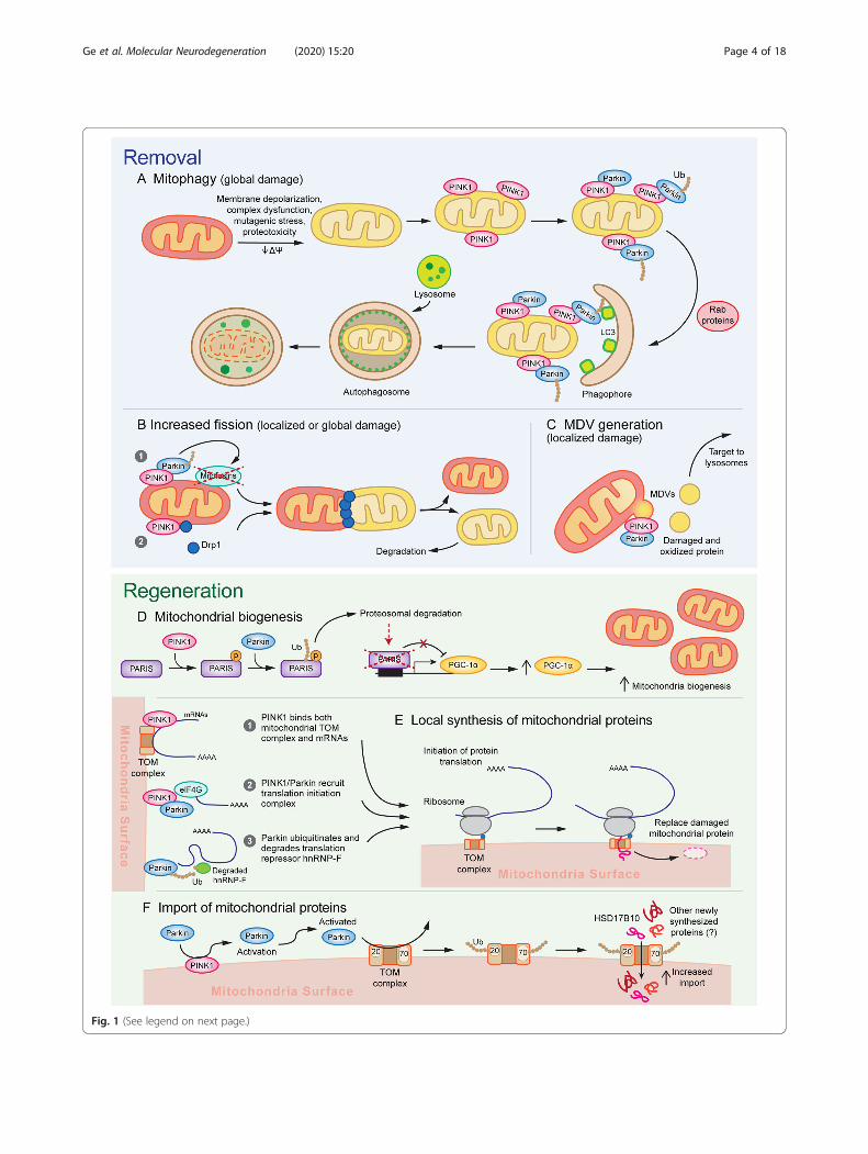
Fig. 1 (See legend on next page.)
Ge et al. Molecular Neurodegeneration (2020) 15:20 Page 4 of 18
PRKN PD (6/17 genetically confirmed PRKN PD, and traceamounts in 1/1 PINK1), suggesting that α-synuclein may bea minor player in these cases [53, 56]. In contrast, theclinical-pathologic features of sporadic PD and othermonogenic causes of PD tend to exhibit significantlygreater variation and wider involvement of other cell popu-lations [54–56]. While it is important to note that the lim-ited availability of autopsy cases may cause anunderestimation of the pathological heterogeneity of PRKNand PINK1 PD, the combined clinical-pathological evidenceof highly selective SNpc DA neuron loss suggests that thesegenes may represent an Achilles heel of SNpc DA neuronsand that studying downstream pathological pathways maybe critical for yielding insights into the vulnerability of thepopulation in PD.
Mechanism of PINK1/Parkin activationPINK1 and Parkin function as the first steps of a signal-ing pathway that activates mitochondrial quality controlpathways in response to mitochondrial damage [57].Under basal conditions, PINK1’s N-terminus is trans-ferred across the OMM to the IMM, with the kinase do-main located closer to the C-terminus protruding outinto the cytosol. PINK1 is then cleaved by IMM-boundproteases and subsequently degraded by the proteasome,leading to undetectable basal levels of PINK1 [58, 59].Stressors such as membrane depolarization, mitochon-drial complex dysfunction, mutagenic stress, and proteo-toxicity lead to accumulation of PINK1 on the OMM byimpairing intermembrane transport of the N-terminusdomain to the IMM. Subsequent homodimerization ofPINK1 on the OMM leads to autophosphorylation,which promotes kinase activation and facilitates bindingto substrates Parkin and ubiquitin [58–61]. Thus,PINK1’s ability to rapidly accumulate and activate in re-sponse to mitochondrial stressors allows it to functionas a sensor of mitochondrial damage.Parkin is an E3 ubiquitin ligase that contains a
ubiquitin-like domain and four RING domains: RING0,RING1, IBR, and RING2 [62]. Its basal activity is
minimal due to intramolecular interactions that blockthe active site and compete with E2 ligase binding [57].Upon mitochondrial injury, PINK1 activates Parkinthrough two mechanisms. First, it phosphorylates ubi-quitin on S65, which competes with an autoinhibitorydomain within Parkin and stabilizes it in an active con-formation. Second, PINK1 directly phosphorylates Par-kin on S65 in Parkin’s ubiquitin-like domain, whichinduces conformational changes that allow for bindingof the charged E2 ligase [57–59, 63–70]. These mecha-nisms increase Parkin’s E3 ubiquitin ligase activity, withboth required for full activation, though it is unclearwhether either mechanism contributes differentially toactivation [63, 66, 71]. Thus, Parkin amplifies a damagedetection signal from PINK1 by facilitating the forma-tion of ubiquitin chains, which recruit more Parkin tothe mitochondria [57].Once recruited to the mitochondria, Parkin exhibits
two waves of ubiquitination: the first wave targets manyouter OMM and mitochondrial matrix proteins withinthe first 2 h of activation, whereas the second wave tar-gets IMM proteins [58, 72, 73]. Parkin has also beenfound to ubiquitinate many cytosolic targets [73], thoughit is unclear whether these targets are phosphorylated bymitochondrial bound Parkin or whether there may becytosolic activation of Parkin. Among these cytosolic tar-gets are AIMP2, whose accumulation leads to PARP1and MIF-dependent cell death of nigral DA neurons;and Parkin Interacting Substrate (PARIS, ZNF746),which causes neurotoxicity by suppressing mitochon-drial biogenesis [74–82]. The diversity of Parkin sub-strates and the formation of multiple types of ubiquitinlinkages (K6, K11, K48, and K63), poses the question ofwhether Parkin ubiquitination may have diverse effectson cellular signaling beyond targeting proteins for deg-radation [18, 73, 83]. Substrate specificity and chain for-mation may show some cell-type dependence. Forexample, a recent proteomic profile of Parkin substratesin HeLa cells and human neurons suggests differences inwhich substrates are targeted, which residues are
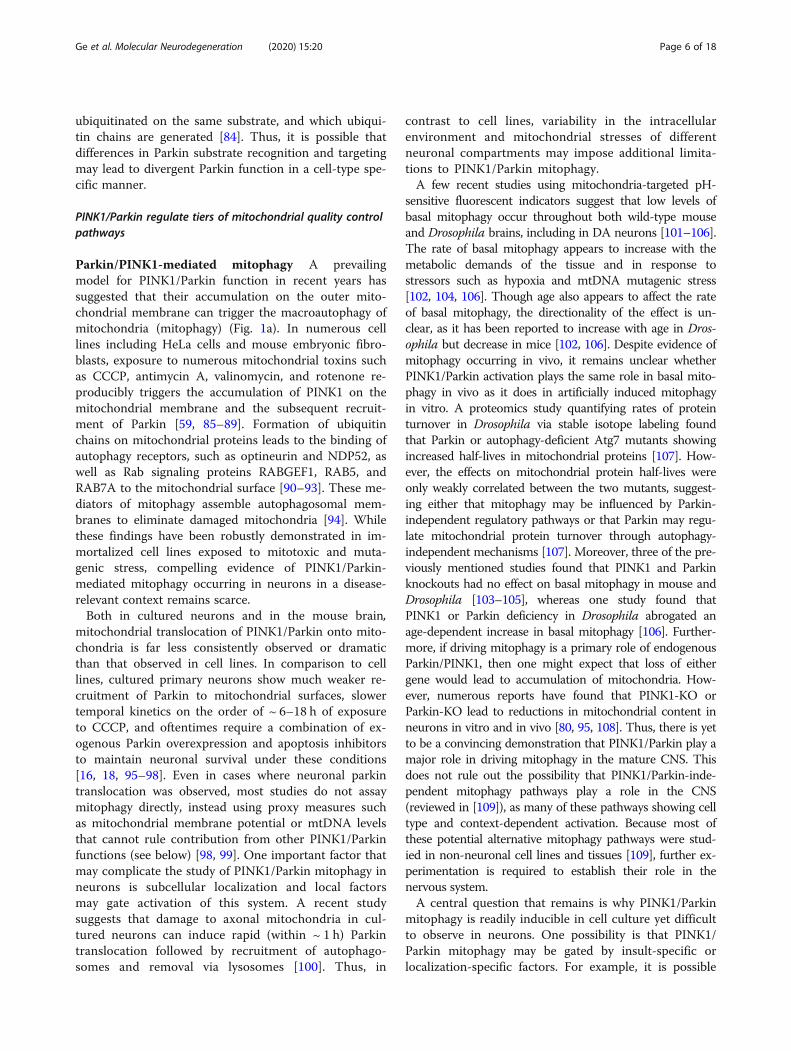
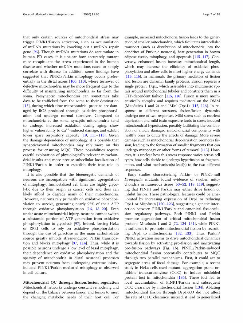
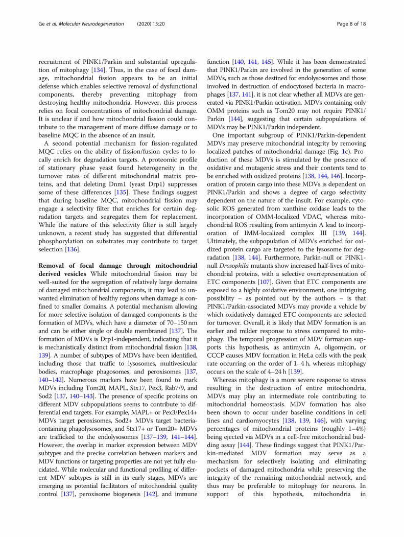
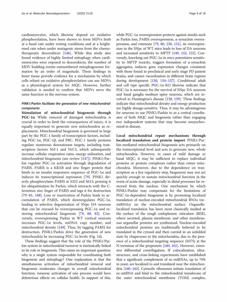
(See figure on previous page.)Fig. 1 A model for the multifunctional role of PINK1/Parkin in mitochondrial quality control. Activation of PINK1/Parkin triggers multiplesequential and parallel mechanisms of a-c mitochondrial removal and d, e mitochondrial regeneration. Different mechanisms of mitochondrialremoval are engaged depending on the severity of damage. a Mitochondria experiencing global/widespread damage undergo mitophagy, inwhich massive PINK1/Parkin activation recruits autophagosome membranes via Rab proteins and LC3 and is subsequently degraded bylysosomes, and b undergo mitochondrial fission caused by PINK1/Parkin dependent mitofusin degradation and Drp1 recruitment. c Focaldamage leads to the activation of mitochondrial fission as well as mediate the Drp1-independent formation of MDVs, which allow for removaland destruction of small pockets of damaged mitochondrial components and limits the nonspecific destruction of functioning subdomains. d Toreplace the mitochondrial components removed through removal mechanisms, PINK1 phosphorylates PARIS and primes it for ubiquitination byParkin. Subsequent proteosomal degradation of PARIS relieves PARIS-mediated transcriptional repression of PGC-1α, thereby stimulatingmitochondrial biogenesis. e Furthermore, recent evidence suggests that PINK1/Parkin may promote local synthesis of nuclear-encodedmitochondrial proteins by bringing mRNAs encoding mitochondrial genes to the mitochondria and promoting translation initiation. f PINK1/Parkin activation further leads to the ubiquitination of TOM complex proteins Tom70 and Tom20, which promotes transport of newly synthesizedproteins into the mitochondria, possibly as a means to facilitate the replacement of damaged protein degraded through other mechanisms
Ge et al. Molecular Neurodegeneration (2020) 15:20 Page 5 of 18
ubiquitinated on the same substrate, and which ubiqui-tin chains are generated [84]. Thus, it is possible thatdifferences in Parkin substrate recognition and targetingmay lead to divergent Parkin function in a cell-type spe-cific manner.
PINK1/Parkin regulate tiers of mitochondrial quality controlpathways
Parkin/PINK1-mediated mitophagy A prevailingmodel for PINK1/Parkin function in recent years hassuggested that their accumulation on the outer mito-chondrial membrane can trigger the macroautophagy ofmitochondria (mitophagy) (Fig. 1a). In numerous celllines including HeLa cells and mouse embryonic fibro-blasts, exposure to numerous mitochondrial toxins suchas CCCP, antimycin A, valinomycin, and rotenone re-producibly triggers the accumulation of PINK1 on themitochondrial membrane and the subsequent recruit-ment of Parkin [59, 85–89]. Formation of ubiquitinchains on mitochondrial proteins leads to the binding ofautophagy receptors, such as optineurin and NDP52, aswell as Rab signaling proteins RABGEF1, RAB5, andRAB7A to the mitochondrial surface [90–93]. These me-diators of mitophagy assemble autophagosomal mem-branes to eliminate damaged mitochondria [94]. Whilethese findings have been robustly demonstrated in im-mortalized cell lines exposed to mitotoxic and muta-genic stress, compelling evidence of PINK1/Parkin-mediated mitophagy occurring in neurons in a disease-relevant context remains scarce.Both in cultured neurons and in the mouse brain,
mitochondrial translocation of PINK1/Parkin onto mito-chondria is far less consistently observed or dramaticthan that observed in cell lines. In comparison to celllines, cultured primary neurons show much weaker re-cruitment of Parkin to mitochondrial surfaces, slowertemporal kinetics on the order of ~ 6–18 h of exposureto CCCP, and oftentimes require a combination of ex-ogenous Parkin overexpression and apoptosis inhibitorsto maintain neuronal survival under these conditions[16, 18, 95–98]. Even in cases where neuronal parkintranslocation was observed, most studies do not assaymitophagy directly, instead using proxy measures suchas mitochondrial membrane potential or mtDNA levelsthat cannot rule contribution from other PINK1/Parkinfunctions (see below) [98, 99]. One important factor thatmay complicate the study of PINK1/Parkin mitophagy inneurons is subcellular localization and local factorsmay gate activation of this system. A recent studysuggests that damage to axonal mitochondria in cul-tured neurons can induce rapid (within ~ 1 h) Parkintranslocation followed by recruitment of autophago-somes and removal via lysosomes [100]. Thus, in
contrast to cell lines, variability in the intracellularenvironment and mitochondrial stresses of differentneuronal compartments may impose additional limita-tions to PINK1/Parkin mitophagy.A few recent studies using mitochondria-targeted pH-
sensitive fluorescent indicators suggest that low levels ofbasal mitophagy occur throughout both wild-type mouseand Drosophila brains, including in DA neurons [101–106].The rate of basal mitophagy appears to increase with themetabolic demands of the tissue and in response tostressors such as hypoxia and mtDNA mutagenic stress[102, 104, 106]. Though age also appears to affect the rateof basal mitophagy, the directionality of the effect is un-clear, as it has been reported to increase with age in Dros-ophila but decrease in mice [102, 106]. Despite evidence ofmitophagy occurring in vivo, it remains unclear whetherPINK1/Parkin activation plays the same role in basal mito-phagy in vivo as it does in artificially induced mitophagyin vitro. A proteomics study quantifying rates of proteinturnover in Drosophila via stable isotope labeling foundthat Parkin or autophagy-deficient Atg7 mutants showingincreased half-lives in mitochondrial proteins [107]. How-ever, the effects on mitochondrial protein half-lives wereonly weakly correlated between the two mutants, suggest-ing either that mitophagy may be influenced by Parkin-independent regulatory pathways or that Parkin may regu-late mitochondrial protein turnover through autophagy-independent mechanisms [107]. Moreover, three of the pre-viously mentioned studies found that PINK1 and Parkinknockouts had no effect on basal mitophagy in mouse andDrosophila [103–105], whereas one study found thatPINK1 or Parkin deficiency in Drosophila abrogated anage-dependent increase in basal mitophagy [106]. Further-more, if driving mitophagy is a primary role of endogenousParkin/PINK1, then one might expect that loss of eithergene would lead to accumulation of mitochondria. How-ever, numerous reports have found that PINK1-KO orParkin-KO lead to reductions in mitochondrial content inneurons in vitro and in vivo [80, 95, 108]. Thus, there is yetto be a convincing demonstration that PINK1/Parkin play amajor role in driving mitophagy in the mature CNS. Thisdoes not rule out the possibility that PINK1/Parkin-inde-pendent mitophagy pathways play a role in the CNS(reviewed in [109]), as many of these pathways showing celltype and context-dependent activation. Because most ofthese potential alternative mitophagy pathways were stud-ied in non-neuronal cell lines and tissues [109], further ex-perimentation is required to establish their role in thenervous system.A central question that remains is why PINK1/Parkin
mitophagy is readily inducible in cell culture yet difficultto observe in neurons. One possibility is that PINK1/Parkin mitophagy may be gated by insult-specific orlocalization-specific factors. For example, it is possible
Ge et al. Molecular Neurodegeneration (2020) 15:20 Page 6 of 18
that only certain sources of mitochondrial stress maytrigger PINK1/Parkin activation, such as accumulationof mtDNA mutations by knocking out a mtDNA repairgene [96]. Though mtDNA mutations do accumulate inhuman PD cases, it is unclear how accurately mutantmice recapitulate the stress experienced in the humandisease and whether mtDNA mutations cause or simplycorrelate with disease. In addition, some findings havesuggested that PINK1/Parkin mitophagy occurs prefer-entially in the distal axons [100, 110], where turnover ofdefective mitochondria may be more frequent due to thedifficulty of maintaining mitochondria so far from thesoma. Presynaptic mitochondria can sometimes takedays to be trafficked from the soma to their destination[15], during which time mitochondrial proteins are dam-aged by ROS produced through oxidative phosphoryl-ation and undergo normal turnover. Compared tomitochondria at the soma, synaptic mitochondria tendto undergo increased oxidation during aging, showhigher vulnerability to Ca2+-induced damage, and exhibitlower spare respiratory capacity [19, 111–113]. Giventhe damage dependence of mitophagy, it is possible thatsynaptic/axonal mitochondria may rely more on thisprocess for ensuring MQC. These possibilities requirecareful exploration of physiologically relevant mitochon-drial insults and more precise subcellular localization ofPINK1/Parkin in order to establish their true role inmitophagy.It is also possible that the bioenergetic demands of
neurons are incompatible with significant upregulationof mitophagy. Immortalized cell lines are highly glyco-lytic due to their origin as cancer cells and thus canlikely afford to degrade many of their mitochondria.However, neurons rely primarily on oxidative phosphor-ylation to survive, generating nearly 95% of their ATPthrough oxidative phosphorylation [16, 18–20]. Evenunder acute mitochondrial injury, neurons cannot switcha substantial portion of ATP generation from oxidativephosphorylation to glycolysis [97]. Indeed, forcing HeLaor RPE1 cells to rely on oxidative phosphorylationthrough the use of galactose as the main carbohydratesource greatly inhibits stress-induced Parkin transloca-tion and blocks mitophagy [97, 114]. Thus, while it ispossible neurons undergo a low level of basal mitophagy,their dependence on oxidative phosphorylation and thesparsity of mitochondria in distal neuronal processesmay prevent neurons from undergoing extreme injury-induced PINK1/Parkin-mediated mitophagy as observedin cell culture.
Mitochondrial QC through fission/fusion regulationMitochondrial networks undergo constant remodeling anddepend on the balance between fission and fusion to meetthe changing metabolic needs of their host cell. For
example, increased mitochondria fission leads to the gener-ation of smaller mitochondria, which facilitates intracellulartransport (such as distribution of mitochondria into thedendrites of Purkinje neurons), heat generation in brownadipose tissue, mitophagy, and apoptosis [115–117]. Con-versely, enhanced fusion increases mitochondrial length,which may increase the efficiency of oxidative phos-phorylation and allow cells to meet higher energy demands[115, 116]. In mammals, the primary mediators of fissionand fusion are dynamin family proteins. Fission requires asingle protein, Drp1, which assembles into multimeric spi-rals around mitochondrial tubules and constricts them in aGTP-dependent fashion [115, 116]. Fusion is more mech-anistically complex and requires mediators on the OMM(Mitofusins 1 and 2) and IMM (Opa1) [115, 116]. In re-sponse to different stressors, fission/fusion dynamicsundergo one of two responses. Mild stress such as nutrientdeprivation and mild toxin exposure leads to stress-inducedmitochondrial hyperfusion, possibly facilitating the combin-ation of mildly damaged mitochondrial components withhealthy ones to dilute the effects of damage. More severedamage such as mitochondrial depolarization promotes fis-sion, leading to the formation of smaller fragments that canundergo mitophagy or other forms of removal [115]. How-ever, it is unclear how this stress response varies across celltypes, how cells decide to undergo hyperfusion or fragmen-tation, and what mechanism(s) lead(s) to the two differentresponses.Early studies characterizing Parkin- or PINK1-null
Drosophila mutants found evidence of swollen mito-chondria in numerous tissue [50–52, 118, 119], suggest-ing that PINK1 and Parkin may either drive fission orinhibit fusion. These pathological features could be ame-liorated by increasing expression of Drp1 or reducingOpa1 or Mitofusin [120–123], suggesting a genetic inter-action between PINK1/Parkin and canonical fission/fu-sion regulatory pathways. Both PINK1 and Parkinpromote degradation of critical mitochondrial fusionproteins Mitofusin 1 and 2 [72, 124–131], while PINK1is sufficient to promote mitochondrial fission by recruit-ing Drp1 to mitochondria [132, 133]. Thus, Parkin/PINK1 activation seems to drive mitochondrial dynamicstowards fission by activating pro-fission and inactivatingpro-fusion pathways (Fig. 1b). PINK1/Parkin-inducedmitochondrial fission potentially contributes to MQCthrough two parallel mechanisms. First, it could act tosegregate areas of focal damage. For example, a recentstudy in HeLa cells used mutant, aggregation-prone or-nithine transcarbamylase (OTC) to induce misfoldedprotein foci in mitochondria [134]. These foci led tolocal accumulation of PINK1/Parkin and subsequentOTC clearance by mitochondrial fission [134]. Ablatingmitochondrial fission through Drp1-KO did not affectthe rate of OTC clearance; instead, it lead to generalized
Ge et al. Molecular Neurodegeneration (2020) 15:20 Page 7 of 18
recruitment of PINK1/Parkin and substantial upregula-tion of mitophagy [134]. Thus, in the case of focal dam-age, mitochondrial fission appears to be an initialdefense which enables selective removal of dysfunctionalcomponents, thereby preventing mitophagy fromdestroying healthy mitochondria. However, this processrelies on focal concentrations of mitochondrial damage.It is unclear if and how mitochondrial fission could con-tribute to the management of more diffuse damage or tobaseline MQC in the absence of an insult.A second potential mechanism for fission-regulated
MQC relies on the ability of fission/fusion cycles to lo-cally enrich for degradation targets. A proteomic profileof stationary phase yeast found heterogeneity in theturnover rates of different mitochondrial matrix pro-teins, and that deleting Dnm1 (yeast Drp1) suppressessome of these differences [135]. These findings suggestthat during baseline MQC, mitochondrial fission mayengage a selectivity filter that enriches for certain deg-radation targets and segregates them for replacement.While the nature of this selectivity filter is still largelyunknown, a recent study has suggested that differentialphosphorylation on substrates may contribute to targetselection [136].
Removal of focal damage through mitochondrialderived vesicles While mitochondrial fission may bewell-suited for the segregation of relatively large domainsof damaged mitochondrial components, it may lead to un-wanted elimination of healthy regions when damage is con-fined to smaller domains. A potential mechanism allowingfor more selective isolation of damaged components is theformation of MDVs, which have a diameter of 70–150 nmand can be either single or double membraned [137]. Theformation of MDVs is Drp1-independent, indicating that itis mechanistically distinct from mitochondrial fission [138,139]. A number of subtypes of MDVs have been identified,including those that traffic to lysosomes, multivesicularbodies, macrophage phagosomes, and peroxisomes [137,140–142]. Numerous markers have been found to markMDVs including Tom20, MAPL, Stx17, Pex3, Rab7/9, andSod2 [137, 140–143]. The presence of specific proteins ondifferent MDV subpopulations seems to contribute to dif-ferential end targets. For example, MAPL+ or Pex3/Pex14+MDVs target peroxisomes, Sod2+ MDVs target bacteria-containing phagolysosomes, and Stx17+ or Tom20+ MDVsare trafficked to the endolysosomes [137–139, 141–144].However, the overlap in marker expression between MDVsubtypes and the precise correlation between markers andMDV functions or targeting properties are not yet fully elu-cidated. While molecular and functional profiling of differ-ent MDV subtypes is still in its early stages, MDVs areemerging as potential facilitators of mitochondrial qualitycontrol [137], peroxisome biogenesis [142], and immune
function [140, 141, 145]. While it has been demonstratedthat PINK1/Parkin are involved in the generation of someMDVs, such as those destined for endolysosomes and thoseinvolved in destruction of endocytosed bacteria in macro-phages [137, 141], it is not clear whether all MDVs are gen-erated via PINK1/Parkin activation. MDVs containing onlyOMM proteins such as Tom20 may not require PINK1/Parkin [144], suggesting that certain subpopulations ofMDVs may be PINK1/Parkin independent.One important subgroup of PINK1/Parkin-dependent
MDVs may preserve mitochondrial integrity by removinglocalized patches of mitochondrial damage (Fig. 1c). Pro-duction of these MDVs is stimulated by the presence ofoxidative and mutagenic stress and their contents tend tobe enriched with oxidized proteins [138, 144, 146]. Incorp-oration of protein cargo into these MDVs is dependent onPINK1/Parkin and shows a degree of cargo selectivitydependent on the nature of the insult. For example, cyto-solic ROS generated from xanthine oxidase leads to theincorporation of OMM-localized VDAC, whereas mito-chondrial ROS resulting from antimycin A lead to incorp-oration of IMM-localized complex III [139, 144].Ultimately, the subpopulation of MDVs enriched for oxi-dized protein cargo are targeted to the lysosome for deg-radation [138, 144]. Furthermore, Parkin-null or PINK1-null Drosophila mutants show increased half-lives of mito-chondrial proteins, with a selective overrepresentation ofETC components [107]. Given that ETC components areexposed to a highly oxidative environment, one intriguingpossibility – as pointed out by the authors – is thatPINK1/Parkin-associated MDVs may provide a vehicle bywhich oxidatively damaged ETC components are selectedfor turnover. Overall, it is likely that MDV formation is anearlier and milder response to stress compared to mito-phagy. The temporal progression of MDV formation sup-ports this hypothesis, as antimycin A, oligomycin, orCCCP causes MDV formation in HeLa cells with the peakrate occurring on the order of 1–4 h, whereas mitophagyoccurs on the scale of 4–24 h [139].Whereas mitophagy is a more severe response to stress
resulting in the destruction of entire mitochondria,MDVs may play an intermediate role contributing tomitochondrial homeostasis. MDV formation has alsobeen shown to occur under baseline conditions in celllines and cardiomyocytes [138, 139, 146], with varyingpercentages of mitochondrial proteins (roughly 1–4%)being ejected via MDVs in a cell-free mitochondrial bud-ding assay [144]. These findings suggest that PINK1/Par-kin-mediated MDV formation may serve as amechanism for selectively isolating and eliminatingpockets of damaged mitochondria while preserving theintegrity of the remaining mitochondrial network, andthus may be preferable to mitophagy for neurons. Insupport of this hypothesis, mitochondria in
Ge et al. Molecular Neurodegeneration (2020) 15:20 Page 8 of 18
cardiomyocytes, which likewise depend on oxidativephosphorylation, have been shown to form MDVs bothat a basal rate under resting conditions and at a height-ened rate when under mutagenic stress from the chemo-therapeutic doxorubicin [146]. While this study alsofound evidence of highly limited mitophagy when cardi-omyocytes were exposed to doxorubicin, the number ofMDV budding events outnumbered mitophagosome for-mation by an order of magnitude. These findings inheart tissue provide evidence for a mechanism by whichcells reliant on oxidative phosphorylation can use MDVsas a physiological system for MQC. However, furthervalidation is needed to confirm that MDVs serve thesame function in the nervous system.
PINK1/Parkin facilitate the generation of new mitochondrialcomponentsStimulation of mitochondrial biogenesis throughPGC-1α While removal of damaged mitochondria iscrucial in order to limit the consequences of injury, it isequally important to generate new mitochondria as re-placement. Mitochondrial biogenesis is governed in largepart by the PGC-1 family of transcription factors, includ-ing PGC-1α, PGC-1β, and PRC. PGC-1 family proteinsregulate numerous downstream targets, including tran-scription factors Nrf-1 and Nrf-2, which subsequentlyincrease cellular respiration rates, energy utilization, andmitochondrial biogenesis (see review [147]). PINK1/Par-kin regulate PGC-1α activation through degradation ofPARIS. PARIS is a KRAB and zinc finger protein thatbinds to an insulin responsive sequence of PGC-1α andinduces its transcriptional repression [79]. PINK1 dir-ectly phosphorylates PARIS at S322 and S613, priming itfor ubiquitination by Parkin, which interacts with the C-terminus zinc finger of PARIS and tags it for destruction[79–81, 148]. Loss or inactivation of Parkin leads to ac-cumulation of PARIS, which downregulates PGC-1α,leading to selective degeneration of SNpc DA neuronsthat can be rescued by overexpressing PGC-1α and re-storing mitochondrial biogenesis [79, 80, 82]. Con-versely, overexpressing Parkin in WT cortical neuronsincreases PGC-1α levels, mtDNA copy number, andmitochondrial density [149]. Thus, by tagging PARIS fordestruction, PINK1/Parkin drive the generation of newmitochondria by increasing PGC-1α levels (Fig. 1d).These findings suggest that the role of the PINK1/Par-
kin system in mitochondrial turnover is intrinsically linkedto its role in biogenesis. This raises an important question:why is a single system responsible for coordinating bothbiogenesis and mitophagy? One explanation is that thesimultaneous activation of mitochondrial removal andbiogenesis moderates changes in overall mitochondrialfunction; runaway activation of one process would havedeleterious effects on cellular health. In support of this,
while PGC-1α overexpression protects against insults suchas Parkin loss, PARIS overexpression, α-synuclein overex-pression, and rotenone [79, 80, 150, 151], its overexpres-sion in the SNpc of WT mice leads to loss of DA neuronsand increased sensitivity to MPTP [149, 152, 153]. Con-versely, knocking out PGC-1α in mice potentiates sensitiv-ity to MPTP toxicity, triggers formation of α-synucleinaggregates, induces gene expression changes consistentwith those found in preclinical and early stage PD patientbrains, and causes vacuolization in different brain regionsduring development [150, 154–157]. Conditional adultand cell type specific PGC-1α-KO likewise indicate thatPGC-1α is necessary for the survival of SNpc DA neuronsand basal ganglia medium spiny neurons, which are in-volved in Huntington’s disease [158, 159]. These findingsindicate that mitochondrial density and energy productionare highly dosage-sensitive. Thus, it may be advantageousfor neurons to use PINK1/Parkin to as a central coordin-ator of both MQC and biogenesis rather than engagingtwo independent systems that may become unsynchro-nized in disease.
Local mitochondrial repair mechanisms throughlocalized translation and protein import PINK1/Par-kin-mediated mitochondrial biogenesis acts primarily onthe transcriptional level and acts to generate new, wholemitochondria. However, in cases of mild damage orbasal MQC, it may be sufficient to replace individualproteins or protein complexes rather than entire mito-chondria. Moreover, due to the dependence on tran-scription as a key regulatory step, biogenesis may not actquickly enough to sustain mitochondrial function in theevent of acute damage, especially in distal processes far re-moved from the nucleus. One mechanism by whichPINK1/Parkin may compensate for the limitations ofPGC-1α-dependent biogenesis is by promoting localizedtranslation of nuclear-encoded mitochondrial RNAs (nc-mtRNAs) on the mitochondrial surface. Organelle-localized translation has been most classically studied atthe surface of the rough endoplasmic reticulum (RER),where secreted, plasma membrane, and other membran-ous organellar proteins are synthesized. Nuclear-encodedmitochondrial proteins are traditionally believed to betranslated in the cytosol and then carried in an unfoldedstate by chaperones to the mitochondria, due to the pres-ence of a mitochondrial targeting sequence (MTS) at theN-terminus of the preprotein [160, 161]. However, exten-sive differential centrifugation, IF colocalization, ultra-structure, and cross-linking experiments have establishedthat a significant complement of nc-mtRNAs, up to 70%in yeast, are localized to and translated near the mitochon-dria [160–163]. Cytosolic ribosomes initiate translation ofnc-mtRNA and bind to the mitochondrial translocase ofthe outer mitochondrial membrane (TOM) complex,
Ge et al. Molecular Neurodegeneration (2020) 15:20 Page 9 of 18
which imports proteins into the mitochondria, via the nas-cent peptide chain in a mechanism reminiscent of the ca-nonical synthesis of secreted and membrane proteins atthe RER [160, 161]. Mitochondria-localized translationhas been mostly studied in yeast and the extent towhich this system is preserved in humans has notbeen thoroughly characterized yet. However, there arereports of specific genes encoding Oxa1, F1B-ATPase,TMEM126A that are translated at the mitochondriain human cell lines [160].Recent studies have suggested that PINK1 and Parkin
may play a role in facilitating localized translation of nc-mtRNAs. Loss of PINK1 in Drosophila neuromusculartissue, human cell lines, and human DA neurons derivedfrom patient iPSCs impairs localization of nc-mtRNAsto mitochondria, including several key ETC components,without affecting total RNA levels of these nc-mtRNAs[164]. Further genetic and biochemical studies describedseveral potential mechanisms by which PINK1/Parkincould affect translation of mitochondria-localized nc-mtRNAs, including PINK1 serving as a binding scaffoldfor bringing together import receptor Tom20 andmRNA 5′ caps, PINK1/Parkin recruiting the translationinitiation complex, and Parkin ubiquitinating and trig-gering degradation of translational repressor hnRNP-F[164]. These findings suggest that local recruitment ofPINK1/Parkin on to mitochondria may induce the syn-thesis of new mitochondrial proteins, perhaps to replacedamaged components removed through mitophagy orMDVs (Fig. 1e). Such a system of localized translationmay be particularly advantageous in neurons as it re-duces the costs of transport and potential protein mis-folding errors from synthesizing proteins exclusively atthe soma. For example, a recent study in cultured super-ior cervical ganglion axons found evidence of transcriptsfrom at least 100 unique nuclear mtRNA genes localizedto the axon [165], while an in vivo experiment pullingdown ribosome-associated mRNA from retinal ganglioncell axons found enrichment of nuclear genes involvedin mitochondrial function such as ETC components inthe axonal compartment [166]. Given the unique archi-tecture and bioenergetic demands of SNpc DA neurons,where the time needed to transport mitochondria to thepresynapse may exceed the half-lives of many mitochon-drial proteins [15], localized translation of nc-mtRNA islikely necessary to maintain mitochondrial function in atemporally and energetically efficient manner.Furthermore, these nuclear encoded mitochondrial
proteins rely on the TOM complex to enter the mito-chondria, a process that recent findings suggest may beregulated by PINK1/Parkin (Fig. 1f). Several proteomicsprofiling and biochemical studies in Drosophila and hu-man cells have found that PINK1-activated Parkin caninduce ubiquitylation of TOM receptor proteins Tom70
and Tom20 [63, 72, 167, 168]. This ubiquitylation mayincrease the import of the endogenous mitochondrialprotein HSD17B10 or a reporter peptide carrying aMTS, with cells carrying PD-associated PINK1 or PRKNshowing impaired import [168, 169]. If these findingsgeneralize to other imported proteins, it would suggestthat PINK1/Parkin may also directly drive protein influxinto the mitochondria through posttranslational regula-tion of the TOM complex. While exciting, these prelim-inary findings require further validation in morecomplex, in vivo mammalian systems. Moreover, furtherstudies into whether ubiquitylation always leads to in-creased import (vs. degradation), and whether thismechanism leads to global increases in protein importor may have target-specific effects (eg. on ETC compo-nents) may yield important insights into the contributionof PINK1/Parkin to mitochondrial protein import. Giventhat PINK1 stabilization serves as a sentinel signal forimport defects [58, 59], the ability of PINK1/Parkin tosubsequently promote mitochondrial import may act asa direct negative feedback mechanism to preserve func-tion by ensuring a steady supply of fresh, undamagedprotein components.These exciting recent findings point to a novel role of
PINK1/Parkin in driving the local supply and replace-ment of mitochondrial proteins without the need to relyon slow transcriptional processes in a distant nucleus.While much work lies ahead to establish the role ofPINK1/Parkin in driving localized translation and pro-tein import in both healthy and disease contexts in themammalian CNS, these mechanisms hint at a degree oftemporal and spatial flexibility in the PINK1/Parkin sys-tem in mitochondrial regeneration that has previouslybeen underappreciated.
Open questions for MQC in PDContribution of PINK1/Parkin MQC dysfunction to sporadicPDWhile it is clear that genetic loss of PINK1/Parkin con-tribute to selective loss of SNpc neurons, these geneticcases represent only a small fraction of PD, which re-mains by and large a sporadic disease with no clear gen-etic etiology [1, 53]. Though plenty of evidence indicatethat mitochondria dysfunction is widespread in sporadicPD cases (summarized above), these alterations are notnecessarily specific to dysfunction of PINK1/ParkinMQC. Thus, the question of whether the mechanisms ofMQC failure delineated in genetic models of PINK1/Par-kin loss translate to the sporadic disease carries majorimplications for how we understand neuronal vulnerabil-ity in sPD.Our understanding of MQC dysfunction in sPD has
arisen largely from surveys of pathological changes inpostmortem patient brains and mechanistic studies
Ge et al. Molecular Neurodegeneration (2020) 15:20 Page 10 of 18
linking α-synuclein aggregation to deficits in the PINK1/Parkin pathway. The two broad classes of potential re-sponses that PINK1/Parkin MQC may exhibit in sPDare either protective activation in response to mitochon-drial damage, or inactivation leading to an additionalpathway of neurodegeneration. While there is evidencethat PINK1 levels are stabilized and increased in PD pa-tient brains [170], Parkin is S-nitrosylated and seques-tered into LBs, leading to reduced availability of solubleParkin to perform its native functions [171–175]. Be-cause Parkin acts downstream of PINK1 activation, itsinactivation in sPD likely blocks the effects of PINK1 ac-cumulation. This is supported by the accumulation ofproteins normally targeted for degradation by thePINK1/Parkin system. Consistent with findings of Parkininactivation in PD brains, protein levels of multiple Par-kin substrates – AIMP2, FBP1, PARIS, PDCD2, STEP61– have been found to be elevated in patient midbrain tis-sue [74–76, 79, 176, 177]. PGC-1α, whose levels wouldbe expected to drop with inactivation of the PINK1/Par-kin pathway, has likewise been found to be downregu-lated in PD brains [150, 155].Evidence that the PINK1/Parkin pathway is inactivated
in PD raises two important questions: how does the
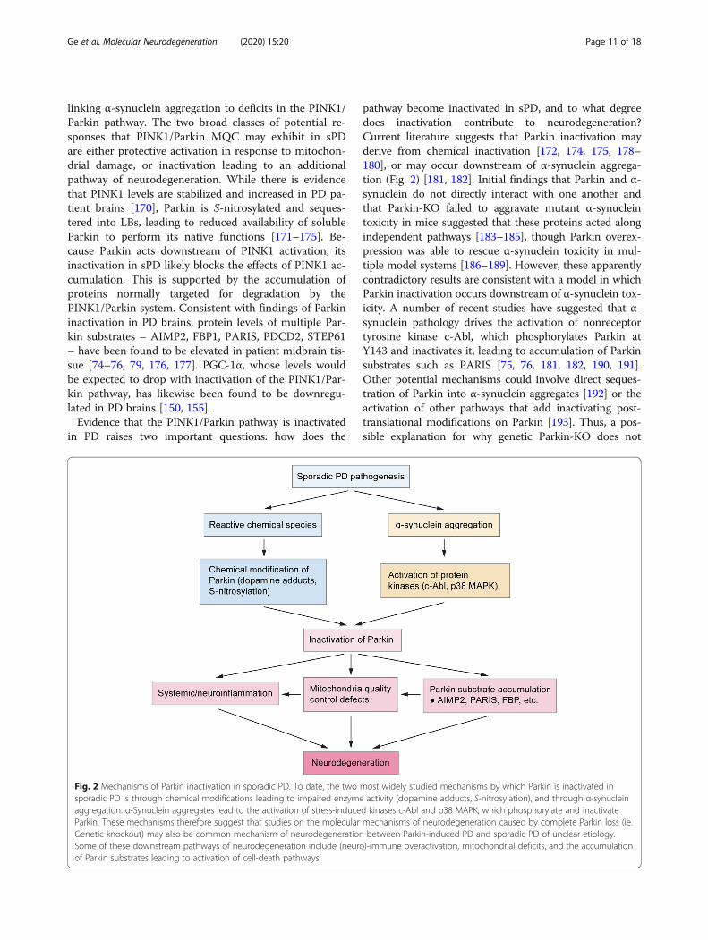
pathway become inactivated in sPD, and to what degreedoes inactivation contribute to neurodegeneration?Current literature suggests that Parkin inactivation mayderive from chemical inactivation [172, 174, 175, 178–180], or may occur downstream of α-synuclein aggrega-tion (Fig. 2) [181, 182]. Initial findings that Parkin and α-synuclein do not directly interact with one another andthat Parkin-KO failed to aggravate mutant α-synucleintoxicity in mice suggested that these proteins acted alongindependent pathways [183–185], though Parkin overex-pression was able to rescue α-synuclein toxicity in mul-tiple model systems [186–189]. However, these apparentlycontradictory results are consistent with a model in whichParkin inactivation occurs downstream of α-synuclein tox-icity. A number of recent studies have suggested that α-synuclein pathology drives the activation of nonreceptortyrosine kinase c-Abl, which phosphorylates Parkin atY143 and inactivates it, leading to accumulation of Parkinsubstrates such as PARIS [75, 76, 181, 182, 190, 191].Other potential mechanisms could involve direct seques-tration of Parkin into α-synuclein aggregates [192] or theactivation of other pathways that add inactivating post-translational modifications on Parkin [193]. Thus, a pos-sible explanation for why genetic Parkin-KO does not
Fig. 2 Mechanisms of Parkin inactivation in sporadic PD. To date, the two most widely studied mechanisms by which Parkin is inactivated insporadic PD is through chemical modifications leading to impaired enzyme activity (dopamine adducts, S-nitrosylation), and through α-synucleinaggregation. α-Synuclein aggregates lead to the activation of stress-induced kinases c-Abl and p38 MAPK, which phosphorylate and inactivateParkin. These mechanisms therefore suggest that studies on the molecular mechanisms of neurodegeneration caused by complete Parkin loss (ie.Genetic knockout) may also be common mechanism of neurodegeneration between Parkin-induced PD and sporadic PD of unclear etiology.Some of these downstream pathways of neurodegeneration include (neuro)-immune overactivation, mitochondrial deficits, and the accumulationof Parkin substrates leading to activation of cell-death pathways
Ge et al. Molecular Neurodegeneration (2020) 15:20 Page 11 of 18
exacerbate α-synuclein toxicity is because it phenocopiesthe Parkin inactivation induced by α-synuclein.An important caveat to many of these studies is the
use of transgenic mice that overexpress mutant α-synuclein, indicating that these mechanisms must be val-idated in a disease model more representative of the hu-man disease. One recent study from our lab attemptedto bridge this gap in knowledge by using the preformedfibrils (PFF) model, a more physiologically accuratemodel system of induced α-synuclein pathology in WTmice. We found that blocking the accumulation of Par-kin substrate PARIS rescued behavioral, molecular, andlifespan deficits as effectively as in transgenic α-synuclein mouse models [181]. Though these findingssuggest that these mechanisms of α-synuclein-inducedinactivation of Parkin are conserved from transgenicmouse models to human PD, more systematic efforts innon-transgenic model systems are needed. Furthermore,while these studies heavily emphasize the role of α-synuclein aggregation in inactivating Parkin (Fig. 2),whether α-synuclein-independent pathways of MQC in-activation such as oxidative damage may also be at playrequires future study. Regardless, these findings impli-cate PINK1/Parkin inactivation not just as a cause of se-lective SNpc degeneration in the small percentage of PDcases associated with monogenic PRKN or PINK1 muta-tions, but also in the sporadic disease driven by α-synuclein aggregation as well.
Mitochondrial dysfunction and neuroinflammationWhile significant work has gone into understandingmitochondrial dysfunction within SNpc neurons in PD,it is less well understood how mitochondrial defects maycontribute to neurodegeneration through non-cell au-tonomous mechanisms. While it is becoming increas-ingly clear that glial dysfunction and neuroinflammationplay an important role in neurodegeneration in PD[194], the degree to which defects in MQC function con-tribute to neuroinflammation is relatively understudied.A few studies suggest that loss of PINK1 or Parkin mayalter glial proliferation, leading to hypersensitized astro-cytes and microglia that have greater levels of basal andtriggered inflammatory cytokine release, nitric oxide(NO) production, and NLRP3 inflammasome activation[26, 195–198]. Co-culturing WT glia with Parkin-KOSNpc DA neurons has been shown to rescue neurondeath and sensitivity to MPP+ observed in pure Parkin-KO co-cultures, suggesting that Parkin deficient gliacontributes to cell death [26].In addition to alterations in the glial inflammatory
profile, compromised PINK1/Parkin MQC may also leadto pathological alterations to the interaction between theCNS and peripheral immune system. For example,PINK1/Parkin may play a role in suppressing
mitochondrial antigen presentation (mitAP) on MHC-Iin macrophages and dendritic cells [140, 145]. Postmor-tem human catecholaminergic neurons as well as cul-tured mouse SNpc neurons can express MHC-Ireceptors, which can be up-regulated over a PINK1-KObackground or in response to infection, inflammatorymediators, oxidative stress, and α-synuclein [145, 199].MitAP caused by loss of PINK1 can lead to brain infil-tration of mitochondrial antigen-specific CD8+ cytotoxicT cells, which then attack SNpc DA neurons [145].Thus, loss of PINK1/Parkin activity could trigger anadaptive immune response against mitochondrial pro-teins and engage the peripheral immune system in animproper assault against the CNS. Furthermore, thesemechanisms may be occurring in a broader milieu ofperipheral immune dysfunction. In macrophages,PINK1/Parkin generate MDVs containing mitochondrialROS that are delivered to bacteria-containing phago-somes [141]. Loss of Parkin impairs bactericidal activityand leads to defective infection clearance, prolonged in-fection course, and elevated cytokine production [141].Furthermore, human subjects with biallelic loss of Parkinshow elevated systemic cytokine levels, with milder in-creases observed in heterozygous subjects [200].These findings suggest that defects in MQC lead to
three interesting effects on immune function that couldcontribute to PD neurodegeneration: an aggravated gliainflammatory phenotype, loss of immune tolerance andpossible autoimmunity against neurons vulnerable inPD, and peripheral immune dysfunction. However, thesemechanisms have largely been studied independently ofone another. We lack an integrated model of how MQCdefects produce (neuro)immune dysfunction and subse-quent neurodegeneration. Furthermore, these mecha-nisms have been demonstrated in the context of globalPINK1/Parkin ablation, whereas MQC defects in sPDmay lead to more CNS-specific and milder immune dys-function. Findings in sporadic PD patients of elevatedsystemic cytokines, CNS immune cell infiltration, and Tcells recognizing α-synuclein peptide do suggest a cer-tain degree of concurrent CNS and peripheral immuneactivation [194, 200, 201], but these studies are inher-ently correlative and give limited insight into the mecha-nisms by which these phenotypes arise. The degree towhich diverse inflammatory mechanisms converge tocause neurodegeneration, and the importance of MQCdefects to these mechanisms in sPD, are important areasof future research.
ConclusionsCell-intrinsic and non-cell autonomous mechanismsEarly studies have long implicated selective vulnerabilityof SNpc DA neurons and mitochondrial dysfunction ascore features of PD. While we now understand that PD
Ge et al. Molecular Neurodegeneration (2020) 15:20 Page 12 of 18
processes are far more distributed across the CNS andmay be driven primarily by prion-like mechanismsspreading α-synuclein aggregates, these non-cell autono-mous mechanisms likely act in concert with cell- andregion-specific factors that lead to selective vulnerabilityto neurodegeneration. Though these cell-intrinsic factorsare likely complex and varied across the different vulner-able subpopulations, in SNpc DA neurons findings overthe last few decades point to the unique mitochondrialchallenges and stresses due to complex cytoarchitectureas a potential major cause. Probing the function ofPINK1/Parkin has led to critical insights into their rolein maintaining mitochondrial integrity and proteostasisin the face of the stressors faced by mitochondria. Theseprotective mechanisms comprise multiple tiers of MQC,such as facilitating mitophagy, regulating fission/fusiondynamics, triggering removal of damaged mitochondrialcomponents through MDV generation, promoting mito-chondrial biogenesis by increasing PGC-1α, and regulat-ing the local translation of mitochondrial genes (Fig. 1),though many of these proposed mechanisms requireconvincing validation in the mammalian CNS. Newerareas of research have begun to establish mechanisms bywhich α-synuclein aggregation causes inactivation ofMQC (Fig. 2), which have clear implications for sPD, aswell as how MQC defects in neurons and non-neuronalcells may contribute to neuroimmune mechanisms ofneurodegeneration.
Critical gaps in understandingDespite the immense progress we have made, criticalgaps in our understanding of PINK1/Parkin MQC re-main. Our understanding of PINK1/Parkin pathways hasbeen built up across a staggering variety of model sys-tems ranging from Drosophila, C. elegans, mice, immor-talized human cell lines, human iPSCs; whether all thesepathways or a specific subset of these pathways is criticalto the survival of human SNpc DA neurons requires fur-ther disambiguation. We have only just begun to eluci-date the organizational principles of these diversemechanisms, and it is likely that subcellular localization,cell-type specific factors, degree of damage, and natureof the damage are important factors governing whichMQC processes become activated and when. For ex-ample, it is likely that MDVs activated in response tofocal damage whereas mitophagy may be required formore severe, global mitochondrial damage. An add-itional possibility is that subcellular localization mayshape dependence on PGC-1α-mediated biogenesis, anucleus-dependent process, versus more spatially re-stricted mechanisms such as localized translation, whichcan operate in compartments far from the nucleus. Es-tablishing the driving principles underlying how PINK1/Parkin juggle these various processes may answer many
of the fundamental questions about cell-type vulnerabil-ity and disease mechanism in sPD.Furthermore, how defects in MQC may interact with
other sources of neuronal vulnerability in PD is anothermajor gap in knowledge. Other populations of neuronsthat selectively degenerate in PD, such as the LC, arerelatively spared in patients with PINK1/PRKN muta-tions [53, 56], indicating that deficiencies in PINK1/Par-kin-mediated MQC are not be the sole determinant ofselective vulnerability. Other proposed causes includeoxidative stress (eg. loss of iron homeostasis), dopaminetoxicity, autonomous pacemaking driving rhythmicCa2+-dependent action potentials, and other vulnerabil-ities in mitochondrial function arising from the size ofaxonal arbors (Reviewed in [4] and [13]). Many of thesemechanisms are clearly interlinked – such as intracellu-lar Ca2+ influx driving mitochondrial Ca2+ uptake lead-ing to increased ATP and ROS production [202, 203] –but where MQC deficits sits in the intertwined networkof vulnerability factors is unclear. Further exploring therelationships within these networks may reveal key hubsthat may prove to be more amenable to disease modify-ing therapeutics.Beyond a neuron-centric view of PD pathogenesis, re-
cent and ongoing studies of non-neuronal MQC defectsand neuroinflammation will bolster our understandingof non-cell autonomous mechanisms of neurodegenera-tion. Finally, further elucidating the mechanistic inter-action of α-synuclein aggregation and PINK1/ParkinMQC inactivation will be critical for establishing the roleof MQC in sPD and synthesizing a more unified under-standing of PD pathogenesis.
Abbreviations(nc-)mtRNA: (nuclear encoded) mitochondrial RNA; CCCP: Carbonyl cyanidem-chlorophenyl hydrazone; CNS: Central nervous system; DA: Dopamine;ETC: Electron transport chain; IMM: Inner mitochondrial membrane; LB: Lewybodies; MDV: Mitochondria-derived vesicle; mitAP: mitochondrial antigenpresentation; MPTP: 1-methyl-4-phenyl-1,2,3,6-tetrahydropyridine;MQC: Mitochondrial quality control; mtDNA: mitochondrial DNA;OMM: Outer mitochondrial membrane; OTC: Ornithine transcarbamylase;PARIS: Parkin Interacting Substrate; PD: PARKINSON’S disease; PFF: Preformedfibrils; RER: Rough endoplasmic reticulum; ROS: Reactive oxygen species;SNpc: Substantia nigra pars compacta; sPD: sporadic Parkinson’s disease;TOM: Translocase of the outer mitochondrial membrane
AcknowledgementsWe thank Audrey H. Effenberger at MIT for discussions and editorialcomments during the preparation of the manuscript. We thank I-Hsun Wufor designing the figures. TMD is the Leonard and Madlyn Abramson Profes-sor in Neurodegenerative Diseases.
Authors’ contributionsPG conducted the literature review, wrote initial draft, and conceived thefigures. All authors contributed to conceiving the outline, reviewing, andediting the manuscript and figures. VLD and TMD approved the manuscriptfor submission. The authors read and approved the final manuscript.
FundingAll authors were supported by grants from the National Institute ofNeurological Disorders and Stroke (NS38377, NS097049, and NS082205) and
Ge et al. Molecular Neurodegeneration (2020) 15:20 Page 13 of 18
the National Institute of Aging (AG059686) JPB Foundation. The authors werealso supported by the Adrienne Helis Malvin Medical Research Foundation andthe Diana Helis Henry Medical Research Foundation through their Parkinson’sDisease Programs (H-1 and M-2014). PG was further supported by a NationalInstitutes of Health Medical Scientist Training Program grant (T32GM007753).
Availability of data and materialsNot applicable.
Ethics approval and consent to participateNot applicable.
Consent for publicationNot applicable.
Competing interestsTMD is a consultant for Mitokinin and owns stock options in the company.TMD and VLD are founders of Valted, LLC and holds an ownership equityinterest in the company. TMD and VLD are founders and hold shares ofstock options as well as equity in, Neuraly, Inc. These arrangements havebeen reviewed and approved by the Johns Hopkins University in accordancewith its conflict of interest policies.
Author details1Neuroregeneration and Stem Cell Programs, Institute for Cell Engineering,Department of Neurology, Department of Physiology, Solomon H. SnyderDepartment of Neuroscience, Department of Pharmacology and MolecularSciences, Johns Hopkins University School of Medicine, 733 North Broadway,Suite 731, Baltimore, MD 21205, USA. 2Adrienne Helis Malvin MedicalResearch Foundation, New Orleans, LA 70130, USA. 3Diana Helis HenryMedical Research Foundation, New Orleans, LA 70130, USA. 4Present address:Department of Brain and Cognitive Sciences, Massachusetts Institute ofTechnology, Cambridge, MA 02139, USA. 5Present address: Picower Institutefor Learning and Memory, Cambridge, MA 02139, USA. 6Present address:Harvard-MIT MD/PhD Program, Harvard Medical School, Boston, MA 02115,USA.
Received: 27 November 2019 Accepted: 13 February 2020
References1. Klein C, Westenberger A. Genetics of Parkinson's disease. Cold Spring Harb
Perspect Med. 2012;2(1):a008888.2. Obeso JA, Stamelou M, Goetz CG, Poewe W, Lang AE, Weintraub D, et al.
Past, present, and future of Parkinson's disease: a special essay on the 200thanniversary of the shaking palsy. Mov Disord. 2017;32(9):1264–310.
3. Braak H, Del Tredici K, Rub U, de Vos RA, Jansen Steur EN, Braak E. Stagingof brain pathology related to sporadic Parkinson's disease. Neurobiol Aging.2003;24(2):197–211.
4. Surmeier DJ, Obeso JA, Halliday GM. Selective neuronal vulnerability inParkinson disease. Nat Rev Neurosci. 2017;18(2):101–13.
5. Li JY, Englund E, Holton JL, Soulet D, Hagell P, Lees AJ, et al. Lewy bodies ingrafted neurons in subjects with Parkinson's disease suggest host-to-graftdisease propagation. Nat Med. 2008;14(5):501–3.
6. Kordower JH, Chu Y, Hauser RA, Freeman TB, Olanow CW. Lewy body-likepathology in long-term embryonic nigral transplants in Parkinson's disease.Nat Med. 2008;14(5):504–6.
7. Volpicelli-Daley LA, Luk KC, Patel TP, Tanik SA, Riddle DM, Stieber A, et al.Exogenous alpha-synuclein fibrils induce Lewy body pathology leading tosynaptic dysfunction and neuron death. Neuron. 2011;72(1):57–71.
8. Luk KC, Kehm V, Carroll J, Zhang B, O'Brien P, Trojanowski JQ, et al.Pathological alpha-synuclein transmission initiates Parkinson-likeneurodegeneration in nontransgenic mice. Science. 2012;338(6109):949–53.
9. Mao X, Ou MT, Karuppagounder SS, Kam TI, Yin X, Xiong Y, et al.Pathological alpha-synuclein transmission initiated by binding lymphocyte-activation gene 3. Science. 2016;353:aah3374.
10. Kim S, Kwon SH, Kam TI, Panicker N, Karuppagounder SS, Lee S, et al.Transneuronal propagation of pathologic alpha-Synuclein from the gut tothe brain models Parkinson's disease. Neuron. 2019;103(4):627–41.e7.
11. Uemura N, Yagi H, Uemura MT, Hatanaka Y, Yamakado H, Takahashi R.Inoculation of alpha-synuclein preformed fibrils into the mouse
gastrointestinal tract induces Lewy body-like aggregates in the brainstemvia the vagus nerve. Mol Neurodegener. 2018;13(1):21.
12. Brundin P, Melki R. Prying into the prion hypothesis for Parkinson's disease.J Neurosci. 2017;37(41):9808–18.
13. Giguere N, Burke Nanni S, Trudeau LE. On cell loss and selectivevulnerability of neuronal populations in Parkinson's disease. Front Neurol.2018;9:455.
14. Bose A, Beal MF. Mitochondrial dysfunction in Parkinson's disease. JNeurochem. 2016;139(Suppl 1):216–31.
15. Misgeld T, Schwarz TL. Mitostasis in neurons: maintaining mitochondria inan extended cellular architecture. Neuron. 2017;96(3):651–66.
16. Grenier K, McLelland GL, Fon EA. Parkin- and PINK1-dependent Mitophagyin neurons: will the real pathway please stand up? Front Neurol. 2013;4:100.
17. Spinelli JB, Haigis MC. The multifaceted contributions of mitochondria tocellular metabolism. Nat Cell Biol. 2018;20(7):745–54.
18. Scarffe LA, Stevens DA, Dawson VL, Dawson TM. Parkin and PINK1: muchmore than mitophagy. Trends Neurosci. 2014;37(6):315–24.
19. Amadoro G, Corsetti V, Florenzano F, Atlante A, Bobba A, Nicolin V, et al.Morphological and bioenergetic demands underlying the mitophagy in post-mitotic neurons: the pink-parkin pathway. Front Aging Neurosci. 2014;6:18.
20. Kann O, Kovacs R. Mitochondria and neuronal activity. Am J Phys Cell Phys.2007;292(2):C641–57.
21. Bolam JP, Pissadaki EK. Living on the edge with too many mouths to feed:why dopamine neurons die. Mov Disord. 2012;27(12):1478–83.
22. Moss J, Bolam JP. A dopaminergic axon lattice in the striatum and itsrelationship with cortical and thalamic terminals. J Neurosci. 2008;28(44):11221–30.
23. Matsuda W, Furuta T, Nakamura KC, Hioki H, Fujiyama F, Arai R, et al. Singlenigrostriatal dopaminergic neurons form widely spread and highly denseaxonal arborizations in the neostriatum. J Neurosci. 2009;29(2):444–53.
24. Anden NE, Hfuxe K, Hamberger B, Hokfelt T. A quantitative study on thenigro-neostriatal dopamine neuron system in the rat. Acta Physiol Scand.1966;67(3):306–12.
25. Pacelli C, Giguere N, Bourque MJ, Levesque M, Slack RS, Trudeau LE.Elevated mitochondrial bioenergetics and axonal Arborization size are keycontributors to the vulnerability of dopamine neurons. Curr Biol. 2015;25(18):2349–60.
26. Giguere N, Pacelli C, Saumure C, Bourque MJ, Matheoud D, Levesque D,et al. Comparative analysis of Parkinson's disease-associated genes in micereveals altered survival and bioenergetics of Parkin-deficient dopamineneurons. J Biol Chem. 2018;293(25):9580–93.
27. Langston JW, Ballard P, Tetrud JW, Irwin I. Chronic parkinsonism in humansdue to a product of meperidine-analog synthesis. Science. 1983;219(4587):979–80.
28. Tanner CM, Kamel F, Ross GW, Hoppin JA, Goldman SM, Korell M, et al.Rotenone, paraquat, and Parkinson's disease. Environ Health Perspect. 2011;119(6):866–72.
29. Dhillon AS, Tarbutton GL, Levin JL, Plotkin GM, Lowry LK, Nalbone JT, et al.Pesticide/environmental exposures and Parkinson's disease in East Texas. JAgromedicine. 2008;13(1):37–48.
30. Dawson TM, Dawson VL. Molecular pathways of neurodegeneration inParkinson's disease. Science. 2003;302(5646):819–22.
31. Schapira AH, Cooper JM, Dexter D, Jenner P, Clark JB, Marsden CD.Mitochondrial complex I deficiency in Parkinson's disease. Lancet. 1989;1(8649):1269.
32. Bindoff LA, Birch-Machin M, Cartlidge NE, Parker WD Jr, Turnbull DM.Mitochondrial function in Parkinson's disease. Lancet. 1989;2(8653):49.
33. Giannoccaro MP, La Morgia C, Rizzo G, Carelli V. Mitochondrial DNA andprimary mitochondrial dysfunction in Parkinson's disease. Mov Disord. 2017;32(3):346–63.
34. Grunewald A, Rygiel KA, Hepplewhite PD, Morris CM, Picard M, Turnbull DM.Mitochondrial DNA depletion in respiratory chain-deficient Parkinsondisease neurons. Ann Neurol. 2016;79(3):366–78.
35. Coxhead J, Kurzawa-Akanbi M, Hussain R, Pyle A, Chinnery P, Hudson G.Somatic mtDNA variation is an important component of Parkinson's disease.Neurobiol Aging. 2016;38:217.e1–6.
36. Dolle C, Flones I, Nido GS, Miletic H, Osuagwu N, Kristoffersen S, et al.Defective mitochondrial DNA homeostasis in the substantia nigra inParkinson disease. Nat Commun. 2016;7:13548.
37. Toulorge D, Schapira AH, Hajj R. Molecular changes in the postmortemparkinsonian brain. J Neurochem. 2016;139(Suppl 1):27–58.
Ge et al. Molecular Neurodegeneration (2020) 15:20 Page 14 of 18
38. Navarro A, Boveris A, Bandez MJ, Sanchez-Pino MJ, Gomez C, Muntane G,et al. Human brain cortex: mitochondrial oxidative damage and adaptiveresponse in Parkinson disease and in dementia with Lewy bodies. FreeRadic Biol Med. 2009;46(12):1574–80.
39. Deng H, Gao K, Jankovic J. The VPS35 gene and Parkinson's disease. MovDisord. 2013;28(5):569–75.
40. Braschi E, Goyon V, Zunino R, Mohanty A, Xu L, McBride HM. Vps35mediates vesicle transport between the mitochondria and peroxisomes.Curr Biol. 2010;20(14):1310–5.
41. Wang W, Wang X, Fujioka H, Hoppel C, Whone AL, Caldwell MA, et al.Parkinson's disease-associated mutant VPS35 causes mitochondrialdysfunction by recycling DLP1 complexes. Nat Med. 2016;22(1):54–63.
42. Tang FL, Liu W, Hu JX, Erion JR, Ye J, Mei L, et al. VPS35 deficiency ormutation causes dopaminergic neuronal loss by impairing mitochondrialfusion and function. Cell Rep. 2015;12(10):1631–43.
43. Verma M, Callio J, Otero PA, Sekler I, Wills ZP, Chu CT. Mitochondrial calciumdysregulation contributes to dendrite degeneration mediated by PD/LBD-associated LRRK2 mutants. J Neurosci. 2017;37(46):11151–65.
44. Howlett EH, Jensen N, Belmonte F, Zafar F, Hu X, Kluss J, et al. LRRK2G2019S-induced mitochondrial DNA damage is LRRK2 kinase dependentand inhibition restores mtDNA integrity in Parkinson's disease. Hum MolGenet. 2017;26(22):4340–51.
45. Ramonet D, Podhajska A, Stafa K, Sonnay S, Trancikova A, Tsika E, et al.PARK9-associated ATP13A2 localizes to intracellular acidic vesicles andregulates cation homeostasis and neuronal integrity. Hum Mol Genet. 2012;21(8):1725–43.
46. Park JS, Koentjoro B, Veivers D, Mackay-Sim A, Sue CM. Parkinson's disease-associated human ATP13A2 (PARK9) deficiency causes zinc dyshomeostasisand mitochondrial dysfunction. Hum Mol Genet. 2014;23(11):2802–15.
47. Park JS, Davis RL, Sue CM. Mitochondrial dysfunction in Parkinson's disease:new mechanistic insights and therapeutic perspectives. Curr NeurolNeurosci Rep. 2018;18(5):21.
48. Kitada T, Asakawa S, Hattori N, Matsumine H, Yamamura Y, Minoshima S,et al. Mutations in the parkin gene cause autosomal recessive juvenileparkinsonism. Nature. 1998;392(6676):605–8.
49. Valente EM, Abou-Sleiman PM, Caputo V, Muqit MM, Harvey K, Gispert S,et al. Hereditary early-onset Parkinson's disease caused by mutations inPINK1. Science. 2004;304(5674):1158–60.
50. Clark IE, Dodson MW, Jiang C, Cao JH, Huh JR, Seol JH, et al. Drosophilapink1 is required for mitochondrial function and interacts genetically withparkin. Nature. 2006;441(7097):1162–6.
51. Park J, Lee SB, Lee S, Kim Y, Song S, Kim S, et al. Mitochondrial dysfunctionin Drosophila PINK1 mutants is complemented by parkin. Nature. 2006;441(7097):1157–61.
52. Yang Y, Gehrke S, Imai Y, Huang Z, Ouyang Y, Wang JW, et al. Mitochondrialpathology and muscle and dopaminergic neuron degeneration caused byinactivation of Drosophila Pink1 is rescued by Parkin. Proc Natl Acad Sci U SA. 2006;103(28):10793–8.
53. Houlden H, Singleton AB. The genetics and neuropathology of Parkinson'sdisease. Acta Neuropathol. 2012;124(3):325–38.
54. Doherty KM, Silveira-Moriyama L, Parkkinen L, Healy DG, Farrell M, MencacciNE, et al. Parkin disease: a clinicopathologic entity? JAMA Neurol. 2013;70(5):571–9.
55. Koros C, Simitsi A, Stefanis L. Genetics of Parkinson's disease: genotype-phenotype correlations. Int Rev Neurobiol. 2017;132:197–231.
56. Schneider SA, Alcalay RN. Neuropathology of genetic synucleinopathieswith parkinsonism: review of the literature. Mov Disord. 2017;32(11):1504–23.
57. Harper JW, Ordureau A, Heo JM. Building and decoding ubiquitin chains formitophagy. Nat Rev Mol Cell Biol. 2018;19(2):93–108.
58. Rub C, Wilkening A, Voos W. Mitochondrial quality control by the Pink1/Parkin system. Cell Tissue Res. 2017;367(1):111–23.
59. Pickrell AM, Youle RJ. The roles of PINK1, parkin, and mitochondrial fidelityin Parkinson's disease. Neuron. 2015;85(2):257–73.
60. Voigt A, Berlemann LA, Winklhofer KF. The mitochondrial kinase PINK1:functions beyond mitophagy. J Neurochem. 2016;139(Suppl 1):232–9.
61. Rasool S, Soya N, Truong L, Croteau N, Lukacs GL, Trempe JF. PINK1autophosphorylation is required for ubiquitin recognition. EMBO Rep. 2018;19:e44891.
62. Trempe JF, Sauve V, Grenier K, Seirafi M, Tang MY, Menade M, et al.Structure of parkin reveals mechanisms for ubiquitin ligase activation.Science. 2013;340(6139):1451–5.
63. Ordureau A, Sarraf SA, Duda DM, Heo JM, Jedrychowski MP, Sviderskiy VO,et al. Quantitative proteomics reveal a feedforward mechanism formitochondrial PARKIN translocation and ubiquitin chain synthesis. Mol Cell.2014;56(3):360–75.
64. Shiba-Fukushima K, Arano T, Matsumoto G, Inoshita T, Yoshida S, IshihamaY, et al. Phosphorylation of mitochondrial polyubiquitin by PINK1 promotesParkin mitochondrial tethering. PLoS Genet. 2014;10(12):e1004861.
65. Walinda E, Morimoto D, Sugase K, Shirakawa M. Dual function ofPhosphoubiquitin in E3 activation of Parkin. J Biol Chem. 2016;291(32):16879–91.
66. Ordureau A, Heo JM, Duda DM, Paulo JA, Olszewski JL, Yanishevski D, et al.Defining roles of PARKIN and ubiquitin phosphorylation by PINK1 inmitochondrial quality control using a ubiquitin replacement strategy. ProcNatl Acad Sci U S A. 2015;112(21):6637–42.
67. Tang MY, Vranas M, Krahn AI, Pundlik S, Trempe JF, Fon EA. Structure-guided mutagenesis reveals a hierarchical mechanism of Parkin activation.Nat Commun. 2017;8:14697.
68. Koyano F, Okatsu K, Kosako H, Tamura Y, Go E, Kimura M, et al. Ubiquitin isphosphorylated by PINK1 to activate parkin. Nature. 2014;510(7503):162–6.
69. Okatsu K, Koyano F, Kimura M, Kosako H, Saeki Y, Tanaka K, et al.Phosphorylated ubiquitin chain is the genuine Parkin receptor. J Cell Biol.2015;209(1):111–28.
70. Pao KC, Stanley M, Han C, Lai YC, Murphy P, Balk K, et al. Probes of ubiquitinE3 ligases enable systematic dissection of parkin activation. Nat Chem Biol.2016;12(5):324–31.
71. Aguirre JD, Dunkerley KM, Mercier P, Shaw GS. Structure of phosphorylatedUBL domain and insights into PINK1-orchestrated parkin activation. ProcNatl Acad Sci U S A. 2017;114(2):298–303.
72. Sarraf SA, Raman M, Guarani-Pereira V, Sowa ME, Huttlin EL, Gygi SP, et al.Landscape of the PARKIN-dependent ubiquitylome in response tomitochondrial depolarization. Nature. 2013;496(7445):372–6.
73. Rose CM, Isasa M, Ordureau A, Prado MA, Beausoleil SA, Jedrychowski MP,et al. Highly multiplexed quantitative mass spectrometry analysis ofUbiquitylomes. Cell Syst. 2016;3(4):395–403.e4.
74. Ko HS, von Coelln R, Sriram SR, Kim SW, Chung KK, Pletnikova O, et al.Accumulation of the authentic parkin substrate aminoacyl-tRNA synthetasecofactor, p38/JTV-1, leads to catecholaminergic cell death. J Neurosci. 2005;25(35):7968–78.
75. Ko HS, Lee Y, Shin JH, Karuppagounder SS, Gadad BS, Koleske AJ, et al.Phosphorylation by the c-Abl protein tyrosine kinase inhibits parkin'subiquitination and protective function. Proc Natl Acad Sci U S A. 2010;107(38):16691–6.
76. Imam SZ, Zhou Q, Yamamoto A, Valente AJ, Ali SF, Bains M, et al. Novelregulation of parkin function through c-Abl-mediated tyrosinephosphorylation: implications for Parkinson's disease. J Neurosci. 2011;31(1):157–63.
77. Lee Y, Karuppagounder SS, Shin JH, Lee YI, Ko HS, Swing D, et al.Parthanatos mediates AIMP2-activated age-dependent dopaminergicneuronal loss. Nat Neurosci. 2013;16(10):1392–400.
78. Wang Y, An R, Umanah GK, Park H, Nambiar K, Eacker SM, et al. A nucleasethat mediates cell death induced by DNA damage and poly(ADP-ribose)polymerase-1. Science. 2016;354:aad6872.
79. Shin JH, Ko HS, Kang H, Lee Y, Lee YI, Pletinkova O, et al. PARIS (ZNF746)repression of PGC-1alpha contributes to neurodegeneration in Parkinson'sdisease. Cell. 2011;144(5):689–702.
80. Stevens DA, Lee Y, Kang HC, Lee BD, Lee YI, Bower A, et al. Parkin loss leadsto PARIS-dependent declines in mitochondrial mass and respiration. ProcNatl Acad Sci U S A. 2015;112(37):11696–701.
81. Siddiqui A, Bhaumik D, Chinta SJ, Rane A, Rajagopalan S, Lieu CA, et al.Mitochondrial quality control via the PGC1alpha-TFEB signaling pathway iscompromised by Parkin Q311X mutation but independently restored byrapamycin. J Neurosci. 2015;35(37):12833–44.
82. Siddiqui A, Rane A, Rajagopalan S, Chinta SJ, Andersen JK. Detrimentaleffects of oxidative losses in parkin activity in a model of sporadicParkinson's disease are attenuated by restoration of PGC1alpha. NeurobiolDis. 2016;93:115–20.
83. Cunningham CN, Baughman JM, Phu L, Tea JS, Yu C, Coons M, et al. USP30and parkin homeostatically regulate atypical ubiquitin chains onmitochondria. Nat Cell Biol. 2015;17(2):160–9.
84. Ordureau A, Paulo JA, Zhang W, Ahfeldt T, Zhang J, Cohn EF, et al.Dynamics of PARKIN-dependent mitochondrial Ubiquitylation in induced
Ge et al. Molecular Neurodegeneration (2020) 15:20 Page 15 of 18
neurons and model systems revealed by digital snapshot proteomics. MolCell. 2018;70(2):211–27.e8.
85. Vives-Bauza C, Zhou C, Huang Y, Cui M, de Vries RL, Kim J, et al. PINK1-dependent recruitment of Parkin to mitochondria in mitophagy. Proc NatlAcad Sci U S A. 2010;107(1):378–83.
86. Narendra D, Tanaka A, Suen DF, Youle RJ. Parkin is recruited selectively toimpaired mitochondria and promotes their autophagy. J Cell Biol. 2008;183(5):795–803.
87. Matsuda N, Sato S, Shiba K, Okatsu K, Saisho K, Gautier CA, et al. PINK1stabilized by mitochondrial depolarization recruits Parkin to damagedmitochondria and activates latent Parkin for mitophagy. J Cell Biol. 2010;189(2):211–21.
88. Geisler S, Holmstrom KM, Skujat D, Fiesel FC, Rothfuss OC, Kahle PJ, et al.PINK1/Parkin-mediated mitophagy is dependent on VDAC1 and p62/SQSTM1. Nat Cell Biol. 2010;12(2):119–31.
89. Narendra DP, Jin SM, Tanaka A, Suen DF, Gautier CA, Shen J, et al. PINK1 isselectively stabilized on impaired mitochondria to activate Parkin. PLoS Biol.2010;8(1):e1000298.
90. Lazarou M, Sliter DA, Kane LA, Sarraf SA, Wang C, Burman JL, et al. Theubiquitin kinase PINK1 recruits autophagy receptors to induce mitophagy.Nature. 2015;524(7565):309–14.
91. Wong YC, Holzbaur EL. Optineurin is an autophagy receptor for damagedmitochondria in parkin-mediated mitophagy that is disrupted by an ALS-linked mutation. Proc Natl Acad Sci U S A. 2014;111(42):E4439–48.
92. Heo JM, Ordureau A, Paulo JA, Rinehart J, Harper JW. The PINK1-PARKINmitochondrial Ubiquitylation pathway drives a program of OPTN/NDP52recruitment and TBK1 activation to promote Mitophagy. Mol Cell. 2015;60(1):7–20.
93. Yamano K, Wang C, Sarraf SA, Munch C, Kikuchi R, Noda NN, et al. EndosomalRab cycles regulate Parkin-mediated mitophagy. Elife. 2018;7:e31326.
94. Palikaras K, Lionaki E, Tavernarakis N. Mechanisms of mitophagy in cellularhomeostasis, physiology and pathology. Nat Cell Biol. 2018;20(9):1013–22.
95. Sung H, Tandarich LC, Nguyen K, Hollenbeck PJ. Compartmentalizedregulation of Parkin-mediated mitochondrial quality control in theDrosophila nervous system in vivo. J Neurosci. 2016;36(28):7375–91.
96. Pickrell AM, Huang CH, Kennedy SR, Ordureau A, Sideris DP, Hoekstra JG,et al. Endogenous Parkin preserves dopaminergic substantia Nigral neuronsfollowing mitochondrial DNA mutagenic stress. Neuron. 2015;87(2):371–81.
97. Van Laar VS, Arnold B, Cassady SJ, Chu CT, Burton EA, Berman SB. Bioenergeticsof neurons inhibit the translocation response of Parkin following rapidmitochondrial depolarization. Hum Mol Genet. 2011;20(5):927–40.
98. Cai Q, Zakaria HM, Simone A, Sheng ZH. Spatial parkin translocation anddegradation of damaged mitochondria via mitophagy in live corticalneurons. Curr Biol. 2012;22(6):545–52.
99. Seibler P, Graziotto J, Jeong H, Simunovic F, Klein C, Krainc D. MitochondrialParkin recruitment is impaired in neurons derived from mutant PINK1induced pluripotent stem cells. J Neurosci. 2011;31(16):5970–6.
100. Ashrafi G, Schlehe JS, LaVoie MJ, Schwarz TL. Mitophagy of damagedmitochondria occurs locally in distal neuronal axons and requires PINK1 andParkin. J Cell Biol. 2014;206(5):655–70.
101. McWilliams TG, Prescott AR, Allen GF, Tamjar J, Munson MJ, Thomson C,et al. Mito-QC illuminates mitophagy and mitochondrial architecture in vivo.J Cell Biol. 2016;214(3):333–45.
102. Sun N, Yun J, Liu J, Malide D, Liu C, Rovira II, et al. Measuring in vivoMitophagy. Mol Cell. 2015;60(4):685–96.
103. Lee JJ, Sanchez-Martinez A, Zarate AM, Beninca C, Mayor U, Clague MJ, et al.Basal mitophagy is widespread in Drosophila but minimally affected by lossof Pink1 or parkin. J Cell Biol. 2018;217(5):1613–22.
104. McWilliams TG, Prescott AR, Montava-Garriga L, Ball G, Singh F, Barini E,et al. Basal Mitophagy occurs independently of PINK1 in mouse tissues ofhigh metabolic demand. Cell Metab. 2018;27(2):439–49.e5.
105. Kim YY, Um JH, Yoon JH, Kim H, Lee DY, Lee YJ, et al. Assessment ofmitophagy in mt-Keima Drosophila revealed an essential role of the PINK1-Parkin pathway in mitophagy induction in vivo. FASEB J. 2019;33(9):9742–51.
106. Cornelissen T, Vilain S, Vints K, Gounko N, Verstreken P, Vandenberghe W.Deficiency of parkin and PINK1 impairs age-dependent mitophagy inDrosophila. Elife. 2018;7:e35878.
107. Vincow ES, Merrihew G, Thomas RE, Shulman NJ, Beyer RP, MacCoss MJ,et al. The PINK1-Parkin pathway promotes both mitophagy and selectiverespiratory chain turnover in vivo. Proc Natl Acad Sci U S A. 2013;110(16):6400–5.
108. Devireddy S, Liu A, Lampe T, Hollenbeck PJ. The Organization ofMitochondrial Quality Control and Life Cycle in the nervous system in vivoin the absence of PINK1. J Neurosci. 2015;35(25):9391–401.
109. Villa E, Marchetti S, Ricci JE. No Parkin zone: Mitophagy without Parkin.Trends Cell Biol. 2018;28(11):882–95.
110. Maday S, Holzbaur EL. Autophagosome biogenesis in primary neuronsfollows an ordered and spatially regulated pathway. Dev Cell. 2014;30(1):71–85.
111. Brown MR, Sullivan PG, Geddes JW. Synaptic mitochondria are moresusceptible to Ca2+overload than nonsynaptic mitochondria. J Biol Chem.2006;281(17):11658–68.
112. Naga KK, Sullivan PG, Geddes JW. High cyclophilin D content of synapticmitochondria results in increased vulnerability to permeability transition. JNeurosci. 2007;27(28):7469–75.
113. Davey GP, Canevari L, Clark JB. Threshold effects in synaptosomal andnonsynaptic mitochondria from hippocampal CA1 and paramedianneocortex brain regions. J Neurochem. 1997;69(6):2564–70.
114. MacVicar TD, Lane JD. Impaired OMA1-dependent cleavage of OPA1 andreduced DRP1 fission activity combine to prevent mitophagy in cells thatare dependent on oxidative phosphorylation. J Cell Sci. 2014;127(Pt 10):2313–25.
115. van der Bliek AM, Shen Q, Kawajiri S. Mechanisms of mitochondrial fissionand fusion. Cold Spring Harb Perspect Biol. 2013;5:a011072.
116. Mishra P, Chan DC. Metabolic regulation of mitochondrial dynamics. J CellBiol. 2016;212(4):379–87.
117. Kageyama Y, Zhang Z, Roda R, Fukaya M, Wakabayashi J, Wakabayashi N,et al. Mitochondrial division ensures the survival of postmitotic neurons bysuppressing oxidative damage. J Cell Biol. 2012;197(4):535–51.
118. Greene JC, Whitworth AJ, Kuo I, Andrews LA, Feany MB, Pallanck LJ.Mitochondrial pathology and apoptotic muscle degeneration in Drosophilaparkin mutants. Proc Natl Acad Sci U S A. 2003;100(7):4078–83.
119. Pesah Y, Pham T, Burgess H, Middlebrooks B, Verstreken P, Zhou Y, et al.Drosophila parkin mutants have decreased mass and cell size and increasedsensitivity to oxygen radical stress. Development. 2004;131(9):2183–94.
120. Poole AC, Thomas RE, Andrews LA, McBride HM, Whitworth AJ, Pallanck LJ.The PINK1/Parkin pathway regulates mitochondrial morphology. Proc NatlAcad Sci U S A. 2008;105(5):1638–43.
121. Deng H, Dodson MW, Huang H, Guo M. The Parkinson's disease genespink1 and parkin promote mitochondrial fission and/or inhibit fusion inDrosophila. Proc Natl Acad Sci U S A. 2008;105(38):14503–8.
122. Park J, Lee G, Chung J. The PINK1-Parkin pathway is involved in theregulation of mitochondrial remodeling process. Biochem Biophys ResCommun. 2009;378(3):518–23.
123. Yang Y, Ouyang Y, Yang L, Beal MF, McQuibban A, Vogel H, et al. Pink1regulates mitochondrial dynamics through interaction with the fission/fusion machinery. Proc Natl Acad Sci U S A. 2008;105(19):7070–5.
124. Poole AC, Thomas RE, Yu S, Vincow ES, Pallanck L. The mitochondrial fusion-promoting factor mitofusin is a substrate of the PINK1/parkin pathway. PLoSOne. 2010;5(4):e10054.
125. Ziviani E, Tao RN, Whitworth AJ. Drosophila parkin requires PINK1 formitochondrial translocation and ubiquitinates mitofusin. Proc Natl Acad SciU S A. 2010;107(11):5018–23.
126. Tanaka A, Cleland MM, Xu S, Narendra DP, Suen DF, Karbowski M, et al.Proteasome and p97 mediate mitophagy and degradation of mitofusinsinduced by Parkin. J Cell Biol. 2010;191(7):1367–80.
127. Gegg ME, Cooper JM, Chau KY, Rojo M, Schapira AH, Taanman JW. Mitofusin 1and mitofusin 2 are ubiquitinated in a PINK1/parkin-dependent manner uponinduction of mitophagy. Hum Mol Genet. 2010;19(24):4861–70.
128. Glauser L, Sonnay S, Stafa K, Moore DJ. Parkin promotes the ubiquitinationand degradation of the mitochondrial fusion factor mitofusin 1. JNeurochem. 2011;118(4):636–45.
129. Yu W, Sun Y, Guo S, Lu B. The PINK1/Parkin pathway regulatesmitochondrial dynamics and function in mammalian hippocampal anddopaminergic neurons. Hum Mol Genet. 2011;20(16):3227–40.
130. Rakovic A, Grunewald A, Kottwitz J, Bruggemann N, Pramstaller PP,Lohmann K, et al. Mutations in PINK1 and Parkin impair ubiquitination ofMitofusins in human fibroblasts. PLoS One. 2011;6(3):e16746.
131. Yamada T, Murata D, Adachi Y, Itoh K, Kameoka S, Igarashi A, et al.Mitochondrial stasis reveals p62-mediated ubiquitination in Parkin-independent Mitophagy and mitigates nonalcoholic fatty liver disease. CellMetab. 2018;28(4):588–604.e5.
Ge et al. Molecular Neurodegeneration (2020) 15:20 Page 16 of 18
132. Buhlman L, Damiano M, Bertolin G, Ferrando-Miguel R, Lombes A, Brice A,et al. Functional interplay between Parkin and Drp1 in mitochondrial fissionand clearance. Biochim Biophys Acta. 2014;1843(9):2012–26.
133. Pryde KR, Smith HL, Chau KY, Schapira AH. PINK1 disables the anti-fissionmachinery to segregate damaged mitochondria for mitophagy. J Cell Biol.2016;213(2):163–71.
134. Burman JL, Pickles S, Wang C, Sekine S, Vargas JNS, Zhang Z, et al.Mitochondrial fission facilitates the selective mitophagy of proteinaggregates. J Cell Biol. 2017;216(10):3231–47.
135. Abeliovich H, Zarei M, Rigbolt KT, Youle RJ, Dengjel J. Involvement ofmitochondrial dynamics in the segregation of mitochondrial matrix proteinsduring stationary phase mitophagy. Nat Commun. 2013;4:2789.
136. Kolitsida P, Zhou J, Rackiewicz M, Nolic V, Dengjel J, Abeliovich H.Phosphorylation of mitochondrial matrix proteins regulates their selectivemitophagic degradation. Proc Natl Acad Sci U S A. 2019;116(41):20517–27.
137. Sugiura A, McLelland GL, Fon EA, McBride HM. A new pathway formitochondrial quality control: mitochondrial-derived vesicles. EMBO J. 2014;33(19):2142–56.
138. Soubannier V, McLelland GL, Zunino R, Braschi E, Rippstein P, Fon EA, et al.A vesicular transport pathway shuttles cargo from mitochondria tolysosomes. Curr Biol. 2012;22(2):135–41.
139. McLelland GL, Soubannier V, Chen CX, McBride HM, Fon EA. Parkin andPINK1 function in a vesicular trafficking pathway regulating mitochondrialquality control. EMBO J. 2014;33(4):282–95.
140. Matheoud D, Sugiura A, Bellemare-Pelletier A, Laplante A, Rondeau C,Chemali M, et al. Parkinson's disease-related proteins PINK1 and Parkinrepress mitochondrial antigen presentation. Cell. 2016;166(2):314–27.
141. Abuaita BH, Schultz TL, O'Riordan MX. Mitochondria-derived vesicles deliverantimicrobial reactive oxygen species to control phagosome-localizedStaphylococcus aureus. Cell Host Microbe. 2018;24(5):625–36.e5.
142. Sugiura A, Mattie S, Prudent J, McBride HM. Newly born peroxisomes are ahybrid of mitochondrial and ER-derived pre-peroxisomes. Nature. 2017;542(7640):251–4.
143. McLelland GL, Lee SA, McBride HM, Fon EA. Syntaxin-17 delivers PINK1/parkin-dependent mitochondrial vesicles to the endolysosomal system. JCell Biol. 2016;214(3):275–91.
144. Soubannier V, Rippstein P, Kaufman BA, Shoubridge EA, McBride HM.Reconstitution of mitochondria derived vesicle formation demonstratesselective enrichment of oxidized cargo. PLoS One. 2012;7(12):e52830.
145. Matheoud D, Cannon T, Voisin A, Penttinen AM, Ramet L, Fahmy AM, et al.Intestinal infection triggers Parkinson's disease-like symptoms in Pink1(−/−)mice. Nature. 2019;571(7766):565–9.
146. Cadete VJ, Deschenes S, Cuillerier A, Brisebois F, Sugiura A, Vincent A, et al.Formation of mitochondrial-derived vesicles is an active and physiologicallyrelevant mitochondrial quality control process in the cardiac system. JPhysiol. 2016;594(18):5343–62.
147. Finck BN, Kelly DP. PGC-1 coactivators: inducible regulators of energymetabolism in health and disease. J Clin Invest. 2006;116(3):615–22.
148. Lee Y, Stevens DA, Kang SU, Jiang H, Lee YI, Ko HS, et al. PINK1 primesParkin-mediated ubiquitination of PARIS in dopaminergic neuronal survival.Cell Rep. 2017;18(4):918–32.
149. Zheng L, Bernard-Marissal N, Moullan N, D'Amico D, Auwerx J, Moore DJ,et al. Parkin functionally interacts with PGC-1alpha to preserve mitochondriaand protect dopaminergic neurons. Hum Mol Genet. 2017;26(3):582–98.
150. Zheng B, Liao Z, Locascio JJ, Lesniak KA, Roderick SS, Watt ML, et al. PGC-1alpha, a potential therapeutic target for early intervention in Parkinson'sdisease. Sci Transl Med. 2010;2(52):52ra73.
151. Ciron C, Zheng L, Bobela W, Knott GW, Leone TC, Kelly DP, et al. PGC-1alpha activity in nigral dopamine neurons determines vulnerability toalpha-synuclein. Acta Neuropathol Commun. 2015;3:16.
152. Clark J, Silvaggi JM, Kiselak T, Zheng K, Clore EL, Dai Y, et al. Pgc-1alphaoverexpression downregulates Pitx3 and increases susceptibility to MPTPtoxicity associated with decreased Bdnf. PLoS One. 2012;7(11):e48925.
153. Ciron C, Lengacher S, Dusonchet J, Aebischer P, Schneider BL. Sustainedexpression of PGC-1alpha in the rat nigrostriatal system selectively impairsdopaminergic function. Hum Mol Genet. 2012;21(8):1861–76.
154. St-Pierre J, Drori S, Uldry M, Silvaggi JM, Rhee J, Jager S, et al. Suppressionof reactive oxygen species and neurodegeneration by the PGC-1transcriptional coactivators. Cell. 2006;127(2):397–408.
155. Eschbach J, von Einem B, Muller K, Bayer H, Scheffold A, Morrison BE, et al.Mutual exacerbation of peroxisome proliferator-activated receptor gamma
coactivator 1alpha deregulation and alpha-synuclein oligomerization. AnnNeurol. 2015;77(1):15–32.
156. Lin J, Wu PH, Tarr PT, Lindenberg KS, St-Pierre J, Zhang CY, et al. Defects inadaptive energy metabolism with CNS-linked hyperactivity in PGC-1alphanull mice. Cell. 2004;119(1):121–35.
157. Leone TC, Lehman JJ, Finck BN, Schaeffer PJ, Wende AR, Boudina S, et al.PGC-1alpha deficiency causes multi-system energy metabolicderangements: muscle dysfunction, abnormal weight control and hepaticsteatosis. PLoS Biol. 2005;3(4):e101.
158. Jiang H, Kang SU, Zhang S, Karuppagounder S, Xu J, Lee YK, et al. AdultConditional Knockout of PGC-1alpha Leads to Loss of Dopamine Neurons.eNeuro. 2016;3:e0183–16.2016.
159. McMeekin LJ, Li Y, Fox SN, Rowe GC, Crossman DK, Day JJ, et al. Cell-specificdeletion of PGC-1alpha from medium spiny neurons causes transcriptionalalterations and age-related motor impairment. J Neurosci. 2018;38(13):3273–86.
160. Lesnik C, Golani-Armon A, Arava Y. Localized translation near themitochondrial outer membrane: An update. RNA Biol. 2015;12(8):801–9.
161. Chacinska A, Koehler CM, Milenkovic D, Lithgow T, Pfanner N. Importingmitochondrial proteins: machineries and mechanisms. Cell. 2009;138(4):628–44.
162. Williams CC, Jan CH, Weissman JS. Targeting and plasticity of mitochondrialproteins revealed by proximity-specific ribosome profiling. Science. 2014;346(6210):748–51.
163. Zhang L, Shimoji M, Thomas B, Moore DJ, Yu SW, Marupudi NI, et al.Mitochondrial localization of the Parkinson's disease related protein DJ-1:implications for pathogenesis. Hum Mol Genet. 2005;14(14):2063–73.
164. Gehrke S, Wu Z, Klinkenberg M, Sun Y, Auburger G, Guo S, et al. PINK1 andParkin control localized translation of respiratory chain component mRNAson mitochondria outer membrane. Cell Metab. 2015;21(1):95–108.
165. Aschrafi A, Kar AN, Gale JR, Elkahloun AG, Vargas JN, Sales N, et al. Aheterogeneous population of nuclear-encoded mitochondrial mRNAs ispresent in the axons of primary sympathetic neurons. Mitochondrion. 2016;30:18–23.
166. Shigeoka T, Jung H, Jung J, Turner-Bridger B, Ohk J, Lin JQ, et al. Dynamicaxonal translation in developing and mature visual circuits. Cell. 2016;166(1):181–92.
167. Martinez A, Lectez B, Ramirez J, Popp O, Sutherland JD, Urbe S, et al.Quantitative proteomic analysis of Parkin substrates in Drosophila neurons.Mol Neurodegener. 2017;12(1):29.
168. Jacoupy M, Hamon-Keromen E, Ordureau A, Erpapazoglou Z, Coge F, CorvolJC, et al. The PINK1 kinase-driven ubiquitin ligase Parkin promotesmitochondrial protein import through the presequence pathway in livingcells. Sci Rep. 2019;9(1):11829.
169. Bertolin G, Jacoupy M, Traver S, Ferrando-Miguel R, Saint Georges T, GrenierK, et al. Parkin maintains mitochondrial levels of the protective Parkinson'sdisease-related enzyme 17-beta hydroxysteroid dehydrogenase type 10. CellDeath Differ. 2015;22(10):1563–76.
170. Muqit MM, Abou-Sleiman PM, Saurin AT, Harvey K, Gandhi S, Deas E, et al.Altered cleavage and localization of PINK1 to aggresomes in the presenceof proteasomal stress. J Neurochem. 2006;98(1):156–69.
171. Schlossmacher MG, Frosch MP, Gai WP, Medina M, Sharma N, Forno L, et al.Parkin localizes to the Lewy bodies of Parkinson disease and dementia withLewy bodies. Am J Pathol. 2002;160(5):1655–67.
172. LaVoie MJ, Ostaszewski BL, Weihofen A, Schlossmacher MG, Selkoe DJ.Dopamine covalently modifies and functionally inactivates parkin. Nat Med.2005;11(11):1214–21.
173. Lonskaya I, Hebron ML, Algarzae NK, Desforges N, Moussa CE. Decreasedparkin solubility is associated with impairment of autophagy in thenigrostriatum of sporadic Parkinson's disease. Neuroscience. 2013;232:90–105.
174. Chung KK, Thomas B, Li X, Pletnikova O, Troncoso JC, Marsh L, et al. S-nitrosylation of parkin regulates ubiquitination and compromises parkin'sprotective function. Science. 2004;304(5675):1328–31.
175. Sunico CR, Nakamura T, Rockenstein E, Mante M, Adame A, Chan SF, et al.S-Nitrosylation of parkin as a novel regulator of p53-mediated neuronal celldeath in sporadic Parkinson's disease. Mol Neurodegener. 2013;8:29.
176. Kurup PK, Xu J, Videira RA, Ononenyi C, Baltazar G, Lombroso PJ, et al.STEP61 is a substrate of the E3 ligase parkin and is upregulated inParkinson's disease. Proc Natl Acad Sci U S A. 2015;112(4):1202–7.
177. Fukae J, Sato S, Shiba K, Sato K, Mori H, Sharp PA, et al. Programmed celldeath-2 isoform1 is ubiquitinated by parkin and increased in the substantianigra of patients with autosomal recessive Parkinson's disease. FEBS Lett.2009;583(3):521–5.
Ge et al. Molecular Neurodegeneration (2020) 15:20 Page 17 of 18
178. Yao D, Gu Z, Nakamura T, Shi ZQ, Ma Y, Gaston B, et al. Nitrosative stresslinked to sporadic Parkinson's disease: S-nitrosylation of parkin regulates itsE3 ubiquitin ligase activity. Proc Natl Acad Sci U S A. 2004;101(29):10810–4.
179. Wong ES, Tan JM, Wang C, Zhang Z, Tay SP, Zaiden N, et al. Relativesensitivity of parkin and other cysteine-containing enzymes to stress-induced solubility alterations. J Biol Chem. 2007;282(16):12310–8.
180. Wang C, Ko HS, Thomas B, Tsang F, Chew KC, Tay SP, et al. Stress-inducedalterations in parkin solubility promote parkin aggregation and compromiseparkin's protective function. Hum Mol Genet. 2005;14(24):3885–97.
181. Brahmachari S, Lee S, Kim S, Yuan C, Karuppagounder SS, Ge P, et al. Parkininteracting substrate zinc finger protein 746 is a pathological mediator inParkinson's disease. Brain. 2019;142(8):2380–401.
182. Brahmachari S, Karuppagounder SS, Ge P, Lee S, Dawson VL, Dawson TM,et al. C-Abl and Parkinson's disease: mechanisms and therapeutic potential.J Park Dis. 2017;7(4):589–601.
183. Chung KK, Zhang Y, Lim KL, Tanaka Y, Huang H, Gao J, et al. Parkinubiquitinates the alpha-synuclein-interacting protein, synphilin-1:implications for Lewy-body formation in Parkinson disease. Nat Med. 2001;7(10):1144–50.
184. Liani E, Eyal A, Avraham E, Shemer R, Szargel R, Berg D, et al. Ubiquitylationof synphilin-1 and alpha-synuclein by SIAH and its presence in cellularinclusions and Lewy bodies imply a role in Parkinson's disease. Proc NatlAcad Sci U S A. 2004;101(15):5500–5.
185. von Coelln R, Thomas B, Andrabi SA, Lim KL, Savitt JM, Saffary R, et al.Inclusion body formation and neurodegeneration are parkin independent ina mouse model of alpha-synucleinopathy. J Neurosci. 2006;26(14):3685–96.
186. Lo Bianco C, Schneider BL, Bauer M, Sajadi A, Brice A, Iwatsubo T, et al.Lentiviral vector delivery of parkin prevents dopaminergic degeneration inan alpha-synuclein rat model of Parkinson's disease. Proc Natl Acad Sci U SA. 2004;101(50):17510–5.
187. Petrucelli L, O'Farrell C, Lockhart PJ, Baptista M, Kehoe K, Vink L, et al. Parkinprotects against the toxicity associated with mutant alpha-synuclein:proteasome dysfunction selectively affects catecholaminergic neurons.Neuron. 2002;36(6):1007–19.
188. Haywood AF, Staveley BE. Parkin counteracts symptoms in a Drosophilamodel of Parkinson's disease. BMC Neurosci. 2004;5:14.
189. Yang Y, Nishimura I, Imai Y, Takahashi R, Lu B. Parkin suppressesdopaminergic neuron-selective neurotoxicity induced by Pael-R inDrosophila. Neuron. 2003;37(6):911–24.
190. Mahul-Mellier AL, Fauvet B, Gysbers A, Dikiy I, Oueslati A, Georgeon S, et al.C-Abl phosphorylates alpha-synuclein and regulates its degradation:implication for alpha-synuclein clearance and contribution to thepathogenesis of Parkinson's disease. Hum Mol Genet. 2014;23(11):2858–79.
191. Brahmachari S, Ge P, Lee SH, Kim D, Karuppagounder SS, Kumar M, et al.Activation of tyrosine kinase c-Abl contributes to alpha-synuclein-inducedneurodegeneration. J Clin Invest. 2016;126(8):2970–88.
192. Kawahara K, Hashimoto M, Bar-On P, Ho GJ, Crews L, Mizuno H, et al. Alpha-Synuclein aggregates interfere with Parkin solubility and distribution: role inthe pathogenesis of Parkinson disease. J Biol Chem. 2008;283(11):6979–87.
193. Chen J, Ren Y, Gui C, Zhao M, Wu X, Mao K, et al. Phosphorylation of Parkinat serine 131 by p38 MAPK promotes mitochondrial dysfunction andneuronal death in mutant A53T alpha-synuclein model of Parkinson'sdisease. Cell Death Dis. 2018;9(6):700.
194. Fuzzati-Armentero MT, Cerri S, Blandini F. Peripheral-central Neuroimmunecrosstalk in Parkinson's disease: what do patients and animal models tell us?Front Neurol. 2019;10:232.
195. Singh K, Han K, Tilve S, Wu K, Geller HM, Sack MN. Parkin targets NOD2 toregulate astrocyte endoplasmic reticulum stress and inflammation. Glia.2018;66(11):2427–37.
196. Mouton-Liger F, Rosazza T, Sepulveda-Diaz J, Ieang A, Hassoun SM, Claire E,et al. Parkin deficiency modulates NLRP3 inflammasome activation byattenuating an A20-dependent negative feedback loop. Glia. 2018;66(8):1736–51.
197. Sun L, Shen R, Agnihotri SK, Chen Y, Huang Z, Bueler H. Lack of PINK1 altersglia innate immune responses and enhances inflammation-induced, nitricoxide-mediated neuron death. Sci Rep. 2018;8(1):383.
198. Solano RM, Casarejos MJ, Menendez-Cuervo J, Rodriguez-Navarro JA. Garciade Yebenes J, Mena MA. Glial dysfunction in parkin null mice: effects ofaging. J Neurosci. 2008;28(3):598–611.
199. Cebrian C, Zucca FA, Mauri P, Steinbeck JA, Studer L, Scherzer CR, et al.MHC-I expression renders catecholaminergic neurons susceptible to T-cell-mediated degeneration. Nat Commun. 2014;5:3633.
200. Sliter DA, Martinez J, Hao L, Chen X, Sun N, Fischer TD, et al. Parkin andPINK1 mitigate STING-induced inflammation. Nature. 2018;561(7722):258–62.
201. Sulzer D, Alcalay RN, Garretti F, Cote L, Kanter E, Agin-Liebes J, et al. T cellsfrom patients with Parkinson's disease recognize alpha-synuclein peptides.Nature. 2017;546(7660):656–61.
202. Guzman JN, Sanchez-Padilla J, Wokosin D, Kondapalli J, Ilijic E, SchumackerPT, et al. Oxidant stress evoked by pacemaking in dopaminergic neurons isattenuated by DJ-1. Nature. 2010;468(7324):696–700.
203. Camello-Almaraz C, Gomez-Pinilla PJ, Pozo MJ, Camello PJ. Mitochondrialreactive oxygen species and Ca2+ signaling. Am J Phys Cell Phys. 2006;291(5):C1082–8.
Publisher’s NoteSpringer Nature remains neutral with regard to jurisdictional claims inpublished maps and institutional affiliations.
Ge et al. Molecular Neurodegeneration (2020) 15:20 Page 18 of 18


![PINK1 import regulation; a fine system to convey ......mitochondria, and the E3 ligase activity of Parkin is acti-vated by binding to phospho-ubiquitin [20, 21]. PINK1 also phosphorylates](https://img.pdfslide.us/doc/110x75/60ff3ba3c386cc67f77a5536/pink1-import-regulation-a-fine-system-to-convey-mitochondria-and-the-e3.jpg)


![Multitasking guardian of mitochondrial quality: Parkin …...the autoubiquitination activity of Parkin [41]. Involvement of Parkin in mitochondrial processes As an E3 ligase, Parkin](https://img.pdfslide.us/doc/110x75/60ff3ba3c386cc67f77a5535/multitasking-guardian-of-mitochondrial-quality-parkin-the-autoubiquitination.jpg)