Embed Size (px)
Citation preview
ARTICLE
PINK1-mediated phosphorylation of LETM1regulates mitochondrial calcium transport andprotects neurons against mitochondrial stressEn Huang1, Dianbo Qu1, Tianwen Huang1, Nicoletta Rizzi2, Wassamon Boonying1, Dorothy Krolak1, Paolo Ciana3,
John Woulfe4, Christine Klein5, Ruth S. Slack1, Daniel Figeys6 & David S. Park 1
Mutations in PTEN-induced kinase 1 (PINK1) result in a recessive familial form of Parkinson’s
disease (PD). PINK1 loss is associated with mitochondrial Ca2+ mishandling, mitochondrial
dysfunction, as well as increased neuronal vulnerability. Here we demonstrate that PINK1
directly interacts with and phosphorylates LETM1 at Thr192 in vitro. Phosphorylated LETM1
or the phospho-mimetic LETM1-T192E increase calcium release in artificial liposomes and
facilitates calcium transport in intact mitochondria. Expression of LETM1-T192E but not
LETM1-wild type (WT) rescues mitochondrial calcium mishandling in PINK1-deficient neu-
rons. Expression of both LETM1-WT and LETM1-T192E protects neurons against
MPP+–MPTP-induced neuronal death in PINK1 WT neurons, whereas only LETM1-T192E
protects neurons under conditions of PINK1 loss. Our findings delineate a mechanism by
which PINK1 regulates mitochondrial Ca2+ level through LETM1 and suggest a model by
which PINK1 loss leads to deficient phosphorylation of LETM1 and impaired mitochondrial
Ca2+ transport..
DOI: 10.1038/s41467-017-01435-1 OPEN
1 University of Ottawa Brain and Mind Research Institute, Department of Cellular and Molecular Medicine, University of Ottawa, Ottawa, ON, Canada K1H8M5. 2 Center of Excellence on Neurodegenerative Diseases, Department of Pharmacological Sciences, University of Milan, Via Balzaretti 9, 20133 Milan,Italy. 3 Center of Excellence on Neurodegenerative Diseases, Department of Oncology and Hemato-Oncology, University of Milan, Via Balzaretti 9, 20133Milan, Italy. 4 Centre for Cancer Therapeutics, Ottawa Hospital Research Institute and Department of Pathology and Laboratory Medicine, University ofOttawa, Ottawa, ON, Canada K1H 8M5. 5 Institute of Neurogenetics, University of Lübeck, Ratzeburger Allee 160, 23538 Lübeck, Germany. 6 Department ofBiochemistry, Microbiology and Immunology, Department of Chemistry and Biomolecular Sciences, and Ottawa Institute of Systems Biology, University ofOttawa, Ottawa, ON, Canada K1H 8M5. Correspondence and requests for materials should be addressed to D.S.P. (email: [email protected])
NATURE COMMUNICATIONS |8: 1399 |DOI: 10.1038/s41467-017-01435-1 |www.nature.com/naturecommunications 1
1234
5678
90
PTEN-induced kinase 1 (PINK1) is a mitochondria targetedserine/threonine kinase. Mutations in PINK1 can causerecessive familial Parkinson’s disease (PD)1, 2. Under basal
conditions, PINK1 is imported into the inner mitochondrialmembrane, where it is processed by mitochondrial proteases3–5.However, upon mitochondrial membrane depolarization ordamage, PINK1 accumulates on the outer membrane of mito-chondria, where it recruits Parkin to trigger a mitophagic path-way of quality control6. Whether PINK1 exerts a biologicalfunction endogenously at the inner mitochondrial membrane isunknown. Previous evidence demonstrates that PINK1 lossresults in mitochondrial dysfunction, reduced complex I activity,increased oxidative damage7, 8, mitochondrial Ca2+ ([Ca2+]m)mishandling and accumulation9–11, and increase in mPTPopening11, 12. One interpretation is that these changes may renderdopaminergic (DA) neurons more vulnerable to stress andthereby contribute to the pathogenesis of PD13, 14.
To better define the mechanism(s) by which PINK1 mayfunction at the mitochondria, we performed a mass spectrometry-based interactomics screen for potential PINK1-interacting pro-teins15, 16. One identified target is the leucine zipper-EF-handcontaining transmembrane protein 1(LETM1), which wasrecently proposed as a Ca2+/H+ antiporter by a genome-wideRNAi screen17. LETM1 is a mitochondrial inner membraneprotein18 and several reports suggest that it mediates mitochon-drial Ca2+ uptake and extrusion in a gradient-dependent man-ner17, 19, 20. In this regard, knockdown of LETM1 compromisesthe rate of mitochondrial Ca2+ uptake and extrusion, leading toan alteration of mitochondrial bioenergetics, metabolic signaling,and sensitization to cell death19–21. However, others have sug-gested that LETM1 plays an essential role in mitochondrial K+
homeostasis by mediating the mitochondrial K+/H+ exchange22.Our study indicates that LETM1 is a substrate of PINK1 and
that LETM1 plays a critical role in protecting neurons fromexogenous stress. Importantly, we also provide evidence thatPINK1-mediated phosphorylation of LETM1 is crucial for itsreported capacity for mitochondrial Ca2+ regulation and neuronalsurvival effects. Taken together, our data provide a compellingmodel by which PINK1 at the inner mitochondrial membraneperforms a critical function of regulating LETM1 function.
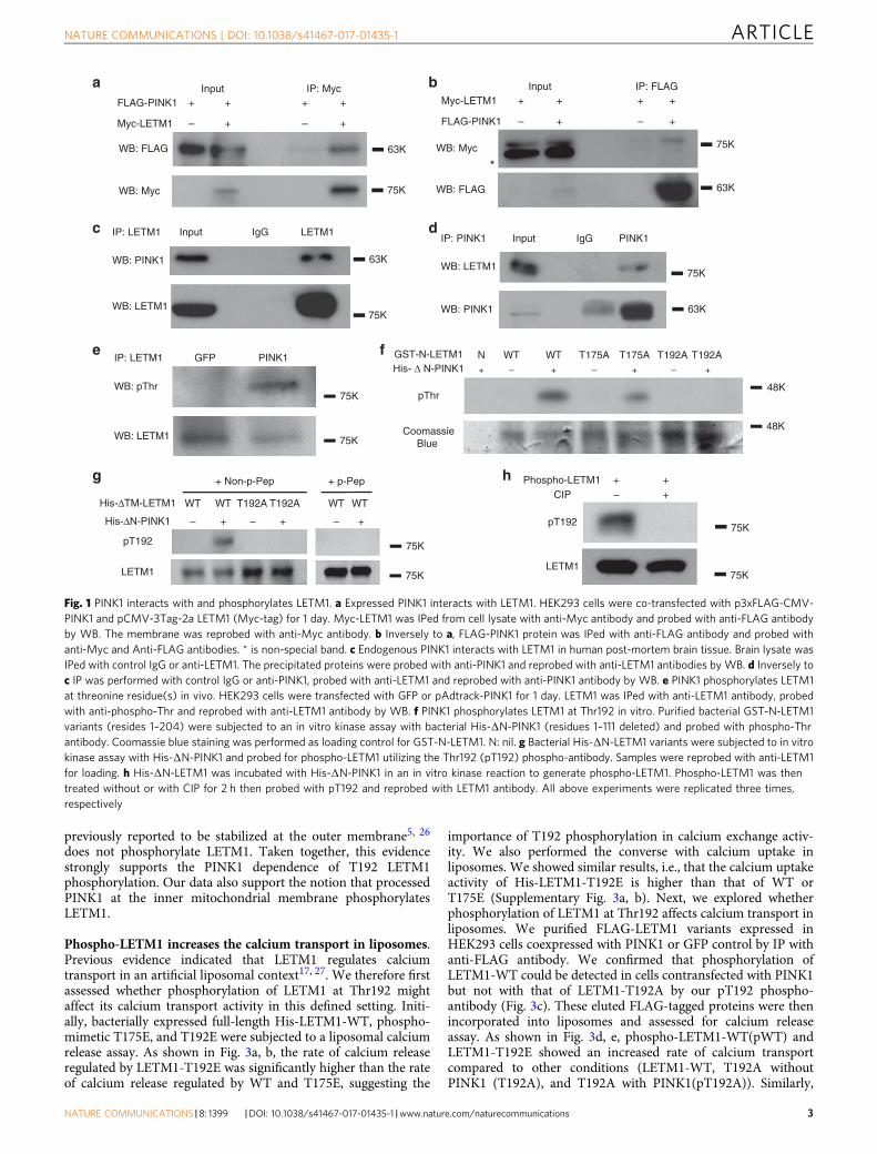
ResultsPINK1 phosphorylates LETM1 at Thr192. We initially identi-fied LETM1 as one of the PINK1-interacting candidates throughan unbiased interaction screen. Briefly, PINK1-FLAG wasexpressed in HEK293 cells and immunoprecipitated. Interactingproteins were determined by mass spectrometry-based inter-actomics as previously reported15, 16. Analysis of the data setsuggested LETM1 as one of the PINK1-interacting candidateswith a Mascot score of 85. To validate this initial finding, weperformed co-immunoprecipitation (Co-IP) analyses withexpression of FLAG-tagged PINK1 and Myc-tagged LETM1 inHEK293 Cells (ATCC). As shown in Fig. 1a, Co-IP for Myc-LETM1 with anti-Myc antibody followed by Western blot (WB)analyses for FLAG-PINK1 with anti-FLAG antibody demon-strated a specific interaction. The reverse Co-IP also supportedthe interaction (Fig. 1b). We further confirmed the interaction ofendogenous PINK1 with LETM1 from human post-mortem brain(Fig. 1c, d) and human SH-SY5Y neuroblastoma cells (Supple-mentary Fig. 1a, b). We next examined whether LETM1 may be akinase substrate of PINK1. To test this, endogenous LETM1 fromHEK293 cells expressing GFP or PINK1 was IPed with an anti-LETM1 antibody and probed with phospho-Thr and phospho-Ser antibodies by WB analyses. The results suggested that LETM1was phosphorylated at threonine residues (Fig. 1e) but not serine
(Supplementary Fig. 1c) in cells expressing PINK1 but not GFP.PINK1 distributes in the outer membrane, inner membrane, andintermembrane space of mitochondria but not in the matrix ofmitochondria4, 23, 24. When localized at the inner mitochondrialmembrane, its kinase domain likely faces the intermembranespace4. LETM1 is also a mitochondrial inner membrane proteinwith a single transmembrane domain. Its N-terminus is exposedto the intermembrane space, whereas its C-terminus is located inthe matrix18, 21. Therefore, we hypothesized that PINK1 mightphosphorylate the N-terminal region of LETM1 (residue 1–204).We searched for potential Thr phosphorylation sites of LETM1 inthis region and identified two potential conserved motifs, Thr175and Thr192, which were conserved in multiple species (Supple-mentary Fig. 2). Of these two sites, Thr192 was conserved in allseven species examined. We constructed and isolated bacteriallyexpressed GST tagged N-terminus of LETM1-WT (residue 1–204,GST-N-LETM1) and mutations with the T175 or T192 sitemutated singly to alanine. These constructs were analyzed by anin vitro kinase assay with His-tagged PINK1 with the N-terminustruncated to promote stability and kinase activity (deleted resi-dues 1–111, His-ΔN-PINK1). The results showed that PINK1phosphorylated the WT and T175A but not T192A fragments,indicating that Thr192 of LETM1 is an appropriate candidatephosphorylation site for PINK1 (Fig. 1f). For further validation,we generated a custom phospho-antibody specific for phos-phorylated LETM1 at Thr192 (pT192). We generated His-taggedLETM1 with the transmembrane portion deleted (His-ΔTM-LETM1) to enhance recovery of bacterially generated LETM1.The pT192 antibody specifically recognized LETM1-WT phos-phorylated by PINK1 but not unphosphorylated LETM1-WT orthe T192A mutant incubated with PINK1 (Fig. 1g). Importantly,the pT192 phospho-antibody generated signal was blocked bypre-incubation with the corresponding phosphopeptide (p-pep-tide) antigen (CNGHTLpT192RRERR) (Fig. 1g, right) but notwith the homologous non-p-peptide (CNGHTLTRRERR)(Fig. 1g, left). In addition, kinase-dead mutant K219M did notphosphorylate LETM1 (Supplementary Fig. 1d). Calf-intestinalalkaline phosphatase (CIP) treatment of phospho-LETM1 elimi-nated the phosphorylation signal (Fig. 1h). These data supportthe specificity of our generated pT192 antibody.
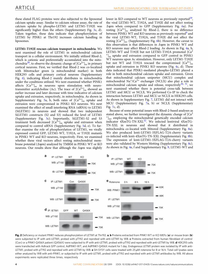
Phospho-LETM1 is decreased in PINK1 KO and Q456Xmutant. To further confirm whether phosphorylation of LETM1at Thr192 is dependent on endogenous PINK1, we utilized ourpT192 phospho-antibody to detect phospho-LETM1 in PINK1WT and knockout (KO) mouse embryo fibroblasts (Fig. 2a) andmouse brain tissues (Fig. 2b). Our results clearly showed thatphosphorylation of LETM1 at Thr192 occurs in a PINK1-dependent manner. Similarly, we analyzed for pT192 signal infibroblasts obtained from a PD patient expressing PINK1-Q456X,a nonsense mutant identified from PD patients with a partialdeletion of the kinase domain resulting in a reduction in its kinaseactivity. LETM1 phosphorylation was dramatically reduced whencompared to normal control individual (Fig. 2c). Conversely,expression of PINK1-Q456X in HEK293 cells also demonstratedreduced phosphorylation of LETM1 at Thr192 when compared toexpression of PINK1 WT (Fig. 2d). Finally, PINK1 is stabilized atthe outer mitochondrial membrane upon mitochondrial stress,topographically eliminating its ability to phosphorylate LETM1.We therefore tested whether mitochondrial damage induced byrotenone would reduce the pT192 LETM1 signal. Rotenonetreatment stabilized full-length PINK1 as previously reported25.Importantly, this was associated with the absence of T192phosphorylation (Fig. 2e). The absence of the T192 signal sug-gests that full-length PINK1 induced by mitochondrial stress
ARTICLE NATURE COMMUNICATIONS | DOI: 10.1038/s41467-017-01435-1
2 NATURE COMMUNICATIONS |8: 1399 |DOI: 10.1038/s41467-017-01435-1 |www.nature.com/naturecommunications
previously reported to be stabilized at the outer membrane5, 26
does not phosphorylate LETM1. Taken together, this evidencestrongly supports the PINK1 dependence of T192 LETM1phosphorylation. Our data also support the notion that processedPINK1 at the inner mitochondrial membrane phosphorylatesLETM1.
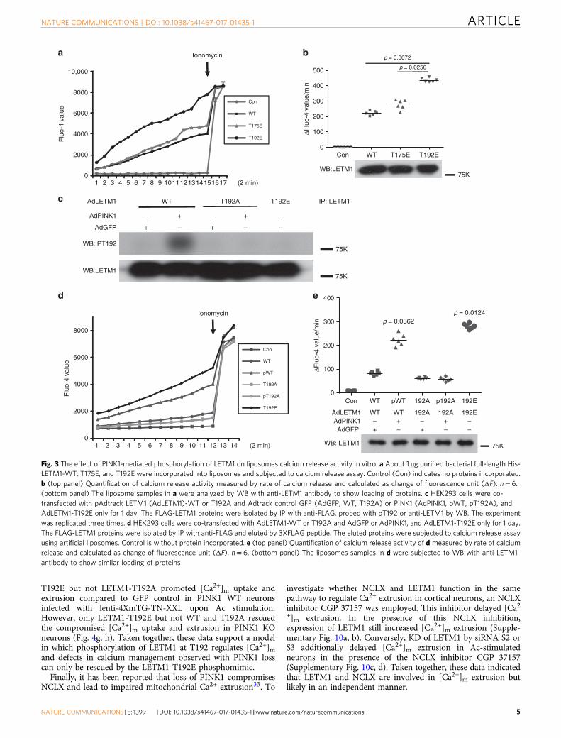
Phospho-LETM1 increases the calcium transport in liposomes.Previous evidence indicated that LETM1 regulates calciumtransport in an artificial liposomal context17, 27. We therefore firstassessed whether phosphorylation of LETM1 at Thr192 mightaffect its calcium transport activity in this defined setting. Initi-ally, bacterially expressed full-length His-LETM1-WT, phospho-mimetic T175E, and T192E were subjected to a liposomal calciumrelease assay. As shown in Fig. 3a, b, the rate of calcium releaseregulated by LETM1-T192E was significantly higher than the rateof calcium release regulated by WT and T175E, suggesting the
importance of T192 phosphorylation in calcium exchange activ-ity. We also performed the converse with calcium uptake inliposomes. We showed similar results, i.e., that the calcium uptakeactivity of His-LETM1-T192E is higher than that of WT orT175E (Supplementary Fig. 3a, b). Next, we explored whetherphosphorylation of LETM1 at Thr192 affects calcium transport inliposomes. We purified FLAG-LETM1 variants expressed inHEK293 cells coexpressed with PINK1 or GFP control by IP withanti-FLAG antibody. We confirmed that phosphorylation ofLETM1-WT could be detected in cells contransfected with PINK1but not with that of LETM1-T192A by our pT192 phospho-antibody (Fig. 3c). These eluted FLAG-tagged proteins were thenincorporated into liposomes and assessed for calcium releaseassay. As shown in Fig. 3d, e, phospho-LETM1-WT(pWT) andLETM1-T192E showed an increased rate of calcium transportcompared to other conditions (LETM1-WT, T192A withoutPINK1 (T192A), and T192A with PINK1(pT192A)). Similarly,
a Input IP: FLAGb
IP: PINK1 Input IgG PINK1
WB: LETM1
WB: PINK1 63K
75K
d
IP: LETM1 GFP PINK1
WB: pThr
WB: LETM1
e f GST-N-LETM1 N WT WT T175A T175A T192A T192AHis- Δ N-PINK1 + – + – + – +
pThr
CoomassieBlue
WT T192AWT T192A WT WT
His-ΔN-PINK1 – + – + – +
pT192
LETM1
+ Non-p-Pep + p-Pep
His-ΔTM-LETM1
g
IP: LETM1 Input IgG LETM1
WB: PINK1
WB: LETM1
63K
75K
Input IP: Myc
c
63K
75K
WB: FLAG
WB: Myc
Myc-LETM1 – + – +
FLAG-PINK1 + + + +
75K
63K
WB: Myc
WB: FLAG
FLAG-PINK1 – + – +
Myc-LETM1 + + + +
h Phospho-LETM1 + +CIP – +
pT192
LETM1
75K
75K
75K
75K
75K
75K
48K
48K
*
Fig. 1 PINK1 interacts with and phosphorylates LETM1. a Expressed PINK1 interacts with LETM1. HEK293 cells were co-transfected with p3xFLAG-CMV-PINK1 and pCMV-3Tag-2a LETM1 (Myc-tag) for 1 day. Myc-LETM1 was IPed from cell lysate with anti-Myc antibody and probed with anti-FLAG antibodyby WB. The membrane was reprobed with anti-Myc antibody. b Inversely to a, FLAG-PINK1 protein was IPed with anti-FLAG antibody and probed withanti-Myc and Anti-FLAG antibodies. * is non-special band. c Endogenous PINK1 interacts with LETM1 in human post-mortem brain tissue. Brain lysate wasIPed with control IgG or anti-LETM1. The precipitated proteins were probed with anti-PINK1 and reprobed with anti-LETM1 antibodies by WB. d Inversely toc IP was performed with control IgG or anti-PINK1, probed with anti-LETM1 and reprobed with anti-PINK1 antibody by WB. e PINK1 phosphorylates LETM1at threonine residue(s) in vivo. HEK293 cells were transfected with GFP or pAdtrack-PINK1 for 1 day. LETM1 was IPed with anti-LETM1 antibody, probedwith anti-phospho-Thr and reprobed with anti-LETM1 antibody by WB. f PINK1 phosphorylates LETM1 at Thr192 in vitro. Purified bacterial GST-N-LETM1variants (resides 1–204) were subjected to an in vitro kinase assay with bacterial His-ΔN-PINK1 (residues 1–111 deleted) and probed with phospho-Thrantibody. Coomassie blue staining was performed as loading control for GST-N-LETM1. N: nil. g Bacterial His-ΔN-LETM1 variants were subjected to in vitrokinase assay with His-ΔN-PINK1 and probed for phospho-LETM1 utilizing the Thr192 (pT192) phospho-antibody. Samples were reprobed with anti-LETM1for loading. h His-ΔN-LETM1 was incubated with His-ΔN-PINK1 in an in vitro kinase reaction to generate phospho-LETM1. Phospho-LETM1 was thentreated without or with CIP for 2 h then probed with pT192 and reprobed with LETM1 antibody. All above experiments were replicated three times,respectively
NATURE COMMUNICATIONS | DOI: 10.1038/s41467-017-01435-1 ARTICLE
NATURE COMMUNICATIONS |8: 1399 |DOI: 10.1038/s41467-017-01435-1 |www.nature.com/naturecommunications 3
these eluted FLAG proteins were also subjected to the liposomalcalcium uptake assay. Similar to calcium release assay, the rate ofcalcium uptake by phospho-LETM1 and LETM1-T192E wassignificantly higher than the others (Supplementary Fig. 3c, d).Taken together, these data indicate that phosphorylation ofLETM1 by PINK1 at Thr192 increases calcium handling inliposomes.
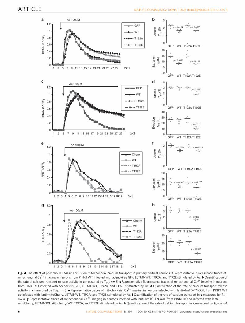
LETM1-T192E rescues calcium transport in mitochondria. Wenext examined the role of LETM1 in mitochondrial calciumtransport in a cellular environment. We initially utilized Rhod-2,which is cationic and preferentially accumulated into the mito-chondria28, to observe the dynamic change of [Ca2+]m in primarycortical neurons. First, we verified that Rhod-2 was co-localizedwith Mitotracker green (a mitochondrial marker) in bothHEK293 cells and primary cortical neurons (SupplementaryFig. 4), indicating Rhod-2 mainly distributes in mitochondriaunder the conditions utilized. We next examined whether PINK1affects [Ca2+]m in neurons upon stimulation with neuro-transmitter acetylcholine (Ac). The trace of [Ca2+]m showed anearlier increase and later decrease with time indicative of calciumuptake and extrusion, respectively, in mitochondria. As shown inSupplementary Fig. 5a, b, both rates of [Ca2+]m uptake andextrusion were compromised in PINK1 KO neurons. We nextexamined the effect of small interfering RNA (siRNA) to LETM1(SiLETM1) in neurons and showed that two independentSiLETM1 constructs (S2 and S3) reduced the level of LETM1(Supplementary Fig. 5c). Importantly, SiLETM1-S2 and S3treatment both decreased [Ca2+]m uptake and extrusion whencompared to control siRNA (Supplementary Fig. 5d, e). To fur-ther examine the role of phosphorylation of LETM1, we virallyexpressed control GFP, LETM1-WT, T192A, or T192E mutantsin PINK1 WT and KO neurons, respectively. First, we examinedwhether these viral vectors affect resting mitochondrial mem-brane potential (Δψm) analyzed by TMRM in PINK1 WT or KOneurons. Our results show that although the Δψm was slightly
lower in KO compared to WT neurons as previously reported29,the viral LETM1-WT, T192A, and T192E did not affect restingΔψm when compared to GFP (Supplementary Fig. 6a). Forresting [Ca2+]m analyzed by Rhod-2, there is no differencebetween PINK1 WT and KO neurons as previously reported9 andthe viral LETM1-WT, T192A, and T192E did not affect theresting [Ca2+]m. (Supplementary Fig. 6b). However, the caveat tothis observation is that differences in Δψm in PINK1 WT andKO neurons may affect Rhod-2 loading. As shown in Fig. 4a, b,LETM1-WT and T192E but not LETM1-T192A promoted [Ca2+]m uptake and extrusion compared to GFP control in PINK1WT neurons upon Ac stimulation. However, only LETM1-T192Ebut not WT and T192A rescued the compromised [Ca2+]muptake and extrusion in PINK1 KO neurons (Fig. 4c, d). Thesedata indicated that PINK1-mediated phospho-LETM1 played arole in both mitochondrial calcium uptake and extrusion. Giventhat mitochondrial calcium uniporter (MCU) complex andmitochondrial Na+/Ca2+ exchanger (NCLX) also play a role inmitochondrial calcium uptake and release, respectively30, 31, wenext examined whether there is potential cross-talk betweenLETM1 and MCU or NCLX. We performed Co-IP to check theinteraction between LETM1 and MCU or NCLX in HEK293 cells.As shown in Supplementary Fig. 7, LETM1 did not interact withMCU (Supplementary Fig. 7a, b) or NCLX (SupplementaryFig. 7c, d).
Because of some potential issues with Rhod-2-based analyses asstated above, we further investigated the dynamic change of [Ca2+]m, employing the mitochondrial genetically encoded calciumindicator 4XmTG-TN-XXL32. We infected lentiviral 4XmTG-TN-XXL in neurons and showed that it distributed inmitochondria co-located with Mitored (Supplementary Fig. 8a).We also produced lenti-LETM1-3XFLAG-T2A-cherry variantsco-infected with lenti-4XmTG-TN-XXL (Supplementary Fig. 8b).The expression of lenti-LETM1-3XFLAG-T2A-cherry variantswere also validated by Western blotting (Supplementary Fig. 8c).As shown in Fig. 4e, f and Supplementary Fig. 9, LETM1-WT and
WB: pT192
WB: LETM1
IP: LETM1 Con
WB: pT192
WB: LETM1
IP: LETM1 WT KOb
c
a
WB: pT192
IB: LETM1
IP: LETM1 WT KO
75K
75K 75K
75K
WB: pT192
WB: LETM1
IP: lETM1 GFP WT Q456Xd
75K
75K
WB: pT192
WB: LETM1
IP: LETM1
WB: PINK1
0 h 8 h 16 hRotenonee
63K
75K
75K
48K
75K
75K
Q456X
Fig. 2 Deficiency or mutant PINK1 reduces phosphorylation of LETM1 at Thr192. a, b Proteins extracted from PINK1 WT or KO MEFs (a) or mouse brain (b)were subjected to IP with anti-LETM1, probed with pT192 and reprobed with anti-LETM1 by WB. c Proteins extracted from human fibroblast of control(Con) or a PINK1-Q456X patient (Q456X) were subjected to IP with anti-LETM1, probed with pT192 and reprobed with anti-LETM1 by WB. d HEK293 cellswere transfected with Adtrack GFP control, AdPINK1-WT, and AdPINK1-Q456X mutant for 1 day. Endogenous LETM1 protein was isolated by IP with anti-LETM1, probed with pT192 and reprobed with anti-LETM1 by WB. e SH-SY5Y cells were treated with 25 µM rotenone for 8 or 16 h. Total cell lysates wereeither analyzed by WB with anti-PINK1, or subjected to IP with anti-LETM1, probed with pT192 and reprobed with anti-LETM1 antibodies by WB. All aboveexperiments were replicated three times, respectively
ARTICLE NATURE COMMUNICATIONS | DOI: 10.1038/s41467-017-01435-1
4 NATURE COMMUNICATIONS |8: 1399 |DOI: 10.1038/s41467-017-01435-1 |www.nature.com/naturecommunications
T192E but not LETM1-T192A promoted [Ca2+]m uptake andextrusion compared to GFP control in PINK1 WT neuronsinfected with lenti-4XmTG-TN-XXL upon Ac stimulation.However, only LETM1-T192E but not WT and T192A rescuedthe compromised [Ca2+]m uptake and extrusion in PINK1 KOneurons (Fig. 4g, h). Taken together, these data support a modelin which phosphorylation of LETM1 at T192 regulates [Ca2+]mand defects in calcium management observed with PINK1 losscan only be rescued by the LETM1-T192E phosphomimic.
Finally, it has been reported that loss of PINK1 compromisesNCLX and lead to impaired mitochondrial Ca2+ extrusion33. To
investigate whether NCLX and LETM1 function in the samepathway to regulate Ca2+ extrusion in cortical neurons, an NCLXinhibitor CGP 37157 was employed. This inhibitor delayed [Ca2+]m extrusion. In the presence of this NCLX inhibition,expression of LETM1 still increased [Ca2+]m extrusion (Supple-mentary Fig. 10a, b). Conversely, KD of LETM1 by siRNA S2 orS3 additionally delayed [Ca2+]m extrusion in Ac-stimulatedneurons in the presence of the NCLX inhibitor CGP 37157(Supplementary Fig. 10c, d). Taken together, these data indicatedthat LETM1 and NCLX are involved in [Ca2+]m extrusion butlikely in an independent manner.
b
AdLETM1 WT T192A T192E
AdPINK1 – + – + –
WB: PT192
WB:LETM1
IP: LETM1
75K
75K
c
AdGFP + – + – –
a
0
2000
4000
6000
8000
10,000
1 2 3 4 5 6 7 8 9 1011121314151617
Flu
o-4
valu
e
Con
ΔFlu
o-4
valu
e/m
in
WT
T175E
T192E
(2 min)
Ionomycin
0
2000
4000
6000
8000
1 2 3 4 5 6 7 8 9 10 11 12 13 14
Flu
o-4
valu
e
Con
WT
pWT
T192A
pT192A
T192E
(2 min)
AdLETM1 WT WT 192A 192A 192EAdPINK1 – + – + –AdGFP + – + – –
WB: LETM1
WB:LETM175K
d
Ionomycin
Con WT T175E T192E0
100
200
300
400
500
Con WT pWT 192A p192A 192E0
100
200
300
400
p = 0.0362p = 0.0124
ΔFlu
o-4
valu
e/m
in
e
p = 0.0072
p = 0.0256
75K
Fig. 3 The effect of PINK1-mediated phosphorylation of LETM1 on liposomes calcium release activity in vitro. a About 1 µg purified bacterial full-length His-LETM1-WT, T175E, and T192E were incorporated into liposomes and subjected to calcium release assay. Control (Con) indicates no proteins incorporated.b (top panel) Quantification of calcium release activity measured by rate of calcium release and calculated as change of fluorescence unit (ΔF). n= 6.(bottom panel) The liposome samples in a were analyzed by WB with anti-LETM1 antibody to show loading of proteins. c HEK293 cells were co-transfected with pAdtrack LETM1 (AdLETM1)-WT or T192A and Adtrack control GFP (AdGFP, WT, T192A) or PINK1 (AdPINK1, pWT, pT192A), andAdLETM1-T192E only for 1 day. The FLAG-LETM1 proteins were isolated by IP with anti-FLAG, probed with pT192 or anti-LETM1 by WB. The experimentwas replicated three times. d HEK293 cells were co-transfected with AdLETM1-WT or T192A and AdGFP or AdPINK1, and AdLETM1-T192E only for 1 day.The FLAG-LETM1 proteins were isolated by IP with anti-FLAG and eluted by 3XFLAG peptide. The eluted proteins were subjected to calcium release assayusing artificial liposomes. Control is without protein incorporated. e (top panel) Quantification of calcium release activity of d measured by rate of calciumrelease and calculated as change of fluorescence unit (ΔF). n= 6. (bottom panel) The liposomes samples in d were subjected to WB with anti-LETM1antibody to show similar loading of proteins
NATURE COMMUNICATIONS | DOI: 10.1038/s41467-017-01435-1 ARTICLE
NATURE COMMUNICATIONS |8: 1399 |DOI: 10.1038/s41467-017-01435-1 |www.nature.com/naturecommunications 5
0
RH
OD
-2 Δ
F/F
0R
HO
D-2
ΔF
/F0
0.2
0.4
0.6
0.8
1
1.2
1 3 5 7 9 11 13 15 17 19 21 23 25 27 29
GFP
WT
T192A
T192E
2XS
a
c
b
e
0
0.2
0.4
0.6
0.8
1
1.2
1 2 3 4 5 6 7 8 9 10 11 12 13 14 15 16 17 18 19
Cherry
WT
T192A
T192E
3XS
Ac 100μM
Ac 100μM
Ac 100μM
0
0.2
0.4
0.6
0.8
1
1.2
1 2 3 4 5 6 7 8 9 10 1112 1314 1516 171819
Cherry
WT
T192A
T192E
3XS
f
0
0.2
0.4
0.6
0.8
1
1.2
1 3 5 7 9 11 13 15 17 19 21 23 25 27 29
GFP
WT
T192A
T192E
2XS
FR
ET
ΔR/R
0F
RE
TΔR
/R0
Ac 100μM
T1/
2(S
)
Upt
ake
T1/
2(S
)
Ext
rusi
on
T1/
2(S
)
Upt
ake
T1/
2(S
)
Ext
rusi
on
T1/
2(S
)T
1/2
(S)
Ext
rusi
on
T1/
2(S
)
Upt
ake
T1/
2(S
)
Ext
rusi
on
h
Upt
ake
d
g
GFP
GFP
WT
WT
T192A
T192A
T192E
T192E
0
20
15
10
5
0
1
2
3
p = 0.0083p = 0.0184
GFP
GFP
GFP
GFP
WT
WT
WT
WT
T192A
T192A
T192A
T192A
T192E
T192E
T192E
T192E
0
20
15
10
5
0
4
3
2
1
0
30
20
10
0
1
2p = 0.0225p = 0.0324
p = 0.0126 p = 0.0138
p = 0.0383
GFP
GFP
WT
WT
T192A
T192A
T192E
T192E
0
40
30
20
10
0
1
2
3
4
p = 0.0117
p = 0.0347 p = 0.0177
p = 0.0280
p = 0.0347
Fig. 4 The effect of phospho-LETM1 at Thr192 on mitochondrial calcium transport in primary cortical neurons. a Representative fluorescence traces ofmitochondrial Ca2+ imaging in neurons from PINK1 WT infected with adenovirus GFP, LETM1-WT, T192A, and T192E stimulated by Ac. b Quantification ofthe rate of calcium transport release activity in a measured by T1/2. n= 5. c Representative fluorescence traces of mitochondrial Ca2+ imaging in neuronsfrom PINK1 KO infected with adenovirus GFP, LETM1-WT, T192A, and T192E stimulated by Ac. d Quantification of the rate of calcium transport releaseactivity in c measured by T1/2. n= 5. e Representative traces of mitochondrial Ca2+ imaging in neurons infected with lenti-4mTG-TN-XXL from PINK1 WTco-infected with lenti-mitoCherry, LETM1-WT, T192A, and T192E stimulated by Ac. f Quantification of the rate of calcium transport in e measured by T1/2.n= 4. g Representative traces of mitochondrial Ca2+ imaging in neurons infected with lenti-4mTG-TN-XXL from PINK1 KO co-infected with lenti-mitoCherry, LETM1-3XFLAG-cherry-WT, T192A, and T192E stimulated by Ac. h Quantification of the rate of calcium transport in g measured by T1/2. n= 4
ARTICLE NATURE COMMUNICATIONS | DOI: 10.1038/s41467-017-01435-1
6 NATURE COMMUNICATIONS |8: 1399 |DOI: 10.1038/s41467-017-01435-1 |www.nature.com/naturecommunications
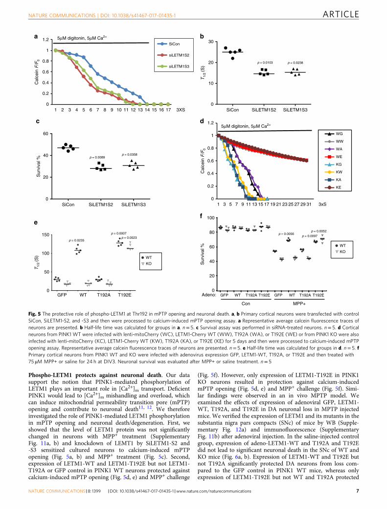
Phospho-LETM1 protects against neuronal death. Our datasupport the notion that PINK1-mediated phosphorylation ofLETM1 plays an important role in [Ca2+]m transport. DeficientPINK1 would lead to [Ca2+]m mishandling and overload, whichcan induce mitochondrial permeability transition pore (mPTP)opening and contribute to neuronal death11, 12. We thereforeinvestigated the role of PINK1-mediated LETM1 phosphorylationin mPTP opening and neuronal death/degeneration. First, weshowed that the level of LETM1 protein was not significantlychanged in neurons with MPP+ treatment (SupplementaryFig. 11a, b) and knockdown of LEMT1 by SiLETM1-S2 and-S3 sensitized cultured neurons to calcium-induced mPTPopening (Fig. 5a, b) and MPP+ treatment (Fig. 5c). Second,expression of LETM1-WT and LETM1-T192E but not LETM1-T192A or GFP control in PINK1 WT neurons protected againstcalcium-induced mPTP opening (Fig. 5d, e) and MPP+ challenge
(Fig. 5f). However, only expression of LETM1-T192E in PINK1KO neurons resulted in protection against calcium-inducedmPTP opening (Fig. 5d, e) and MPP+ challenge (Fig. 5f). Simi-lar findings were observed in an in vivo MPTP model. Weexamined the effects of expression of adenoviral GFP, LETM1-WT, T192A, and T192E in DA neuronal loss in MPTP injectedmice. We verified the expression of LETM1 and its mutants in thesubstantia nigra pars compacts (SNc) of mice by WB (Supple-mentary Fig. 12a) and immunofluorescence (SupplementaryFig. 11b) after adenoviral injection. In the saline-injected controlgroup, expression of adeno-LETM1-WT and T192A and T192Edid not lead to significant neuronal death in the SNc of WT andKO mice (Fig. 6a, b). Expression of LETM1-WT and T192E butnot T192A significantly protected DA neurons from loss com-pared to the GFP control in PINK1 WT mice, whereas onlyexpression of LETM1-T192E but not WT and T192A protected
a
Sur
viva
l %
Con MPP+
GFP WT T192A T192E GFP WT T192A T192EAdeno:GFP WT T192A T192E
T1/
2 (S
)
d
0
0.2
0.4
0.6
0.8
1
1.2
1 3 5 7 9 11 13 15 17 19 21 23 25 27 29 31
WG
WW
WA
WE
KG
KW
KA
KE
5μM digitonin, 5μM Ca2+
3xSC
alce
in F
/F0
0
0.2
0.4
0.6
0.8
1
1.2
1 2 3 4 5 6 7 8 9 10 11 12 13 14 15 16 17
SiCon
siLETM1S2
siLETM1S3
3XS
Cal
cein
F/F
05μM digitonin, 5μM Ca2+
ef
p = 0.0103 p = 0.0238
c
Sur
viva
l %
p = 0.0089p = 0.0358
p = 0.0007
p = 0.0235p = 0.0023
T1/
2 (S
)
b
SiCon SiLETM1S2 SiLETM1S30
10
20
30
SiCon SiLETM1S2 SiLETM1S3
WT
KO
0
20
40
60
0
50
100
150
0
20
40
60
80
100
p = 0.0056p = 0.0052
p = 0.0097
WT
KO
Fig. 5 The protective role of phospho-LETM1 at Thr192 in mPTP opening and neuronal death. a, b Primary cortical neurons were transfected with controlSiCon, SiLETM1-S2, and -S3 and then were processed to calcium-induced mPTP opening assay. a Representative average calcein fluorescence traces ofneurons are presented. b Half-life time was calculated for groups in a. n= 5. c Survival assay was performed in siRNA-treated neurons. n= 5. d Corticalneurons from PINK1 WT were infected with lenti-mitoCherry (WC), LETM1-Cherry WT (WW), T192A (WA), or T192E (WE) or from PINK1 KO were alsoinfected with lenti-mitoCherry (KC), LETM1-Cherry WT (KW), T192A (KA), or T192E (KE) for 5 days and then were processed to calcium-induced mPTPopening assay. Representative average calcein fluorescence traces of neurons are presented. n= 5. e Half-life time was calculated for groups in d. n= 5. fPrimary cortical neurons from PINK1 WT and KO were infected with adenovirus expression GFP, LETM1-WT, T192A, or T192E and then treated with75 µM MPP+ or saline for 24 h at DIV3. Neuronal survival was evaluated after MPP+ or saline treatment. n= 5
NATURE COMMUNICATIONS | DOI: 10.1038/s41467-017-01435-1 ARTICLE
NATURE COMMUNICATIONS |8: 1399 |DOI: 10.1038/s41467-017-01435-1 |www.nature.com/naturecommunications 7
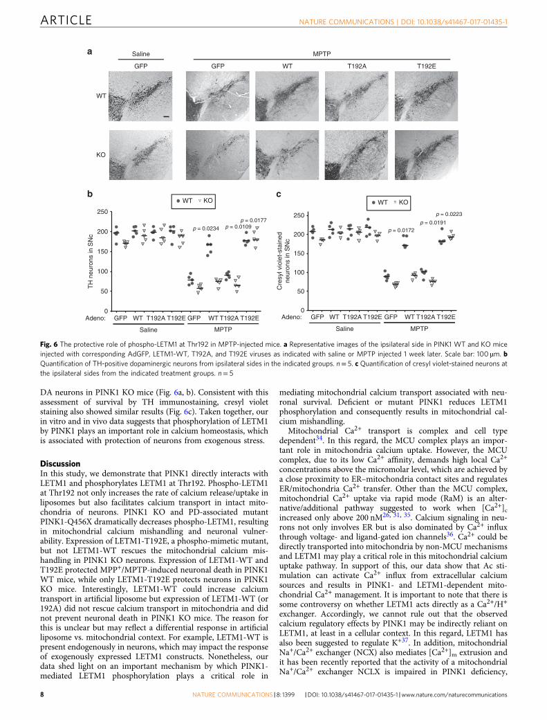
DA neurons in PINK1 KO mice (Fig. 6a, b). Consistent with thisassessment of survival by TH immunostaining, cresyl violetstaining also showed similar results (Fig. 6c). Taken together, ourin vitro and in vivo data suggests that phosphorylation of LETM1by PINK1 plays an important role in calcium homeostasis, whichis associated with protection of neurons from exogenous stress.
DiscussionIn this study, we demonstrate that PINK1 directly interacts withLETM1 and phosphorylates LETM1 at Thr192. Phospho-LETM1at Thr192 not only increases the rate of calcium release/uptake inliposomes but also facilitates calcium transport in intact mito-chondria of neurons. PINK1 KO and PD-associated mutantPINK1-Q456X dramatically decreases phospho-LETM1, resultingin mitochondrial calcium mishandling and neuronal vulner-ability. Expression of LETM1-T192E, a phospho-mimetic mutant,but not LETM1-WT rescues the mitochondrial calcium mis-handling in PINK1 KO neurons. Expression of LETM1-WT andT192E protected MPP+/MPTP-induced neuronal death in PINK1WT mice, while only LETM1-T192E protects neurons in PINK1KO mice. Interestingly, LETM1-WT could increase calciumtransport in artificial liposome but expression of LETM1-WT (or192A) did not rescue calcium transport in mitochondria and didnot prevent neuronal death in PINK1 KO mice. The reason forthis is unclear but may reflect a differential response in artificialliposome vs. mitochondrial context. For example, LETM1-WT ispresent endogenously in neurons, which may impact the responseof exogenously expressed LETM1 constructs. Nonetheless, ourdata shed light on an important mechanism by which PINK1-mediated LETM1 phosphorylation plays a critical role in
mediating mitochondrial calcium transport associated with neu-ronal survival. Deficient or mutant PINK1 reduces LETM1phosphorylation and consequently results in mitochondrial cal-cium mishandling.
Mitochondrial Ca2+ transport is complex and cell typedependent34. In this regard, the MCU complex plays an impor-tant role in mitochondria calcium uptake. However, the MCUcomplex, due to its low Ca2+ affinity, demands high local Ca2+
concentrations above the micromolar level, which are achieved bya close proximity to ER–mitochondria contact sites and regulatesER/mitochondria Ca2+ transfer. Other than the MCU complex,mitochondrial Ca2+ uptake via rapid mode (RaM) is an alter-native/additional pathway suggested to work when [Ca2+]cincreased only above 200 nM26, 31, 35. Calcium signaling in neu-rons not only involves ER but is also dominated by Ca2+ influxthrough voltage- and ligand-gated ion channels36. Ca2+ could bedirectly transported into mitochondria by non-MCU mechanismsand LETM1 may play a critical role in this mitochondrial calciumuptake pathway. In support of this, our data show that Ac sti-mulation can activate Ca2+ influx from extracellular calciumsources and results in PINK1- and LETM1-dependent mito-chondrial Ca2+ management. It is important to note that there issome controversy on whether LETM1 acts directly as a Ca2+/H+
exchanger. Accordingly, we cannot rule out that the observedcalcium regulatory effects by PINK1 may be indirectly reliant onLETM1, at least in a cellular context. In this regard, LETM1 hasalso been suggested to regulate K+37. In addition, mitochondrialNa+/Ca2+ exchanger (NCX) also mediates [Ca2+]m extrusion andit has been recently reported that the activity of a mitochondrialNa+/Ca2+ exchanger NCLX is impaired in PINK1 deficiency,
c
WT
KO
a
Adeno: Adeno:
GFP GFP WT T192A T192E
Saline MPTP
0GFP GFPGFP GFPWT WTWT WT
Saline SalineMPTP MPTP
T192A T192AT192A T192AT192E T192ET192E T192E
TH
neu
rons
in S
Nc
Cre
syl v
iole
t-st
aine
dne
uron
s in
SN
c50
100
150
200
250 250
200
150
100
50
0
p = 0.0172p = 0.0191
p = 0.0223
b
p = 0.0234 p = 0.0109p = 0.0177
WT WTKO KO
Fig. 6 The protective role of phospho-LETM1 at Thr192 in MPTP-injected mice. a Representative images of the ipsilateral side in PINK1 WT and KO miceinjected with corresponding AdGFP, LETM1-WT, T192A, and T192E viruses as indicated with saline or MPTP injected 1 week later. Scale bar: 100 µm. bQuantification of TH-positive dopaminergic neurons from ipsilateral sides in the indicated groups. n= 5. c Quantification of cresyl violet-stained neurons atthe ipsilateral sides from the indicated treatment groups. n= 5
ARTICLE NATURE COMMUNICATIONS | DOI: 10.1038/s41467-017-01435-1
8 NATURE COMMUNICATIONS |8: 1399 |DOI: 10.1038/s41467-017-01435-1 |www.nature.com/naturecommunications
which can be rescued by PKA33. However, our data support thatLETM1 and NCLX are involved in [Ca2+]m extrusion in amanner that is likely independent. Nonetheless, our data stronglysupport a model by which PINK1 regulation of LETM1 is criticalfor neuronal death induced by mitochondrial stress both in vitroand in vivo.
Neurodegenerative disorders including PD exhibit dysregula-tion of calcium homeostasis due to excitotoxicity, perturbedenergy metabolism and oxidative stress, and compromised cel-lular calcium-regulating systems38. It has been suggested thatadult DA neurons engage L-type channels permeable to calciumto drive pacemaking, leading to elevated intracellular calcium andneuronal vulnerability39. In stressed neurons, extracellular cal-cium enters the cell via calcium permeable channels (i.e., gluta-mate receptors) elevating [Ca2+]C and [Ca2+]m38. Accordingly,one interesting model is that PINK1-mediated phosphorylation ofLETM1 at Thr192 could facilitate [Ca2+]m extrusion to preventsustained [Ca2+]m accumulation and protect neurons from stressand death. At least in models of mitochondrial stress, this appearsto hold true. However, an important caveat is that MPP+/MPTPmodels of degeneration may not accurately reflect the true age-dependent processes in human PD. In this regard, the study ofdegenerative processes in model rodent systems is hampered bythe lack of robust DA degeneration with respect to genetic formsof PD like with PINK1 loss40. However, it is intriguing to spec-ulate that because of the proposed pacemaking capacity of DAneurons, a PINK1-LETM1 regulatory mechanism may be parti-cularly critical in PD. In this model (Supplementary Fig. 13), wehypothesize that mutant or deficient PINK1 results in decreasedphosphorylation of LETM1, consequently accelerating [Ca2+]mmishandling and overload, rendering neurons vulnerable tostress.
MethodsAntibodies. The following antibodies were used: anti-FLAG (Sigma, Cat: F3165,dilution 1:10,000), anti-Myc (Abcam, Ab9106, 1:2500), anti-Phosphothreonine(pThr, Thermo Fisher, 71-8200, 1:2500); anti-Phosphoserine (pSer, Thermo Fisher,61–8100, 1:2500); anti-LETM1 (Santa Cruz, mouse sc-514136, 1:2500 or NovusBiologicals, rabbit NBP1-33433, 1:10,000). Anti-PINK1 (Abgent, mouse AM6406a,1:5000 and Novus Biologicals, rabbit BC100–494, 1:5000), anti-Tyrosine hydro-xylase (TH, EMD Millipore, AB152, 1:1000), anti-MCU (Cell Signaling, 14997,1:10,000), anti-NCLX (Abcam, ab136975, 1:5000). Special custom phospho-LETM1 at Thr192 (pT192) polyclonal antibody was generated and purified from arabbit immunized with carrier protein-conjugated phosphopeptide,CNGHTLpT192RRERR (residues 187–197 of human LETM1, NP_036450.1), usingstandard protocols by Biogenes (Berlin, Germany, dilution 1:2500).
Plasmid construction and virus production. The full open reading frame com-plimentary DNA clone of human LETM1 was purchased from Origene and wascloned into pCMV-3Tag-2a (Myc-Tag) and sequence was verified by DNAsequencing. LETM1 mutants were generated using QuickChange Site-DirectedMutagensis Kit (Stratagene, USA). LETM1 variants were engineered into thepGEX4T-1 or pET28a vector for preparation of GST- or His-tag fusion proteins41.LETM1 variants and PINK1 were cloned to pAdtrack-CMV with 3XFLAG at C-terminus for preparation of recombinant adenovirus as previously described42. Theviral constructs expressing 3XFLAG-LETM1 variants and GFP are controlled byseparate CMV promoters. The adenoviruses were produced using pAdEasy systemas previously described43.
Bacterial protein purification and in vitro kinase assay. For production ofbacterial recombinant protein, transmembrane domains in amino-acid residues1–111 of PINK1 were deleted by plasmid cloning and designated ΔN-PINK1. OnlyN-terminus (amino-acid residues 1–204) of LETM1 without transmembranedomains and C-terminus were produced by cloning and designated N-LETM1. Thetransmembrane domain (TM; amino-acid residue 205–227) of human LETM1were deleted and designated ΔTM-LETM1. All GST- or His-tagged fusion proteinswere expressed in Escherichia coli and purified using Sepharose 4B agarose (GEHealthcare, USA) or Ni-NTA Agarose (Qiagen Inc, Canada) respectively, asper manufacturer’s instruction. The PINK1 kinase assay with LETM1 variants wasperformed as previously described44, 45. Briefly, 10 µg GST-N-LETM1 or His-ΔTM-LETM1 variants with 2 µg His-ΔN-PINK1 was incubated at 30 °C for 6 h in40 µl of reaction buffer containing 50 mM Tris-HCL (pH 7.4), 10 mM MgCl2, 2
mM MnCl2, 0.5 mM Calcium, 2 mM DTT and 0.1 mM ATP. The reactions weresubjected to WB with pSer, pThr, or pT192 antibodies.
Immunoprecipitation. Cells or samples were harvested in lysis buffer (50 mMTris-HCl (pH 7.4), 100 mM NaCl, 2 mM EDTA, 1 mM DTT, 1% Triton X-100, and0.1% SDS) supplemented with protease inhibitors. IPs were performed throughincubation of 2 µg antibodies with lysates (2 mg protein) at 4 °C for 2 h followed byincubation with protein A beads (Sigma, USA) at 4 °C for 2 h. The washed sampleswere analyzed by Western blotting using TrueBlot secondary antibodies (RocklandImmunochemicals Inc). Some IPed FLAG-tagged proteins were eluted with3XFLAG peptide (Sigma) according to the manufacturer’s instructions.
Artificial liposomes. The calcium transport activity of LETM1 was measured ascalcium released from artificial liposomes as described previously with minormodification17, 27, 46. Liposomes were reconstituted by mixing 100 mg/ml asolectin,10 mg/ml cardiolipin in solubilization solution (Tris 50 mM, pH 7.4, 2% Triton-100, 2 mM calcium, EDTA 0.5 mM) and sonicated until the solution was clear.Combined with varied purified LETM1 proteins, the liposomes were incubated atroom temperature for 1 h. Detergent was then removed by passing AmberliteXAD-2 (Sigma) columns three times. Extraliposomal solutions were changed bypassing eight times through Sephadex G-25 (GE Healthcare) columns swelled withTE buffer (Tris 50 mM/EDTA 0.5M). The free [Ca2+] was measured (excitation at488 nm and emission at 530 nm) in a plate reader by loading 10 μM Fluo-4 pen-tapotassium salt (Invitrogen). The calcium uptake assay in liposome was alsoperformed. Liposomes were reconstituted by mixing 20 mg/ml 3:1 POPE (850757P;Avanti Lipids Polar)/POPG (840457P, Avanti Lipids Polar) in solubilizationsolution (Tris 50 mM, pH 7.4, 2% Triton-100, EDTA 1mM). The rest of theprocedure is similar to the calcium release assay. Extraliposomal solutions con-tained Tris 50 mM/EDTA 0.5 mM. Ca2+ was added to extraliposomal solutionsto create the initial 750 nM free [Ca2+] (CaCl2 0.47 mM/EDTA 0.5 mM) and thenthe free [Ca2+] was measured (excitation at 488 nm and emission at 530 nm) ina plate reader by loading 10 μM Fluo-4 pentapotassium salt (Invitrogen). [Ca2+]was calculated using WEBMAXC (http://www.stanford.edu/~cpatton/webmaxcS.htm).
Neuronal cultures with survival assay and RNA interference. Primary cultureof mouse cortical neurons was carried out as described previously47, 48. Forinfection, 24 h after initial plating the cortical neurons were infected with adeno-virus (MOI 100). Two days after infection, cortical neurons were subjected tosurvival determination following exposure to 75 μM MPP+ for 24 h. For survival ofinfected neurons, GFP-positive neurons were assessed for nuclear integrity asdetermined by Hoechst 33258 (0.5 ng/ml) staining. Neurons with punctate orcondensed nuclei were assessed as dead. Survival was expressed as the percentageof live cells to total cells. SiRNA to mouse LETM1 was produced utilizing theSilencer siRNA Construction Kit (AM1620, Thermo Fisher Scientific) asper manufacturer’s instruction. The sequences of sense strand siRNA to LETM1and scramble control are: S1 (5′-UGAGGAAAUCAUGC-GUUUU-3′); S2 (5′-GGAGGAGAUUGACAUCCUC-3′); S3 (5′-GCAAAUCAAGCACA-UUCCA-3′);scramble control siRNA (5′-GUAGCACGCGUA ACUGUCU-3′). Neurons weretransfected with 30 pmol/well (24 well plate) siRNA to mouse LETM1 (SiLETM1)using Lipofectamine 2000 (Invitrogen, USA) for 2 days and re-transfected once at3 days.
TMRE staining for mitochondrial membrane potential. Cortical neurons wereloaded with 200 nM tetramethylrodamine methylester (TMRM; T669; ThermoFisher) in imaging buffer (20 mM HEPES, 135 mM NaCl, 5 mM KCl, 1 mM CaCl2,30 mM D-glucose, 10 mM succinate, pH 7.4) for 30 min at room temperature, andthe dye was present during the imaging experiment analyzed by a Zeiss 510 metaconfocal microscope.
Mitochondrial Ca2+ imaging in individual neurons. Mitochondrial Ca2+ imagingin individual primary cortical neurons employed Rhod-2 dye, which is cationic andpreferentially accumulated in the mitochondria28. Cortical neurons at DIV3–4 oncoverslip infected with adenovirus were loaded with Rhod-2-AM (Invitrogen) 1 µMand 0.01% (w/v) pluronic acid in imaging buffer (20 mM HEPES, 135 mM NaCl, 5mM KCl, 1 mM CaCl2, 30 mM D-glucose, 10 mM succinate, pH 7.4) at roomtemperature for 30 min. Subsequently, cells were washed with imaging buffer andfurther incubated for 60 min at 37 °C for de-esterification of dye. MitochondrialCa2+ imaging experiments were performed in the imaging chamber (WarnerInstruments) at 37 °C with Zeiss AxioObserver. D1 Microscope with ×40 objective.Rhod-2 was excited at 543 nm, and emission measured at 580 nm. The increasedmitochondrial calcium was stimulated by 100 µM Ac. The images were captured ina time lapse model at 2s intervals for 2 min and neurons were stimulated at the10th frame of recording. Relative increase in fluorescence intensity was normalizedby fluorescence at the resting condition (ΔF/F0). Data were generated and analyzedwith Zeiss microscope Axiovision rel 4.8 software. The rate of uptake or extrusionof mitochondrial calcium was expressed as T1/2 calculated by Logarithmic (LOG)function formatted as T1/2= Exp[(Y1/2–b)/a] in Microsoft Excel 2010. Data were
NATURE COMMUNICATIONS | DOI: 10.1038/s41467-017-01435-1 ARTICLE
NATURE COMMUNICATIONS |8: 1399 |DOI: 10.1038/s41467-017-01435-1 |www.nature.com/naturecommunications 9
presented as the traces of average values for all data points generated from aminimum of 20 neurons per experiment and at least five independent experiments.
FRET-based mitochondrial calcium imaging. TN-XXL is a genetically encodedFRET calcium sensor and based on the calcium binding protein troponin C linkingcyan fluorescent protein (CFP) and Citrine Cp174 fluorescent protein (one YFP).We produced a mitochondrial targeted 4XmTG-TN-XXL construct, where fourmitochondrial targeting sequences (mTGs) derived from the subunit VIII ofhuman cytochrome c oxidase were fused with the N-terminal of TN-XXL. 4XmTG-TN-XXL (kindly provided by Dr. Oliver Griesbeck, Max Planck Institute ofNeurobiology, Martinsried, Germany) was subcloned into lentiviral pLVX-AcGFP1-N1 vector (Clotech, Cat No: 632154) with deletion of GFP. We alsogenerated the mito-targetted PLVX-4XmTG-Cherry as control and PLVX-LETM1-3XFLAG-T2A-Cherry WT, T192A, and T192E constructs with deletion of GFP.T2A is a self-cleaving 2A peptide37. Lentiviruses were generated by PLVX, pPAX2,and pMD2.G system. Lenti-4XmTG-TN-XXL were co-infected with lenti-4XmTG-Cherry, LETM1-3XFLAG-T2A-Cherry WT, T192A, and T192E in neurons (MOI10) at DIV 1 for 5–7 days before calcium imaging.
For FRET-based mitochondrial calcium imaging, all FRET microscopicobservations were performed on a Zeiss 510 meta confocal microscope at 37 °C.Donor (CFP) was excited at 458 nm and detected in a bandwidth of 470–500 nm(CFP channel), whereas the excitation at 514 nm and emission at 505–550 nm wereused for detecting acceptor (YFP) (YFP channel). For FRET, the excitation was at458 nm and detection at 505–550 nm (FRET channel). CFP and FRET emissionchannels were recorded simultaneously by passing through a long-pass dichroicmirror (515 nm). The images were captured in a time-lapse model at 3s intervalsfor 2 min and neurons were stimulated at the 5th frame of recording by 100 µM Ac.Images were analyzed using Image J software. The Intensity of FRET/CFP wascalculated as FRET ratio. Relative increase in FRET ratio was normalized by theresting condition (ΔR/R0). The rate of uptake or extrusion of mitochondrialcalcium was expressed as T1/2 calculated by LOG function formatted as T1/2= Exp[(Y1/2–b)/a] in Microsoft Excel 2010. Data were presented as the traces of averagevalues for all data points generated from a minimum of 10 neurons per experimentand at least four independent experiments.
Measurement of the mPTP opening. mPTP opening was assessed by thequenching of calcein fluorescence with cobalt30. PINK1 WT or KO neurons wereloaded with Calcein-AM (1 μM, Molecular Probes) at 37 °C for 15 min and CoCl2(2 mM, Sigma) was added for further 30 min. Neurons were washed and thenpermeabilized with 5 μM digitonin in 5 μM Ca2+ in imaging buffer (20 mMHEPES, 135 mM NaCl, 5 mM KCl, 30 mM D-glucose, 10 mM succinate, pH 7.4) toinduce mPTP opening. Images were captured with the Zeiss AxioObserver. D1Microscope in a time-lapse model at 3s intervals for 2 min. Relative calceinfluorescence intensity was normalized to the resting condition (ΔF/F0) and T1/2was calculated. Data were generated and analyzed with the Zeiss microscopeAxiovision rel 4.8 software.
Animals with viral delivery and MPTP administration. PINK1-deficient mice(maintained on a C57BL/6 background) and wild-type littermates were generatedby breeding their heterozygous counterparts. Genotyping information was reportedpreviously49. Mice were housed two to five animals per cage with a 12-h light/darkcycle (lights on from 0700 to 1900 h) at constant temperature (23 °C) with adlibitum access to food and water. All animal experiments conformed to theguidelines set forth by the Canadian Council for the Use and Care of Animals inResearch (CCAC) and the Canadian Institutes for Health Research (CIHR) andwith approval from the University of Ottawa Animal Care Committee.
We have previously shown that adenoviruses were targeted unilaterally to DAneurons of SNc from the striatum by retrograde transport using a well-establishedadenoviral-mediated gene delivery approach14. About 52 male PINK1 WT and 52male KO mice of 8–12 weeks were randomly allocated to four groups, respectively.A single unilateral injection of each virus expressing GFP, LETM1-WT, T192A, orT192E (2 µl, 1 × 10⁷ particles per µl) was delivered to the right striatum (0.5 mmrostral, 2.2 mm right of bregma, and 3.4 mm below the skull surface) of 8-week-oldmale mice using a syringe pump system 7 days before MPTP administration. Toexamine the viral expression in TH neurons, the samples extracted from SNc ofpost 7 days virus-injected mice were subjected to Western blotting. Coronalsections (14 µm thickness) of the ventral midbrain were double labeled usingspecific primary antibody to GFP (Abcam) or TH (Millipore).
Mice received one intraperitoneal injection of MPTP·HCl per day (30 mg/kg,Sigma, USA) for 5 consecutive days50–53. Control mice received an equivalentvolume of 0.9% saline. Assessment of dopamine neuron survival was performedblindly 2 weeks after the start of the MPTP injection by immunohistochemicalanalyses.
Immunohistochemistry and immunofluorescence. Mice were perfused trans-cardially and brains were fixed in 4% paraformaldehyde and cryoprotected aspreviously described54. Free floating serial coronal sections (14 µm thickness) of theventral midbrain were collected. Sections were then incubated with TH antibody(1:1000) for 24 h at 4 °C. Immunoreactivity was visualized by using anavidin–biotin complex peroxidase/3,3′-diaminobenzidine (DAB) reaction. For
double-labeling immunofluorescence, samples were double labeled with primaryantibody to GFP (Abcam) or TH (Millipore) and visualized using either Alex-488-conjugated anti-rabbit IgG (1:200) or Alex-594-conjugated anti-mouse IgG (1:200).
Quantification of dopaminergic neuronal loss. The number of DA (TH positive)neurons was only counted from the sections in the region containing the medialterminal nucleus (MTN) because this region has been previously shown to expressthe highest level of virus-mediated gene expression after intrastriatal infection50.We also used the MTN as a landmark to evaluate consistent levels of SNc. Neuronsin the ipsilateral and contralateral side to the viral injection were assessed aspreviously described50. At least three sections per animal were analyzed. In parallel,cresyl violet staining was performed to validate determination of nigral counts aspreviously described50. The anatomical localization of SNc was referenced by THstaining.
Statistical analysis. Statistical differences between multiple groups of data were allanalyzed with nonparametric Kruskal–Wallis one-way ANOVA test usingGraphPad Prism v7.03 program. The data are presented as median. Exact p-value isindicated in the graph if p< 0.05.
Data availability. All data generated or analyzed during this study are eitherincluded in this published article (and its Supplementary Information files) oravailable from the authors.
Received: 21 November 2016 Accepted: 18 September 2017
References1. Valente, E. M. et al. Hereditary early-onset Parkinson’s disease caused by
mutations in PINK1. Science 304, 1158–1160 (2004).2. Corti, O., Lesage, S. & Brice, A. What genetics tells us about the causes and
mechanisms of Parkinson’s disease. Physiol. Rev. 91, 1161–1218 (2011).3. Deas, E. et al. PINK1 cleavage at position A103 by the mitochondrial protease
PARL. Hum. Mol. Genet. 20, 867–879 (2011).4. Fallaize, D., Chin, L. S. & Li, L. Differential submitochondrial localization of
PINK1 as a molecular switch for mediating distinct mitochondrial signalingpathways. Cell Signal. 27, 2543–2554 (2015).
5. Jin, S. M. et al. Mitochondrial membrane potential regulates PINK1 import andproteolytic destabilization by PARL. J. Cell Biol. 191, 933–942 (2010).
6. Pickrell, A. M. & Youle, R. J. The roles of PINK1, parkin, and mitochondrialfidelity in Parkinson’s disease. Neuron 85, 257–273 (2015).
7. Hoepken, H. H. et al. Mitochondrial dysfunction, peroxidation damage andchanges in glutathione metabolism in PARK6. Neurobiol. Dis. 25, 401–411(2007).
8. Morais, V. A. et al. PINK1 loss-of-function mutations affect mitochondrialcomplex I activity via NdufA10 ubiquinone uncoupling. Science 344, 203–207(2014).
9. Heeman, B. et al. Depletion of PINK1 affects mitochondrial metabolism,calcium homeostasis and energy maintenance. J. Cell. Sci. 124, 1115–1125(2011).
10. Marongiu, R. et al. Mutant Pink1 induces mitochondrial dysfunction in aneuronal cell model of Parkinson’s disease by disturbing calcium flux. J.Neurochem. 108, 1561–1574 (2009).
11. Gandhi, S. et al. PINK1-associated Parkinson’s disease is caused by neuronalvulnerability to calcium-induced cell death. Mol. Cell 33, 627–638 (2009).
12. Gautier, C. A. et al. Regulation of mitochondrial permeability transition pore byPINK1. Mol. Neurodegener. 7, 22 (2012).
13. Gautier, C. A., Kitada, T. & Shen, J. Loss of PINK1 causes mitochondrialfunctional defects and increased sensitivity to oxidative stress. Proc. Natl Acad.Sci. USA 105, 11364–11369 (2008).
14. Haque, M. E. et al. Cytoplasmic Pink1 activity protects neurons fromdopaminergic neurotoxin MPTP. Proc. Natl Acad. Sci. USA 105, 1716–1721(2008).
15. Qu, D. et al. BAG2 gene-mediated regulation of PINK1 protein is critical formitochondrial translocation of PARKIN and neuronal survival. J. Biol. Chem.290, 30441–30452 (2015).
16. Ewing, R. M. et al. Large-scale mapping of human protein-protein interactionsby mass spectrometry. Mol. Syst. Biol. 3, 89 (2007).
17. Jiang, D., Zhao, L. & Clapham, D. E. Genome-wide RNAi screen identifiesLetm1 as a mitochondrial Ca2+/H+ antiporter. Science 326, 144–147 (2009).
18. Tamai, S. et al. Characterization of the mitochondrial protein LETM1, whichmaintains the mitochondrial tubular shapes and interacts with the AAA-ATPase BCS1L. J. Cell. Sci. 121, 2588–2600 (2008).
19. Jiang, D., Zhao, L., Clish, C. B. & Clapham, D. E. Letm1, the mitochondrialCa2+/H+ antiporter, is essential for normal glucose metabolism and alters
ARTICLE NATURE COMMUNICATIONS | DOI: 10.1038/s41467-017-01435-1
10 NATURE COMMUNICATIONS |8: 1399 |DOI: 10.1038/s41467-017-01435-1 |www.nature.com/naturecommunications
brain function in Wolf-Hirschhorn syndrome. Proc. Natl Acad. Sci. USA 110,E2249–2254 (2013).
20. Doonan, P. J. et al. LETM1-dependent mitochondrial Ca2+ flux modulatescellular bioenergetics and proliferation. FASEB J. 28, 4936–4949 (2014).
21. Dimmer, K. S. et al. LETM1, deleted in Wolf-Hirschhorn syndrome is requiredfor normal mitochondrial morphology and cellular viability. Hum. Mol. Genet.17, 201–214 (2008).
22. McQuibban, A. G. et al. A drosophila mutant of LETM1, a candidate gene forseizures in Wolf-Hirschhorn syndrome. Hum. Mol. Genet. 19, 987–1000(2010).
23. Pridgeon, J. W., Olzmann, J. A., Chin, L. S. & Li, L. PINK1 protects againstoxidative stress by phosphorylating mitochondrial chaperone TRAP1. PLoSBiol. 5, e172 (2007).
24. Becker, D., Richter, J., Tocilescu, M. A., Przedborski, S. & Voos, W. Pink1kinase and its membrane potential (Deltapsi)-dependent cleavage product bothlocalize to outer mitochondrial membrane by unique targeting mode. J. Biol.Chem. 287, 22969–22987 (2012).
25. Okatsu, K. et al. PINK1 autophosphorylation upon membrane potentialdissipation is essential for Parkin recruitment to damaged mitochondria. Nat.Commun. 3, 1016 (2012).
26. Narendra, D. P. et al. PINK1 is selectively stabilized on impaired mitochondriato activate Parkin. PLoS Biol. 8, e1000298 (2010).
27. Tsai, M. F., Jiang, D., Zhao, L., Clapham, D. & Miller, C. Functionalreconstitution of the mitochondrial Ca2+/H+antiporter Letm1. J. Gen. Physiol.143, 67–73 (2014).
28. Minta, A., Kao, J. P. & Tsien, R. Y. Fluorescent indicators for cytosolic calciumbased on rhodamine and fluorescein chromophores. J. Biol. Chem. 264,8171–8178 (1989).
29. Gautier, C. A., et al. Regulation of mitochondrial permeability transition poreby PINK1. Mol. Neurodegener. 7, 22 (2012).
30. De Stefani, D., Raffaello, A., Teardo, E., Szabo, I. & Rizzuto, R. A forty-kilodalton protein of the inner membrane is the mitochondrial calciumuniporter. Nature 476, 336–340 (2011).
31. Baughman, J. M. et al. Integrative genomics identifies MCU as an essentialcomponent of the mitochondrial calcium uniporter. Nature 476, 341–345(2011).
32. Mank, M. et al. A genetically encoded calcium indicator for chronic in vivotwo-photon imaging. Nat. Methods 5, 805–811 (2008).
33. Kostic, M. et al. PKA phosphorylation of NCLX reverses mitochondrial calciumoverload and depolarization, promoting survival of PINK1-deficientdopaminergic neurons. Cell Rep. 13, 376–386 (2015).
34. Duchen, M. R., Verkhratsky, A. & Muallem, S. Mitochondria and calcium inhealth and disease. Cell Calcium 44, 1–5 (2008).
35. Rowland, A. A. & Voeltz, G. K. Endoplasmic reticulum-mitochondria contacts:function of the junction. Nat. Rev. Mol. Cell Biol. 13, 607–625 (2012).
36. Duchen, M. R. Mitochondria, calcium-dependent neuronal death andneurodegenerative disease. Pflugers Arch. 464, 111–121 (2012).
37. Nowikovsky, K. et al. The LETM1/YOL027 gene family encodes a factor of themitochondrial K+ homeostasis with a potential role in the Wolf-Hirschhornsyndrome. J. Biol. Chem. 279, 30307–30315 (2004).
38. Nicholls, D. G. Mitochondrial calcium function and dysfunction in the centralnervous system. Biochim. Biophys. Acta 1787, 1416–1424 (2009).
39. Surmeier, D. J. & Schumacker, P. T. Calcium, bioenergetics, and neuronalvulnerability in Parkinson’s disease. J. Biol. Chem. 288, 10736–10741 (2013).
40. Kitada, T., Tong, Y., Gautier, C. A. & Shen, J. Absence of nigral degeneration inaged parkin/DJ-1/PINK1 triple knockout mice. J. Neurochem. 111, 696–702(2009).
41. Qu, D. et al. The protein SET binds the neuronal Cdk5 activator p35nck5a andmodulates Cdk5/p35nck5a activity. J. Biol. Chem. 277, 7324–7332 (2002).
42. He, T. C. et al. A simplified system for generating recombinant adenoviruses.Proc. Natl Acad. Sci. USA 95, 2509–2514 (1998).
43. Sedarous, M. et al. Calpains mediate p53 activation and neuronal death evokedby DNA damage. J. Biol. Chem. 278, 26031–26038 (2003).
44. Qu, D. et al. Role of Cdk5-mediated phosphorylation of Prx2 in MPTP toxicityand Parkinson’s disease. Neuron 55, 37–52 (2007).
45. Sha, D., Chin, L. S. & Li, L. Phosphorylation of parkin by Parkinson disease-linked kinase PINK1 activates parkin E3 ligase function and NF-kappaBsignaling. Hum. Mol. Genet. 19, 352–363 (2010).
46. Ahn, T., Yun, C. H., Chae, H. Z., Kim, H. R. & Chae, H. J. Ca2+/H+ antiporter-like activity of human recombinant Bax inhibitor-1 reconstituted intoliposomes. FEBS J. 276, 2285–2291 (2009).
47. Xiang, H. et al. Evidence for p53-mediated modulation of neuronal viability. J.Neurosci. 16, 6753–6765 (1996).
48. Fortin, A. et al. APAF1 is a key transcriptional target for p53 in the regulationof neuronal cell death. J. Cell Biol. 155, 207–216 (2001).
49. Hallows, J. L., Chen, K., DePinho, R. A. & Vincent, I. Decreased cyclin-dependent kinase 5 (cdk5) activity is accompanied by redistribution of cdk5and cytoskeletal proteins and increased cytoskeletal protein phosphorylation inp35 null mice. J. Neurosci. 23, 10633–10644 (2003).
50. Crocker, S. J. et al. c-Jun mediates axotomy-induced dopamine neuron deathin vivo. Proc. Natl Acad. Sci. USA 98, 13385–13390 (2001).
51. Smith, P. D. et al. Cyclin-dependent kinase 5 is a mediator of dopaminergicneuron loss in a mouse model of Parkinson’s disease. Proc. Natl Acad. Sci. USA100, 13650–13655 (2003).
52. Kalia, S. K. et al. BAG5 inhibits Parkin and enhances dopaminergic neurondegeneration. Neuron 44, 931–945 (2004).
53. Kim, R. H. et al. Hypersensitivity of DJ-1-deficient mice to 1-methyl-4-phenyl-1,2,3,6-tetrahydropyrindine (MPTP) and oxidative stress. Proc. Natl Acad. Sci.USA 102, 5215–5220 (2005).
54. Crocker, S. J. et al. Inhibition of calpains prevents neuronal and behavioraldeficits in an MPTP mouse model of Parkinson’s disease. J. Neurosci. 23,4081–4091 (2003).
AcknowledgementsWe thank Dr. Jie Shen (Harvard Medical School) for the PINK1 knockout mouse strain,and Dr. Ming-Feng Tsai (Brandeis University) for critical reading of the manuscript. D.S.P. was supported by Canadian Institute of Heath Research (CIHR), The W. GarfieldWeston Foundation, Parkinson Society Canada, Parkinson Research Consortium, Centreof Excellence in Neurodegeneration (CoEN), EU Joint Programme—NeurodegenerativeDisease Research (JPND), Krembil/Brain Canada Foundation and Ontario Brain Insti-tute. C.K. is supported by the DFG (KL1134/11-1) and the Hermann and Lilly SchillingFoundation. D.S.P. is the career investigator of Heart and Stroke Foundation of Ontario.
Author contributionsE.H., D.Q., P.C., J.W., C.K., R.S.S., D.F., and D.S.P. designed research; E.H. performedmost of the experimental work; D.Q., T.H., N.R., W.B., D.K. provided technical support;E.H. and D.S.P. analyzed data and wrote the manuscript; D.S.P. supervised the project.
Additional informationSupplementary Information accompanies this paper at doi:10.1038/s41467-017-01435-1.
Competing interests: The authors declare no competing financial interests.
Reprints and permission information is available online at http://npg.nature.com/reprintsandpermissions/
Publisher's note: Springer Nature remains neutral with regard to jurisdictional claims inpublished maps and institutional affiliations.
Open Access This article is licensed under a Creative CommonsAttribution 4.0 International License, which permits use, sharing,
adaptation, distribution and reproduction in any medium or format, as long as you giveappropriate credit to the original author(s) and the source, provide a link to the CreativeCommons license, and indicate if changes were made. The images or other third partymaterial in this article are included in the article’s Creative Commons license, unlessindicated otherwise in a credit line to the material. If material is not included in thearticle’s Creative Commons license and your intended use is not permitted by statutoryregulation or exceeds the permitted use, you will need to obtain permission directly fromthe copyright holder. To view a copy of this license, visit http://creativecommons.org/licenses/by/4.0/.
© The Author(s) 2017
NATURE COMMUNICATIONS | DOI: 10.1038/s41467-017-01435-1 ARTICLE
NATURE COMMUNICATIONS |8: 1399 |DOI: 10.1038/s41467-017-01435-1 |www.nature.com/naturecommunications 11