Embed Size (px)
Citation preview
- 91 -
Effects of Long-Term Stimulation on Skeletal Muscle PhenotypeExpression and Collagen/Fibrillin Distribution
Dennis R. Trumble, Changping Duan, and James A. Magovern
Cardiothoracic Surgery Research, Allegheny-Singer Research Institute, Depart-ment of Surgery, Allegheny General Hospital, Pittsburgh, Pennsylvania
AbstractThe effects of chronic electrical stimulation on muscle fiber phenotype and metabolism arewell known, but its impact on the extracellular matrix is poorly understood. Material pro-perties of skeletal muscle are largely influenced by the viscoelastic properties of connectivetissues which occupy the interstitium. Changes in collagen and fibrillin content may therefo-re play a key role in muscular adaptation processes. Latissimus dorsi (LD) of eight rabbitswere used to study muscular adaptation to long-term electrical conditioning. Muscles wereconditioned using burst stimuli delivered over 6 or 12 weeks. Contralateral LD were used ascontrol. Stimulation produced marked reductions in maximum isometric force, but impro-ved endurance capacity due to increased percent cross sectional area (CSA) occupied byslow-twitch oxidative muscle fibers. Stimulation also increased percent CSA occupied bytype I collagen and fibrillin. In contrast, the amount of type III collagen and fast-twitch gly-colytic fibers decreased in stimulated muscle. These data suggest that muscular adaptationto long-term stimulation includes both alterations in fiber type expression and remodeling ofthe extracellular matrix.Key words: collagen, extracellular matrix, fibrillin, skeletal muscle, stimulation.
Basic Appl Myol 11 (2): 91-98, 2001
Skeletal muscle is highly adaptable in that its metabolicand contractile characteristics are largely regulated by itspattern of use. Muscles used intermittently against largeloads contain mostly thick, fast-twitch glycolytic (FG) fi-bers which are powerful but prone to fatigue. Musclesused against lighter loads over prolonged periods retainthinner, slow-twitch oxidative (SO) fibers which are lesspowerful but more fatigue-resistant. Several studies haveshown that muscle phenotype can be manipulated viachronic electrical stimulation to enhance fatigue resistanceat the expense of contractile power [14, 17, 23]. Thisproperty (termed “muscle plasticity”) has prompted manyresearchers to investigate the possibility of using condi-tioned skeletal muscle as an endogenous power source forcardiac assist [1, 16, 24].
Although many aspects of muscular adaptation to long-term stimulation are well-known, several facets remainunexplored. Changes in fiber-type, metabolic activity,muscle mass, contractile strength, and speed have beendocumented in numerous reports [6, 22, 25], but little at-tention has been paid to alterations within the extracellularmatrix. This is an important aspect of the adaptation proc-ess since the mechanical properties of muscle tissue are
heavily influenced by the interstitium which binds indi-vidual muscle cells into a cohesive unit.
Collagen types I and III are the major forms of intersti-tial collagen found in both skeletal muscle and heart tissue[8, 18] and have been shown to strongly influence tissueextensibility [2]. Type III collagen is also the main com-ponent of connective tissue in non-capillary blood vessels[7] and hence, collagen staining techniques can be used toidentify small vessels in the muscle interstitium [4]. Anetwork of elastic fibers in the extracellular matrix pro-vides muscle tissue with the resilience required to recoilafter a transient stretch. These flexible fibers comprise anamorphous core of elastin surrounded by microfibrils, ofwhich fibrillin is the primary element [10]. The amountand relative proportion of these interstitial componentshas been shown to change with exercise training [9].Hence, it is reasonable to hypothesize that chronic electri-cal stimulation might also alter collagen and fibrillin con-tent in skeletal muscle.
This study is designed to test this hypothesis usingelectrically-stimulated rabbit latissimus dorsi (LD) mus-cle. The objective is to quantify changes in LD mor-phology (both intracellular and extracellular) brought
Skeletal muscle alterations with stimulation
- 92 -
about by the conditioning process and to determine howthese structural changes relate to muscle function andlong-term work capacity.
Materials and methods
Animal model and surgical procedure
Experiments were performed on eight female NewZealand white rabbits (2.5-3.5 kg) in accordance withthe “Principles of Laboratory Animal Care” formulatedby the National Society for Medical Research and the“Guide for the Care and Use of Laboratory Animals”prepared by the Institution of Laboratory Animal Re-sources and published by the National Institutes ofHealth (NIH publication 85-23, revised 1985). Rabbitswere randomly assigned to one of two groups of four: 1)6-week stimulation (6-wk), and 2) 12-week stimulation(12-wk). Sterile techniques were used for all operativeprocedures. Medtronic Itrel neuromuscular stimulators(Model 7420) were used with custom paraneural leads(U.S. Pat. No. 5,158,097) to condition the muscles.Animals were pre-anesthetized with xylazine and keta-mine, intubated, and placed on a ventilator. Anesthesiawas maintained with a mixture of 50% oxygen, 48-49%nitrous oxide, and 1-2% isoflurane. Acepromazine andatropine were given to control secretions. EKG moni-toring was performed in each case. A lateral incisionwas made in the skin and subcutaneous muscle along theleft hemithorax. Hemostasis was achieved without theuse of electrocautery. The proximal medial border of theLD muscle was dissected free from adjacent structuresand elevated from the chest wall by gentle retraction.The thoracodorsal nerve branch entering the insertion ofthe LD muscle was identified and the cathode of theparaneural electrode sutured to the muscle fibers adja-cent to the neural bundle. The LD muscle was secured inits native location in preparation for in situ stimulation.The stimulator was tunneled into the abdominal subcu-taneous tissue and the incision closed in the standardfashion. Antibiotics were given preoperatively and forthree days postoperatively.
Stimulation parameters
The left LD from each animal was stimulated for ei-ther 6 or 12 weeks while the right LD served as an un-stimulated, paired control. Muscles were conditionedusing burst stimuli delivered at a rate of 53 pulse trainsper minute for 24 hours/day. Burst frequencies were in-creased gradually from 10 to 25 Hz over the first fourweeks to prevent stimulation-induced muscle damage.Other stimulator settings remained constant throughoutthe training period and were set as follows: pulse am-plitude = 2.0 volts; pulse width = 210 µsec; burst dura-tion = 250 msec; inter-burst interval = 880 msec.
Muscle mechanics testing
Upon completion of the stimulation protocol, the ani-mals received premedication similar to that used for sur-gery, without endotracheal intubation. Muscle peak iso-metric force was tested over five contractions with thesame stimulation pattern used for muscle conditioning(each burst comprising 7 pulses separated by an intervalof 40 msec). Muscle contractile energetics and fatigueresistance were determined under the same stimulationconditions for 40 minutes. Each study employed a cus-tom skeletal muscle ergometer designed to impose iso-metric or near isotonic loading conditions on the muscleas described previously [5].
This apparatus enables the measurement of linear mo-tion under conditions of active shortening and passiveextension. The muscle load comprised a stack of weightsattached to the LD by a thin cord traversing a stationarypulley. Motion was measured with a pair of sonomicro-metry crystals mounted beneath the rack on telescoping,fluid-filled pipettes. Forces were measured through astrain gauge mounted between the load and the muscle.The load level was chosen to approximate the peak ten-sion generated by the normal human myocardium(which ranges from about 1.0-2.0 N/cm2). Because thecross-sectional area of the muscles under investigationaverage 1.5-2.0 cm2, the afterload applied during thesetests was 1.5 N/cm2 x 1.75 cm2
, or 2.6 Newtons (roughly265 gf). To ensure that this load was transmitted directlyto the LD, the lead cable of the ergometer was securedto the LD origin via Teflon felt sutured to the cable.Each animal was held in position to minimize body dis-placement in response to muscle contraction against theload.
Tissue preparation
Muscles harvested from both sides of all animals un-derwent the same set of studies. Two biopsies (0.5-1.0cm2) were taken from the distal, proximal, anterolateral,and posterolateral areas of LD muscle. These sampleswere used for histochemistry, immunofluorescence andbiochemistry determinations.
Histochemical analysis for muscle fiber types
Samples were frozen in 2-methylbutane cooled withliquid nitrogen. Serial 8-mm cross sections were cut on acryostat microtome at 21°C, mounted on a coverslip,and air dried. Cross sections were subsequently stainedfor ATPase activity (preincubation at pH 4.3, 4.55, and10.4) for fiber type identification and nicotinamide ade-nine dinucleotide tetrazolium reductase (NADH-TR)activity as an indicator of oxidative potential [3, 11].Fibers were identified as fast-twitch glycolytic (FG),fast-twitch oxidative glycolytic (FOG), and slow-twitchoxidative (SO). Identification of 300-500 fibers fromeach sample were made and presented as percentages.Fiber cross sectional area was measured using myosin
Skeletal muscle alterations with stimulation
- 93 -
ATPase stained with preincubation pH 4.55. Muscle crosssections were divided into 4-5 evenly spaced regions, de-pending on the size of the sample. Representative fasci-cles with fibers cut perpendicular to their long axes weremeasured with the use of an OPTIMAS 6 image process-ing system. This system consists of a microscope with anattached camera coupled to a Compaq personal computer,optical mouse, and an image processor.
Immunofluorescent analysis for collagen and fibrillin
Fresh frozen sections were cut on a cryostat, placed onslides and put into –20°C acetone fixative for 10 minutes.The fixative was removed with a 10 minute wash in (1X)PBS. The slides were then placed into a humidity chamberand the tissue blocked with (1X) PBS containing 5%normal goat serum at room temperature with gentle rota-tion. The block was tapped-off by (1X) PBS washing.Three kinds of antibodies were used: monoclonal anti-type I collagen (Sigma C-2456), monoclonal anti-type IIIcollagen (Calbiochem CP19), and polyclonal anti-fibrillin(EPC PR210). The tissues were incubated with antibody(or 4% BSA/PBS alone for negative control) for threehours at room temperature in a humidity chamber. Un-bound primary antibodies were removed with 3-fold (I X)PBS washing. Tissues were then incubated with goat anti-mouse secondary antibody (Sigma F-4143) conjugated toFITC for 30 minutes at room temperature. Following 8-fold washing and fixing with mounting medium, tissueswere examined by fluorescent microscopy using 450-490nm filter [21]. The OPTIMAS 6 image processing systemwas used to determine the location of these proteins andmeasure the percent of muscle CSA made fluorescent viathis technique. The relative proportion of the muscle crosssection made fluorescent via antibody binding was used asan indicator of change in collagen/fibrillin content. Be-cause this technique has not been validated for measuringabsolute amounts of these proteins present in skeletalmuscle, changes in fluorescence were taken only as grossindicators of alterations in collagen and fibrillin concen-trations.
Biochemical analysis for metabolic enzymes
Samples were frozen and pulverized in liquid nitrogenfor enzyme analysis. Enzymes involved in glycolysis(phosphofructokinase [PFK] and lactate dehydrogenase[LDH]), terminal oxidation (citrate synthase [CS] andmalate dehydrogenase [MDH]) and β-oxidation (3-hydroxyacyl CoA dehydrogenase [HADH]) were deter-mined spectrophotometrically and in duplicate. Enzymeactivities were expressed as mmol/g-muscle/min [12, 13,15, 20].
Statistical analysis
Results are expressed as means ± SE unless otherwisestated. Data were analyzed using one-tailed ANOVAwith non-repeating measures and Duncan's multiple
range test. The use of one-tailed analyses is justifiedsince the direction of change for those parameters testedare well documented from prior studies of electricallyconditioned skeletal muscle [3, 6, 14, 23]. Differenceswere considered significant at the p < 0.05 level.
Results
Isometric contraction and fatigue resistance
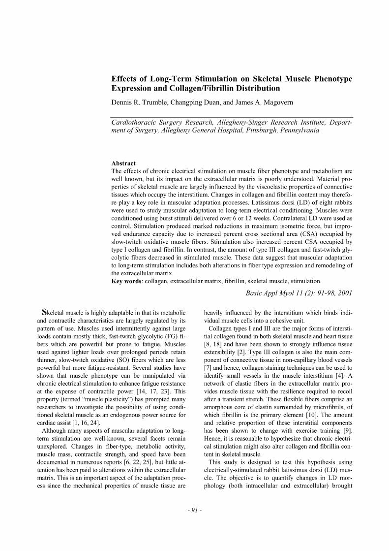
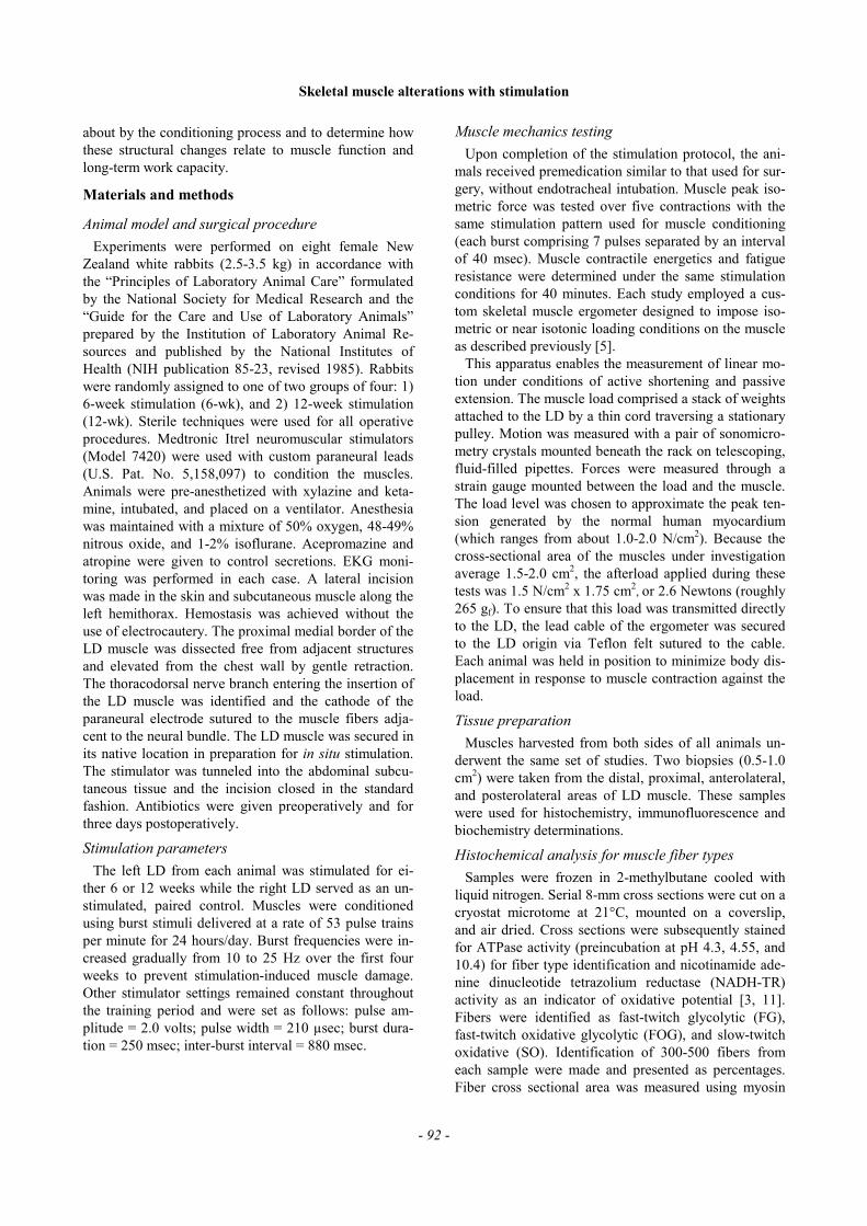
Muscle stimulated for either 6 or 12 weeks produced amarked reduction in maximum isometric force (Figure 1).Comparison of maximum isometric force generation withcontrol muscles shown a 44.3% overall decrease in the 6-week group and a 48.8% decrease in the 12-week group.Strength differences were not significant between the twostimulation groups. Stimulated muscle groups displayedsignificantly improved endurance capacity relative tocontrol, as illustrated in Figure 2. Stroke work was re-duced by 67% after the first five minutes of testing andthen dropped 94% by the ten-minute mark in the controlgroup. At the conclusion of the 40 minutes test, controlmuscles retained only 2% of their initial work capacity.Conditioned muscles were fatigued by 8% in the 6-weekgroup and 18% in the 12-week group after the first fiveminutes of testing and retained 66% (6-wk) and 57% (12-wk) of their initial stroke work at 40 minutes.
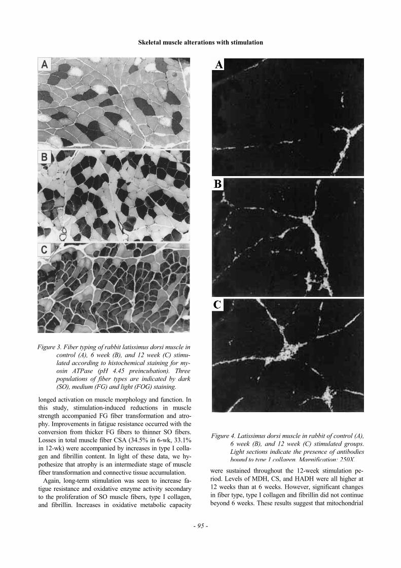
Muscle fiber types
Muscle fiber composition and cross-sectional area dataare shown in Table 1. The percentage of SO fibers inboth 6-wk and 12-wk groups was higher than control.Conversely, the percentage of FG fibers in 6-wk and 12-wk groups was lower than control. Differences in FOGfibers were not significant between stimulated and con-
Figure 1. Peak isometric force generated by controland stimulated muscles. Long-term stimulationdecreased force generation by 44.3% in the 6-wk group and by 48.8% in the 12-wk grouprelative to control. Strength differences werenot significant between the two stimulationgroups. *Statistically significant change rela-tive to control (P<0.05).
Skeletal muscle alterations with stimulation
trol groups. Typical fiber-type distributions for controland conditioned groups are shown in Figure 3. Therewere no differences in the CSA of SO fibers among thethree groups. However, the CSA of FOG fibers was de-creased in the 6-wk group and the CSA of FG fibers wasreduced in both 6-wk and 12-wk groups.
Metabolic enzyme analysis
Muscle enzyme activities measured in LD muscle arepresented in Table 2. PFK and LDH activities were re-duced by both 6-wk and 12-wk stimulation. Conversely,MDH was enhanced in both stimulation groups. Signifi-cant increases in CS and HADH activities were seenonly in the 12-wk group.
Collagen and fibrillin analysis
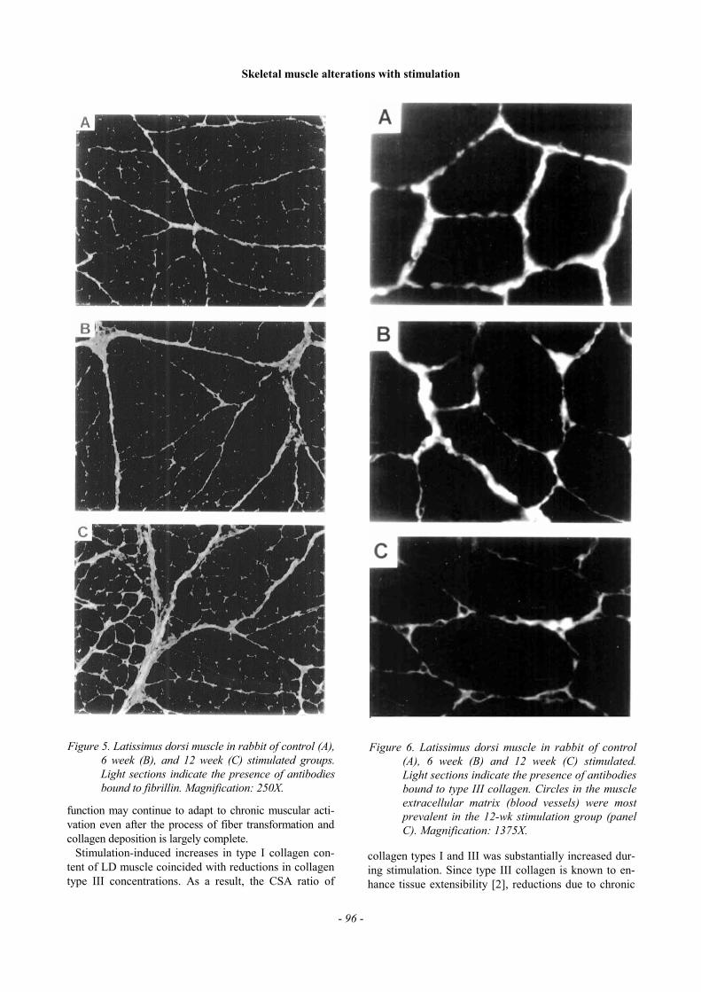
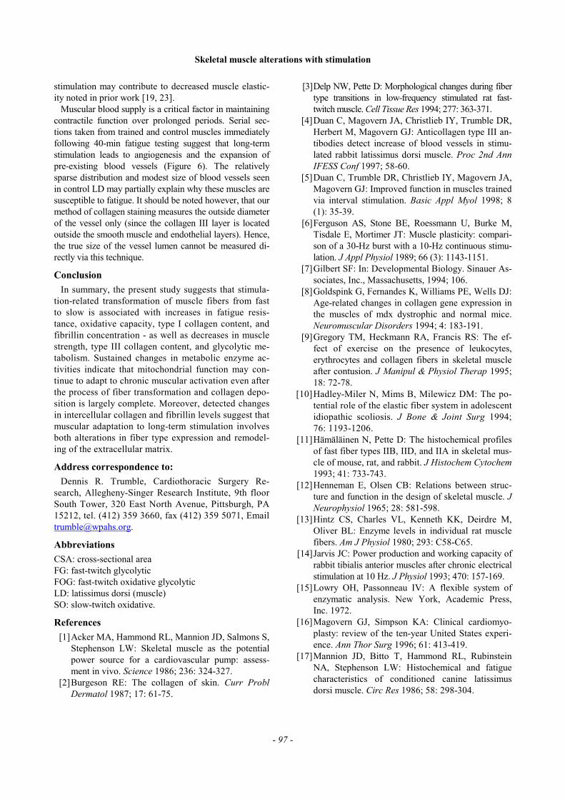
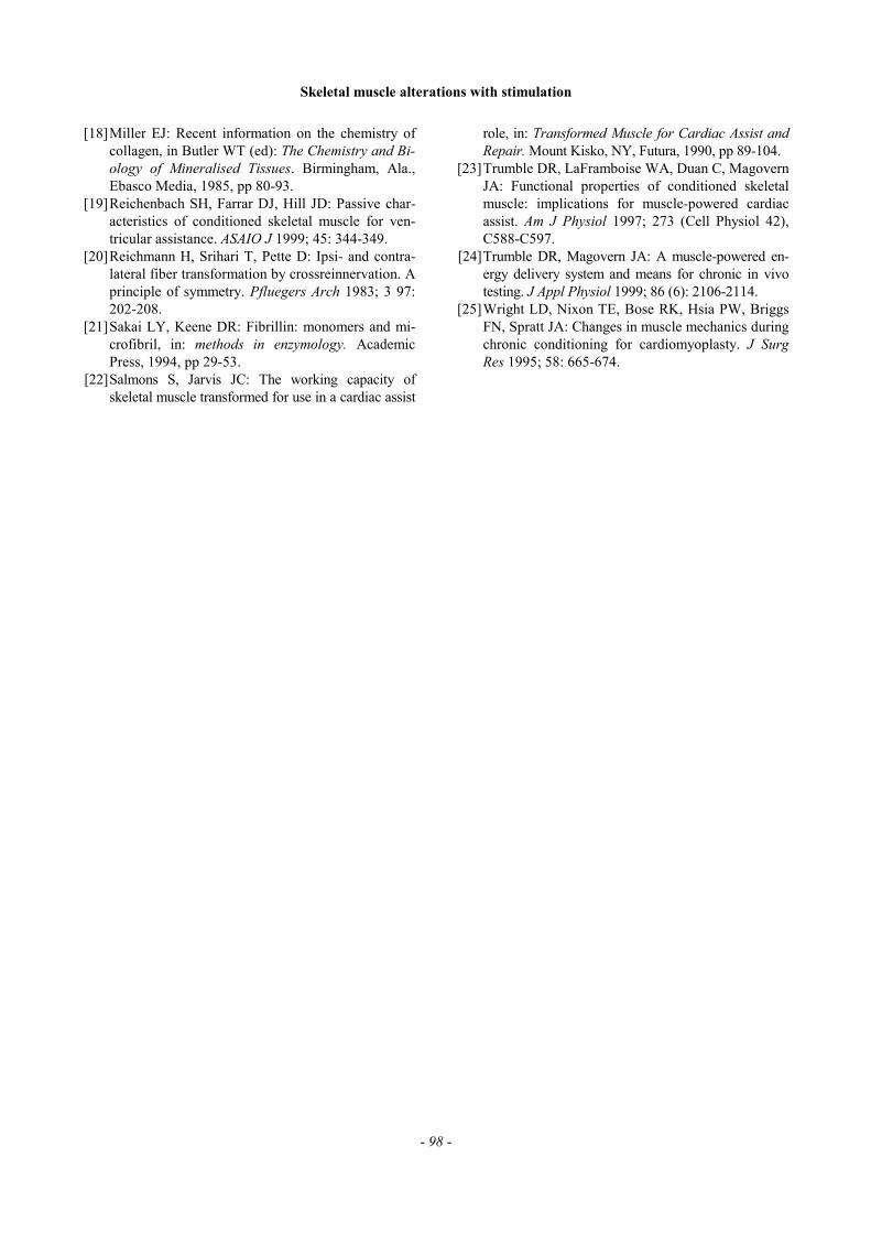
The percentage of muscle CSA seen to fluoresce dueto antibodies bound to type I collagen was lower incontrol LD than in both 6-wk and 12-wk groups, indi-cating an increase in type I collagen content with stimu-lation (Figure 4). Fibrillin concentrations also appearedto increase in response to LD stimulation in both 6-wkand 12-wk stimulated groups (Figure 5). Conversely, theproportion of type III collagen decreased with stimula-tion, total antibody fluorescence appearing lowest in the6-wk group. Some segments of type III collagen formedcircles in the extracellular matrix, (indicative of smallblood vessels), which were seen to both proliferate andexpand after 6 weeks of stimulation (Figure 6). Thenumber and size of these circles were seen to increasefurther after 12 weeks of burst stimulation.
Discussion
The purpose of this study was to determine adaptationsin muscle function, fiber type, metabolic characteristics,and collagen and fibrillin content due to long-termstimulation in rabbit LD muscle. Control LD muscleswere composed mainly of FG fibers (62.3%, Table 1).FG fibers occupied an even greater proportion of musclefiber CSA in control muscles (68.9%) due to their largercross sectional area. Any changes in FG fibers shouldtranslate directly to functional and structural changes inthe LD muscle. Our results support this hypothesis,since decreases in FG fibers corresponded with reduc-tions of isometric force, improvements in fatigue resis-tance, increases in type I collagen and fibrillin, and de-creases in glycolytic enzyme activities.
Observed changes in muscle mechanics, myofiber dis-tribution, collagen volume, and fibrillin content have
Figure 2. Relative endurance characteristics of controland conditioned LD muscles. Stroke work isnormalized to initial work levels and is chartedover 40-minutes of continuous activation at 53contractions per minute. The two conditionedmuscle groups displayed significantly improvedendurance capacities, retaining 66% (6-wk) and57% (12-wk) of their initial stroke work com-pared to 2% for control. *Statistically significantchange relative to control (P<0.05).
Table 1. Muscle fiber type and cross-sectional area of rabbit LD muscle in control, 6-wk, and 12-wk burst stimulation.
Percent Muscle CSA Occupied Mean CSA of Fibers (mm2)
control 6-week 12-week control 6-week 12-week
SO 17.9±2.4 59.0±5.8* 62.3±6.7* 2724±241 2233±140 2321±193FOG 19.9±2.2 28.3±2.4 28.5±7.0 3068±140 2376+138* 2465+57FG 62.3±4.3 12.8±4.4* 9.3±2.2* 3904± 102 2526±179* 2294±150*
Data are mean ± SD. * Statistically significant change relative to control (p < 0.05).
yielded new information concerning the influence of pro-
-
Table 2. Muscle enzyme activities of rabbit LD muscle in control, 6-wk, and 12-wk burst stimulation.
PFK LDH MDH HADH CS
control 31.8±5.9 474.8±68.4 114.0±10.8 4.0±0.6 6.6+0.76-week 9.0±2.4* 139.0±22.8* 157.0±11.7* 3.9+0.3 10.4+1.2
12-week 11.6±13.3* 162.5±26.5* 234.5±13.0*= 6.0±0.6*= 24. l±2.1*=
Data are mean ± SD. Muscle enzyme activities are expressed as µmol/g/min. * Statistically significant change relative to control (p < 0.05). = Statistically significant change relative to 6-wk stimulation (p < 0.05).
- 94 -
Skeletal muscle alterations with stimulation
- 95 -
longed activation on muscle morphology and function. Inthis study, stimulation-induced reductions in musclestrength accompanied FG fiber transformation and atro-phy. Improvements in fatigue resistance occurred with theconversion from thicker FG fibers to thinner SO fibers.Losses in total muscle fiber CSA (34.5% in 6-wk, 33.1%in 12-wk) were accompanied by increases in type I colla-gen and fibrillin content. In light of these data, we hy-pothesize that atrophy is an intermediate stage of musclefiber transformation and connective tissue accumulation.
Again, long-term stimulation was seen to increase fa-tigue resistance and oxidative enzyme activity secondaryto the proliferation of SO muscle fibers, type I collagen,and fibrillin. Increases in oxidative metabolic capacity
were sustained throughout the 12-week stimulation pe-riod. Levels of MDH, CS, and HADH were all higher at12 weeks than at 6 weeks. However, significant changesin fiber type, type I collagen and fibrillin did not continuebeyond 6 weeks. These results suggest that mitochondrial
Figure 3. Fiber typing of rabbit latissimus dorsi muscle incontrol (A), 6 week (B), and 12 week (C) stimu-lated according to histochemical staining for my-osin ATPase (pH 4.45 preincubation). Threepopulations of fiber types are indicated by dark(SO), medium (FG) and light (FOG) staining.
Figure 4. Latissimus dorsi muscle in rabbit of control (A),6 week (B), and 12 week (C) stimulated groups.Light sections indicate the presence of antibodiesbound to type 1 collagen. Magnification: 250X.
Skeletal muscle alterations with stimulation
- 96 -
function may continue to adapt to chronic muscular acti-vation even after the process of fiber transformation andcollagen deposition is largely complete.
Stimulation-induced increases in type I collagen con-tent of LD muscle coincided with reductions in collagentype III concentrations. As a result, the CSA ratio of
collagen types I and III was substantially increased dur-ing stimulation. Since type III collagen is known to en-hance tissue extensibility [2], reductions due to chronic
Figure 6. Latissimus dorsi muscle in rabbit of control(A), 6 week (B) and 12 week (C) stimulated.Light sections indicate the presence of antibodiesbound to type III collagen. Circles in the muscleextracellular matrix (blood vessels) were mostprevalent in the 12-wk stimulation group (panelC). Magnification: 1375X.
Figure 5. Latissimus dorsi muscle in rabbit of control (A),6 week (B), and 12 week (C) stimulated groups.Light sections indicate the presence of antibodiesbound to fibrillin. Magnification: 250X.
Skeletal muscle alterations with stimulation
- 97 -
stimulation may contribute to decreased muscle elastic-ity noted in prior work [19, 23].
Muscular blood supply is a critical factor in maintainingcontractile function over prolonged periods. Serial sec-tions taken from trained and control muscles immediatelyfollowing 40-min fatigue testing suggest that long-termstimulation leads to angiogenesis and the expansion ofpre-existing blood vessels (Figure 6). The relativelysparse distribution and modest size of blood vessels seenin control LD may partially explain why these muscles aresusceptible to fatigue. It should be noted however, that ourmethod of collagen staining measures the outside diameterof the vessel only (since the collagen III layer is locatedoutside the smooth muscle and endothelial layers). Hence,the true size of the vessel lumen cannot be measured di-rectly via this technique.
Conclusion
In summary, the present study suggests that stimula-tion-related transformation of muscle fibers from fastto slow is associated with increases in fatigue resis-tance, oxidative capacity, type I collagen content, andfibrillin concentration - as well as decreases in musclestrength, type III collagen content, and glycolytic me-tabolism. Sustained changes in metabolic enzyme ac-tivities indicate that mitochondrial function may con-tinue to adapt to chronic muscular activation even afterthe process of fiber transformation and collagen depo-sition is largely complete. Moreover, detected changesin intercellular collagen and fibrillin levels suggest thatmuscular adaptation to long-term stimulation involvesboth alterations in fiber type expression and remodel-ing of the extracellular matrix.
Address correspondence to:
Dennis R. Trumble, Cardiothoracic Surgery Re-search, Allegheny-Singer Research Institute, 9th floorSouth Tower, 320 East North Avenue, Pittsburgh, PA15212, tel. (412) 359 3660, fax (412) 359 5071, [email protected].
Abbreviations
CSA: cross-sectional areaFG: fast-twitch glycolyticFOG: fast-twitch oxidative glycolyticLD: latissimus dorsi (muscle)SO: slow-twitch oxidative.
References
[1] Acker MA, Hammond RL, Mannion JD, Salmons S,Stephenson LW: Skeletal muscle as the potentialpower source for a cardiovascular pump: assess-ment in vivo. Science 1986; 236: 324-327. [2] Burgeson RE: The collagen of skin. Curr ProblDermatol 1987; 17: 61-75.
[3] Delp NW, Pette D: Morphological changes during fibertype transitions in low-frequency stimulated rat fast-twitch muscle. Cell Tissue Res 1994; 277: 363-371. [4] Duan C, Magovern JA, Christlieb IY, Trumble DR,Herbert M, Magovern GJ: Anticollagen type III an-tibodies detect increase of blood vessels in stimu-lated rabbit latissimus dorsi muscle. Proc 2nd AnnIFESS Conf 1997; 58-60. [5] Duan C, Trumble DR, Christlieb IY, Magovern JA,Magovern GJ: Improved function in muscles trainedvia interval stimulation. Basic Appl Myol 1998; 8(1): 35-39. [6] Ferguson AS, Stone BE, Roessmann U, Burke M,Tisdale E, Mortimer JT: Muscle plasticity: compari-son of a 30-Hz burst with a 10-Hz continuous stimu-lation. J Appl Physiol 1989; 66 (3): 1143-1151. [7] Gilbert SF: In: Developmental Biology. Sinauer As-sociates, Inc., Massachusetts, 1994; 106. [8] Goldspink G, Fernandes K, Williams PE, Wells DJ:Age-related changes in collagen gene expression inthe muscles of mdx dystrophic and normal mice.Neuromuscular Disorders 1994; 4: 183-191. [9] Gregory TM, Heckmann RA, Francis RS: The ef-fect of exercise on the presence of leukocytes,erythrocytes and collagen fibers in skeletal muscleafter contusion. J Manipul & Physiol Therap 1995;18: 72-78. [10] Hadley-Miler N, Mims B, Milewicz DM: The po-tential role of the elastic fiber system in adolescentidiopathic scoliosis. J Bone & Joint Surg 1994;76: 1193-1206. [11] Hämäläinen N, Pette D: The histochemical profilesof fast fiber types IIB, IID, and IIA in skeletal mus-cle of mouse, rat, and rabbit. J Histochem Cytochem1993; 41: 733-743. [12] Henneman E, Olsen CB: Relations between struc-ture and function in the design of skeletal muscle. JNeurophysiol 1965; 28: 581-598. [13] Hintz CS, Charles VL, Kenneth KK, Deirdre M,Oliver BL: Enzyme levels in individual rat musclefibers. Am J Physiol 1980; 293: C58-C65. [14] Jarvis JC: Power production and working capacity ofrabbit tibialis anterior muscles after chronic electricalstimulation at 10 Hz. J Physiol 1993; 470: 157-169. [15] Lowry OH, Passonneau IV: A flexible system ofenzymatic analysis. New York, Academic Press,Inc. 1972. [16] Magovern GJ, Simpson KA: Clinical cardiomyo-plasty: review of the ten-year United States experi-ence. Ann Thor Surg 1996; 61: 413-419. [17] Mannion JD, Bitto T, Hammond RL, RubinsteinNA, Stephenson LW: Histochemical and fatiguecharacteristics of conditioned canine latissimusdorsi muscle. Circ Res 1986; 58: 298-304.
Skeletal muscle alterations with stimulation
- 98 -
[18] Miller EJ: Recent information on the chemistry ofcollagen, in Butler WT (ed): The Chemistry and Bi-ology of Mineralised Tissues. Birmingham, Ala.,Ebasco Media, 1985, pp 80-93. [19] Reichenbach SH, Farrar DJ, Hill JD: Passive char-acteristics of conditioned skeletal muscle for ven-tricular assistance. ASAIO J 1999; 45: 344-349. [20] Reichmann H, Srihari T, Pette D: Ipsi- and contra-lateral fiber transformation by crossreinnervation. Aprinciple of symmetry. Pfluegers Arch 1983; 3 97:202-208. [21] Sakai LY, Keene DR: Fibrillin: monomers and mi-crofibril, in: methods in enzymology. AcademicPress, 1994, pp 29-53. [22] Salmons S, Jarvis JC: The working capacity ofskeletal muscle transformed for use in a cardiac assist
role, in: Transformed Muscle for Cardiac Assist andRepair. Mount Kisko, NY, Futura, 1990, pp 89-104. [23] Trumble DR, LaFramboise WA, Duan C, MagovernJA: Functional properties of conditioned skeletalmuscle: implications for muscle-powered cardiacassist. Am J Physiol 1997; 273 (Cell Physiol 42),C588-C597. [24] Trumble DR, Magovern JA: A muscle-powered en-ergy delivery system and means for chronic in vivotesting. J Appl Physiol 1999; 86 (6): 2106-2114. [25] Wright LD, Nixon TE, Bose RK, Hsia PW, BriggsFN, Spratt JA: Changes in muscle mechanics duringchronic conditioning for cardiomyoplasty. J SurgRes 1995; 58: 665-674.