Embed Size (px)
Citation preview
Clinical Practice
The effects of ULF–TENS stimulation ongnathology: the state of the art
Nicolae Chipaila, Fabrizio Sgolastra, Alessandro Spadaro, Davide Pietropaoli,Chiara Masci, Ruggero Cattaneo, Annalisa Monaco
Department of Life, Health and Environmental Sciences, Unit of Gnathology, University of L’Aquila, L’Aquila,Italy
Aims: The aim of this study was to evaluate the state of the art in the current literature regarding the effect ofultra low frequency–transcutaneous electrical nerve stimulation (ULF–TENS) on patients with tempor-omandibular disorders (TMD).Methodology: The authors reviewed the literature through a thorough manual and electronic research onPubMed database (using the Medical Subject Headings thesaurus) and subsequent analysis of all thefound papers regarding the effect of TENS on TMD patients. No randomized controlled trials on theinvestigated topic were found. Only eight papers regarding controlled clinical trials (CCT) were selectedaccording to the search strategy selection criteria.Results: According to the available literature and the authors’ experience, ULF–TENS seems to be a validsupport in the management of TMD patients, but also a ‘provocative’ tool, so its application should alwaysbe monitored by electromyographic and electrognathographic analysis (before and after TENS).Conclusions: Further clinical studies (mainly randomized controlled trials) on ULF–TENS application inneuromuscular gnathology are needed.
Keywords: TENS, TMD patients, Masticatory muscles
IntroductionTemporomandibular disorders (TMD): definitionand characteristicsTMD comprise a group of musculoskeletal disorders,
affecting alterations in the structure and/or function
of the temporomandibular joints, masticatory mus-
cles, dentition, and supporting structures.1
Currently, two classification systems are adopted as
the reference standard, both for clinical practice and
the design of scientific studies. Both systems have a
marked clinical guidance, and are based mainly on
symptoms such as pain in the preauricular area and/or
in the masticatory muscles system, abnormalities in
mandibular movements, and joint sounds, such as
clicks and/or crackles during the excursions. The
diagnostic reference classifications are: (1) the one
established by the American Academy of Orofacial
Pain2 in collaboration with the International Headache
Society, and (2) the one that is represented by the
Research Diagnostic Criteria for Temporomandibular
Disorders.3
The latter is the classification system used by most
researchers, and frames the TMD from a dual point of
view, clinical and psychosocial, and refers to the least
operator dependent investigation criteria, and there-
fore, is based on standardized objectification criteria.3–5
According to the most recent literature reviews,
the prevalence of TMD in the general population
reported by several authors varies between 12 and
60%, and the data from different studies are therefore
very much influenced by the sampling methodology
and diagnostic criteria adopted.6,7 The most affected
are women between 25 and 45 years old, with a
prevalence more than double that of men.
The etiology and pathogenesis of TMD are still
poorly understood.8 However, in the pathogenic
sequence of masticatory system disorders, it is possible
to identify the presence of an event able to alter the
threshold of individual physiological tolerance, and
to induce the onset of symptoms. The ‘physiologic
tolerance’ represents the limit of individual adapt-
ability to a certain degree of functional interference.
Correspondence to: Dr Nicolae Chipaila, University of L’Aquila,Department of Life, Health and Environmental Sciences, Unit ofGnathology, Via Vetoio 1, 67100, L’Aquila, Italy. Email: [email protected]
118� W. S. Maney & Son Ltd 2014DOI 10.1179/0886963413Z.00000000018 CRANIOH: The Journal of Craniomandibular & Sleep Practice 2014 VOL. 32 NO. 2
However, this parameter cannot be investigated
scientifically, since it is extremely modulated by
different factors. These events may be local (e.g.
change in proprioceptive input after trauma or
parafunction and central excitatory effect of deep
pain) or systemic (represented mainly by stress).6
Currently, the scientific community agrees to bring
the development of TMD to a multifactorial etiology,
which combine several factors, including the main
ones: psychological, occlusal, parafunctional, trau-
matic, hormonal, postural, neuromuscular.9–15
The diagnosis of TMD is generally based on clinical
examination of the patient; however, there are other
diagnostic tools, such as surface electromyography
(sEMG) and computerized electrognathography, which
provide substantial information about the muscle
activity and jaw movements’ dynamics on the stoma-
tognathic system unit.
The pain reduction and the normal jaw function
restoration are the main goals of conservative
management of TMD.16
Some of the conservative therapies proposed for
the treatment of TMD are: physical therapy, cogni-
tive-behavioral techniques, drug therapy, occlusal
splints, occlusal arrangement, acupuncture, low level
laser therapy (LLLT), and transcutaneous electrical
nerve stimulation (TENS).1,17–22
The aim of this work is to evaluate the state of the
art regarding the effects of TENS on TMD patients.
TENS: definitionTENS is a technique that involves the application of
an electrical stimulus on the major nerves. In relation
to the parameters used (amplitude and impulse
duration), it is possible to act on the major nerves
through a targeted neurophysiological mechanism.
How to use the term ‘TENS’Generally, when we use the term ‘TENS’ in
rehabilitative medicine we refer to antalgic TENS
(pain suppressing TENS). In dentistry, we use the
term ‘TENS’ framing it as a neuromuscular TENS
(functional muscle stimulation). The characteristics
of each of the two stimulations will be described
below.
Antalgic TENSIn general medicine, TENS is a therapeutic modality,
mainly used for the treatment of musculoskeletal
pain, as it promotes analgesic effects.
The idea of using a source of electrical stimulation
as a therapy in reducing the allergic phenomena is not
a recent concept in the world of medicine. In fact, as
Katch23 stated in his work of 1986, as early as 46 AD
Scribonius Largus, the court physician of the Roman
emperor Claudius proposed the use of electric eels in
the treatment of headaches and gout.
Two types of TENS are used clinically, low
frequency TENS (frequency of stimulation ,10 Hz,
LF) and high frequency TENS (frequency of
stimulation .50 Hz, HF). In clinical and research
practice, frequencies ,5 Hz for the low frequency
and .100 Hz for the high frequency are employed.
It must be pointed out that within the frequency,
electrostimulation classification under 1000 Hz is
considered low frequency. Thus, both the ‘low’ and
‘high’ frequency of stimulation for analgesic use are
subclasses of the more general low-frequency stimu-
lation. Hence the term ultra low frequency (ULF)
attributed to stimulation with TENS frequency less
than 4 Hz.
The distinction in the field of antalgic TENS
between low and high frequency has followed a
physiopathogenetic principle. It was believed that the
mechanism of action of LF and HF TENS on the
pain was quite different.24 From the clinical finding,
which is not always detectable, it was found that the
TENS effect is obtainable with different stimulation
modalities.25,26
The LF–TENS analgesia, obtained with 20–
45 minutes administration times, was longer lasting
after the suspension of the pulse, while the HF–TENS
analgesia was rapid in setting up but quickly stopped
once stimulation was suspended.24–26
LF–TENS stimulation, to be able to evoke the
movement of the joint segment, must be administered
to such an amplitude as to feel uncomfortable by the
subject who receives it. Therefore, the low frequency
stimulation would be conducted on the small caliber
sensory fibers, which carry the pain and deep
sensibility.24–26 Once in the posterior horn of the
medulla, the second order activated neuron would
project at the level of various areas of the trunk,
including the periaqueductal gray and other areas
throughout defined Substance Producing Analgesia
coding for an endorphin analgesic effect.27,28 The
endorphin path needs a longer time to be activated,
but is active for longer, as long as the endorphins
have been degraded and have ceased their activity.
HF–TENS is not felt by the subject, except as a
slight tingling sensation. The feeling is not unplea-
sant; indeed, it is sometimes perceived as pleasant.
It is assumed that the stimulation, which does not
cause pain, is conducted along sensory fibers of large
caliber. The second order medullary neuron would be
controlled by an inhibitor interneuron. The activa-
tion of this interneuron inhibits the transmission of
the second order neuron, while its inhibition would
Chipaila et al. Effects of ULF–TENS stimulation on gnathology
CRANIOH: The Journal of Craniomandibular & Sleep Practice 2014 VOL. 32 NO. 2 119
lead to a greater freedom of transmission of the
second order neuron. The HF–TENS would act on
this interneuron. In fact, the large caliber fibers would
send an excitatory collateral to the inhibitor inter-
neuron, which, once activated, would lead to the
transmission inhibition by the spinal cord second
order neuron. Any information on this second-order
neuron should come from the periphery, for example,
conveyed by nociceptive fibers, and would be blocked
because the second-order neuron was inhibited by the
interneuron excited by pulses (HF–TENS) running
on the fibers of large caliber: the gate closes.
The phenomenon would be ‘direct’ and the feed-
back could be active only in the presence of
stimulation of large nerve fibers. The antalgic effect
would cease at the time of HF–TENS cessation.
Generally, high frequency and low intensity
characteristics of stimulation are those that allow us
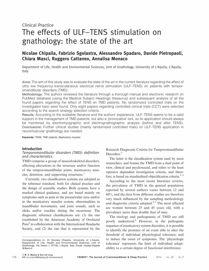
to call this kind of application ‘conventional’ (Fig. 1).
Conventional TENS works via Melzack and Wall’s
‘gate control’ theory,29 by the activation of peripheral
inhibition systems of nociceptive stimuli, and by
stimulating production and release of endogenous
opioids, neuropeptides and neurotransmitters with
analgesic action. Neither action mechanism would
have a simple symptomatic action, but would act with
synergy and graduality, obtaining a sort of ‘reset’ of
the nociceptive system. Other action mechanisms,
such as the metabolic recovery of the muscular tissue
and the unloading reflex, hypnosis and stress
analgesia, exteroceptive suppression and counter
irritation, endogenous inhibition and sympathetic
activity reduction have also been suggested by
Galletti et al.30
Neuromuscular TENSRegarding the purely muscular component, it has
been argued that the impulse is conducted by dromic
and antidromic methods. The dromic method would
mean that, once it meets the motor nerve trunk, the
current is conveyed to the periphery in order to
achieve the innervated muscle fibers. The effect of this
path would lead to contraction of the muscle itself
and, if the stimulus amplitude is sufficient to
determine the activation of a sufficient number of
motor units and if the muscle ancillary articular
heads are free, there may be a shift of the body
segment. Specifically, a contraction of the lip, the
eyelid or the raising of the jaw by the action of the
VIIth or the Vth pair of cranial nerves could occur.
The effect due to the muscle contraction implies,
then, a series of side effects related to the activation
of the reflexes circuits mediated by neuromuscular
spindles, by the stretching of the tendons, which in
turn contribute to modulate the effects of the second,
third or hundredth pulse generated by TENS.
Often these aspects are neglected in the explanation
of the TENS effect. This may be a mistake, because
even at this level, one should recognize the consider-
able complexity of action and central connection that
should not be overlooked in considering the effects,
even purely muscular of transcutaneous electrical
nerve stimulation.
The second path covered by the impulse is defined
antidromic. In short, the impulse besides being
conducted towards the muscular periphery, would
be conducted to the motor nuclei of the involved
nerves. This would bring the direct activation of
motor neurons and, through them, the impulse would
then be carried to the muscles.
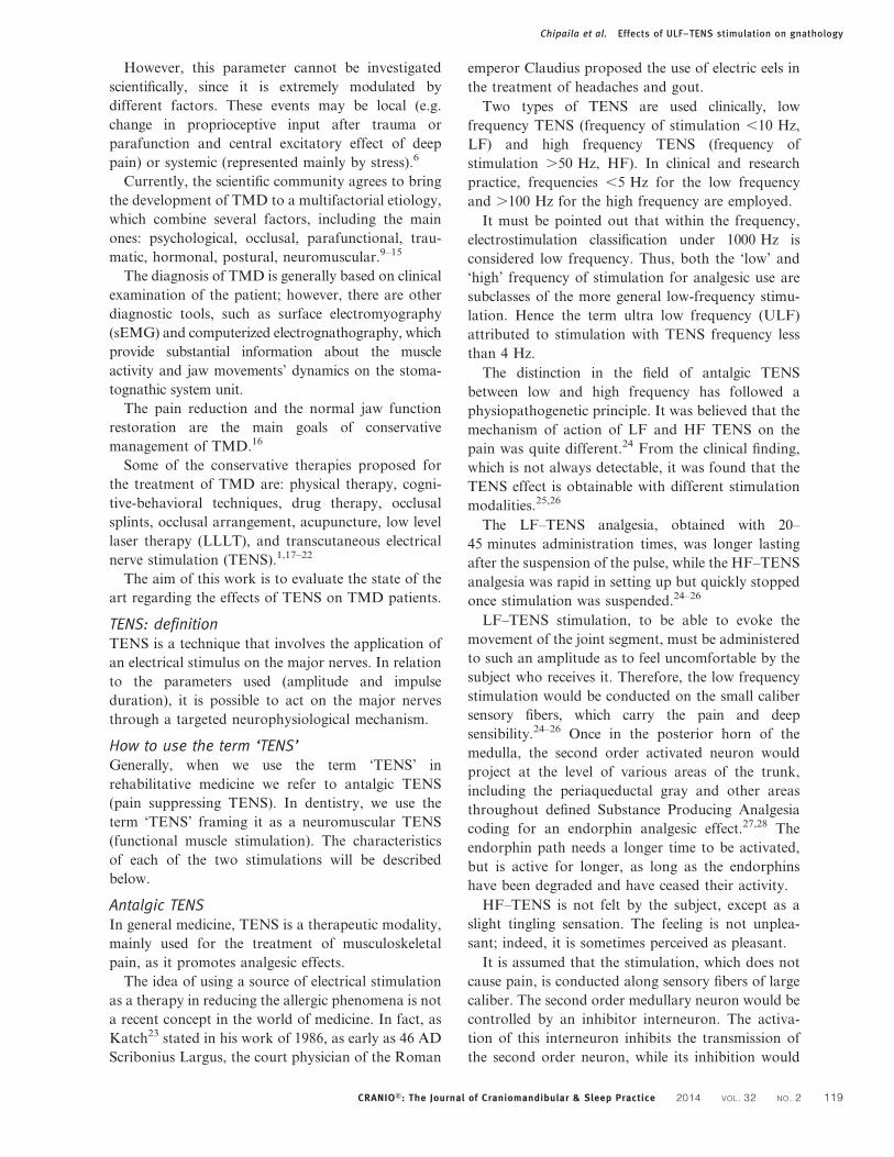
Neuromuscular TENS or ULF–TENS was pro-
posed for the first time by Jankelson31 in 1969, and is
currently studied and has been used as a TMD
therapeutic strategy by several authors.22,32,33 It
consists in the application of three electrodes, two
active electrodes (anode), positioned on the skin
overlying the sigmoid notch of the jaw, immediately
Figure 1 Conventional TENS stimulator device.
Chipaila et al. Effects of ULF–TENS stimulation on gnathology
120 CRANIOH: The Journal of Craniomandibular & Sleep Practice 2014 VOL. 32 NO. 2
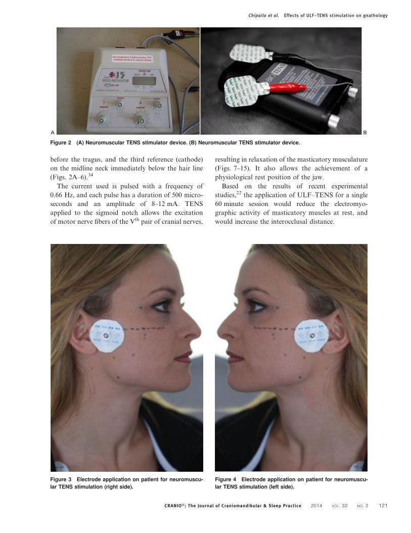
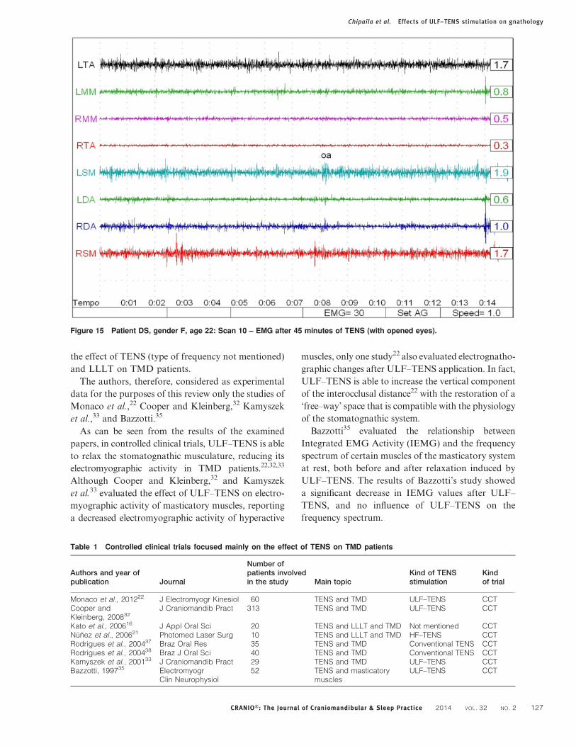
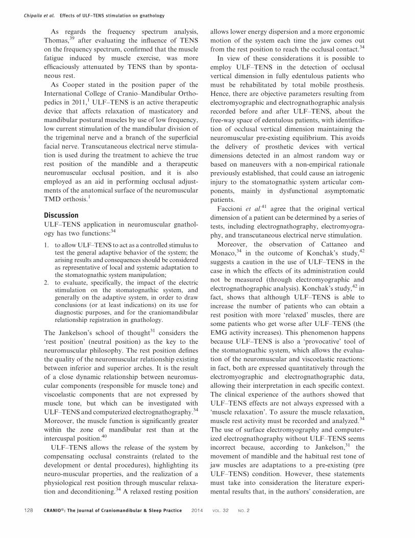
before the tragus, and the third reference (cathode)
on the midline neck immediately below the hair line
(Figs. 2A–6).34
The current used is pulsed with a frequency of
0.66 Hz, and each pulse has a duration of 500 micro-
seconds and an amplitude of 8–12 mA. TENS
applied to the sigmoid notch allows the excitation
of motor nerve fibers of the Vth pair of cranial nerves,
resulting in relaxation of the masticatory musculature
(Figs. 7–15). It also allows the achievement of a
physiological rest position of the jaw.
Based on the results of recent experimental
studies,22 the application of ULF–TENS for a single
60 minute session would reduce the electromyo-
graphic activity of masticatory muscles at rest, and
would increase the interocclusal distance.
Figure 3 Electrode application on patient for neuromuscu-
lar TENS stimulation (right side).
Figure 4 Electrode application on patient for neuromuscu-
lar TENS stimulation (left side).
Figure 2 (A) Neuromuscular TENS stimulator device. (B) Neuromuscular TENS stimulator device.
Chipaila et al. Effects of ULF–TENS stimulation on gnathology
CRANIOH: The Journal of Craniomandibular & Sleep Practice 2014 VOL. 32 NO. 2 121
Several authors have evaluated the effect of ULF–
TENS on electromyographic activity of masticatory
muscles in TMD patients.
Cooper and Kleinberg32 concluded that ULF and
low amplitude TENS applied for 60 minutes in TMD
patients is able to relax the masticatory musculature
and facilitate the detection of a physiological rest
position of the jaw. Kamyszek et al.33 evaluated the
effects of ULF–TENS applied for 30–40 minutes in
TMD patients with and without hyperactivity muscle
at rest, with similar results in terms of reduction of
EMG activity of the stomatognathic musculature, in
agreement with the results of Bazzotti.35
Didier et al.36 suggest the use of ULF–TENS in
deprogramming the masticatory musculature aimed
to identify the physiological rest position of the jaw in
patients with chronic daily headache who must be
subjected to neuromuscular therapy with occlusal
devices. They stress the importance of ULF–TENS in
deprogramming the muscles of the stomatognathic
system in chronic daily headache patients with
significant discrepancy of mandibular position, and
the role of electrognathographic exams in high-
lighting this discrepancy.36
Materials and MethodsFor the current study, the PubMed database was
used. The search strategy in the PubMed database
was applied to the thesaurus Medical Subject
Headings and it involved the following terms: ‘tens
and tmd’ (n518 articles found), ‘temporomandibular
disorders and tens’ (n51937 articles found), ‘trancu-
taneous electrical nerve stimulation and temporo-
mandibular disorders’ (n567 articles found),
‘trancutaneous electrical nerve stimulation and
TMD’ (n515 articles found), ‘craniomandibular
disorders and transcutaneous electrical nerve stimu-
lation’ (n569 articles found), ‘craniomandibular
disorders and tens’ (n570 articles found), ‘tens and
dentistry’ (n5238 articles found), ‘transcutaneous
electrical nerve stimulation and dentistry’ (n5201
articles found).
All the articles selected from the PubMed database
were manually analyzed by title, keywords, and
abstract. The articles concerning the application of
TENS in patients with bruxism or clenching were
excluded from this review, as these events are anxious
parafunctions with increased tension of the mastica-
tory muscles, not classifiable as TMDs.
No randomized controlled trials on TENS applica-
tion in TMD patients were found.
Only eight papers (CCT) found approval by the
authors according to their relevancy on the debated
topic (Table 1), so they were thoroughly examined.
ResultsExamining the selected works, it was found that only
four studies22,32,33,35 have investigated the effect of
ULF–TENS on TMD patients: two studies37,38 (but
with the same authors) evaluated the effect of
conventional TENS on pain, masticatory muscle
activity and activation pattern of TMD patients,
one study21 focused on the effect of HF–TENS and
LLLT on TMD patients, and one study16 evaluatedFigure 6 Patient after electrodes application for TENS and
electromyography (EMG).
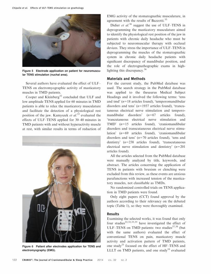
Figure 5 Electrode application on patient for neuromuscu-
lar TENS stimulation (nuchal area).
Chipaila et al. Effects of ULF–TENS stimulation on gnathology
122 CRANIOH: The Journal of Craniomandibular & Sleep Practice 2014 VOL. 32 NO. 2
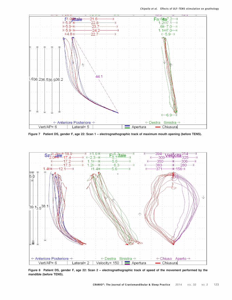
Figure 8 Patient DS, gender F, age 22: Scan 2 – electrognathographic track of speed of the movement performed by the
mandible (before TENS).
Figure 7 Patient DS, gender F, age 22: Scan 1 – electrognathographic track of maximum mouth opening (before TENS).
Chipaila et al. Effects of ULF–TENS stimulation on gnathology
CRANIOH: The Journal of Craniomandibular & Sleep Practice 2014 VOL. 32 NO. 2 123
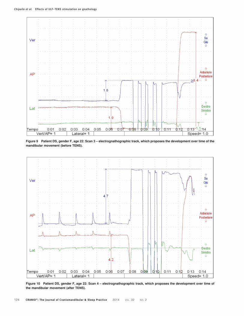
Figure 10 Patient DS, gender F, age 22: Scan 4 – electrognathographic track, which proposes the development over time of
the mandibular movement (after TENS).
Figure 9 Patient DS, gender F, age 22: Scan 3 – electrognathographic track, which proposes the development over time of the
mandibular movement (before TENS).
Chipaila et al. Effects of ULF–TENS stimulation on gnathology
124 CRANIOH: The Journal of Craniomandibular & Sleep Practice 2014 VOL. 32 NO. 2
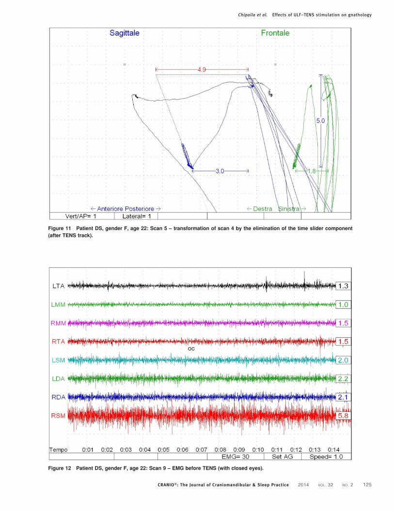
Figure 12 Patient DS, gender F, age 22: Scan 9 – EMG before TENS (with closed eyes).
Figure 11 Patient DS, gender F, age 22: Scan 5 – transformation of scan 4 by the elimination of the time slider component
(after TENS track).
Chipaila et al. Effects of ULF–TENS stimulation on gnathology
CRANIOH: The Journal of Craniomandibular & Sleep Practice 2014 VOL. 32 NO. 2 125
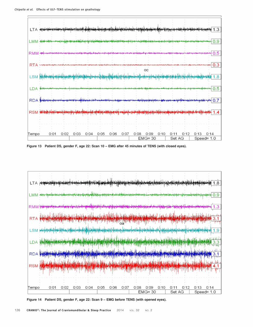
Figure 14 Patient DS, gender F, age 22: Scan 9 – EMG before TENS (with opened eyes).
Figure 13 Patient DS, gender F, age 22: Scan 10 – EMG after 45 minutes of TENS (with closed eyes).
Chipaila et al. Effects of ULF–TENS stimulation on gnathology
126 CRANIOH: The Journal of Craniomandibular & Sleep Practice 2014 VOL. 32 NO. 2
the effect of TENS (type of frequency not mentioned)
and LLLT on TMD patients.
The authors, therefore, considered as experimental
data for the purposes of this review only the studies of
Monaco et al.,22 Cooper and Kleinberg,32 Kamyszek
et al.,33 and Bazzotti.35
As can be seen from the results of the examined
papers, in controlled clinical trials, ULF–TENS is able
to relax the stomatognathic musculature, reducing its
electromyographic activity in TMD patients.22,32,33
Although Cooper and Kleinberg,32 and Kamyszek
et al.33 evaluated the effect of ULF–TENS on electro-
myographic activity of masticatory muscles, reporting
a decreased electromyographic activity of hyperactive
muscles, only one study22 also evaluated electrognatho-
graphic changes after ULF–TENS application. In fact,
ULF–TENS is able to increase the vertical component
of the interocclusal distance22 with the restoration of a
‘free–way’ space that is compatible with the physiology
of the stomatognathic system.
Bazzotti35 evaluated the relationship between
Integrated EMG Activity (IEMG) and the frequency
spectrum of certain muscles of the masticatory system
at rest, both before and after relaxation induced by
ULF–TENS. The results of Bazzotti’s study showed
a significant decrease in IEMG values after ULF–
TENS, and no influence of ULF–TENS on the
frequency spectrum.
Table 1 Controlled clinical trials focused mainly on the effect of TENS on TMD patients
Authors and year ofpublication Journal
Number ofpatients involvedin the study Main topic
Kind of TENSstimulation
Kindof trial
Monaco et al., 201222 J Electromyogr Kinesiol 60 TENS and TMD ULF–TENS CCTCooper andKleinberg, 200832
J Craniomandib Pract 313 TENS and TMD ULF–TENS CCT
Kato et al., 200616 J Appl Oral Sci 20 TENS and LLLT and TMD Not mentioned CCTNunez et al., 200621 Photomed Laser Surg 10 TENS and LLLT and TMD HF–TENS CCTRodrigues et al., 200437 Braz Oral Res 35 TENS and TMD Conventional TENS CCTRodrigues et al., 200438 Braz J Oral Sci 40 TENS and TMD Conventional TENS CCTKamyszek et al., 200133 J Craniomandib Pract 29 TENS and TMD ULF–TENS CCTBazzotti, 199735 Electromyogr
Clin Neurophysiol52 TENS and masticatory
musclesULF–TENS CCT
Figure 15 Patient DS, gender F, age 22: Scan 10 – EMG after 45 minutes of TENS (with opened eyes).
Chipaila et al. Effects of ULF–TENS stimulation on gnathology
CRANIOH: The Journal of Craniomandibular & Sleep Practice 2014 VOL. 32 NO. 2 127
As regards the frequency spectrum analysis,
Thomas,39 after evaluating the influence of TENS
on the frequency spectrum, confirmed that the muscle
fatigue induced by muscle exercise, was more
efficaciously attenuated by TENS than by sponta-
neous rest.
As Cooper stated in the position paper of the
International College of Cranio–Mandibular Ortho-
pedics in 2011,1 ULF–TENS is an active therapeutic
device that affects relaxation of masticatory and
mandibular postural muscles by use of low frequency,
low current stimulation of the mandibular division of
the trigeminal nerve and a branch of the superficial
facial nerve. Transcutaneous electrical nerve stimula-
tion is used during the treatment to achieve the true
rest position of the mandible and a therapeutic
neuromuscular occlusal position, and it is also
employed as an aid in performing occlusal adjust-
ments of the anatomical surface of the neuromuscular
TMD orthosis.1
DiscussionULF–TENS application in neuromuscular gnathol-
ogy has two functions:34
1. to allow ULF–TENS to act as a controlled stimulus totest the general adaptive behavior of the system; thearising results and consequences should be consideredas representative of local and systemic adaptation tothe stomatognathic system manipulation;
2. to evaluate, specifically, the impact of the electricstimulation on the stomatognathic system, andgenerally on the adaptive system, in order to drawconclusions (or at least indications) on its use fordiagnostic purposes, and for the craniomandibularrelationship registration in gnathology.
The Jankelson’s school of thought31 considers the
‘rest position’ (neutral position) as the key to the
neuromuscular philosophy. The rest position defines
the quality of the neuromuscular relationship existing
between inferior and superior arches. It is the result
of a close dynamic relationship between neuromus-
cular components (responsible for muscle tone) and
viscoelastic components that are not expressed by
muscle tone, but which can be investigated with
ULF–TENS and computerized electrognathography.34
Moreover, the muscle function is significantly greater
within the zone of mandibular rest than at the
intercuspal position.40
ULF–TENS allows the release of the system by
compensating occlusal constraints (related to the
development or dental procedures), highlighting its
neuro-muscular properties, and the realization of a
physiological rest position through muscular relaxa-
tion and deconditioning.34 A relaxed resting position
allows lower energy dispersion and a more ergonomic
motion of the system each time the jaw comes out
from the rest position to reach the occlusal contact.34
In view of these considerations it is possible to
employ ULF–TENS in the detection of occlusal
vertical dimension in fully edentulous patients who
must be rehabilitated by total mobile prosthesis.
Hence, there are objective parameters resulting from
electromyographic and electrognathographic analysis
recorded before and after ULF–TENS, about the
free-way space of edentulous patients, with identifica-
tion of occlusal vertical dimension maintaining the
neuromuscular pre-existing equilibrium. This avoids
the delivery of prosthetic devices with vertical
dimensions detected in an almost random way or
based on maneuvers with a non-empirical rationale
previously established, that could cause an iatrogenic
injury to the stomatognathic system articular com-
ponents, mainly in dysfunctional asymptomatic
patients.
Faccioni et al.41 agree that the original vertical
dimension of a patient can be determined by a series of
tests, including electrognathography, electromyogra-
phy, and transcutaneous electrical nerve stimulation.
Moreover, the observation of Cattaneo and
Monaco,34 in the outcome of Konchak’s study,42
suggests a caution in the use of ULF–TENS in the
case in which the effects of its administration could
not be measured (through electromyographic and
electrognathographic analysis). Konchak’s study,42 in
fact, shows that although ULF–TENS is able to
increase the number of patients who can obtain a
rest position with more ‘relaxed’ muscles, there are
some patients who get worse after ULF–TENS (the
EMG activity increases). This phenomenon happens
because ULF–TENS is also a ‘provocative’ tool of
the stomatognathic system, which allows the evalua-
tion of the neuromuscular and viscoelastic reactions:
in fact, both are expressed quantitatively through the
electromyographic and electrognathographic data,
allowing their interpretation in each specific context.
The clinical experience of the authors showed that
ULF–TENS effects are not always expressed with a
‘muscle relaxation’. To assure the muscle relaxation,
muscle rest activity must be recorded and analyzed.34
The use of surface electromyography and computer-
ized electrognathography without ULF–TENS seems
incorrect because, according to Jankelson,31 the
movement of mandible and the habitual rest tone of
jaw muscles are adaptations to a pre-existing (pre
ULF–TENS) condition. However, these statements
must take into consideration the literature experi-
mental results that, in the authors’ consideration, are
Chipaila et al. Effects of ULF–TENS stimulation on gnathology
128 CRANIOH: The Journal of Craniomandibular & Sleep Practice 2014 VOL. 32 NO. 2
enough for drawing conclusions, at least about the
current state of the art of TENS effect on TMD
patients. The authors also suggest further clinical and
experimental investigations about this topic, to
support the current findings.
ConclusionsAccording to examined papers, the authors can
conclude that:
1. TENS is a therapeutic tool able to relax thestomatognathic muscles, to reduce the electromyo-graphic activity of masticatory muscles at rest, and toincrease the vertical component of the interocclusaldistance in TMD patients.
2. TENS is able to facilitate the detection of aphysiological rest position of the jaw.
3. The effects of TENS must be checked with sEMGand electrognathography.
4. Further studies are needed regarding the applica-tion of TENS in neuromuscular gnathology, mainlyrandomized controlled trials.
Disclaimer statementsContributors All the authors contributed actively to
this study: NC conceived, designed and wrote the
paper, FS, AS, DP and CM performed the Pubmed
search and articles analysis, and RC and AM
reviewed the paper.
Funding These authors have no support or funding
to report.
Conflicts of interest The authors declare that they
have no conflict of interests.
Ethics approval This study was performed in
accordance with the ethical principles of the World
Medical Association’s Declaration of Helsinki.
References1 Cooper BC. Temporomandibular disorders: A position paper
of the International College of Cranio-Mandibular Orthopedics(ICCMO). J Craniomandib Pract. 2011;29(3):237–44.
2 American Academy of Orofacial Pain: Assessment of orofacialpain disorders. In: Okeson JP, editor. Orofacial pain: guidelinesfor assessment, diagnosis, and management. Chicago, IL:Quintessence; 1996. p. 19–44.
3 Dworkin SF, Leresche L. Research diagnostic criteria fortemporomandibular disorders: review, criteria, examinationsand specifications, critique. J Craniomand Disord. 1992;6:301–55.
4 National Institute of Health Technology Assessment ConferenceStatement: Management of temporomandibular disorders. J AmDent Assoc. 1996;127:1595–603.
5 Plesh O, Sinisi SE, Crawford PB, Gansky SA. Diagnoses basedon the Research Diagnostic Criteria for TemporomandibularDisorders in a biracial population of young women. J OrofacPain. 2005;19(1):65–75.
6 Manfredini D, Piccotti F, Ferronato G, Guarda-Nardini L.Age peaks of different RDC/TMD diagnoses in a patientpopulation. J Dent. 2010;38(5):392–9.
7 Goncalves DA, Dal Fabbro AL, Campos JA, Bigal ME, SpecialiJG. Symptoms of temporomandibular disorders in the popula-tion: an epidemiological study. J Orofac Pain. 2010;24(3):270–8.
8 Sessle BJ. Evolution of the research diagnostic criteria fortemporomandibular disorders. J Orofac Pain. 2010;24(1):5.
9 Mackie A, Lyons K. The role of occlusion in temporomandib-ular disorders-a review of the literature. N Z Dent J.2008;104(2):54–9.
10 Maixner W, Greenspan JD, Dubner R, Bair E, Mulkey F,Miller V, et al. Potential autonomic risk factors for chronicTMD: descriptive data and empirically identified domains fromthe OPPERA case-control study. J Pain. 2011;12(11):T75–91.
11 Fillingim RB, Ohrbach R, Greenspan JD, Knott C, Dubner R,Bair E, et al. Potential psychosocial risk factors for chronicTMD: descriptive data and empirically identified domains fromthe OPPERA case-control study. J Pain. 2011;12(11):T46–60.
12 Monaco A, Cozzolino V, Cattaneo R, Cutilli T, Spadaro A.Osteopathic manipulative treatment (OMT) effects on man-dibular kinetics: kinesiographic study. Eur J Paediatr Dent.2008;9(1):37–42.
13 Monaco A, Cattaneo R, Spadaro A, D’Andrea P, Marzo G,Gatto R: Ocular correction effects on EMG activity ofstomatognathic muscles in children with functional mandibularlateral- deviation: a case control study. Eur J Paediatr Dent.2006;7(2):81–8.
14 Monaco A, Cattaneo R, Spadaro A, Giannoni M, Di MartinoS, Gatto R. Visual input effect on EMG activity of masticatoryand postural muscles in healthy and in myopic children. Eur JPaediatr Dent. 2006;7(1):18–22.
15 Monaco A, Cattaneo R, Mesin L, Ciarrocchi I, Sgolastra F,Pietropaoli D. Dysregulation of the autonomous nervoussystem in patients with temporomandibular disorder: apupillometric study. PLoS One. 2012;7(9):e45424.
16 Kato MT, Kogawa EM, Santos CN, Conti PC. TENS and low-level laser therapy in the management of temporomandibulardisorders. J Appl Oral Sci. 2006;14(2):130–5.
17 Madani AS, Mirmortazavi A. Comparison of three treatmentoptions for painful temporomandibular joint clicking. J OralSci. 2011;53(3):349–54.
18 Dym H, Israel H. Diagnosis and treatment of temporoman-dibular disorders. Dent Clin North Am. 2012;56(1):149–61, ix.
19 Inchingolo F, Tatullo M, Marrelli M, Inchingolo AM, TarulloA, Inchingolo AD, et al. Combined occlusal and pharmacolo-gical therapy in the treatment of temporo-mandibular dis-orders. Eur Rev Med Pharmacol Sci. 2011;15(11):1296–300.
20 Petrucci A, Sgolastra F, Gatto R, Mattei A, Monaco A.Effectiveness of low-level laser therapy in temporomandibulardisorders: a systematic review and meta-analysis. J Orofac Pain.2011;25(4):298–307.
21 Nunez SC, Garcez AS, Suzuki SS, Ribeiro MS. Management ofmouth opening in patients with temporomandibular disordersthrough low-level laser therapy and transcutaneous electricalneural stimulation. Photomed Laser Surg. 2006;24(1):45–9.
22 Monaco A, Sgolastra F, Ciarrocchi I, Cattaneo R. Effect oftranscutaneous electrical nervous stimulation on electromyo-graphic and kinesiographic activity of patients with temporoman-dibular disorders: a placebo-controlled study. J ElectromyogrKinesiol. 2012;22(3):463–8.
23 Katch EM. Application of transcutaneous electrical nervestimulation in dentistry. Anesth Prog. 1986;33(3):156–60.
24 Sluka KA, Walsh D. Transcutaneous electrical nerve stimula-tion: basic science mechanisms and clinical effectiveness. J Pain.2003;4(3):109–21.
25 King EW, Sluka KA. The effect of varying frequency andintensity of transcutaneous electrical nerve stimulation onsecondary mechanical hyperalgesia in an animal model ofinflammation. J Pain. 2001;2(2):128–33.
26 Gopalkrishnan P, Sluka KA. Effect of varying frequency,intensity, and pulse duration of transcutaneous electrical nervestimulation on primary hyperalgesia in inflamed rats. ArchPhys Med Rehabil. 2000;81(7):984–90.
27 Bandler R, Keay KA, Floyd N, Price J. Central circuitsmediating patterned autonomic activity during active vs.passive emotional coping. Brain Res Bull. 2000;53(1):95–104.
28 Waters AJ, Lumb BM. Descending control of spinal nociceptionfrom the periaqueductal grey distinguishes between neurons withand without C-fibre inputs. Pain. 2008;134(1–2):32–40.
29 Melzack R, Wall PD. Pain mechanisms: a new theory. Science.1965;150(3699):971–9.
Chipaila et al. Effects of ULF–TENS stimulation on gnathology
CRANIOH: The Journal of Craniomandibular & Sleep Practice 2014 VOL. 32 NO. 2 129
30 Galletti SP, Bergamini M, Pantaleo T. Highlights in the subjectof low frequency-high intensity TENS (review). MinervaStomatol. 1995;44(9):421–9.
31 Jankelson B. Electronic control of muscle contraction-a newclinical era in occlusion and prosthodontics. Sci Educ Bull.1969;2(1):29–31.
32 Cooper BC, Kleinberg I. Establishment of a temporomandib-ular physiological state with neuromuscular orthosis treatmentaffects reduction of TMD symptoms in 313 patients. JCraniomandib Pract. 2008;26(2):104–17.
33 Kamyszek G, Ketcham R, Garcia R, Radke J.Electromyographic evidence of reduced muscle activity whenULF-TENS is applied to the Vth and VIIth cranial nerves. JCraniomandib Pract. 2001;19(3):162–8.
34 Cattaneo R, Monaco A. Elettromiografia e chinesiografia perla clinica odontoiatrica. Principi di odontoiatria neuro miofasciale. San Benedetto del Tronto: Futura Publishing Society;2007.
35 Bazzotti L. Electromyography tension and frequency spectrumanalysis at rest of some masticatory muscles, before and afterTENS. Electromyogr. Clin. Neurophysiol. 1997;37:365–78.
36 Didier H, Marchetti C, Borromeo G, Tullo V, D’Amico D,Bussone G, et al. Chronic daily headache: suggestion for theneuromuscular oral therapy. Neurol Sci. 2011;32(1):S161–4.
37 Rodrigues D, Siriani AO, Berzin F. Effect of conventionalTENS on pain and electromyographic activity of masticatorymuscles in TMD patients. Braz Oral Res. 2004;18(4):290–5.
38 Rodrigues D, Siriani AO, Berzin F. Effect of tens on theactivation pattern of the masticatory muscles in TMD patients.Braz J Oral Sci. 2004;3(10):510–5.
39 Thomas NR. The effect of fatigue and TENS on the EMGmean power frequency. Front oral physiology. Vol. 7. Basel:Karger; 1990. p. 162.
40 Hickman DM, Stauber W. Mapping mandibular rest inhumans utilizing electromyographic patterns from masticatorymuscles. J Craniomandib Pract. 2007;25(4):264–72.
41 Faccioni F, Laino A, Papadia D. Rehabilitation of partiallyedentulous patient with loss of vertical dimension. ProgOrthod. 2004;5:4–17.
42 Konchak P, Thomas N, Lanigan D, Devon R. Free way spacemeasurement using mandibular kinesiograph and EMG beforeand after TENS. Angle Orthod. 1988;58:343–50.
Chipaila et al. Effects of ULF–TENS stimulation on gnathology
130 CRANIOH: The Journal of Craniomandibular & Sleep Practice 2014 VOL. 32 NO. 2