Embed Size (px)
Citation preview

Effects of non-invasive brain stimulation on post-stroke dysphagia: Asystematic review and meta-analysis of randomized controlled trials
Jessica M. Pisegna a,b,!, Asako Kaneoka a,b, William G. Pearson Jr. c, Sandeep Kumar d, Susan E. Langmore a,b
a Boston University Medical Center, FGH Building 820 Harrison Ave., Boston, MA 02118, United Statesb Boston University, Sargent College, 635 Commonwealth Ave., Boston, MA 02215, United Statesc Georgia Regents University, 1120 15th St., Augusta, GA 30912, United Statesd Beth Israel Deaconess Medical Center, 330 Brookline Ave., Boston, MA 02215, United States
a r t i c l e i n f o
Article history:Accepted 25 April 2015Available online xxxx
Keywords:Transcranial magnetic stimulation (TMS)Transcranial direct current stimulation(tDCS)Non-invasive brain stimulationUnilateral lesionPost-stroke populationDysphagia
h i g h l i g h t s
! We synthesize evidence for non-invasive brain stimulation on post-stroke dysphagia.! Transcranial direct current stimulation (tDCS) and repetitive transcranial magnetic stimulation
(rTMS) significantly increased swallowing outcomes in stroke patients.! Stimulating the unaffected hemisphere resulted in larger and significant effect sizes.
a b s t r a c t
Objective: The primary aim of this review is to evaluate the effects of non-invasive brain stimulation onpost-stroke dysphagia.Methods: Thirteen databases were systematically searched through July 2014. Studies had to meetpre-specified inclusion and exclusion criteria. Each study’s methodological quality was examined.Effect sizes were calculated from extracted data and combined for an overall summary statistic.Results: Eight randomized controlled trials were included. These trials revealed a significant, moderatepooled effect size (0.55; 95% CI = 0.17, 0.93; p = 0.004). Studies stimulating the affected hemispherehad a combined effect size of 0.33 (95% CI = "0.52, 1.18; p = 0.44), while studies stimulating the unaf-fected had a much larger, significant pooled effect size (0.70; 95% CI = 0.25, 1.15; p = 0.002). Atlong-term follow up, three studies demonstrated a large but non-significant pooled effect size (0.81,p = 0.11).Conclusions: This review found evidence for the efficacy of non-invasive brain stimulation on post-strokedysphagia. A greater effect size resulted when stimulating the unaffected rather than the affected hemi-sphere. This finding is in agreement with previous studies implicating the plasticity of cortical neurons inthe unaffected hemisphere.Significance: Non-invasive brain stimulation appears to assist cortical reorganization in post-stroke dys-phagia but emerging factors highlight the need for more data.! 2015 International Federation of Clinical Neurophysiology. Published by Elsevier Ireland Ltd. All rights
reserved.
1. Introduction
Studies report that 50–81% of people who have a stroke experi-ence swallowing problems (Hamdy, 2010; Meng et al., 2000). This
impact is staggering when the cost implications and morbidity ofpost-stroke dysphagia are considered. Stroke patients with dys-phagia cost more to treat (about $4,510 more per patient than astroke patient without dysphagia) because their hospital stay isnearly doubled, they require more therapy, and they have morecomplications with worse outcomes (Bonilha et al., 2014; Altmanet al., 2010). Further, mortality is significantly higher in strokepatients with dysphagia; they have a 2.6-fold increased rate ofdeath (Sharma et al., 2001; Smithard et al., 1996).
http://dx.doi.org/10.1016/j.clinph.2015.04.0691388-2457/! 2015 International Federation of Clinical Neurophysiology. Published by Elsevier Ireland Ltd. All rights reserved.
! Corresponding author at: 474 Massachusetts Ave., Unit 2, Boston, MA 02118,United States. Tel.: +1 617 448 0061.
E-mail addresses: [email protected] (J.M. Pisegna), [email protected] (A. Kaneoka),[email protected] (W.G. Pearson), [email protected] (S. Kumar), [email protected] (S.E. Langmore).
Clinical Neurophysiology xxx (2015) xxx–xxx
Contents lists available at ScienceDirect
Clinical Neurophysiology
journal homepage: www.elsevier .com/locate /c l inph
Please cite this article in press as: Pisegna JM et al. Effects of non-invasive brain stimulation on post-stroke dysphagia: A systematic review and meta-anal-ysis of randomized controlled trials. Clin Neurophysiol (2015), http://dx.doi.org/10.1016/j.clinph.2015.04.069

1.1. The need for a novel approach
Considering these statistics, the lack of an effective and quickrehabilitation for post-stroke dysphagia is surprising. Relying onnatural recovery is a slow and incomplete approach.Compensatory strategies, such as prescribing thickened liquidsand tucking the chin, are likely to negatively impact the patient’squality of life or, with non-compliance, lead to a negative outcome.Further, exercise for dysphagia requires weeks of intensive trainingbefore sufficient strengthening occurs (Burkhead et al., 2007). Amore efficient rehabilitation is needed.
Researchers have looked to non-invasive brain stimulation as ameans to rehabilitate dysphagia, and various small studies haveinvestigated whether non-invasive brain stimulation could be usedas a treatment for post-stroke dysphagia. The state of the researchis at the point where a synthesis of the extant literature would helpto elucidate this treatment’s overall effect.
The purpose of this systematic review and meta-analysis is toreview non-invasive brain stimulation on post-stroke dysphagiaby examining evidence produced by randomized controlled trialsand synthesizing their results. The research question is: Are theeffects of non-invasive brain stimulation on swallowing inpost-stroke dysphagic patients positive, and what can be learnedabout the best use of these technologies to improve outcomes?Variables of interest include: hemispheric targets in swallowinginnervation, duration of stimulation, stimulation modality, andlong-term follow up.
1.2. Review of tDCS and TMS
Non-invasive brain stimulation is based on the principle of neu-roplasticity, best defined as changes in neuronal pathways toincrease neural functioning via synaptogenesis, reorganization,and network strengthening and suppression. The two most com-monly used techniques are tDCS and TMS.
1.2.1. Transcranial direct current stimulation (tDCS)Transcranial direct current stimulation (tDCS) provides a steady
flow of low-intensity, electrical current between a positive andnegative electrode placed strategically to target an area of the cor-tex. Anodal tDCS increases the excitability of cortical neurons byshifting the polarity of their resting membrane potential, therebyincreasing the chance of depolarization. It has short-term effectsthat are mediated through changes in membrane potentials viasodium and calcium channels and other processes like GABAergicinhibition (Stagg et al., 2009; Ardolino et al., 2005; Islam et al.,1995). It also has been shown to have longer lasting effects, whichoccur through N-methyl-D-aspartate (NMDA) receptors, seen inlong term potentiation and long term depression, via neurotrophicfactors such as brain-derived neurotrophic factor (BDNF) (Fritschet al., 2010; Liebetanz et al., 2002).
The effects of the short-term and long-term mechanisms havebeen witnessed from one hour up to weeks after the stimulation(Brunoni et al., 2012; Priori, 2003). For these reasons, tDCS has aposited therapeutic application to post-stroke rehabilitation.
1.2.2. Transcranial magnetic stimulation (TMS)Another technique is transcranial magnetic stimulation (TMS).
Here, a copper wire coil is placed over the targeted area of the cor-tex. During TMS, a brief, high current pulse is produced in a coil ofwire, which in turn produces a magnetic field with lines of fluxtraversing perpendicularly to the plane of the coil. At the rightstrength, it can cause depolarization of the targeted neurons.Repetitive TMS (rTMS) is simply the repeated application of TMS.Pulses at a low frequency (#1 Hz) have an inhibitory effect by
slowing neuronal excitability. On the other hand, pulses at a highfrequency (P3 Hz) increase the excitability of the neurons.
Studies have demonstrated that the neurophysiological effectsof TMS include short-term effects via voltage-gated channels andsodium and calcium flow velocity (Wagner et al., 2007;Theodore, 2003). Other studies have demonstrated its influenceon neurotransmitters. TMS has demonstrated an increase in gluta-mate and a decrease in GABAA (Ridding and Rothwell, 2007;Michael et al., 2003; Zangen and Hyodo, 2002). As in tDCS,post-stimulatory effects of TMS lasting beyond the treatment ses-sion have been documented. Longer lasting effects are likely due tofactors like increases in NMDA-receptor activation (Quartarone,2013).
1.2.3. tDCS versus TMSBoth tDCS and TMS are relatively safe forms of non-invasive
brain stimulation. The word ‘relatively’ is preferred because eventhough there is no reason to suspect harm from the low-intensityprotocols, much is unknown about the limits of current density,repeated applications, and long-term safety. Common safety con-cerns include seizures, scalp irritation or burns, and a localizedheadache or discomfort. For more detailed discussions about safetyconcerns, the reader is referred to other publications (tDCS: Biksonet al., 2009; Nitsche et al., 2003; Priori, 2003; McCreery et al., 1990;Agnew and McCreery, 1987; TMS: Rossi et al., 2009; Machii et al.,2006; Wassermann, 1998).
No studies investigating motor improvement have documenteddrastically different outcomes between the two techniques(Takeuchi and Izumi, 2012). Both have the potential to be per-formed as sham stimulation, an important quality for clinical trials(although Fregni and Pascual-Leone, 2007 suggest that TMS ismore difficult to produce as an active sham). The two techniquescan also be adjusted to upregulate, downregulate, and target differ-ent areas of the cortex.
However, there are several important differences between thetechniques. First, and most clearly, TMS is magnetic stimulationresulting from rapidly changing magnetic fields, and tDCS is elec-tric, driven by a battery-powered device. Second, TMS generatesdepolarization whereas tDCS only modifies the excitability thresh-old of targeted neurons. This can be seen by a level of current den-sity nearly 30 times as intense (A/m2) with TMS than with tDCS atthe level of cortical grey matter (Wagner et al., 2007). Third, thewire coils for rTMS focus the magnetic field, compared to the wideelectrodes used for tDCS. TDCS has been shown to provide a widerspread of current density magnitudes, suggesting that more tissuereceives the stimulation with tDCS than with TMS (Wagner et al.,2007). It should be noted, however, that spread does still occurin rTMS. Fourth, models have shown that the skull shunts tDCScurrents across the scalp’s surface. TMS currents appear to reachtheir maximum current density slightly deeper at the level of thecerebral spinal fluid (Wagner et al., 2007). Lastly, TMS can beapplied in a fraction of a second with one pulse whereas tDCS doesnot have this capability. Of particular interest to this review is howtDCS and TMS, looked at together and separately, influence dys-phagia in the post-stroke population.
1.3. Variables of interest in non-invasive brain stimulation
1.3.1. Hemispheric targets in swallowing innervationAlthough swallowing is a bilaterally innervated process, strong
evidence by multiple researchers suggests that there is lateraliza-tion to a dominant hemisphere (Lowell et al., 2012; Li et al.,2009; Malandraki et al., 2009; Hamdy et al., 1998a, 1997; Hamdyet al., 1996; Robbins et al., 1993; Barer, 1989; Robbins andLevine, 1988; Gordon et al., 1987). A lesion in the dominant hemi-sphere is likely to result in oropharyngeal dysphagia leaving intact,
2 J.M. Pisegna et al. / Clinical Neurophysiology xxx (2015) xxx–xxx
Please cite this article in press as: Pisegna JM et al. Effects of non-invasive brain stimulation on post-stroke dysphagia: A systematic review and meta-anal-ysis of randomized controlled trials. Clin Neurophysiol (2015), http://dx.doi.org/10.1016/j.clinph.2015.04.069

but weaker, projections from the non-dominant side (Teismannet al., 2011; Li et al., 2009; Khedr et al., 2008; Hamdy et al.,1998b, 1997, 1996). Multiple studies have shown thatre-organizing and increasing the strength of the contralesionalhemispheric projections help to rehabilitate dysphagia (Parket al., 2013; Michou et al., 2012; Teismann et al., 2011; Fraseret al., 2002).
Stimulating the lesioned or unlesioned hemisphere remains acontroversial topic, as evidence is mixed as to which method bestoptimizes the recovery of post-stroke dysphagia. That is, somestudies have stimulated the lesioned hemisphere (Yang et al.,2012; Khedr et al., 2009). This is believed to either restore outputfrom the lesioned side (as it does for corticospinal pathwaysPomervoy et al., 2007) or counteract suppressive effects from thecontralesional hemisphere. Other studies aim to inhibit the intact,contralesional projections that are believed to be hyperactivepost-stroke (Yun et al., 2011; Verin and Leroi, 2009). The theorybehind this approach is that there is increased transcallosal inhibi-tion that occurs after stroke and decreasing it helps to recover theswallow. And yet other studies have stimulated the contralesionalhemisphere as a means to encourage excitability and plasticity inwhat is believed to be the ‘weaker side’ (Vasant et al., 2014; Parket al., 2013; Kumar et al., 2011). Clearly, research is still investigat-ing the mechanisms at play in lesioned or contralesional hemi-spheric stimulation.
1.3.2. Duration of stimulationWhile published studies have not generally provided a ratio-
nale, it is likely that the choice of stimulation duration is madeconsidering safety guidelines. In general, tDCS studies tend toapply stimulation for 5–30 min and rTMS for 5–20 min, althoughrTMS duration depends on the number of pulses and how manytrains of pulses. It is unclear how this parameter contributes torehabilitation. A review of anodal tDCS to the motor cortex inhealthy and stroke participants suggested larger effects with13 min of stimulation than 10 min (Bastani and Jaberzadeh,2012). No studies could be found investigating the influence ofrTMS duration per session on outcomes. However, studies haveshown that the pattern of rTMS pulses can influence outcomes.In fact, long continuous theta bursts 40 seconds long have beenshown to produce effects opposite of the excitatory results seenwith 2-second intermittent bursts: longer trains of rTMS stimula-tion were more suppressive in the long term (Cantarero et al.,2013; Huang et al., 2005).
The rationale for duration in terms of the number of days ofstimulation is even more unclear. Study protocols have rangedfrom 1 to 20 days in daily or twice daily sessions without any sta-ted rationale (Wagner et al., 2007). On the whole, there is limiteddata to clarify the impact of stimulation duration on outcomes,in both time per session and number of days.
1.3.3. Stimulation modalityAnother consideration of unknown influence is tDCS versus
TMS. The question here is if there is a difference in outcomesdepending on the stimulation type. Until now, no studies haveattempted to answer this question despite a myriad of reviewscomparing the two techniques. This may be because they are toodifferent to be compared, namely in their stimulation type,strength, focal beam, and duration. TMS has parameters like fre-quency, intensity, and number of pulses that distinguish it fromtDCS parameters such as the amplitude and stimulation duration(see Section 1.2.3). In these ways, the applications are not compa-rable. Yet this study suggests that a realistic question is which typeof stimulation should be used? In 2012, a review of non-invasivebrain stimulation on post-stroke motor recovery posed this ques-tion (Takeuchi and Izumi, 2012), as did a more recent review
(Simonetta-Moreau, 2014). Neither article found an answer. Thisreview will stratify the identified studies by stimulation type as ameans to begin a discussion to address this question.
1.3.4. Long-term follow upSeveral studies have investigated the lasting effects of
non-invasive brain stimulation on post-stroke motor outcomesand have reported results in favor of the extended effects from6 days to even 6 months after stimulation (Khedr et al., 2013;Hesse et al., 2011; DiLazzaro et al., 2010; Kim et al., 2009; Boggioet al., 2007). Caution must be taken before jumping to conclusions,however, because multiple syntheses of these data have yieldednon-significant results, although they trend in a positive direction(Ludemann-Podubecka et al., 2014; Marquez et al., 2013; Bastaniand Jaberzadeh, 2012).
To date, only a handful of studies have reported on long-termoutcome measures specifically related to swallowing (Park et al.,2013; Shigematsu et al., 2013; Yang et al., 2012; Khedr et al.,2009). It has been suggested that ‘‘repeated sessions, with cumula-tive effect, seem to be superior to a single session, and are neededto induce a sustained effect’’ (Fregni and Pascual-Leone, 2007, p.390). On the other hand, two reviews have noted that there is noevidence to suggest that non-invasive brain stimulation is capableof long-lasting effects and, if it was, then it would be unethical touse it on healthy subjects (Doeltgen, 2014; Ridding and Rothwell,2007). Non-invasive brain stimulation clearly has created morequestions than answers due to its multifaceted variables as simpleas type, duration, and long-term efficacy.
2. Methods
2.1. Search strategy
The lead author searched the following 13 electronic databasesfrom their inception to July 2014: ASHA journals, CINHAL,Cochrane database of systematic reviews, Embase, PEDro,ProQuest, PSYCHInfo, PUBMED, RehabData, Science Direct,Scopus, TRIP, and Web of Science. Google Scholar,ClinicalTrials.gov, and www.controlled-trials.com were alsosearched. Additionally, literature was identified by citation track-ing in reference lists from identified papers. A hand search of allrelevant references and authors was also completed. In an effortto identify all eligible trials and grey literature, the author con-tacted researchers in the field to obtain the most current informa-tion regarding data and ongoing studies and searched relevantspecial interest groups for possible studies. The following key-words were used in combinations for database searches: dysphagia,swallowing, deglutition, transcranial neurostimulation, cortical stimu-lation, brain, rehabilitation, swallow, transcranial magnetic stimula-tion (TMS), transcranial direct current stimulation (tDCS), andnon-invasive brain stimulation. See Appendix A for an example ofa specific search term and subject headings.
2.2. Selection criteria
Two clinician reviewers, blinded from one another’s results,included or excluded retrieved studies by screening titles andabstracts using pre-specified inclusion and exclusion criteria:
2.2.1. Inclusion criteria
1. The study used tDCS, TMS, or rTMS as an intervention forhuman adults with post-stroke dysphagia.
2. Study outcomes related to swallowing: swallowing physiologymeasurements, functional outcomes of swallowing, quality of
J.M. Pisegna et al. / Clinical Neurophysiology xxx (2015) xxx–xxx 3
Please cite this article in press as: Pisegna JM et al. Effects of non-invasive brain stimulation on post-stroke dysphagia: A systematic review and meta-anal-ysis of randomized controlled trials. Clin Neurophysiol (2015), http://dx.doi.org/10.1016/j.clinph.2015.04.069

life related to swallowing, diet scales, dysphagia symptomscales, or health outcomes related to swallowing.
2.2.2. Exclusion criteria
1. The subjects’ pathology was something other than unilateralstroke (i.e., brainstem infarction).
2. The subjects had pre-existing muscular or neurologic disordersor pre-existing dysphagia (i.e., Parkinson’s disease).
3. The participants were healthy (i.e., pathology was induced via asimulated lesion).
4. The article was not a randomized controlled trial.5. The swallowing outcomes involved only esophageal measures.6. The study received a PEDro scale rating of ‘‘poor,’’ defined as 3
or less, or the abstract reported 75% or less of the items onthe ‘PRISMA for Abstracts Checklist.’
7. Not enough data was reported or able to be calculated afterattempts to contact the corresponding author.
2.3. Process of identification
Ambiguous titles and abstracts were sent to full-text review soas not to erroneously exclude potential studies. All languages wereincluded. Of note, studies were not excluded if the subjectsreceived a combination of therapeutic interventions (i.e. concur-rent non-invasive brain stimulation and another secondary inter-vention). This is because even though paired stimulation presentsa confounding variable, researchers are in a state of clinical equi-poise about treating patients with transcranial neurostimulationalone and are ethically obligated to provide some other form oftreatment to enrolled subjects. Further, non-invasive brain stimu-lation is rarely used alone and inclusion of paired stimuli increasesthe sample size and allows for greater generalization.
Fig. 1 displays the flow chart of studies in the PRISMA format(Preferred Reporting Items for Systematic Reviews andMeta-Analyses).
2.4. Quality assessment
Two reviewers read each of the final included full-text articlesand independently assigned a quality marker to each using the val-idated Physiotherapy Evidence Database (PEDro) scale (Moseleyet al., 2002; Maher et al., 2003). The maximum score on this scale
is 10. A final score of 9 to 10 is considered excellent, 6 to 8 is con-sidered good, 4 to 5 is fair, and 3 or below is poor (Foley et al.,2002). This review only accepted studies rated 4 or higher. If thestudy was only reported as an abstract, reviewers used the‘PRISMA for Abstract Checklist’ (Beller et al., 2013). This reviewonly included eligible abstracts containing at least 9/12 items(75%) from the Abstract Checklist as a threshold for quality. Thereviewers were blinded to the cutoff values and any discrepanciesin scoring were resolved in a consensus meeting.
2.5. Data extraction
Data was extracted from each article for the meta-analyses.Specifically, the extracted data included: patient characteristics(age, gender, stroke type); stimulation type, location, intensityand amount; outcome measures; and the sample size, groupmeans, and standard deviations for each outcome. Where datawas not provided, attempts were made to contact the correspond-ing author. In instances where results were only presented in fig-ures and the authors did not report further information despiteattempts to contact them, a Plot Digitizer program was used toextract values (Huwaldt, 2011). This program digitizes uploadedfigures by calibrating the image’s axes. Data points can then beextracted by clicking on any data point on the figure. If the studydid not report enough quantifiable results and the authors didnot respond to requests, then the study was excluded.
2.6. Statistical analyses
The process of selecting and screening studies relied on assess-ing the level of agreement between two raters, otherwise known asthe Kappa coefficient. This statistic expresses percent agreement,accounting for chance. These calculations were performed on SASsoftware (SAS Institute Inc, 2010).
The effect of each study was calculated into an effect size index,a summary statistic indicative of the magnitude of a treatmenteffect. More specifically, the effect size refers to the standardizedmean difference (SMD) of each study’s results. Because problemscan arise when small, non-parametric distributions are used withthe traditional SMD called Cohen’s d, this study used Hedges’(adjusted) g, which partially adjusts these problems. Hedge’s gexpresses the size of the intervention effect between two groupsrelative to the variability observed in that study (Deeks et al.,
1,549 identified through dasearching and through other
990 potential titles and abstscreened
172 full-text articles revie
7 studies identified for final inclusion
tabase sources
racts 818 excluded upon further review
• Were not a study • Did not address one or m
clinical questions • Did not meet inclusion crit
wed
165 studies excluded because: • They met exclusion criteri• Data was not able to be e• Data was duplicated in an
559 duplicates excluded (internal o
because they:
ore of the
teria
ia xtracted other study
r external)
Fig. 1. Process for identification of included studies (PRISMA: Preferred Reporting Items for Systematic Reviews and Meta-Analyses).
4 J.M. Pisegna et al. / Clinical Neurophysiology xxx (2015) xxx–xxx
Please cite this article in press as: Pisegna JM et al. Effects of non-invasive brain stimulation on post-stroke dysphagia: A systematic review and meta-anal-ysis of randomized controlled trials. Clin Neurophysiol (2015), http://dx.doi.org/10.1016/j.clinph.2015.04.069

2008). It is useful when outcomes are non-parametric or measuredin a variety of ways. For this review, the SMD (specifically, Hedges’adjusted g) was calculated using the following equations:
SMD $ f!Xe " !Xc
s
where f $ 4%ne&nc"2'"44%ne&nc"2'"1 and S $
!!!!!!!!!!!!!!!!!!!!!!!!!!!!!%ne"1'S2
e&%nc"1'S2c
ne&nc"2
q(e = experimental
group, c = control group, !x $ mean, n = number of subjects, s = stan-dard deviation).
In interpreting effect size values, a rating scale for the SMD wasused: less than 0.4 was small, 0.40–0.69 was moderate, and 0.70 orgreater was large (Higgins and Green, 2008). In conjunction witheffect size interpretations, a p-value of 0.05 or less was consideredstatistically significant. It must be noted, however, that the p-valueis hindered by a small sample size and may conflate instanceswhere an effect size is large but not significant in terms of ap-value. The discussion section contains attempts to explain suchinstances. There are limitations to using the SMD, but it is the rec-ommended statistic of Cochrane reviews and is the most applicablestatistic for these analyses.
The relevant study data (group sample size, group mean differ-ences, and pooled standard deviations) were entered into RevMan5, computer software used for performing meta-analyses and pre-senting the results graphically (Review Manager, 2014). In all stud-ies, the outcome variables were treated continuously. Where notreported, standard deviations were calculated using SD = SE/(
pn)
or using the PlotDigitizer. The pooled SD was calculated using:
n $
!!!!!!!!!!!!!!!!!!!!!!!!!!!!!!!!!!!!!!!!!!!!!!!!!!!%n1 " 1'S2
1 & %n1 " 1'S22'
n1 & n2 " 2
s
where n = number of subjects and s = standard deviation of groups 1(experimental) and 2 (control).
To pool the effect sizes, RevMan was used to compute aweighted average of all of the studies’ effect sizes. This weightedaverage will hereafter be referred to as the pooled effect size.This meta-analysis was performed using a random-effects model.The I2 statistic, useful in suggesting how impacting the heterogene-ity may be by describing the percentage of variation across studies,was used to assess statistical heterogeneity. Here, an I2 less than25%, 25–75%, and greater than 75% was regarded as low, moderate,and high, respectively (Higgins et al., 2003). Results were consid-ered in light of the amount of the calculated statistical and clinicalheterogeneity.
3. Results
3.1. Identification and selection of studies
Collectively, from all 13 databases, the search yielded a total of1,549 studies. Using the RefWorks exact duplicate finder, the leadauthor excluded 559 duplicate studies (4 internal duplicates and555 external duplicates). Thus, 990 studies were subjected to theinitial screening. Two clinician reviewers, blinded from oneanother’s results, included or excluded each of the 990 potentialinclusions by screening titles and abstracts. The two screenersidentified potential studies with 0.98 agreement (simple Kappacoefficient). The disputed citations (n = 6) were resolved with ameeting, resulting in 100% consensus of the 818 exclusions andleaving 172 studies. Upon further full-text review of the 172 studies,the lead author subsequently excluded 165 of them because theymet certain exclusion criteria, data could not be extracted, or datawas duplicated in another included study. A total of 7 studies(involving 8 trials) were ultimately identified for inclusion, demon-strated in Fig. 1. Kim et al. (2011) investigated 2 treatment arms,
each with independent subjects in addition to a sham arm, thuscontributing two trials and in this review. They are labeled A andB for the purposes of clarity.
3.2. Quality assessment
Two reviewers rated the quality of each of the included studiesusing the PEDro scale with 0.54 agreement. Disagreements wereresolved in a consensus meeting and, after consultation with thePEDro scale authors, 100% agreement was achieved. The qualityscores of the included clinical studies ranged from 4 to 9 (mean6.13), which indicated a ‘good’ overall quality score for controlledclinical trials (see Table A1). No studies were excluded due to ‘poor’quality (a PEDro score of 3 or less). No abstracts were included dueto a lack of data and a lack of responses from authors, therefore the‘PRISMA for Abstract Checklist’ was not used.
3.3. Description of studies
3.3.1. ParticipantsFrom all of the included studies, a total of 146 patients with
post-stroke dysphagia received brain stimulation. About 55% ofthe participants were male (n = 81). The average age was57.1 years old. All patients had suffered a stroke, the majority ofwhich were ischemic strokes (n = 97 ischemic, n = 25 hemorrhagic,n = 20 other). The time post-stroke varied greatly, from 24 h to40 months. All subjects had some indication of dysphagia,although the severity was not elaborated upon other than baselinemeasures, and the method of dysphagia assessment also varied;two studies used clinical assessments and the five others usedvideofluoroscopy. Table A2 in the Appendix A provides moredetails.
Three studies investigated tDCS, totaling 50 patients whoreceived this intervention (n = 41 ischemic, n = 9 hemorrhagic).Four studies investigated rTMS, totaling 92 patients who receivedthis stimulation (n = 56 ischemic, n = 16 hemorrhagic, n = 20other).
3.3.2. OutcomesThe outcome measures differed across trials (see Table A3). One
of the most widely used scales in the field of dysphagia is thePenetration–Aspiration Scale (PAS), a scale of increasing severityfrom 1 to 8 (Rosenbek et al., 1996). Of the included trials for thismeta-analysis, three used it as one of their outcome measures(Michou et al., 2014, Park et al., 2013; Kim et al., 2011). Of note,Michou et al. (2014) used the cumulative PAS scores for each sub-ject. Another outcome measure, used by three trials (Michou et al.,2014; Park et al., 2013; Yang et al., 2012), was the functional dys-phagia scale (FDS), a scale of increasing severity from 1 to 100 indi-cating various characteristics of the oral and pharyngeal stages,although two versions were used (Han et al., 2008; Han et al.,2001). Finally, the dysphagia outcome severity scale (DOSS;O’Neil et al., 1999), a scale of decreasing severity from 1 to 7,was used as an outcome measure for Kumar et al. (2011) andShigematsu et al. (2013). This scale indicates diet, independencelevel, and type of nutrition. Khedr et al. (2009) used an outcomecalled the ‘‘dysphagic outcome severity scale’’ that appeared tobe an unvalidated scale, different from the DOSS. The scale usedby Khedr et al. (2009) rated patients on awareness of their dyspha-gia from 1 to 4 in increasing severity (Parker et al., 2004).
When there was more than one outcome, the outcome that wasan ordinal scale was used as an attempt to maintain uniformity.Therefore, certain outcomes such as motor evoked potentials(MEPs) and timing measures were not analyzed because an ordinalscale was the preferred outcome to encourage similarity acrossstudies. Further, because the directionality of the scales differed
J.M. Pisegna et al. / Clinical Neurophysiology xxx (2015) xxx–xxx 5
Please cite this article in press as: Pisegna JM et al. Effects of non-invasive brain stimulation on post-stroke dysphagia: A systematic review and meta-anal-ysis of randomized controlled trials. Clin Neurophysiol (2015), http://dx.doi.org/10.1016/j.clinph.2015.04.069

(i.e., a greater number on one scale indicates improvementwhereas a greater number on another scale indicates decline),some effect sizes were multiplied by "1 to allow for uniformityof scale direction across all trials.
Lastly, outcome measures were made at different times. All stud-ies recorded measurements at baseline, defined as the onset of thestimulation treatment, but the number of days of treatment variedfrom one session to 5 days to 10 days (5 consecutive days, 2 daysoff, then 5 more consecutive days). Three studies followed patientsfor long-term outcomes, which were also at varied time points (afourth reported long-term outcomes but data point values couldnot be obtained). Even though the included trials lasted for differentlengths of time, their ‘‘post-baseline measures’’ were groupedtogether and arbitrarily defined as less than one hour after the inter-vention ended. If a trial had an assessment at a time point greaterthan one hour after the intervention ended, those outcomes mea-sures were defined as ‘‘long term’’ (see Table A4 in the Appendix A).
3.3.3. Electric field orientation and densityTable 1 highlights the details of each study’s stimulation proto-
col. Studies using rTMS used widely different protocols. One stim-ulated the unaffected hemisphere with high-frequency stimulation(5 Hz) at 90% of the resting threshold (Park et al., 2013). One stim-ulated the affected hemisphere at 5 Hz at 100% and the other stim-ulated the unaffected hemisphere with low-frequency stimulation(1 Hz) at 100% (Kim et al., 2011). Another stimulated the affectedhemisphere with high-frequency stimulation (3 Hz) at 120%
(Khedr et al., 2009). And the most recent study stimulated theunaffected hemisphere with high-frequency stimulation (5 Hz) at90% of the resting motor threshold (Michou et al., 2014). The coilwas nearly the same size and same figure-8 shape across allrTMS studies. The studies report targeting cortical areas includingthe pharyngeal, mylohyoid, and esophageal motor cortex.
Two of the three tDCS studies stimulated the unaffected hemi-sphere, one at 1 mA and the other at 2 mA. The study that stimu-lated the affected hemisphere used 1 mA of voltage. The targetedcortical area for these three studies was the ‘‘swallowing motorcortex’’ or the ‘‘pharyngeal motor cortex.’’ This area is assumedto refer to the midinferior lateral section of the primary motor cor-tex, as reported by the respective authors (Table 1). The size andplacement of the anodal and reference electrodes were similaracross the three studies.
3.3.4. Duration of stimulationThe duration of stimulation varied from 1 to 10 days of treat-
ment with 10–30 min of stimulation each day (Table 1). No authorprovided a rationale for the selected regimen.
3.4. Synthesized data analyses
3.4.1. Overall summary effectThere is an overall significant, moderate size of effect in favor of
transcranial neurostimulation on post-stroke dysphagia (pooledeffect size = 0.55; 95% CI = 0.17, 0.93; p = 0.004, see Fig. 2). The
Table 1Parameters of stimulation type, schedule, intensity, and location from all included trials.
Study Stimulation Schedule Hemisphere Location Size
Michou et al. (2014) rTMS - 1 day, 5 blocks of 50- 5 Hz at 90%
Unaffected Pharyngeal motor cortex 70 mmfigure-8 coil
Park et al. (2013) rTMS - 10 days, 10 min/day- 5 Hz at 90%
Unaffected Pharyngeal cortical ‘‘hot spot’’ 70 mmfigure-8 coil
Kim et al. (2011) rTMS (3 groups) - 10 days, 20 min/day- (A) High: 5 Hz 100%- (B) Low: 1 Hz 100%
(A) Affected(B) Unaffected Sham: Affected
Mylohyoid cortical ‘‘hot spot’’ 90 mmfigure-8 coil
Khedr et al. (2009) rTMS - 5 days, 10 min/day- 3 Hz at 120%
Affected Esophageal motor cortex 90 mmfigure-8 coil
Shigematsu et al. (2013) tDCS - 10 days, 20 min/day- 1 mA anodal
Unaffected Pharyngeal motor cortex 5 ( 7 cm2
(both electrodes)Yang et al. (2012) tDCS - 10 days, 20 min/day
- 1 mA anodalAffected Pharyngeal motor cortex 5 ( 5 cm2
(both electrodes)Kumar et al. (2011) tDCS - 5 days, 30 min/day
- 2 mA anodalUnaffected Swallowing motor cortex Anode: 3 ( 5 cm2
Referent: 5 ( 6 cm2
rTMS, repetitive transcranial magnetic stimulation; tDCS, transcranial direct current stimulation; % reported under Schedule is that of resting motor threshold.
Fig. 2. Calculated effect sizes (standardized mean differences) from baseline to post-baseline of all included trials and the total pooled effect size of all trials combined. ‘Mean’represents post values minus baseline; ‘SD’ (standard deviation) represents baseline SD and post SD pooled together; ‘Total’ indicates the number of subjects in each group.Forest plot: the size of the green square indicates sample size and is crossed by a line indicating the 95% confidence interval (CI); The large black diamond is the pooledestimate of effect size of all trials combined; The effects sizes of 5 of the 7 studies had to be multiplied by "1 to adjust for directionality of their outcome scales (all but Kumaret al. (2011) and Shigematsu et al. (2013)); Kim et al. (2011) investigated 2 treatments arms in addition to a sham arm, A: high-frequency stimulation to the affectedhemisphere and B: low-frequency stimulation to the unaffected hemisphere on independent subjects.
6 J.M. Pisegna et al. / Clinical Neurophysiology xxx (2015) xxx–xxx
Please cite this article in press as: Pisegna JM et al. Effects of non-invasive brain stimulation on post-stroke dysphagia: A systematic review and meta-anal-ysis of randomized controlled trials. Clin Neurophysiol (2015), http://dx.doi.org/10.1016/j.clinph.2015.04.069

statistical heterogeneity of the combined trials is considered low,I2 = 20%.
Three studies were found to have small, negative effect sizes(Michou et al., 2014; Yang et al., 2012; Kim et al., 2011). Five stud-ies had moderate to large, positive effect sizes ranging from 0.55 to1.15 but only two were considered statistically significant (seeTable 2).
3.4.2. Stimulation typeSubgroup analyses were performed to look for emerging factors.
When considering the three tDCS trials alone, there was a moder-ate but non-significant pooled effect size (0.52, p = 0.12) favoringthe stimulation intervention (Fig. 3A). The five trials investigatingrTMS on post-stroke dysphagia demonstrated a similar, but signif-icant, pooled effect size (0.56, p = 0.03; see Fig. 3B).
3.4.3. Affected vs. unaffected hemispheric stimulationEffects of non-invasive brain stimulation, either rTMS or tDCS,
to the affected (lesioned) hemisphere demonstrated a small,non-significant pooled effect size of 0.33 across the three applica-ble trials (Fig. 4A). They contrast the larger effect size of stimula-tion to the unaffected hemisphere across four trials (0.70; 95%CI = 0.25, 1.15; p = 0.002), seen in Fig. 4B. The trials stimulatingthe unaffected hemisphere had considerably less statistical hetero-geneity (I2 = 0%) than those stimulating the affected hemisphere(I2 = 62%).
3.4.4. Stimulation durationThis meta-analysis found brain stimulation lasting 10 minutes
or less to have a similar, moderate but non-significant pooled effect
size (0.64; 95% CI = -0.02,1.29; p = 0.06) when compared to stimu-lation lasting 20 to 30 min (0.49; 95% CI = "0.02,1.01; p = 0.06).These subgroups are demonstrated in Figs. 5A and B and havemoderate statistical heterogeneity (I2 = 28% and 30%, respectively).
3.4.5. Long-term follow-upOnly three trials followed patients for what was considered to
be long-term follow-up (Fig. 6). Park et al. (2013) re-assessedpatients at 2 weeks and found a moderate but non-significanteffect size still in place (SMD = 0.38). This is just slightly less thanthe study’s immediate effect size of 0.55. Yang et al. (2012) demon-strated a small, non-significant effect size of 0.37 at their 3-monthfollow-up, contrasting with the negative effect size of "0.13 seenimmediately after stimulation. Finally, Shigematsu et al. (2013)showed a very large and significant effect size of 1.74 when theyre-assessed patients at 1 month. All together, these studies suggestthat the pooled effects of non-invasive brain stimulation werelarge at long-term follow-up (0.81; 95% CI = "0.06, 1.68). Thisresult should be considered in light of moderate heterogeneity(I2 = 54%) and a lack of statistical significance (p = 0.07).
4. Discussion
Post-stroke dysphagia is not only costly, but potentially fataland is experienced in at least one out of every two stroke patients.Many studies have investigated whether non-invasive brain stim-ulation could be used as a treatment to rehabilitate dysphagia.Randomized controlled trials that have investigated non-invasivebrain stimulation as a treatment for stroke-related dysphagia havebeen small and therefore inadequate in providing reliable esti-mates of treatment effects on their own. A systematic review isvery helpful in this context to synthesize the results from these tri-als and to sum up the best available research on this topic.However, no review to date has specifically synthesized the effectsof non-invasive brain stimulation on post-stroke dysphagia. Thepurpose of this systematic review and meta-analysis was to sum-marize and synthesize the findings of the best evidence, to date.
Seven randomized controlled trials met this review’s inclusionand exclusion criteria. One trial contained two eligible treatmentarms done on independent subjects. Thus, eight trials wereincluded. The synthesized findings demonstrate that the use ofnon-invasive brain stimulation facilitated recovery in post-strokedysphagia. When combining the 8 trials, a moderate and signifi-cant pooled effect size emerged (0.55, 95% CI = 0.17, 0.93;p = 0.004). This meta-analysis standardized outcomes and com-bined multiple small studies, allowing for a bigger picture: in 95out of 100 meta-analyses done on this very same research
Table 2Calculated effect sizes (standardized mean differences) of included studies and theirdesignated magnitudes.
Study Effect size 95% CI Magnitude+
Yang et al. (2012) "0.13 ("1.12, 0.85) SmallKim et al. (2011)HIGH FREQ "0.09 ("0.96, 0.79) SmallMichou et al. (2014) "0.03 ("1.17, 1.10) SmallPark et al. (2013) 0.55 ("0.39, 1.50) ModerateShigematsu et al. (2013) 0.84 ("0.08, 1.77) LargeKumar et al. (2011) 0.86 ("0.25, 1.97) LargeKim et al. (2011)LOW FREQ 1.09* (0.13, 2.04) LargeKhedr et al. (2009) 1.15* (0.31, 1.99) Large
Pooled = 0.55 (0.17, 0.93)
+ Higgins and Green, 2008; CI, confidence interval; SMD, standardized meandifference.
* These two studies were the only significant effect sizes, as their confidenceintervals did not include zero.
Fig. 3. (A) Effect sizes of tDCS trials on post-stroke dysphagia; (B) Effect sizes of rTMS trials on post-stroke dysphagia. Forest plots: the green square size indicates sample sizeand is crossed by a line indicating the 95% confidence interval (CI); The large black diamond is the pooled effect size of the combined trials; The effects sizes of all studies (butKumar et al. (2011) and Shigematsu et al. (2013)) were multiplied by "1 to adjust for directionality of their outcome scales; ⁄Kim et al. (2011) investigated 2 treatments armsin addition to a sham arm, A: high-frequency stimulation to the affected hemisphere and B: low-frequency stimulation to the unaffected hemisphere on independentsubjects. (For interpretation of the references to colour in this figure legend, the reader is referred to the web version of this article.)
J.M. Pisegna et al. / Clinical Neurophysiology xxx (2015) xxx–xxx 7
Please cite this article in press as: Pisegna JM et al. Effects of non-invasive brain stimulation on post-stroke dysphagia: A systematic review and meta-anal-ysis of randomized controlled trials. Clin Neurophysiol (2015), http://dx.doi.org/10.1016/j.clinph.2015.04.069

question, one should expect to see an effect size of an improve-ment in dysphagia ranging from 0.17 to 0.93.
The pooled effect size of 0.55 reached significance and is muchgreater than what has been seen in other meta-analyses of motorstudies of the limbs. Two recent meta-analyses found smallerand non-significant effect sizes when pooling data from anodaltranscranial direct current stimulation on post-stroke motor per-formance (Marquez et al., 2013; Bastani and Jaberzadeh, 2012).They both calculated non-significant effect sizes of 0.05 and 0.39,respectively. The larger effect size from the present study mayhave to do with the bilateral cortical representation of swallowing,
where it is possible to exploit the relatively intact networks in theunlesioned hemisphere for plastic changes. This study also high-lights several factors that emerged across trial outcomes, discussedbelow.
4.1. tDCS vs. rTMS
One factor to consider is the type of stimulation: transcranialdirect current stimulation (tDCS) versus repetitive transcranialmagnetic stimulation (rTMS). This is the first study to comparethe two techniques in a quantitative manner as a means to address
Fig. 4. (A) Effect sizes of studies stimulating the affected (lesioned) hemisphere and their pooled effect size; (B) Effect sizes of studies stimulating the unaffected(contralesional) hemisphere and their pooled effect size. Forest plots: the green square size indicates sample size and is crossed by a line indicating the 95% confidenceinterval (CI); The large black diamond is the pooled effect size of the combined trials; The effects sizes of all studies (but Kumar et al. (2011)) and Shigematsu et al. (2013))were multiplied by "1 to adjust for directionality of their outcome scales; ⁄Kim et al. (2011) investigated 2 treatments arms in addition to a sham arm, A: high-frequencystimulation to the affected hemisphere and B: low-frequency stimulation to the unaffected hemisphere on independent subjects. (For interpretation of the references tocolour in this figure legend, the reader is referred to the web version of this article.)
Fig. 5. Effect sizes of trials that provided (A) about 10 min or less of stimulation; (B) 20–30 min of stimulation and the pooled effect size of each group. Forest plots: the greensquare size indicates sample size and is crossed by a line indicating the 95% confidence interval (CI); The large black diamond is the pooled effect size of the combined trials;The effects sizes of all studies (but Kumar et al. (2011) and Shigematsu et al. (2013)) were multiplied by "1 to adjust for directionality of their outcome scales; ⁄Kim et al.(2011) investigated 2 treatments arms in addition to a sham arm, A: high-frequency stimulation to the affected hemisphere and B: low-frequency stimulation to theunaffected hemisphere on independent subjects. (For interpretation of the references to colour in this figure legend, the reader is referred to the web version of this article.)
Fig. 6. Effect sizes at long-term follow-up. Forest plots: the green square size indicates sample size and is crossed by a line indicating the 95% confidence interval (CI); Thelarge black diamond is the pooled effect size of the combined trials; The effects sizes of Yang et al., 2012 and Park et al., 2013 were multiplied by "1 to adjust for directionalityof their outcome scales. (For interpretation of the references to colour in this figure legend, the reader is referred to the web version of this article.)
8 J.M. Pisegna et al. / Clinical Neurophysiology xxx (2015) xxx–xxx
Please cite this article in press as: Pisegna JM et al. Effects of non-invasive brain stimulation on post-stroke dysphagia: A systematic review and meta-anal-ysis of randomized controlled trials. Clin Neurophysiol (2015), http://dx.doi.org/10.1016/j.clinph.2015.04.069

the question: Which type of stimulation should be used? Here,tDCS and rTMS showed similar pooled effect sizes on swallowingoutcomes (0.52 and 0.56, respectively), but there are too many dif-ferences across study designs to draw any definitive conclusions.The question should also be posed in a clinical light: Which typeof stimulation should be used for which patient? To answer thisquestion, neurophysiological outcomes and patient characteristicsmust be carefully considered. Unfortunately, the evidence in theextant literature is not abundant enough to address this importantquestion. The only conclusion that this review can make is that,considering the technique differences and confounding variables,no evidence emerged to suggest major differences in efficacybetween the two techniques.
One variable confounding these findings is hemispheric stimu-lation discussed below in 4.2. The pooled effect size for all tDCSstudies was 0.52 but when excluding the 1 study that stimulatedthe affected hemisphere, the effect size jumped to 0.85 (95%CI = 0.14, 1.56; p = 0.02). For rTMS studies, excluding the 2 studiesthat stimulated the affected hemisphere changed the effect sizevery little from 0.56 to 0.59 (95% CI = "0.02, 1.20; p = 0.06). Thus,when controlling for hemispheric stimulation, tDCS studiesdemonstrated a larger and significant effect size than rTMS studies.One possible explanation is that tDCS activates a larger corticalarea thereby stimulating more of the swallowing cortical network.
4.2. Hemispheric stimulation
Another important factor is the issue of which hemisphere tostimulate. Trials stimulating the unaffected hemisphere demon-strated a larger, and significant, pooled effect size than trials stim-ulating the affected hemisphere (see Fig. 4). In fact, one can saythat when stimulation is applied to the unaffected hemisphere,in 95 out of 100 trials there will be a positive effect ranging from0.15 to 1.35. Such a significant result (p = 0.002) is highly informa-tive for both procedural knowledge and furthering the understand-ing of the mechanisms behind non-invasive brain stimulation. Thisresult supports the findings of earlier studies that also found anincrease in the strength of the unaffected hemisphere helps torehabilitate swallowing function (Mistry et al., 2012; Teismannet al., 2011; Singh et al., 2009; Hamdy et al., 1998b). Further,Kumar and colleagues (2011) suggest that non-invasive brain stim-ulation may be an ‘‘augmentation effect of the naturally occurringchanges in the unaffected swallowing cortex’’ (2011, p. 1038).Swallowing is bilaterally innervated and plasticity of the unaf-fected hemisphere likely facilitates recovery of the swallow.
Khedr et al. (2009) followed 10 of their enrolled subjects whoreceived rTMS (not sham) and at 1-month post-stimulation foundsignificant increases in the excitability of the corticobulbar projec-tions in both hemispheres despite stimulation to only the affectedhemisphere. Similarly, one subject in Yang and colleague’s (2012)study demonstrated an increase in glucose metabolism in the unaf-fected hemisphere despite stimulation to the affected hemisphere.As Hamdy and colleagues demonstrated (1998b), an increased cor-tical representation of the swallowing mechanism in the unaf-fected hemisphere is associated with recovery of the swallow.
The present review corroborates these results mentionedabove and two of the trials best exemplify this point. They wereperformed using the same excitatory stimulation (5 Hz rTMS for10 days at 90–100% of resting motor threshold) on very similarpopulations (an average age of late 60’s to early 70’s, timepost-stoke both averaged from 1 to 2 months post-onset) withthe same outcome (Penetration–Aspiration Scale [PAS] assessedby videofluoroscopy). The main difference between the twostudies was the hemispheric stimulation. Kim et al. (2011a)stimulated the affected hemisphere while Park et al. (2013)stimulated the unaffected hemisphere. Their effect sizes mimic
that found in the overall meta-analysis: stimulating the unaf-fected hemisphere produced a larger magnitude of improvementin dysphagia (lower PAS scores) than stimulating the affectedhemisphere (Figure A5). However, other variables likely playeda role in the opposing outcomes. One such variable is stimulationduration. Kim et al. (2011) stimulated for 20 min per day whilePark et al. (2013) stimulated for 10 min per day. This interestingconfounder is discussed further in Section 4.3. Other variablesthat likely affected the outcome are: lesion type, location, andsize, stroke type, and study methodological design (a 4/10 versusa 9/10 on the PEDro scale, respectively).
It should be noted that Corti, Patten, and Triggs conducted areview in 2012 of rTMS on the motor cortex in post-strokepatients, concluding that excitatory stimulation to the affectedhemisphere is an effective approach. Two important differencesmay explain the discrepancies found between their review andthe present review. One, they included non-controlled studies,which contain bias due to a lack of a control group. Two, theyanalyzed motor outcomes of the arm and hand. Swallowing fol-lows a more complex system of bilateral innervation than theunilateral corticospinal pathways. These two reasons may explainthe contrasting results.
Another issue related to hemispheric stimulation is excitatoryversus inhibitory stimulation. Only one study (Kim et al., 2011b)used inhibitory stimulation to the unaffected hemisphere andthey showed positive results (see Fig. 4B). Authors have sug-gested that up-regulation of the unaffected hemisphere willincrease pharyngeal representation at that stimulation sitethereby improving the swallow (Park et al., 2013; Kumar et al.,2011; Hamdy et al., 1998b). Other authors have introduceddown-regulation of the unaffected hemisphere to purportedlydecreased transcallosal inhibition, thus improving swallowingfunction (Verin and Leroi, 2009) and other motor movementssuch as hand strength (Ludemann-Podubecka et al., 2014;Marquez et al., 2013). The limited results of the present reviewsuggest that both excitatory and inhibitory stimulation of theunaffected hemisphere improve dysphagia. It remains unclearwhat is occurring on a neuronal level but the difference in thepresent review’s findings are likely due to the bilateral innerva-tion qualities of swallowing.
4.3. Stimulation duration
This review also looked at the effect of stimulation duration.Overall, there was little difference in size of effect between trialsthat stimulated for 10 min or less and those that stimulated for20–30 min (Fig. 5). However, conclusions should not be drawnfrom this result due to the multitude of confounding variables.What is valuable, on the other hand, is what was expected to beseen and why it’s not apparent.
Previous studies show that more stimulation is not necessarilybetter. Over activation of N-methyl-D-aspartate (NMDA) afterstroke, especially in the early stages, may be detrimental(Adeyemo et al., 2012). Other studies have documented ceilingeffects and an eventual decrease in MEP amplitude with prolongedstimulation (Batsikadze et al., 2013; Monte-Silva et al., 2013).Therefore, one may expect to see smaller or reversed effects withlonger stimulation.
On the other hand, some researchers conclude that greaterstimulation for longer duration is needed for dysphagia recovery.Yang et al. (2012) stimulated with anodal tDCS at 1 mA for20 min per day for 10 days and saw no difference between theirstimulation and sham group at post-baseline. They concluded thatlonger stimulation like 30 minutes per day done by Kumar et al.(2011) resulted in more positive results because their study usedthe same electrode placements with anodal tDCS at 2 mA for
J.M. Pisegna et al. / Clinical Neurophysiology xxx (2015) xxx–xxx 9
Please cite this article in press as: Pisegna JM et al. Effects of non-invasive brain stimulation on post-stroke dysphagia: A systematic review and meta-anal-ysis of randomized controlled trials. Clin Neurophysiol (2015), http://dx.doi.org/10.1016/j.clinph.2015.04.069

30 min per day for 5 days. While it is tempting to make conclusionshere about stimulation intensity and duration, multiple variablespreclude such inferences. For one, the authors of the two studiesstimulated different hemispheres. Further, the protocols alsodiffered in patient-specific electrode positioning, lesion size, andoutcome scales. Therefore it is difficult to make conclusions aboutstimulation duration from the present studies due to theirdifferences.
In the bigger picture, one could argue that tDCS and rTMSshould not be grouped together to address stimulation duration,as was done in the present review. They require different amountsof time to create certain responses. And perhaps this is the reasonfor the null outcome. Evaluation for duration should be stratifiedby treatment modality, but there is currently not enough data toanalyze it in this fashion. Therefore these results do not shed lighton how stimulation duration relates to outcomes in post-strokedysphagia, but rather bring to light variables that should be consid-ered in future research.
4.4. Long-term follow-up
This review found three studies that investigated long-termefficacy. Clinically, this is the most important implication ofnon-invasive brain stimulation. Patients need to improve theirswallow not only immediately after the stimulation session butin the long-term, as well. It is difficult to draw conclusions fromthe three small trials that present with conflicting results at differ-ent time points post-stimulation (2 weeks, 1 month, 3 months, seeFig. 6), as well as moderate statistical heterogeneity. The three tri-als’ effect sizes were small to large in magnitude, although onlyone out of the three studies was statistically significant(Shigematsu et al., 2013). Another meta-analysis found similarmixed results at follow-up on motor function in stroke patients;only 2 out of the 4 studies reported significant long-term improve-ments (Marquez et al., 2013).
Literature supports the theory that non-invasive brain stimula-tion increases synapse transmission strength and rate lastingbeyond treatment sessions. While it is tempting to highlight allthree positive effect sizes from the included trials, the one statisti-cally significant trial may have skewed the overall pooled effectsize (see Fig. 6). Therefore the general result of this subgroup doesnot definitely support long-term effects of transcranial neurostim-ulation. Rather, a non-significant trend was seen in the direction ofa positive effect size. If there is, indeed, a long-term effect, then theuse of non-invasive brain stimulation on healthy subjects shouldbe questioned (Doeltgen, 2014; Ridding and Rothwell, 2007).Little is known about the difference in plasticity between healthysubjects and stroke patients and what variables influence the out-comes. There is a need for more data.
4.5. Strengths and limitations
4.5.1. StrengthsThis review included an extensive search of 13 databases,
including grey literature, without limitations on language or pub-lication date. The inclusion and exclusion criteria were defined apriori and the review’s focus was appropriately narrow. Theauthors aimed to include only good-quality studies by using RCTsand systematically rating each one. Further, this review is the firstto estimate an overall effect of non-invasive brain stimulation onpost-stroke dysphagia while highlighting important factors forconsideration.
Publication bias is an important consideration in meta-analysesto assess for any bias toward significant results. Such an analysiscan be performed with a simple funnel plot. Publication bias is sug-gested by asymmetry around the pooled effect size, usually in a
positive direction. Here, the studies lie somewhat symmetricallyaround the pooled effect size, indicated by the vertical line inFig. 7, suggesting little publication bias in this review.
4.5.2. LimitationsThere are multiple limitations that must be addressed. First, the
included studies were heterogeneous in their treatment protocoland outcome measurements. One study in particular, Khedr et al.(2009), may have shown such a large effect size due to several fac-tors influencing the precision of their results. One, the outcomewas an unvalidated scale measuring patient awareness of dyspha-gia, and two, they targeted the swallowing motor cortex by mea-suring esophageal motor-evoked potentials. Additionally, the PlotDigitizer was used to extract the post-baseline values and standarddeviations. In these ways it is possible that the study’s effect size isnot a precise estimate of their stimulation treatment, especiallyconsidering their results, which greatly contrast other studies thatstimulated the affected hemisphere. However, when excluding thisstudy from the analysis, the pooled effect size only slightlydropped from 0.55 to 0.44 –still considered moderate– andremained significant (p = 0.02).
Second, patient characteristics differed across studies. Stroketype (ischemic and hemorrhagic) and time post-onset (combiningacute and chronic stroke patients) are just a few of the diverse vari-ables that could have confounded the results. However, no matterwhat the lesion type, it is presumed that the dysphagias were neu-rogenic and therefore should benefit from interventions promotingneuroplasticity. Kim et al. (2011) included 2 subjects with trau-matic brain injury, which was exclusionary criterion. It was thejudgment of the lead author to allow the study to remain due tothe strength of this study, overall.
Third, some studies combined the non-invasive brain stimula-tion with other actions (i.e., pharyngeal electrical stimulation, aneffortful swallow). This review did not exclude any randomizedcontrolled trial if it provided paired stimulation. Certainly, by com-bining the stimulation with another form of intervention, a con-founding variable comes into play: the type and strength of thesecondary action. While some research has demonstrated thatpaired stimulation recovers function better than one interventionused alone, this does not take away from the weight of evidenceindicating the benefit of neurostimulation (Michou et al., 2012;Celnik et al., 2009). Future research should continue to investigatehow, and which, paired interventions work optimally in recoveringswallowing function in the patient.
Finally, it is useful to discuss factors in the studies that did notfavor stimulation (Michou et al., 2014; Yang et al., 2012; Kim et al.,
0.55 Fig. 7. Funnel plot of the 8 included trials assessing publication bias. SE = Standarderror, SMD = Standardized mean difference.
10 J.M. Pisegna et al. / Clinical Neurophysiology xxx (2015) xxx–xxx
Please cite this article in press as: Pisegna JM et al. Effects of non-invasive brain stimulation on post-stroke dysphagia: A systematic review and meta-anal-ysis of randomized controlled trials. Clin Neurophysiol (2015), http://dx.doi.org/10.1016/j.clinph.2015.04.069

2011). These three studies had a near-zero effect size, suggestingthat there was no difference in the magnitude of effect betweenthe stimulation group and the control group. Two of the studiesreceived a low PEDro score of 4, suggesting only ‘fair’ methodolog-ical quality. Yang et al. (2012) used swallowing training that varieddepending on each patient’s swallowing function. Michou et al.(2014) showed no effect size, however this study reported onlyone session of treatment. One session may not be enough to regis-ter a measureable benefit to the patient. These factors should beconsidered when these three studies are grouped with the otherstudies in this analysis. In fact, when these three studies areremoved from the overall analysis, the measure of variability dueto heterogeneity rather than chance (the I2 statistic) drops from20% to 0% and the effect size jumps from 0.55 to 0.91, suggestingthat these three studies may be limiting a more accurate effectsize. However, these studies did meet all of the eligibility criteriaand deserve more reflection in light of an intervention that is stillnot fully understood.
4.6. Conclusion
This review found evidence for the efficacy of non-invasivebrain stimulation on post-stroke dysphagia but largerrandomized-controlled trials are needed to better understand itseffect on post-stroke dysphagia. Future studies should enroll alarge, homogeneous population and be well controlled.Researchers should pay careful attention to which hemispherethey select to stimulate and what outcome measures best addresstheir research question. Studies would benefit from documentingneurophysiological outcomes that may provide insight into thebehavioral changes. Other research questions may want to con-sider if non-invasive brain stimulation is best for their patientand why. For instance, peripheral electrical stimulation (PES) hasalso shown promising results (Michou et al., 2014).
Many may wonder if non-invasive brain stimulation is ready tobe used clinically. While this is not the question addressed by thepresent review, the authors believe the answer is no for two rea-sons. One, while the results of this review are generally in favorof non-invasive brain stimulation, specific and definitive conclu-sions cannot be made from only eight small and clinically hetero-geneous trials. Two, there are not enough safety measures in placefor the intervention to become a clinical tool. Even though no stud-ies demonstrated statistically significant negative effect sizes oradverse outcomes, safety measures need to be more fully devel-oped and put in place to protect patients before non-invasive brainstimulation can be considered for clinical use. Based on this pre-liminary review, non-invasive brain stimulation facilitated recov-ery in post-stroke dysphagia but, in our opinion, should not yetbe considered for clinical use outside of clinical trials.
Acknowledgements
This manuscript is based on research conducted by Jessica M.Pisegna, a PhD candidate at Boston University, Boston, MA. Thisproject had no external funding. We are grateful to Lisa Philpottsfor teaching us her expert methods in systematic searching. Theauthors would also like to thank Marika Muttilainen for participat-ing in the screening process.
All authors, except for the second author, receive partial salaryfunding by the fourth author’s NIH grant, NIDCD1R01DC012584-01A1. Research reported in this publication/pre-sentation was supported by the National Institute On DeafnessAnd Other Communication Disorders of the National Institutes ofHealth under Award Number R01DC012584. The content is solelythe responsibility of the authors and does not necessarily represent
the official views of the National Institutes of Health. Conflict ofinterest: The authors declare no conflict of interest.
Appendix A. Supplementary data
Supplementary data associated with this article can be found, inthe online version, at http://dx.doi.org/10.1016/j.clinph.2015.04.069.
References
*Included in the meta-analysis
Adeyemo BO, Simis M, Duarte Macea D, Fregni F. Systematic review of parametersof stimulation, clinical trial design characteristics, and motor outcomes in non-invasive brain stimulation in stroke. Front Psychiatry 2012;3(88):1–27.
Agnew W, McCreery D. Considerations for safety in the use of extracranialstimulation for motor evoked potentials. Neurosurgery 1987;20:143–7.
Altman KW, Yu G, Schaefer SD. Consequence of dysphagia in the hospitalizedpatient: Impact on prognosis and hospital resources. Arch Otolaryngol HeadNeck Surg 2010;135(8):784–9.
Ardolino G, Bossi B, Barbieri S, Priori A. Non-synaptic mechanisms underlie theafter-effects of cathodal transcutaneous direct current stimulation of thehuman brain. J Physiol 2005;568(2):653–63.
Barer DH. The natural history and functional consequences of dysphagia afterhemispheric stroke. J Neurol Neurosurg Psychiatry 1989;52:236–41.
Batsikadze G, Moliadze V, Paulus W, Kuo MF, Nitsche MA. Partially non-linearstimulation intensity-dependent effects of direct current stimulation on motorcortex excitability in humans. J Physiol 2013;591:1987–2000.
Bastani A, Jaberzadeh S. Does anodal transcranial direct current stimulationenhance excitability of the motor cortex and motor function in healthyindividuals and subjects with stroke: a systematic review and meta-analysis.Clin Neurophysiol 2012;123:644–57.
Beller EM, Glasziou PP, Altman DG, Hopewell S, Bastian H, Chalmers I, et al. PRISMAfor abstracts: reporting systematic reviews in journal and conference abstracts.PLoS Med 2013;6(7):1–6.
Bikson M, Datta A, Elwassif M. Establishing safety limits for transcranial directcurrent stimulation. Clin Neurophysiol 2009;120(6):1033–4.
Brunoni AR, Nitsche MA, Bolognini N, Bikson M, Wagner T, Merabet L, et al. Clinicalresearch with transcranial direct current stimulation (tDCS): challenges andfuture directions. Brain Stimul 2012;5(3):175–95.
Burkhead LM, Sapienza CM, Rosenbek JC. Strength-training exercise in dysphagiarehabilitation: principals, procedures, and directions for future research.Dysphagia 2007;22:251–65.
Boggio PS, Nunes A, Rigonatti SP, Nitsche MA, Pascual-Leone A, Fregni F. Repeatedsessions of noninvasive brain DC stimulation is associated with motor functionimprovement in stroke patients. Restor Neurol Neurosci 2007;25(2):123–9.
Bonilha HS, Simpson AN, Ellis C, Mauldin P, Martin-Harris B, Simpson K. The one-year attributable cost of post-stroke dysphagia. Dysphagia 2014;29:545–52.
Cantarero G, Lloyd A, Celnik P. Reversal of long-term potentiation-like plasticityprocesses after motor learning disrupts skill retention. J Neurosci2013;33(31):12862–9.
Celnik P, Paik NJ, Vandermeeren Y, Dimyan M, Cohen LG. Effects of combinedperipheral nerve stimulation and brain polarization on performance of a motorsequence task after chronic stroke. Stroke 2009;40:1764–71.
Corti M, Patten CM, Triggs W. Repetitive transcranial magnetic stimulation of motorcortex after stroke: a focused review. Am J Phys Med Rehabil 2012;91:254–70.
Deeks JJ, Higgins JPT, Altman DG. (Eds.). Chapter 9: Analysing data and undertakingmeta-analyses. In: Higgins JPT, Green S. editors, Cochrane Handbook forSystematic Reviews of Interventions. The Cochrane Collaboration; 2008.Version 5.0.1.
DiLazzaro V, Profice P, Pilato F, Dileone M, Olivero A, Ziemann U. The effects ofmotor cortex rTMS on corticospinal descending activity. Clin Neurophysiol2010;121(4):464–73.
Doeltgen SH. Noninvasive brain stimulation in swallowing rehabilitation: how canthe evidence base inform practice? SIG 13 Perspectives on Swallowing andSwallowing Disorders. Dysphagia 2014;23:5–22.
Foley NC, Teasell RW, Bhogal SK, Speechley MR. Stroke rehabilitation evidence-based review: methodology. Top Stroke Rehabil 2002;10(1):1–7.
Fraser C, Power M, Hamdy S, Rothwell J, Hobday D, Hollander I, et al. Drivingplasticity in human adult motor cortex is associated with improved motorfunction after brain injury. Neuron 2002;34:831–40.
Fregni F, Pascual-Leone A. Technology insight: Non-invasive brain stimulation inneurology-perspectives on the therapeutic potential of rTMS and tDCS. Nat RevNeurol 2007;3:383–93.
Fritsch B, Reis J, Martinowich K, Schambra HM, Ji Y, Cohen LG, et al. Direct currentstimulation promotes BDNF-dependent synaptic plasticity: potentialimplications for motor learning. Neuron 2010;66(2):198–204.
Gordon C, Hewer RL, Wade DT. Dysphagia in acute stroke. Brit Med J1987;295:411–4.
J.M. Pisegna et al. / Clinical Neurophysiology xxx (2015) xxx–xxx 11
Please cite this article in press as: Pisegna JM et al. Effects of non-invasive brain stimulation on post-stroke dysphagia: A systematic review and meta-anal-ysis of randomized controlled trials. Clin Neurophysiol (2015), http://dx.doi.org/10.1016/j.clinph.2015.04.069

Hamdy S, Aziz Q, Rothwell JC, Singh KD, Barlow J, Hughes DG, et al. The corticaltopography of human swallowing musculature in health and disease. Nat Med1996;2(11):1217–24.
Hamdy S, Aziz Q, Rothwell JC, Crone R, Hughes D, Tallis RC, et al. Explainingoropharyngeal dysphagia after unilateral hemispheric stroke. Lancet1997;350:686–92.
Hamdy S, Aziz Q, Rothwell JC, Hobson A, Thompson DG. Sensorimotor modulationof human cortical swallowing pathways. J Physiol 1998a;506:857–66.
Hamdy S, Aziz Q, Rothwell JC, Power M, Singh KD, Nicholson DA, et al. Recovery ofswallowing after dysphagic stroke relates to functional reorganization in theintact motor cortex. Gastroenterology 1998b;115:1104–12.
Hamdy S. Role of Neurostimulation and neuroplasticity in the rehabilitation ofdysphagia after stroke. SIG 13 Perspectives on Swallowing and SwallowingDisorders. Dysphagia 2010;19:3–9.
Han TR, Paik NJ, Park JW, Kwon BS. The prediction of persistent dysphagia beyondsix months after stroke. Dysphagia 2008;23:59–64.
Han TR, Paik NJ, Park JW. Quantifying swallowing function after stroke: a functionaldysphagia scale based on videofluoroscopic studies. Arch Phys Med Rehabil2001;82(5):677–82.
Hesse S, Waldner A, Mehrholz J, Tomelleri C, Pohl M, Werner C. Combinedtranscranial direct current stimulation and robot-assisted arm training insubacute stroke patients: an exploratory, randomized multicenter trial.Neurorehabil Neural Repair 2011;25:838–46.
Higgins J, Green S. Cochrane handbook for systematic reviews of interventions: theCochrane collaboration. http://www.cochrane-handbook.org: 2008.
Higgins JPT, Thompson SG, Deeks JJ, Altman DG. Measuring inconsistency in meta-analyses. Brit Med J 2003;327(7414):557–60.
Huang YZ, Edwards MJ, Rounis E, Bhatia KP, Rothwell JC. Theta burst stimulation ofthe human motor cortex. Neuron 2005;45(2):201–6.
Huwaldt JA. Plot Digitizer. Accessed from http://plotdigitizer.sourceforge.net/. June3, 2011.
Islam N, Aftabuddin M, Moriwaki A, Hattori Y, Hori Y. Increase in the calcium levelfollowing anodal polarization in the rat brain. Brain Res 1995;684:206–8.
Khedr EM, Shawky OA, El-Hammady DH, Rothwell JC, Darwish ES, Mostafa OM,Tohamy AM. Effect of anodal versus cathodal transcranial direct currentstimulation on stroke rehabilitation: a pilot randomized controlled trials.Neurorehabil Neural Repair 2013;27:592–601.
* Khedr EM, Abo-Elfetoh N, Rothwell JC. Treatment of post-stroke dysphagia withrepetitive transcranial magnetic stimulation. Acta Neurol Scand2009;119:155–61.
Khedr EM, Abo-Elfetoh N, Ahmed MA, Kamel NF, Farook M, El Karn MF. Dysphagiaand hemispheric stroke: a transcranial magnetic study. Clin Neurophysiol2008;38:235–42.
* Kim LC, Chun MH, Kim BR, Lee SJ. Effect of repetitive transcranial magneticstimulation on patients with brain injury and dysphagia. Ann Rehabil Med2011;35:765–71.
Kim DY, Ohn SH, Yang EJ, Park CI, Jung KJ. Enhancing motor performance by anodaltranscranial direct current stimulation in subacute stroke patients. Am J PhysMed Rehabil 2009;88:829–86.
* Kumar S, Wagner C, Frayne C, Zhu L, Selim M, Feng W, Schlaung G. Noninvasivebrain stimulation may improve stroke-related dysphagia: a pilot study. Stroke2011;42:1035–40.
Liebetanz D, Nitsche MA, Tergau F, Paulus W. Pharmacological approach to synapticand membrane mechanisms of DC-induced neuroplasticity in man. Brain2002;125:2238–47.
Li S, Luo C, Yu B, Yan B, Gong Q, He C, He L, Huang X, Yao D, Lui S, Tang H, Chen Q,Zeng Y, Zhou D. Functional magnetic resonance imaging study on dysphagiaafter unilateral hemispheric stroke: a preliminary study. J Neurosurg Psychiatry2009;80(12):1320–9.
Lowell SY, Reynolds RC, Chen G, Horwitz B, Ludlow CL. Functional connectivity andlaterality of the motor and sensory components in the volitional swallowingnetwork. Exp Brain Res 2012;219:85–96.
Ludemann-Podubecka J, Bosl K, Rothhardt S, Verheyden G, Nowak DA. Transcranialdirect current stimulation for motor recovery of upper limb function afterstroke. Neurosci Biobehav Rev 2014;47:245–59.
Machii K, Cohen D, Ramos-Estebanez C, Pascual-Leone A. Safety of rTMS to non-motor cortical areas in healthy participants and patients. Clin Neurophys2006;117:455–71.
Maher CG, Sherrington C, Herbert RD, Moseley AM, Elkins M. Reliability of thePEDro scale for rating quality of randomized controlled trials. Phys Ther2003;83(8):713–21.
Malandraki GA, Sutton BP, Perlman AL, Karampinos DC, Conway C. Neural activationof swallowing and swallowing-related tasks in healthy young adults: anattempt to separate the components of deglutition. Hum Brain Mapp2009;30:3209–26.
Marquez J, van Vliet P, McElduff P, Lagopoulos J, Parsons M. Transcranial directcurrent stimulation (tDCS): does it have merit in stroke rehabilitation? Asystematic review. Int J Stroke 2013;10:306–16.
McCreery DB, Agnew WF, Yuen TG, Bullara L. Charge density and charge per phaseas cofactors in neural injury induced by electrical stimulation. IEEE TransBiomed Eng 1990;37(10):996–1001.
Meng NH, Wang TG, Lien IN. Dysphagia in patients with brainstem stroke:incidence and outcome. Am J Phys Med Rehabil 2000;79(2):170–5.
Michael N, Gosling M, Reutemann M, Kersting A, Heindel W, Arolt V, Pfleiderer B.Metabolic changes after repetitive transcranial magnetic stimulation (rTMS) onthe left prefrontal cortex: a sham-controlled proton magnetic resonance
spectroscopy (1H MRS) study of healthy brain. Eur J Neurosci2003;17(11):2462–8.
* Michou E, Mistry S, Jefferson S, Tyrrell P, Hamdy S. Characterizing the mechanismsof central and peripheral forms of neurostimulation in chronic dysphagic strokepatients. Brain Stimul 2014;7:66–73.
Michou E, Mistry S, Jefferson S, Singh S, Rothwell JC, Hamdy S. Targeting unlesionedpharyngeal motor cortex improves swallowing in healthy individuals and afterdysphagic stroke. Gastroenterology 2012;142:29–38.
Mistry S, Vasant DH, Michou E, Hamdy S. The novel brain stimulation interventionof transcranial direct current stimulation restores brain and swallowingfunction after ‘virtual-lesion’ to human pharyngeal motor cortex.Gastroenterology 2012;142(5):S70. http://dx.doi.org/10.1016/S0016-5085(12)60271-6.
Monte-Silva K, Kuo MF, Hessenthaler S, Fresnoza S, Liebetanz D, Paulus W, NitscheMA. Induction of late LTP-like plasticity in the human motor cortex by repeatednon-invasive brain stimulation. Brain stimul 2013;6(3):424–32.
Moseley AM, Herbert RD, Sherrington C, Maher CG. Evidence for physiotherapypractice: a survey of the Physiotherapy Evidence Database (PEDro). Aust JPhysiother 2002;48(1):43–9.
Nitsche MA, Nitsche MS, Klein CC, Tergau F, Rothwell JC, Paulus W. Level of action ofcathodal DC polarisation induced inhibition of the human motor cortex. ClinNeurophys 2003;114:600–4.
O’Neil KH, Purdy M, Falk J, Gallo L. The Dysphagia Outcome and Severity Scale.Dysphagia 1999;14(3):139–45.
* Park JW, Oh JC, Lee JW, Yeo JS, Ryu KH. The effect of 5 Hz high-frequency rTMS overcontralesional pharyngeal motor cortex in post-stroke oropharyngealdysphagia: A randomized controlled study. J Neurogastroenterol Motil2013;25:e250–324.
Parker C, Power M, Hamdy S, Bowen A, Tyrrell P, Thompson DG. Awareness ofDysphagia by patients following stroke predicts swallowing performance.Dysphagia 2004;19:28–35.
Pomervoy VM, Cloud G, Tallis RC, Donaldsom C, Nayak V, Miller S. Transcranialmagnetic stimulation and muscle contraction to enhance stroke recovery: arandomized proof-of-principle and feasibility investigation. NeurorehabilNeural Repair 2007;21(6):509–17.
Priori A. Brain polarization in humans: a reappraisal of an old tool for prolongednon-invasive modulation of brain excitability. Clin Neurophysiol2003;114:589–95.
Quartarone A. Repetitive transcranial magnetic stimulation enhances BDNF-TrkBsignaling in both brain and lymphocyte. Clin Neurophysiol 2013;124(10):e51.
Review Manager (RevMan) [Computer program]. Version 5.3.3. Copenhagen,Denmark: The Nordic Cochrane Centre, The Cochrane Collaboration; 2014.
Ridding MC, Rothwell JC. Is there a future for therapeutic use of transcranialmagnetic stimulation? Nat Rev Neurosci 2007;8:559–67.
Robbins J, Levine RL. Swallowing after unilateral stroke of the cerebral cortex:preliminary experience. Dysphagia 1988;3:11–7.
Robbins J, Levine RL, Maser A, Rosenbek JC, Kempster GB. Swallowing afterunilateral stroke of the cerebral cortex. Arch Phys Med Rehabil1993;74(12):1295–300.
Rosenbek JC, Robbins JA, Roecker EB, Coyle JL, Wood JL. A Penetration–AspirationScale. Dysphagia 1996;11:93–8.
Rossi S, Hallett M, Rossini PM, Pascual-Leone A. The safety of TMS consensus group.Safety, ethical considerations, and application guidelines for the use oftranscranial magnetic stimulation in clinical practice and research. ClinNeurophysiol 2009;120(12):2008–39.
SAS Institute Inc. [Computer program]. SAS 9.3. Crary, NC: SAS Institute Inc.; 2010.* Shigematsu T, Fujishima I, Ohno K. Transcranial direct current stimulation
improves swallowing function in stroke patients. Neurorehabil Neural Repair2013;27:363–9.
Sharma JC, Fletcher S, Vassallo M, Ross I. What influences outcome of stroke—pyrexia or dysphagia? Int J Clin Pract 2001;55(1):17–20.
Simonetta-Moreau M. Non-invasive brain stimulation (NIBS) and motor recoveryafter stroke. Ann Phys Rehabil Med 2014;57(8):530–42.
Singh S, Mistry S, Jefferson SJ, Davies K, Rothwell J, Williams J, Hamdy S. A magneticresonance spectroscopy study of brain glutamate in a model of plasticity inhuman swallowing motor cortex. Gastroenterology 2009;136(2):417–24.
Smithard DG, O’Neill PA, Park C, Morris J, Wyatt R, England R, Martin DF.Complications and outcome after acute stroke: Does dysphagia matter?Stroke 1996;27:1200–4.
Stagg CJ, Best JG, Stephenson MC, O’Shea J, Wylezinska M, Kincses ZT, et al. Polarity-sensitive modulation of cortical neurotransmitters by transcranial stimulation. JNeurosci 2009;29(16):5202–6.
Takeuchi N, Izumi SI. Noninvasive brain stimulation for motor recovery after stroke:mechanisms and future views. Neural Plast 2012;2012:1–10.
Teismann IK, Suntrup S, Warnecke T, Steinsträter O, Fischer M, Flöel A, et al. Corticalswallowing processing in early subacute stroke. BMC Neurol 2011;11:1–13.
Theodore WH. Transcranial magnetic stimulation in epilepsy. Epilepsy Curr2003;3(6):191–7.
Verin E, Leroi AM. Poststroke dysphagia rehabilitation by repetitive transcranialmagnetic stimulation: a noncontrolled pilot study. Dysphagia 2009;24:204–10.
Vasant DH, Mistry S, Michou E, Jefferson S, Rothewell JC, Hamdy S. Transcranialdirect current stimulation reverses neurophysiological and behavioural effectsof focal inhibition of human pharyngeal motor cortex on swallowing. J Physiol2014;592(4):695–709.
Wagner T, Valero-Cabre A, Pascual-Leon A. Noninvasive human brain stimulation.Annu Rev Biomed Eng 2007;9:527–65.
12 J.M. Pisegna et al. / Clinical Neurophysiology xxx (2015) xxx–xxx
Please cite this article in press as: Pisegna JM et al. Effects of non-invasive brain stimulation on post-stroke dysphagia: A systematic review and meta-anal-ysis of randomized controlled trials. Clin Neurophysiol (2015), http://dx.doi.org/10.1016/j.clinph.2015.04.069
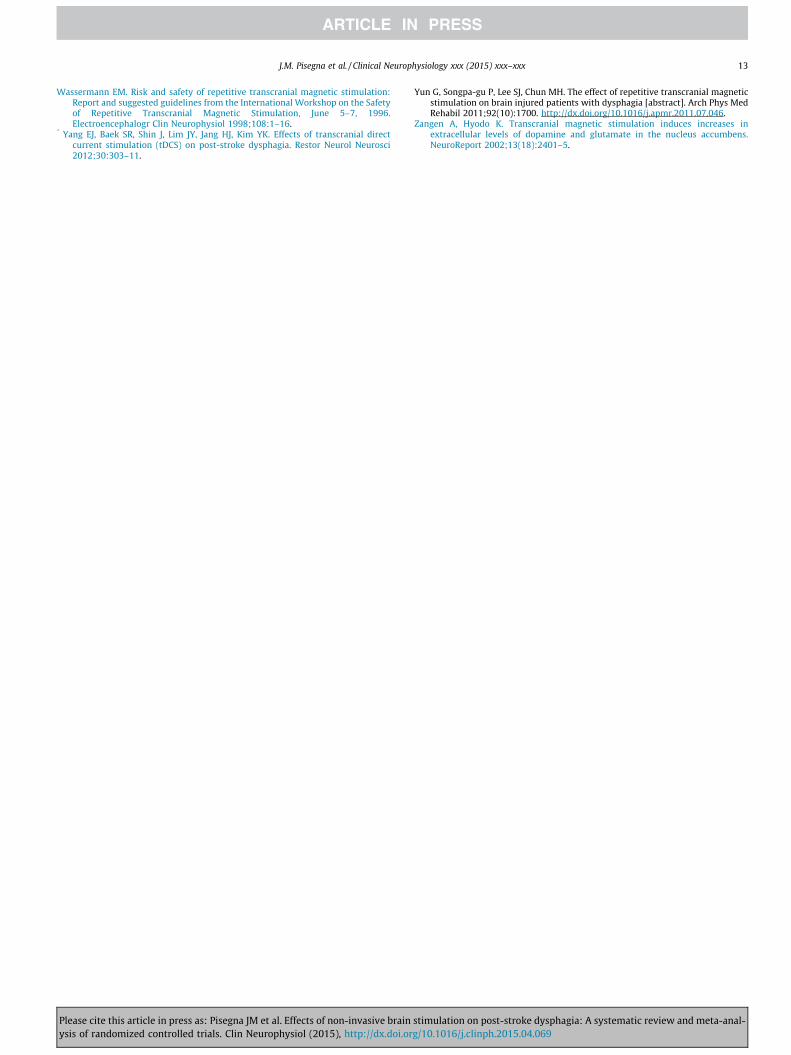
Wassermann EM. Risk and safety of repetitive transcranial magnetic stimulation:Report and suggested guidelines from the International Workshop on the Safetyof Repetitive Transcranial Magnetic Stimulation, June 5–7, 1996.Electroencephalogr Clin Neurophysiol 1998;108:1–16.
* Yang EJ, Baek SR, Shin J, Lim JY, Jang HJ, Kim YK. Effects of transcranial directcurrent stimulation (tDCS) on post-stroke dysphagia. Restor Neurol Neurosci2012;30:303–11.
Yun G, Songpa-gu P, Lee SJ, Chun MH. The effect of repetitive transcranial magneticstimulation on brain injured patients with dysphagia [abstract]. Arch Phys MedRehabil 2011;92(10):1700. http://dx.doi.org/10.1016/j.apmr.2011.07.046.
Zangen A, Hyodo K. Transcranial magnetic stimulation induces increases inextracellular levels of dopamine and glutamate in the nucleus accumbens.NeuroReport 2002;13(18):2401–5.
J.M. Pisegna et al. / Clinical Neurophysiology xxx (2015) xxx–xxx 13
Please cite this article in press as: Pisegna JM et al. Effects of non-invasive brain stimulation on post-stroke dysphagia: A systematic review and meta-anal-ysis of randomized controlled trials. Clin Neurophysiol (2015), http://dx.doi.org/10.1016/j.clinph.2015.04.069