Embed Size (px)
Citation preview
Effects of 20-Hydroxyecdysone and other Hormones on Egg
Development, and Identification of a Vitellin-Binding Protein in the
Ovary of the Tick, Amblyomma hebraeum
Kevin J. Friesen and W. Reuben Kaufman*
Department of Biological Sciences, University of Alberta
Edmonton, Alberta, Canada T6G 2E9
*Author for correspondence: Tel. 1-780-492-1279; Fax. 1-780-492-9234;
E-mail: [email protected]
Submitted to Journal of Insect Physiology, October 27, 2003
Revised March 12, 2004
Abstract
Partially-fed adult female Amblyomma hebraeum ticks were injected with 20-hydroxyecdysone
(20E; up to 43 µg/g body weight [bw]), juvenile hormone III (JH III; up to 100 g/g bw), bovine insulin
(up to 2000 mU/g bw), or triiodothyronine (up to 200 ng/g bw) in an attempt to stimulate vitellogenesis.
Of these, only 20E stimulated synthesis and release of vitellogenin (Vg). Immunoblot analysis revealed
that Vg-synthesis occurred in the fat body. However, consistent with earlier observations suggesting
that a distinct signal may be required for Vg-uptake, there was no significant Vg-uptake by oocytes of
partially-fed, 20E-treated ticks. Because Vg-uptake commonly occurs via receptor-mediated
endocytosis (i.e., a specific Vg-receptor), we attempted to identify a vitellin (Vt)-binding protein in
ovaries of engorged female ticks. A single 86 kDa Vt-binding protein was identified, even under
reducing conditions (2-mercaptoethanol), by a ligand-blotting technique. Sodium salt of suramin (5
mM) inhibited binding of Vt to the 86 kDa protein. However, this protein was also detected in ovaries
from small partially-fed ticks (50-100 mg), suggesting that the inability of 20E to stimulate Vg-uptake in
partially-fed ticks may not have been due to the absence of a Vg-receptor.
Keywords: ixodid ticks, Amblyomma hebraeum, vitellogenesis, vitellogenin, vitellin, 20-
hydroxyecdysone, juvenile hormone, vitellogenin-receptor, insulin, triiodothyronine (T3)
1. Introduction
In insects, synthesis of vitellogenin (Vg) is regulated by the juvenile hormones (JHs), and/or the
ecdysteroids (Nijhout, 1994). Much less is known about vitellogenesis in ticks and other chelicerate
arthropods (Kaufman, 1997; Chang and Kaufman, in press). A series of experiments by Chinzei et al.
(1992) on the argasid tick, Ornithodoros moubata, indicates that Vg-synthesis is initiated by a
‗vitellogenesis inducing factor‘ (VIF); VIF is a neuropeptide that acts on some unknown tissue to cause
the release of a ‗fat body stimulating factor‘ (FSF). FSF is the vitellogenic hormone. The identity of
FSF has not been established, but an ecdysteroid hormone has been implicated (Taylor et al., 2000;
Ogihara, 2003). The weight of current evidence is in favor of an ecdysteroid being the vitellogenic
hormone in ixodid ticks:
Following engorgement, the rise in hemolymph ecdysteroid–titer correlates with the period of egg
development in A. hebraeum (Kaufman, 1991; Friesen and Kaufman, 2002) and in Ixodes scapularis
(James et al., 1997).
Injections of 20E into small partially-fed ticks (A. hebraeum) stimulates enlargement of fat body,
possibly a reflection of accelerated Vg-synthesis (Lunke and Kaufman, 1993). However, slow
infusion of 20E into partially-fed ticks did not stimulate Vg-uptake by oocytes in the latter study.
20E, but not the JH-analogue, methoprene, stimulates Vg-synthesis by Dermacentor variabilis fat
body in vitro (Sankhon et al., 1999). 20E also stimulates Vg-synthesis when injected into A.
hebraeum (Friesen and Kaufman, 2002).
Numerous neurosecretory centers have been identified in the synganglion (= tick CNS)
(reviewed by Binnington and Obenchain, 1982). Neurosecretory activity in general seems to increase
shortly after a blood meal, and then declines during egg production (Eisen et al., 1973; Shanbaky et al.,
1990), suggesting that a neuropeptide may be part of the pathway controlling vitellogenesis (Chinzei et
al., 1992). Likewise, Lomas et al. (1997) demonstrated that a peptide from the synganglion stimulates
ecdysteroid-synthesis in A. hebraeum. While no specific neurosecretory products have been fully
characterized in ticks, antibodies against bovine insulin bind to three neurosecretory regions in the
syngangion of Ornithodoros parkeri (Zhu et al., 1991). Vertebrate insulin has up to 68% nucleotide
sequence homology to the ecdysteroidogenic peptide, bombyxin (Iwami et al., 1989). Both bovine and
porcine insulins, acting through insulin-like receptors, can stimulate ecdysteroid-synthesis in isolated
ovaries of the mosquito, Aedes aegypti (Graf et al., 1997; Riehle and Brown, 1999).
Engorged argasid ticks treated with precocene, an inhibitor of JH synthesis (Retnakaran et al.,
1985), produced and laid fewer eggs than untreated engorged ticks, an effect which was partially
reversed by topical application of JH (Pound and Oliver, 1979). Moreover, JH and a variety of JH-like
compounds stimulated oviposition in fed, virgin argasid ticks (Connat et al., 1983). However, Taylor et
al. (1991a) demonstrated that JH did not stimulate Vg-synthesis in unfed, virgin argasid ticks, even
though vitellogenesis did occur in such ticks when treated with pyrethroid insecticides. Finally,
although extracts of Boophilus microplus have JH-like bioactivity (Connat, 1987), no known JH has
been identified in ticks by gas chromatography/mass spectrometry (Connat, 1987; Neese et al., 2000).
Taylor et al. (1991b) concluded that, whereas JH appears not to stimulate Vg-synthesis in argasid ticks,
it may have stimulated ovulation and/or oviposition in those studies in which ticks were treated with
exogenous JH (eg: Pound and Oliver, 1979; Connat et al., 1983; Connat, 1987).
In some insects, thyroxine interacts with JH-receptors and mimics JH effects. For example, the
protein and RNA content of the fat body in Bombyx mori increases following injection of thyroxine
(Chaudhuri and Medda, 1987). Likewise, both thyroxine, and triiodothyronine (T3) bind to putative JH-
receptors on the follicular epithelium of Rhodnius prolixus and Locusta migratoria, and thus stimulate
patency of the follicular cell layer in the same manner as JH (Davey and Gordon, 1996; Kim et al.,
1999). Consequently, here we test whether T3might also stimulate vitellogenesis in ticks.
In this study, the effects of 20E, insulin, JH-III, and T3 on vitellogenesis were examined in A.
hebraeum. As shown previously, 20E does not stimulate Vg-uptake in the oocytes of partially-fed ticks
(Lunke and Kaufman, 1993), even though 20E stimulates Vg-synthesis in vivo (Friesen and Kaufman,
2002). The possibility arises that up-regulation of a Vg-receptor (VgR) in the ovary is controlled by a
factor distinct from the vitellogenic hormone, a factor which is absent in small partially-fed ticks. Thus,
in this study we also attempted to identify a putative VgR and determine when it appears in the ovary
during the feeding cycle.
2. Materials and Methods
2.1 Ticks
Our colony of A. hebraeum was kept in darkness at 27°C and >95% relative humidity. Tick
feeding occurred on rabbits as described by Kaufman and Phillips (1973). Ticks were allowed to
engorge and detach spontaneously (weight range 900-3500 mg), or were forcibly removed from the host
below the approximate ‗critical weight‘ necessary to trigger vitellogenesis (< 250 mg; Kaufman and
Lomas, 1996; Weiss and Kaufman, 2001). Just prior to conducting experiments, ticks were rinsed in tap
water and weighed.
2.2 Hormone treatments
For injection of hormones, stock solutions were made as follows: 20E (Sigma, St. Louis, MO,
USA) was dissolved in 70% ethanol to make a 5 mg/ml solution; bovine insulin (Sigma) was dissolved
in dilute HCl (pH 2.5) to a final concentration of 5 mg/ml; T3 (Sigma) was dissolved in 1 N NaOH to a
final concentration of 4 mg/ml. On the day of injection, the stock solutions were diluted to working
concentration in sterile-filtered medium TC199 (Gibco, Burlington, ON, Canada) containing 50 µg/ml
gentamicin (Sigma). Experimental ticks were surface sterilized in 70% ethanol for 1 min, and injected
through the camerostomal fold with 5 µl (for insulin and T3 treatments) or 10 µl (for 20E treatment) per
100 mg body weight (bw) via a 30 gauge needle fitted to an AGLA micrometer syringe unit (Wellcome
Reagents Ltd., Beckenham, Kent, England). Final doses of these hormones were within the range
0.0043-43 µg 20E/g bw, 20-2000 mU insulin/g bw, and 100-200 ng T3/g bw. Vehicle-injected and/or
non-injected ticks served as controls. Second and third bolus injections were given to each tick on days
2 and 5. Ticks were kept under colony conditions until their hemolymph and tissues were collected on
day 10 (see below).
For topical application, JH III (Sigma) was dissolved in acetone to a concentration of 10 mg/ml;
final concentrations for treatments were 2, 5, and 10 mg/ml. On days 0, 2, and 5 post-removal, partially-
fed ticks received a volume of 1 l/100 mg bw JH III solution applied to the dorsal cuticle. Hence, the
final doses were 20, 50, and 100 µg JH III/g bw. Control ticks were treated with 1 l/100 mg bw
acetone. Ticks were stored under colony conditions until day 6, when hemolymph and tissue samples
were collected (see below).
2.3 Collection of tissue and hemolymph samples
Tick survival was determined at the time of dissection. Apart from those ticks injected with the
highest dose of 20E (43,000 ng/g bw), 40% of which died by day 10, the survival of all other groups was
greater than 96 %
For collection of hemolymph and ovaries, ticks were stuck to disposable petri dishes with
cyanoacrylate glue and cooled in a refrigerator for 20 min. Cooling inhibits gut contraction, thereby
reducing the risk of piercing the delicate gut wall and contaminating the hemolymph (Kaufman, 1991). A
small incision was made in the cuticle, and hemolymph was collected in a calibrated glass micropipette;
exact sample volume was calculated from the length of the fluid column. Samples were diluted at least
1:10 in 100% methanol for radioimmunoassay (RIA), or 1:4 in phosphate-buffered saline (PBS; 35 mM
NaH2PO4, 60 mM Na2HPO4, 150 mM NaCl, pH 7.0) for electrophoresis or ELISA. Samples for RIA were
stored at -15 C and all other samples were stored at -70 C until analyzed.
After collection of hemolymph, ticks were flooded with a modified Hank‘s balanced saline (200
mM NaCl, 8.9 mM D-glucose, 5.4 mM KCl, 1.3 mM CaCl2, 0.4 mM MgSO4, 0.44 mM KH2PO4, 0.35
mM Na2HPO4, 27 µM phenol red, pH 7.2) and the dorsal cuticle removed using a microscalpel. Gut,
salivary glands, Malpighian tubules, trachea, reproductive tract, and fat body were removed and
immediately frozen on dry ice for immunoblot analysis.
Unlike most insects, ticks possess a single, U-shaped, tubular ovary, terminating in a pair of
oviducts. In metastriate ixodid ticks (including A. hebraeum), these oviducts converge to an
inconspicuous common oviduct, which in turn leads to the vagina (Diehl et al., 1982). A lateral groove
runs the length of the ovary. Oocyte development is asynchronous: immature oocytes are always found
within the lateral groove throughout the extneded period of oogenesis, whereas mature ones are
suspended from the ovarian wall opposite the groove. In common with all chelicerate arthropods, no
follicular epithelium surrounds the oocytes (Kaufman, 1997; Chang and Kaufman, in press).
Ovaries were dissected out, and the length of the long axis of the eight apparently largest ovoid
oocytes was measured using an ocular micrometer fitted to a compound microscope. The mean value
for the eight oocytes was recorded for each tick. The ovaries were then gently blotted and weighed.
Prior to analysis, all tissues were homogenized in PBS containing protease inhibitors (Protease Inhibitor
Cocktail Set 1, Calbiochem, La Jolla, CA), centrifuged at 13,000 X g for 5 min and the pellet discarded.
Supernatants were stored at -70 C for further analysis.
2.4 Assay for salivary gland degeneration
Because 20E triggers salivary gland degeneration (Kaufman, 1991), we also measured salivary
gland function in this study using the technique of Harris and Kaufman (1984). Briefly, paired salivary
glands were excised from each tick 5 or 10 days post-engorgement, and the main duct ligated using a
very fine strand of silk thread (Dermalon , Syneture, Norwalk, CT); the silk strand weighed less than
the sensitivity of the balance [10 µg,]). The glands were gently blotted, weighed to the nearest 10 µg
and incubated in medium TC 199 (Gibco) containing 10 µM dopamine (Sigma) for 10 min, blotted
again, and re-weighed. Because dopamine stimulates salivary fluid secretion and the fluid cannot escape
through the ligated duct, any fluid transported from the bathing medium accumulates within the acinar
lumen and results in an increase in salivary gland weight. The net weight increase is thus a direct
measure of fluid secretory competence. Loss of fluid secretory competence compared to controls is thus
a quantitative measure of salivary gland degeneration (Harris and Kaufman, 1984).
2.5 Preparation of partially-purified vitellin (Vt)
Vt was partially-purified from the ovaries of day 10 engorged ticks as described previously
(Friesen and Kaufman, 2002). Briefly, an ovary homogenate was passed through a gel filtration column
(Superose 6B, Pharmacia; now Pfizer, Kirkland, QUE, Canada) and then an ion exchange column
(DEAE-Sephacel, Pharmacia). The flow rate (1-3 ml/min) of the columns was maintained by a
peristaltic pump. Tick Vg contains a heme moiety (Sonenshine, 1991), so the fractions eluted from each
column which contained large amounts of both heme and protein (determined by spectrophotometry;
Kaufman et al, 1986) were pooled and concentrated using Centriprep™ centrifuge tubes (Amicon,
Beverly, MA, USA). The protein concentration of the resulting extract was measured using the Bradford
assay (Bradford, 1976; Bradford reagent kit; Sigma), using bovine serum albumin (BSA; Sigma) as a
standard, and the extract frozen at -70 C in 20 µl aliquots.
2.6 Electrophoresis, immunoblotting, and ligand blotting
Proteins from tissue homogenates were separated by SDS-PAGE (sodium dodecyl sulfate
polyacrylamide gel electrophoresis; Laemmlli, 1970) under reducing or non-reducing conditions.
Samples (all at 100 µg, except for Fig. 3 where it was 30 µg) were diluted in electrophoresis sample
buffer (60 mM Tris-HCl pH 6.8, 10% (v/v) glycerol, 0.0025% (w/v) bromophenol blue, 2% (w/v) SDS,
with or without 2-mercaptoethanol (5%) for reducing gels and non-reducing gels, respectively) prior to
being applied to the gels. Electrophoresis was performed using mini-Protean equipment (Bio-Rad,
Hercules, CA). All gels consisted of a 3% stacking gel, and a 7.5% resolving gel. Electrophoresis was
performed at 110V until the dye front reached the bottom of the gels. Proteins were then transferred to
polyvinylidene difluoride membrane (PVDF; BioRad) for immunoblotting. Transfers to PVDF
membrane were for 1 h at 0.3-0.5 mA. Antibodies used for immunoblots and ELISA (see below) were
prepared against A. hebraeum Vg, which consists of two hemolymph polypeptides, Vg211 and Vg148,
identified according to criteria presented by Friesen and Kaufman (2002). Membranes containing
transferred proteins were first kept overnight in blocking buffer (5% skim milk, 2% BSA, in Tris-
buffered saline [TTBS: 60 mM Tris-HCl, 0.3 M NaCl, 0.04% (v/v) Tween 20, pH 7.5]). For antibody
reactions, strips were then placed in blocking buffer containing either anti-Vg211 (1:500) or anti-Vg148
(1:250), or both. After 1 h, strips were washed three times for 5 min with blocking buffer, then placed in
secondary antibody (alkaline phosphatase (AP)-conjugated, goat anti-rabbit IgG; BioRad) diluted 1:2000
in blocking buffer. One hour later, strips were washed three times for 5 min in TTBS and then stained
for approximately 20 min using an AP-conjugate substrate kit (BioRad).
In order to identify the putative VgR, ligand blotting was performed in a manner similar to that
described by previous authors (Dhadialla et al., 1992; Sappington et al., 1995; Romano and Limatola,
2000). Briefly, protein resolved by SDS-PAGE was transferred to PVDF membrane as described above
for immunoblots, and then blocked for 30 min in blocking buffer. The membrane containing transferred
proteins was then incubated overnight at 4 C with 1 mg/ml Vt (partially purified from day 10 engorged
ovaries) in dilute blocking buffer (2.5% skim milk, 1% BSA, TTBS). Control strips were incubated
with only dilute blocking buffer. In some cases, the sodium salt of suramin (5 mM; Sigma) was added
to the Vt solution to test for inhibition of Vt binding (Dhadialla et al., 1992; Sappington et al., 1995).
Following overnight incubation, the membrane was washed and treated with a combination of anti-
Vg211 and anti-Vg148 antibodies, as described above for immunoblots. The latter method detects both
endogenous Vg and/or Vt in ovary homogenates as well as any protein which binds exogenous Vt. Thus,
for a polypeptide to be designated as a ‗Vt-binding protein', it must appear in blots exposed to
exogenous Vt but not in control blots.
Immunoblots and ligand blots were converted to digital format using a Hewlett-Packard
hpscanjet 3570c benchtop scanner and Adobe Photoshop software. All images were scanned at a
resolution of 600 pixels/inch. Mobilities of polypeptide bands were then measured using NIH Image
software (National Institute of Health, U.S.A.), and the relative mobility of each band calculated in
relation to the molecular weight standards. In the case of Figure 7, some background discoloration was
removed using Adobe Photoshop .
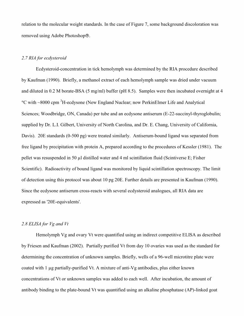
2.7 RIA for ecdysteroid
Ecdysteroid-concentration in tick hemolymph was determined by the RIA procedure described
by Kaufman (1990). Briefly, a methanol extract of each hemolymph sample was dried under vacuum
and diluted in 0.2 M borate-BSA (5 mg/ml) buffer (pH 8.5). Samples were then incubated overnight at 4
C with ~8000 cpm 3H-ecdysone (New England Nuclear; now PerkinElmer Life and Analytical
Sciences; Woodbridge, ON, Canada) per tube and an ecdysone antiserum (E-22-succinyl-thyroglobulin;
supplied by Dr. L.I. Gilbert, University of North Carolina, and Dr. E. Chang, University of California,
Davis). 20E standards (0-500 pg) were treated similarly. Antiserum-bound ligand was separated from
free ligand by precipitation with protein A, prepared according to the procedures of Kessler (1981). The
pellet was resuspended in 50 µl distilled water and 4 ml scintillation fluid (Scintiverse E; Fisher
Scientific). Radioactivity of bound ligand was monitored by liquid scintillation spectroscopy. The limit
of detection using this protocol was about 10 pg 20E. Further details are presented in Kaufman (1990).
Since the ecdysone antiserum cross-reacts with several ecdysteroid analogues, all RIA data are
expressed as '20E-equivalents'.
2.8 ELISA for Vg and Vt
Hemolymph Vg and ovary Vt were quantified using an indirect competitive ELISA as described
by Friesen and Kaufman (2002). Partially purified Vt from day 10 ovaries was used as the standard for
determining the concentration of unknown samples. Briefly, wells of a 96-well microtitre plate were
coated with 1 µg partially-purified Vt. A mixture of anti-Vg antibodies, plus either known
concentrations of Vt or unknown samples was added to each well. After incubation, the amount of
antibody binding to the plate-bound Vt was quantified using an alkaline phosphatase (AP)-linked goat
anti-rabbit secondary antibody (BioRad) and an AP color substrate kit (p-nitrophenylphosphate and
diethanolamine kit; BioRad). The color reaction was quantified by measuring the absorbance of each
well at 405 nm using a microtitre plate reader (Bio-Tek Instruments, Winooski, VN). Absorbance
values of known Vt-concentration were plotted on a logarithmic scale to create a standard curve from
which concentration of Vg or Vt in unknown samples were calculated and reported as ‗Vt-equivalents‘.
The sensitivity of this ELISA to A. hebraeum Vg was approximately 5 ng of Vt-equivalents
2.9 Statistical analysis
Results are reported as mean SEM (n). Statistical analysis was done using Statview 4.02
(Abacus Concepts, Berkely California). Differences among treatments were analyzed using a 1-way
analysis of variance (ANOVA). Statistical significance is indicated as follows: (*) 0.01<p<0.05; (**)
0.001<p<0.01; (***) p<0.001.
3. Results
3.1 Effect of 20E
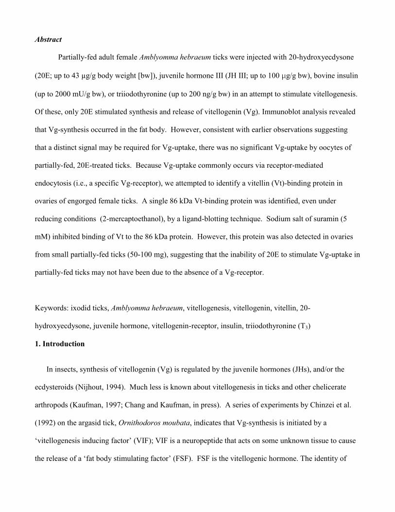
Injection of 20E had no significant effect on ovary weight (Fig. 1A) or oocyte length (Fig. 1B) in
partially-fed ticks by 5 or 10 days post-treatment. However, 20E caused a marked increase in
hemolymph Vg-concentration (Fig. 1C). Vg-concentration was highest on day 5 at 18.3 3.8 mg/ml
(approximately 30 times the day 5 EtOH control) and on day 10 at 35.0 5.5 mg/ml (approximately 20
times the day 10 EtOH control), following 3 bolus injections of 4,300 ng 20E/g bw. On both days 5 and
10, ticks treated with 43,000 ng 20E/g bw had significantly lower hemolymph Vg-concentrations than
those treated with 4,300 ng 20E/g bw (p < 0.0001). Moreover, when dissecting day 10 ticks that had
been treated with the highest dose of 20E, we noticed that the hypodermis had fallen away from the
surrounding cuticle, suggesting a very abnormal type of apolysis.
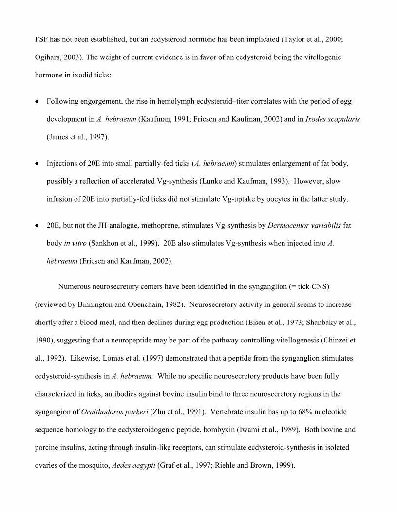
Salivary gland weight and salivary gland fluid secretory competence decreased in 20E-treated
partially-fed ticks (Fig. 2). Compared to untreated controls, EtOH caused a significant increase in
salivary gland fluid secretory competence by day 10 (p < 0.009; Fig. 2B). However, at both day 5 and
day 10, salivary gland fluid secretory competence was significantly lower at 430 ng 20E/g bw and above
than both untreated and EtOH controls (Fig. 2B).
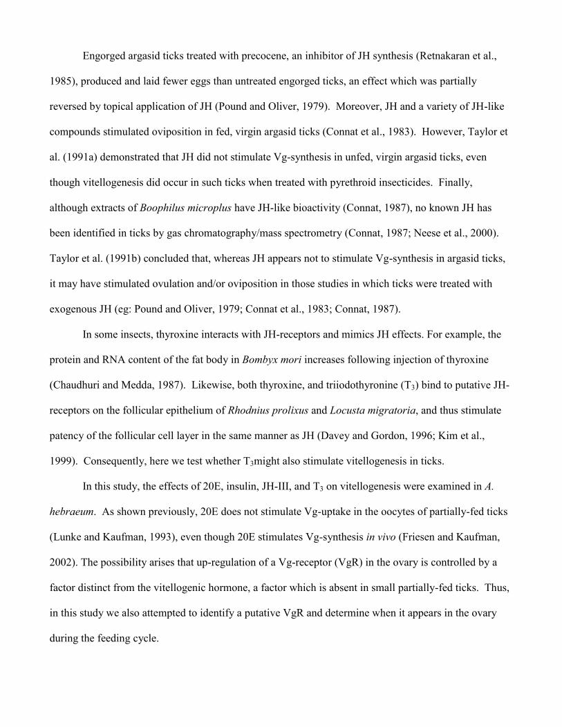
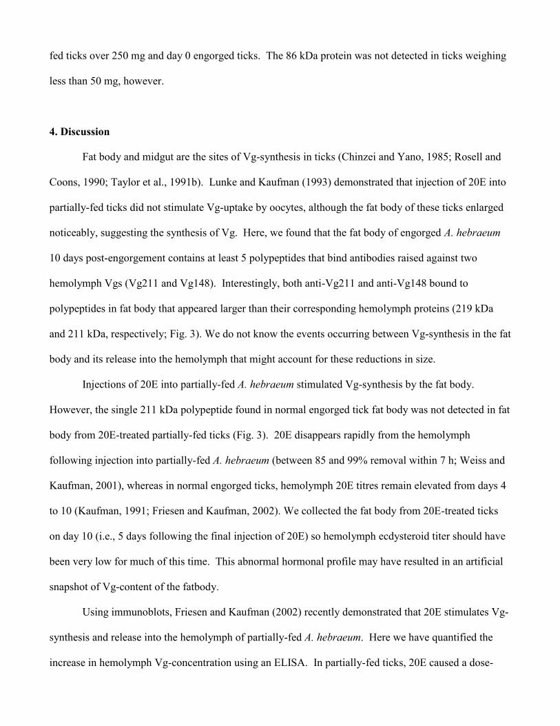
Anti-Vg211 antibody (Friesen and Kaufman, 2002) bound to a single 219 kDa polypeptide that
was present in the fat body of both day 10 engorged and 20E-treated partially-fed ticks (Fig. 3). Anti-
Vg148 antibody bound to three polypeptides of 211, 169, and 78 kDa in fat body from engorged ticks.
The 211 and 169 kDa polypeptides did not appear in fat body from 20E-treated (4,300 ng 20E/g bw)
partially-fed ticks. However, the 78 kDa polypeptide appeared more intense in 20E-treated ticks than in
engorged ticks. No polypeptides from day 0 engorged and EtOH-injected partially-fed tick fat bodies
were specific to anti-Vg antibodies (Fig 3).
3.2 Effect of insulin
Compared to vehicle-injected controls, multiple injections of bovine insulin into partially-fed
ticks had no significant effect on ovary weight (n=6-15), salivary gland fluid secretory competence
(n=12-24), hemolymph Vg-concentration (n=4-14), or hemolymph 20E-concentration (n=2-10) by days
5 or 10 (data not shown). The data for Vg-concentration was highly variable, however.
3.3 Effects of JH III and T3
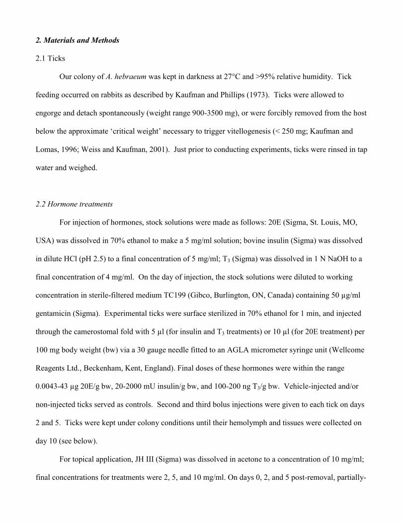
Topical application of partially-fed ticks with JH III had no significant effect on ovary weight
(Fig. 4A), or hemolymph 20E- and Vg-concentrations (Fig. 4B) by day 6 post-treatment. Salivary
glands of ticks treated with 10 g JH/g bw weighed significantly less than acetone-treated controls by
day 6 (1.88 0.09 mg in JH-treated glands and 2.64 0.19 mg in control glands; Fig. 4C). Salivary
gland fluid secretory competence was greater in ticks treated with 2 g JH/g bw than in acetone treated
controls (3.21 0.19 mg/gland/10 min for JH-treated glands and 1.37 0.13 mg/gland/10 min for
controls). The latter effect was not apparent at 5 or 10 g JH/g bw, however (Fig. 4D).
There were no apparent effects of T3 treatment on ovary weight (n=3-4), hemolymph 20E-
concentration (n=2-4), hemolymph Vg-concentration (n=2-4), or salivary gland fluid secretory
competence (n=5-7) by day 5 or 10 (data not shown).
3.4 Identification of a Vt-binding protein
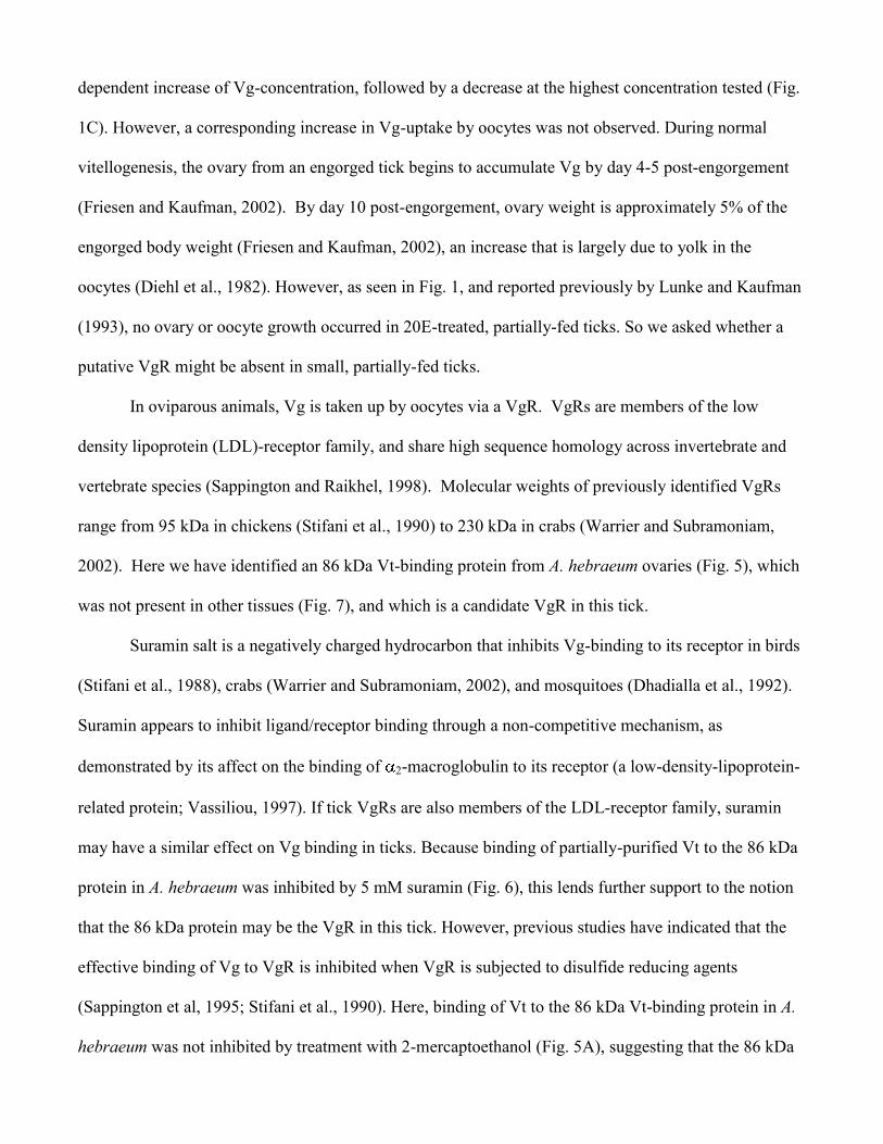
Immunoblots of days 2-4 engorged ovary homogenate were incubated first with partially purified
Vt from day 10 engorged ovaries, and then by anti-Vg antibodies, to test for Vt-binding proteins. In
addition to Vt already present in the eggs, days 2-4 ovary homogenates contained only a single
detectable polypeptide (86 kDa) that bound to Vt (Fig. 5B). Subjecting ovary homogenates to non-
reducing conditions did not appear to affect the size or Vt-binding function of this polypeptide (Fig. 5A);
however, this polypeptide band appeared fainter under non-reducing conditions than under reducing
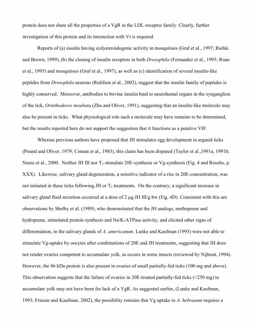
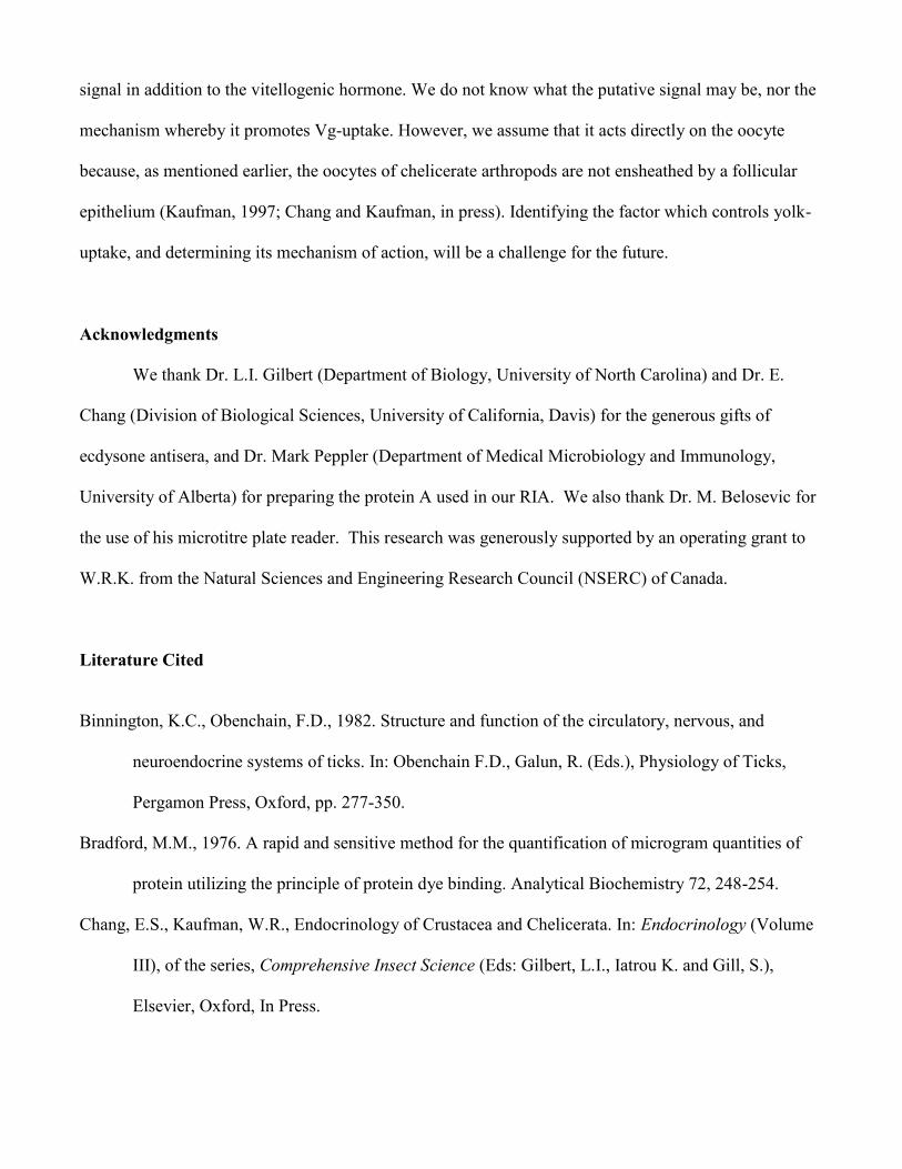
conditions. Vt-binding to the 86 kDa polypeptide was attenuated by 5 mM suramin (Fig. 6). Moreover,
the 86 kDa polypeptide was not detected in tissues other than ovaries, including the reproductive tract
(seminal receptical, uterus, accessory glands), midgut, salivary glands, Malpighian tubules, and trachea
(Fig. 7).
The 86 kDa Vt-binding protein was detected by ligand blotting in ovaries of engorged ticks up to
day 4, but not on day 8 (Fig. 8). We do not interpret the lack of detection of this band on day 8 to a
disappearance of the protein. All lanes of the gel were loaded with 100 µg protein (see methods).
Because Vt is so abundant on day 8, the 86 kDa binding protein may have been present in too small an
amount to show up on the gel. The 86 kDa band on ligand blots was visible in ovary homogenates from
partially-fed ticks between 100-150 mg, and appeared more intense in ovary homogenates of partially-
fed ticks over 250 mg and day 0 engorged ticks. The 86 kDa protein was not detected in ticks weighing
less than 50 mg, however.
4. Discussion
Fat body and midgut are the sites of Vg-synthesis in ticks (Chinzei and Yano, 1985; Rosell and
Coons, 1990; Taylor et al., 1991b). Lunke and Kaufman (1993) demonstrated that injection of 20E into
partially-fed ticks did not stimulate Vg-uptake by oocytes, although the fat body of these ticks enlarged
noticeably, suggesting the synthesis of Vg. Here, we found that the fat body of engorged A. hebraeum
10 days post-engorgement contains at least 5 polypeptides that bind antibodies raised against two
hemolymph Vgs (Vg211 and Vg148). Interestingly, both anti-Vg211 and anti-Vg148 bound to
polypeptides in fat body that appeared larger than their corresponding hemolymph proteins (219 kDa
and 211 kDa, respectively; Fig. 3). We do not know the events occurring between Vg-synthesis in the fat
body and its release into the hemolymph that might account for these reductions in size.
Injections of 20E into partially-fed A. hebraeum stimulated Vg-synthesis by the fat body.
However, the single 211 kDa polypeptide found in normal engorged tick fat body was not detected in fat
body from 20E-treated partially-fed ticks (Fig. 3). 20E disappears rapidly from the hemolymph
following injection into partially-fed A. hebraeum (between 85 and 99% removal within 7 h; Weiss and
Kaufman, 2001), whereas in normal engorged ticks, hemolymph 20E titres remain elevated from days 4
to 10 (Kaufman, 1991; Friesen and Kaufman, 2002). We collected the fat body from 20E-treated ticks
on day 10 (i.e., 5 days following the final injection of 20E) so hemolymph ecdysteroid titer should have
been very low for much of this time. This abnormal hormonal profile may have resulted in an artificial
snapshot of Vg-content of the fatbody.
Using immunoblots, Friesen and Kaufman (2002) recently demonstrated that 20E stimulates Vg-
synthesis and release into the hemolymph of partially-fed A. hebraeum. Here we have quantified the
increase in hemolymph Vg-concentration using an ELISA. In partially-fed ticks, 20E caused a dose-
dependent increase of Vg-concentration, followed by a decrease at the highest concentration tested (Fig.
1C). However, a corresponding increase in Vg-uptake by oocytes was not observed. During normal
vitellogenesis, the ovary from an engorged tick begins to accumulate Vg by day 4-5 post-engorgement
(Friesen and Kaufman, 2002). By day 10 post-engorgement, ovary weight is approximately 5% of the
engorged body weight (Friesen and Kaufman, 2002), an increase that is largely due to yolk in the
oocytes (Diehl et al., 1982). However, as seen in Fig. 1, and reported previously by Lunke and Kaufman
(1993), no ovary or oocyte growth occurred in 20E-treated, partially-fed ticks. So we asked whether a
putative VgR might be absent in small, partially-fed ticks.
In oviparous animals, Vg is taken up by oocytes via a VgR. VgRs are members of the low
density lipoprotein (LDL)-receptor family, and share high sequence homology across invertebrate and
vertebrate species (Sappington and Raikhel, 1998). Molecular weights of previously identified VgRs
range from 95 kDa in chickens (Stifani et al., 1990) to 230 kDa in crabs (Warrier and Subramoniam,
2002). Here we have identified an 86 kDa Vt-binding protein from A. hebraeum ovaries (Fig. 5), which
was not present in other tissues (Fig. 7), and which is a candidate VgR in this tick.
Suramin salt is a negatively charged hydrocarbon that inhibits Vg-binding to its receptor in birds
(Stifani et al., 1988), crabs (Warrier and Subramoniam, 2002), and mosquitoes (Dhadialla et al., 1992).
Suramin appears to inhibit ligand/receptor binding through a non-competitive mechanism, as
demonstrated by its affect on the binding of 2-macroglobulin to its receptor (a low-density-lipoprotein-
related protein; Vassiliou, 1997). If tick VgRs are also members of the LDL-receptor family, suramin
may have a similar effect on Vg binding in ticks. Because binding of partially-purified Vt to the 86 kDa
protein in A. hebraeum was inhibited by 5 mM suramin (Fig. 6), this lends further support to the notion
that the 86 kDa protein may be the VgR in this tick. However, previous studies have indicated that the
effective binding of Vg to VgR is inhibited when VgR is subjected to disulfide reducing agents
(Sappington et al, 1995; Stifani et al., 1990). Here, binding of Vt to the 86 kDa Vt-binding protein in A.
hebraeum was not inhibited by treatment with 2-mercaptoethanol (Fig. 5A), suggesting that the 86 kDa
protein does not share all the properties of a VgR in the LDL-receptor family. Clearly, further
investigation of this protein and its interaction with Vt is required.
Reports of (a) insulin having ecdysteroidogenic activity in mosquitoes (Graf et al., 1997; Riehle
and Brown, 1999), (b) the cloning of insulin receptors in both Drosophila (Fernandez et al., 1995; Ruan
et al., 1995) and mosquitoes (Graf et al., 1997), as well as (c) identification of several insulin-like
peptides from Drosophila neurons (Rulifson et al., 2002), suggest that the insulin family of peptides is
highly conserved. Moreover, antibodies to bovine insulin bind to neurohemal organs in the synganglion
of the tick, Ornithodoros moubata (Zhu and Oliver, 1991), suggesting that an insulin-like molecule may
also be present in ticks. What physiological role such a molecule may have remains to be determined,
but the results reported here do not support the suggestion that it functions as a putative VIF.
Whereas previous authors have proposed that JH stimulates egg development in argasid ticks
(Pound and Oliver, 1979; Connat et al., 1983), this claim has been disputed (Taylor et al.,1991a, 1991b;
Neese et al., 2000. Neither JH III nor T3 stimulate 20E-synthesis or Vg-synthesis (Fig. 4 and Results, p.
XXX). Likewise, salivary gland degeneration, a sensitive indicator of a rise in 20E-concentration, was
not initiated in these ticks following JH or T3 treatments. On the contrary, a significant increase in
salivary gland fluid secretion occurred at a dose of 2 µg JH III/g bw (Fig. 4D). Consistent with this are
observations by Shelby et al. (1989), who demonstrated that the JH analogs, methoprene and
hydroprene, stimulated protein synthesis and Na/K-ATPase activity, and elicited other signs of
differentiation, in the salivary glands of A. americanum. Lunke and Kaufman (1993) were not able to
stimulate Vg-uptake by oocytes after combinations of 20E and JH treatments, suggesting that JH does
not render ovaries competent to accumulate yolk, as occurs in some insects (reviewed by Nijhout, 1994).
However, the 86 kDa protein is also present in ovaries of small partially-fed ticks (100 mg and above).
This observation suggests that the failure of ovaries in 20E-treated partially-fed ticks (<250 mg) to
accumulate yolk may not have been for lack of a VgR. As suggested earlier, (Lunke and Kaufman,
1993; Friesen and Kaufman, 2002), the possibility remains that Vg uptake in A. hebraeum requires a
signal in addition to the vitellogenic hormone. We do not know what the putative signal may be, nor the
mechanism whereby it promotes Vg-uptake. However, we assume that it acts directly on the oocyte
because, as mentioned earlier, the oocytes of chelicerate arthropods are not ensheathed by a follicular
epithelium (Kaufman, 1997; Chang and Kaufman, in press). Identifying the factor which controls yolk-
uptake, and determining its mechanism of action, will be a challenge for the future.
Acknowledgments
We thank Dr. L.I. Gilbert (Department of Biology, University of North Carolina) and Dr. E.
Chang (Division of Biological Sciences, University of California, Davis) for the generous gifts of
ecdysone antisera, and Dr. Mark Peppler (Department of Medical Microbiology and Immunology,
University of Alberta) for preparing the protein A used in our RIA. We also thank Dr. M. Belosevic for
the use of his microtitre plate reader. This research was generously supported by an operating grant to
W.R.K. from the Natural Sciences and Engineering Research Council (NSERC) of Canada.
Literature Cited
Binnington, K.C., Obenchain, F.D., 1982. Structure and function of the circulatory, nervous, and
neuroendocrine systems of ticks. In: Obenchain F.D., Galun, R. (Eds.), Physiology of Ticks,
Pergamon Press, Oxford, pp. 277-350.
Bradford, M.M., 1976. A rapid and sensitive method for the quantification of microgram quantities of
protein utilizing the principle of protein dye binding. Analytical Biochemistry 72, 248-254.
Chang, E.S., Kaufman, W.R., Endocrinology of Crustacea and Chelicerata. In: Endocrinology (Volume
III), of the series, Comprehensive Insect Science (Eds: Gilbert, L.I., Iatrou K. and Gill, S.),
Elsevier, Oxford, In Press.
Chaudhuri, A., Medda, A.K., 1987. Thyroxine-induced alterations in protein and nucleic acid contents of
fat body of female silkworms during different developmental stages. Insect Science and its
Application 8, 43-48.
Chinzei, Y., Taylor, D., Miura, K., Ando, K., 1992. Vitellogenesis induction by synganglion factor in
adult female tick, Ornithodoros moubata (Acari: Argasidae). Journal of the Acarological Society
of Japan 1, 15-26.
Chinzei, Y., Yano, I., 1985. Fat body is the site of vitellogenin synthesis in the soft tick, Ornithodoros
moubata. Journal of Comparative Physiology B 155, 671-678.
Connat, J.-L., 1987. Aspects Endocrinologiques de la Physiologie du Developement et de la
Reproduction chez les Tiques. Thèse de doctorat D'Etat (87/DIJO/5005), Dijon, France.
Connat, J.-L., Ducommun, J., Diehl, P.-A., 1983. Juvenile hormone-like substances can induce
vitellogenesis in the tick Ornithodoros moubata (Acarina: Argasidae). International Journal of
Invertebrate Reproduction 6, 285-294.
Davey, K.G., Gordon, D.R.B., 1996. Fenoxycarb and thyroid hormones have JH-like effects on the
follicle cells of Locusta migratoria in vitro. Archives of Insect Biochemistry and Physiology 32,
613-622.
Dhadialla, T.S., Hays, A.R., Raikhel, A.S., 1992. Characterization of the solubilized mosquito
vitellogenin receptor. Insect Biochemistry and Molecular Biology 22, 803-816.
Diehl, P.-A., Aeschlimann, A., Obenchain, F.D., 1982. Tick reproduction: oogenesis and oviposition. In:
Obenchain F.D., Galun, R. (Eds.), Physiology of Ticks, Pergamon Press, Oxford, pp. 277-350.
Eisen, Y., Warburg, M.R., Galun, R., 1973. Neurosecretory activity as related to feeding and oogenesis
in the fowl-tick Argas persicus (Oken). General and Comparative Endocrinology 21, 331-340.
Fernandez, R., Tabarini, D., Azpiazu, N., Frasch, M., Schlessinger, J., 1995. The Drosophila insulin
receptor homolog: a gene essential for embryonic development encodes two receptor isoforms
with different signaling potential. EMBO J. 14, 3373-3384.
Friesen, K.J., Kaufman, W.R., 2002. Quantification of vitellogenesis and its control by 20-
hydroxyecdysone in the ixodid tick, Amblyomma hebraeum. Journal of Insect Physiology 48,
773-782.
Graf, R., Neuenshwander, S., Brown, M.R., Ackermann, U., 1997. Insulin-mediated secretion of
ecdysteroids from mosquito ovaries and molecular cloning of the insulin receptor homologue
from ovaries of bloodfed Aedes aegypti. Insect Molecular Biology 6, 151-163.
Harris, R.A., Kaufman, W.R., 1984. Neural involvement in the control of salivary gland degeneration in
the ixodid tick, Amblyomma hebraeum. Journal of Experimental Biology 109, 281-290.
Iwami, M., Kawakami, A., Ishizaki, H., Takahashi, S.Y., Adachi, T., Suzuki, Y., Nagasawa, H., Suzuki,
A., 1989. Cloning of a gene encoding bombyxin, an insulin-like brain secretory peptide of the
silkmoth Bombyx mori with prothroracicotropic activity. Development, Growth and
Differentiation 31, 31-37.
James, A.M., Zhu, X.X., Oliver, Jr. J.H., 1997. Vitellogenin and ecdysteroid titers in Ixodes scapularis
during vitellogenesis. Journal of Parasitology 83, 559-563.
Kaufman, W.R., 1990. Effect of 20-hydroxyecdysone on the salivary gland of the male tick,
Amblyomma hebraeum. Experimental and Applied Acarology 9, 87-95.
Kaufman, W.R., 1991. Correlation between haemolymph ecdysteroid titre, salivary gland degeneration
and ovarian development in the ixodid tick, Amblyomma hebraeum Koch. Journal of Insect
Physiology 37, 95-99.
Kaufman, W.R., 1997. Chapter 7: Arthropoda—Chelicerata, In: Adiyodi, K.G., Adiyodi, R.G. (Eds.),
Reproductive Biology of Invertebrates, Oxford and IBH Publishing Co. Pvt. Ltd., New Delhi,
vol. 8, pp. 211-245.
Kaufman, W.R., Lomas, L.O., 1996. ―Male factors‖ in ticks: their role in feeding and egg development.
Invertebrate Reproduction and Development 30, 191-198.
Kaufman, W.R., Phillips, J.E., 1973. Ion and water balance in the ixodid tick, Dermacentor andersoni: I.
Routes of ion and water excretion. Journal of Experimental Biology 58, 523-536.
Kaufman, W.R., Ungarian, S.G., Noga, A.E., 1986. The effect of avermectins on feeding, salivary fluid
secretion, and fecundity in some ixodid ticks. Experimental and Applied Acarology 2, 1-18.
Kessler, S.W., 1981. Use of protein A-bearing Staphylococcus for the immunoprecipitation and isolation
of antigens from cells. Methods in Enzymology 73, 422.
Kim, Y., Davari, E.D., Sevala, V., Davey, K.G., 1999. Functional binding of a vertebrate hormone, L-
3,5,3‘-triiodothyronine (T3), on insect follicle cell membranes. Insect Biochemistry and
Molecular Biology 29, 943-950.
Laemmli, U.K., 1970. Cleavage of structural proteins during the assembly of the head of bacteriophage
T4. Nature 227, 680-685.
Lomas, L.O., Turner P.C., Rees, H.H., 1997. A novel neuropeptide-endocrine interaction controlling
ecdysteroid production in ixodid ticks. Proceedings of the Royal Society of London B 264, 589-
596.
Lunke, M.D., Kaufman, W.R., 1993. Hormonal control of ovarian development in the tick Amblyomma
hebraeum Koch (Acari: Ixodidae). Invertebrate Reproduction and Development 23, 25-38.
Neese, P.A., Sonenshine, D.E., Kallapur, V.L., Apperson, C.S., Roe, R.M., 2000. Absence of insect
juvenile hormones in the American dog tick, Dermacentor variabilis (Say) (Acari: Ixodidae),
and in Ornithodoros parkeri Cooley (Acari: Argasidae). Journal of Insect Physiology 46, 477-
490.
Nijhout, H.F., 1994. Insect Hormones. Princeton University Press, Princeton, New Jersey.
Ogihara K. 2003. Ecdysteroid Hormone Titer and Expression of Ecdysone Receptor mRNA as Related
to Vitellogenesis in the Soft Tick, Ornithodoros moubata (Acari: Argasidae). Master of
Agricultural Science thesis, University of Tsukuba.
Pound, J.M., Oliver, Jr. J.H., 1979. Juvenile hormone: evidence of its role in the reproduction of ticks.
Science 206, 355-357.Retnakaran, A. Granett, J., Ennis, T., 1985. Insect Growth Regulators, In:
Kerkut, G.I., Gilbert, L.I. (Eds.), Comprehensive Insect Physiology, Biochemistry, and
Pharmacology, Pergamon Press, Oxford, vol. 12, pp. 529-602.
Riehle, M.A., Brown, M.R., 1999. Insulin stimulates ecdysteroid production through a conserved
signaling cascade in the mosquito Aedes aegypti. Insect Biochemistry and Molecular Biology 29,
855-860.
Romano, M., Limatola, E., 2000. Oocyte plasma membrane proteins and the appearance of vitellogenin
binding protein during oocyte growth in the lizard Podarcis sicula. General and Comparative
Endocrinology 118, 383-392.
Rosell, R., Coons, L.B., 1990. Quantification of vitellogenin in the hemolymph and localization of
vitellogenin in selected organs of adult female Dermacentor variabilis. Advances in Invertebrate
Reproduction 5, 559-564.
Ruan, Y., Chen, C., Cao, Y., Garofalo, R.S., 1995. The Drosophila insulin receptor contains a novel
carboxyl-terminal extension likely to play an important role in signal transduction. Journal of
Biological Chemistry 270, 4236-4243.
Rulifson, E.J., Seung, K.K., Nusse, R., 2002. Ablation of insulin-producing neurons in flies: growth and
diabetic phenotypes. Science 296, 1118-1120.
Sankhon, N., Lockey, T., Rosell, R.C., Rothschild, M., Coons, L., 1999. Effect of methoprene and 20-
hydroxyecdysone on vitellogenin production in cultured fat bodies and backless explants from
unfed female Dermacentor variabilis. Journal of Insect Physiology 45, 755-761.
Sappington, T.W., Hays, A.R., Raikhel, A.S., 1995. Mosquito vitellogenin receptor: purification,
developmental and biochemical characterization. Insect Biochemistry and Molecular Biology 25,
807-817.
Sappington, T.W., Raikhel, A.S., 1998. Molecular characteristics of insect vitellogenins and vitellogenin
receptors. Insect Biochemistry and Molecular Biology 28, 277-300.
Shanbaky, N.M., El-Said, A., Helmy, N., 1990. Changes in neurosecretory cell activity in female Argas
(Argas) hermanni (Acari: Argasidae). Journal of Medical Entomology 27, 975-981.
Shelby, K.S., Kocan, K.M., Bantle, J.A., Sauer, J.R., 1989. Effect of methoprene and 20-
hydroxyecdysone on salivary gland development of the lone star tick, Amblyomma americanum
(L.). Journal of Insect Physiology 35, 313-320.
Sonenshine, D.E., 1991. Biology of Ticks, Volume 1, Oxford University Press, Oxford.
Stifani, S., George, R., Schneider, W.J., 1988. Solubilization and characterisation of the chicken oocyte
vitellogenin receptor. Biochemical Journal 250, 467-475.
Stifani, S., Barber, D.L., Nimpf, J., Schneider, W.J., 1990. A single chicken oocyte plasma membrane
protein mediates uptake of very low density lipoprotein and vitellogenin. PNAS USA 87, 1955-
1959.
Taylor, D., Chinzei, Y., Ito, K. Higuchi, N., Ando, K., 1991a. Stimulation of vitellogenesis by
pyrethroids in mated and virgin female adults, male adults, and fourth instar females of
Ornithodoros moubata. Journal of Medical Entomology 28, 322-329.
Taylor, D., Chinzei, Y., Miura, K., Ando, K., 1991b. Vitellogenin synthesis, processing and hormonal
regulation in the tick Ornithodoros parkeri (Acari: Argasidae). Insect Biochemistry 21, 723-733.
Taylor D., Nakajima Y. and Chinzei Y. 2000. Ecdysteroids and vitellogenesis in the soft tick,
Ornithodoros moubata (Acari: Argasidae). In: Proceedings of the 3rd
International Conference
"Ticks and Tick-borne Pathogens: Into the 21st Century" (ed. M. Kazimírová, M. Labuda and
P.A. Nuttall), pp. 223-227. Institute of Zoology, Slovak Academy of Sciences.
Vassiliou, G., 1997. Pharmacological concentrations of suramin inhibit the binding of 2-macroglobulin
to its cell-surface receptor. European Journal of Biochemistry 250, 320-325.
Warrier, S., Subramoniam, T., 2002. Receptor mediated yolk uptake in the crab Scylla serrata:
crustacean vitellogenin receptor recognizes related mammalian serum lipoproteins. Molecular
Reproduction and Development 61, 536-548.
Weiss, B.L., Kaufman, W.R., 2001. The relationship between ‗critical weight; and 20-hydroxyecdysone
in the female ixodid tick, Amblyomma hebraeum. Journal of Insect Physiology 47, 1261-1267.
Zhu, X.X., Oliver, Jr. J.H., 1991. Immunocytochemical localization of an insulin-like substance in the
synganglion of the tick Ornithodoros parkeri (Acari: Argasidae). Experimental and Applied
Acarology13, 153-158.
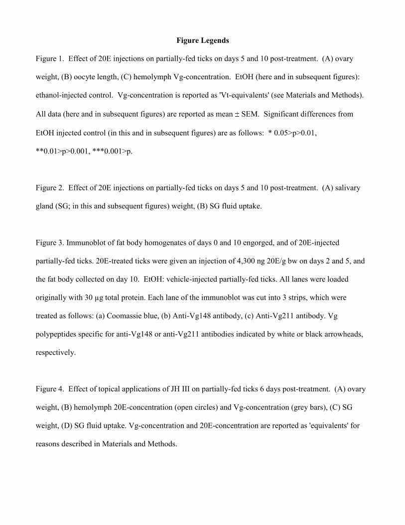
Figure Legends
Figure 1. Effect of 20E injections on partially-fed ticks on days 5 and 10 post-treatment. (A) ovary
weight, (B) oocyte length, (C) hemolymph Vg-concentration. EtOH (here and in subsequent figures):
ethanol-injected control. Vg-concentration is reported as 'Vt-equivalents' (see Materials and Methods).
All data (here and in subsequent figures) are reported as mean SEM. Significant differences from
EtOH injected control (in this and in subsequent figures) are as follows: * 0.05>p>0.01,
**0.01>p>0.001, ***0.001>p.
Figure 2. Effect of 20E injections on partially-fed ticks on days 5 and 10 post-treatment. (A) salivary
gland (SG; in this and subsequent figures) weight, (B) SG fluid uptake.
Figure 3. Immunoblot of fat body homogenates of days 0 and 10 engorged, and of 20E-injected
partially-fed ticks. 20E-treated ticks were given an injection of 4,300 ng 20E/g bw on days 2 and 5, and
the fat body collected on day 10. EtOH: vehicle-injected partially-fed ticks. All lanes were loaded
originally with 30 µg total protein. Each lane of the immunoblot was cut into 3 strips, which were
treated as follows: (a) Coomassie blue, (b) Anti-Vg148 antibody, (c) Anti-Vg211 antibody. Vg
polypeptides specific for anti-Vg148 or anti-Vg211 antibodies indicated by white or black arrowheads,
respectively.
Figure 4. Effect of topical applications of JH III on partially-fed ticks 6 days post-treatment. (A) ovary
weight, (B) hemolymph 20E-concentration (open circles) and Vg-concentration (grey bars), (C) SG
weight, (D) SG fluid uptake. Vg-concentration and 20E-concentration are reported as 'equivalents' for
reasons described in Materials and Methods.
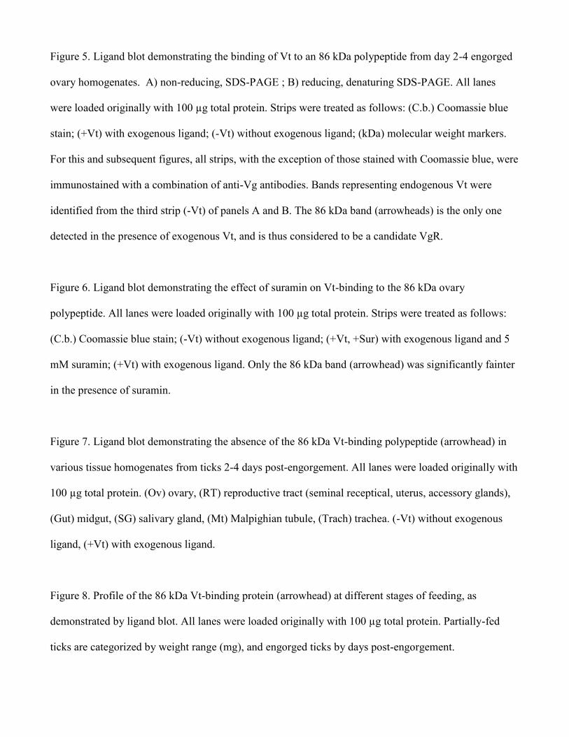
Figure 5. Ligand blot demonstrating the binding of Vt to an 86 kDa polypeptide from day 2-4 engorged
ovary homogenates. A) non-reducing, SDS-PAGE ; B) reducing, denaturing SDS-PAGE. All lanes
were loaded originally with 100 µg total protein. Strips were treated as follows: (C.b.) Coomassie blue
stain; (+Vt) with exogenous ligand; (-Vt) without exogenous ligand; (kDa) molecular weight markers.
For this and subsequent figures, all strips, with the exception of those stained with Coomassie blue, were
immunostained with a combination of anti-Vg antibodies. Bands representing endogenous Vt were
identified from the third strip (-Vt) of panels A and B. The 86 kDa band (arrowheads) is the only one
detected in the presence of exogenous Vt, and is thus considered to be a candidate VgR.
Figure 6. Ligand blot demonstrating the effect of suramin on Vt-binding to the 86 kDa ovary
polypeptide. All lanes were loaded originally with 100 µg total protein. Strips were treated as follows:
(C.b.) Coomassie blue stain; (-Vt) without exogenous ligand; (+Vt, +Sur) with exogenous ligand and 5
mM suramin; (+Vt) with exogenous ligand. Only the 86 kDa band (arrowhead) was significantly fainter
in the presence of suramin.
Figure 7. Ligand blot demonstrating the absence of the 86 kDa Vt-binding polypeptide (arrowhead) in
various tissue homogenates from ticks 2-4 days post-engorgement. All lanes were loaded originally with
100 µg total protein. (Ov) ovary, (RT) reproductive tract (seminal receptical, uterus, accessory glands),
(Gut) midgut, (SG) salivary gland, (Mt) Malpighian tubule, (Trach) trachea. (-Vt) without exogenous
ligand, (+Vt) with exogenous ligand.
Figure 8. Profile of the 86 kDa Vt-binding protein (arrowhead) at different stages of feeding, as
demonstrated by ligand blot. All lanes were loaded originally with 100 µg total protein. Partially-fed
ticks are categorized by weight range (mg), and engorged ticks by days post-engorgement.
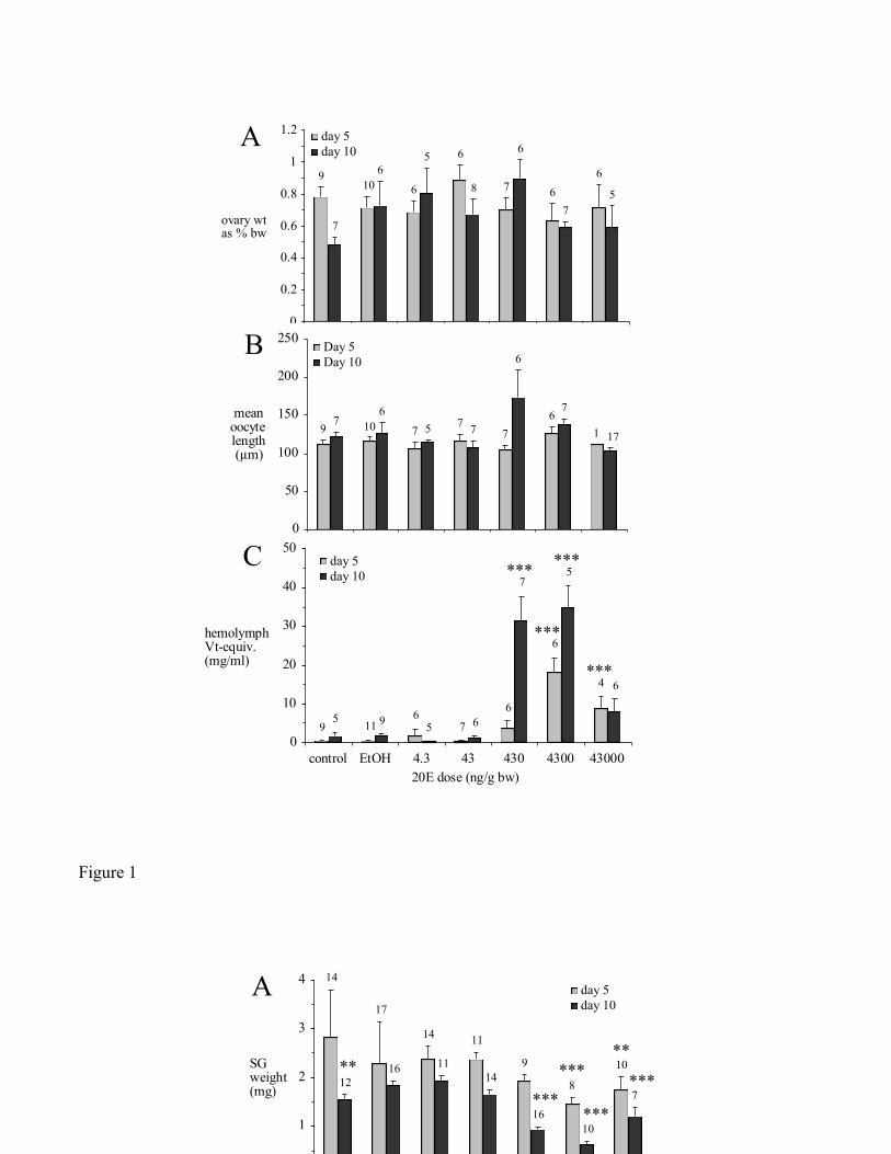
Figure 1
0
0.2
0.4
0.6
0.8
1
1.2
control EtOH 4.3 43 430 4300 43000
20E dose (ng/g bw)
ovary wt as % bw
day 5
day 10A
9
7
6
6
6
6
6
6
5
5
7
7
8
10
0
50
100
150
200
250
control EtOH 4.3 43 430 4300 43000
20E dose (ng/g bw)
mean oocyte length (µm)
Day 5
Day 10B
9
7
7
7
7
7
6
10
6
6
5
7
1
17
0
10
20
30
40
50
control EtOH 4.3 43 430 4300 43000
20E dose (ng/g bw)
hemolymph Vt-equiv. (mg/ml)
day 5
day 10
C ***
***
***
***
9
9
5
11
5
6
6
6
6
6
7
7
5
4
0
1
2
3
4
control EtOH 4.3 43 430 4300 43000
20E dose (ng/g bw)
SG weight (mg)
day 5
day 10A 14
14
14
12
17
16
16
11
11
9
10
10
8
7
** **
***
***
***
***
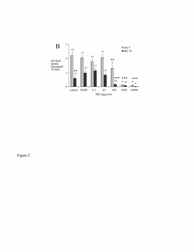
Figure 2
0
1
2
3
control EtOH 4.3 43 430 4300 43000
20E (ng/g bw)
SG fluid uptake (mg/gland/10 min)
day 5
day 10
B
*** *** ***
**
**
11
14
14
6
13
11
14
14
10
12
9
8
9
17
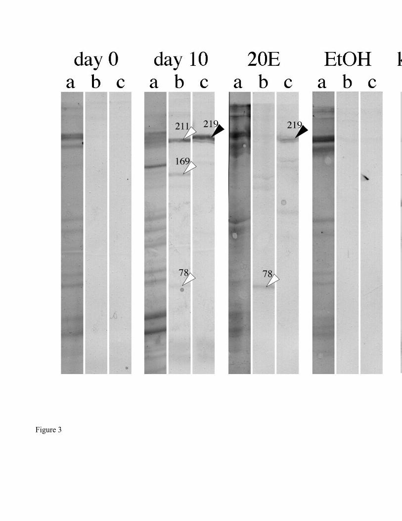
Figure 3
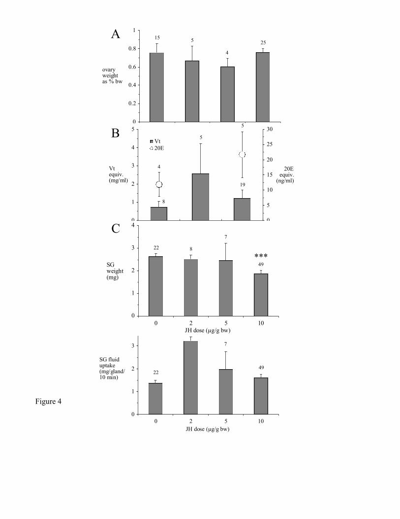
Figure 4
0
0.2
0.4
0.6
0.8
1
0 2 5 10
JH dose (µg/g bw)
ovary weight as % bw
A 15
25
5
4
0
1
2
3
4
0 2 5 10
JH dose (µg/g bw)
SG fluid uptake (mg/gland/10 min)
D
22
49
7
8
***
0
1
2
3
4
5
0 2 10JH dose (µg/g bw)
Vt equiv. (mg/ml)
0
5
10
15
20
25
30
20E equiv.
(ng/ml)
Vt
20E
B
4
5
5
8
19
0
1
2
3
4
0 2 5 10
JH dose (µg/g bw)
SG weight (mg)
C
22
49
7
8
***
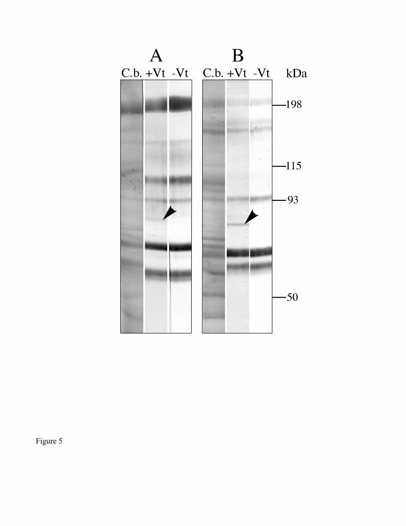
Figure 5
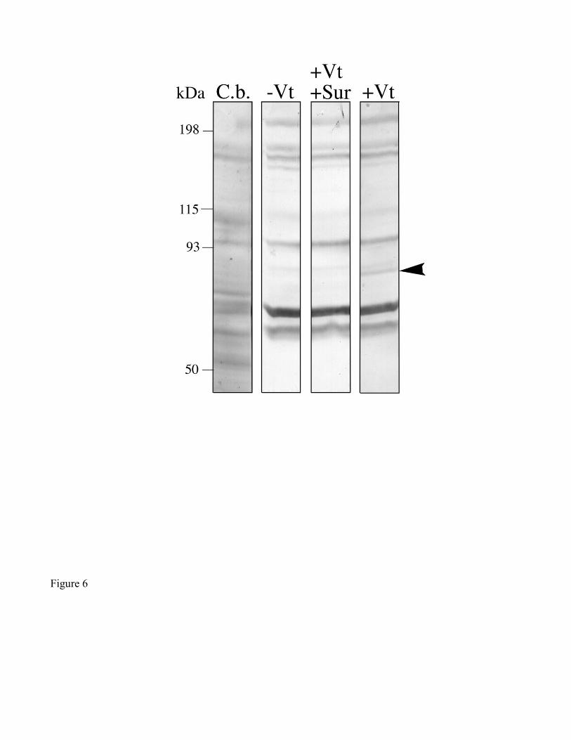
Figure 6
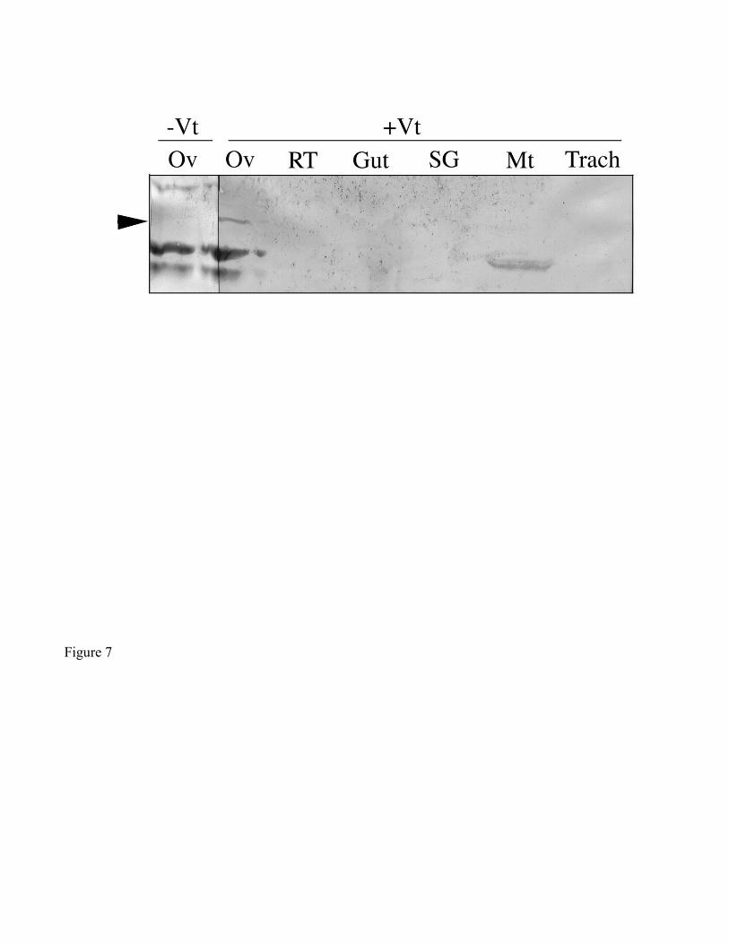
Figure 7
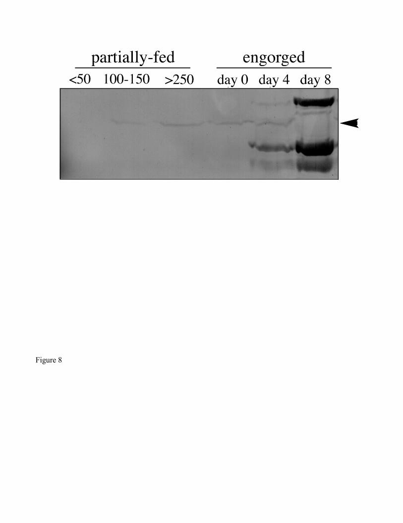
Figure 8