Embed Size (px)
Citation preview
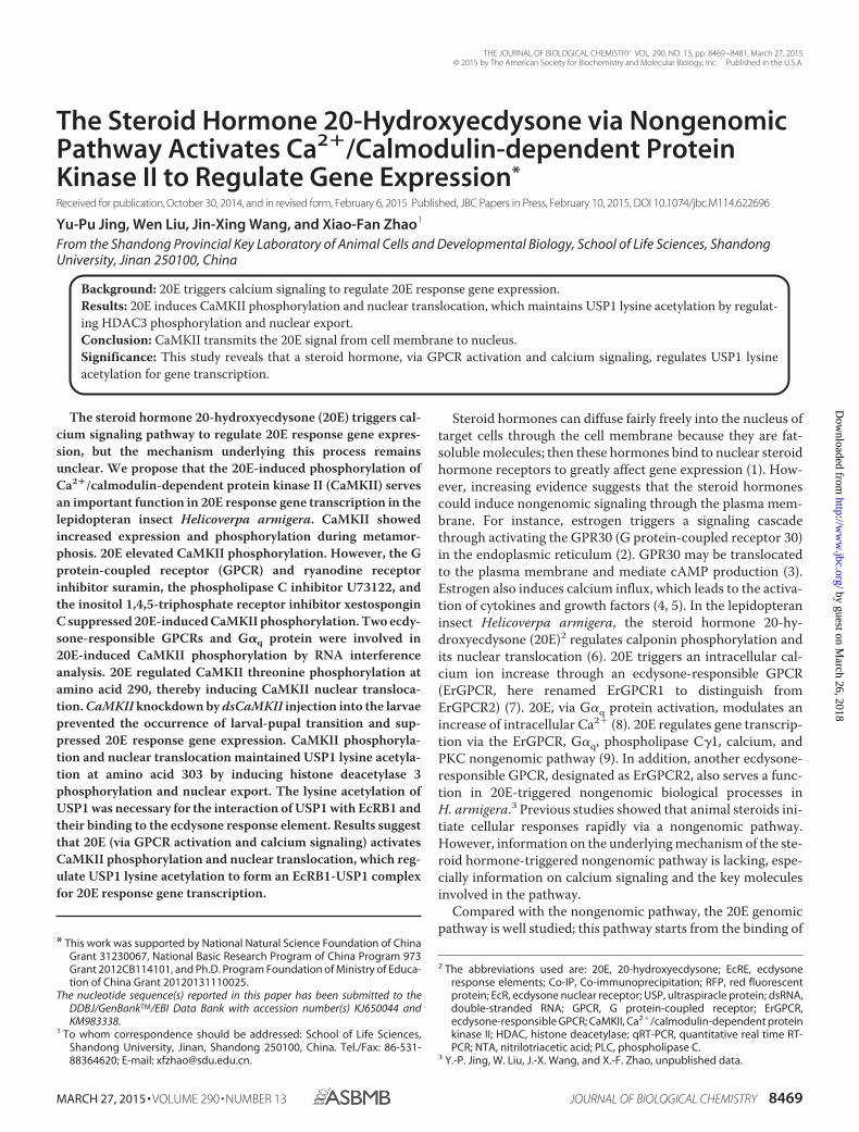
The Steroid Hormone 20-Hydroxyecdysone via NongenomicPathway Activates Ca2�/Calmodulin-dependent ProteinKinase II to Regulate Gene Expression*
Received for publication, October 30, 2014, and in revised form, February 6, 2015 Published, JBC Papers in Press, February 10, 2015, DOI 10.1074/jbc.M114.622696
Yu-Pu Jing, Wen Liu, Jin-Xing Wang, and Xiao-Fan Zhao1
From the Shandong Provincial Key Laboratory of Animal Cells and Developmental Biology, School of Life Sciences, ShandongUniversity, Jinan 250100, China
Background: 20E triggers calcium signaling to regulate 20E response gene expression.Results: 20E induces CaMKII phosphorylation and nuclear translocation, which maintains USP1 lysine acetylation by regulat-ing HDAC3 phosphorylation and nuclear export.Conclusion: CaMKII transmits the 20E signal from cell membrane to nucleus.Significance: This study reveals that a steroid hormone, via GPCR activation and calcium signaling, regulates USP1 lysineacetylation for gene transcription.
The steroid hormone 20-hydroxyecdysone (20E) triggers cal-cium signaling pathway to regulate 20E response gene expres-sion, but the mechanism underlying this process remainsunclear. We propose that the 20E-induced phosphorylation ofCa2�/calmodulin-dependent protein kinase II (CaMKII) servesan important function in 20E response gene transcription in thelepidopteran insect Helicoverpa armigera. CaMKII showedincreased expression and phosphorylation during metamor-phosis. 20E elevated CaMKII phosphorylation. However, the Gprotein-coupled receptor (GPCR) and ryanodine receptorinhibitor suramin, the phospholipase C inhibitor U73122, andthe inositol 1,4,5-triphosphate receptor inhibitor xestosponginC suppressed 20E-induced CaMKII phosphorylation. Two ecdy-sone-responsible GPCRs and G�q protein were involved in20E-induced CaMKII phosphorylation by RNA interferenceanalysis. 20E regulated CaMKII threonine phosphorylation atamino acid 290, thereby inducing CaMKII nuclear transloca-tion. CaMKII knockdown by dsCaMKII injection into the larvaeprevented the occurrence of larval-pupal transition and sup-pressed 20E response gene expression. CaMKII phosphoryla-tion and nuclear translocation maintained USP1 lysine acetyla-tion at amino acid 303 by inducing histone deacetylase 3phosphorylation and nuclear export. The lysine acetylation ofUSP1 was necessary for the interaction of USP1 with EcRB1 andtheir binding to the ecdysone response element. Results suggestthat 20E (via GPCR activation and calcium signaling) activatesCaMKII phosphorylation and nuclear translocation, which reg-ulate USP1 lysine acetylation to form an EcRB1-USP1 complexfor 20E response gene transcription.
Steroid hormones can diffuse fairly freely into the nucleus oftarget cells through the cell membrane because they are fat-soluble molecules; then these hormones bind to nuclear steroidhormone receptors to greatly affect gene expression (1). How-ever, increasing evidence suggests that the steroid hormonescould induce nongenomic signaling through the plasma mem-brane. For instance, estrogen triggers a signaling cascadethrough activating the GPR30 (G protein-coupled receptor 30)in the endoplasmic reticulum (2). GPR30 may be translocatedto the plasma membrane and mediate cAMP production (3).Estrogen also induces calcium influx, which leads to the activa-tion of cytokines and growth factors (4, 5). In the lepidopteraninsect Helicoverpa armigera, the steroid hormone 20-hy-droxyecdysone (20E)2 regulates calponin phosphorylation andits nuclear translocation (6). 20E triggers an intracellular cal-cium ion increase through an ecdysone-responsible GPCR(ErGPCR, here renamed ErGPCR1 to distinguish fromErGPCR2) (7). 20E, via G�q protein activation, modulates anincrease of intracellular Ca2� (8). 20E regulates gene transcrip-tion via the ErGPCR, G�q, phospholipase C�1, calcium, andPKC nongenomic pathway (9). In addition, another ecdysone-responsible GPCR, designated as ErGPCR2, also serves a func-tion in 20E-triggered nongenomic biological processes inH. armigera.3 Previous studies showed that animal steroids ini-tiate cellular responses rapidly via a nongenomic pathway.However, information on the underlying mechanism of the ste-roid hormone-triggered nongenomic pathway is lacking, espe-cially information on calcium signaling and the key moleculesinvolved in the pathway.
Compared with the nongenomic pathway, the 20E genomicpathway is well studied; this pathway starts from the binding of
* This work was supported by National Natural Science Foundation of ChinaGrant 31230067, National Basic Research Program of China Program 973Grant 2012CB114101, and Ph.D. Program Foundation of Ministry of Educa-tion of China Grant 20120131110025.
The nucleotide sequence(s) reported in this paper has been submitted to theDDBJ/GenBankTM/EBI Data Bank with accession number(s) KJ650044 andKM983338.
1 To whom correspondence should be addressed: School of Life Sciences,Shandong University, Jinan, Shandong 250100, China. Tel./Fax: 86-531-88364620; E-mail: [email protected].
2 The abbreviations used are: 20E, 20-hydroxyecdysone; EcRE, ecdysoneresponse elements; Co-IP, Co-immunoprecipitation; RFP, red fluorescentprotein; EcR, ecdysone nuclear receptor; USP, ultraspiracle protein; dsRNA,double-stranded RNA; GPCR, G protein-coupled receptor; ErGPCR,ecdysone-responsible GPCR; CaMKII, Ca2�/calmodulin-dependent proteinkinase II; HDAC, histone deacetylase; qRT-PCR, quantitative real time RT-PCR; NTA, nitrilotriacetic acid; PLC, phospholipase C.
3 Y.-P. Jing, W. Liu, J.-X. Wang, and X.-F. Zhao, unpublished data.
THE JOURNAL OF BIOLOGICAL CHEMISTRY VOL. 290, NO. 13, pp. 8469 –8481, March 27, 2015© 2015 by The American Society for Biochemistry and Molecular Biology, Inc. Published in the U.S.A.
MARCH 27, 2015 • VOLUME 290 • NUMBER 13 JOURNAL OF BIOLOGICAL CHEMISTRY 8469
by guest on March 26, 2018
http://ww
w.jbc.org/
Dow
nloaded from
20E up to the nuclear ecdysone receptor (EcR), after which aheterodimeric transcriptional EcR-USP complex with theultraspiracle protein (USP) is formed (10). The formation oftranscriptional complex EcRB1-USP1 is regulated by 20E vianongenomic signaling (11, 12). The EcR-USP complex can bindto ecdysone response elements (EcRE), which are located at the5� promoter region of 20E response genes for initiating genetranscription (12, 13). These 20E response genes include thetranscription factor Broad (Br) in Bombyx mori (13), alsoknown as Br-C (broad complex) (14), and the transcription fac-tor HR3 (hormone receptor 3) in H. armigera (12). Br initiatesmetamorphosis in Manduca sexta and Drosophila melano-gaster (15). Broad Z7 (BrZ7) promotes larval-pupal transition inH. armigera (16). HR3 in M. sexta is the early delay gene in the20E genomic pathway (17) and is recognized as a central regu-lator in 20E-driven developmental switches during insectdevelopment and metamorphosis (18). HR3 could also mediatethe expressions of EcRB1 and USP1 in Aedes aegypti (19). TheEcRE of H. armigera HR3 and the red fluorescence protein(RFP) are used to construct the 20E response reporter plasmid,which can be used to detect 20E-induced EcRB1-USP1-depen-dent gene transcription in the genomic pathway (12). Thesestudies provide a basis for further study of the mechanismunderlying the nongenomic pathway and the connectionbetween genomic and nongenomic pathways.
The Ca2�/calmodulin-dependent protein kinase II (CaMKII),a serine/threonine kinase, serves an important function in cal-cium signaling (20). CaMKII can be activated by Ca2� and cal-modulin, and activation leads to the autophosphorylation ofCaMKII at amino acid threonine 287 (or 286 in different iso-forms) in mammalian cells (21). CaMKII, which may be locatedin the cytosol, cytoskeleton (22), and nucleus (23), responds tothe elevation of intracellular calcium ion concentration (24)and mediates a variety of biological processes, including neu-rotransmitter synthesis (25), neurotransmitter exocytosis (26),and ion channel regulation in mammalian (27) and insect cells(28). CaMKII induces histone deacetylase 4 (HDAC4) phos-phorylation and nuclear export, which keep the target proteinacetylation regulated by histone acetyltransferases (29). Theacetylation of histone catalyzed by histone acetyltransferasesresults in loose nucleosomes structure and promotes gene acti-vation (30). By contrast, the deacetylation of histone catalyzedby HDACs leads to chromatin condensation and transcrip-tional repression (31). In addition, HDACs can regulate a vari-ety of cellular processes by regulating a variety of non-histoneprotein deacetylations, some of which are transcription factorsand co-regulators, e.g. nuclear receptor corepressor SMRT(silencing mediator of retinoid and thyroid hormone receptors)(32) and MEF2 (myocyte enhancer factor 2) (33). Therefore,CaMKII can be used as a target in studies on the nongenomicpathway and those on the connection between the genomic andnongenomic pathways of the steroid hormone.
We examined the CaMKII expression profile and hormonalregulation on the CaMKII expression level, nuclear transloca-tion, and phosphorylation. We also studied the mechanism bywhich CaMKII regulated the 20E response gene expression.20E promoted CaMKII phosphorylation via GPCR, G�q, phos-pholipase C (PLC), and calcium signaling. The phosphorylated
CaMKII transferred into the nucleus to induce HDAC3 phos-phorylation and translocation from the nucleus to the cytosol,which maintained USP1 lysine acetylation. The acetylation ofUSP1 was necessary for the formation of the 20E-inducedEcRB1-USP1 transcription complex. Our results suggest that20E regulates CaMKII phosphorylation via a nongenomic path-way for gene transcription in the genomic pathway.
EXPERIMENTAL PROCEDURES
Chemicals—The following reagents were purchased for anal-yses: pET-32a vector system (Promega Corporation, Madison,WI), pIEx-4-His vector system (containing a His tag) (providedby Dr. Marek Jindra, Biology Center, Academy of Sciences ofthe Czech Republic), restriction enzymes (Thermo Fisher Sci-entific, Lithuania), DNA polymerase (TransGen Biotech, Bei-jing, China), Unizol reagent (Biostar, Shanghai, China), proteinA resin (GenScript, Piscataway, NJ), first strand cDNA synthe-sis kit (BioTeke Corporation, Beijing, China), 20E (Sigma), PCRprimers (Sangon Biotech, Shanghai, China), gene sequencing(BGI, Shenzhen, China), and UltraSYBR Mixture (With ROX)(Beijing ComWin Biotech Co. Ltd., Beijing, China). Otherchemicals were of analytical reagent grade and were purchasedin China.
Insect—The cotton bollworms (H. armigera) were fed on anartificial diet at 27 � 1 °C and exposed to a light/dark photope-riod of 14 h/10 h, as described by Zhao et al. (34). The cottonbollworms were obtained from the Wuhan Institute of Virologyof the Chinese Academy of Sciences (Wuhan, China).
Cell Culture—The HaEpi cell line, a Helicoverpa epidermalcell line, was obtained from the H. armigera integument andhas been well characterized previously. This cell line has beenused as a platform to investigate hormonal regulation duringlepidopteran insect development. HaEpi cells were developedas a loosely attached monolayer and were maintained at 27 �1 °C with Grace’s medium containing 10% FBS (Invitrogen)(35).
Bioinformatics Analysis—CaMKII was obtained by tran-scriptome sequencing of the HaEpi cells cDNA library, whichwas established in our laboratory (GenBankTM accession no.KJ650044). Protein translation and prediction were achievedusing ExPASy software. cDNA and encoded protein were ana-lyzed by performing a BLAST search in the NCBI database.
Preparation of Antiserum against CaMKII—By using the cor-responding primers (Table 1), the cDNA fragment encoding apart of the CaMKII was amplified from H. armigera and wasinserted into the expression vector pET-32a (�). The recombi-nant plasmid was transformed into Escherichia coli DH5� cellsand then isolated and transformed into E. coli Rosetta hostcells. Isopropyl-�-D-thiogalactopyranoside (0.5 mM) was usedto induce the production of target proteins by the host cells inthe LB medium (containing 1% tryptone, 0.5% yeast extract,and 1% NaCl). The recombinant CaMKII protein was purifiedusing a Ni2�-NTA affinity column (GE Healthcare) and used asantigen for antiserum preparation. The antiserum specificitywas examined by immunoblotting analysis.
Western Blot—The total protein of cells or larvae tissue wasextracted using TBS, which contained 50 mM Tris-HCl (pH7.5), 150 mM NaCl, and 1 mM phenylmethanesulfonyl fluoride.
20E Regulates Gene Expression through CaMKII Signaling
8470 JOURNAL OF BIOLOGICAL CHEMISTRY VOLUME 290 • NUMBER 13 • MARCH 27, 2015
by guest on March 26, 2018
http://ww
w.jbc.org/
Dow
nloaded from
The protein was centrifuged at 10,000 � g at 4 °C for 10 min.The supernatant was collected, and the protein concentrationwas measured according to Bradford’s method. The 20 �g oftotal protein of each sample was loaded on 7.5% to 12.5% SDS-PAGE and electrophoretically transferred onto a nitrocellulosemembrane. The membrane was incubated with a blockingbuffer (2% skim milk in TBS) for 1 h at room temperature, after
which primary antibodies were diluted with blocking buffer for2– 4 h. The membrane was washed thrice with TBST (0.02%Tween in TBS) for 10 min each time, and the second antibody ofalkaline phosphatase-conjugated AffiniPure horse anti-rabbit/anti-mouse IgG was diluted to 1:10,000 with the same blockingbuffer. The membrane was washed with TBST thrice (for 10min each time) and subsequently washed with TBS thrice (for 5
TABLE 1Oligonucleotide sequences of PCR primers
Primer name Oligonucleotide sequence (5�3 3�)
RNA interferenceCaMKII-RNAi F 5�-gcgtaatacgactcactatagggattggaagacgacgatttgg-3�CaMKII-RNAi R 5�-gcgtaatacgactcactatagggtgccatatgcgagtctcttg-3�ErGPCR1-RNAi F 5�-gcgtaatacgactcactataggtcggaggaggcgaaggag-3�ErGPCR1-RNAi R 5�-gcgtaatacgactcactatagggtgttcgccgcagtcaaa-3�ErGPCR2-RNAi F 5�-gcgtaatacgactcactatagggttcatccttctaacggtggc-3�ErGPCR2-RNAi R 5�-gcgtaatacgactcactatagggtcgcttcatcttcgctatct-3�GFP-RNAi F 5�-gcgtaatacgactcactataggtggtcccaattctcgtggaac-3�GFP-RNAi R 5�-gcgtaatacgactcactataggcttgaagttgaccttgatgcc-3�G�q-RNAi F 5�-gcgtaatacgactcactataggtcggaggaggcgaaggag-3�G�q-RNAi R 5�-gcgtaatacgactcactatagggtgttcgccgcagtcaaa-3�HDAC3-RNAi F 5�-gcgtaatacgactcactatagggccgactcgttagctggagac-3�HDAC3-RNAi R 5�-gcgtaatacgactcactatagggttggctgattctcacgtctg-3�HDAC4-RNAi F 5�-gcgtaatacgactcactatagggccaccgaagtcaagcagaag-3�HDAC4-RNAi R 5�-gcgtaatacgactcactataggggtcttgcgcagagggtagtc-3�HDAC6-RNAi F 5�-gcgtaatacgactcactatagggcggttctagtggtgggaaaa-3�HDAC6-RNAi R 5�-gcgtaatacgactcactatagggtgcaccatactgagaggtcg-3�
OverexpressionCaMKII-OE F 5�-tactcagagctcatggcaaatccaaaccgtgaa-3�CaMKII-OE R 5�-tactcaagatctgggagcgatggaaatgtattgc-3�EcRB1-OE F 5�-tactcacaattggatgagacgccgctggtataac-3�EcRB1-OE R 5�-tactcaggcgcgccgagagcgccggcgagtccgccac-3�HDAC3-OE F 5�-tactcagagctcatgactcaacataaagtagct-3�HDAC3-OE R 5�-tactcaagatctgtggctccttgttctcaact-3�USP1-OE F 5�-tactcagagctcatgatggagccctcgagagat-3�USP1-OE R 5�-tactcaggcgcgccgacatcatgttggtgtctat-3�
qRT-PCRBrZ7-qRT F 5�-ggtgactgtccttactgcggcat-3�BrZ7-qRT R 5�-ttaattcctttgaccatgact-3�CaMKII-qRT F 5�-atcgataacacgccgactcat-3�CaMKII-qRT R 5�-gtattgccgtccacttgttgt-3�EcRB1-qRT F 5�-aattgcccgtcagtacga-3�EcRB1-qRT R 5�-tgagcttctcattgagga-3�ErGPCR1-qRT F 5�-aaacggttcacctactacgc-3�ErGPCR1-qRT R 5�-cgcttcatcttcgctatct-3�ErGPCR2-qRT F 5�-cgagggtcaagtctgaggtt-3�ErGPCR2-qRT R 5�-tattattagtcgtggtggta-3�G�q-qRT F 5�-ggcagttgcgaaaggac-3�G�q-qRT R 5�-tctgagttggacggatt-3�HDAC3-qRT F 5�-gtgggagatgattgtccagtct-3�HDAC3-qRTR 5�-ggtcggtagaactccatgacat-3�HDAC4-qRT F 5�-caagaaggacaagcacgagca-3�HDAC4-qRT R 5�-ggacttcacgatgccccagt-3�HDAC6-qRT F 5�-ggtgtccgcatttagactcct-3�HDAC6-qRT R 5�-cgtaaagaagagagttgtcca-3�HR3-qRT F 5�-tcaagcacctcaacagcagcccta-3�HR3-qRT R 5�-gactttgctgatgtcaccctccgc-3�USP1-qRT F 5�-ggtcctgacagcaatgtt-3�USP1-qRT R 5�-ttccagctccagctgactgaag-3��-Actin-qRT F 5�-cctggtattgctgaccgtatgc-3��-Actin-qRT R 5�-ctgttggaaggtggagagggaa-3�
ChIP assayEcRB1-P F 5�-atattcgaatcgttggcg-3�EcRB1-P R 5�-cagtgtaattaagagaca-3�
Prokaryotic expressionCaMKII-exp F 5�-tactcagaattccaagagaccgtagactgcttg-3�CaMKII-exp R 5�-tactcactcgagggctgcagaagggactag-3�
Site-directed mutagenesisCaMKII T290A F 5�-aggcaagaggccgtagactgc-3�CaMKII T290A R 5�-gcagtctacggcctcttgcct-3�USP1 K58R F 5�-agtggttccaggcacctctgt-3�USP1 K58R R 5�-acagaggtgcctggaaccact-3�USP1 K71R F 5�-gcgtcgggaagacattatgga-3�USP1 K71R R 5�-tccataatgtcttcccgacgc-3�USP1 K303R F 5�-ctgtcgctgaggatgcgcagt-3�USP1 K303R R 5�-actgcgcatcctcagcgacag-3�
20E Regulates Gene Expression through CaMKII Signaling
MARCH 27, 2015 • VOLUME 290 • NUMBER 13 JOURNAL OF BIOLOGICAL CHEMISTRY 8471
by guest on March 26, 2018
http://ww
w.jbc.org/
Dow
nloaded from

min each time). The target signals were visualized using p-nitroblue tetrazolium/5-bromo-4-chloro-3-indolyl phosphate (10ml of TBS, 45 �l of 5% nitro blue tetrazolium, and 35 �l of 5%5-bromo-4-chloro-3-indolyl phosphate) after incubation in thedark for 10 min.
Quantitative Real Time RT-PCR Analysis—Total RNA wasextracted using Unizol reagent, and the first-strand cDNA wassynthesized using a first-strand cDNA synthesis kit accordingto the manufacturer’s instructions. The qRT-PCR analysis wasperformed using a CFX96TM real time system (Bio-Rad) with afinal volume of 10 �l through the following steps: 95 °C initialdenaturation for 15 min, 40 cycles of 95 °C for 15 s and 60 °C for60 s, 78 °C for 2 s for plate reading, and melting curve analysisfrom 65 °C to 95 °C. �-Actin was used as reference gene to nor-malize the gene expression. The experiments were repeatedthrice, and the data were analyzed using the 2���CT method.
Hormonal regulation in H. armigera larvae—20E was ini-tially dissolved in DMSO to prepare a 10 mg/ml solution andsubsequently diluted with sterile PBS (140 mM NaCl and 10 mM
sodium phosphate, pH 7.4) at 1:100. The sixth instar larvae at6 h were injected with 20E at 500 ng/larva. The controls weretreated with an equivalent amount of DMSO at the same stage.The total protein was isolated from the integument, midgut,and fat body of larvae (3 to 5 larvae) after the insects weretreated with 20E for 0.25, 0.5, 1, 3, or 6 h. The proteins wereused for Western blot analysis.
RNAi in the HaEpi Cell Line—The MEGAscriptTM RNAi kit(Ambion, Austin, TX) was used to generate dsRNA. ThedsRNA was transcribed from PCR templates of the CaMKII (orother genes) at 37 °C for 4 h (primers are shown in Table 1),according to the manufacturer’s instructions. DNase I was usedto remove DNA from the dsRNA solution. dsGFP was tran-scribed utilizing GFP DNA as a template and used as a nonspe-cific RNAi control. The dsRNA concentration was determinedby spectrophotometrical analysis at 260 nm. For the transfec-tion of dsRNA into the cell line, HaEpi cells were seeded in6-well plates with 5 � 105 cells/well. The RNAfectin transfec-tion reagent (Tiangen, Beijing, China) was used for dsRNAtransfection according to the manufacturer’s instructions. Thefinal dsRNA concentration was 1 �g/ml in the medium withoutFBS. After incubation at 27 °C for 6 –12 h, the cells were replen-ished with a complete medium and were used for experiments.
RNAi in H. armigera Larvae—The H. armigera larvae wereselected for dsCaMKII injection (dsGFP injection as control) at6 h after the appearance of the sixth instar. The larvae wererandomly separated into two groups with 30 larvae/group,and three independent experiments were performed. Eachlarva in the experimental group was injected with dsCaMKII(1 �g), whereas each larva in the negative control group wasinjected with dsGFP. The statistical data of the larval-pupaltransition phenotypes of each group were obtained. mRNAwas extracted from the sixth instar larvae at 120 h for qRT-PCR analysis.
Overexpression and Phosphorylation Analysis of CaMKII—The CaMKII ORF or mutation (obtained by site-directedmutagenesis of CaMKII in vitro) was amplified from H. armig-era using corresponding primers (Table 1) and then insertedinto the pIEx-4-His vector or pIEx-4-RFP-His vector (pIEx-4-
His vector fusing with RFP). HaEpi cells were maintained at80% confluence under normal growth conditions, as previouslydescribed by Shao et al. (35). The reconstructed plasmids weretransfected into the cells for 6 –12 h with the help of the DNA-fectin transfection reagent. Subsequently, the cells were replen-ished with a complete medium. After being cultured in fullnutrient culture medium for 24 h, 20E was added to the cellsat a final concentration of 2 �M, according to the methodused in our previous work. The control cells received anequivalent volume of DMSO, which was used as a solvent for20E. The pIEx-4-RFP-His/pIEx-4-His vector was transfectedand served as the negative control. The � phosphatase (Mil-lipore, Temecula, CA) treatment was conducted using pro-tein extracts from HaEpi cells treated with 2 �M 20E for 0.5 h.The gel concentration of SDS-PAGE was 7.5%. �-Actin wasused as the protein control using antiserum against �-actinin H. armigera.
Co-immunoprecipitation (Co-IP)—The ORFs of EcRB1 (Gen-BankTM accession no. EU526831) and USP1 (EU526832) wereamplified from H. armigera using corresponding primers(Table 1) and were then inserted into the pIEx-4-RFP-His andpIEx-4-His vectors, respectively. The reconstructed plasmidswere transfected into the cells, as described above. The anti-body against RFP (1 �l) and PBS (400 �l) was incubated withprotein A resin for 30 min at room temperature. The resin waswashed with 500 �l of PBS thrice. HaEpi cells were transfectedwith dsCaMKII for RNA interference. The dsGFP was used asthe negative control, as previously shown. The cells weretreated with 20E for 1 h, and DMSO was used as the control.The protein was extracted from cells using radioimmunopre-cipitation assay buffer containing 0.1 M Tris-HCl buffer (pH8.0), 150 mM NaCl, and 1% Nonidet P-40. The supernatant washarvested by centrifugation at 12,000 � g for 10 min (4 °C). Thesupernatant was added to protein A resin to eliminate nonspe-cific binding and harvested by centrifugation. The supernatantwas added to the resin-antibody complex and incubated for2– 4 h with gentle shaking at 4 °C. The resin was harvested bycentrifugation and washed with PBS thrice. Lastly, the resin wastreated with SDS-PAGE loading buffer and boiled for 10 min.After centrifugation at 12,000 � g for 10 min (4 °C), the proteinsamples were loaded onto SDS-PAGE for Western blot analysisusing the antibody against RFP (Zhongshan, Beijing, China)and His (Zhongshan, Beijing, China) against EcRB1-RFP andUSP-His, respectively.
Mutation and Lysine Acetylation Analysis of USP1—HaEpicells were maintained at 80% confluence under normal growthconditions in the 6-well plates. The USP1-His plasmids orthe mutations (USP1-K58R-His, USP1-K71R-His, and USP1-K303R-His plasmids), which were obtained by site-directedmutagenesis of USP1 in vitro, were transfected into the cells, aspreviously shown. The cells were treated with 20E for 1 h, andDMSO was used as the control. The protein was extracted fromthe cells using radioimmunoprecipitation assay buffer, and thesupernatant was harvested by centrifugation and purified usinga Ni2�-NTA affinity column. The purified protein was used forWestern blot analysis with the antibody against acetyl lysine(Immunechem, Nanning, China).
20E Regulates Gene Expression through CaMKII Signaling
8472 JOURNAL OF BIOLOGICAL CHEMISTRY VOLUME 290 • NUMBER 13 • MARCH 27, 2015
by guest on March 26, 2018
http://ww
w.jbc.org/
Dow
nloaded from

HDAC3 Phosphorylation Levels Detection—HDAC3-RFP-His was overexpressed in HaEpi cells, and the cells were trans-fected with dsCaMKII (1 �g/ml in the medium) for 12 h. ThedsGFP was the nonspecific dsRNA control. Finally, the cellswere treated with 20E at 2 �M for 0.5 h. Equal volumes ofDMSO were used for the negative control. HDAC3-RFP-Hiswas purified by Ni2�-NTA affinity column for detecting phos-phorylation levels. The number of moles of phosphorus permole of HDAC3-RFP-His was determined using the Phospho-protein phosphate estimation assay kit (Sangon Biotech,Shanghai, China) based on the alkaline hydrolysis of phosphatefrom seryl and threonyl residues in phosphoproteins. Thereleased phosphate was quantified in a 96-well microplateaccording to the manufacturer’s instructions.
Chromatin Immunoprecipitation Assay—The USP1-His,USP1-K303R-His, or pIEx-4-His (as negative control) plasmidswere transfected into the cells. Subsequently, the cells weretreated with 20E for 3 h. DMSO treatment was used as thecontrol. Formaldehyde (37%) was added to the cells at a finalconcentration of 0.5% for cross-linking at 37 °C for 10 min.Glycine was subsequently added at a final concentration of 125mM at room temperature for 10 min. The cells were washedtwice with ice-cold PBS, harvested by centrifugation, and thensuspended in 200 �l of SDS lysis buffer containing 1% SDS, 10mM EDTA, and 50 mM Tris-HCl (pH 8.1). Ultrasonication wasperformed to break up the genomic DNA into 200 –1,000-bpfragments. After centrifugation, the supernatant was pre-cleared with protein A resin at 4 °C for 1 h. After centrifugation,20 �l of supernatant was used as an input sample for qRT-PCR(negative control sample). The remaining supernatant (180 �l)was incubated with anti-His antibody for 12 h. The protein Aresin was added to collect the protein and DNA complex at4 °C. The complex was washed as follows: once with low saltbuffer containing 200 mM Tris-HCl (pH 8.0), 2 mM EDTA, 150mM NaCl, 0.1% SDS, and 1.0% Triton X-100; once with high saltwash buffer containing 20 mM Tris-HCl (pH 8.0), 2 mM EDTA,500 mM NaCl, 0.1% SDS, and 1.0% Triton X-100; once with LiClwash buffer containing 10 mM Tris-HCl (pH 8.0), 250 mM LiCl,1 mM EDTA, 1% Nonidet P-40, and 1% deoxycholate; and twicewith Tris-EDTA buffer containing 10 mM Tris-HCl (pH 8.0)and 1 mM EDTA. The proteins were eluted with buffer (1% SDSand 100 mM NaHCO3). The DNA-protein cross-links werereversed at 65 °C overnight and treated with RNase A and pro-teinase K at 56 °C for 2 h. DNA was purified and analyzed byqRT-PCR to detect the EcRB1-USP1-binding element inH. armigera HR3 promoter using the EcRB1PF/EcRB1PRprimers shown in the Table 1.
RESULTS
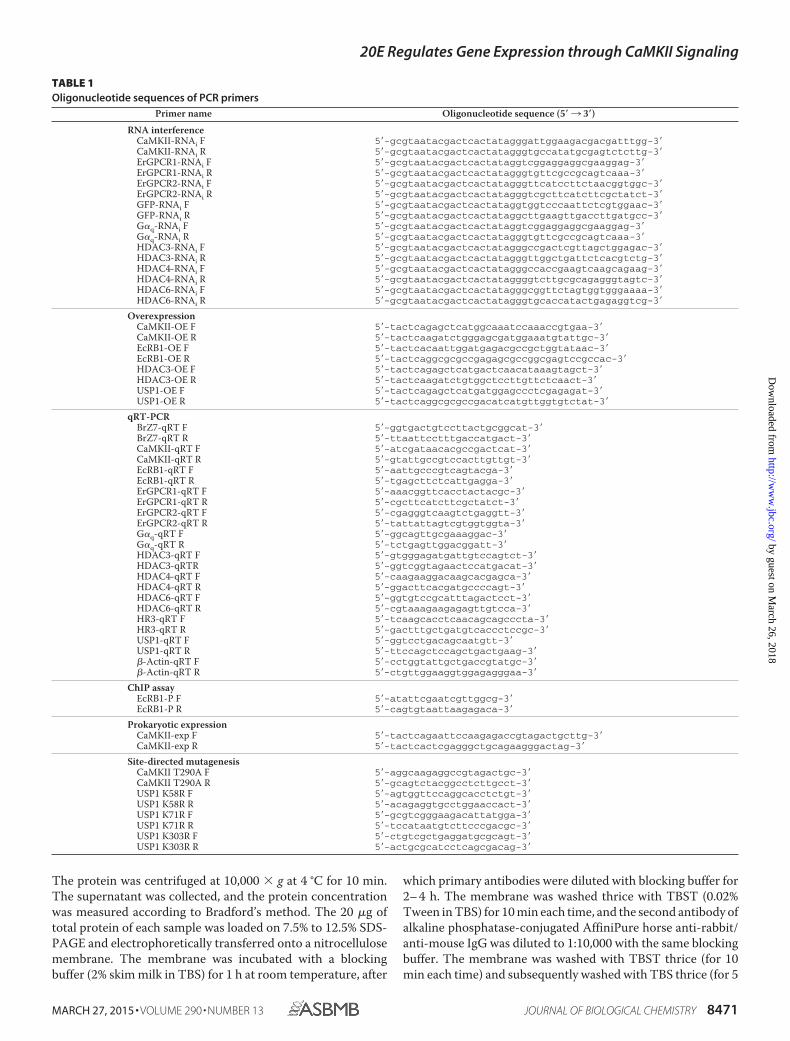
CaMKII Expression and Phosphorylation Are Increased dur-ing the Metamorphic Stage—CaMKII protein was detected astwo bands in various tissues by Western blot with a polyclonalantibody against H. armigera CaMKII. The relative intensity ofthe upper band was intensified in comparison with the lowerband at the metamorphic stage. CaMKII protein was increasedat the fifth instar molting and sixth instar metamorphosisstages of the different tissues, i.e. integument, midgut, and fatbody. The results of qRT-PCR showed that the mRNA levels of
CaMKII also increased at fifth instar molting and sixth instarmetamorphic stages (Fig. 1A). The abovementioned resultsshowed that the expression profile of CaMKII is in agreementwith the 20E titer in Lepidoptera (10), thereby implying thatCaMKII plays a role in metamorphosis.
To test whether the upper band was post-translationallymodified after induction by 20E, the sixth instar larvae at 6 hwere injected with 20E or DMSO (solvent control). The expres-sion levels of the upper band were significantly up-regulated inthe three tissues (compared with the DMSO control) by 20Einduction for 15 min (Fig. 1B). In addition, the molecularweight of the upper band was degraded to the molecular weightof the lower band by � phosphatase treatment (Fig. 1C), therebyindicating that the upper band is the phosphorylated CaMKII.These results suggest that CaMKII expression and phosphory-lation levels are up-regulated by 20E in the metamorphic stage.
To address the pathway of 20E-regulating CaMKII phosphor-ylation, the intracellular calcium concentration increase inHaEpi cells was blocked by inhibitors, including the GPCRs andryanodine receptor inhibitor suramin, receptor tyrosine kinaseinhibitor SU6668, PLC inhibitor U73122, and inositol 1,4,5-triphosphate receptor inhibitor xestospongin C (XeC). Toexclude the effects of gene transcription induced by 20E, thetime of 20E induction was limited to 30 min. Compared withthe DMSO treatment, 20E increased the phosphorylation levelof CaMKII. Suramin, U73122, and XeC significantly inhibited20E-increased CaMKII phosphorylation, but inhibition was notobserved under the SU6668 treatment. Suramin blocked bothGPCR (36) and ryanodine receptor signaling (37). Therefore,the two ecdysone-responsible GPCRs (ErGPCR1 and ErG-PCR2) and G�q protein were knocked down by RNAi in HaEpicells to address the involvement of GPCR in 20E-inducedCaMKII phosphorylation. The knockdown of ErGPCR1,ErGPCR2, and G�q suppressed 20E-induced CaMKII phosphory-lation (Fig. 2A). The efficacy of RNAi was confirmed by semi-quantitative RT-PCR analysis (Fig. 2A, panel a). These resultssuggest that 20E regulates CaMKII phosphorylation throughthe GPCR, G�q, PLC, and calcium signaling.
The Thr287 is a key phosphorylation site for its activation inmammalian CaMKII (21), and the Thr290 in H. armigeraCaMKII is the conserved threonine with mammalian Thr287 byhomologous analysis. To understand the consequence of theCaMKII phosphorylation, CaMKII and its mutant CaMKII-T290A (threonine was mutated to alanine at 290 site) wereoverexpressed by pIEx-4-RFP-His vector in the HaEpi cells. Inthe DMSO control, CaMKII-RFP-His was mainly distributed inthe cytoplasm and partially translocated into the nucleus by 20Einduction in 0.5 h. However, when threonine was mutated toalanine at the 290 site, CaMKII-T290A-RFP-His was not trans-located into the nucleus by 20E induction. The overexpressionof RFP alone by pIEx-4-RFP-His plasmid did not change sub-cellular location under 20E (Fig. 2B). Western blot showed thatthe nuclear located CaMKII was phosphorylated, whereas thecytoplasmic located CaMKII was not phosphorylated (Fig. 2C).The 20E-induced phosphorylation site of CaMKII at Thr290 wasconfirmed with the overexpression of CaMKII-RFP-His andCaMKII-T290A-RFP-His in HaEpi cells, as shown by theresults of Western blot analysis (Fig. 2D). The overexpression of
20E Regulates Gene Expression through CaMKII Signaling
MARCH 27, 2015 • VOLUME 290 • NUMBER 13 JOURNAL OF BIOLOGICAL CHEMISTRY 8473
by guest on March 26, 2018
http://ww
w.jbc.org/
Dow
nloaded from
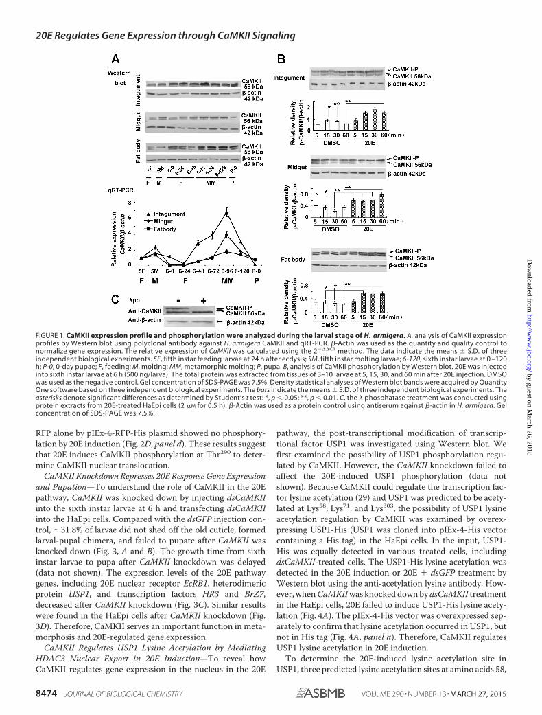
RFP alone by pIEx-4-RFP-His plasmid showed no phosphory-lation by 20E induction (Fig. 2D, panel d). These results suggestthat 20E induces CaMKII phosphorylation at Thr290 to deter-mine CaMKII nuclear translocation.
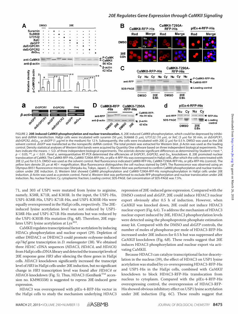
CaMKII Knockdown Represses 20E Response Gene Expressionand Pupation—To understand the role of CaMKII in the 20Epathway, CaMKII was knocked down by injecting dsCaMKIIinto the sixth instar larvae at 6 h and transfecting dsCaMKIIinto the HaEpi cells. Compared with the dsGFP injection con-trol, �31.8% of larvae did not shed off the old cuticle, formedlarval-pupal chimera, and failed to pupate after CaMKII wasknocked down (Fig. 3, A and B). The growth time from sixthinstar larvae to pupa after CaMKII knockdown was delayed(data not shown). The expression levels of the 20E pathwaygenes, including 20E nuclear receptor EcRB1, heterodimericprotein USP1, and transcription factors HR3 and BrZ7,decreased after CaMKII knockdown (Fig. 3C). Similar resultswere found in the HaEpi cells after CaMKII knockdown (Fig.3D). Therefore, CaMKII serves an important function in meta-morphosis and 20E-regulated gene expression.
CaMKII Regulates USP1 Lysine Acetylation by MediatingHDAC3 Nuclear Export in 20E Induction—To reveal howCaMKII regulates gene expression in the nucleus in the 20E
pathway, the post-transcriptional modification of transcrip-tional factor USP1 was investigated using Western blot. Wefirst examined the possibility of USP1 phosphorylation regu-lated by CaMKII. However, the CaMKII knockdown failed toaffect the 20E-induced USP1 phosphorylation (data notshown). Because CaMKII could regulate the transcription fac-tor lysine acetylation (29) and USP1 was predicted to be acety-lated at Lys58, Lys71, and Lys303, the possibility of USP1 lysineacetylation regulation by CaMKII was examined by overex-pressing USP1-His (USP1 was cloned into pIEx-4-His vectorcontaining a His tag) in the HaEpi cells. In the input, USP1-His was equally detected in various treated cells, includingdsCaMKII-treated cells. The USP1-His lysine acetylation wasdetected in the 20E induction or 20E � dsGFP treatment byWestern blot using the anti-acetylation lysine antibody. How-ever, when CaMKII was knocked down by dsCaMKII treatmentin the HaEpi cells, 20E failed to induce USP1-His lysine acety-lation (Fig. 4A). The pIEx-4-His vector was overexpressed sep-arately to confirm that lysine acetylation occurred in USP1, butnot in His tag (Fig. 4A, panel a). Therefore, CaMKII regulatesUSP1 lysine acetylation in 20E induction.
To determine the 20E-induced lysine acetylation site inUSP1, three predicted lysine acetylation sites at amino acids 58,
FIGURE 1. CaMKII expression profile and phosphorylation were analyzed during the larval stage of H. armigera. A, analysis of CaMKII expressionprofiles by Western blot using polyclonal antibody against H. armigera CaMKII and qRT-PCR. �-Actin was used as the quantity and quality control tonormalize gene expression. The relative expression of CaMKII was calculated using the 2���CT method. The data indicate the means � S.D. of threeindependent biological experiments. 5F, fifth instar feeding larvae at 24 h after ecdysis; 5M, fifth instar molting larvae; 6-120, sixth instar larvae at 0 –120h; P-0, 0-day pupae; F, feeding; M, molting; MM, metamorphic molting; P, pupa. B, analysis of CaMKII phosphorylation by Western blot. 20E was injectedinto sixth instar larvae at 6 h (500 ng/larva). The total protein was extracted from tissues of 3–10 larvae at 5, 15, 30, and 60 min after 20E injection. DMSOwas used as the negative control. Gel concentration of SDS-PAGE was 7.5%. Density statistical analyses of Western blot bands were acquired by QuantityOne software based on three independent biological experiments. The bars indicate the means � S.D. of three independent biological experiments. Theasterisks denote significant differences as determined by Student’s t test: *, p 0.05; **, p 0.01. C, the � phosphatase treatment was conducted usingprotein extracts from 20E-treated HaEpi cells (2 �M for 0.5 h). �-Actin was used as a protein control using antiserum against �-actin in H. armigera. Gelconcentration of SDS-PAGE was 7.5%.
20E Regulates Gene Expression through CaMKII Signaling
8474 JOURNAL OF BIOLOGICAL CHEMISTRY VOLUME 290 • NUMBER 13 • MARCH 27, 2015
by guest on March 26, 2018
http://ww
w.jbc.org/
Dow
nloaded from
71, and 303 of USP1 were mutated from lysine to arginine,namely, K58R, K71R, and K303R. In the input, the USP1-His,USP1-K58R-His, USP1-K71R-His, and USP1-K303R-His wereequally overexpressed in the HaEpi cells, respectively. The 20E-induced lysine acetylation level was not reduced by USP1-K58R-His and USP1-K71R-His mutations but was reduced bythe USP1-K303R-His mutation (Fig. 4B). Therefore, 20E regu-lates USP1 lysine acetylation at Lys303.
CaMKII regulates transcriptional factor acetylation by inducingHDACs phosphorylation and nuclear export (29). Depletion ofeither DHDAC1 or DHDAC3 could promote ecdysone-inducedeip74ef gene transcription in D. melanogaster (38). We obtainedthree HDAC cDNA sequences (HDAC3, HDAC4, and HDAC6)from HaEpi cells cDNA library and detected the transcript levels of20E response gene HR3 after silencing the three genes in HaEpicells. HDAC3 knockdown significantly increased the transcriptlevel of HR3 in HaEpi cells under 20E induction, but no significantchange in HR3 transcription level was found after HDAC4 orHDAC6 knockdown (Fig. 5). Thus, HDAC3 (GenBankTM acces-sion no. KM983338) is suggested to repress 20E-induced geneexpression.
HDAC3 was overexpressed with pIEx-4-RFP-His vector inthe HaEpi cells to study the mechanism underlying HDAC3
repression of 20E-induced gene expression. Compared with theDMSO control and dsGFP, 20E could induce HDAC3 nuclearexport obviously after 0.5 h of induction. However, whenCaMKII was knocked down, 20E could not induce HDAC3nuclear export (Fig. 6A). To address the mechanism of HDAC3nuclear export induced by 20E, HDAC3 phosphorylation levelswere detected using the phosphoprotein phosphate estimationassay kit. Compared with the DMSO and dsGFP controls, thenumber of moles of phosphorus per mole of HDAC3-RFP-Hisincreased under 20E induces for 0.5 h but was suppressed afterCaMKII knockdown (Fig. 6B). These results suggest that 20Einduces HDAC3 phosphorylation and nuclear export via acti-vating CaMKII.
Because HDAC3 can catalyze transcriptional factor deacety-lation in the nucleus (39), the effect of HDAC3 on USP1 lysineacetylation was studied by co-overexpressing HDAC3-RFP-Hisand USP1-His in the HaEpi cells, combined with CaMKIIknockdown to block HDAC3-RFP-His translocation fromnucleus to cytoplasm. Compared with the pIEx-4-RFP-Hisoverexpressing control, the overexpression of HDAC3-RFP-His showed obvious inhibitory effect on USP1 lysine acetylationunder 20E induction (Fig. 6C). These results suggest that
FIGURE 2. 20E-induced CaMKII phosphorylation and nuclear translocation. A, 20E induced CaMKII phosphorylation, which could be depressed by inhibi-tors and dsRNA transfection. HaEpi cells were incubated with suramin (50 �M), SU6668 (5 �M), U73122 (10 �M), or XeC (3 �M) for 30 min, or dsErGPCR1,dsErGPCR2, dsG�q, or dsGFP (1 �g/ml in the medium) for 12 h. Subsequently, the cells were incubated with 20E (2 �M) for 0.5 h. DMSO was used as the 20Esolvent control. dsGFP was transfected as the nonspecific dsRNA control. The total protein was extracted for Western blot. �-Actin was used as the loadingcontrol. Density statistical analyses of Western blot bands were acquired by Quantity One software based on three independent biological experiments. Thebars indicate the means � S.D. of three independent biological experiments. The asterisks denote significant differences as determined by Student’s t test: *,p 0.05; **, p 0.01. Panel a, semiquantitative RT-PCR determined the efficiencies of ErGPCR1, ErGPCR2, and G�q knockdown. B, 20E promoted nucleartranslocation of CaMKII. The CaMKII-RFP-His, CaMKII-T290A-RFP-His, or pIEx-4-RFP-His was overexpressed in HaEpi cells, after which the cells were treated with20E (2 �M) for 0.5 h. DMSO was used as the solvent control. Red fluorescence indicated CaMKII-RFP-His, CaMKII-T290A-RFP-His, or pIEx-RFP-His (control). Theyellow bars denote 20 �M at 40� magnification. Blue fluorescence distinguishes the cell nucleus stained by DAPI. The fluorescence was observed using anOlympus BX51 fluorescence microscope (Shinjuku-ku, Tokyo, Japan). C, Western blot was performed to confirm CaMKII phosphorylation and nuclear translo-cation under 20E induction. D, Western blot showed CaMKII phosphorylation and CaMKII-T290A-RFP-His nonphosphorylation in HaEpi cells under 20Einduction. �-Actin was used as a protein control. Panel d, Western blot was performed to exclude RFP phosphorylation and nuclear translocation under 20Einduction. Nu, nuclear fraction; Cy, cytoplasmic fraction; Loading control, SDS-PAGE. Gel concentration of SDS-PAGE was 7.5%.
20E Regulates Gene Expression through CaMKII Signaling
MARCH 27, 2015 • VOLUME 290 • NUMBER 13 JOURNAL OF BIOLOGICAL CHEMISTRY 8475
by guest on March 26, 2018
http://ww
w.jbc.org/
Dow
nloaded from
CaMKII regulates USP1 lysine acetylation by inducing HDAC3nuclear export during 20E induction.
USP1 Acetylation Determines the Interaction between USP1and EcRB1 and Their Binding to EcRE—To address the mech-anism of USP1-K303 acetylation serving in the 20E responsegene transcription, the protein interaction between EcRB1 andUSP1 was examined. In the input, EcRB1-RFP-His and USP1-His were simultaneously and equally overexpressed in theHaEpi cells, including cells with CaMKII knockdown. In theco-immunoprecipitation (Co-IP) using an antibody againstRFP to precipitate EcRB1-RFP-His, USP1-His band wasdetected increasingly in the 20E-induced cells compared withthe DMSO control. However, when CaMKII was knockeddown, USP1-His band was lighter than that in the dsGFP con-trol during 20E induction (Fig. 7A). pIEx-4-RFP-His and pIEx-4-His overexpressions were controlled to exclude the possibil-ity of protein interaction caused by His or RFP tag (Fig. 7A,panel a). These results suggest that 20E regulates the interac-tion between EcRB1-RFP-His and USP1-His through activatingCaMKII.
To confirm the involvement of the 20E-induced USP1-Hislysine acetylation in the interaction between EcRB1-RFP-Hisand USP1-His, USP1-His and USP1-K303R-His were sepa-rately overexpressed in the HaEpi cells, as shown in the input.USP1-His interacting with EcRB1-RFP-His was detected inCo-IP using an antibody against RFP, whereas USP1-K303R-
His failed to interact with EcRB1 under 20E induction (Fig. 7B).Furthermore, USP1-His could bind to the EcRE under20E induction by ChIP analysis. However, USP1-K303R-Hisreduced the ability of binding to EcRE. pIEx-4-His was overex-pressed as a control to exclude the possibility of His tag bindingto EcRE (Fig. 7C). Therefore, USP1-K303 acetylation isrequired for the formation of EcRB1-USP1 transcriptionalcomplex and increases the ability of the complex to bind to theEcRE in the 20E pathway.
DISCUSSION
20E Induces a Rapid Increase of Ca2� Ion Concentration inthe Cytoplasm of the HaEpi Cell Line through ErGPCR Signal-ing (7). However, the function and mechanism of the increase ofintracellular calcium triggered by 20E nongenomic signalingpathway are still unclear. It is well known that USP1 can bephosphorylated by PKC through ErGPCR and PLC signalingpathways (9, 40). However, information on 20E-induced lysineacetylation of USP1 has been limited. The present studyshowed that 20E regulates CaMKII phosphorylation at theThr290 site for nuclear translocation via ErGPCR, G�q, PLC,and calcium signaling. Subsequently, the phosphorylated CaM-KII regulates USP1 lysine acetylation at the Lys303 site throughinducing HDAC3 phosphorylation and nuclear export, which isnecessary for the interaction between USP1 and EcRB1 andtheir binding to EcRE. Therefore, nongenomic pathway is
FIGURE 3. CaMKII knockdown blocked larval-pupal transition and 20E response gene expression. A, insect phenotypes after CaMKII and GFP (negativecontrol) knockdown (dsCaMKII or dsGFP was injected into sixth instar larvae at 6 h). The photograph was captured at pupal stage using a Nikon E995 digitalcamera (Hiyoda-ku, Tokyo, Japan). B, statistical analysis of phenotypes after CaMKII and GFP knockdown. Abnormal pupae display larval-pupal chimeras. dsGFPwas used as nonspecific dsRNA control. The bars indicate the means � S.D. of three independent biological experiments (with 30 larvae individuals perreplication). The asterisks denotes significant differences as determined by Student’s t test: *, p 0.05. C, expression levels of 20E-induced genes in larvalmidgut after knockdown of CaMKII and GFP (negative control). dsCaMKII or dsGFP was injected into sixth instar larvae at 6 h, and then the total mRNA wasisolated from larval midgut at 6 –120 h for qRT-PCR analysis. D, expression levels of 20E-induced genes after CaMKII and GFP (negative control) knockdown inHaEpi cells. Cells were transfected with dsCaMKII (1 �g/ml in the medium) for 12 h and subsequently treated with 20E (2 �M) for 6 h. An equivalent volume ofDMSO was applied to cells as solvent control for 20E. In the experiments of C and D, dsGFP and �-actin were used as nonspecific dsRNA and quantitative control,respectively. The relative expression of 20E-induced genes was calculated by qRT-PCR analysis using the 2���CT method. The bars indicate the means � S.D.of three independent biological experiments. The asterisks denote significant differences as determined by Student’s t test: *, p 0.05; **, p 0.01.
20E Regulates Gene Expression through CaMKII Signaling
8476 JOURNAL OF BIOLOGICAL CHEMISTRY VOLUME 290 • NUMBER 13 • MARCH 27, 2015
by guest on March 26, 2018
http://ww
w.jbc.org/
Dow
nloaded from

involved in 20E-induced EcRB1-USP1-dependent transcrip-tion initiation in genomic pathway triggered by 20E.
20E Induces CaMKII Phosphorylation and Nuclear Translo-cation via Nongenomic Pathway—CaMKII is a multimericprotein, and each monomer has three domains, i.e. N-terminalcatalytic domain, regulatory region, and C-terminal subunitassociation domain. The catalytic domain and regulatory
region are tightly associated at basal resting state, therebyresulting in autoinhibition of the kinase activity of CaMKII (41).The C-terminal subunit association domain is responsiblefor the assembly of subunits into large multimers (8 –14 sub-units) that can comprise one or several different isoforms (42).The Thr287 in the regulatory region of mammalian CaMKII canbe rapidly autophosphorylated by the binding of calcium andcalmodulin (Ca2�/CaM), followed by the phosphorylation ofthe secondary sites (Thr305 and Thr306); thus, CaMKII is sus-tainably activated (21). CaMKII in H. armigera also had anN-terminal serine-threonine kinase catalytic domain (aminoacids 17–275), a regulatory region (amino acids 281–318), and aC terminus (amino acids 388 –510). Thr290 was conserved withmammalian CaMKII autophosphorylation site Thr287, andThr309 and Thr310 were conserved with mammalian CaMKIICa2�/CaM binding sites Thr305 and Thr306 in the regulatoryregion. Previous studies have proved that 20E triggers the rapidincrease of intracellular calcium ion concentration throughErGPCR and PLC signaling pathways (7, 9), and such a phe-nomenon is one of the important features of nongenomic path-way. In the present study, we further demonstrated that 20Einduced CaMKII phosphorylation at the Thr290 site in 30 minvia ErGPCRs, G�q, PLC, and calcium signaling pathways,thereby illustrating the output of the increase of intracellularcalcium ion under 20E induction.
CaMKII is localized in specific tissues and subcellular com-partments with different functions (21). CaMKII contained anuclear localization signal in many species (43). However,CaMKII in D. melanogaster was detected without canonicalnuclear localization signal in nuclear extracts (21). Similarly,nuclear localization signal was not detected in the amino acidsequence of H. armigera CaMKII. Nonetheless, CaMKII-RFP-His could still be partially transported into the nucleus in aphosphorylated form during 20E induction. Results suggestthat CaMKII nuclear translocation is dependent on its phos-phorylation in 20E-induced signaling, which is confirmed bysite-directed mutant CaMKII-T290A.
20E Regulates USP1 Lysine Acetylation for the Gene Tran-scription Initiation via CaMKII—CaMKII regulates gene tran-scription initiation via directly phosphorylating transcriptionfactors, such as cAMP response element binding protein (44)and serum response factor (45). However, in the present study,the direct phosphorylation of USP1 mediated by CaMKII wasnot detected. CaMKII, as a serine/threonine kinase in nucleus,can also phosphorylate HDAC4 and promote its outflux fromthe nucleus (44). In the nucleus, HDAC4 binds to transcriptionfactors, such as serum response factor, and induces histonedeacetylation. Histone deacetylation promotes chromatin con-densation and favors transcriptional repression (29), whereashistone acetylation relaxes the nucleosome structure and favorsgene activation (30). In addition, as a corepressor, HDAC4 canregulate deacetylation of transcription factors, such as tran-scription factor forkhead box protein O (46). In Drosophila S2cells, the RNAi-mediated depletion of the various HDACs(DHDAC1, DHDAC2, DHDAC3, DHDAC4, and DHDACX)revealed that only the depletion of HDAC1 or HDAC3 affectedtranscription, and depletion of HDAC3 caused the up-regula-tion of 29 genes, including the ecdysone-induced eip74ef gene
FIGURE 4. CaMKII regulated USP1 lysine acetylation under 20E induction.A, 20E induce USP1 lysine acetylation through CaMKII. USP1-His was overex-pressed in HaEpi cells. Subsequently, the cells were treated with 20E (2 �M) for1 h. DMSO was used as the negative control. Input, protein expression levelsof CaMKII, USP1-His, and �-actin in HaEpi cells, which were detected by West-ern blot using antibody anti-CaMKII, anti-His, and anti-�-actin, respectively.Gel concentration of SDS-PAGE was 10%. USP1-His lysine acetylation wasdetected by Western blot using anti-Ac-Lys antibody after being purified byNi2�-NTA affinity column. Panel a, His tag was overexpressed by the plasmidpIEx-4-His as a control, and the lysine acetylation was detected by the samemethods as described in A. B, identification of the 20E-induced acetylationsite in USP1. Plasmid of USP1-His, USP1-K58R-His, USP1-K71R-His, or USP1-K303R-His was overexpressed in HaEpi cells, after which the cells were treatedwith 20E (2 �M) for 1 h. DMSO was used as the solvent control. Input, expres-sion levels of USP1-His, USP1-K58R-His, USP1-K71R-His, and USP1-K303R-Hisin HaEpi cells were detected by Western blot using antibody against His tag.�-Actin was used as loading control. The gel concentration of SDS-PAGE was10%. Density statistical analyses of Western blot bands were acquired byQuantity One software based on three independent biological experiments.The bars indicate the means � S.D. of three independent biological experi-ments. The asterisks denote significant differences as determined byStudent’s t test: **, p 0.01.
20E Regulates Gene Expression through CaMKII Signaling
MARCH 27, 2015 • VOLUME 290 • NUMBER 13 JOURNAL OF BIOLOGICAL CHEMISTRY 8477
by guest on March 26, 2018
http://ww
w.jbc.org/
Dow
nloaded from

(38). Similar to HDAC4, HDAC3 can also function for manysequence-specific transcription factors, including NF-�B (47),SMAD7 (mothers against decapentaplegic homolog 7) (48),and c-Jun (49). HDAC3 belongs to a multimolecular complexthat contains nuclear receptor corepressor and SMRT proteinsubunit, which are required for many nuclear hormone recep-
tors involved in physiological action (50, 51). In Drosophila,SMRT-related and ecdysone receptor interacting factor, whichshares a similar function and regional homology to the verte-brate nuclear corepressors SMRT and nuclear receptor core-pressor, can interact with EcR and can mediate transcriptionrepression by interacting with Sin3A, a repressor known to
FIGURE 5. 20E induced HR3 transcription through HDAC3. Cells were transfected with dsHDAC3, dsHDAC4, or dsHDAC6 (1 �g/ml in the medium) for 12 h andsubsequently treated with 20E (2 �M) for 6 h. An equivalent volume of DMSO was applied to cells as solvent control for 20E, and dsGFP was used as nonspecificdsRNA. Total mRNA was isolated for qRT-PCR analysis using the 2���CT method. The bars indicate the means � S.D. of three independent biological experi-ments. The asterisks denote significant differences as determined by Student’s t test: *, p 0.05; **, p 0.01.
FIGURE 6. 20E induced HDAC3 phosphorylation and nuclear export through CaMKII to maintain USP1 lysine acetylation. A, 20E regulated HDAC3subcellular location through activating CaMKII in HaEpi cells. The HDAC3-RFP-His or pIEx-4-RFP-His (negative control) was overexpressed in HaEpi cells, and thecells were transfected with dsCaMKII or dsGFP (1 �g/ml in the medium) for 12 h. dsGFP was used as the nonspecific dsRNA control. The cells were treated with20E (2 �M) for 0.5 h for immunocytochemical localization analysis. An equivalent volume of DMSO was applied to cells as solvent control for 20E. Redfluorescence indicated HDAC3-RFP-His or pIEx-4-RFP-His. The yellow bars denote 20 �M at 40� magnification. Blue fluorescence distinguished the cell nucleusstained by DAPI. The fluorescence was observed using an Olympus BX51 fluorescence microscope. B, 20E induced HDAC3 phosphorylation, which could bedepressed by dsCaMKII treatment in HaEpi cells. The number of moles of phosphorus per mole of HDAC3-RFP-His was determined using a phosphoproteinphosphate estimation assay kit after HDAC3-RFP-His was purified by Ni2�-NTA affinity column. HDAC3-RFP-His was overexpressed in HaEpi cells, after whichthe cells were treated with dsCaMKII and treated with 2 �M 20E or an equivalent amount of DMSO for 0.5 h. pIEx-4-RFP-His was overexpressed as the control.Significant difference was determined by Student’s t test based on three independent biological experiments: *, p 0.05. C, 20E-induced USP1 lysineacetylation was depressed by HDAC3 in the nucleus. USP1-His and HDAC3-RFP-His were co-overexpressed in HaEpi cells, after which the cells were treated withdsCaMKII and treated with 20E (2 �M for 1 h). pIEx-4-RFP, USP1-His co-overexpression, and dsGFP were used as the negative controls. USP1-His was isolated byNi2�-NTA affinity column and detected by Western blot using antibody anti-His and anti-Ac-Lys. Input, protein expression levels of CaMKII, HDAC3-RFP-His,RFP-tag, USP1-His, and �-actin in HaEpi cells were detected by Western blot using antibody anti-CaMKII, anti-RFP, anti-His, and anti-�-actin, respectively. Gelconcentration of SDS-PAGE was 10%. Density statistical analyses of Western blot bands of Ac-Lys-USP1-His/USP1-His were acquired by Quantity One softwarebased on three independent biological experiments. The bars indicate the means � S.D. of three independent biological experiments. The asterisks denotessignificant differences as determined by Student’s t test: *, p 0.05.
20E Regulates Gene Expression through CaMKII Signaling
8478 JOURNAL OF BIOLOGICAL CHEMISTRY VOLUME 290 • NUMBER 13 • MARCH 27, 2015
by guest on March 26, 2018
http://ww
w.jbc.org/
Dow
nloaded from

form a complex with the histone deacetylase Rpd3/HDAC (52).We observed that 20E regulated USP1 lysine acetylation toform EcRB1 and USP1 heterodimer by activating CaMKII.Meanwhile, 20E could also induce HDAC3 phosphorylationand nuclear export, and CaMKII silencing reduced HDAC3phosphorylation and translocation from nucleus to cytoplasmin HaEpi cells. In addition, HDAC3 overexpression reducedUSP1 lysine acetylation in HaEpi cells under 20E induction,and HDAC3 knockdown could enhance HR3 transcriptincrease induced by 20E. HDAC3 may be involved in theEcR-mediated transcription repression via the depression ofUSP1 lysine acetylation and can be regulated by CaMKIIunder 20E induction.
20E regulates the formation of heterodimer EcRB1-USP1 forgene transcription initiation (10). A previous study showed thatUSP1 was phosphorylated by PKC, and this modification wasnecessary for USP1 to bind to EcRB1 in HaEpi cells under 20Einduction (9). The CaMKII knockdown had no effect on the20E-induced USP1 phosphorylation but down-regulated USP1lysine acetylation at the Lys303 site and the formation of EcRB1-USP1 transcriptional complex. USP1 phosphorylation andlysine acetylation are two separate processes that contribute tothe gene transcription regulation in the 20E pathway.
Conclusion—20E induces CaMKII phosphorylation throughErGPCRs, G�q, PLC, and calcium signaling pathways. Thephosphorylated CaMKII is partially translocated into the
FIGURE 7. 20E, via activating CaMKII, regulated USP1 lysine acetylation to determine its interaction with EcRB1. A, CaMKII regulated the formation ofEcRB1 and USP1 heterodimer under 20E induction. USP1-His and EcRB1-RFP-His were overexpressed in HaEpi cells, after which the cells were transfected withdsCaMKII and dsGFP (1 �g/ml in the medium) for 12 h prior to incubation with 20E (2 �M) for 3 h. DMSO was used as the solvent control. Input, protein expressionlevels of CaMKII, EcRB1-RFP-His, USP1-His, and �-actin in HaEpi cells were detected by Western blot using antibody anti-CaMKII, anti-RFP, anti-His, andanti-�-actin, respectively. Co-IP, EcRB1-RFP-His was immunoprecipitated with antibody against RFP, and the co-precipitated USP1-His was detected byWestern blot using antibody anti-His. Gel concentration of SDS-PAGE was 10%. Density statistical analyses of Western blot bands were acquired by QuantityOne software based on three independent biological experiments. The bars indicate the means � S.D. of three independent biological experiments. Theasterisks denotes significant differences, as determined by Student’s t test: *, p 0.05. Panel a, pIEx-4-His and pIEx-4-RFP-His were overexpressed in HaEpi cellsas the negative controls. The cells were treated, and the proteins were detected by the same methods used in A. B, USP1 lysine acetylation was necessary toform the EcRB1 and USP1 heterodimer. USP1-His, USP1-K303R-His, and EcRB1-RFP-His were overexpressed in HaEpi cells, after which the cells were treated with20E (2 �M) for 3 h. DMSO was used as solvent control. Input, protein expression levels of EcRB1-RFP-His, USP1-His, USP1-K303R-His, and �-actin in HaEpi cellswere detected by Western blot using antibody anti-RFP, anti-His, and anti-�-actin, respectively. Co-IP, EcRB1-RFP-His was immunoprecipitated with antibodyagainst RFP, and the co-precipitated USP1-His or USP1-K303R-His was detected by Western blot using antibody anti-His. Gel concentration of SDS-PAGE was10%. Density statistical analyses of Western blot bands were acquired by Quantity One software based on three independent biological experiments. The barsindicate the means � S.D. of three independent biological experiments. The asterisks denote significant differences as determined by Student’s t test: **, p 0.01. C, ChIP and qRT-PCR analyses of the binding capabilities of USP1-His and USP1-K303R-His to EcRE in the HR3 promoter by anti-His precipitation. USP1-Hisor USP1-K303R-His was overexpressed in HaEpi cells, after which the cells were treated with 20E (2 �M) for 3 h. DMSO was used as solvent control. Expressionlevels of USP1-His, USP1-K303R-His, and His tag in HaEpi cells were detected by Western blot using antibody anti-His. �-Actin was used as loading control.pIEx-4-His was overexpressed and used as negative control for ChIP analysis. Gel concentration of SDS-PAGE was 10%. The relative expression was calculatedby qRT-PCR with the DNA template in the precipitates by antibody anti-His. The bars indicate the means � S.D. of three independent biological experiments.The asterisks denotes significant difference as determined by Student’s t test: **, p 0.01.
20E Regulates Gene Expression through CaMKII Signaling
MARCH 27, 2015 • VOLUME 290 • NUMBER 13 JOURNAL OF BIOLOGICAL CHEMISTRY 8479
by guest on March 26, 2018
http://ww
w.jbc.org/
Dow
nloaded from

nucleus. CaMKII regulates USP1 lysine acetylation via induc-tion of HDAC3 phosphorylation and nuclear export. USP1lysine acetylation is necessary in the formation of EcRB1-USP1transcription complex and its binding to EcRE, which promotesgene transcription initiation in the 20E pathway (Fig. 8).
REFERENCES1. Thompson, E. B. (1995) Steroid hormones: membrane transporters of
steroid hormones. Curr. Biol. 5, 730 –7322. Meldrum, D. R. (2007) G-protein-coupled receptor 30 mediates estrogen’s
nongenomic effects after hemorrhagic shock and trauma. Am. J. Pathol.170, 1148 –1151
3. Sandén, C., Broselid, S., Cornmark, L., Andersson, K., Daszkiewicz-Nilsson, J., Mårtensson, U. E., Olde, B., and Leeb-Lundberg, L. M. (2011) Gprotein-coupled estrogen receptor 1/G protein-coupled receptor 30 local-izes in the plasma membrane and traffics intracellularly on cytokeratinintermediate filaments. Mol. Pharmacol. 79, 400 – 410
4. Falkenstein, E., Tillmann, H. C., Christ, M., Feuring, M., and Wehling, M.(2000) Multiple actions of steroid hormones: a focus on rapid, non-genomic effects. Pharmacol. Rev. 52, 513–556
5. Davis, P. J., Tillmann, H. C., Davis, F. B., and Wehling, M. (2002) Compar-ison of the mechanisms of nongenomic actions of thyroid hormone andsteroid hormones. J. Endocrinol. Invest. 25, 377–388
6. Liu, P. C., Wang, J. X., Song, Q. S., and Zhao, X. F. (2011) The participationof calponin in the cross talk between 20-hydroxyecdysone and juvenilehormone signaling pathways by phosphorylation variation. PLoS One 6,e19776
7. Cai, M. J., Dong, D. J., Wang, Y., Liu, P. C., Liu, W., Wang, J. X., and Zhao,X. F. (2014) G-protein-coupled receptor participates in 20-hydroxyecdy-sone signaling on the plasma membrane. Cell Commun Signal 12, 9
8. Ren, J., Li, X. R., Liu, P. C., Cai, M. J., Liu, W., Wang, J. X., and Zhao, X. F.(2014) G-protein �q participates in the steroid hormone 20-hydroxyecdy-sone nongenomic signal transduction. J. Steroid Biochem. Mol. Biol. 144,313–323
9. Liu, W., Cai, M. J., Zheng, C. C., Wang, J. X., and Zhao, X. F. (2014)Phospholipase C�1 connects the cell membrane pathway to the nuclearreceptor pathway in insect steroid hormone signaling. J. Biol. Chem. 289,13026 –13041
10. Riddiford, L. M., Hiruma, K., Zhou, X., and Nelson, C. A. (2003) Insightsinto the molecular basis of the hormonal control of molting and metamor-phosis from Manduca sexta and Drosophila melanogaster. Insect Biochem.Mol. Biol. 33, 1327–1338
11. Liu, W., Zhang, F. X., Cai, M. J., Zhao, W. L., Li, X. R., Wang, J. X., andZhao, X. F. (2013) The hormone-dependent function of Hsp90 in thecrosstalk between 20-hydroxyecdysone and juvenile hormone signalingpathways in insects is determined by differential phosphorylation andprotein interactions. Biochim. Biophys. Acta 1830, 5184 –5192
12. Liu, W., Cai, M. J., Wang, J. X., and Zhao, X. F. (2014) In a nongenomicaction, steroid hormone 20-hydroxyecdysone induces phosphorylation ofcyclin-dependent kinase 10 to promote gene transcription. Endocrinology155, 1738 –1750
13. Nishita, Y. (2014) Ecdysone response elements in the distal promoter ofthe Bombyx broad-complex gene, BmBR-C. Insect Mol. Biol. 23, 341–356
14. Emery, I. F., Bedian, V., and Guild, G. M. (1994) Differential expression ofbroad-complex transcription factors may forecast tissue-specific develop-mental fates during Drosophila metamorphosis. Development 120,3275–3287
15. Erezyilmaz, D. F., Riddiford, L. M., and Truman, J. W. (2006) The pupalspecifier broad directs progressive morphogenesis in a direct-developinginsect. Proc. Natl. Acad. Sci. U.S.A. 103, 6925– 6930
16. Cai, M. J., Liu, W., Pei, X. Y., Li, X. R., He, H. J., Wang, J. X., and Zhao, X. F.(2014) Juvenile hormone prevents 20-hydroxyecdysone-induced meta-morphosis by regulating the phosphorylation of a newly identified broadprotein. J. biol. chem. 289, 26630 –26641
17. Lan, Q., Hiruma, K., Hu, X., Jindra, M., and Riddiford, L. M. (1999) Acti-vation of a delayed-early gene encoding MHR3 by the ecdysone receptorheterodimer EcR-B1-USP-1 but not by EcR-B1-USP-2. Mol. Cell. Biol. 19,4897– 4906
18. Lam, G. T., Jiang, C., and Thummel, C. S. (1997) Coordination of larvaland prepupal gene expression by the DHR3 orphan receptor during Dro-sophila metamorphosis. Development 124, 1757–1769
19. Mane-Padros, D., Cruz, J., Cheng, A., and Raikhel, A. S. (2012) A criticalrole of the nuclear receptor HR3 in regulation of gonadotrophic cycles ofthe mosquito Aedes aegypti. PLoS One 7, e45019
20. Schulman, H., and Greengard, P. (1978) Ca2�-dependent protein phos-phorylation system in membranes from various tissues, and its activationby “calcium-dependent regulator.” Proc. Natl. Acad. Sci. U.S.A. 75,5432–5436
21. Griffith, L. C., Lu, C. S., and Sun, X. X. (2003) CaMKII, an enzyme on themove: regulation of temporospatial localization. Mol. Interv. 3, 386 – 403
22. Lin, Y. C., and Redmond, L. (2008) CaMKII� binding to stable F-actin invivo regulates F-actin filament stability. Proc. Natl. Acad. Sci. U.S.A. 105,15791–15796
23. Loweth, J. A., Baker, L. K., Guptaa, T., Guillory, A. M., and Vezina, P.(2008) Inhibition of CaMKII in the nucleus accumbens shell decreasesenhanced amphetamine intake in sensitized rats. Neurosci. Lett. 444,157–160
24. Lisman, J. (1994) The CaM kinase II hypothesis for the storage of synapticmemory. Trends Neurosci. 17, 406 – 412
25. Yamauchi, T., and Fujisawa, H. (1981) Tyrosine 3-monoxygenase is phos-phorylated by Ca2�-, calmodulin-dependent protein kinase, followed byactivation by activator protein. Biochem. Biophys. Res. Commun. 100,807– 813
26. Kennedy, M. B., and Greengard, P. (1981) Two calcium/calmodulin-de-pendent protein kinases, which are highly concentrated in brain, phos-phorylate protein I at distinct sites. Proc. Natl. Acad. Sci. U.S.A. 78,1293–1297
27. Pitt, G. S. (2007) Calmodulin and CaMKII as molecular switches for car-diac ion channels. Cardiovasc. Res. 73, 641– 647
28. Lu, H., Leung, H. T., Wang, N., Pak, W. L., and Shieh, B. H. (2009) Role ofCa2�/calmodulin-dependent protein kinase II in Drosophila photorecep-tors. J. Biol. Chem. 284, 11100 –11109
29. Backs, J., Song, K., Bezprozvannaya, S., Chang, S., and Olson, E. N. (2006)CaM kinase II selectively signals to histone deacetylase 4 during car-diomyocyte hypertrophy. J. Clin. Invest. 116, 1853–1864
30. Roth, S. Y., Denu, J. M., and Allis, C. D. (2001) Histone acetyltransferases.
FIGURE 8. Explanation for the role of CaMKII involved in the 20E path-ways. 20E induced CaMKII phosphorylation via ErGPCRs, G�q, PLC, and cal-cium signaling pathways. Phosphorylated CaMKII enters the nucleus to reg-ulate USP1 lysine acetylation via induction of HDAC3 phosphorylation andnuclear export. The lysine-acetylated USP1 interacts with EcRB1 to form theEcRB1-USP1 transcriptional complex, which binds with EcRE to initiate genestranscription in the 20E pathway.
20E Regulates Gene Expression through CaMKII Signaling
8480 JOURNAL OF BIOLOGICAL CHEMISTRY VOLUME 290 • NUMBER 13 • MARCH 27, 2015
by guest on March 26, 2018
http://ww
w.jbc.org/
Dow
nloaded from

Annu. Rev. Biochem. 70, 81–12031. Backs, J., and Olson, E. N. (2006) Control of cardiac growth by histone
acetylation/deacetylation. Circ. Res. 98, 15–2432. Takeuchi, M., Ishida, A., Kameshita, I., Kitani, T., Okuno, S., and Fujisawa,
H. (2001) Identification and characterization of CaMKP-N, nuclearcalmodulin-dependent protein kinase phosphatase. J. Biochem. 130,833– 840
33. Miska, E. A., Karlsson, C., Langley, E., Nielsen, S. J., Pines, J., and Kouza-rides, T. (1999) HDAC4 deacetylase associates with and represses theMEF2 transcription factor. EMBO J. 18, 5099 –5107
34. Zhao, X. F., Wang, J. X., and Wang, Y. C. (1998) Purification and charac-terization of a cysteine proteinase from eggs of the cotton boll worm,Helicoverpa armigera. Insect Biochem. Mol. Biol. 28, 259 –264
35. Shao, H. L., Zheng, W. W., Liu, P. C., Wang, Q., Wang, J. X., and Zhao, X. F.(2008) Establishment of a new cell line from lepidopteran epidermis andhormonal regulation on the genes. PLoS One 3, e3127
36. Abbracchio, M. P., Burnstock, G., Boeynaems, J. M., Barnard, E. A., Boyer,J. L., Kennedy, C., Knight, G. E., Fumagalli, M., Gachet, C., Jacobson, K. A.,and Weisman, G. A. (2006) International Union of Pharmacology LVIII:update on the P2Y G protein-coupled nucleotide receptors: from molec-ular mechanisms and pathophysiology to therapy. Pharmacol. Rev. 58,281–341
37. Wolner, I., Kassack, M. U., Ullmann, H., Karel, A., and Hohenegger, M.(2005) Use-dependent inhibition of the skeletal muscle ryanodine recep-tor by the suramin analogue NF676. Br. J. Pharmacol. 146, 525–533
38. Foglietti, C., Filocamo, G., Cundari, E., De Rinaldis, E., Lahm, A., Cortese,R., and Steinkühler, C. (2006) Dissecting the biological functions of Dro-sophila histone deacetylases by RNA interference and transcriptional pro-filing. J. Biol. Chem. 281, 17968 –17976
39. Chen, L. F., and Greene, W. C. (2003) Regulation of distinct biologicalactivities of the NF-�B transcription factor complex by acetylation. J. Mol.Med. 81, 549 –557
40. Sun, X., and Song, Q. (2006) PKC-mediated USP phosphorylation is re-quired for 20E-induced gene expression in the salivary glands of Drosoph-ila melanogaster. Arch. Insect Biochem. Physiol. 62, 116 –127
41. Erickson, J. R. (2014) Mechanisms of CaMKII activation in the heart. Front
Pharmacol. 5, 5942. Braun, A. P., and Schulman, H. (1995) The multifunctional calcium/cal-
modulin-dependent protein kinase: from form to function. Annu. Rev.Physiol. 57, 417– 445
43. Hudmon, A., and Schulman, H. (2002) Neuronal Ca2�/calmodulin-de-pendent protein kinase II: the role of structure and autoregulation in cel-lular function. Annu. Rev. Biochem. 71, 473–510
44. Kreusser, M. M., and Backs, J. (2014) Integrated mechanisms of CaMKII-dependent ventricular remodeling. Front. Pharmacol. 5, 36
45. Flück, M., Booth, F. W., and Waxham, M. N. (2000) Skeletal muscle CaM-KII enriches in nuclei and phosphorylates myogenic factor SRF at multiplesites. Biochem. Biophys. Res. Commun. 270, 488 – 494
46. Mihaylova, M. M., Vasquez, D. S., Ravnskjaer, K., Denechaud, P. D., Yu,R. T., Alvarez, J. G., Downes, M., Evans, R. M., Montminy, M., and Shaw,R. J. (2011) Class IIa histone deacetylases are hormone-activated regula-tors of FOXO and mammalian glucose homeostasis. Cell 145, 607– 621
47. Baek, S. H., Ohgi, K. A., Rose, D. W., Koo, E. H., Glass, C. K., and Rosenfeld,M. G. (2002) Exchange of N-CoR corepressor and Tip60 coactivator com-plexes links gene expression by NF-�B and beta-amyloid precursor pro-tein. Cell 110, 55– 67
48. Tabata, T., Kokura, K., Ten Dijke, P., and Ishii, S. (2009) Ski co-repressorcomplexes maintain the basal repressed state of the TGF-beta target gene,SMAD7, via HDAC3 and PRMT5. Genes Cells 14, 17–28
49. Weiss, C., Schneider, S., Wagner, E. F., Zhang, X., Seto, E., and Bohmann,D. (2003) JNK phosphorylation relieves HDAC3-dependent suppressionof the transcriptional activity of c-Jun. EMBO J. 22, 3686 –3695
50. Yoon, H. G., Chan, D. W., Huang, Z. Q., Li, J., Fondell, J. D., Qin, J., andWong, J. (2003) Purification and functional characterization of the humanN-CoR complex: the roles of HDAC3, TBL1 and TBLR1. EMBO J. 22,1336 –1346
51. Yoon, H.-G., Chan, D. W., Reynolds, A. B., Qin, J., and Wong, J. (2003)N-CoR mediates DNA methylation-dependent repression through amethyl CpG binding protein Kaiso. Mol. Cell 12, 723–734
52. Tsai, C. C., Kao, H. Y., Yao, T. P., McKeown, M., and Evans, R. M. (1999)SMRTER, a Drosophila nuclear receptor coregulator, reveals that EcR-mediated repression is critical for development. Mol. Cell 4, 175–186
20E Regulates Gene Expression through CaMKII Signaling
MARCH 27, 2015 • VOLUME 290 • NUMBER 13 JOURNAL OF BIOLOGICAL CHEMISTRY 8481
by guest on March 26, 2018
http://ww
w.jbc.org/
Dow
nloaded from
Yu-Pu Jing, Wen Liu, Jin-Xing Wang and Xiao-Fan Zhao/Calmodulin-dependent Protein Kinase II to Regulate Gene Expression2+
The Steroid Hormone 20-Hydroxyecdysone via Nongenomic Pathway Activates Ca
doi: 10.1074/jbc.M114.622696 originally published online February 10, 20152015, 290:8469-8481.J. Biol. Chem.
10.1074/jbc.M114.622696Access the most updated version of this article at doi:
Alerts:
When a correction for this article is posted•
When this article is cited•
to choose from all of JBC's e-mail alertsClick here
http://www.jbc.org/content/290/13/8469.full.html#ref-list-1
This article cites 52 references, 18 of which can be accessed free at
by guest on March 26, 2018
http://ww
w.jbc.org/
Dow
nloaded from