Embed Size (px)
Citation preview
Submitted 7 February 2018Accepted 7 July 2018Published 31 July 2018
Corresponding authorGenciana Terova,[email protected]
Academic editorLuis Conceicão
Additional Information andDeclarations can be found onpage 20
DOI 10.7717/peerj.5355
Copyright2018 Rimoldi et al.
Distributed underCreative Commons CC-BY 4.0
OPEN ACCESS
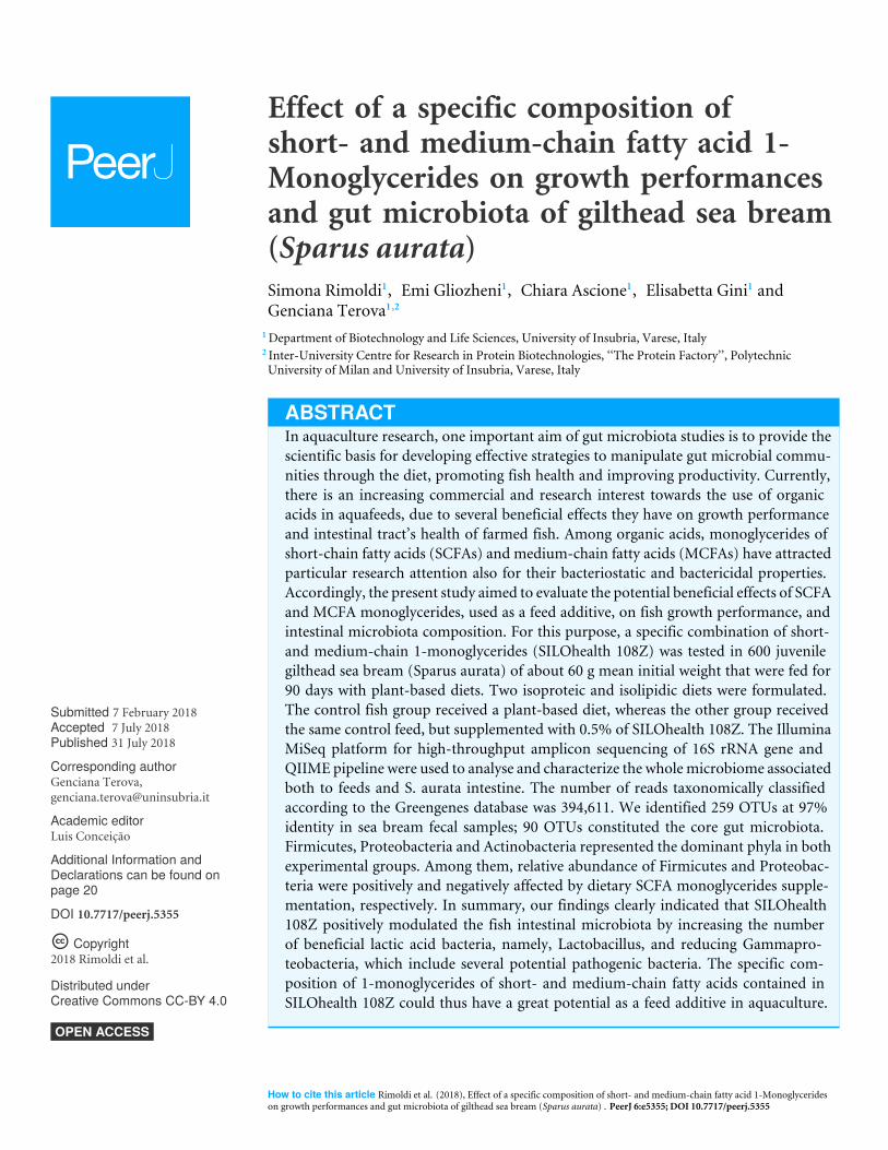
Effect of a specific composition ofshort- and medium-chain fatty acid 1-Monoglycerides on growth performancesand gut microbiota of gilthead sea bream(Sparus aurata)Simona Rimoldi1, Emi Gliozheni1, Chiara Ascione1, Elisabetta Gini1 andGenciana Terova1,2
1Department of Biotechnology and Life Sciences, University of Insubria, Varese, Italy2 Inter-University Centre for Research in Protein Biotechnologies, ‘‘The Protein Factory’’, PolytechnicUniversity of Milan and University of Insubria, Varese, Italy
ABSTRACTIn aquaculture research, one important aim of gut microbiota studies is to provide thescientific basis for developing effective strategies to manipulate gut microbial commu-nities through the diet, promoting fish health and improving productivity. Currently,there is an increasing commercial and research interest towards the use of organicacids in aquafeeds, due to several beneficial effects they have on growth performanceand intestinal tract’s health of farmed fish. Among organic acids, monoglycerides ofshort-chain fatty acids (SCFAs) and medium-chain fatty acids (MCFAs) have attractedparticular research attention also for their bacteriostatic and bactericidal properties.Accordingly, the present study aimed to evaluate the potential beneficial effects of SCFAand MCFA monoglycerides, used as a feed additive, on fish growth performance, andintestinal microbiota composition. For this purpose, a specific combination of short-and medium-chain 1-monoglycerides (SILOhealth 108Z) was tested in 600 juvenilegilthead sea bream (Sparus aurata) of about 60 g mean initial weight that were fed for90 days with plant-based diets. Two isoproteic and isolipidic diets were formulated.The control fish group received a plant-based diet, whereas the other group receivedthe same control feed, but supplemented with 0.5% of SILOhealth 108Z. The IlluminaMiSeq platform for high-throughput amplicon sequencing of 16S rRNA gene andQIIME pipeline were used to analyse and characterize the wholemicrobiome associatedboth to feeds and S. aurata intestine. The number of reads taxonomically classifiedaccording to the Greengenes database was 394,611. We identified 259 OTUs at 97%identity in sea bream fecal samples; 90 OTUs constituted the core gut microbiota.Firmicutes, Proteobacteria and Actinobacteria represented the dominant phyla in bothexperimental groups. Among them, relative abundance of Firmicutes and Proteobac-teria were positively and negatively affected by dietary SCFA monoglycerides supple-mentation, respectively. In summary, our findings clearly indicated that SILOhealth108Z positively modulated the fish intestinal microbiota by increasing the numberof beneficial lactic acid bacteria, namely, Lactobacillus, and reducing Gammapro-teobacteria, which include several potential pathogenic bacteria. The specific com-position of 1-monoglycerides of short- and medium-chain fatty acids contained inSILOhealth 108Z could thus have a great potential as a feed additive in aquaculture.
How to cite this article Rimoldi et al. (2018), Effect of a specific composition of short- and medium-chain fatty acid 1-Monoglycerideson growth performances and gut microbiota of gilthead sea bream (Sparus aurata) . PeerJ 6:e5355; DOI 10.7717/peerj.5355
Subjects Aquaculture, Fisheries and Fish Science, Marine BiologyKeywords Next-generation sequencing, Metagenomics, Gut microbiome, Feed additive,Aquaculture, 1-Monoglycerides, SILOhealth 108Z, 16S rRNA gene
INTRODUCTIONAquaculture, with an average annual rate of 8 percent, is probably the fastest-growingfood-producing sector in the world. It provides nearly 50 percent of the seafood consumedby humans (FAO, 2014) and the World Bank projects that aquaculture will increase toprovide 2/3 of the world’s fish in 2030. Currently, about 68% and 88% of the demand forfishmeal (FM) and fish oil (FO), respectively, comes from aquaculture (Naylor et al., 2009).However, with most wild fish capture fisheries at or above maximum sustainable yield,aquaculture can no longer rely on oceanic resources for the manufacturing of aquafeedsand such feed options are simply not sustainable (Naylor et al., 2000). To defray risingcosts and avert ecological harm, commercial feed producers and fish farmers have madesubstantial efforts to reduce the proportion of FM and FO in aquaculture feed, by replacingground-up forage fish with terrestrial plants (Tacon & Metian, 2008; Gatlin et al., 2007).However, the use of vegetable feedstuff in aquafeed production has several drawbacksthat are related to the low level of indispensable amino acids (in particular lysine andmethionine) and to the presence of a wide variety of anti-nutritional factors that coulddamage the intestine, thus reducing nutrient absorption, and consequently, affecting fishgrowth and resistance to stress and diseases (Zhang et al., 2013; Penn et al., 2011; Santigosaet al., 2011; Francis, Makkar & Becker, 2001).
For this reason, nutritionists and feed manufacturers are investing great effort to findfeed additives that could prevent or alleviate the adverse effects at the gut level of plant-basedingredients that are actually used in fish diet formulations.
Here, the most promising feed additives seem to be organic acids that are compoundswith acidic properties associated with their carboxyl group (−COOH) (Lim et al., 2015).
Among them, short- and medium-chain fatty acids (SCFAs and MCFAs) are known toplay a central role as energy-source for enterocytes. SCFAs are fatty acids with aliphatictails of one to six carbon atoms, the most common being acetic (C2), propionic (C3), andbutyric (C4) acid, whereas MCFA comprise fatty acids with seven to 12 carbon atoms.SCFAs are produced within the intestinal lumen by bacterial fermentation of undigesteddietary carbohydrates and fibers (cellulose, hemicellulose, pectin). Contrariwise, MCFAsmainly arise from dietary triglycerides and natural sources of MCFAs are generally coconutoil, palm kernel oil, and milk. The use of SCFAs as additive in aquafeeds and theirimpact on fish growth, nutrient utilization, and disease resistance were recently reviewed(Ng & Koh, 2017). Among SCFAs, butyric acid has received particular attention for itsvarious well-documented beneficial effects on the health of intestinal tract and peripheraltissues in human and farmed animals, including fish (Guilloteau et al., 2010; Mátis et al.,2013; Robles et al., 2013; Liu et al., 2014). Butyrate represents a major energy source forenterocytes and is involved in maintaining gut mucosal health, playing a central role inenhancing epithelial cell proliferation and differentiation and in improving the intestinal
Rimoldi et al. (2018), PeerJ, DOI 10.7717/peerj.5355 2/27
absorption (Gálfi & Neogrády, 2001; Wong et al., 2006; Canani et al., 2011). Butyrate hasanti-inflammatory properties and the potential to stimulate the immune system, too(Vinolo et al., 2011; Hamer et al., 2008; Terova et al., 2016; Rimoldi et al., 2016; Tian et al.,2017). However, the data on the effect of butyric acid and its salts (sodium butyrate) onthe growth performance of cultured fish and crustaceans are still controversial. In juvenilecommon carp (Cyprinus carpio) (Liu et al., 2014), and Pacific white shrimp (Litopenaeusvannamei) (Da Silva et al., 2016), butyrate supplementation positively affected the growthperformance. On the other hand, a dietary supplementation of a mixture of SCFAs,containing butyrate, did not significantly improve growth rate or feed utilization inAtlantic salmon (Salmo salar), rainbow trout (Oncorhynchus mykiss), and European seabass (Bjerkeng, Storebakken & Wathne, 1999; Gao et al., 2011; Terova et al., 2016). Recently,Simó-Mirabet et al. (2017) reported that sodium salt of coconut fatty acid distillate,particularly rich in lauric acid (C12), increased feed intake, improved gut development andnutrient absorption, thus enhancing growth rate of gilthead sea bream (Sparus aurata).Moreover,MCFAs have been suggested to have a role in immunological response regulation(Wang et al., 2006). Organic acids, their salts or combinations thereof, are commonlyknown as acidifiers and are used as storage preservatives in terrestrial livestock feeds aswell as in aquafeeds (Ng & Koh, 2017). Due to their capacity to reduce pH, they inhibitmicrobial growth and diminish a possible contamination of feed by pathogenic organismssuch as Salmonella and Escherichia coli (Lückstädt, 2008; Van Immerseel et al., 2003; VanImmerseel et al., 2004; Skřivanová et al., 2009). The mechanism of action of SCFAs andMCFAs differs from that of antibiotics. Salsali, Parker & Sattar (2008) firstly proposed thatSCFAs and MCFAs bacteriostatic and bactericidal activities could be due to the ability ofthe undissociated form of the acid to penetrate the bacterial cell wall and, once inside,to dissociate releasing protons, thereby lowering the cytoplasmic pH. Consequently, thebacteriummust redirect its energy towards the efflux of the excess protons, thus exhaustingcell metabolism and leading to lower bacterial cell growth and even to cell death (Salsali,Parker & Sattar, 2008; Hismiogullari et al., 2008; Ng & Koh, 2017). In the digestive tract,organic acids cause a pH reduction in the intestine via the delivery of H+ ions (Lim etal., 2015). Actually, in fish, dietary administration of acidifiers inhibits overgrowth ofpH-sensitive pathogenic bacteria favouring the growth of beneficial intestinal flora (Zhouet al., 2009; Hoseinifar, Sun & Caipang, 2017; Abu Elala & Ragaa, 2015; Ringøet al., 2016;Da Silva et al., 2013; Da Silva et al., 2016; Anuta et al., 2011; De Schryver et al., 2010; Liuet al., 2014; Piazzon et al., 2017). Indeed, although the bacteriostatic activity of organicacids is preserved at the intestinal level, their bactericidal efficacy is limited because ofthe intestinal pH. Being weak acids with modest pKas of approximately 3.6 to 4.7, themajority of organic acids at neutral or slightly alkaline pH, are present as anions ratherthan as undissociated forms (free acids) that are assumed to penetrate the lipid membrane,destroying the bacterial cell (Yoon et al., 2018).
Dietary free organic acids and their salts have also the disadvantage to be easily absorbedby the upper digestive tract, thus limiting their delivery to the desired target, i.e., lowerintestinal tract, where they exert the aforementioned beneficial actions.
Rimoldi et al. (2018), PeerJ, DOI 10.7717/peerj.5355 3/27
On the contrary, monoglycerides, which are esters formed by glycerol and one moleculeof fatty acid, have no such drawbacks. The great advantage of monoglycerides is thatorganic acid is released from the glycerol backbone only under the action of intestinallipases. This means that SCFA or MCFA remains protected from absorption in the uppergastrointestinal tract and could reach the final portion of intestine, where it would exert itsmajor functions (Sampugna et al., 1967; Namkung et al., 2011). Moreover, monoglyceridespossess a more effective antimicrobial activity than the corresponding free fatty acids, sincetheir efficacy is independent from environmental pH (Bergsson et al., 2001; Sun, O’Connor& Roberton, 2003; Thormar, Hilmarsson & Bergsson, 2006). Due to their amphipathicproperties, monoglycerides show a membrane-lytic action, which leads to bacterialmembrane destabilization and pore formation. Membrane-destabilizing activity causesincreased cell permeability and cell lysis, leading to inhibition of growth and cell death(Yoon et al., 2018). MCFA monoglycerides are able to penetrate also the peptidoglycanlayer of Gram-positive bacteria’s cell wall (Bergsson et al., 2001).
Up to date, antimicrobial and growth-promoting action of monoglycerides havebeen widely investigated in poultry (Bedford & Gong, 2018; Yang et al., 2018; Jahanian &Golshadi, 2015; Leeson et al., 2005), whereas in fish their effects have been poorly explored.Accordingly, the present study aimed to evaluate the potential beneficial effects of dietarySCFA and MCFA monoglycerides on fish growth performances and intestinal microbiotacomposition. For this purpose, a specific synergic combination of 1-monoglycerides ofshort- andmedium-chain fatty acids (SILOhealth 108Z), commercially available from SILOSpA, Florence, Italy (http://www.silohealth.com/), was tested in juvenile gilthead sea bream(Sparus aurata) fed a plant-based diet. The Illumina MiSeq platform for high-throughputsequencing of 16S rRNA gene was utilized to analyse and characterize the whole gutmicrobiome of gilthead sea bream.
MATERIALS AND METHODSEthics statementThis study was carried out in strict accordance with the recommendations in the Guidefor the Care and Use of Laboratory Animals of the indoor experimental facility of CivitaIttica (Civitavecchia, Italy), and in accordance with EU Directive 2010/63/E U for animalexperiments. TheCommittee on the Ethics of Animal Experiments of the same experimentalfacility approved all of the study protocols (approval n. 120/2008-A of 03/09/2008 (Art.12of D.Lgs.116/92)). Fish handling was performed under tricaine methanesulfonate (MS222)anesthesia, and all effort was made to minimize discomfort, stress, and pain to the fish.
Experimental dietsThe two experimental diets were formulated and manufactured by VRM S.r.l. Naturalleva(Verona, Italy). Feeds were prepared using small-scale machinery for mixing ingredientsand preparing pellets of 3.0 mm in diameter. The formulation and proximate compositionof diets are shown in Tables 1 and 2. The diets were isoenergetic (17.5 MJ kg−1),isoproteic (50%), and isolipidi c (16%), fully satisfying the gilthead sea bream nutritionaldemands (Table 2). The control group (CTRL) received a commercial plant-based diet;
Rimoldi et al. (2018), PeerJ, DOI 10.7717/peerj.5355 4/27
Table 1 Formulation (g kg−1 diet) of experimental diets.
Ingredient CTRL Sh108
Fish meal 280.0 280.0Corn gluten 220.0 220.0Guar germ meal 132.0 132.0Soybean seed meal 120.0 120.0Wheat middlings 120.0 120.0Fish oil (94%) 64.5 62.4Rapeseed oil 44.3 41.4DL-methionine 4.5 4.5Lysine hydrochloride 2.7 2.7Taurine 4.5 4.5Vitamin C (stay-C 35) 0.6 0.6Vitamin and mineral premixa 7.0 7.0SILOhealth108 – 5.0
Notes.aVitamin and mineral premix (quantities in 1 kg of mix): Vitamin A, 4,000,000 IU; Vitamin D3, 800,000 IU; Vitamin C, 25,000mg; Vitamin E, 15,000 mg; Inositol, 15,000 mg; Niacin, 12,000 mg; Choline chloride, 6,000 mg; Calcium Pantothenate, 3,000mg; Vitamin B1, 2,000 mg; Vitamin B3, 2,000 mg; Vitamin B6, 1,800 mg; Biotin, 100 mg; Manganese, 9,000 mg; Zinc, 8,000mg; Iron, 7,000 mg; Copper, 1,400 mg; Cobalt, 160 mg; Iodine 120 mg; Anticaking & Antioxidant+ carrier, making up to1,000 g.
Table 2 Proximate composition (g kg−1 diet) of the experimental diets.
DIET
CTRL Sh108
Moisture 42.1 42.1Crude protein 500.0 500.0Crude lipids 160.0 160.0Crude fibre 19.6 19.6NFE 213.3 213.3Ash 65.0 65.0DP 403.9 403.9DE (MJ kg −1) 17.5 17.5DP/DE (g MJ −1) 22.9 23.0EPA 12.3 11.8DHA 8.2 7.8n−3/n−6 1.3 1.3DHA/EPA 0.6 0.6
Notes.NFE, Nitrogen-free extract; DP, digestible protein; DE, digestible energy; EPA, Eicosapentaenoic acid; DHA, Docosahex-aenoic acid; n−3, omega-3 fatty acids; n−6, omega-6 fatty acids.
the treated group (Sh108) received the same control feed but it was supplementedwith 0.5% of SILOhealth 108Z commercially available from SILO SpA, Florence, Italy(http://www.silohealth.com/). SILOhealth 108Z is composed of a specific combinationof 1-monoglycerides of short- and medium-chain fatty acids (from C3 to C12), in which1-monobutyrin represents 65% of total blend (Table 3).
Rimoldi et al. (2018), PeerJ, DOI 10.7717/peerj.5355 5/27
Table 3 Fatty acid composition (%) of SILOhealth 108Z.
Fatty acid Quantity (%)
C3:0 Propionic acid 20C4:0 Butyric acid 65C6:0, C7:0, C8:0, C9:0, C12 Blend of caproic, heptanoic, caprylic, lauric acid 15
Table 4 Growth and feed efficiency indices. Final mean body weight, specific growth rate (SGR), relative growth rate (RGR), biological feedconversion ratio (bFCR), and economic feed conversion ratio (eFCR) values of sea bream fed with two experimental diets (CTRL and Sh108). Theweight data represent the mean value± SD (n = 300 fish/per diet). SGR, RGR, bFCR, and eFCR were tank-based determined (n = 3) and reportedas mean± SD. Different letters indicate statistically significant differences between groups (Student’s t -test, P < 0.05).
Diet Initial weight Final weight SGR (% day−1) RGR (%) bFCR eFCR
CTRL 60.56± 1.44 126.84± 1.90 0.75± 0.01 109.49± 2.49 1.53± 0.05 1.55±0.05a
Sh108 60.50± 0.70 129.39± 1.12 0.77± 0.01 113.88± 3.27 1.47± 0.01 1.48±0.01b
Fish and feeding trialSix hundred juvenile gilthead sea bream of about 60 g mean initial body weight (Table 4)were randomly distributed into six fiberglass tanks of 2m3 each (100 fish/tank) at the indoorexperimental facility of Civita Ittica (Civitavecchia, Italy). The tanks were supplied withfiltered sea water (salinity of 37 mg/l) at a temperature and average dissolved oxygen levelof 21.2± 1.4 ◦C and 11.7± 0.6 mg/l, respectively. Fish were kept under a 12:12 h light:darkphotoperiod regimen. Feeding rate was restricted to 2.0% of biomass during the feedingexperiment based on four-weekly fish weight measurements. During the experiment thatlasted 90 days, fish in triplicate groups (three tanks/diet) were fed with their respective diettwice a day (7:00 am and 4:00 pm) for 6 days per week, except Sunday. Feed consumption(g) in each tank was estimated from the difference between feed delivered into the tankand uneaten feed. Uneaten feed was collected from the bottom of the tank one hour aftereach meal by siphoning, dried at 70 ◦C and then weighed. Fish mortality was checked andrecorded every day. At the end of the feeding trial, all fish in the tank were individuallyweighed and measured for their length. Specific growth rate (SGR), relative growth rate(RGR), and biological and economic feed conversion ratio (bFCR and eFCR, respectively)values were calculated. The bFCR is the net amount of feed used to produce one kg of fish,whereas the eFCR considers all the feed used, meaning that the effects of feed losses andmortalities are included (Robb & Crampton, 2013).
The each ratio values were calculated using the following formulas:
bFCR=Total feed /(Final weight(Wt)+mass mortality)− Initial weight (W0)
eFCR=Total feed /(Final weight(Wt)− Initial weight (W0))
SGR= 100 × (lnWt/lnW0)/Days
RGR= 100 ×(Wt −W0)/W0.
The day of fecal sampling, fish were fed at 6:00 am and after 6 h from the last meal,six fish/diet (2 fish/tank) were randomly collected and euthanized with an overdose(320 mg/L at 22 ◦C) of anesthetic (tricaine-methasulfonate MS-222). To avoid gut content
Rimoldi et al. (2018), PeerJ, DOI 10.7717/peerj.5355 6/27
contamination by the body surface microflora during dissection, external abdominalsurface of each fish was wiped thoroughly with a sterile 70◦ alcohol moistened cotton withan area of 10 cm2. Then, with the aid of sterile scissors and forceps, the entire intestine(excluding pyloric ceca) was exposed from the ventral side and aseptically removed. Thefecal content was obtained by squeezing out and scrapping the intestinal mucosa with asterile spatula, in order to collect both, the digesta- and the mucosa-associated microbiota.The fecal samples were immediately frozen in dry ice and stored at minus 80 ◦C until themetagenomics analysis.
Microbial DNA extractionTwo hundred and fifty mg of intestinal content from each fish (12 × 250 mg samples intotal) and 200 mg of each dietary pellet (2 × 200 mg samples in total) were processed forDNA extraction using DNeasy PowerSoil Kit (Qiagen, Milan, Italy). The bacterial cells weredisrupted via high-speed shaking in plastic tubes with stainless steel beads (TissueLyser II,Qiagen, Milan, Italy) for 2 min at 25 Hz. Total DNA was then extracted according to themanufacturer’s instructions. A sample with only lysis buffer was processed in parallel tothe biological samples as a negative control to check if external DNA contamination wasintroduced during the extraction procedure. Bacterial DNA concentration was measuredspectrophotometrically by using NanoDropTM 2000 Spectrophotometer (Thermo FisherScientific, Monza, Italy) and then stored at −20 ◦C until further processing.
16S rRNA gene library preparation and sequencingThe 16S ribosomal RNA gene library was prepared according to the Illumina protocol ‘‘16SMetagenomic Sequencing Library Preparation’’ (#15044223 rev.B). PCR amplificationsof the V3-V4 region of the 16S rRNA gene were carried out in 25-µl reactionscontaining bacterial DNA (500 ng), buffer (10X), dNTPs (0.2 mM), MgSO4 (1.5mM), Platinum
R©Taq DNA Polymerase High Fidelity (1U) (Thermo Fisher Scientific,
Monza, Italy), forward primer (5′-CCTACGGGNBGCASCAG-3′), and reverse primer(5′-GACTACNVGGGTATCTAATCC-3′) (400 nM each). The universal primers used wereselected by Takahashi et al. (2014) and were designed with Illumina adapters at their 5′ end.All the procedure for 16S rRNA gene library preparation and sequencing is described indetail in Rimoldi et al. (2018). However, briefly, PCR cycling conditions for 16S rRNAgene amplification were 94 ◦C for 1 min, 30 cycles of 94 ◦C for 30 s, 55 ◦C for 1 min,and 68 ◦C for 1.30 min, with a final extension step at 68 ◦C for 10 min. The resulting sizeof 16S rRNA gene amplicons was about 550 bp. Dual indices and Illumina sequencingadapters (P5 and P7) were then attached to the amplicons using Nextera XT Index Kit(Illumina, San Diego, CA, USA), according to manufacturer’s instructions, to produce thefinal libraries. Final libraries were quantified by quantitative PCR (qPCR) using KAPALibrary Quantification Kits for Illumina R© platforms (Kapa Biosystems Ltd., Dorset, UK)and a set of six diluted DNA standards to generate a standard curve. Final libraries werepooled in equimolar amounts, denatured and diluted to 6 pM. Before loading onto theMiSeq flow cell, 15% of the PhiX control library was combined with the amplicon library.Sequencing was performed on an Illumina MiSeq platform using v3 reagent and a 2×300
Rimoldi et al. (2018), PeerJ, DOI 10.7717/peerj.5355 7/27
bp paired end protocol, according to the manufacturer’s instructions (Illumina, San Diego,CA, USA).
Sequencing raw data analysisRaw sequences were processed using the open-source bioinformatics pipeline QIIMEv1.9.1 (Caporaso et al., 2010) by BMR Genomics NGS service (Padova, Italy). Sequenceswere trimmed using Trimmomatic v0.32. Only reads above 36 nucleotides in length wereincluded in the downstream analysis. The remaining sequences were grouped by dietaccording to their barcodes. For original amplicon reconstruction, overlapping R1 and R2paired reads were joined using FLASH v1.2.11 software (http://sourceforge.net/projects/flashpage) and filtered for base quality (Q > 30). Amplicons were dereplicated, sorted, andclustered at ≥ 97% identity. Amplicon clusters (Operational Taxonomic Units, OTUs)were then identified against reference QIIME-formatted Greengenes database v.13.8(http://greengenes.lbl.gov) by using QIIME script ‘pick_closed_reference_otus.py’ andonly the OTUs that represented at least 0.005% of total reads were kept. The taxonomicalclassification was performed down to species level. To determine the abundance ofeach bacterial taxon, OTUs obtained from each sample were binned according to theirconsensus sequences, and the final OTU-table output files, in txt and biom format, werecreated using ‘summarize_taxa_through_plots.py’ custom script. OTUs assigned to thephylumCyanobacteria (classChloroplast ) were removed from the analysis as potential plantcontaminants, as described in Rimoldi et al. (2018). Reads of mitochondrial or eukaryoticorigin were also excluded.
Alpha and beta diversity statistics were performed as described in Rimoldi et al. (2018).Alpha diversity metrics were calculated based on a rarefied OTU table using ‘observedspecies’, ‘Chao1 index’ (species richness estimator), ‘Shannon’s diversity index’, ‘Good’scoverage’, and ‘PDwhole tree’. OTUs diversity among sample communities (beta diversity)was assessed by applying weighted (presence/absence/abundance matrix) and unweighted(presence/absence matrix) UniFrac distance matrices (Lozupone & Knight, 2005; Lozuponeet al., 2007). The distance matrices were visualized by principal coordinate analysis (PCoA)three-dimensional plots.
The common core microbiome (OTUs shared, regardless of the diet, and foundin at least five out of the six samples per dietary group) was identified using the‘compute_core_microbiome.py’ script. The Venn diagrams representing the results ofthe core microbiota were drawn using the web tool http://bioinformatics.psb.ugent.be/webtools/Venn/.
StatisticsAll data were presented as means ± standard deviation. The number of reads acrosssamples was normalized by sample size and the relative abundance (%) of each taxon wascalculated. Only those taxa with an overall abundance of more than 1% (up to order)and more than 0.5% at family and genus level were considered for statistical analysis.Before being statistically analysed, the resulting microbial profiles were calculated as theangular transformation (arcsine of the square root). All data were tested for normality and
Rimoldi et al. (2018), PeerJ, DOI 10.7717/peerj.5355 8/27
homogeneity of variances by Shapiro–Wilk’s and Levene’s test, respectively. Differencesbetween two groups were analysed by unpaired Student’s t -test or non-parametric Mann–Whitney U test, depending if the data were or not normal distributed. Welch’s t -test wasused instead of Student’s t -test when variances were unequal between groups. Statisticalsignificance was set at P < 0.05. Correction of multiple testing was done using Benjamini–Hochberg False Discovery Rate (FDR) method with a false discovery rate (Q) set to 0.20.All analyses were performed using Past3 software (Hammer, Harper & Ryan, 2001). Toverify the significance of differences in the beta diversity of bacterial communities, analysisof similarities (ANOSIM), and permutational multivariate analysis of variance (adonisfunction) were performed with 999 permutations. Both tests were accomplished usingQIIME script ‘compare_categories.py’.
RESULTSFish growth performance and feeding conversionDuring the 90 days of the feeding trial, the mortality rate was lower than 1%. Specifically,two fish of CTRL and four fish of Sh108 group died during the first week of feeding trial,with no further mortalities recorded for the rest of the test. Fish growth performanceindexes such as SGR, and RGR did not reveal any significant differences between controland SILOhealth 108Z-supplemented dietary groups, meaning that all fish grew efficiently,regardless of the fatty acid monoglycerides supplementation. At the end of the feeding trial,all fish doubled their body mass reaching a final mean body weight of 126.84± 1.90 g, and129.39 ± 1.12 g in CTRL and Sh108 group, respectively. On the contrary, economic FCRdiffered between two groups, resulting lower in fish fed diet Sh108 (Table 4).
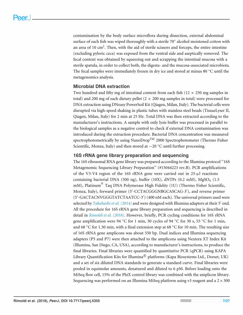
Characterization of microbial communities of the dietsBacterial communities associated to feeds were analysed using the QIIME pipeline, whichrevealed that the two microbial profiles were qualitatively and quantitatively equivalent.After filtering for quality, trimming length, and generating consensus lineages, the numberof reads taxonomically classified according to the Greengenes database was 47,791 and44,483 for CTRL and Sh108 diet, respectively. The total number of OTUs at 97% identityfound in CTRL and Sh108 feed samples amounted to 193 and 188, respectively. The overallamount of reads of eukaryotic origin was around 70%. The microbial profiles of feedsamples at the phylum, family, and genus taxonomic level are reported in Figs. 1A–1C.The most abundant bacterial taxa (relative abundance >1%) were mainly comprised of 3phyla, four classes, six orders, seven families, eight genera, and eight species (Figs. 1A–1C;Dataset S1).
QIIME data analysis and taxonomic characterization of gutmicrobiomeThe twelve fecal samples were processed via Illumina MiSeq platform and analysed usingthe QIIME pipeline. During bioinformatics analysis process, two CTRL samples werediscarded following OTU-picking step, due to their inadequate number of sequences. Thetotal number of reads taxonomically classified according to the Greengenes database was
Rimoldi et al. (2018), PeerJ, DOI 10.7717/peerj.5355 9/27
Figure 1 Bacterial relative abundance (%) in the feeds. The amount (%) of the most prevalent bacte-ria in CTRL and Sh108 feeds at (A) phylum; (B) family, and (C) genus level. Only bacteria with an overallabundance of ≥ 1% (at genus level) and ≥ 0.5% (at family and genus level), were reported. Bacteria withlower abundance were pooled and indicated as ‘‘Others’’.
Full-size DOI: 10.7717/peerj.5355/fig-1
Rimoldi et al. (2018), PeerJ, DOI 10.7717/peerj.5355 10/27
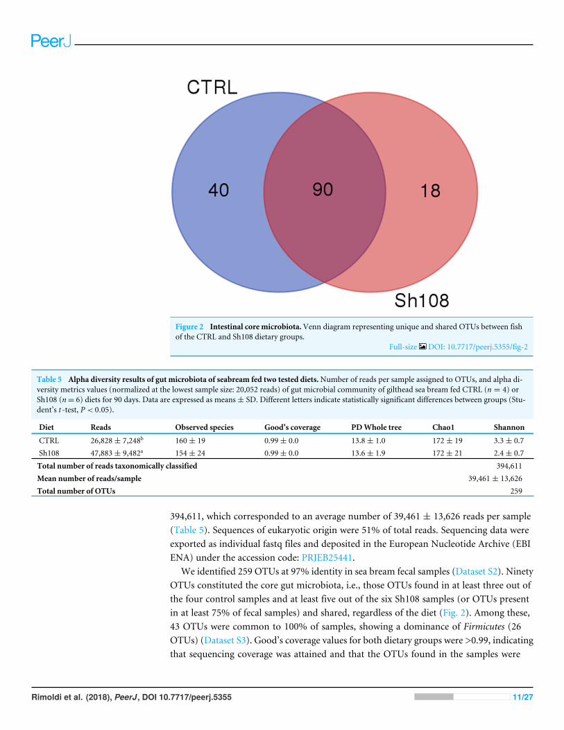
Figure 2 Intestinal core microbiota.Venn diagram representing unique and shared OTUs between fishof the CTRL and Sh108 dietary groups.
Full-size DOI: 10.7717/peerj.5355/fig-2
Table 5 Alpha diversity results of gut microbiota of seabream fed two tested diets.Number of reads per sample assigned to OTUs, and alpha di-versity metrics values (normalized at the lowest sample size: 20,052 reads) of gut microbial community of gilthead sea bream fed CTRL (n = 4) orSh108 (n= 6) diets for 90 days. Data are expressed as means± SD. Different letters indicate statistically significant differences between groups (Stu-dent’s t -test, P < 0.05).
Diet Reads Observed species Good’s coverage PDWhole tree Chao1 Shannon
CTRL 26,828± 7,248b 160± 19 0.99± 0.0 13.8± 1.0 172± 19 3.3± 0.7Sh108 47,883± 9,482a 154± 24 0.99± 0.0 13.6± 1.9 172± 21 2.4± 0.7
Total number of reads taxonomically classified 394,611
Mean number of reads/sample 39,461± 13,626
Total number of OTUs 259
394,611, which corresponded to an average number of 39,461 ± 13,626 reads per sample(Table 5). Sequences of eukaryotic origin were 51% of total reads. Sequencing data wereexported as individual fastq files and deposited in the European Nucleotide Archive (EBIENA) under the accession code: PRJEB25441.
We identified 259 OTUs at 97% identity in sea bream fecal samples (Dataset S2). NinetyOTUs constituted the core gut microbiota, i.e., those OTUs found in at least three out ofthe four control samples and at least five out of the six Sh108 samples (or OTUs presentin at least 75% of fecal samples) and shared, regardless of the diet (Fig. 2). Among these,43 OTUs were common to 100% of samples, showing a dominance of Firmicutes (26OTUs) (Dataset S3). Good’s coverage values for both dietary groups were >0.99, indicatingthat sequencing coverage was attained and that the OTUs found in the samples were
Rimoldi et al. (2018), PeerJ, DOI 10.7717/peerj.5355 11/27
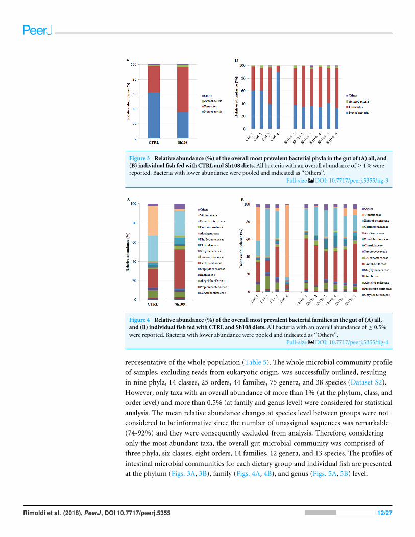
Figure 3 Relative abundance (%) of the overall most prevalent bacterial phyla in the gut of (A) all, and(B) individual fish fed with CTRL and Sh108 diets. All bacteria with an overall abundance of ≥ 1% werereported. Bacteria with lower abundance were pooled and indicated as ‘‘Others’’.
Full-size DOI: 10.7717/peerj.5355/fig-3
Figure 4 Relative abundance (%) of the overall most prevalent bacterial families in the gut of (A) all,and (B) individual fish fed with CTRL and Sh108 diets. All bacteria with an overall abundance of ≥ 0.5%were reported. Bacteria with lower abundance were pooled and indicated as ‘‘Others’’.
Full-size DOI: 10.7717/peerj.5355/fig-4
representative of the whole population (Table 5). The whole microbial community profileof samples, excluding reads from eukaryotic origin, was successfully outlined, resultingin nine phyla, 14 classes, 25 orders, 44 families, 75 genera, and 38 species (Dataset S2).However, only taxa with an overall abundance of more than 1% (at the phylum, class, andorder level) and more than 0.5% (at family and genus level) were considered for statisticalanalysis. The mean relative abundance changes at species level between groups were notconsidered to be informative since the number of unassigned sequences was remarkable(74-92%) and they were consequently excluded from analysis. Therefore, consideringonly the most abundant taxa, the overall gut microbial community was comprised ofthree phyla, six classes, eight orders, 14 families, 12 genera, and 13 species. The profiles ofintestinal microbial communities for each dietary group and individual fish are presentedat the phylum (Figs. 3A, 3B), family (Figs. 4A, 4B), and genus (Figs. 5A, 5B) level.
Rimoldi et al. (2018), PeerJ, DOI 10.7717/peerj.5355 12/27
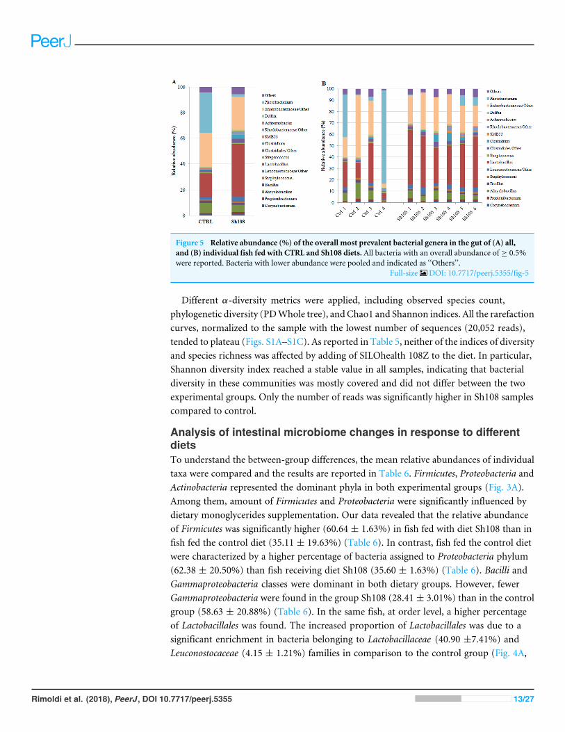
Figure 5 Relative abundance (%) of the overall most prevalent bacterial genera in the gut of (A) all,and (B) individual fish fed with CTRL and Sh108 diets. All bacteria with an overall abundance of ≥ 0.5%were reported. Bacteria with lower abundance were pooled and indicated as ‘‘Others’’.
Full-size DOI: 10.7717/peerj.5355/fig-5
Different α-diversity metrics were applied, including observed species count,phylogenetic diversity (PDWhole tree), andChao1 and Shannon indices. All the rarefactioncurves, normalized to the sample with the lowest number of sequences (20,052 reads),tended to plateau (Figs. S1A–S1C). As reported in Table 5, neither of the indices of diversityand species richness was affected by adding of SILOhealth 108Z to the diet. In particular,Shannon diversity index reached a stable value in all samples, indicating that bacterialdiversity in these communities was mostly covered and did not differ between the twoexperimental groups. Only the number of reads was significantly higher in Sh108 samplescompared to control.
Analysis of intestinal microbiome changes in response to differentdietsTo understand the between-group differences, the mean relative abundances of individualtaxa were compared and the results are reported in Table 6. Firmicutes, Proteobacteria andActinobacteria represented the dominant phyla in both experimental groups (Fig. 3A).Among them, amount of Firmicutes and Proteobacteria were significantly influenced bydietary monoglycerides supplementation. Our data revealed that the relative abundanceof Firmicutes was significantly higher (60.64 ± 1.63%) in fish fed with diet Sh108 than infish fed the control diet (35.11 ± 19.63%) (Table 6). In contrast, fish fed the control dietwere characterized by a higher percentage of bacteria assigned to Proteobacteria phylum(62.38 ± 20.50%) than fish receiving diet Sh108 (35.60 ± 1.63%) (Table 6). Bacilli andGammaproteobacteria classes were dominant in both dietary groups. However, fewerGammaproteobacteria were found in the group Sh108 (28.41 ± 3.01%) than in the controlgroup (58.63 ± 20.88%) (Table 6). In the same fish, at order level, a higher percentageof Lactobacillales was found. The increased proportion of Lactobacillales was due to asignificant enrichment in bacteria belonging to Lactobacillaceae (40.90 ±7.41%) andLeuconostocaceae (4.15 ± 1.21%) families in comparison to the control group (Fig. 4A,
Rimoldi et al. (2018), PeerJ, DOI 10.7717/peerj.5355 13/27
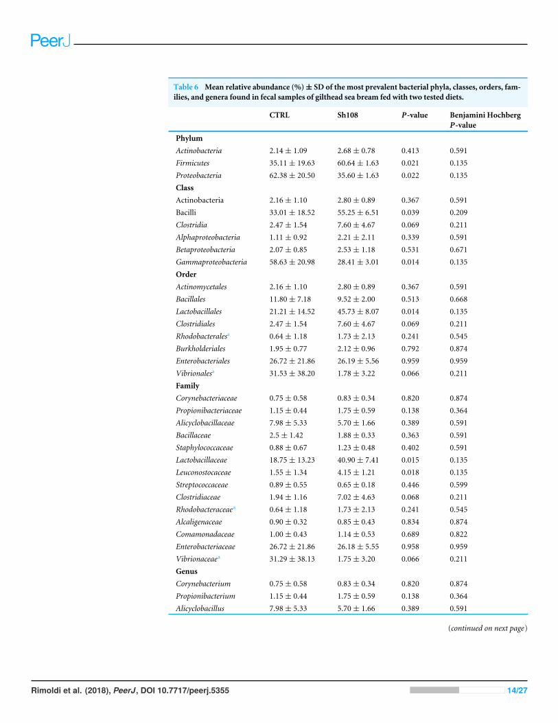
Table 6 Mean relative abundance (%)± SD of the most prevalent bacterial phyla, classes, orders, fam-ilies, and genera found in fecal samples of gilthead sea bream fed with two tested diets.
CTRL Sh108 P-value Benjamini HochbergP-value
PhylumActinobacteria 2.14± 1.09 2.68± 0.78 0.413 0.591Firmicutes 35.11± 19.63 60.64± 1.63 0.021 0.135Proteobacteria 62.38± 20.50 35.60± 1.63 0.022 0.135ClassActinobacteria 2.16± 1.10 2.80± 0.89 0.367 0.591Bacilli 33.01± 18.52 55.25± 6.51 0.039 0.209Clostridia 2.47± 1.54 7.60± 4.67 0.069 0.211Alphaproteobacteria 1.11± 0.92 2.21± 2.11 0.339 0.591Betaproteobacteria 2.07± 0.85 2.53± 1.18 0.531 0.671Gammaproteobacteria 58.63± 20.98 28.41± 3.01 0.014 0.135OrderActinomycetales 2.16± 1.10 2.80± 0.89 0.367 0.591Bacillales 11.80± 7.18 9.52± 2.00 0.513 0.668Lactobacillales 21.21± 14.52 45.73± 8.07 0.014 0.135Clostridiales 2.47± 1.54 7.60± 4.67 0.069 0.211Rhodobacteralesa 0.64± 1.18 1.73± 2.13 0.241 0.545Burkholderiales 1.95± 0.77 2.12± 0.96 0.792 0.874Enterobacteriales 26.72± 21.86 26.19± 5.56 0.959 0.959Vibrionalesa 31.53± 38.20 1.78± 3.22 0.066 0.211FamilyCorynebacteriaceae 0.75± 0.58 0.83± 0.34 0.820 0.874Propionibacteriaceae 1.15± 0.44 1.75± 0.59 0.138 0.364Alicyclobacillaceae 7.98± 5.33 5.70± 1.66 0.389 0.591Bacillaceae 2.5± 1.42 1.88± 0.33 0.363 0.591Staphylococcaceae 0.88± 0.67 1.23± 0.48 0.402 0.591Lactobacillaceae 18.75± 13.23 40.90± 7.41 0.015 0.135Leuconostocaceae 1.55± 1.34 4.15± 1.21 0.018 0.135Streptococcaceae 0.89± 0.55 0.65± 0.18 0.446 0.599Clostridiaceae 1.94± 1.16 7.02± 4.63 0.068 0.211Rhodobacteraceaea 0.64± 1.18 1.73± 2.13 0.241 0.545Alcaligenaceae 0.90± 0.32 0.85± 0.43 0.834 0.874Comamonadaceae 1.00± 0.43 1.14± 0.53 0.689 0.822Enterobacteriaceae 26.72± 21.86 26.18± 5.55 0.958 0.959Vibrionaceaea 31.29± 38.13 1.75± 3.20 0.066 0.211GenusCorynebacterium 0.75± 0.58 0.83± 0.34 0.820 0.874Propionibacterium 1.15± 0.44 1.75± 0.59 0.138 0.364Alicyclobacillus 7.98± 5.33 5.70± 1.66 0.389 0.591
(continued on next page)
Rimoldi et al. (2018), PeerJ, DOI 10.7717/peerj.5355 14/27
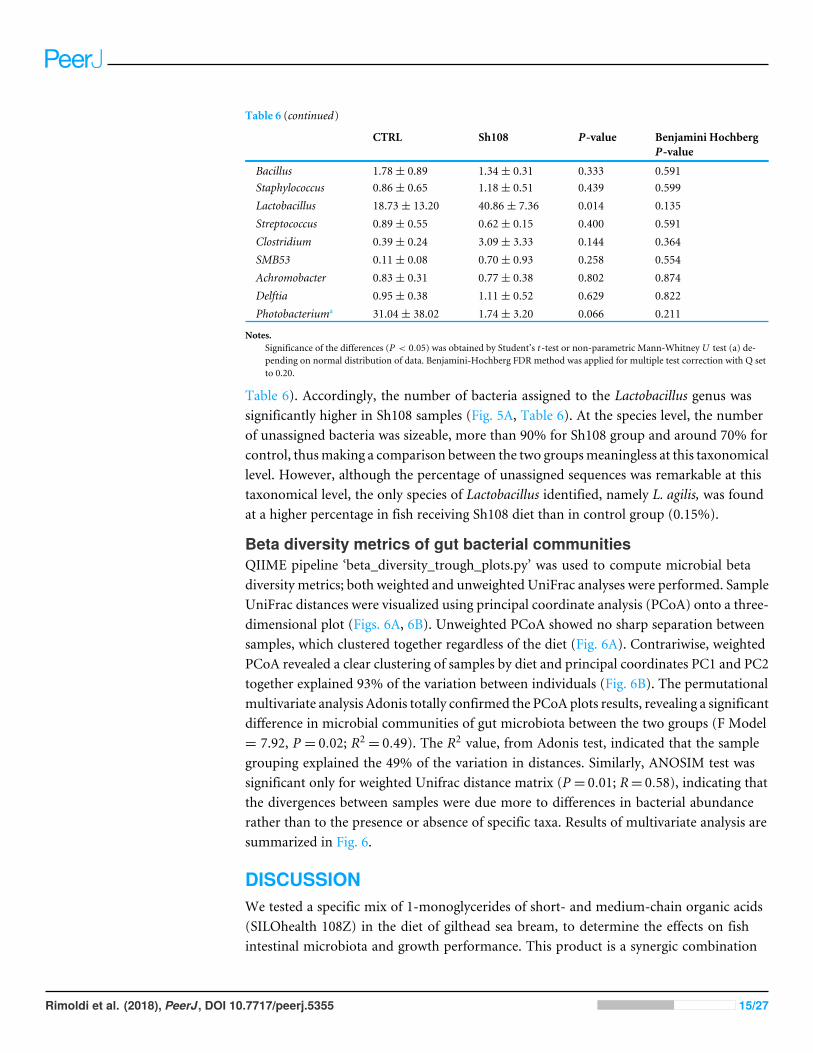
Table 6 (continued)
CTRL Sh108 P-value Benjamini HochbergP-value
Bacillus 1.78± 0.89 1.34± 0.31 0.333 0.591Staphylococcus 0.86± 0.65 1.18± 0.51 0.439 0.599Lactobacillus 18.73± 13.20 40.86± 7.36 0.014 0.135Streptococcus 0.89± 0.55 0.62± 0.15 0.400 0.591Clostridium 0.39± 0.24 3.09± 3.33 0.144 0.364SMB53 0.11± 0.08 0.70± 0.93 0.258 0.554Achromobacter 0.83± 0.31 0.77± 0.38 0.802 0.874Delftia 0.95± 0.38 1.11± 0.52 0.629 0.822Photobacteriuma 31.04± 38.02 1.74± 3.20 0.066 0.211
Notes.Significance of the differences (P < 0.05) was obtained by Student’s t -test or non-parametric Mann-Whitney U test (a) de-pending on normal distribution of data. Benjamini-Hochberg FDR method was applied for multiple test correction with Q setto 0.20.
Table 6). Accordingly, the number of bacteria assigned to the Lactobacillus genus wassignificantly higher in Sh108 samples (Fig. 5A, Table 6). At the species level, the numberof unassigned bacteria was sizeable, more than 90% for Sh108 group and around 70% forcontrol, thusmaking a comparison between the two groupsmeaningless at this taxonomicallevel. However, although the percentage of unassigned sequences was remarkable at thistaxonomical level, the only species of Lactobacillus identified, namely L. agilis, was foundat a higher percentage in fish receiving Sh108 diet than in control group (0.15%).
Beta diversity metrics of gut bacterial communitiesQIIME pipeline ‘beta_diversity_trough_plots.py’ was used to compute microbial betadiversity metrics; both weighted and unweighted UniFrac analyses were performed. SampleUniFrac distances were visualized using principal coordinate analysis (PCoA) onto a three-dimensional plot (Figs. 6A, 6B). Unweighted PCoA showed no sharp separation betweensamples, which clustered together regardless of the diet (Fig. 6A). Contrariwise, weightedPCoA revealed a clear clustering of samples by diet and principal coordinates PC1 and PC2together explained 93% of the variation between individuals (Fig. 6B). The permutationalmultivariate analysis Adonis totally confirmed the PCoA plots results, revealing a significantdifference in microbial communities of gut microbiota between the two groups (F Model= 7.92, P = 0.02; R2
= 0.49). The R2 value, from Adonis test, indicated that the samplegrouping explained the 49% of the variation in distances. Similarly, ANOSIM test wassignificant only for weighted Unifrac distance matrix (P = 0.01; R= 0.58), indicating thatthe divergences between samples were due more to differences in bacterial abundancerather than to the presence or absence of specific taxa. Results of multivariate analysis aresummarized in Fig. 6.
DISCUSSIONWe tested a specific mix of 1-monoglycerides of short- and medium-chain organic acids(SILOhealth 108Z) in the diet of gilthead sea bream, to determine the effects on fishintestinal microbiota and growth performance. This product is a synergic combination
Rimoldi et al. (2018), PeerJ, DOI 10.7717/peerj.5355 15/27
Figure 6 Beta diversity metrics. Principal Coordinate Analysis of (A) Unweighted, and (B) WeightedUnifrac distances of gut microbial communities associated to two experimental diets. Each dot representsan individual sample plots according to its microbial profile at genus level. Results of Permutational mul-tivariate analysis of variance (adonis function) and Analysis of similarity (ANOSIM) are reported next tothe PCoA plot to which they are referred. Significance was set at P < 0.05.
Full-size DOI: 10.7717/peerj.5355/fig-6
of short and medium chain 1-monoglycerides (from C3 to C12), particularly rich inmonobutyrin. It has been widely demonstrated that butyrate, despite being the leastabundant of the three-primary gastrointestinal SCFAs (acetate, propionate and butyrate),exerts important protective and anti-inflammatory functions in the gut of several fishspecies, ultimately enhancing gut health and improving fish performance (Benedito-Palos et al., 2016; Liu et al., 2014; Terova et al., 2016; Rimoldi et al., 2016). These previous,promising results prompted the idea that, as feed additive, butyric acid monoglycerides,could represent an effective strategy to improve fish growth performance, feed conversion,and disease resistance by promoting the establishment of a healthy intestinal microbiota.Indeed, esterification with glycerol protect butyric acid from being absorbed in the upperpart of the digestive system targeting its release in the deeper tracts of intestine wherebutyrate would exert its major functions.
Use of monoglycerides as feed additive has been widely investigated in poultry (Bedford& Gong, 2018; Yang et al., 2018; Jahanian & Golshadi, 2015; Leeson et al., 2005). On thecontrary, research dealing with their use in aquaculture is very scarce to date, despite theincreasing commercial interest in the use of SCFAs and MCFAs in aquafeeds for farmedfish species. In this perspective, our findings represent a first contribution which couldhelp to fill this knowledge gap.
We tested a dietary inclusion level of 0.5% for SILOhealth 108Z. This inclusion level waschosen based on studies conducted in Pacific white shrimp (Penaeus vannamei) and whitesturgeon (Acipenser transmontanus) that were recently presented at some aquacultureconferences by Parini & Paoli (2016), and Parini (2016). The authors of these studiesreported that the inclusion of 0.5% of SILOhealth 108Z in shrimp feed increased SGR andimproved FCR, whereas in sturgeon infected with Aeromonas hydrophila, the addition of0.8% of SILOhealth 108Z to the diet, improved fish growth performance, and increased thesurvival rate. However, considering that no bacterial challenge was planned in our studyfor gilthead sea bream, a nutritional dosage of 0.5% of SILOhealth 108Z was decided to beincluded in the diet of this species.
Rimoldi et al. (2018), PeerJ, DOI 10.7717/peerj.5355 16/27
The dietary supplementation of 0.5% SILOhealth 108Z did not significantly improve fishgrowth performance. However, even if not significant, SGR mean value of fish receivingSh108 diet showed an improvement of 3% in comparison to control fish. Interestingly,even if the biological FCR did not differ between two groups, the economic FCR value waslower (improved) in fish fed with Sh108 diet. The eFCR is a very strong tool for farmersand feed companies to monitor the performance of feeds as it takes into account not onlythe nutritional value of the feed, but also the health status of the fish (Robb & Crampton,2013). Indeed, factors well outside the control of the feed quality, such as fish disease andmortalities, can strongly affect eFCR and in order to reduce (improve) the eFCR, farmersshould follow a series of corrective actions as described in Robb & Crampton (2013).
Similarly to the present study, no consistent effects in growth rates were observed inrainbow trout (Gao et al., 2011), European sea bass (Terova et al., 2016; Rimoldi et al.,2016) or gilthead sea bream fed dietary butyrate (Benedito-Palos et al., 2016). On the otherhand, a diet supplemented with medium-chain fatty acids in the form of a sodium salt ofcoconut fatty acid distillate enhanced the overall feed intake and growth rates of sea bream(Simó-Mirabet et al., 2017). As suggested by Ng & Koh (2011), in addition to the amountof organic acid included in the diet, various factors may influence fish growth, includingorganic acid type, fish species and age, diet composition, and farming condition, whichcould explain these apparently conflicting and inconsistent results reported in literature.
A precious contribution to our understanding of the controversial mechanism of actionof organic acids could come from studies of fish gut microbiota. Recently, the advent ofnext-generation sequencing (NGS) technologies has substantially improved our knowledgeof changes in the gut microbial ecosystem in fish, in response to a variety of factors,including diet. To the best of our knowledge, this study represents the first investigationon the effects of dietary 1-monoglycerides on gut bacterial community of gilthead seabream. In agreement with previous metagenomics studies conducted on the same fishspecies, our results indicated that Firmicutes and Proteobacteria were the most dominantphyla of the gut microbiome regardless of the diet (Parma et al., 2016; Estruch et al., 2015).Similarly, Piazzon et al. (2017) found a dominance of Proteobacteria in intestine of juvenilesea bream unrelated to the diet; however, compared to our findings, the relative abundanceof Firmicutes was much lower, from 0.5% to 27.9%. This divergence could be related tothe fact that Piazzon and colleagues (2017) investigated only changes in the autochthonousbacterial community, whereas we considered both the luminal- (allochthonous) andmucosa-associated communities (autochthonous). Actually, Firmicutes are generally thedominant phylum of transient microbial community in the distal intestine with a relativeabundance of around 70% (Parma et al., 2016; Estruch et al., 2015).
Although we did not observe an overall effect of 0.5% SILOhealth dietarysupplementation on the bacterial richness and diversity, the composition of gut microbiotain terms of relative abundance of specific taxa, was significantly influenced by the dietarytreatment. As revealed by weighted UniFrac PCoA of bacterial communities, there wasa significant relationship between diet type and microbiota associated to fish intestine.Weighted UniFrac β-diversity measurement showed a clear clustering of samples bydiet, statistically validated by ANOSIM and adonis test. Our data revealed that including
Rimoldi et al. (2018), PeerJ, DOI 10.7717/peerj.5355 17/27
SILOhealth 108Z in the diet was associated with a higher Firmicutes:Proteobacteria ratiothan in the control diet, which instead favoured, the presence of Proteobacteria. Specifically,adding 1-monoglycerides to the diet induced a twofold increase in intestinal Firmicutesrelative abundance as compared to the control diet. A similar trend was described in seabream following butyrate dietary administration (Piazzon et al., 2017), but in this case a139-fold increase with respect to the control diet was registered. The Firmicutes phylumincludes different genera of lactic acid bacteria such as Streptococcus, Lactobacillus, andLeuconostoc. They are generally thought to be beneficial microorganisms associated with ahealthy intestinal epithelium and are often used as probiotics for fish and other vertebrates;therefore, an increase in their number is mostly considered desirable (Kim, Bhatnagar &Kang, 2012; Askarian et al., 2011; Ringø& Gatesoupe, 1998). Moreover, Firmicutes includeseveral bacterial genera, which play an important role in degrading otherwise indigestiblecarbohydrates, such as resistant starch and dietary fiber, thus contributing to a moreefficient food energy utilization. In particular, the relative abundance of lactic acidbacteria belonging to the Leuconostocaceae and Lactobacillaceae families, the latter mainlyrepresented by Lactobacillus genus, were positively affected by our tested feed additive.In agreement with our findings, dietary Na-butyrate supplementation increased theabundance of Lactobacillus and decreased the number of harmful bacteria Aeromonas andEscherichia coli in the intestine of grass carp (Ctenopharyngodon idella) (Tian et al., 2017).Similarly, the lactic acid bacteria, but not the total intestinal bacterial count, significantlyincreased in common carp fry fed different levels of a blend of SCFAs (Hoseinifar, Sun &Caipang, 2017). Furthermore, it has been reported that the supplementation of potassiumdiformate to plant protein-based diets stimulated the colonization of some lactic acidbacteria in the gut of tilapia (Oreochromis niloticus) (Abu Elala & Ragaa, 2015) and hybridtilapia (Oreochromis niloticus ♀ × Oreochromis aureus ♂) (Zhou et al., 2009), whereasbutyrate supplementation at 0.4% in a plant-based diet, induced a partial reversion togut microbial phenotype of fish fed control diet (based on fishmeal and fish oil), with adecrease in Photobacterium (Piazzon et al., 2017). A similar effect was found in our samples;indeed, two fish of the control group showed very high percentage of this bacterial genus,whereas the relative abundance of Photobacterium was definitely less in all samples ofSh108 group. Actually, besides Firmicutes, the number of Proteobacteria, in particularGammaproteobacteria, was affected by adding SILOhealth 108Z to the diet. Indeed, seabream fed with Sh108 diet showed a reduced percentage of this taxon in comparison tocontrol group. The dominance of Proteobacteria phylum in gut microbiome has beendescribed in several marine carnivorous fish (Sullam et al., 2012), including gilthead seabream (Kormas et al., 2014; Piazzon et al., 2017; Estruch et al., 2015). However, the mostabundant Proteobacteria harboured in the gut of sea bream from either a wild populationor fed conventional fishmeal-based diets, are usually Betaproteobacteria (Desai et al., 2012)and not Gammaproteobacteria, as in the present study. Generally, a high amount ofGammaproteobacteria has been associated with vegetable ingredients in the diet (Piazzonet al., 2017; Desai et al., 2012; Estruch et al., 2015). Indeed, the Gammaproteobacteria classincludes several species of bacteria, belonging, for example, to Photobacterium genus,capable to degrade cellulose. However, the Proteobacteria phylum includes also many
Rimoldi et al. (2018), PeerJ, DOI 10.7717/peerj.5355 18/27
potential pathogenic genera, such as Pseudomonas, the same Photobacterium, and Vibrio.Therefore, when this phylum represents the dominant clade of intestinal microflora, itmight indicate an alteration in the gut microbiota balance. An imbalanced microbiota,could negatively affect the intestinal immune mechanisms, thus contributing to easierdevelopment of diseases in fish (Savas, Kubilay & Basmaz, 2005). In the present study, 0.5%of organic acid monoglycerides in the diet was sufficient to significantly reduce the amountof Proteobacteria in the intestine of gilthead sea bream and, at the same time, to favour theproliferation of Firmicutes. Interestingly, Kollanoor and colleagues (2007) demonstrated invitro antibacterial activity of caprylic acid (C9) and itsmonoglyceride that is a component ofSILOhealth 108Z blend, against fish pathogens, including Edwardsiella species that belongto Gammaproteobacteria class. Additionally, low concentrations of SILOhealth 108Z (from0.01% to 0.1%) inhibited growth of pathogenic bacteria in vitro, without inhibiting thebeneficial Lactobacillus plantarum and Lactobacillus acidophilus (Parini & Paoli, 2016).This in vitro test proved that SILOhealth 108Z selectively exerts antibacterial action againstVibrio parahaemolyticus, Vibrio mimicus, Aeromonas salmonicida, Aeromonas hydrophila,Bacillus cereus, and Photobacterium damselae. Accordingly, the inclusion of SILOhealth108Z in white sturgeon, rohu (Labeo rohita) and shrimp diets reduced the mortalitycaused by pathogenic bacteria A. hydrophila and V. parahaemolyticus (Parini, 2016). Theantimicrobial action of SILOhealth 108Z is strictly related to the amphipathic structureof monoglycerides that enables them to interact with cell membranes of several entericpathogenic bacteria, thus altering membrane integrity and causing inhibition of bacterialgrowth up to cell death (Yoon et al., 2018; Salsali, Parker & Sattar, 2008).
In this regard, even Lactobacilli could have an active role in host defense againstpathogenic bacterial invasion at the intestinal level. It is known that lactic acid bacteriainhibit the growth of pathogens by producing antibacterial compounds, such as lacticacid, hydrogen peroxide, and bacteriocins and by releasing biosurfactants. These are astructurally diverse group of surface-active compounds synthesized by microorganismsand characterized by amphipathic nature. Biosurfactants enhance the solubility ofwater-insoluble compounds, facilitating their uptake into the cell. They participate inprocesses such as biofilm formation and defense against other microorganisms by affectingmicroorganisms’ adhesion to different surfaces and exhibiting antibacterial activity. Inour study, L. agilis was the only species of Lactobacillus present in small amounts in fishfed Sh108 diet, but not in fish fed the control diet. Also of interest, it has been recentlyreported that this bacterial species has the ability to produce a biosurfactant compound,which is a glycoprotein with antimicrobial and anti-adhesive activities that are effectiveagainst pathogens such as Staphylococcus aureus, Streptococcus agalactiae and Pseudomonasaeruginosa (Gudiña et al., 2015).
CONCLUSIONSIn summary, the present study indicated that there were no differences in growthperformance between gilthead sea bream fed the diet supplemented with 0.5% ofSILOhealth 108Z and fish fed the control diet. Economic feed conversion ratio (eFCR) was,
Rimoldi et al. (2018), PeerJ, DOI 10.7717/peerj.5355 19/27
instead, significantly improved by dietary administration of 1-monoglycerides.Our findingsclearly indicated that SILOhealth 108Z positively modulated the fish intestinal microbiotaby increasing the relative abundance of beneficial lactic acid bacteria, namely, Lactobacillus.Therefore, the specific composition of 1-monoglycerides of short- and medium-chain fattyacid contained in SILOhealth 108Z has great potential as a feed additive in aquaculture. Thepresent study provides a further confirmation that it possible through diet manipulationto obtain positive effects on gut microbiota, which is known to have a very important rolein growth performance, feed conversion, and disease resistance of farmed fish. However,further experiments are needed to elucidate which feed ingredients have the highest impacton changes in the gutmicrobiota and how these changes can interact with hostmetabolism.
ACKNOWLEDGEMENTSEmi Gliozheni and Chiara Ascione are PhD students of the ‘‘Dottorato in Biotecnologie,Bioscienze e Tecnologie chirurgiche’’ at the ‘‘Università degli Studi dell’Insubria’’, Varese,Italy.
ADDITIONAL INFORMATION AND DECLARATIONS
FundingThis work was supported by the AGER project Fine Feed for Fish (4F), Rif. nr. 2016-01-01,and by Silo International S.r.l. The funders had no role in study design, data collection andanalysis, decision to publish, or preparation of the manuscript.
Grant DisclosuresThe following grant information was disclosed by the authors:AGER project Fine Feed for Fish (4F), Rif. nr. 2016-01-01.Silo International S.r.l.
Competing InterestsThe authors declare there are no competing interests.
Author Contributions• Simona Rimoldi analyzed the data, authored or reviewed drafts of the paper, approvedthe final draft.• Emi Gliozheni performed the experiments, prepared figures and/or tables, authored orreviewed drafts of the paper, approved the final draft.• Chiara Ascione performed the experiments, analyzed the data, prepared figures and/ortables, authored or reviewed drafts of the paper, approved the final draft.• Elisabetta Gini analyzed the data, prepared figures and/or tables, authored or revieweddrafts of the paper, approved the final draft.• Genciana Terova conceived and designed the experiments, contributed reagents/mate-rials/analysis tools, authored or reviewed drafts of the paper, approved the final draft.
Rimoldi et al. (2018), PeerJ, DOI 10.7717/peerj.5355 20/27
Animal EthicsThe following information was supplied relating to ethical approvals (i.e., approving bodyand any reference numbers):
This study was carried out in strict accordance with the recommendations in theGuide for the Care and Use of Laboratory Animals of the indoor experimental facilityof Civita Ittica (Civitavecchia, Italy), and in accordance with EU Directive 2010/63/EUfor animal experiments. The Committee on the Ethics of Animal Experiments of thesame experimental facility approved all of the study protocols [approval n. 120/2008-A of03/09/2008 (Art.12 of D.Lgs.116/92).
DNA DepositionThe following information was supplied regarding the deposition of DNA sequences:
Sequencing data has been deposited in European Nucleotide Archive (EBI ENA) underthe accession code: PRJEB25441.
Data AvailabilityThe following information was supplied regarding data availability:
All the raw data are included in the Tables, SupplementalMaterials and Figshare: Terova,Genciana, and 0000-0002-1995-263x. ‘‘Sea Bream Gut Bacterial DNA Sequencing’’. 25 July2018. Web. 26 July 2018. DOI: 10.6084/m9.figshare.5858310.v1.
Supplemental InformationSupplemental information for this article can be found online at http://dx.doi.org/10.7717/peerj.5355#supplemental-information.
REFERENCESAbu Elala NM, Ragaa NM. 2015. Eubiotic effect of a dietary acidifier (potassium difor-
mate) on the health status of cultured Oreochromis niloticus. Journal of AdvancedResearch 6:621–629 DOI 10.1016/j.jare.2014.02.008.
Anuta JD, Buentello A, Patnaik S, Lawrence AL, Mustafa A, HumeME, Gatlin DM,KempMC. 2011. Effect of dietary supplementation of acidic calcium sulfate(Vitoxal) on growth, survival, immune response and gut microbiota of the pacificwhite shrimp, Litopenaeus vannamei. Journal of the World Aquaculture Society42(6):834–844 DOI 10.1111/j.1749-7345.2011.00519.x.
Askarian F, Kousha A, SalmaW, Ringø E. 2011. The effect of lactic acid bacteriaadministration on growth, digestive enzyme activity and gut microbiota in Persiansturgeon (Acipenser persicus) and beluga (Huso huso) fry. Aquaculture Nutrition17:488–497 DOI 10.1111/j.1365-2095.2010.00826.x.
Bedford A, Gong J. 2018. Implications of butyrate and its derivatives for gut health andanimal production. Animal Nutrition 4(2):151–159 DOI 10.1016/j.aninu.2017.08.010.
Benedito-Palos L, Ballester-Lozano GF, Simó P, Karalazos V, Ortiz Á, Calduch-Giner J, Pérez-Sánchez J. 2016. Lasting effects of butyrate and low FM/FO diets
Rimoldi et al. (2018), PeerJ, DOI 10.7717/peerj.5355 21/27
on growth performance, blood haematology/biochemistry and molecular growth-relatedmarkers in gilthead sea bream (Sparus aurata). Aquaculture 454:8–18DOI 10.1016/j.aquaculture.2015.12.008.
Bergsson G, Arnfinnsson J, Steingrímsson Ó, Thormar H. 2001. Killing of gram-positive cocci by fatty acids and monoglycerides. APMIS 109:670–678DOI 10.1034/j.1600-0463.2001.d01-131.x.
Bjerkeng B, Storebakken T,Wathne E. 1999. Cholesterol and short-chain fatty acidsin diets for Atlantic salmon Salmo salar (L.): effects on growth, organ indices,macronutrient digestibility, and fatty acid composition. Aquaculture Nutrition5(3):181–191 DOI 10.1046/j.1365-2095.1999.00103.x.
Canani RB, CostanzoMD, Leone L, Pedata M, Meli R, Calignano A. 2011. Potentialbeneficial effects of butyrate in intestinal and extraintestinal diseases.World Journalof Gastroenterology 17:1519–1528 DOI 10.3748/wjg.v17.i12.1519.
Caporaso JG, Kuczynski J, Stombaugh J, Bittinger K, Bushman FD, Costello EK, FiererN, Peña AG, Goodrich JK, Gordon JI, Huttley GA, Kelley ST, Knights D, Koenig JE,Ley RE, Lozupone CA, McDonald D, Muegge BD, PirrungM, Reeder J, SevinskyJR, Turnbaugh PJ, WaltersWA,Widmann J, Yatsunenko T, Zaneveld J, KnightR. 2010. QIIME allows analysis of high-throughput community sequencing data.Nature Methods 7:335–336 DOI 10.1038/nmeth.f.303.
Da Silva BC, Vieira FN, Mouriño JLP, Bolivar N, Seiffert WQ. 2016. Butyrate andpropionate improve the growth performance of Litopenaeus vannamei. AquacultureResearch 47(2):612–623 DOI 10.1111/are.12520.
Da Silva BC, Vieira F do N, Mouriño JLP, Ferreira GS, Seiffert WQ. 2013. Salts oforganic acids selection by multiple characteristics for marine shrimp nutrition.Aquaculture 384–387:104–110.
De Schryver P, Sinha AK, Kunwar PS, Baruah K, VerstraeteW, Boon N, De Boeck G,Bossier P. 2010. Poly- β-hydroxybutyrate (PHB) increases growth performanceand intestinal bacterial range-weighted richness in juvenile European sea bass,Dicentrarchus labrax . Applied Microbiology and Biotechnology 86(5):1535–1541DOI 10.1007/s00253-009-2414-9.
Desai AR, Links MG, Collins SA, Mansfield GS, DrewMD, Van Kessel AG, Hill JE.2012. Effects of plant-based diets on the distal gut microbiome of rainbow trout(Oncorhynchus mykiss). Aquaculture 350–353:134–142.
Estruch G, ColladoMC, Peñaranda DS, Tomás Vidal A, Jover CerdáM, Pérez MartínezG, Martinez-Llorens S. 2015. Impact of fishmeal replacement in diets forgilthead sea bream (Sparus aurata) on the gastrointestinal microbiota deter-mined by pyrosequencing the 16S rRNA Gene. PLOS ONE 10(8):e0136389DOI 10.1371/journal.pone.0136389.
Food and Agriculture Organization (FAO). 2014. The state of world fisheries andaquaculture. Rome, 223. In: FAO Fisheries and Aquaculture Department [online].Available at www.fao.org/3/a-i3720e.pdf .
Francis G, Makkar HPS, Becker K. 2001. Antinutritional factors present in plant-derived alternate fish feed ingredients and their effects in fish. Aquaculture 199(3–4):197–227 DOI 10.1016/S0044-8486(01)00526-9.
Rimoldi et al. (2018), PeerJ, DOI 10.7717/peerj.5355 22/27
Gálfi P, Neogrády S. 2001. The pH-dependent inhibitory action of n-butyrate ongastrointestinal epithelial cell division. Food Research International 34(7):581–586DOI 10.1016/S0963-9969(01)00075-8.
Gao Y, Storebakken T, Shearer KD, PennM, ØverlandM. 2011. Supplemen-tation of fishmeal and plant protein-based diets for rainbow trout with amixture of sodium formate and butyrate. Aquaculture 311(1–4):233–240DOI 10.1016/j.aquaculture.2010.11.048.
Gatlin DM, Barrows FT, Brown P, Dabrowski K, Gaylord TG, Hardy RW, Herman E,Hu G, Krogdahl Å, Nelson R, Overturf K, Rust M, SealeyW, Skonberg D, SouzaEJ, Stone D,Wilson R,Wurtele E. 2007. Expanding the utilization of sustainableplant products in aquafeeds: a review. Aquaculture Research 38(6):551–579DOI 10.1111/j.1365-2109.2007.01704.x.
Gudiña EJ, Fernandes EC, Teixeira JA, Rodrigues LR. 2015. Antimicrobial and anti-adhesive activities of cell-bound biosurfactant from Lactobacillus agilis CCUG31450.RSC Advances 5:90960–90968 DOI 10.1039/C5RA11659G.
Guilloteau P, Martin L, Eeckhaut V, Ducatelle R, Zabielski R, Van Immerseel F. 2010.From the gut to the peripheral tissues: the multiple effects of butyrate. NutritionResearch Reviews 23(2):366–384 DOI 10.1017/S0954422410000247.
Hamer HM, Jonkers D, Venema K, Vanhoutvin S, Troost FJ, Brummer R-J. 2008.Review article: the role of butyrate on colonic function. Alimentary Pharmacology& Therapeutics 27(2):104–119.
Hammer Ø, Harper DAT, Ryan PD. 2001. PAST: paleontological statistics softwarepackage for education and data analysis. Palaeontologia Electronica 4(1):Article 4.
Hismiogullari SE, Hismiogullari AA, Sahin F, Toksoy OE, Yenice S, Karasartova D.2008. Investigation of antibacterial and cytotoxic effects of organic acids includingascorbic acid, lactic acid and acetic acids on mammalian cells. Journal of Animal andVeterinary Advances 7(6):681–684.
Hoseinifar SH, Sun YZ, Caipang CM. 2017. Short-chain fatty acids as feed supplementsfor sustainable aquaculture: an updated view. Aquaculture Research 48(4):1380–1391DOI 10.1111/are.13239.
Jahanian R, Golshadi M. 2015. Effect of dietary supplementation of butyric acidglycerides on performance, immunological responses, ileal microflora, and nutrientdigestibility in laying hens fed different basal diets. Livestock Science 178:228–236DOI 10.1016/j.livsci.2015.05.038.
Kim SK, Bhatnagar I, Kang KH. 2012. Development of marine probiotics: prospects andapproach. Advances in Food and Nutrition Research 65:353–362DOI 10.1016/B978-0-12-416003-3.00023-8.
Kollanoor A, Vasudevan P, Nair MKM, Hoagland TH, Venkitanarayanan K. 2007.Inactivation of bacterial fish pathogens by mediumchain lipid molecules (caprylicacid, monocaprylin and sodium caprylate). Aquaculture Research 38:1293–1300DOI 10.1111/j.1365-2109.2007.01799.x.
Kormas KA, Meziti A, Mente E, Frentzos A. 2014. Dietary differences are reflected onthe gut prokaryotic community structure of wild and commercially reared sea bream(Sparus aurata).Microbiology Open 3(5):718–728 DOI 10.1002/mbo3.202.
Rimoldi et al. (2018), PeerJ, DOI 10.7717/peerj.5355 23/27
Leeson S, Namkung H, Antongiovanni M, Lee EH. 2005. Effect of butyric acid on theperformance and carcass yield of broiler chickens. Poultry Science 84:1418–1422DOI 10.1093/ps/84.9.1418.
Lim CC, Lückstdt CD,Webster P, Kesius. 2015. Organic acids and their salts. In: LeeCS, Lim C, Gatlin DM, Webster CD, eds. Dietary nutrients, additives and fish health.Hoboken: Wiley-Blackwell, 305–315.
LiuW, Yang Y, Zhang J, Gatlin DM, Ringo E, Zhou Z. 2014. Effects of dietary mi-croencapsulated sodium butyrate on growth, intestinal mucosal morphology,immune response and adhesive bacteria in juvenile common carp (Cyprinus carpio)pre-fed with or without oxidised oil. British Journal of Nutrition 112(1):15–29DOI 10.1017/S0007114514000610.
Lozupone CA, HamadyM, Kelley ST, Knight R. 2007. Quantitative and qualitativeβ diversity measures lead to different insights into factors that structure mi-crobial communities. Applied and Environmental Microbiology 73:1576–1585DOI 10.1128/AEM.01996-06.
Lozupone C, Knight R. 2005. UniFrac: a new phylogenetic method for comparingmicrobial communities. Applied and Environmental Microbiology 71:8228–8235DOI 10.1128/AEM.71.12.8228-8235.2005.
Lückstädt C. 2008. The use of acidifiers in fish nutrition. CAB reviews. Perspectivesin Agriculture, Veterinary Science, Nutrition and Natural Resources 3:Article 44DOI 10.1079/PAVSNNR20083044.
Mátis G, Neogrády Z, Csikó G, Kulcsár A, Kenéz Á, Huber K. 2013. Effects of orallyapplied butyrate bolus on histone acetylation and cytochrome P450 enzyme activityin the liver of chicken—a randomized controlled trial. Nutrition & Metabolism10(1):Article 12 DOI 10.1186/1743-7075-10-12.
Namkung H, Yu H, Gong J, Leeson S. 2011. Antimicrobial activity of butyrate glyceridestoward salmonella typhimurium and clostridium perfringens. Poultry Science90(10):2217–2222 DOI 10.3382/ps.2011-01498.
Naylor LR, Goldburg J, Primavera HJ, Kautsky N, Beveridge MCM, Clay J, FolkeC, Lubchenco J, Mooney H, Troell M. 2000. Effect of aquaculture on world fishsupplies. Nature 405:1017–1024 DOI 10.1038/35016500.
Naylor RL, Hardy RW, Bureau DP, Chiu A, Elliott M, Farrell AP, Forster L, GatlinDM, Goldburg RJ, Hua K, Nichols PD. 2009. Feeding aquaculture in an era offinite resources. Proceedings of the National Academy of Sciences 106:15103–15110DOI 10.1073/pnas.0905235106.
NgWK, Koh CB. 2011. Application of organic acids in aquafeeds: impacts on fishgrowth, nutrient utilization and disease resistance. In: Lückstädt C, ed. Standards foracidifiers—principles for the use of organic acids in animal nutrition. Proceeding of the1st international acidifier summit. Nottingham: Nottingham University Press, 49–58.
NgWK, Koh CB. 2017. The utilization and mode of action of organic acids inthe feeds of cultured aquatic animals. Reviews in Aquaculture 9:342–368DOI 10.1111/raq.12141.
Parini M. 2016. Species independent efficacy of a specific composition of Monoglyc-erides of short and medium chain fatty acids in preventing EMS and controlling
Rimoldi et al. (2018), PeerJ, DOI 10.7717/peerj.5355 24/27
Vibrio parahaemolyticus, Aeromonas spp., Streptococcus uberis, Flavobacterium spp.,Yersinia ruckery, Pseudomonas spp and Bacillus cereus affecting shrimps and otheraquatic species. In: Book of abstracts: 2nd Global Summit on Aquaculture & Fisheries.Kuala Lumpur: OMICS International.
Parini M, Paoli A. 2016. Efficacy of a specific composition of 1-monoglycerides of short-and medium-chain fatty acids in controlling Aeromonas salmonicida, Yersinia ruckeri,Flavobacterium psychrophilum in salmonids and Vibrio parahaemolyticus and Vibrioharveyi affecting shrimp. In: Book of abstracts: Aquaculture Europe 2016. Edinburgh:OMICS International.
Parma L, Candela M, Soverini M, Turroni S, Consolandi C, Brigidi P, Mandrioli L,Sirri R, Fontanillas R, Gatta PP, Bonaldo A. 2016. Next-generation sequencingcharacterization of the gut bacterial community of gilthead sea bream (Sparusaurata, L.) fed low fishmeal based diets with increasing soybean meal levels. AnimalFeed Science and Technology 222:204–216 DOI 10.1016/j.anifeedsci.2016.10.022.
PennMH, Bendiksen EA, Campbell P, Krogdahl AS. 2011.High level of dietary peaprotein concentrate induces enteropathy in Atlantic salmon (Salmo salar L.).Aquaculture 310(3–4):267–273 DOI 10.1016/j.aquaculture.2010.10.040.
PiazzonMC, Calduch-Giner JA, Fouz B, Estensoro I, Simó-Mirabet P, Puyalto M,Karalazos V, Palenzuela O, Sitjà-Bobadilla A, Pérez-Sánchez J. 2017. Undercontrol: how a dietary additive can restore the gut microbiome and proteomicprofile, and improve disease resilience in a marine teleostean fish fed vegetable diets.Microbiome 5:Article 164 DOI 10.1186/s40168-017-0390-3.
Rimoldi S, Finzi G, Ceccotti C, Girardello R, Grimaldi A, Ascione C, Terova G. 2016.Butyrate and taurine exert a mitigating effect on the inflamed distal intestine ofEuropean sea bass fed with a high percentage of soybean meal. Fisheries and AquaticSciences 19(1):1–14 DOI 10.1186/s41240-016-0002-3.
Rimoldi S, Terova G, Ascione C, Giannico R, Brambilla F. 2018. Next generationsequencing for gut microbiome characterization in rainbow trout (Oncorhynchusmykiss) fed animal byproduct meals as an alternative to fishmeal protein sources.PLOS ONE 13(3):e0193652 DOI 10.1371/journal.pone.0193652.
Ringø E, Gatesoupe FJ. 1998. Lactic acid bacteria in fish: a review. Aquaculture 160(3–4):177–203.
Ringø E, Zhou Z, Vecino JLG,Wadsworth S, Romero J, Krogdahl Å, Olsen RE,Dimitroglou A, Foey A, Davies S, OwenM, Lauzon HL, Martinsen LL, SchryverDE, Bossier P, Sperstad S, Merrifield DL. 2016. Effect of dietary components onthe gut microbiota of aquatic animals. A never-ending story? Aquaculture Nutrition22:219–282 DOI 10.1111/anu.12346.
Robb DHF, Crampton VO. 2013. On-farm feeding and feed management: perspectivesfrom the fish feed industry. On-farm feeding and feed management in aquaculture.In: Hasan MR, New MB, eds. FAO Fisheries and Aquaculture Technical Paper No. 583.Rome: FAO, 489–518.
Robles R, Lozano AB, Sevilla A, Márquez L, Nuez-OrtínW,Moyano F. 2013. Effectof partially protected butyrate used as feed additive on growth and intestinal
Rimoldi et al. (2018), PeerJ, DOI 10.7717/peerj.5355 25/27
metabolism in sea bream (Sparus aurata). Fish Physiology and Biochemistry39(6):1567–1580 DOI 10.1007/s10695-013-9809-3.
Salsali H, ParkerWJ, Sattar SA. 2008. The effect of volatile fatty acids on the inactivationof Clostridium perfringens in anaerobic digestion.World Journal of Microbiology andBiotechnology 24(5):659–665 DOI 10.1007/s11274-007-9514-4.
Sampugna J, Quinn JG, Pitas RE, Carpenter DL, Jensen RG. 1967. Digestion of butyrateglycerides by pancreatic lipase. Lipids 2(5):397–402 DOI 10.1007/BF02531853.
Santigosa E, García-Meilań I, ValentínMV, Navarro I, Pérez-Sańchez J, GallardoMA.2011. Plant oils’ inclusion in high fish meal-substituted diets: effect on digestion andnutrient absorption in gilthead sea bream (Sparus aurata L.). Aquaculture Research42:962–974 DOI 10.1111/j.1365-2109.2010.02679.x.
Savas S, Kubilay A, Basmaz N. 2005. Effect of bacterial load in feeds on intestinalmicroflora of seabream (Sparus aurata) larvae and juveniles. Israeli Journal ofAquaculture 57:3–9.
Simó-Mirabet P, PiazzonMC, Calduch-Giner JA, Ortiz Á, Puyalto M, Sitjà-BobadillaA, Pérez Sánchez J. 2017. Sodium salt medium-chain fatty acids and Bacillus-basedprobiotic strategies to improve growth and intestinal health of gilthead sea bream(Sparus aurata). PeerJ 5:e4001 DOI 10.7717/peerj.4001.
Skřivanová E, Molatová Z, Skřivanová V, MarounekM. 2009. Inhibitory activity ofrabbit milk and medium-chain fatty acids against enteropathogenic Escherichia coliO128. Veterinary Microbiology 135(3–4):358–362 DOI 10.1016/j.vetmic.2008.09.083.
Sullam KE, Essinger SD, Lozupone CA, O’ConnorMP, Rosen GL, Knight R, KilhamSS, Russell JA. 2012. Environmental and ecological factors that shape the gutbacterial communities of fish: a meta-analysis.Molecular Ecology 21:3363–3378DOI 10.1111/j.1365-294X.2012.05552.x.
Sun CQ, O’Connor CJ, Roberton AM. 2003. Antibacterial actions of fatty acids andmonoglycerides against Helicobacter pylori. FEMS Immunology and MedicalMicrobiology 36:9–17 DOI 10.1016/S0928-8244(03)00008-7.
Tacon AGJ, MetianM. 2008. Global overview on the use of fish meal and fish oil inindustrially compounded aquafeeds: trends and future prospects. Aquaculture285(1–4):146–158 DOI 10.1016/j.aquaculture.2008.08.015.
Takahashi S, Tomita J, Nishioka K, Hisada T, NishijimaM. 2014. Develop-ment of a prokaryotic universal primer for simultaneous analysis of bacteriaand archaea using next-generation sequencing. PLOS ONE 9(8):e105592DOI 10.1371/journal.pone.0105592.
Terova G, Díaz N, Rimoldi S, Ceccotti C, Gliozheni E, Piferrer F. 2016. Effects ofsodium butyrate treatment on histone modifications and the expression of genesrelated to epigenetic regulatory mechanisms and immune response in European SeaBass (Dicentrarchus Labrax) fed a plant-based diet. PLOS ONE 11(7):1–20.
Thormar H, Hilmarsson H, Bergsson G. 2006. Stable concentrated emulsionsof the 1-monoglyceride of capric acid (monocaprin) with microbial activ-ities against the food-borne bacteria Campylobacter jejuni, Salmonella spp.and Escherichia coli. Applied and Environmental Microbiology 72:522–526DOI 10.1128/AEM.72.1.522-526.2006.
Rimoldi et al. (2018), PeerJ, DOI 10.7717/peerj.5355 26/27
Tian L, Zhou X-Q, JiangW-D, Liu Y,Wu P, Jiang J, Kuang S-Y, Tang L, Tang xx,Zhang Y-A, Xie F, Feng L. 2017. Sodium butyrate improved intestinal immunefunction associated with NF-kB and p38MAPK signaling pathways in younggrass carp (Ctenopharyngodon idella). Fish & Shellfish Immunology 66:548–563DOI 10.1016/j.fsi.2017.05.049.
Van Immerseel F, De Buck J, Pasmansa F, Velge P, Bottreau E, Fievez V, HaesebrouckF, Ducatelle R. 2003. Invasion of Salmonella enteritidis in avian intestinal epithelialcells in vitro is influenced by short-chain fatty acids. International Journal of FoodMicrobiology 85:237–248 DOI 10.1016/S0168-1605(02)00542-1.
Van Immerseel F, Fievez V, De Buck J, Pasmans F, Martel A, Haesebrouck F, DucatelleR. 2004.Microencapsulated short-chain fatty acids in feed modify colonization andinvasion early after infection with salmonella enteritidis in young chickens. PoultryScience 83:69–74 DOI 10.1093/ps/83.1.69.
VinoloMAR, Rodrigues HG, Nachbar RT, Curi R. 2011. Regulation of inflammation byshort chain fatty acids. Nutrients 3(10):858–876 DOI 10.3390/nu3100858.
Wang J, Wu X, Simonavicius N, Tian H, Ling L. 2006.Medium-chain fatty acidsas ligands for orphan G protein-coupled receptor GPR84. Journal of BiologicalChemistry 281:34457–34464 DOI 10.1074/jbc.M608019200.
Wong JMW, De Souza R, Kendall CWC, EmamA, Jenkins DJA. 2006. Colonic health:fermentation and short chain fatty acids. Journal of Clinical Gastroenterology40(3):235–243 DOI 10.1097/00004836-200603000-00015.
Yang X, Yin F, Yang Y, Lepp D, Yu H, Ruan ZH, Yang CH, Yin Y, Hou Y, Lee-son S, Gong J. 2018. Dietary butyrate glycerides modulate intestinal micro-biota composition and serum metabolites in broilers. Scientific Reports 8:4940DOI 10.1038/s41598-018-22565-6.
Yoon BK, Jackman JA, Valle-González ER, Cho N-J. 2018. Antibacterial free fattyacids and monoglycerides: biological activities, experimental testing, and thera-peutic applications. International Journal of Molecular Sciences 19(4):Article 1114DOI 10.3390/ijms19041114.
Zhang J-X, Guo L-Y, Feng L, JiangW-D, Kuang S-Y, Liu Y, Hu K, Jiang J, Shu-HongL, Tang L, Xiap-Qiu Z. 2013. Soybean b-conglycinin induces inflammation andoxidation and causes dysfunction of intestinal digestion and absorption in fish. PLOSONE 8(3):e58115 DOI 10.1371/journal.pone.0058115.
Zhou Z, Liu Y, He S, Shi P, Gao X, Yao B, Ringo E. 2009. Effects of dietary potassiumdiformate (KDF) on growth diformate (KDF) on growth performance, feed conver-sion and intestinal bacterial community of hybrid tilapia (Oreochromis niloticus xO. aureus). Aquaculture 291:89–94 DOI 10.1016/j.aquaculture.2009.02.043.
Rimoldi et al. (2018), PeerJ, DOI 10.7717/peerj.5355 27/27