Embed Size (px)
Citation preview
Surface & Coatings Technology 206 (2012) 4601–4605
Contents lists available at SciVerse ScienceDirect
Surface & Coatings Technology
j ourna l homepage: www.e lsev ie r .com/ locate /sur fcoat
Effect of a bioactive glass-ceramic on the apatite nucleation on titanium surfacemodified by micro-arc oxidation
Paulo Soares a,⁎, Carlos A.H. Laurindo a, Ricardo D. Torres a, Neide K. Kuromoto b,Oscar Peitl c, Edgar D. Zanotto c
a Mechanical Engineering Department, Polytechnic School, Pontifícia Universidade Católica do Paraná, 80215‐901 Curitiba (PR), Brazilb Physics Department, Universidade Federal do Paraná, 81531‐990 Curitiba (PR), Brazilc Materials Engineering Department, Universidade Federal de São Carlos, 13565‐905 São Carlos (SP), Brazil
⁎ Corresponding author at: Pontifícia Universidade CaPolitécnica, Departamento de Engenharia Mecânica, ParConceição. 1155, Prado Velho, 80215‐901, Curitiba, P1346; fax: +55 41 3271 1349.
E-mail address: [email protected] (P. Soares).
0257-8972/$ – see front matter © 2012 Elsevier B.V. Alldoi:10.1016/j.surfcoat.2012.05.019
a b s t r a c t
a r t i c l e i n f oArticle history:Received 5 November 2011Accepted in revised form 5 May 2012Available online 14 May 2012
Keywords:Bioactive glass-ceramicTitaniumMicro-arc oxidationHydroxyapatite formation
The aim of this work was to evaluate the ability of a bioactive glass-ceramic to induce the apatite nucleationon the titanium oxide layer produced by micro-arc oxidation. “In vitro” tests were carried out on a simulatedbody fluid solution in two different manners: one group was soaked in the SBF, while the other group wassoaked together with the bioactive glass-ceramic. Results revealed that after 7 days, the specimens soakedin SBF were covered with an amorphous calcium phosphate layer, while the specimens soaked in SBF plusglass-ceramic formed a crystalline apatite layer, suggesting thus, that the glass-ceramic provides silanolgroups that accelerated the hydroxyapatite apatite precipitation on the anodic TiO2 layer.
© 2012 Elsevier B.V. All rights reserved.
1. Introduction
Bioactive ceramics such as Bioglass [1], Cerabone A/W [2], andBiosilicato [3], have the properties to spontaneously bond and integratewith living bone forming a biologically active bone‐like apatite layer ontheir surfaces. But they are not appropriate to replace bones when sub-jected to load due to their poor mechanical properties [4]. On the otherhand, the use of titanium and its alloys are widely used in biomedicaldevices due to its good biocompatibility, corrosion resistance, goodfatigue strength, high fracture toughness and elastic modulus near tothat of human cortical bones [5]. However, Ti and its alloys are notbioactive [6]. To improve the bone bonding ability of titanium implants,several methods have been used to modify the titanium surfaces in-cluding deposition of bioactive coatings [7]. One of the most acceptedand commercialized bioactive coating material is the hydroxyapatite(HA) applied by plasma spray [8], but its final composition, as well asits adhesion to substrate, is not well controlled [9]. Bioglass coatingsdeposited by plasma spray or enameling fail due to the poor adhesionof the coating and/or degradation of the glass properties during thecoating procedure [10,11].
Another technique extensively used to improve the surface inter-action of titanium with living tissues is the micro-arc oxidation
tólica do Paraná, PUCPR, EscolaqTec, Bloco III, Rua ImaculadaR, Brazil. Tel.: +55 41 3271
rights reserved.
(MAO). Using this technique, it is possible to obtain a tailored surface,with desired roughness, composition and porous size, by controllingthe electrolyte, applied voltage and/or current, and oxidation time[12–18]. Besides the improvement of osseointegration, such surfacesmay enhance the anchorage of bioceramics coatings.
The mechanism of apatite formation on the modified titanium hasattracted considerable interest, once it may provide informationabout bioactive surface functionalization of non-bioactive materials. Itis known that the mechanism of apatite formation on these bio-ceramics depends on the corrosion and precipitation processes leadingto the formation of functional groups when soaked in a simulated bodyfluid (SBF). These groups have specific structures with negativecharges, and induce apatite formation via formation of an amorphouscalcium phosphate [19–21]. The fundamental understanding of thismechanism provides the guidelines for designing novel bioactive mate-rials with different surface properties.
The aim of this study was to evaluate the ability of a bioactiveglass-ceramic as an induction agent for the formation of apatitelayer on oxidized titanium samples when soaked in simulated bodyfluid (SBF). This work will give us a better understanding of howthe application of the glass-ceramic acts as a bioactive coating on ananchoring substrate such as porous titanium oxide layer.
2. Material and methods
Commercially pure titanium (cp-Ti) plates (20×10×0.9) mm,ASTM grade 2, were mechanically ground using #1000 SiC paper,
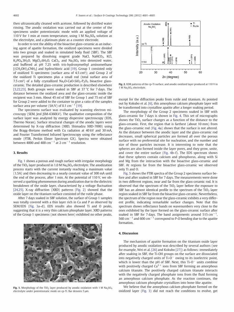
Fig. 2. XDR patterns of the cp-Ti surface, and anodic oxidized layer produced at 110 V in1 M Na2SO4 electrolyte.
4602 P. Soares et al. / Surface & Coatings Technology 206 (2012) 4601–4605
then ultrasonically cleaned with acetone, followed by distilled waterrinsing. The anodic oxidation was carried out at the center of thespecimens under potentiostatic mode with an applied voltage of110 V for 1 min at room temperature, using 1 M Na2SO4 solution asthe electrolyte, and a platinum plate as a counter electrode.
In order to test the ability of the bioactive glass-ceramic as an induc-ing agent of apatite formation, the oxidized specimens were dividedinto two groups and soaked in simulated body fluid (SBF). The SBFwas prepared by dissolving reagent grade NaCl, NaHCO3, KCl,K2PO4.3H2O, MgCl2.6H2O, CaCl2, and Na2SO4 into deionized water,and buffered at pH 7.25 with tris-hydroxymethyl aminomethane[(CH2OH)3CNH2] and hydrochloric acid [19]. Group 1 consisted onlyof oxidized Ti specimens (surface area of 4.5 cm²); and Group 2 ofthe oxidized Ti specimens plus a small rod (total surface area of7.5 cm²) of a fully crystallized Na2O-CaO-SiO2-P2O5 bioactive glass-ceramic. The detailed glass-ceramic production is described elsewhere[3,22,23]. Both groups were soaked in SBF at 37 °C for 7 days. Thedistance between the oxidized area and the glass-ceramic inside therecipient was 3 mm. About 45 ml of SBF for Group 1 and 75 ml of SBFfor Group 2 were added to the container to give a ratio of the samplessurface area per volume (SA/V) of 0.1 cm−1 [19].
The specimens surface was evaluated by scanning electron mi-croscopy (SEM, Jeol JSM-6360LV). The qualitative composition of thesurface layer was analyzed by energy dispersive spectroscopy (EDS,Thermo Noran). Surface structural changes of the anodic layers weredetermined by X-ray diffraction (XRD, Shimadzu XRD-7000) usingthe Bragg–Bretano method with Cu radiation at 40 kV and 30 mA,and Fourier Transformed Infrared Spectroscopy using the reflectancemode (FTIR, Perkin Elmer Spectrum GX). Spectra were obtainedbetween 4000 and 400 cm−1 at 2 cm−1 resolution.
3. Results
Fig. 1 shows a porous and rough surface with irregular morphologyof the TiO2 layer produced in 1.0 M Na2SO4 electrolyte. The anodizationprocess starts with the current instantly reaching a maximum value(1.5A) and then decreasing to a nearly constant value of 300 mA untilthe end of the process, after 1 min. At the potential of 110 V, we ob-served a sparking phenomenon during anodization due to the dielectricbreakdown of the oxide layer, characterized by a voltage fluctuation[24,25]. X-ray diffraction (XRD) patterns (Fig. 2) showed that theoxide layer on the titanium surface consisted of the rutile phase.
After 7 days soaked in SBF solution, the surface of Group 1 sampleswas totally covered with a thin layer rich in Ca and P as observed bySEM/EDS (Fig. 3a–d). EDS results also showed Ti and O peaks,suggesting that it is a very thin calcium phosphate layer. XRD patternsof the Group 1 specimens (not shown here) exhibited no other peaks,
Fig. 1. Morphology of the TiO2 layer produced by anodic oxidation with 1 M Na2SO4
electrolyte under potentiostatic mode on cp-Ti. Bar denotes 5 μm.
except for the diffraction peaks from rutile and titanium. As pointedout by Kokubo et al. [6], this amorphous calcium phosphate layer willbe transformed into crystalline apatite after a longer soaking period.
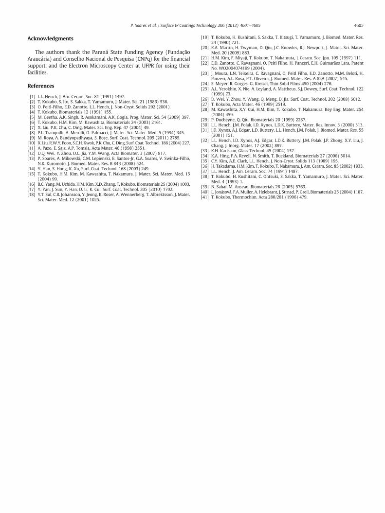
The morphology of the Group 2 specimens soaked in SBF withglass-ceramic for 7 days is shown in Fig. 4. This set of micrographsshows the TiO2 surface changes as a function of the distance to theglass-ceramic. First, the region that is farthest (about 10 mm) fromthe glass-ceramic rod (Fig. 4a) shows that the surface is not altered.As the distance between the anodic layer and the glass-ceramic roddecreases, small spherical particles are formed all over the poroussurface with no preferential site for nucleation, and the number andsize of those particles increase. It is interesting to note that thespheres are also formed inside the layer pores, and they grow, unite,and cover the entire surface (Fig. 4b–f). The EDS spectrum showsthat these spheres contain calcium and phosphorus, along with Siand Mg from the interaction with the bioactive glass-ceramic andSBF. At regions far from the bioactive glass-ceramic we observedonly Ti and O.
Fig. 5 shows the FTIR spectra of the Group 2 specimens surface be-fore and after soaked in SBF for 7 days. The measurements were donein two different regions, near and far from the glass-ceramic rod. It isobserved that the spectrum of the TiO2 layer before the exposure toSBF has an almost identical profile to the spectrum of the TiO2 layerregion soaked in SBF far from the bioactive glass-ceramic. Nevertheless,the spectrum of the region near the glass-ceramic exhibits a very differ-ent profile, indicating remarkable surface changes. Note that thisspectrum shows reflectance bands on wavenumbers very close to theones exhibited by the layer formed on the glass-ceramic surface aftersoaked in SBF for 7 days. The band assignments around 515 cm−1,560 cm−1 and 600 cm−1 correspond to P-O bending due to the apatiteformation.
4. Discussion
The mechanism of apatite formation on the titanium oxide layerproduced by anodic oxidation was described by several authors (seefor example, Wei et al. [26] and Kokubo [27]) as follows: immediatelyafter soaking in SBF, the Ti-OH groups on the surface are dissociatedinto negatively charged units of Ti-O− owing to its isoelectric point,which is lower than the pH of SBF. Next, this Ti-O− units combinewith positively charged Ca2+ ions from SBF forming an amorphouscalcium titanate. The positively charged calcium titanate interactswith the negatively charged phosphate ions from the fluid formingan amorphous calcium phosphate. As the reaction continues, theamorphous calcium phosphate crystallizes into bone‐like apatite.
We believe that the amorphous calcium phosphate formed on theoxidized layer (Fig. 3) did not reach the conditions to evolve, get
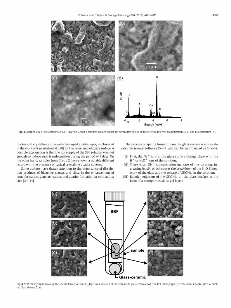
Fig. 3. Morphology of the amorphous Ca-P layer on Group 1 samples surface soaked for seven days in SBF solution, with different magnification (a–c) and EDS spectrum (d).
4603P. Soares et al. / Surface & Coatings Technology 206 (2012) 4601–4605
thicker and crystallize into a well-developed apatite layer, as observedin thework of Kawashita et al. [28] for the same kind of oxide surface. Apossible explanation is that the ion supply of the SBF solution was notenough to induce such transformation during the period of 7 days. Onthe other hand, samples from Group 2 have shown a notably differentresult, with the presence of typical crystalline apatite spheres.
Some authors have drawn attention to the importance of dissolu-tion products of bioactive glasses and silica in the enhancement ofbone formation, gene activation, and apatite formation in vitro and invivo [29–34].
Fig. 4. SEM micrographs showing the apatite formation on TiO2 layer as a function of the dirod. Bars denote 5 μm.
The process of apatite formation on the glass surface was investi-gated by several authors [35–37] and can be summarized as follows:
(i) First, the Na+ ions of the glass surface change place with theH+ or H3O+ ions of the solution;
(ii) There is an OH− concentration increase of the solution, in-creasing its pH, which causes the breakdown of the Si-O-Si net-work of the glass and the release of Si(OH)4 in the solution;
(iii) Repolymerization of the Si(OH)4 on the glass surface in theform of a nanoporous silica-gel layer;
stance to glass-ceramic rod. The last micrograph (f) is the nearest to the glass-ceramic
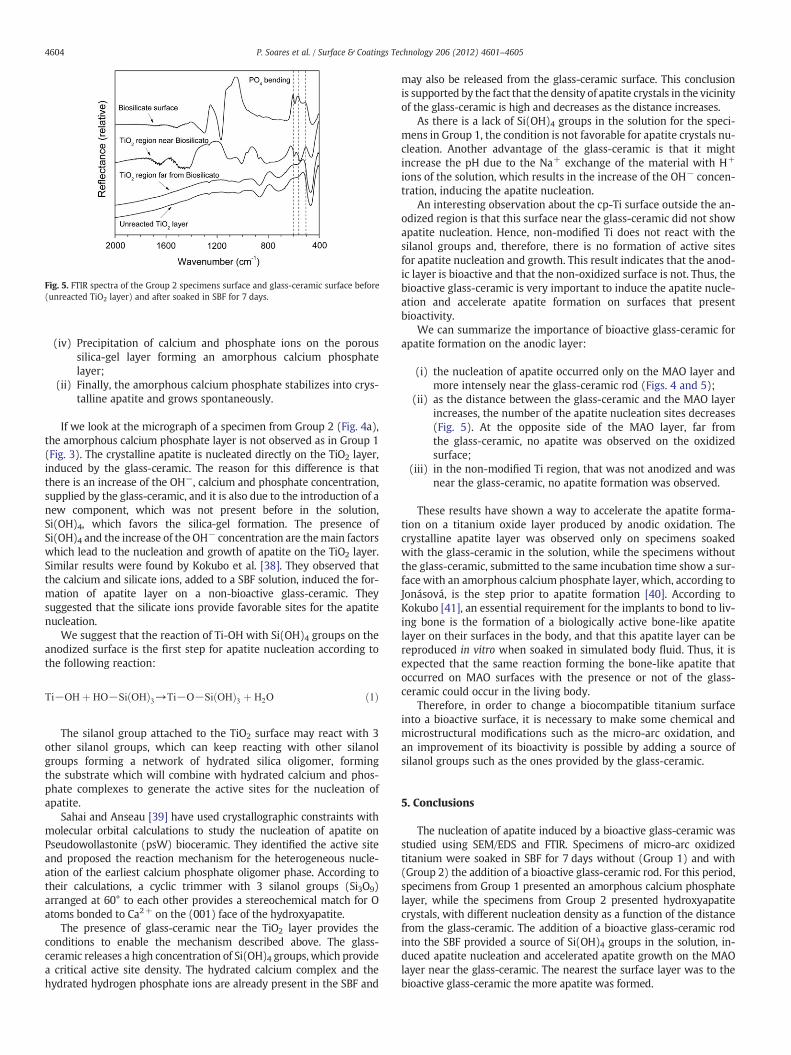
Fig. 5. FTIR spectra of the Group 2 specimens surface and glass-ceramic surface before(unreacted TiO2 layer) and after soaked in SBF for 7 days.
4604 P. Soares et al. / Surface & Coatings Technology 206 (2012) 4601–4605
(iv) Precipitation of calcium and phosphate ions on the poroussilica-gel layer forming an amorphous calcium phosphatelayer;
(ii) Finally, the amorphous calcium phosphate stabilizes into crys-talline apatite and grows spontaneously.
If we look at the micrograph of a specimen from Group 2 (Fig. 4a),the amorphous calcium phosphate layer is not observed as in Group 1(Fig. 3). The crystalline apatite is nucleated directly on the TiO2 layer,induced by the glass-ceramic. The reason for this difference is thatthere is an increase of the OH−, calcium and phosphate concentration,supplied by the glass-ceramic, and it is also due to the introduction of anew component, which was not present before in the solution,Si(OH)4, which favors the silica-gel formation. The presence ofSi(OH)4 and the increase of the OH− concentration are themain factorswhich lead to the nucleation and growth of apatite on the TiO2 layer.Similar results were found by Kokubo et al. [38]. They observed thatthe calcium and silicate ions, added to a SBF solution, induced the for-mation of apatite layer on a non-bioactive glass-ceramic. Theysuggested that the silicate ions provide favorable sites for the apatitenucleation.
We suggest that the reaction of Ti-OH with Si(OH)4 groups on theanodized surface is the first step for apatite nucleation according tothe following reaction:
Ti−OH þ HO−SiðOHÞ3→Ti−O−SiðOHÞ3 þ H2O ð1Þ
The silanol group attached to the TiO2 surface may react with 3other silanol groups, which can keep reacting with other silanolgroups forming a network of hydrated silica oligomer, formingthe substrate which will combine with hydrated calcium and phos-phate complexes to generate the active sites for the nucleation ofapatite.
Sahai and Anseau [39] have used crystallographic constraints withmolecular orbital calculations to study the nucleation of apatite onPseudowollastonite (psW) bioceramic. They identified the active siteand proposed the reaction mechanism for the heterogeneous nucle-ation of the earliest calcium phosphate oligomer phase. According totheir calculations, a cyclic trimmer with 3 silanol groups (Si3O9)arranged at 60° to each other provides a stereochemical match for Oatoms bonded to Ca2+ on the (001) face of the hydroxyapatite.
The presence of glass-ceramic near the TiO2 layer provides theconditions to enable the mechanism described above. The glass-ceramic releases a high concentration of Si(OH)4 groups, which providea critical active site density. The hydrated calcium complex and thehydrated hydrogen phosphate ions are already present in the SBF and
may also be released from the glass-ceramic surface. This conclusionis supported by the fact that the density of apatite crystals in the vicinityof the glass-ceramic is high and decreases as the distance increases.
As there is a lack of Si(OH)4 groups in the solution for the speci-mens in Group 1, the condition is not favorable for apatite crystals nu-cleation. Another advantage of the glass-ceramic is that it mightincrease the pH due to the Na+ exchange of the material with H+
ions of the solution, which results in the increase of the OH− concen-tration, inducing the apatite nucleation.
An interesting observation about the cp-Ti surface outside the an-odized region is that this surface near the glass-ceramic did not showapatite nucleation. Hence, non-modified Ti does not react with thesilanol groups and, therefore, there is no formation of active sitesfor apatite nucleation and growth. This result indicates that the anod-ic layer is bioactive and that the non-oxidized surface is not. Thus, thebioactive glass-ceramic is very important to induce the apatite nucle-ation and accelerate apatite formation on surfaces that presentbioactivity.
We can summarize the importance of bioactive glass-ceramic forapatite formation on the anodic layer:
(i) the nucleation of apatite occurred only on the MAO layer andmore intensely near the glass-ceramic rod (Figs. 4 and 5);
(ii) as the distance between the glass-ceramic and the MAO layerincreases, the number of the apatite nucleation sites decreases(Fig. 5). At the opposite side of the MAO layer, far fromthe glass-ceramic, no apatite was observed on the oxidizedsurface;
(iii) in the non-modified Ti region, that was not anodized and wasnear the glass-ceramic, no apatite formation was observed.
These results have shown a way to accelerate the apatite forma-tion on a titanium oxide layer produced by anodic oxidation. Thecrystalline apatite layer was observed only on specimens soakedwith the glass-ceramic in the solution, while the specimens withoutthe glass-ceramic, submitted to the same incubation time show a sur-face with an amorphous calcium phosphate layer, which, according toJonásová, is the step prior to apatite formation [40]. According toKokubo [41], an essential requirement for the implants to bond to liv-ing bone is the formation of a biologically active bone-like apatitelayer on their surfaces in the body, and that this apatite layer can bereproduced in vitro when soaked in simulated body fluid. Thus, it isexpected that the same reaction forming the bone-like apatite thatoccurred on MAO surfaces with the presence or not of the glass-ceramic could occur in the living body.
Therefore, in order to change a biocompatible titanium surfaceinto a bioactive surface, it is necessary to make some chemical andmicrostructural modifications such as the micro-arc oxidation, andan improvement of its bioactivity is possible by adding a source ofsilanol groups such as the ones provided by the glass-ceramic.
5. Conclusions
The nucleation of apatite induced by a bioactive glass-ceramic wasstudied using SEM/EDS and FTIR. Specimens of micro-arc oxidizedtitanium were soaked in SBF for 7 days without (Group 1) and with(Group 2) the addition of a bioactive glass-ceramic rod. For this period,specimens from Group 1 presented an amorphous calcium phosphatelayer, while the specimens from Group 2 presented hydroxyapatitecrystals, with different nucleation density as a function of the distancefrom the glass-ceramic. The addition of a bioactive glass-ceramic rodinto the SBF provided a source of Si(OH)4 groups in the solution, in-duced apatite nucleation and accelerated apatite growth on the MAOlayer near the glass-ceramic. The nearest the surface layer was to thebioactive glass-ceramic the more apatite was formed.
4605P. Soares et al. / Surface & Coatings Technology 206 (2012) 4601–4605
Acknowledgments
The authors thank the Paraná State Funding Agency (FundaçãoAraucária) and Conselho Nacional de Pesquisa (CNPq) for the financialsupport, and the Electron Microscopy Center at UFPR for using theirfacilities.
References
[1] L.L. Hench, J. Am. Ceram. Soc. 81 (1991) 1497.[2] T. Kokubo, S. Ito, S. Sakka, T. Yamamuro, J. Mater. Sci. 21 (1986) 536.[3] O. Peitl-Filho, E.D. Zanotto, L.L. Hench, J. Non-Cryst. Solids 292 (2001).[4] T. Kokubo, Biomaterials 12 (1991) 155.[5] M. Geetha, A.K. Singh, R. Asokamani, A.K. Gogia, Prog. Mater. Sci. 54 (2009) 397.[6] T. Kokubo, H.M. Kim, M. Kawashita, Biomaterials 24 (2003) 2161.[7] X. Liu, P.K. Chu, C. Ding, Mater. Sci. Eng. Rep. 47 (2004) 49.[8] P.L. Tranquilli, A. Merolli, O. Palmacci, J. Mater. Sci. Mater. Med. 5 (1994) 345.[9] M. Roya, A. Bandyopadhyaya, S. Bose, Surf. Coat. Technol. 205 (2011) 2785.
[10] X. Liu, R.W.Y. Poon, S.C.H. Kwok, P.K. Chu, C. Ding, Surf. Coat. Technol. 186 (2004) 227.[11] A. Pazo, E. Saiz, A.P. Tomsia, Acta Mater. 46 (1998) 2551.[12] D.Q. Wei, Y. Zhou, D.C. Jia, Y.M. Wang, Acta Biomater. 3 (2007) 817.[13] P. Soares, A. Mikowski, C.M. Lepienski, E. Santos-Jr, G.A. Soares, V. Swinka-Filho,
N.K. Kuromoto, J. Biomed. Mater. Res. B 84B (2008) 524.[14] Y. Han, S. Hong, K. Xu, Surf. Coat. Technol. 168 (2003) 249.[15] T. Kokubo, H.M. Kim, M. Kawashita, T. Nakamura, J. Mater. Sci. Mater. Med. 15
(2004) 99.[16] B.C. Yang, M. Uchida, H.M. Kim, X.D. Zhang, T. Kokubo, Biomaterials 25 (2004) 1003.[17] Y. Yan, J. Sun, Y. Han, D. Li, K. Cui, Surf. Coat. Technol. 205 (2010) 1702.[18] Y.T. Sul, C.B. Johansson, Y. Jeong, K. Roser, A. Wennerberg, T. Albrektsson, J. Mater.
Sci. Mater. Med. 12 (2001) 1025.
[19] T. Kokubo, H. Kushitani, S. Sakka, T. Kitsugi, T. Yamamuro, J. Biomed. Mater. Res.24 (1990) 721.
[20] R.A. Martin, H. Twyman, D. Qiu, J.C. Knowles, R.J. Newport, J. Mater. Sci. Mater.Med. 20 (2009) 883.
[21] H.M. Kim, F. Miyaji, T. Kokubo, T. Nakamuta, J. Ceram. Soc. Jpn. 105 (1997) 111.[22] E.D. Zanotto, C. Ravagnani, O. Peitl Filho, H. Panzeri, E.H. Guimarães Lara, Patent
No. WO2004074199 (2004).[23] J. Moura, L.N. Teixeira, C. Ravagnani, O. Peitl Filho, E.D. Zanotto, M.M. Beloti, H.
Panzeri, A.L. Rosa, P.T. Oliveira, J. Biomed. Mater. Res. A 82A (2007) 545.[24] S. Meyer, R. Gorges, G. Kreisel, Thin Solid Films 450 (2004) 276.[25] A.L. Yerokhin, X. Nie, A. Leyland, A. Mattheus, S.J. Dowey, Surf. Coat. Technol. 122
(1999) 73.[26] D. Wei, Y. Zhou, Y. Wang, Q. Meng, D. Jia, Surf. Coat. Technol. 202 (2008) 5012.[27] T. Kokubo, Acta Mater. 46 (1999) 2519.[28] M. Kawashita, X.Y. Cui, H.M. Kim, T. Kokubo, T. Nakamura, Key Eng. Mater. 254
(2004) 459.[29] P. Ducheyne, Q. Qiu, Biomaterials 20 (1999) 2287.[30] L.L. Hench, J.M. Polak, I.D. Xynos, L.D.K. Buttery, Mater. Res. Innov. 3 (2000) 313.[31] I.D. Xynos, A.J. Edgar, L.D. Buttery, L.L. Hench, J.M. Polak, J. Biomed. Mater. Res. 55
(2001) 151.[32] L.L. Hench, I.D. Xynos, A.J. Edgar, L.D.K. Buttery, J.M. Polak, J.P. Zhong, X.Y. Liu, J.
Chang, J. Inorg. Mater. 17 (2002) 897.[33] K.H. Karlsson, Glass Technol. 45 (2004) 157.[34] K.A. Hing, P.A. Revell, N. Smith, T. Buckland, Biomaterials 27 (2006) 5014.[35] C.Y. Kim, A.E. Clark, L.L. Hench, J. Non-Cryst. Solids 113 (1989) 195.[36] H. Takadama, H.M. Kim, T. Kokubo, T. Nakamura, J. Am. Ceram. Soc. 85 (2002) 1933.[37] L.L. Hench, J. Am. Ceram. Soc. 74 (1991) 1487.[38] T. Kokubo, H. Kushitani, C. Ohtsuki, S. Sakka, T. Yamamuro, J. Mater. Sci. Mater.
Med. 4 (1993) 1.[39] N. Sahai, M. Anseau, Biomaterials 26 (2005) 5763.[40] L. Jonásová, F.A.Muller, A. Helebrant, J. Strnad, P. Greil, Biomaterials 25 (2004) 1187.[41] T. Kokubo, Thermochim. Acta 280/281 (1996) 479.

![Temperature‐dependent Nucleation and Growth of Dendrite‐Free … · nucleation, chronoamperometry has been used to model heterogeneous nucleation behavior.[10] Therefore, we further](https://img.pdfslide.us/doc/110x75/5ecedb8e0e2bd5210370ca09/temperatureadependent-nucleation-and-growth-of-dendriteafree-nucleation-chronoamperometry.jpg)