Embed Size (px)
Citation preview
RESEARCH ARTICLE
Eco-friendly drugs from the marine environment:spongeweed-synthesized silver nanoparticles are highly effectiveon Plasmodium falciparum and its vector Anopheles stephensi,with little non-target effects on predatory copepods
Kadarkarai Murugan1,2& Chellasamy Panneerselvam3
& Jayapal Subramaniam1&
Pari Madhiyazhagan1& Jiang-Shiou Hwang4 & Lan Wang5 & Devakumar Dinesh1
&
Udaiyan Suresh1& Mathath Roni1 & Akon Higuchi6 & Marcello Nicoletti7 &
Giovanni Benelli8,9
Received: 13 April 2016 /Accepted: 4 May 2016 /Published online: 16 May 2016# Springer-Verlag Berlin Heidelberg 2016
Abstract Mosquitoes act as vectors of devastating pathogensand parasites, representing a key threat for millions of humansand animals worldwide. The control of mosquito-borne dis-eases is facing a number of crucial challenges, including theemergence of artemisinin and chloroquine resistance inPlasmodium parasites, as well as the presence of mosquitovectors resistant to synthetic and microbial pesticides.Therefore, eco-friendly tools are urgently required. Here, asynergic approach relying to nanotechnologies and biologicalcontrol strategies is proposed. The marine environment is anoutstanding reservoir of bioactive natural products, whichhave many applications against pests, parasites, and patho-gens. We proposed a novel method of seaweed-mediated syn-thesis of silver nanoparticles (AgNP) using the spongeweedCodium tomentosum, acting as a reducing and capping agent.AgNP were characterized by UV–Vis spectroscopy, Fouriertransform infrared (FTIR) spectroscopy, scanning electronmi-croscopy (SEM), energy-dispersive X-ray spectroscopy
(EDX), andX-ray diffraction (XRD). Inmosquitocidal assays,the 50% lethal concentration (LC50) ofC. tomentosum extractagainst Anopheles stephensi ranged from 255.1 (larva I) to487.1 ppm (pupa). LC50 of C. tomentosum-synthesizedAgNP ranged from 18.1 (larva I) to 40.7 ppm (pupa). In lab-oratory, the predation efficiency of Mesocyclops aspericorniscopepods against A. stephensi larvae was 81, 65, 17, and 9 %(I, II, III, and IV instar, respectively). In AgNP contaminatedenvironment, predation was not affected; 83, 66, 19, and 11 %(I, II, III, and IV). The anti-plasmodial activity ofC. tomentosum extract and spongeweed-synthesized AgNPwas evaluated against CQ-resistant (CQ-r) and CQ-sensitive(CQ-s) strains of Plasmodium falciparum. Fifty percent inhib-itory concentration (IC50) ofC. tomentosumwere 51.34 μg/ml(CQ-s) and 65.17 μg/ml (CQ-r); C. tomentosum-synthesizedAgNP achieved IC50 of 72.45 μg/ml (CQ-s) and 76.08 μg/ml(CQ-r). Furthermore, low doses of the AgNP inhibited thegrowth of Bacillus subtilis, Klebsiella pneumoniae, and
Responsible editor: Philippe Garrigues
* Giovanni [email protected]; [email protected]
1 Division of Entomology, Department of Zoology, School of LifeSciences, Bharathiar University, Coimbatore, Tamil Nadu 641046,India
2 Department of Biotechnology, Thiruvalluvar University, Serkkadu,Vellore, Tamil Nadu 632115, India
3 Biology Department, Faculty of Science, University of Tabuk,Tabuk, Saudi Arabia
4 Institute of Marine Biology, National Taiwan Ocean University,Keelung 20224, Taiwan
5 School of Life Science and Technology, Shanxi University,Taiyuan 030006, China
6 Department of Chemical and Materials Engineering, NationalCentral University, No. 300 Jhongli, Taoyuan 32001, Taiwan
7 Department of Environmental Biology, Sapienza University ofRome, Piazzale Aldo Moro 5, 00185 Rome, Italy
8 Insect Behaviour Group, Department of Agriculture, Food andEnvironment, University of Pisa, via del Borghetto 80,56124 Pisa, Italy
9 The BioRobotics Institute, Sant’Anna School of Advanced Studies,Viale Rinaldo Piaggio 34, 56025 Pontedera, Italy
Environ Sci Pollut Res (2016) 23:16671–16685DOI 10.1007/s11356-016-6832-9
Salmonella typhi, using the agar disk diffusion and minimuminhibitory concentration protocol. Overall, C. tomentosummetabolites and spongeweed-synthesized AgNP may be po-tential candidates to develop novel and effective tools in thefight against Plasmodium parasites and their mosquito vec-tors. The employ of ultra-low doses of nanomosquitocides insynergy with cyclopoid crustaceans seems a promising greenroute for effective mosquito control programs.
Keywords Anopheles stephensi .Codium tomentosum .
Malaria .Mosquito-bornediseases .Plasmodium falciparum .
Mesocyclops aspericornis . Anti-bacterial activity
Introduction
Arthropods are dangerous vectors of deadly pathogens andparasites (Mehlhorn et al. 2012; Benelli 2015a). Among them,mosquitoes (Diptera: Culicidae) represent a key threat for mil-lions of humans and animals worldwide. Their medical andveterinary importance is mainly due to the fact that their act asvectors for a number of pathogens and parasites of publichealth relevance, including malaria, avian malaria, yellow fe-ver, dengue, Japanese encephalitis, Zika virus, Rift Valleyfever, Western equine encephalomyelitis, bancroftian andbrugian filariae, canine heartworm disease (Dirofilariaimmitis), and setariosis (Setaria spp.) (Benelli and Mehlhorn2016; Benelli et al. 2016).
Malaria is caused by Plasmodium parasites, mainlyPlasmodium falciparum, Plasmodium vivax, Plasmodiumovale, and Plasmodium malariae. In addition, periodic reportshighlighted the presence of simian malaria parasites found inhumans, most of them implicating Plasmodium knowlesi.Plasmodium parasites are vectored to vertebrates through thebites of infected Anopheles mosquitoes, which bite mainlybetween dusk and dawn. According to the latest estimates,there were about 198 million cases of malaria in 2013 andan estimated 584,000 deaths. In Africa, most deaths occuramong children living where a child dies every minute frommalaria, although mortality rates among children have beenreduced by an estimated 58 % since 2000 (Mehlhorn 2008;WHO 2016). In addition, besides the fall of malaria infectionrates worldwide, with special reference to sub-Saharan Africa,2015 was an annus mirabilis for malaria control, due to theNobel Prize to the Chinese scientist Youyou Tu for the dis-covery of artemisinin and the development of the first vaccineagainst P. falciparum malaria [i.e., RTS,S/AS01 (RTS,S)](Benelli and Mehlhorn 2016).
However, malaria prevention and control is facing anumber of crucial challenges, including the emergenceof artemisinin and chloroquine resistance in Plasmodiumparasites (Benelli and Mehlhorn 2016), as well as thepresence of mosquito strains resistant to synthetic and
microbial pesticides (Hemingway and Ranson 2000;Aziz et al. 2016; Naqquash et al. 2016). Therefore, eco-friendly tools are urgently required (Rajkumar and Jebanesan2008; Azizullah et al. 2014; Benelli et al. 2015a, b, c; Pavela2015a, b; Govindarajan and Benelli 2016). Notably, botani-cals have been used by human communities in different partsof the world as mosquitocidals, adult repellents, and oviposi-tion deterrents, against a wide number of mosquito species(e.g., Benelli 2015a, b). People entering into regions wheredengue, malaria, or yellow fever risks exist may protect them-selves by use of plant-derived repellents (Mehlhorn et al.2012; Amer and Mehlhorn 2006a, b). On the other hand,people living in endemic regions have to protect themselvesby several strategies at the same time, since infection rates ofmosquitoes may be extremely high (Amer and Mehlhorn2006c, d; Semmler et al. 2009; Benelli 2015a).
Biological control of mosquito vectors using predatorycopepods, tadpoles, and fishes also received attention(Marten 2000; Murugan et al. 2015a, b, c; Subramaniamet al. 2016a, b). Predation is an important factor for themaintenance of trophic equilibrium of ecological commu-nities, including aquatic ones. Good examples are odonateyoung instars, water bugs, tadpoles, fishes, crabs, and co-pepods acting as natural enemies of mosquito young in-stars (Bowatte et al. 2013; Murugan et al. 2015d, e, f, g2016). Cyclopoid copepods are crucial invertebrate pred-ators in zooplankton communities, and are frequentlypresent in lakes, drains, ponds, and reservoirs (Changand Hanazato 2003; Kumar and Hwang 2006). Thecyclopoid Mesocyclops aspericornis is a generalist preda-tor that preys on a wide spectrum of food types, rangingfrom rotifers, cladocerans, mosquito larvae, and fishyoung instars (Kumar et al. 2012). More generally, severalspecies of copepods, including M. aspericornis, Mesocyclopsguangxiensis, Mesocyclops longisetus, and Mesocyclopsthermocyclopoides, have been reported as potential biologicalcontrol agents against mosquitoes (Rawlins et al. 1997;Manrique-Saide et al. 1998; Schaper 1999; Murugan et al.2015b).
Nanotechnology has the potential to revolutionize a widearray of applications in the fields of catalysis, sensors, opto-electronics, magnetic devices, drug delivery, anti-microbials,pest management, and parasitology (Scrinis and Lyons 2007;Haverkamp 2010, Govindarajan et al. 2016; Kumar et al.2016). The utilization of plants for nanoparticle synthesiscan be advantageous over other biological processes, becauseit eliminates the elaborate process of maintaining cell culturesand can also be suitably scaled up for large-scale nanoparticlesynthesis (Shankar et al. 2004; Chandramohan et al.2016; Jaganathan et al. 2016). Recently, several plants havebeen screened for efficient and rapid extracellular synthesis ofmetal nanoparticles with different mosquitocidal and anti-splamodial properties (Benelli 2016a, b). The marine
16672 Environ Sci Pollut Res (2016) 23:16671–16685
environment is an outstanding reservoir of bioactive naturalproducts, which have many applications against pests, para-sites, and pathogens (Kalimuthu et al. 2014; Murugan et al.2016). Concerning the seaweed-mediated synthesis ofnanomosquitocides and anti-plasmodial drugs, good exam-ples included seaweed extracts of Sargassum muticum(Madhiyazhagan et al. 2015), Hypnea musciformis (Roniet al. 2015), Ulva lactuca (Murugan et al. 2015h), andCentroceras clavulatum (Murugan et al. 2016).
Codium tomentosum is a species of green seaweeds inthe family Codiaceae. Its common names include velvethorn and spongeweed. The spongeweed is native to thenorth east Atlantic Ocean from the British Isles south-wards to the Azores and Cape Verde. It has also beenrecorded around the coasts of Africa and in various otherparts of the world. A lectin named tomentine has been isolatedby affinity chromatography from C. tomentosum. It showsN-acetylglucosamine-specific activity and has been found tobe rich in glycine, threonine, and valin. To the best of ourknowledge, there is no information about the insecticidaland anti-plasmodial potential of the spongeweed (WRMS1967; Valentao et al. 2010).
In this research, we proposed spongeweed-mediated syn-thesis of silver nanoparticles (AgNP) using C. tomentosum asa reducing and capping agent. AgNP were characterized byUV–Vis spectroscopy, Fourier transform infrared (FTIR)spectroscopy, scanning electron microscopy (SEM), energy-dispersive x-ray spectroscopy (EDX), and x-ray diffraction(XRD). The potential of the C. tomentosum extract andspongeweed-synthesized AgNP were evaluated againstchloroquine-sensitive (CQ-s) and chloroquine resistant (CQ-r) strains of P. falciparum, as well as against the larvae andpupae of the malaria vector Anopheles stephensi. The anti-oxidant and anti-bacteric potential of C. tomentosum andAgNP were also assessed. Concerning non-target effect ofAgNP in the aquatic environment, the predatory efficiencyof M. aspericornis copepods was evaluated in standard labo-ratory conditions and post-treatment with ultra-low doses ofspongeweed-synthesized AgNP (i.e., 1 ppm).
Materials and methods
Plant material
C. tomentosum spongeweeds was collected in the Gulf ofMannar (Tamil Nadu, India, latitude from 8° 47′ to 9° 15′N; longitude from 78° 12′ to 79° 14′ E). C. tomentosumspongeweeds were washed with tap water and shade-driedat room temperature for 5 days. Voucher specimenswere stored in our laboratories and are available uponrequest.
DPPH radical scavenging assay
The anti-oxidant activity of the ethanol extract was deter-mined in terms of hydrogen donating or radical scavengingability using the stable radical 2,2-diphenyl-1-picrylhydrazyl(DPPH), according to the methods by Blois (1958) andPanneerselvam et al. (2016). Sample extract at various con-centrations were taken and the volume was adjusted to 100 μlwith ethanol. About 5 ml of a 0.1 mM ethanolic solution ofDPPH was added to the aliquots of samples and butylatedhydroxytoluene (BHT) and rutin were used as standards.Negative control was prepared by adding 100 μl of ethanolin 5ml of 0.1 mMethanolic solution of DPPH. The tubes wereallowed to stand for 20 min at 27 °C. The absorbance of thesample was measured at 517 nm against the blank (ethanol).Radical scavenging activity of the samples was expressed as50 % inhibitory concentration (IC50) values, which are theconcentrations required to inhibit 50 % of DPPH.
Synthesis and characterization of silver nanoparticles
C. tomentosum aqueous extract was prepared adding 10 g ofwashed and finely cut spongeweeds in a 300-ml Erlenmeyerflask filled with 100 ml of sterilized double-distilled water andthen boiling the mixture for 5 min, before finally decanting it.The extract was filtered using Whatman filter paper no. 1,stored at −4 °C and tested within 5 days. The filtrate wastreated with aqueous 1 mM AgNO3 solution in anErlenmeyer flask and incubated at room temperature. Abrown-yellow solution indicated the formation of AgNP,s ince aqueous s i lver ions were reduced by theC. tomentosum extract generating stable AgNP in water.Silver nitrate was purchased from the Precision ScientificCo. (Coimbatore, India). Green synthesis of AgNP wasconfirmed by sampling the reaction mixture at regularintervals and the absorption maxima was scanned byUV–Vis, at the wavelength of 200–800 nm in UV-3600Shimadzu spectrophotometer at 1-nm resolution.Furthermore, the reaction mixture was subjected to centri-fugation at 15,000 rpm for 20 min, resulting pellet wasdissolved in deionized water and filtered throughMillipore filter (0.45 μm). An aliquot of this filtrate con-taining AgNP was used for scanning electron microscopy(SEM), Fourier transform infrared (FTIR) spectroscopy,x-ray diffraction (XRD) analysis, and energy-dispersivex-ray (EDX) spectroscopy. The structure and compositionof freeze-dried purified AgNP was analyzed by using a 10kV ultra high-resolution scanning electron microscopewith 25 μl of sample was sputter coated on copper stuband the images of AgNP were studied using a FEIQUANTA-200 SEM. The surface groups of the AgNPwere qualitatively confirmed by FTIR spectroscopy,with spectra recorded by a PerkinElmer Spectrum 2000
Environ Sci Pollut Res (2016) 23:16671–16685 16673
FTIR spectrophotometer. EDX assays confirmed the pres-ence of metals in analyzed samples (Murugan et al.2015a, b).
Anopheles stephensi rearing
The eggs of A. stephensi were collected from water reser-voirs in Coimbatore (Tamil Nadu, India) using an BO^type brush. Batches of 100–110 eggs were transferred to18 cm× 13 cm×4 cm enamel trays containing 500 ml ofwater, where eggs were allowed to hatch in laboratoryconditions (27 ± 2 °C and 75–85 % R.H.; 14:10 (L/D)photoperiod). A. stephensi larvae were fed daily with 5 gof ground dog biscuits (Pedigree, USA) and hydrolyzedyeast (Sigma-Aldrich, Germany) in a 3:1 ratio (Dineshet al. 2015). Newly emerged larvae, pupae, and adultswere collected and used in the experiments.
Mesocyclops aspericornis rearing
M. aspericornis adults were collected from a pond(Coimbatore, India) using a mesh net. All collected sampleswere identified as M. aspericornis by Dr. Y. Ranga Reddy(Department of Zoology, Acharya Nagarjuna University,India) (Murugan et al. 2015b). M. aspericornis was rearedfollowing the method reported by Kosiyachinda et al.(2003). Isofemale lines were established from gravid femalesand maintained at Department of Zoology, BharathiarUniversity (Coimbatore, India). Gravid females from differentisofemale lines were pooled and mass reared in dechlorinatedwater (pH 7) in fish tanks (15 l) at 27 ± 1 °C and naturalphotoperiod. Food was Paramecium spp. prepared fromboiled rice straw water extract, and commercial powdered fishfood.
Larvicidal and pupicidal toxicity in laboratory conditions
Twenty-five A. stephensi larvae (I, II, III, or IV instar) orpupae were placed for 24 h in a glass beaker filled with250 ml of dechlorinated water plus the desired concen-tration of C. tomentosum extract (100, 200, 300, 400,and 500 ppm) or green-synthesized AgNP (10, 20, 30,40, and 50 ppm). Larval food (0.5 mg) was provided foreach tested concentration (Kovendan et al. 2012). Eachconcentration was replicated five times against all in-stars. Control mosquitoes were exposed for 24 h to thecorresponding concentration of the solvent. Percentagemortality was calculated as follows:
Percentage mortality ¼ ðnumber of dead individuals=number of treated individualsÞ � 100
In vitro cultivation of Plasmodium falciparum
Following Murugan et al. (2015h, i), CQ-sensitive (CQ-s)strain 3D7 and CQ-resistant (CQ-r) strain INDO ofP. falciparum were used in in vitro blood stage culture to testthe anti-malarial efficacy of C. tomentosum ethanol extracts.The culture was maintained at G. Kuppusamy NaiduMemorial Hospital (Coimbatore, India). P. falciparum culturewas maintained according to the method described by Tragerand Jensen (1976), with minor modifications. P. falciparum(3D7) cultures were maintained in fresh O+ve human erythro-cytes suspended at 4 % hematocrit in RPMI 1640 (Sigma)containing 0.2 % sodium bicarbonate, 0.5 % albumax,45 μg/l hypoxanthine, and 50 μg/l gentamycin and incubatedat 37 °C under a gas mixture of 5 % O2, 5 % CO2, and 90 %N2. Every day, infected erythrocytes were transferred into afresh complete medium to propagate the culture. ForP. falciparum (INDO strain) in culture medium, Albumaxwas replaced by 10 % pooled human serum.
Anti-plasmodial assays
Control stock solutions of CQwere prepared in water (Milli-Qgrade). The tested extract and the AgNP suspension wereprepared in dimethyl sulfoxide (DMSO). All stocks were di-luted with culture medium to achieve the required concentra-tions. In all cases except CQ, the final solution contained0.4 % DMSO (which was found to be non-toxic to the para-site). Then, the seaweed extract and AgNP were placed in 96-well flat-bottom tissue culture-grade plates.
The extract of C. tomentosum and AgNP were evaluatedfor anti-malarial activity against P. falciparum strains 3D7 andINDO. For drug screening, SYBR green I-based fluorescenceassay was used following the method by Smilkstein et al.(2004) and Murugan et al. (2015h, i). Sorbitol-synchronizedparasites were incubated under normal culture conditions at2% hematocrit and 1% parasitemia in the absence or presenceof increasing concentrations of the tested drugs. CQ was usedas positive control. After 48 h of incubation, 100 μl of SYBRGreen I solution {0.2 μl of 10,000 X SYBR Green I(Invitrogen)/ml} in lysis buffer [Tris (20 mM; pH 7.5),EDTA (5 mM), saponin (0.008 %; w/v), and Triton X-100(0.08 %; v/v)] was added to each well and mixed gently twicewith a multi-channel pipette and incubated in the dark at 37 °Cfor 1 h. Fluorescence was measured with a Victor fluorescencemulti-well plate reader (PerkinElmer) with excitation andemission wavelength bands centered at 485 and 530 nm, re-spectively. The fluorescence counts were plotted against thedrug concentration and the 50 % inhibitory concentration(IC50) was determined by an analysis of dose–responsecurves. Results were validated microscopically by the exam-ination of Giemsa-stained smears of extract-treated parasitecultures (Bagavan et al. 2011; Murugan et al. 2015h).
16674 Environ Sci Pollut Res (2016) 23:16671–16685
Predation of Mesocyclops aspericornis against Anophelesstephensi larvae
The predation efficiency of M. aspericornis was assessedagainst A. stephensi larvae. For each instar, 100 mosquitoeswere introduced, with 10 copepods, in a glass beaker contain-ing 250 ml of dechlorinated water. Mosquito larvae were re-placed daily with new ones. For each mosquito instar, fourreplicates were conducted. Control was 250 ml ofdechlorinated water without copepods. All beakers werechecked after 1, 2, 3, 4, and 5 days and the number of preyconsumed by copepods was recorded. Predatory efficiencywas calculated using the following formula:
Predatory efficiency ¼ ½ðnumber of consumed mosquitoes=number of predatorsÞ=total number
of mosquitoes� � 100
Predation of Mesocyclops aspericornis against Anophelesstephensi larvae post-treatment with silver nanoparticles
Here, the predation efficiency of M. aspericornis adults wasassessed against A. stephensi larvae, after a mosquitocidaltreatment with AgNP. For each instar, 100 mosquitoes wereintroduced with 10 copepods in a glass beaker filled with250 ml of dechlorinated water plus the desired concentrationof C. tomentosum-synthesized AgNP (i.e., one third of the 50% lethal concentration (LC50) calculated against first instarlarvae of A. stephensi, 1 ppm). Mosquito larvae were replaceddaily with new ones. For each mosquito instar four replicateswere conducted. Control was dechlorinated water plus AgNP,without copepods. All beakers were checked after 1, 2, 3, 4,and 5 days and the number of prey consumed by copepodswas recorded. Predatory efficiency was calculated using theabove-mentioned formula.
Anti-bacterial activity
C. tomentosum-synthesized AgNP were tested againstBacillus subtilis, Klebsiella pneumoniae and Salmonellatyphi. All bacteria strains were provided by Microbial TypeCulture, Collection and Gene Bank Institute of MicrobialTechnology, Sector 39-A, Chandigarh-160036 (India). Forall species, bacterial cultures 18–24-h old were used for thepreparation of testing cultures. All bacteria were grown innutrient broth: peptone (5 g/l), hydrolyzed yeast extract(1.50 g/l), beef extract (1.50 g/l), and sodium chloride (5 g/l), pH 7.4. Thirteen grams of nutrient broth was suspendedinto 100 ml of distilled water. Twenty-five milliliters of nutri-ent broth was transferred in each of four conical glass flasksand autoclaved at 121 °C for 15 min (15 psi). Then, eachbacterial strain was inoculated and incubated at 37 °C for
24 h. After this phase, the culture attained 2×10⁻⁶ cfu/mland was used for anti-bacterial assays. The anti-bacterial ac-tivity of C. tomentosum-synthesized AgNP was assessedusing the agar disk diffusion method (Dinesh et al. 2015).The tested bacteria strains were swabbed on Muller-Hintonagar medium plates. Three sterilized filter paper disks treatedwith three different concentrations of the tested compoundswere inserted in each plate. The plates were incubated at 37 °Cfor 24 h. After the incubation, the zones of inhibition radiuswere measured using a photomicroscope (Leica ES2,Germany).
Data analysis
SPSS software package 16.0 version was used for all analyses.Data from DPPH radical scavenging assay and mosquitocidalexperiments were analyzed by probit analysis, calculatingIC50 and LC50 values, respectively (Finney 1971). In anti-plasmodial assays, values were expressed as percentagegrowth inhibition. The concentration causing 50 % inhibitionof parasite growth (IC50) was calculated from the drugconcentration-response curves. Bacteria inhibition growth da-ta were transformed into arcsine√proportion values and ana-lyzed using a two-way ANOVAwith three factors (i.e., testeddose and targeted species). Means were separated usingTukey’s HSD test (P<0.05).
Copepod predation data were analyzed by JMP 7 using aweighted generalized linear model with one fixed factor:y=Xß+ ε where y is the vector of the observations (i.e., thenumber of consumed preys), X is the incidence matrix, ß is thevector of fixed effects (i.e., the targeted instar), and ε is thevector of the random residual effect. A probability level ofP<0.05 was used for the significance of differences betweenvalues.
Results and discussion
Synthesis and bio-physical characterization of silvernanoparticles
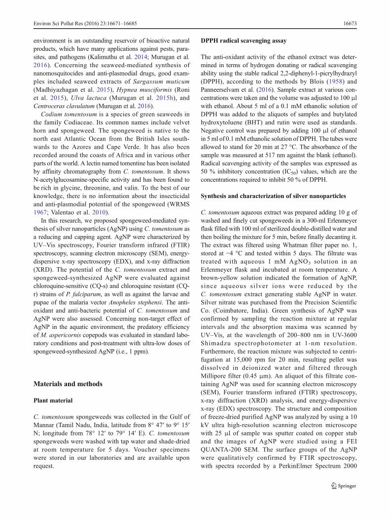
In our experiments, UV–Vis spectrum showed a maximumabsorbance peak at 420 nm, which increased over time duringthe incubation of silver nitrate with the C. tomentosum extract(Fig. 1). The broadness of the peak is a good indicator of thesize of the nanoparticles. As the particle size increases, thepeak becomes narrower, with a decreased bandwidth (Petitet al. 1993; Kong and Jang 2006). It is generally recognizedthat UV–Vis spectroscopy could be used to examine the sizeand shape-controlled nanoparticles in aqueous suspensions(Shrivastava and Dash 2010). As a confirmation, theC. tomentosum frond extract without AgNO3 did not showany change in color over time. C. tomentosum-mediated
Environ Sci Pollut Res (2016) 23:16671–16685 16675
reduction of silver ions to silver nanoparticles was linked withchanges in the UV–Vis spectra. The appearance of theyellowish-brown color was an indication of the synthesis ofcolloidal AgNP in the medium. The dark color may be due tothe excitation of surface plasmon vibrations, typical of theAgNP (Ahmad et al. 2003; Dinesh et al. 2015). In agreementwith our results, a peak with maximum absorption at 410 nmcharacterized the synthesis of Aloe vera-fabricated AgNP(Dinesh et al. 2015), while peaks with maximum absorptionat 450 and 420 nm were observed forMoringa oleifera-fabri-cated AgNP (Sujitha et al. 2015) and Phyllanthus niruri-syn-thesized AgNP (Suresh et al. 2015), respectively.
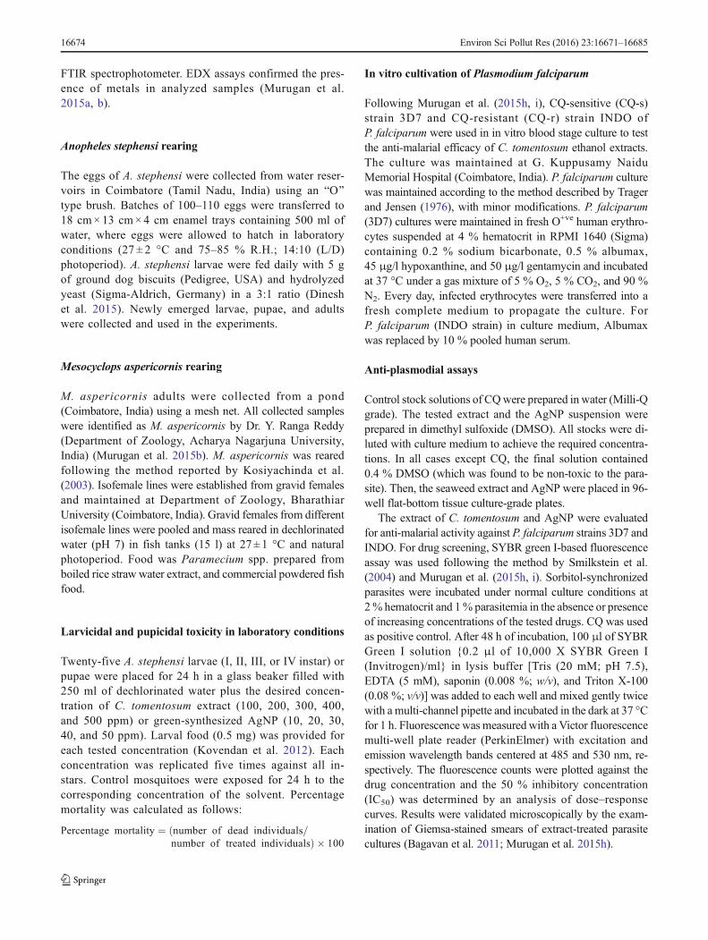
In XRD analyses, Bragg’s reflections corresponding to the(111), (200), (220), (311), and (222) sets of lattice planes wereobserved, showing that the AgNP were crystalline in nature(Fig. 2). XRD results suggest that crystallization of the bio-organic phase occurred on the surface of the AgNP. The XRDpattern observed in this study was consistent with previ-ous reports (Bar et al. 2009), including seaweed-mediatedprocesses (Madhiyazhagan et al. 2015). Sathyavathi et al.(2010) reported diffraction peaks at 44.50°, 52.20°, and76.7° 2θ, which correspond to the (111), (200), and (220)facets of the face-centered cubic crystal structure. Dubeyet al. (2009) studied the size of silver nanocrystals asestimated from the full width at half-maximum of (111)peak of silver using the Scherrer’s formula was 20–60 nm.
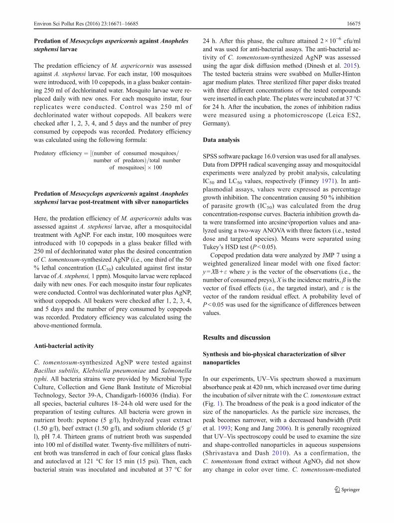
FTIR spectroscopy shed light on the identity of biomole-cules from C. tomentosum, which may be responsible for syn-thesis and stabilization of AgNP. The FTIR spectrum of AgNPprepared from the C. tomentosum extract showed peaks at3496, 2970, 1498, 1479, and 593/cm (Fig. 3). The broad in-tense band close to 3402/cm may be assigned to the N–H
stretching frequency arising from peptide linkages(Mukherjee et al. 2008). The presence of peak at 3416/cmcould be ascribed to O–H group in polyphenols or proteins/enzymes or polysaccharides (Susanto et al. 2009). The peaklocated at 1640 cm-1 could be assigned to the C=O stretchingin carboxyl groups or C=N bending in amide groups (Bankaraet al. 2010). The strong band at 1635/cm may be due to amideI vibrations, corresponding to stretching of carbonyl groups inamide linkages (Nagajyothi et al. 2014). FTIR peaks corre-sponding to aromatic rings, geminal methyls, and ether link-ages may indicate the presence of flavones and terpenoidsresponsible for the stabilization of the AgNP (Nabikhan etal. 2010). FTIR spectroscopy revealed that the carbonylgroups from amino acid residues have the stronger ability tobind metal, indicating that the proteins could form a cappinglayer on AgNP, preventing agglomeration and thereby stabi-lizing the medium (Benelli 2016a).
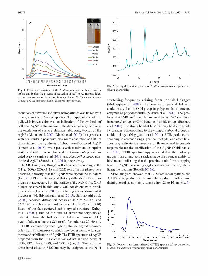
SEM analyses showed that C. tomentosum-synthesizedAgNPs were predominantly irregular in shape, with a largedistribution of sizes, mainly ranging from 20 to 40 nm (Fig. 4).
0
0.5
1
1.5
2
2.5
3
200
226
252
278
304
330
356
382
408
434
460
486
512
538
564
590
616
642
668
694
720
746
772
798
120min90min60min30min15min5min
Absorba
nce
Wavenumber (nm)
a bc
Fig. 1 Chromatic variation of the Codium tomentosum leaf extract abefore and b after the process of reduction of Ag+ to Ag nanoparticles.c UV-visualization of the absorption spectra of Codium tomentosum-synthesized Ag nanoparticles at different time intervals
20 40 60 80
200
400
600
800
1000
1200
1400
Inte
nsity
2 Theta
Fig. 2 X-ray diffraction pattern of Codium tomentosum-synthesizedsilver nanoparticles
Fig. 3 Fourier transform infrared (FTIR) spectra of vacuum-driedCodium tomentosum-synthesized silver nanoparticles
16676 Environ Sci Pollut Res (2016) 23:16671–16685
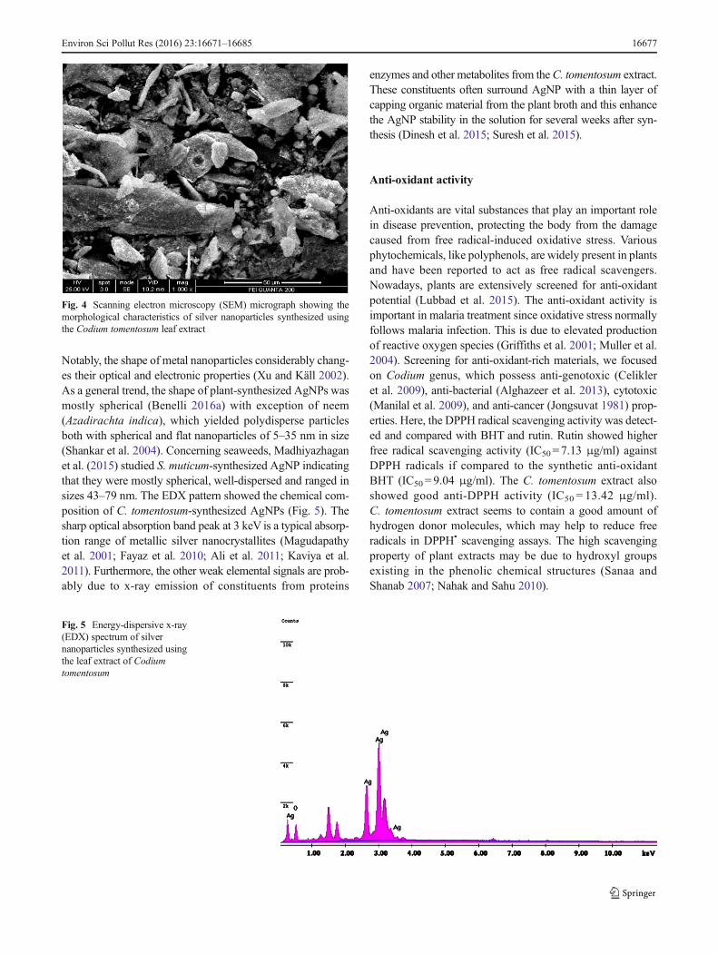
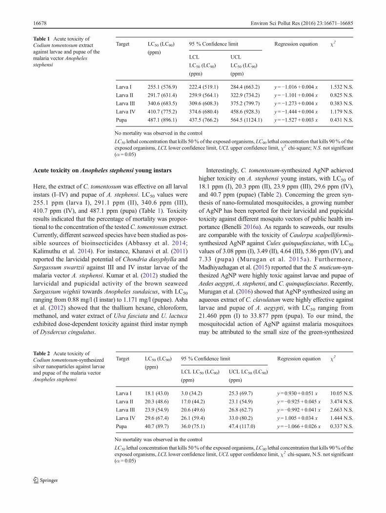
Notably, the shape of metal nanoparticles considerably chang-es their optical and electronic properties (Xu and Käll 2002).As a general trend, the shape of plant-synthesized AgNPs wasmostly spherical (Benelli 2016a) with exception of neem(Azadirachta indica), which yielded polydisperse particlesboth with spherical and flat nanoparticles of 5–35 nm in size(Shankar et al. 2004). Concerning seaweeds, Madhiyazhaganet al. (2015) studied S. muticum-synthesized AgNP indicatingthat they were mostly spherical, well-dispersed and ranged insizes 43–79 nm. The EDX pattern showed the chemical com-position of C. tomentosum-synthesized AgNPs (Fig. 5). Thesharp optical absorption band peak at 3 keV is a typical absorp-tion range of metallic silver nanocrystallites (Magudapathyet al. 2001; Fayaz et al. 2010; Ali et al. 2011; Kaviya et al.2011). Furthermore, the other weak elemental signals are prob-ably due to x-ray emission of constituents from proteins
enzymes and other metabolites from theC. tomentosum extract.These constituents often surround AgNP with a thin layer ofcapping organic material from the plant broth and this enhancethe AgNP stability in the solution for several weeks after syn-thesis (Dinesh et al. 2015; Suresh et al. 2015).
Anti-oxidant activity
Anti-oxidants are vital substances that play an important rolein disease prevention, protecting the body from the damagecaused from free radical-induced oxidative stress. Variousphytochemicals, like polyphenols, are widely present in plantsand have been reported to act as free radical scavengers.Nowadays, plants are extensively screened for anti-oxidantpotential (Lubbad et al. 2015). The anti-oxidant activity isimportant in malaria treatment since oxidative stress normallyfollows malaria infection. This is due to elevated productionof reactive oxygen species (Griffiths et al. 2001; Muller et al.2004). Screening for anti-oxidant-rich materials, we focusedon Codium genus, which possess anti-genotoxic (Celikleret al. 2009), anti-bacterial (Alghazeer et al. 2013), cytotoxic(Manilal et al. 2009), and anti-cancer (Jongsuvat 1981) prop-erties. Here, the DPPH radical scavenging activity was detect-ed and compared with BHT and rutin. Rutin showed higherfree radical scavenging activity (IC50= 7.13 μg/ml) againstDPPH radicals if compared to the synthetic anti-oxidantBHT (IC50 = 9.04 μg/ml). The C. tomentosum extract alsoshowed good anti-DPPH activity (IC50 = 13.42 μg/ml).C. tomentosum extract seems to contain a good amount ofhydrogen donor molecules, which may help to reduce freeradicals in DPPH• scavenging assays. The high scavengingproperty of plant extracts may be due to hydroxyl groupsexisting in the phenolic chemical structures (Sanaa andShanab 2007; Nahak and Sahu 2010).
Fig. 4 Scanning electron microscopy (SEM) micrograph showing themorphological characteristics of silver nanoparticles synthesized usingthe Codium tomentosum leaf extract
Fig. 5 Energy-dispersive x-ray(EDX) spectrum of silvernanoparticles synthesized usingthe leaf extract of Codiumtomentosum
Environ Sci Pollut Res (2016) 23:16671–16685 16677
Acute toxicity on Anopheles stephensi young instars
Here, the extract of C. tomentosum was effective on all larvalinstars (I–IV) and pupae of A. stephensi. LC50 values were255.1 ppm (larva I), 291.1 ppm (II), 340.6 ppm (III),410.7 ppm (IV), and 487.1 ppm (pupa) (Table 1). Toxicityresults indicated that the percentage of mortality was propor-tional to the concentration of the testedC. tomentosum extract.Currently, different seaweed species have been studied as pos-sible sources of bioinsecticides (Abbassy et al. 2014;Kalimuthu et al. 2014). For instance, Khanavi et al. (2011)reported the larvicidal potential of Chondria dasyphylla andSargassum swartzii against III and IV instar larvae of themalaria vector A. stephensi. Kumar et al. (2012) studied thelarvicidal and pupicidal activity of the brown seaweedSargassum wightii towards Anopheles sundaicus, with LC50
ranging from 0.88 mg/l (I instar) to 1.171 mg/l (pupae). Ashaet al. (2012) showed that the thallium hexane, chloroform,methanol, and water extract of Ulva fasciata and U. lactucaexhibited dose-dependent toxicity against third instar nymphof Dysdercus cingulatus.
Interestingly, C. tomentosum-synthesized AgNP achievedhigher toxicity on A. stephensi young instars, with LC50 of18.1 ppm (I), 20.3 ppm (II), 23.9 ppm (III), 29.6 ppm (IV),and 40.7 ppm (pupae) (Table 2). Concerning the green syn-thesis of nano-formulated mosquitocides, a growing numberof AgNP has been reported for their larvicidal and pupicidaltoxicity against different mosquito vectors of public health im-portance (Benelli 2016a). As regards to seaweeds, our resultsare comparable with the toxicity of Caulerpa scalpelliformis-synthesized AgNP against Culex quinquefasciatus, with LC50
values of 3.08 ppm (I), 3.49 (II), 4.64 (III), 5.86 ppm (IV), and7.33 (pupa) (Murugan et al. 2015a). Furthermore,Madhiyazhagan et al. (2015) reported that the S. muticum-syn-thesized AgNP were highly toxic against larvae and pupae ofAedes aegypti, A. stephensi, and C. quinquefasciatus. Recently,Murugan et al. (2016) showed that AgNP synthesized using anaqueous extract of C. clavulatum were highly effective againstlarvae and pupae of A. aegypti, with LC50 ranging from21.460 ppm (I) to 33.877 ppm (pupa). To our mind, themosquitocidal action of AgNP against malaria mosquitoesmay be attributed to the small size of the green-synthesized
Table 1 Acute toxicity ofCodium tomentosum extractagainst larvae and pupae of themalaria vector Anophelesstephensi
Target LC50 (LC90)
(ppm)
95 % Confidence limit Regression equation χ2
LCL
LC50 (LC90)
(ppm)
UCL
LC50 (LC90)
(ppm)
Larva I 255.1 (576.9) 222.4 (519.1) 284.4 (663.2) y=−1.016 + 0.004 x 1.532 N.S.
Larva II 291.7 (631.4) 259.9 (564.1) 322.9 (734.2) y=−1.101 + 0.004 x 0.825 N.S.
Larva III 340.6 (683.5) 309.6 (608.3) 375.2 (799.7) y=−1.273 + 0.004 x 0.383 N.S.
Larva IV 410.7 (775.2) 374.6 (680.4) 458.6 (928.3) y=−1.444 + 0.004 x 1.179 N.S.
Pupa 487.1 (896.1) 437.5 (766.2) 564.5 (1124.1) y=−1.527 + 0.003 x 0.431 N.S.
No mortality was observed in the control
LC50 lethal concentration that kills 50% of the exposed organisms, LC90 lethal concentration that kills 90% of theexposed organisms, LCL lower confidence limit, UCL upper confidence limit, χ2 chi-square; N.S. not significant(α= 0.05)
Table 2 Acute toxicity ofCodium tomentosum-synthesizedsilver nanoparticles against larvaeand pupae of the malaria vectorAnopheles stephensi
Target LC50 (LC90)
(ppm)
95 % Confidence limit Regression equation χ2
LCL LC50 (LC90)
(ppm)
UCL LC50 (LC90)
(ppm)
Larva I 18.1 (43.0) 3.0 (34.2) 25.3 (69.7) y= 0.930 + 0.051 x 10.05 N.S.
Larva II 20.3 (48.6) 17.0 (44.2) 23.1 (54.9) y=−0.925 + 0.045 x 3.474 N.S.
Larva III 23.9 (54.9) 20.6 (49.6) 26.8 (62.7) y=−0.992 + 0.041 x 2.663 N.S.
Larva IV 29.6 (67.4) 26.1 (59.4) 33.0 (80.2) y= 1.005 + 0.034 x 1.444 N.S.
Pupa 40.7 (89.7) 36.0 (75.1) 47.4 (117.0) y=−1.066 + 0.026 x 0.337 N.S.
No mortality was observed in the control
LC50 lethal concentration that kills 50% of the exposed organisms, LC90 lethal concentration that kills 90% of theexposed organisms, LCL lower confidence limit,UCL upper confidence limit, χ2 chi-square, N.S. not significant(α= 0.05)
16678 Environ Sci Pollut Res (2016) 23:16671–16685
nanoparticles, which allows passage through the insect cuticleand into individual cells where they interfere with molting andother physiological processes (Arjunan et al. 2012; Muruganet al. 2015b; Benelli 2016b).
Copepod predation post-treatmentwith spongeweed-synthesized silver nanoparticles
M. aspericornis adults actively predate A. stephensi young lar-val instars. The predatory efficiency per copepod per day was8.1, 6.5, 1.7, and 0.9 larvae (larva I, II, III, and IV, respectively)(Table 3). Our results are in agreement with previous evidenceson other species. Indeed, adult copepods belonging to otherspecies have been found effective to control young larval instarsof different mosquitoes, including the arbovirus vectors Aedesalbopictus and A. aegypti, while little predation rates have beenobserved against late instar larvae (Kay et al. 1992; Schreiberet al. 1993; Marten et al. 1994; Murugan et al. 2015a, b).
Post-treatment with spongeweed-synthesized AgNP, thepredatory efficiency of a single M. aspericornis per day wascomparable to standard laboratory conditions, i.e., 8.3, 6.6, 1.9,and 1.1 larvae (larva I, II, III, and IV, respectively) (Table 4).Also, in this experiment, copepods were effective predators offirst and second instars of mosquitoes, while they are not active
control agents against late larval instars. The higher predationrates of M. aspericornis against A. stephensi young larvae maybe due to the impact of nanoparticles treatment on the preyorganism, since they can affect the physiological and metabolicactivities, thus motility. This has been hypothesized also byMurugan et al. (2011), reporting higher predation rates of thecopepodM. aspericornis against A. aegypti, post-treatment withneem seed kernel extract. In addition, Murugan et al.(2015a) showed that seaweed-synthesized AgNPs did notreduce the predation of copepod M. longisetus against thefilariasis vector C. quinquefasciatus. Furthermore, Muruganet al. (2015b) showed that very low doses (i.e., 1 ppm) oflemongrass-synthesized gold nanoparticles may help to controlmalaria and dengue vectors boosting early instar mosquitolarvae predation by M. aspericornis. Overall, we hypothesizethat low doses of green-synthesized AgNPs reduce motility ofmosquito larvae, thus enhancing the predation success ofmosquito natural enemies (Murugan et al. 2015c,d,e;Subramaniam et al. 2015).
Anti-plasmodial assays
In Asian traditional medicine, marine plants are used to curemany of the infectious and non infectious diseases
Table 3 Predation of thecopepodMesocyclopsaspericornis against larvae ofAnopheles stephensi
Targetedprey
Consumed preys (n) Totalpredation(n)
Consumedpreys percopepod perday (n)
Control Day 1 Day 2 Day 3 Day 4 Day 5
Larva I 0 89± 1.1 85± 4.7 80 ± 6.6 78 ± 5.0 75 ± 3.8 407 8.1a
Larva II 0 75± 5.9 72± 4.1 65 ± 3.9 60 ± 3.6 55 ± 5.0 327 6.5b
Larva III 0 25± 3.6 21± 4.6 17 ± 3.9 13 ± 2.0 9 ± 1.9 85 1.7c
Larva IV 0 16± 3.6 13± 3.6 10 ± 3.8 6 ± 0.9 3 ± 0.4 48 0.9d
Predation rates are means ± SD of four replicates (10 copepods vs. 100 mosquitoes per replicate)
Control was water without copepods
Within the column, means followed by the same letter(s) are not significantly different (generalized linear model,P< 0.05)
Table 4 Predation of thecopepodMesocyclopsaspericornis against larvae ofAnopheles stephensi after atreatment with seaweed-synthesized silver nanoparticles(1 ppm)
Targetedprey
Consumed preys (n) Totalpredation(n)
Consumedpreys percopepod perday (n)
Control Day 1 Day 2 Day 3 Day 4 Day 5
Larva I 0 93± 6.6 90± 4.1 85 ± 4.2 78 ± 6.09 73± 5.1 419 8.3a
Larva II 0 83± 6.2 68± 8.3 65 ± 4.0 60 ± 5.2 58± 3.0 334 6.6b
Larva III 0 24± 3.5 22± 3.5 19 ± 4.9 17 ± 3.06 13± 2.9 95 1.9c
Larva IV 0 15± 0.9 14± 0.6 13 ± 0.7 8 ± 0.8 5 ± 1.9 55 1.1d
Predation rates are means ± SD of four replicates (10 copepods vs. 100 mosquitoes per replicate)
Control was water without copepods
Within the column, means followed by the same letter(s) are not significantly different (generalized linear model,P< 0.05)
Environ Sci Pollut Res (2016) 23:16671–16685 16679
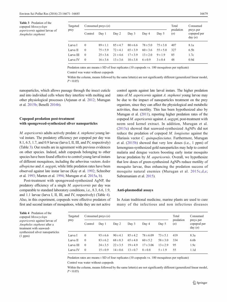
(Bandaranayake 1998). Several seaweeds have shown a widerange of bioactive properties. Recent research elucidates thatplant metabolites are a useful arsenal in the fight against ma-laria. For instance, Ravikumar et al. (2011b) reported thatseaweeds are a good source of compounds which can be usedfor the development of anti-plasmodial drugs. Here, the anti-plasmodial activity of C. tomentosum extract andC. tomentosum-synthesized AgNP was evaluated againstCQ-resistant (CQ-r) and CQ-sensitive (CQ-s) strains ofP. falciparum when compared to chloroquine. C. tomentosumIC50 were 51.34 μg/ml (CQ-s) and 65.17 μg/ml (CQ-r), whileC. tomentosum-synthesized AgNP IC50 were 72.45 μg/ml(CQ-s) and 76.08 μg/ml (CQ-r). IC50 of choloroquine were80.00μg/ml (CQ-s) and 85.00μg/ml (CQ-r) (Fig. 6). In agree-ment with our findings, Ravikumar et al. (2011a) studied thatthe methanolic extracts of two marine plants Chaetomorphaantennina and Aegiceras corniculatum showed maximumsuppression of parasitemia, 63.50 ± 0.408 % at 1.5 mg/mlagainst P. falciparum. Kamaraj et al. (2012) reported that theleaf extract of Eclipta prostrata showed moderateanti-plasmodial activity against P. falciparum with the IC50
value of 30 μg/ml. Recently, Murugan et al. (2015i) studiedthat the anti-plasmodial activity of Senna occidentalis and
Ocimum basilicum against CQ-resistant (CQ-r) and CQ-sensitive (CQ-s) strains of P. falciparum; IC50 ofS. occidentalis were 48.80 μg/ml (CQ-s) and 54.28 μg/ml(CQ-r), while O. basilicum IC50 were 68.14 μg/ml (CQ-s)and 67.27 μg/ml (CQ-r). Furthermore, to the best of ourknowledge, the anti-plasmodial activity of green-synthesizedmetal nanoparticles has been scarcely studied (Benelli 2016a,b; Benelli and Mehlhorn 2016). For example, Murugan et al.(2015h) reported that anti-plasmodial activity of seaweed-synthesized AgNP against CQ-resistant (CQ-r) and CQ-sensitive (CQ-s) strains of P. falciparum, IC50 of U. lactucaextract were 57.26 μg/ml (CQ-s) and 66.36 μg/ml (CQ-r),while U. lactuca-synthesized AgNP IC50 were 76.33 μg/ml(CQ-s) and 79.13 μg/ml (CQ-r). In addition, AgNP synthe-sized using Catharanthus roseus, and Couroupita guianensisare active against blood stages of P. falciparum(Ponarulselvam et al. 2012, 2015; Subramaniam et al.2016b). Notably, the anti-plasmodial property of the above-mentioned plant extracts may be attributed to the presence ofphytochemicals conferring protective and anti-oxidative activ-ity against oxidative stress induced in the host parasitized redblood cells by the malarial parasites (Becker et al. 2004;Nethengwe et al. 2012).
Fig. 6 Growth inhibition ofchloroquine-resistant andchloroquine-sensitive strains ofPlasmodium falciparum post-treatment with chloroquine,C. tomentosum extract andseaweed-synthesized silvernanoparticles
Table 5 Growth inhibitioninduced by Codium tomentosum-synthesized silver nanoparticlesagainst three pathogen bacteria
Treatment (mg/ml) Inhibition zone (mm)
Bacillus subtilis Klebsiella pneumoniae Salmonella typhi
Ag nanoparticles 50 12.42 ± 0.56b 11.22 ± 0.23b 14.64 ± 0.43b
Ag nanoparticles 100 16.80 ± 0.98c 15.48± 0.52c 17.46 ± 0.64c
Ag nanoparticles 150 18.36 ± 1.02d 17.68± 0.28d 20.24 ± 0.84d
Tetracycline 100 5.20 ± 0.78a 6.22± 0.54a 4.48 ± 0.96a
Values are mean± standard deviation of four replicates
Tetracycline was tested as positive control
Growth inhibition zones were not recorded in negative control, where no anti-biotic drugs were administered
Within each column, different letters indicate significant differences (ANOVA, Tukey’s HSD test, P< 0.05)
16680 Environ Sci Pollut Res (2016) 23:16671–16685
Anti-bacterial activity
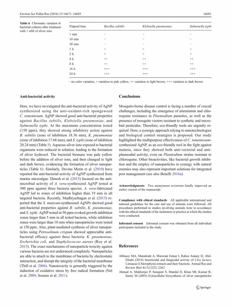
Here, we have investigated the anti-bacterial activity of AgNPsynthesized using the anti-oxidant-rich spongeweedC. tomentosum. AgNP showed good anti-bacterial propertiesagainst Bacillus subtilis, Klebsiella pneumoniae, andSalmonella typhi. At the maximum concentration tested(150 ppm), they showed strong inhibitory action againstB. subtilis (zone of inhibition 18.36 mm), K. pneumoniae(zone of inhibition 17.68 mm), and S. typhi (zone of inhibition20.24 mm) (Table 5). Aqueous silver ions exposed to bacterialorganisms were reduced in solution, leading to the formationof silver hydrosol. The bacterial biomass was pale yellowbefore the addition of silver ions, and then changed to lightand dark brown, evidencing the formation of silver nanopar-ticles (Table 6). Similarly, Devina Merin et al. (2010) havereported the anti-bacterial activity of AgNP synthesized frommarine microalgae. Dinesh et al. (2015) focused on the anti-microbial activity of A. vera-synthesized AgNP tested at300 ppm against three bacteria species. A. vera-fabricatedAgNP led to zones of inhibition higher than 19 mm in alltargeted bacteria. Recently, Madhiyazhagan et al. (2015) re-ported that the S. muticum-synthesized AgNPs showed goodanti-bacterial properties against B: subtilis, K. pneumoniae,and S. typhi. AgNP tested at 50 ppm evoked growth inhibitionzones larger than 5 mm in all tested bacteria, while inhibitionzones were larger than 10 mmwhen nanoparticles were testedat 150 ppm. Also, plant-mediated synthesis of silver nanopar-ticles using Petroselinum crispum showed appreciable anti-bacterial efficacy against three bacteria: K. pneumoniae,Escherichia coli, and Staphylococcus aureus (Roy et al.2015). The exact mechanisms of nanoparticle toxicity againstvarious bacteria are not understood completely. Nanoparticlesare able to attach to the membrane of bacteria by electrostaticinteraction, and disrupt the integrity of the bacterial membrane(Thill et al. 2006). Nanotoxicity is generally triggered by theinduction of oxidative stress by free radical formation (Nelet al. 2009; Soenen et al. 2011).
Conclusions
Mosquito-borne disease control is facing a number of crucialchallenges, including the emergence of artemisinin and chlo-roquine resistance in Plasmodium parasites, as well as thepresence of mosquito vectors resistant to synthetic and micro-bial pesticides. Therefore, eco-friendly tools are urgently re-quired. Here, a synergic approach relying to nanotechnologiesand biological control strategies is proposed. Our studyhighlighted the multipurpose effectiveness of C. tomentosum-synthesized AgNP, as an eco-friendly tool in the fight againstmalaria, since they showed both anti-vectorial and anti-plasmodial activity, even on Plasmodium strains resistant tochloroquine. Other bioactivities, like bacterial growth inhibi-tion and the employ of nanoparticles in synergy with naturalenemies may also represent important solutions for integratedpest management (see also Benelli 2016a).
Acknowledgments Two anonymous reviewers kindly improved anearlier version of the manuscript.
Compliance with ethical standards All applicable international andnational guidelines for the care and use of animals were followed. Allprocedures performed in studies involving animals were in accordancewith the ethical standards of the institution or practice at which the studieswere conducted.
Informed consent Informed consent was obtained from all individualparticipants included in the study.
References
Abbassy MA, Mamdouh A, Marzouk Entsar I, Rabea Amany D, Abd-Elnabi (2014) Insecticidal and fungicidal activity of Ulva lactucaLinnaeus (Chlorophyta) extracts and their fractions. Annual Res andReview Biol 4(13):2252–2262
Ahmad A, Mukherjee P, Senapati S, Mandal D, Khan MI, Kumar R,Sastry M (2003) Extracellular biosynthesis of silver nanoparticles
Table 6 Chromatic variation inbacterial cultures after treatmentwith 1 mM of silver ions
Elapsed time Bacillus subtilis Klebsiella pneumoniae Salmonella typhi
1 min − − −10 min − − −30 min − − −1 h + + +
2 h + + +
4 h ++ ++ ++
8 h ++ ++ ++
16 h +++ +++ +++
24 h +++ +++ +++
− no color variation, + variation to pale yellow, ++ variation to light brown, +++ variation to dark brown
Environ Sci Pollut Res (2016) 23:16671–16685 16681
using the fungus Fusarium oxysporum. Colloids Surf B:Biointerfaces 28:313–318
Alghazeer R, Whida F, Abduelrhman E, Gammoudi F, Naili M (2013)In vitro antibacterial activity of alkaloid extracts from green, red andbrown macroalgae from western coast of Libya. Afr J Biotechnol12(51):7086–7091
Ali DM, Sasikala M, Gunasekaran M, Thajuddin N (2011) Biosynthesisand characterization of silver nanoparticles using marine cyanobac-terium, Oscillatoria willei. Digest J Nanomater Biostruct 6(2):385–390
AmerA,Mehlhorn H (2006a) Repellency effect of forty-one essential oilsagainst Aedes, Anopheles and Culex mosquitoes. Parasitol Res 99:478–490
Amer A, Mehlhorn H (2006b) The sensilla of Aedes and Anophelesmosquitoes and their importance in repellency. Parasitol Res 99:491–499
Amer A, Mehlhorn H (2006c) Larvicidal effects of various essential oilsagainst Aedes, Anopheles, and Culex larvae (Diptera, Culicidae).Parasitol Res 99:466–472
Amer A,Mehlhorn H (2006d) Persistency of larvicidal effects of plant oilextracts under different storage conditions. Parasitol Res 99:473–477
Arjunan NK, Murugan K, Rejeeth C, Madhiyazhagan P, Barnard DR(2012) Green synthesis of silver nanoparticles for the control ofmosquito vectors of malaria, filariasis and dengue. Vector BorneZoontic Dis 12:262–268
Asha A, Martin Rathi J, Patric Raja D, Sahayaraj K (2012) Biocidalactivity of twomarine green algal extracts against third instar nymphof Dysdercus cingulatus (Fab) (Hemiptera: Pyrrhocoridae). JBiopest 5:129–134
Aziz AT, Mahyoub JA, Rehman H, Saggu S,Murugan K, PanneerselvamC, Nicoletti M, Wei H, Canale A, Benelli G (2016) Insecticidesusceptibility in larval populations of the West Nile vector Culexpipiens L. (Diptera:Culicidae) in Saudi Arabia. Asia Pac J TropBiomed doi:10.1016/j.apjtb.2015.12.017
Azizullah A, Rehman ZU, Ali I, Murad W, Muhammad N, Ullah W,Hader D-P (2014) Chlorophyll derivatives can be an efficient weap-on in the fight against dengue. Parasitol Res 113:4321–4326
Bagavan A, Rahuman AA, Kaushik NK, Sahal D (2011) In vitro antima-larial activity of medicinal plant extracts against Plasmodiumfalciparum. Parasitol Res 108:15–22
Bandaranayake WM (1998) Traditional and medicinal uses of man-groves. Mang Salt Marsh 2:33–148
Bankara A, Joshi B, Ravi Kumara A, Zinjardea S (2010) Banana peelextract mediated novel route for the synthesis of silver nanoparticles.J Colloids Surf A: Physicochem Eng 368:58–63
Bar H, Dipak Bhui KR, Gobinda Sahoo P, Sarkar P (2009) Green syn-thesis of silver nanoparticles using latex of Jatropha curcas.Colloids Surf A Physicochem Eng Asp 339:134–139
Becker K, Tilley L, Vennerstrom JL, Roberts D, Rorerson S, Ginsburg H(2004) Oxidative stress in malaria parasite-infected erythrocytes:host-parasite interactions. Int J Parasitol 34:163–189
Benelli G (2015a) Research in mosquito control: current challenges for abrighter future. Parasitol Res 114:2801–2805
Benelli G (2015b) Plant-borne ovicides in the fight against mosquitovectors of medical and veterinary importance: a systematic review.Parasitol Res 114:3201–3212
Benelli G (2016a) Plant-mediated biosynthesis of nanoparticles as anemerging tool against mosquitoes of medical and veterinary impor-tance: a review. Parasitol Res 115: 23-34
Benelli G (2016b) Plant-mediated synthesis of nanoparticles: a newer andsafer tool against mosquito-borne diseases? Asia Pacif J TropBiomed. doi:10.1016/j.apjtb.2015.10.015
Benelli G, Mehlhorn H (2016) Declining malaria, rising dengue and Zikavirus: insights for mosquito vector control. Parasitol Res doi:10.1007/s00436-016-4971-z
Benelli G, Bedini S, Cosci F, Toniolo C, Conti B, Nicoletti M (2015a)Larvicidal and ovi-deterrent properties of neem oil and fractionsagainst the filariasis vector Aedes albopictus (Diptera: Culicidae):a bioactivity survey across production sites. Parasitol Res 114:227–236
Benelli G, Bedini S, Flamini G, Cosci F, Cioni PL, Amira S, Benchikh F,Laouer H, Di Giuseppe G, Conti B (2015b) Mediterranean essentialoils as effective weapons against the West Nile vector Culex pipiensand the Echinostoma intermediate host Physella acuta: what hap-pens around? An acute toxicity survey on non-target mayflies.Parasitol Res. doi:10.1007/s00436-014-4267-0
Benelli G, Murugan K, Panneerselvam C, Madhiyazhagan P, Conti B,Nicoletti M (2015c) Old ingredients for a new recipe? Neemcake, alow-cost botanical by-product in the fight against mosquito-bornediseases. Parasitol Res 114:391–397
Benelli G, Lo Iacono A, Canale A, Mehlhorn H (2016) Mosquito vectorsand the spread of cancer: an overlooked connection? Parasitol Res.doi:10.1007/s00436-016-5037-y
Blois MS (1958) Antioxidants determination by the use of a stable freeradical. Nat 4617:1199–1200
Bowatte G, Perera P, Senevirathne G, Meegaskumbura S,Meegaskumbura M (2013) Tadpoles as dengue mosquito (Aedesaegypti) egg predators. Biol Control 67:469–474
Celikler S, Vatan O, Yildiz G, Bilaloglu R (2009) Evaluation of anti-oxidative, genotoxic and antigenotoxic potency of Codiumtomentosum Stackhouse ethanolic extract in human lymphocytesin vitro. Food Chem Toxicol 47(4):796–801
Chandramohan B, Murugan K, Panneerselvam C, Madhiyazhagan P,Chandirasekar R, Dinesh D, Mahesh Kumar P, Kovendan K,Suresh U, Subramaniam J, Rajaganesh R, Aziz AT, Syuhei B,Saleh Alsalhi M, Devanesan S, Nicoletti M, Wei H, Benelli G(2016) Characterization and mosquitocidal potential of neem cake-synthesized silver nanoparticles: genotoxicity and impact on preda-tion efficiency of mosquito natural enemies. Parasitol Res 115:1015–1025
Chang KH, Hanazato T (2003) Vulnerability of cladoceran species topredation by the copepod Mesocyclops leuckarti: laboratory obser-vations on the behavioral interaction between predator and prey.Freshw Biol 48:476–484
Devina Merin D, Prakash S, Valentine Bhimba B (2010) Antibacterialscreening of silver nanoparticles synthesized by marine micro alga.Asian Pacific J Trop Med 3:797–799
Dinesh D,Murugan K, Madhiyazhagan P, PanneerselvamC, Nicoletti M,Jiang W, Benelli G, Chandramohan B, Suresh U (2015)Mosquitocidal and antibacterial activity of green-synthesized silvernanoparticles from Aloe vera extracts: towards an effective toolagainst the malaria vector Anopheles stephensi? Parasitol Res 114:1519–29
Dubey M, Bhadauria S, Kushwah BS (2009) Green synthesis ofnanosilver particles from extract of Eucalyptus hybrida (Safeda)leaf. Dig J Nanomater Biostruct 4(3):537–543
Fayaz AM, Balaji K, Girilal MYR, Kalaichelvan PT, Venketesan R(2010) Biogenic synthesis of silver nanoparticles and their synergis-tic effect with antibiotics: a study against gram-positive and gramnegative bacteria. Nanomed Nanotechnol Biol Med 6:103–109
Finney DJ (1971) Probit analysis. Cambridge University, London, pp 68–78
Govindarajan M, Benelli G (2016) α-humulene and β-elemene fromSyzygium zeylanicum (Myrtaceae) essential oil: highly effectiveand eco-friendly larvicides against Anopheles subpictus, Aedesalbopictus and Culex tritaeniorhynchus (Diptera: Culicidae).. doi:10.1007/s00436-016-5025-2, Parasitol Res
Govindarajan M, Rajeswary M, Veerakumar K, Muthukumaran U, HotiSL, Mehlhorn H, Barnard DR, Benelli G (2016) Novel synthesis ofsilver nanoparticles using Bauhinia variegata: a recent eco-friendlyapproach for mosquito control. Parasitology Research 115:723–733
16682 Environ Sci Pollut Res (2016) 23:16671–16685
Griffiths MJ, Ndungu F, Baird KL, Muller DPR, Marsh K, Charles RJ,Newton C (2001) Oxidative stress and erythrocyte damage inKenyan children with severe Plasmodium falciparum malaria. Br JHaematol 113:486–491
Haverkamp RG (2010) Ten years of nanoparticle research in Australiaand New Zealand. Part Sci Technol 28:1–40
Hemingway J, Ranson H (2000) Insecticide resistance in insect vectors ofhuman disease. Annu Rev Entomol 45:371–391
Jaganathan A, Murugan K, Panneerselvam C, Madhiyazhagan P, DineshD, Vadivalagan C, Aziz AT, Chandramohan B, Suresh U,Rajaganesh R, Subramaniam J, Nicoletti M, Higuchi A, AlarfajAA, Munusamy MA, Kumar S, Benelli G (2016) Earthworm-mediated synthesis of silver nanoparticles: a potent tool against he-patocellular carcinoma, pathogenic bacteria, Plasmodium parasitesand malaria mosquitoes. Parasitol Int 65:276–284
Jongsuvat Y (1981) Investigation of anticancer from Acanthus ilicifolius.Dissertation, Chulalongkorn University, Thailand, M.Sc
Kalimuthu K, Lin SM, Tseng LC, Murugan K, Hwang J-S (2014)Bioefficacy potential of seaweed Gracilaria firma with copepod,Megacyclops formosanus for the control larvae of dengue vectorAedes aegypti. Hydrobiologia 741:113–123
Kamaraj C, Kaushik NK, RahumanAA,Mohanakrishnan D, Bagavan A,Elango G, Zahir AA, Santhoshkumar T,Marimuthu S, Jayaseelan C,Kirthi AV, Rajakumar G, Velayutham K, Sahal D (2012)Antimalarial activities of medicinal plants traditionally used in thevillages of Dharmapuri regions of South India. J Ethnopharmacol141(3):796–802
Kaviya S, Santhanalakshmi J, Viswanathan B, Muthumary J, SrinivasanK (2011) Biosynthesis of silver nanoparticles using Citrus sinensispeel extract and its antibacterial activity. Spectrochim Acta Part A:Mol Biomol Spectrosc 79:594–598
Kay BH, Cabral CP, Sleigh AC, Brown MD, Ribeiro ZM, VasconcelosAW (1992) Laboratory evaluations of Mesocyclops (Cyclopoida:Copepoda) for mosquito control. J Med Entomol 29:599–602
Khanavi M, Toulabi PB, Abai MR, Sadati N, Hadjiakhoondi F,Hadjiakhoondi A, Vatandoost H (2011) Larvicidal activity of marinealgae, Sargassum swartzii andChondria dasyphylla, against malariavector Anopheles stephensi. J Vector Borne Dis 48:241–244
Kong H, Jang J (2006) One-step fabrication of silver nanoparticle em-bedded polymer nanofibers by radical-mediated dispersion poly-merization. Chem Comm 8:3010–3012
Kosiyachinda P, Bhumiratana A, Kittayapong P (2003) Enhancement ofthe efficacy of a combination of Mesocyclops aspericornis andBacillus thuringiensis var. israelensis by community-based productsin controlling Aedes aegypti larvae in Thailand. Am J TropMedHyg69:206–212
Kovendan K, Murugan K, Vincent S, Barnard DR (2012) Studies onlarvicidal and pupicidal activity of Leucas aspera Willd(Lamiaceae) and bacterial insecticide, Bacillus sphaericus againstmalarial vector Anopheles stephensi Liston (Diptera: Culicidae).Parasitol Res 110:195–203
Kumar R, Hwang JS (2006) Larvicidal efficiency of aquatic predators: aperspective for mosquito biocontrol. Zool Stud 45:447–466
Kumar KP, Murugan K, Kovendan K, Kumar AN, Hwang JS, BarnardDR (2012) Combined effect of seaweed (Sargassum wightii) andBacillus thuringiensis var. israelensis on the coastal mosquitoAnopheles sundaicus, in Tamil Nadu India. Sci Asia 38:141–146
Kumar PM, Murugan K, Madhiyazhagan P, Kovendan K, Amerasan D,Chandramohan B, Dinesh D, Suresh U, Nicoletti M, Alsalhi MS,Devanesan S, Wei H, Kalimuthu K, Hwang J-S, Lo Iacono A,Benelli G (2016) Biosynthesis, characterization, and acute toxicityof Berberis tinctoria-fabricated silver nanoparticles against theAsian tiger mosquito, Aedes albopictus, and the mosquito predatorsToxorhynchites splendens and Mesocyclops thermocyclopoides.Parasitol Res 115(2):751–759
Lubbad MY, Al-Quraishy S, Dkhil MA (2015) Antimalarial and antiox-idant activities of Indigofera oblongifolia onPlasmodium chabaudi-induced spleen tissue injury in mice. Parasitol Res 114:3431–3438
Madhiyazhagan P, Murugan K, Naresh Kumar A, Nataraj T, Dinesh D,Panneerselvam C, Subramaniam J, Mahesh Kumar P, Suresh U,Roni M, Nicoletti M, Alarfaj AA, Higuchi A, Munusamy MA,Benelli G (2015) Sargassum muticum-synthesized silver nanoparti-cles: an effective control tool against mosquito vectors and bacterialpathogens. Parasitol Res. doi:10.1007/s00436-015-4671-0
Magudapathy P, Gangopadhyay P, Panigrahi BK, Nair KGM, Dhara S(2001) Electrical transport studies of Ag nanoclusters embedded inglass matrix. Physica 299:142–146
Manilal A, Sujith S, Seghal Kiran G, Selvin J, Shakir C, Gandhimathi R,Nataraja Panikkar MV (2009) Biopotentials of seaweeds collectedfrom southwest coast of India. J Mar Sci Technolo 17(1):67–73
Manrique-Saide PS, Ibanez-Bernal D-GH, Parra Tabla V (1998)Mesocyclops longisetus effects on survivorship of Aedes aegyptiimmature stages in car tyres. Med Vet Entomol 12:386–390
Marten GG (2000) Dengue hemorrhagic fever, mosquitoes, and cope-pods. J Policy Studies (Japan) 9:131–141
MartenGG, Bordes ES, NguyenM (1994) Use of cyclopoid copepods formosquito control. Hydrobiologia 292 293:491–496
Mehlhorn H (2008) Encyclopedia of parasitology, 3rd edn. Springer,Heidelberg
Mehlhorn H, Al-Rasheid KA, Al-Quraishy S, Abdel-Ghaffar F (2012)Research and increase of expertise in arachno-entomology are ur-gently needed. Parasitol Res 110:259–265
Mukherjee P, Roy M, Mandal BP, Dey GK, Mukherjee PK, Ghatak J,Tyagia K, Kale SP (2008) Green synthesis of highly stabilized nano-crystalline silver particles by a non-pathogenic and agriculturallyimportant fungus T. asperellum. Nanotechnol 19:075103
Muller FL, Liu Y, Van Remmen H (2004) Complex III releases superox-ide to both sides of the inner mitochondrial membrane. J Biol Chem279:49064–49073
Murugan K, Hwang SJ, Kovendan K, Kumar KP, Vasugi C, Kumar AN(2011) Use of plant products and copepods for control of the denguevector, Aedes aegypti. Hydrobiologia 666:331–338
Murugan K, Benelli G, Suganya A, Dinesh D, Panneerselvam C,Nicoletti M, Hwang JS, Mahesh Kumar P, Subramaniam J, SureshU (2015a) Toxicity of seaweed-synthesized silver nanoparticlesagainst the filariasis vector Culex quinquefasciatus and its impacton predation efficiency of the cyclopoid crustacean Mesocyclopslongisetus. Parasitol Res 114:2243-2253
Murugan K, Benelli G, Panneerselvam C, Subramaniam J, Jeyalalitha T,Dinesh D, Nicoletti M, Hwang JS, Suresh U, Madhiyazhagan P(2015b) Cymbopogon citratus-synthesized gold nanoparticles boostthe predation efficiency of copepod Mesocyclops aspericornisagainst malaria and dengue mosquitoes. Exp Parasitol 153:129–138
Murugan K, Priyanka V, Dinesh D, Madhiyazhagan P, Panneerselvam C,Subramaniam J, Suresh U, Chandramohan B, Roni M, Nicoletti M,Alarfaj AA, Higuchi A, Munusamy MA, Khater HF, Messing RH,Benelli G (2015c) Enhanced predation by Asian bullfrog tadpoles,Hoplobatrachus tigerinus, against the dengue vector Aedes aegyptiin an aquatic environment treated with mosquitocidal nanoparticles.Parasitol Res 114:3601-3610
Murugan K, Dinesh D, Jenil Kumar P, PanneerselvamC, Subramaniam J,Madhiyazhagan P, Suresh U, Nicoletti M, Alarfaj AA, MunusamyMA, Higuchi A, Mehlhorn H, Benelli G (2015d) Datura metel-synthesized silver nanoparticles magnify predation of dragonflynymphs against the malaria vector Anopheles stephensi. ParasitolRes 114:4645-4654.
Murugan K, Venus JSE, Panneerselvam C, Bedini S, Conti B, NicolettiM, Kumar Sarkar S, Hwang JS, Subramaniam J, Madhiyazhagan P,Mahesh Kumar P, Dinesh D, Suresh U, Benelli G (2015e)Biosynthesis, mosquitocidal and antibacterial properties ofToddalia asiatica-synthesized silver nanoparticles: do they impact
Environ Sci Pollut Res (2016) 23:16671–16685 16683
predation of guppy Poecilia reticulata against the filariasis mosquitoCulex quinquefasciatus? Environ Sci Pollut Res 2:17053-17064
Murugan K, Dinesh D, Paulpandi M, Dakhellah Meqbel Althbyani A,Subramaniam J, Madhiyazhagan P, Wang L, Suresh U, MaheshKumar P, Mohan J, Rajaganesh R, Wei H, Kalimuthu K, ParajuleeMN, Mehlhorn H, Benelli G (2015f) Nanoparticles in the fightagainst mosquito-borne diseases: bioactivity of Bruguieracylindrica-synthesized nanoparticles against dengue virus DEN-2(in vitro) and its mosquito vector Aedes aegypti (Diptera:Culicidae). Parasitol Res 114:4349–4361
Murugan K, Aamina LM, Panneerselvam C, Dinesh D, Suresh U,Subramaniam J, Madhiyazhagan P, Hwang JS, Wang L, NicolettiM, Benelli G (2015g) Aristolochia indica green-synthesized silvernanoparticles: a sustainable control tool against the malaria vectorAnopheles stephensi? Res Vet Sci 102:127-135
Murugan K, Samidoss CM, Panneerselvam C, Higuchi A, Roni M,Suresh U, Chandramohan B, Subramaniam J, Madhiyazhagan P,Dinesh D, Rajaganesh R, Alarfaj AA, Nicoletti M, Kumar S, WeiH, Canale A, Mehlhorn H, Benelli G (2015h) Seaweed-synthesizedsilver nanoparticles: an eco-friendly tool in the fight againstPlasmodium falciparum and its vector Anopheles stephensi?Parasitol Res 114(11):4087-97
Murugan K, Aarthi N, Kovendan K, Panneerselvam C,Chandramohan B,Mahesh Kumar P, Amerasan D, Paulpandi M,Chandirasekar R,Dinesh D, Suresh U, Subramaniam J, Higuchi A, Alarfaj AA,Nicoletti M, Mehlhorn H, Benelli G (2015i) Mosquitocidal andantiplasmodial activity of Senna occidentalis (Cassiae) andOcimum basilicum (Lamiaceae) from Maruthamalai hills againstAnopheles stephensi and Plasmodium falciparum. Parasitol Res114(10):3657-64
Murugan K, Aruna P, Panneerselvam C,Madhiyazhagan P, Paulpandi M,Subramaniam J, Rajaganesh R, Wei H, Saleh Alsalhi M, DevanesanS, Nicoletti M, Syuhei B, Canale A, Benelli G (2016) Fightingarboviral diseases: low toxicity on mammalian cells, dengue growthinhibition (in vitro) and mosquitocidal activity of Centrocerasclavulatum-synthesized silver nanoparticles. Parasitol Res 115:651–662
Nabikhan A, Kandasamy K, Raj A, Alikunhi NM (2010) Synthesis ofantimicrobial silver nanoparticles by callus and leaf extracts fromsaltmarsh plant, Sesuvium portulacastrum (L). Colloids Surf B:Biointer 79:488–493
Nagajyothi PC, Sreekanth TVM, Lee JL, Lee KD (2014) Mycosynthesis:antibacterial, antioxidant and antiproliferative activities of silvernanoparticles synthesized from Inonotus obliquus (Chaga mush-room) extract. J Photochem Photobiol B Biol 130:299–304
Nahak G, Sahu RK (2010) In vitro antioxidative activity of Azadirachtaindica and Melia azedarach leaves by DPPH scavenging assay.Nature and Sci 8(4):22–28
Nel AE et al (2009) Understanding biophysicochemical interactions at thenano-bio interface. Nat Mater 8:543–557
NethengweMF, Opoku AR, Dludla PV, Madida KT, Shonhai A, Smith P,Singh M (2012) Larvicidal, antipyretic and antiplasmodial activityof some Zulu medicinal plants. J Med Plants Res 6(7):1255–1262
Panneerselvam C, Murugan K, Roni M, Aziz AT, Suresh U, RajaganeshR, Madhiyazhagan P, Subramaniam J, Dinesh D, Nicoletti M,Higuchi A, Alarfaj AA, Munusamy MA, Kumar S, Desneux N,Benelli G (2016) Fern-synthesized nanoparticles in the fight againstmalaria: LC/MS analysis of Pteridium aquilinum leaf extract andbiosynthesis of silver nanoparticles with high mosquitocidal andantiplasmodial activity. Parasitol Res 115:997–1013
Pavela R (2015a) Essential oils for the development of eco-friendly mos-quito larvicides: a review. Ind Crops Prod 76:174–187
Pavela R (2015b) Acute toxicity and synergistic and antagonistic effectsof the aromatic compounds of some essential oils against Culexquinquefasciatus Say larvae. Parasitol Res doi:10.1007/s00436-015-4614-9
Petit C, Lixon P, Pileni MP (1993) In situ synthesis of silver nanoclusterin AOT reverse micelles. J Phys Chem 97:12974–12983
Ponarulselvam S, PanneerselvamC,Murugan K, Aarthi N, Kalimuthu K,Thangamani S (2012) Synthesis of silver nanoparticles using leavesof Catharanthus roseus Linn. G. Donn and their antiplasmodialactivities. Asian Pacific J Trop Biomed 2:574–580
Rajkumar S, Jebanesan A (2008) Bioactivity of flavonoid compounds fromPoncirus trifoliata L. (Family: Rutaceae) against the dengue vector,Aedes aegypti L. (Diptera: Culicidae). Parasitol Res 104(1):19–25
Ravikumar S, Ali MS, Beula JM (2011a) Mosquito larvicidal efficacy ofseaweed extracts against dengue vector of Aedes aegypti. Asia Pac JTrop Biomed 1:S143–S146
Ravikumar S, Ramanathan G, Inbaneson SJ, Ramu A (2011a)Antiplasmodial activity of two marine polyherbal preparations fromChaetomorpha antennina and Aegiceras corniculatum againstPlasmodium falciparum. Parasitol Res 108:107-113
Rawlins SC, Martinez R, Wiltshire S, Clarke D, Prabhakar P, Spinks M(1997) Evaluation of Caribbean strains of Macrocyclops andMesocyclops (Cyclopoida: Cyclopidae) as biological control toolsfor the dengue vector Aedes aegypti. J AmMosq Control Assoc 13:18–23
Roni M, Murugan K, Panneerselvam C, Subramaniam J, Nicoletti M,Madhiyazhagan P, Dinesh D, Suresh U, Khater HF, Wei H, CanaleA, Alarfaj AA, Munusamy MA, Higuchi A, Benelli G (2015)Characterization and biotoxicity of Hypnea musciformis-synthe-sized silver nanoparticles as potential eco-friendly control toolagainst Aedes aegypti and Plutella xylostella. Ecotoxicol EnvironSaf 121:31–38
Roy K, Sarkar CK, Ghosh CK (2015) Plant-mediated synthesis of silvernanoparticles using parsley (Petroselinum crispum) leaf extract:spectral analysis of the particles and antibacterial study. ApplNanosci (2015) 5:945–951
Sanaa M, Shanab S (2007) Antioxidant and antibiotic activities of somesea weeds (Egyptian isolates). Int J Agr Biol 9(2):220–225
Sathyavathi R, Balamurali Krishna M, Venugopal Rao S, Saritha R,Narayana Rao D (2010) Biosynthesis of silver nanoparticles usingCoriandrum sativum leaf extract and their application in nonlinearoptics. Adv Sci Lett 3:1–6
Schaper S (1999) Evaluation of Costa Rican copepods (Crustacea:Eudecapoda) for larval Aedes aegypti control with special referenceto Mesocyclops thermocyclopoides. J Am Mosq Control Assoc 15:510–519
Schreiber ET, Thrner WL, Lopez AM, Hallmon CE, Marten GG (1993)Evaluation of two cyclopoid copepods for Aedes albopictus controlin tires in the panhandle of Florida at low introduction rates. J FlaMosq Control Assoc 64:7317
Scrinis G, Lyons K (2007) The emerging nano-corporate paradigm: nano-technology and the transformation of nature, food and agri-foodsystems. Int J Sociol Food Agric 15(2):22–44
Semmler M, Abdel-Ghaffar F, Al-Rasheid KAS, Mehlhorn H (2009)Nature helps: from research to products against blood sucking ar-thropods. Parasitol Res 105:1483–1487
Shankar S, Rai S, Ahmad A, Sastry M (2004) Rapid synthesis of Au, Ag,and bimetallic Au core-Ag shell nanoparticles using Neem(Azadirachta indica) leaf broth. J Colloid Interface Sci 275:496–502
Shrivastava S, Dash D (2010) Label-free colorimetric estimation of pro-teins using nanoparticles of silver. Nano-Micro Lett 2:164–168
Smilkstein M, Sriwilaijaroen N, Kelly JX, Wilairat P, Riscoe M (2004)Simple and inexpensive fluorescence-based technique for highthrough put antimalarial drug screening. Antimicrob AgentsChemother 48:1803–6
Soenen SJ et al (2011) Cellular toxicity of inorganic nanoparticles: com-mon aspects and guidelines for improved nanotoxicity evaluation.Nano Today 6:446–465
Subramaniam J, Murugan K, Panneerselvam C, Kovendan K,Madhiyazhagan P, Mahesh Kumar P, Dinesh D, Chandramohan B
16684 Environ Sci Pollut Res (2016) 23:16671–16685
Suresh U, Nicoletti M, Higuchi A, Hwang JS, Kumar S, Alarfaj AA,Munusamy MA, Messing RH (2015) Eco-friendly control of malar-ia and arbovirus vectors using the mosquitofish Gambusia affinisand ultra-low dosages ofMimusops elengi-synthesized silver nano-particles: towards an integrative approach? Environ Sci Pollut Res.22:20067–20083. doi:10.1007/s11356-015-5253-5
Subramaniam J, Murugan K, Panneerselvam C, Kovendan K,Madhiyazhagan P, Mahesh Kumar P, Dinesh D, Chandramohan B,Suresh U, Nicoletti M, Higuchi A, Hwang JS, Kumar S, Alarfaj AA,Munusamy MA, Messing RH, Benelli G (2016a) Eco-friendly con-trol of malaria and arbovirus vectors using the mosquitofishGambusia affinis and ultra-low dosages ofMimusops elengi-synthe-sized silver nanoparticles: towards an integrative approach? EnvironSci Poll Res. doi:10.1007/s11356-015-5253-5
Subramaniam J, Murugan K, Panneerselvam C, Kovendan K,Madhiyazhagan P, Dinesh D, Mahesh Kumar P, Chandramohan B(2016b) Multipurpose effectiveness of Couroupita guianensis-syn-thesized gold nanoparticles: high antiplasmodial potential, field ef-ficacy against malaria vectors and synergy with Aplocheilus lineatuspredators. Environ Sci Poll Res. doi:10.1007/s11356-015-6007-0
Sujitha V, Murugan K, Paulpandi M, Panneerselvam C, Suresh U, RoniM, Nicoletti M, Higuchi A, Madhiyazhagan P, Subramaniam J,Dinesh D, Vadivalagan C, Chandramohan B, Alarfaj AA,Munusamy MA, Barnard DR, Benelli G (2015) Green synthesizedsilver nanoparticles as a novel control tool against dengue virus
(DEN-2) and its primary vector Aedes aegypti. Parasitol Res 114:3315–3325
Suresh U, Murugan K, Benelli G, Nicoletti M, Barnard DR,Panneerselvam C, Mahesh Kumar P, Subramaniam J, Dinesh D,Chandramohan B (2015) Tackling the growing threat of dengue:Phyllanthus niruri-mediated synthesis of silver nanoparticles andtheir mosquitocidal properties against the dengue vector Aedesaegypti (Diptera: Culicidae). Parasitol Res 114:1551–1562
Susanto H, Feng Y, Ulbricht M (2009) Fouling behavior of aqueoussolutions of polyphenolic compounds during ultrafiltration. J FoodEng 91:333–340
Thill A et al (2006) Cytotoxicity of CeO2 nanoparticles for Escherichiacoli. Physico-chemical insight of the cytotoxicity mechanism.Environ Sci Technol 40:6151–6156
TragerW, Jensen J (1976) Humanmalaria parasites in continuous culture.Science 193:673–675
Valentao P, Pedro T, Daniela G, Paula GDP, Teresa M, Paula AB (2010)Codium tomentosum and Plocamium cartilagineum: chemistry andantioxidant potential. Food Chem 119:1359–1368
WHO (2016) Malaria fact sheet., Updated January 2016WRMS (1967) Codium tomentosum—Stackhouse, 1797 World Register
of Marine Species., Retrieved 2011-09-17Xu H, Käll M (2002) Morphology effects on the optical properties of
silver nanoparticles. J Nanosci Nanotech 4:254–259
Environ Sci Pollut Res (2016) 23:16671–16685 16685