Embed Size (px)
Citation preview
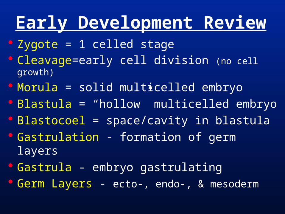
Early Development Review Zygote = 1 celled stage Cleavage=early cell division (no cell growth)
Morula = solid multicelled embryo Blastula = “hollow” multicelled embryo Blastocoel = space/cavity in blastula Gastrulation - formation of germ layers Gastrula - embryo gastrulating Germ Layers - ecto-, endo-, & mesoderm
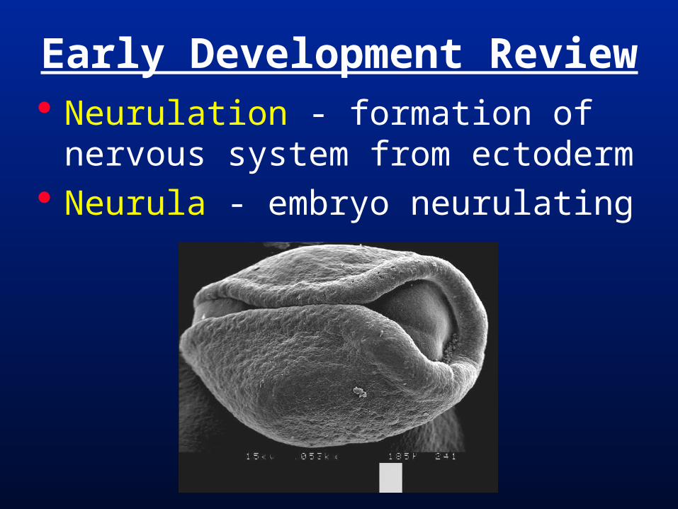
Early Development Review Neurulation - formation of nervous
system from ectoderm Neurula - embryo neurulating
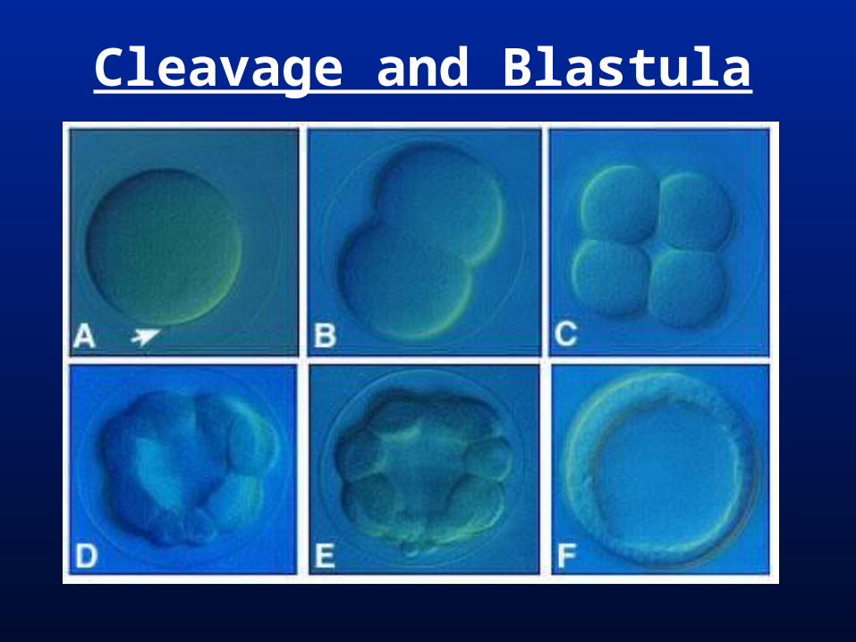
Cleavage and Blastula
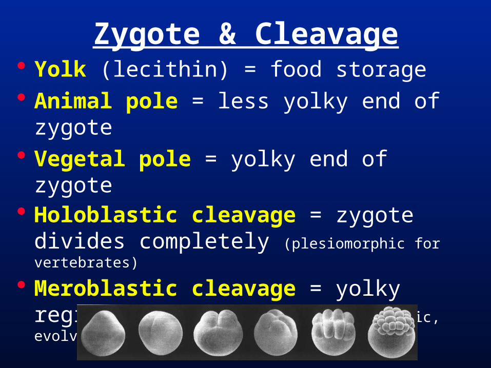
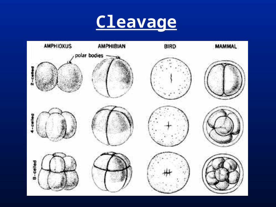
Zygote & Cleavage Yolk (lecithin) = food storage Animal pole = less yolky end of zygote Vegetal pole = yolky end of zygote Holoblastic cleavage = zygote divides
completely (plesiomorphic for vertebrates)
Meroblastic cleavage = yolky region does not divide (apomorphic, evolved at least 3 times in vertebrates)
Cleavage
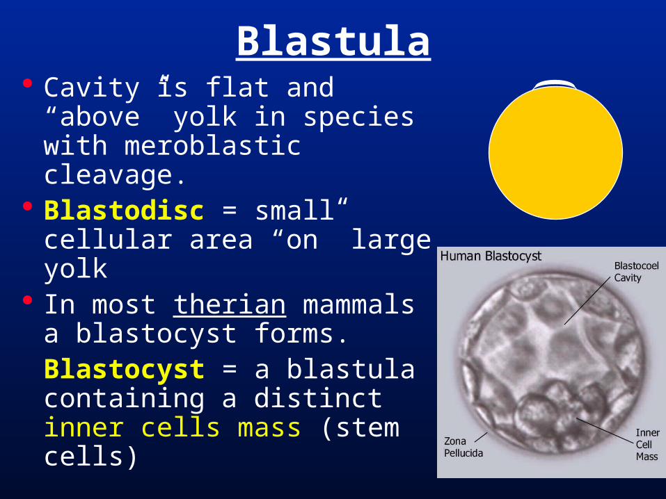
Blastula Cavity is flat and “above”
yolk in species with meroblastic cleavage.
Blastodisc = small cellular area “on” large yolk
In most therian mammals a blastocyst forms.Blastocyst = a blastula containing a distinct inner cells mass (stem cells)
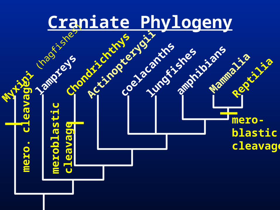
Craniate Phylogeny
Myx
ini (
hagf
ishes
)
lam
preys
Chondrichth
ys
Actin
optery
gii
coel
acan
ths
lungfis
hes
amphib
ians
Mam
mal
ia
Reptil
iam
ero
bla
stic
clea
vag
e
mero-blasticcleavage
mer
o. c
leav
age
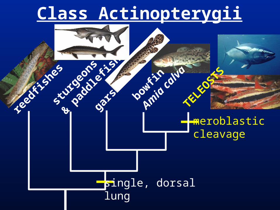
Class Actinopterygii
meroblasticcleavage
sturg
eons
& pad
dlefis
h
gars bowfin
Amia
cal
va
TELEOSTS
reed
fishes
single, dorsallung
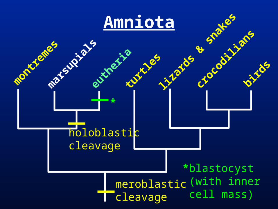
Amniota
montre
mes
mar
supia
ls
euth
eria
turtl
es
lizar
ds & s
nakes
croco
dilian
s
birds
meroblastic cleavage
holoblasticcleavage
*blastocyst (with inner cell mass)
*
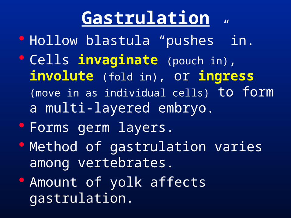
Gastrulation Hollow blastula “pushes” in. Cells invaginate (pouch in), involute
(fold in), or ingress (move in as individual
cells) to form a multi-layered embryo. Forms germ layers. Method of gastrulation varies among
vertebrates. Amount of yolk affects gastrulation.
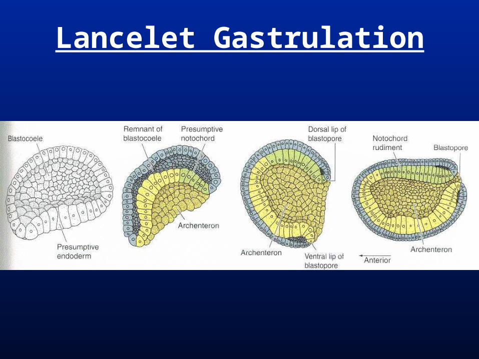
Lancelet Gastrulation
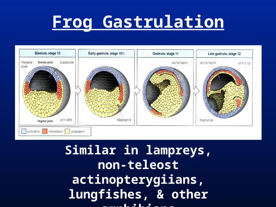
Frog Gastrulation
Similar in lampreys, non-teleost actinopterygiians,
lungfishes, & other amphibians
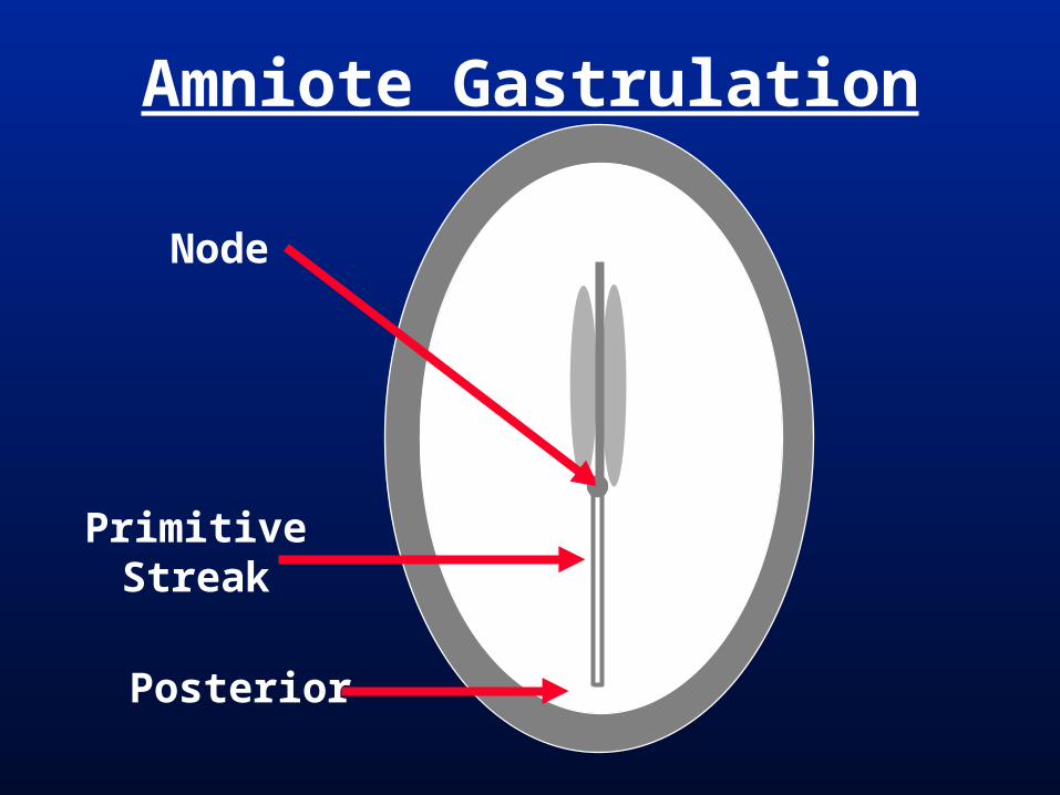
Amniote Gastrulation Primitive Streak = elongate region of
ingressing cells in an amniote gastrula Primitive streak gastrulation modified
gastrulation for a large volume of yolk.
Small disk (blastodisc) of cells on top of a large yolk.
Therian Mammals = little yolk but still retain primitive streak gastrulation.
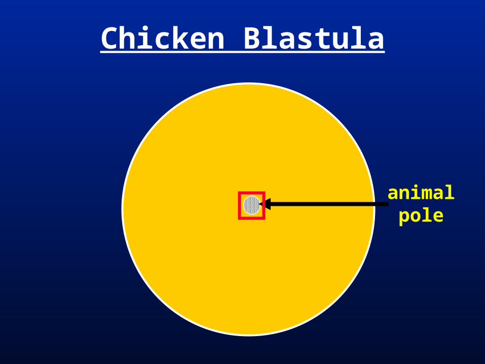
Chicken Blastula
animalpole
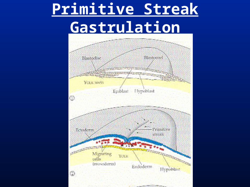
Primitive Streak Gastrulation
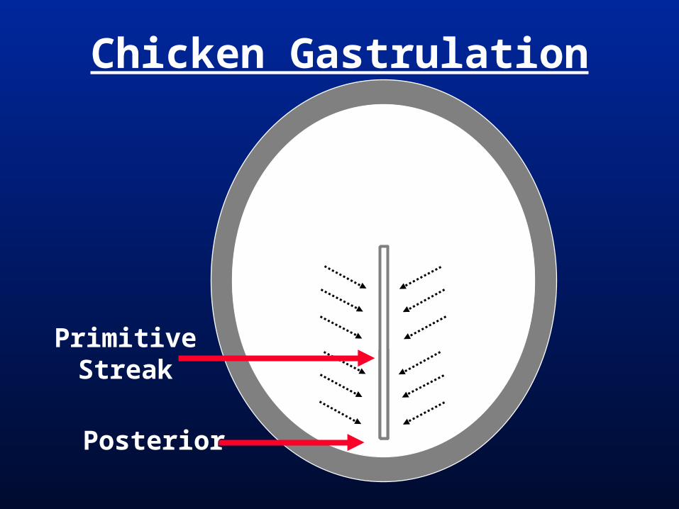
Chicken Gastrulation
Posterior
PrimitiveStreak
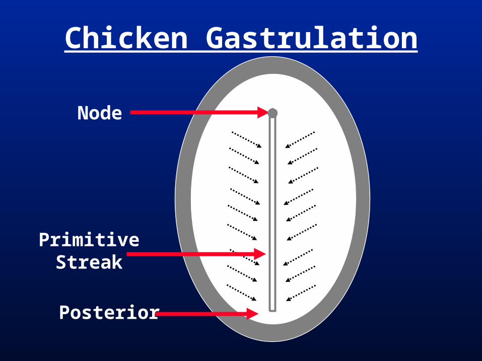
Chicken Gastrulation
Node
Posterior
PrimitiveStreak
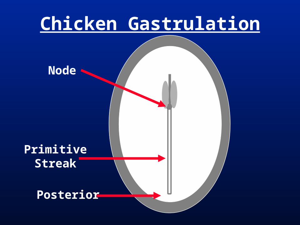
Chicken Gastrulation
Node
Posterior
PrimitiveStreak

Chicken Gastrulation
Posterior
PrimitiveStreak
Node
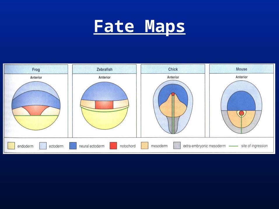
Fate Maps
Craniate Phylogeny
Myx
ini (
hagf
ishes
)
lam
preys
Chondrichth
ys
Actin
optery
gii
coel
acan
ths
lungfis
hes
amphib
ians
Mam
mal
ia
Reptil
ia
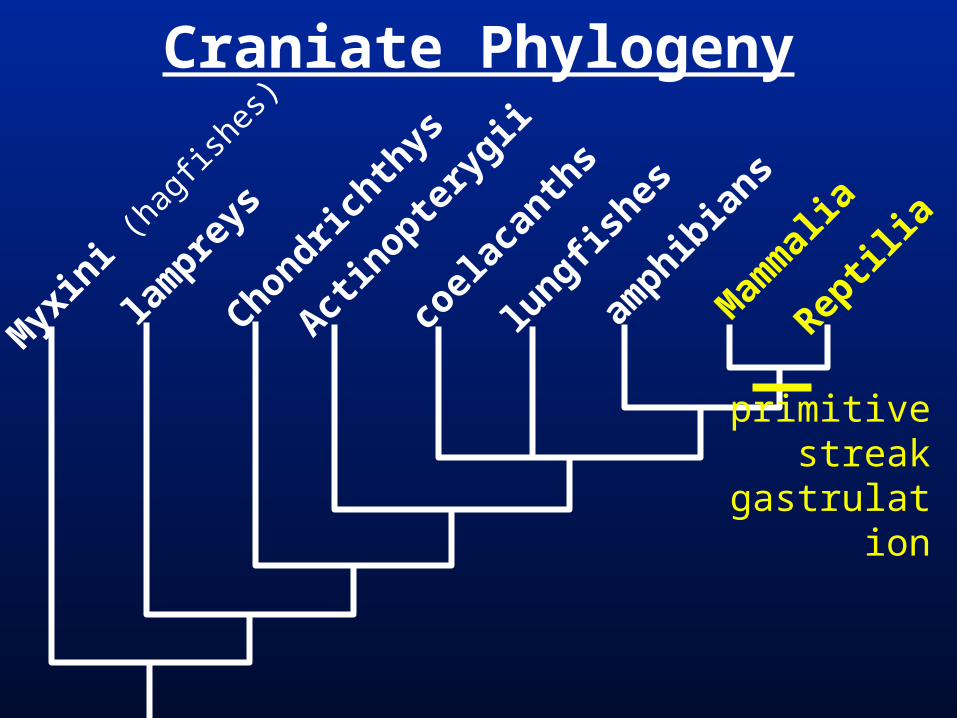
primitivestreak
gastrulation
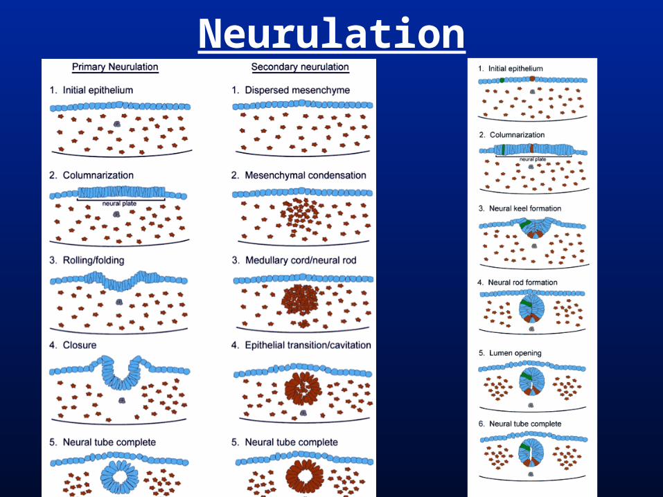
Neurulation Mesodermal notochord signals overlying
ectodermal epithelium to form hollow neural tube.
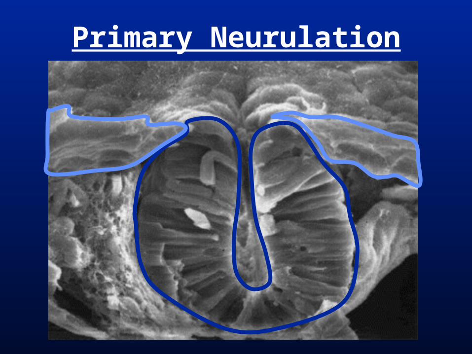
Neural tube central nervous system. Primary neurulation = ectodermal
epithelium infolds anteriorly. Secondary neurulation = occurs in the
post-anal tail of (all) vertebrates; mesenchymal mesoderm cells form a rod-shaped mass that cavitates.
Neurulation
Primary Neurulation

Primitive Streak Gastrulation
Amniote Gastrulation
Posterior
PrimitiveStreak
Node
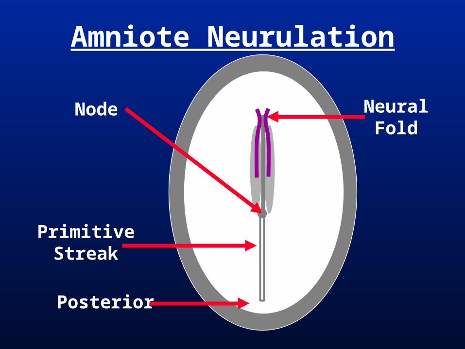
Amniote Neurulation
NeuralFold
Posterior
PrimitiveStreak
Node
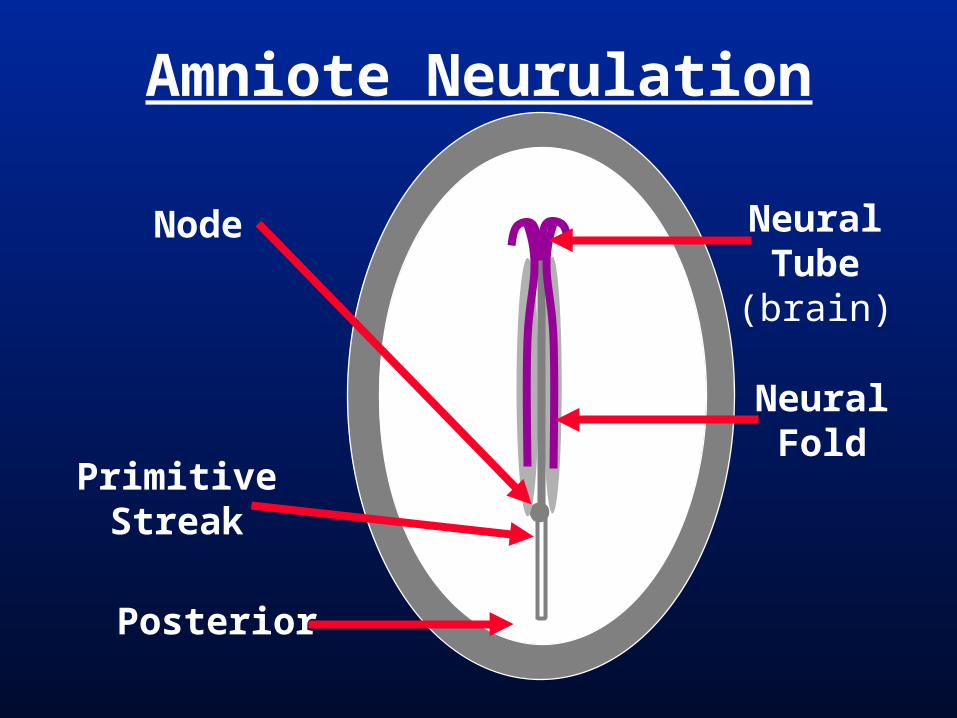
Amniote Neurulation
NeuralTube(brain)
NeuralFold
Posterior
PrimitiveStreak
Node
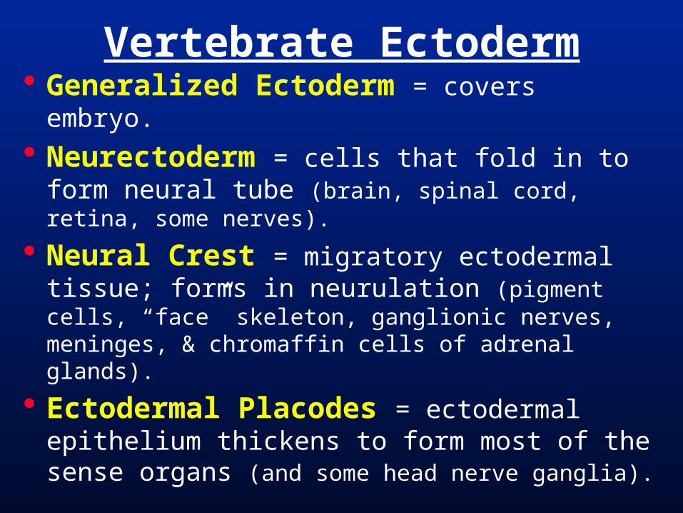
Vertebrate Ectoderm Generalized Ectoderm = covers embryo. Neurectoderm = cells that fold in to form
neural tube (brain, spinal cord, retina, some nerves).
Neural Crest = migratory ectodermal tissue; forms in neurulation (pigment cells, “face” skeleton, ganglionic nerves, meninges, & chromaffin cells of adrenal glands).
Ectodermal Placodes = ectodermal epithelium thickens to form most of the sense organs (and some head nerve ganglia).
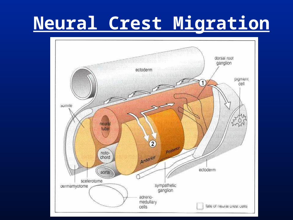
Neural Crest Migration
Craniate Phylogeny
Myx
ini (
hagf
ishes
)
lam
preys
Chondrichth
ys
Actin
optery
gii
coel
acan
ths
lungfis
hes
amphib
ians
Mam
mal
ia
Reptil
ia
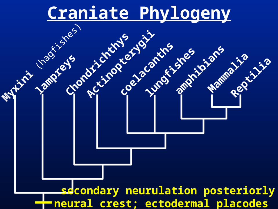
secondary neurulation posteriorlyneural crest; ectodermal placodes
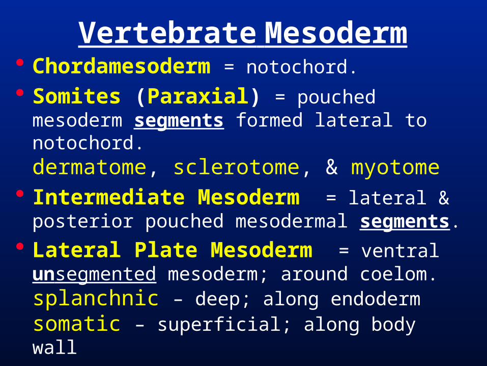
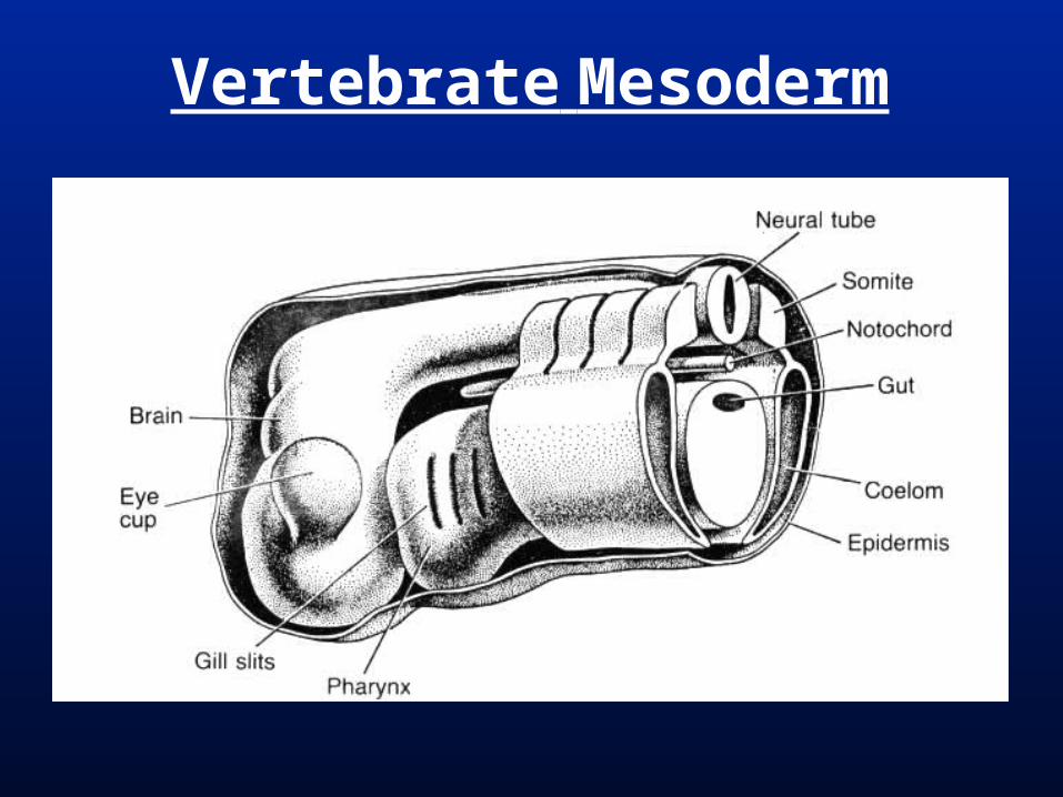
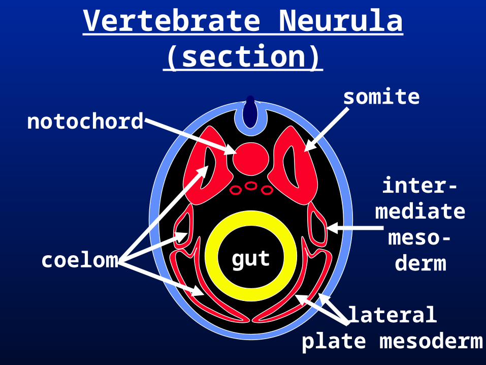
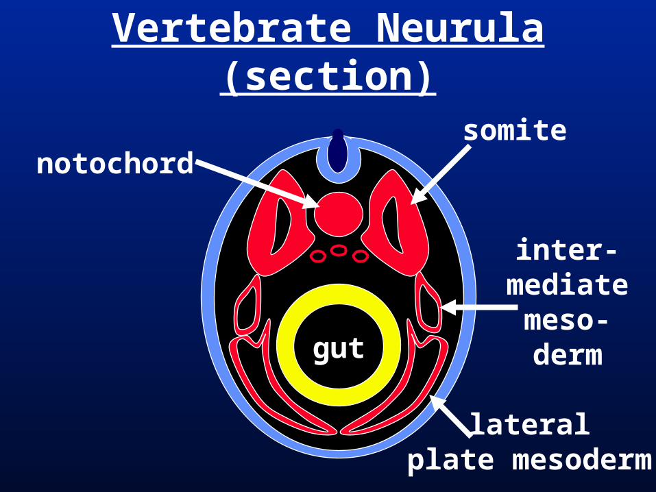
Vertebrate Mesoderm Chordamesoderm = notochord.
Somites (Paraxial) = pouched mesoderm segments formed lateral to notochord.dermatome, sclerotome, & myotome
Intermediate Mesoderm = lateral & posterior pouched mesodermal segments.
Lateral Plate Mesoderm = ventral unsegmented mesoderm; around coelom.splanchnic – deep; along endodermsomatic – superficial; along body wall
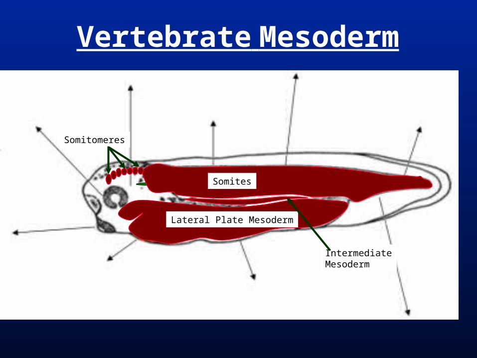
Vertebrate Mesoderm
Lateral Plate Mesoderm
Somites
Somitomeres
IntermediateMesoderm
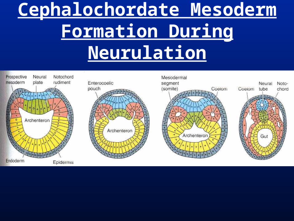
Cephalochordate Mesoderm Formation During Neurulation

Vertebrate Mesoderm
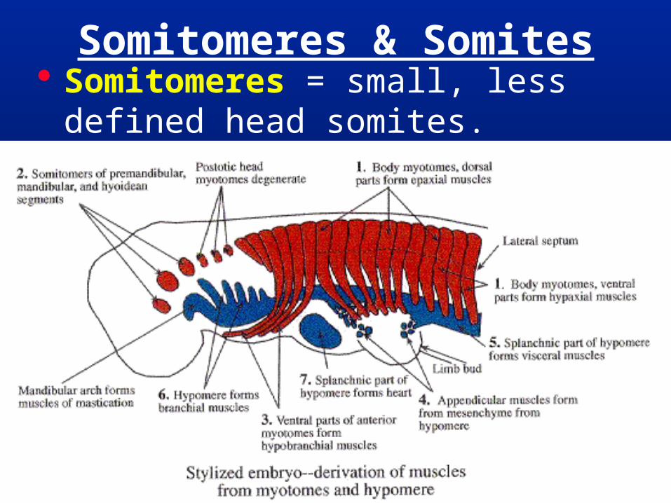
Somitomeres & Somites Somitomeres = small, less defined
head somites.
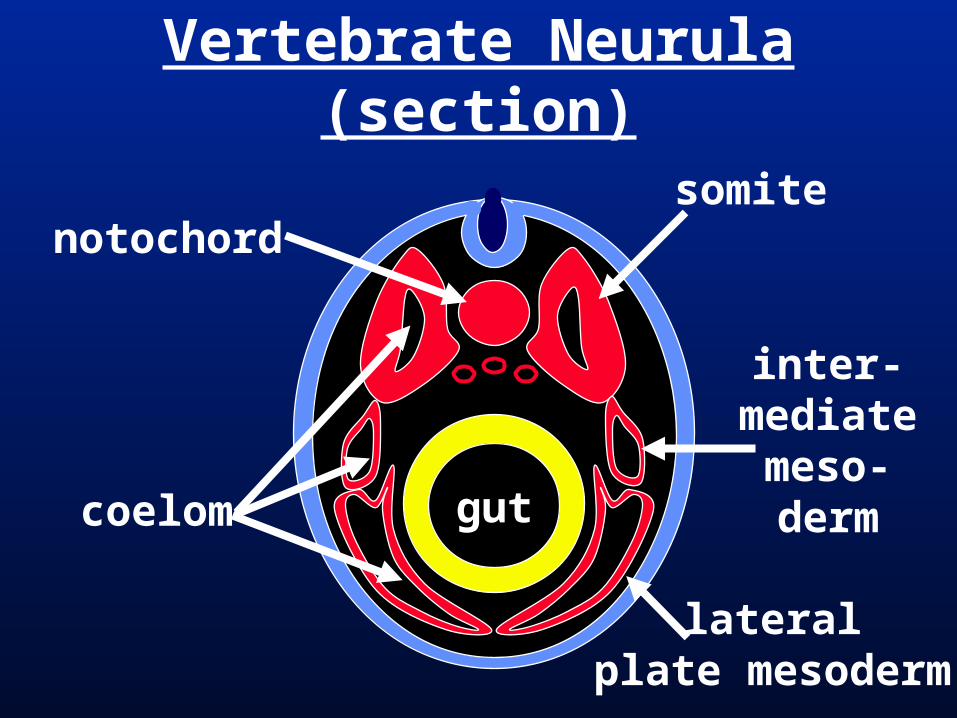
Vertebrate Neurula (section)
notochord
coelom
somite
inter-mediatemeso-derm
lateralplate mesoderm
gut
Vertebrate Mesoderm
Vertebrate Neurula (section)
notochord
coelom gut
somite
inter-mediatemeso-derm
lateralplate mesoderm
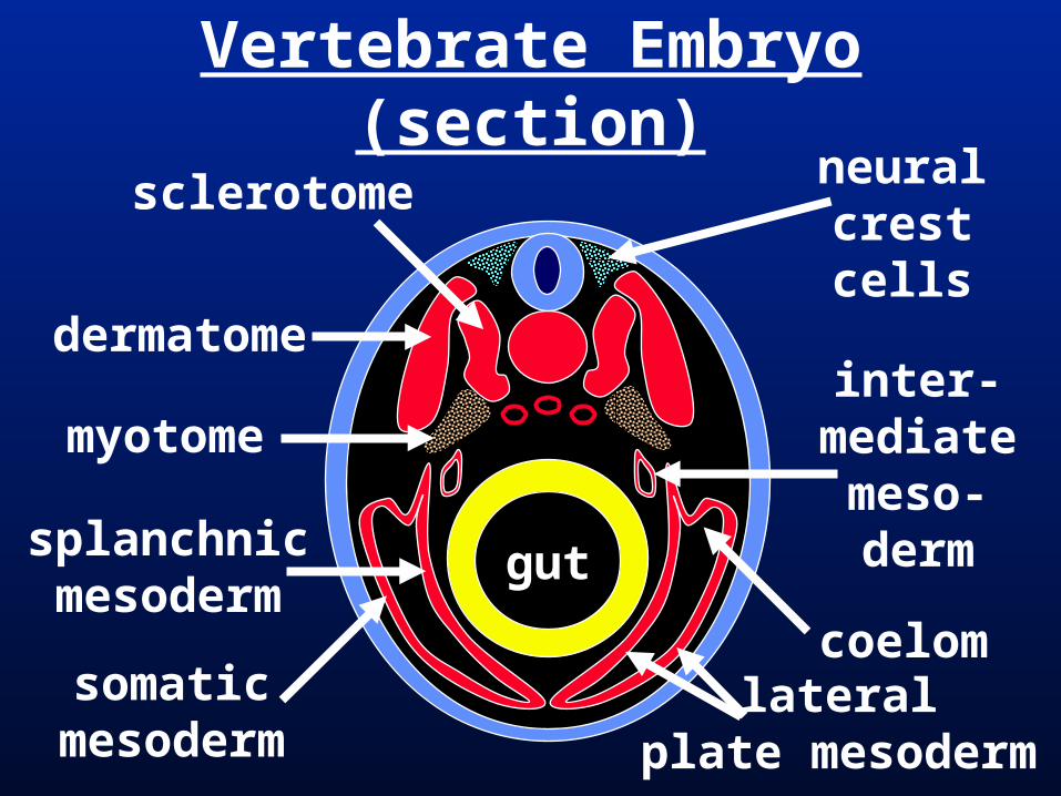
Vertebrate Embryo (section)neuralcrestcells
dermatome
sclerotome
myotome
splanchnicmesoderm
somaticmesoderm
coelom
gut
inter-mediatemeso-derm
lateralplate mesoderm
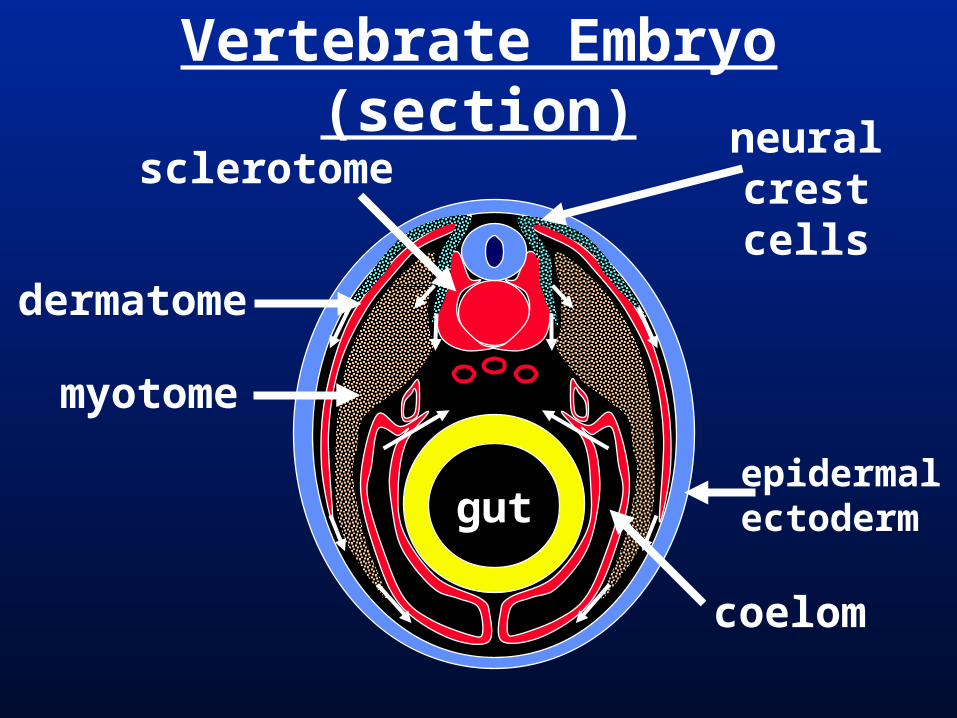
Vertebrate Embryo (section)
coelom
neuralcrestcells
dermatome
sclerotome
myotome
gutepidermalectoderm
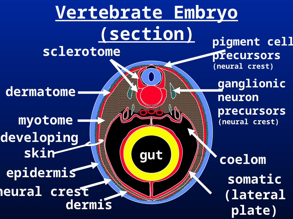
Vertebrate Embryo (section)
gut
pigment cell precursors(neural crest)
ganglionicneuron precursors(neural crest)
dermatome
sclerotome
myotomedeveloping
skin
dermis
epidermis
neural crest
coelom
somatic(lateral plate)
mesoderm
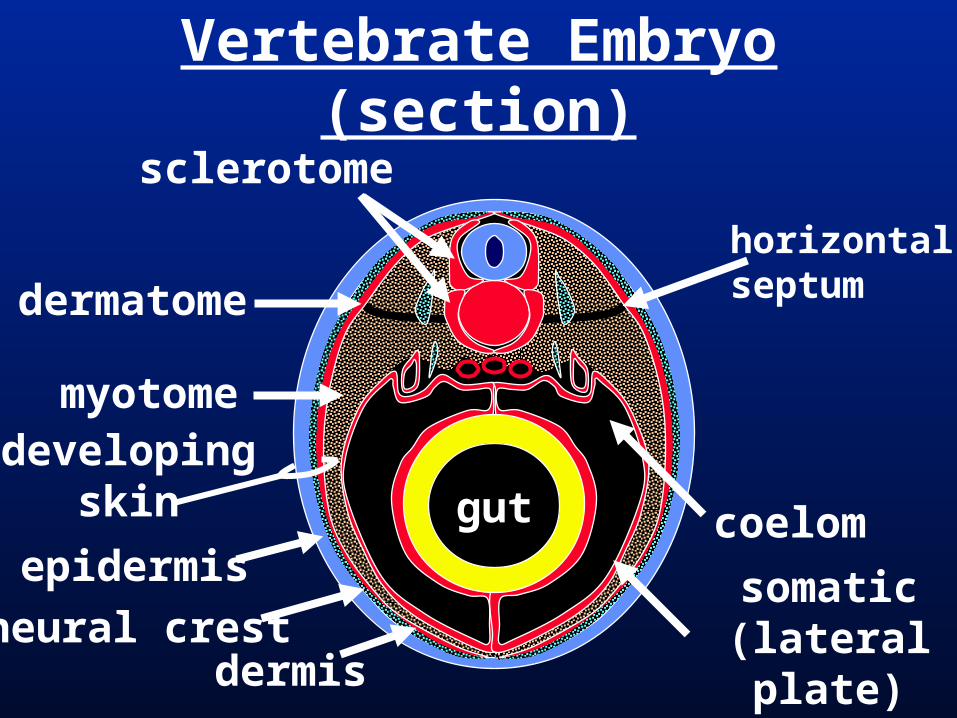
Vertebrate Embryo (section)
gut
dermatome
sclerotome
myotome
horizontalseptum
developingskin
dermis
epidermis
neural crest
coelom
somatic(lateral plate)
mesoderm
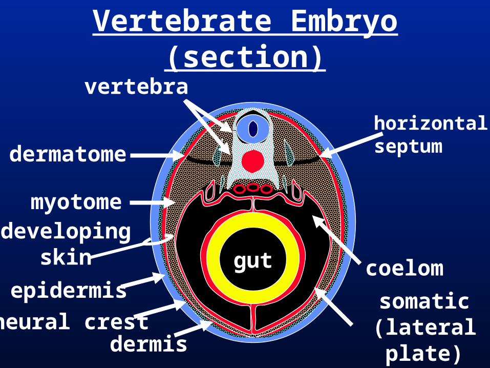
Vertebrate Embryo (section)
gut
dermatome
vertebra
myotomedeveloping
skin
dermis
epidermis
neural crest
horizontalseptum
coelom
somatic(lateral plate)
mesoderm
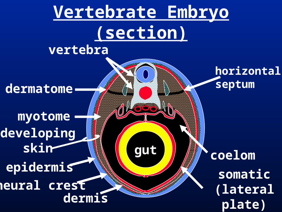
Vertebrate Embryo (section)
gut
dermatome
vertebra
myotome
horizontalseptum
coelom
somatic(lateral plate)
mesoderm
developingskin
dermis
epidermis
neural crest
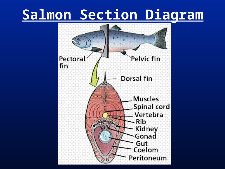
Salmon Section Diagram
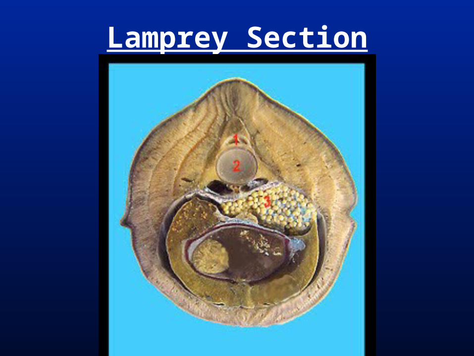
Lamprey Section
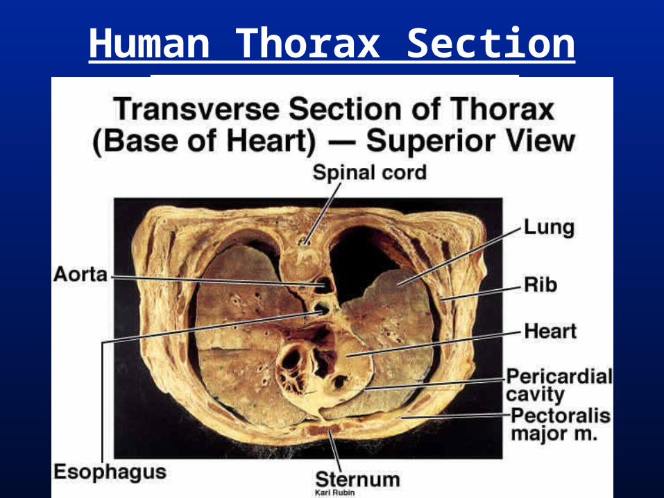
Human Thorax Section
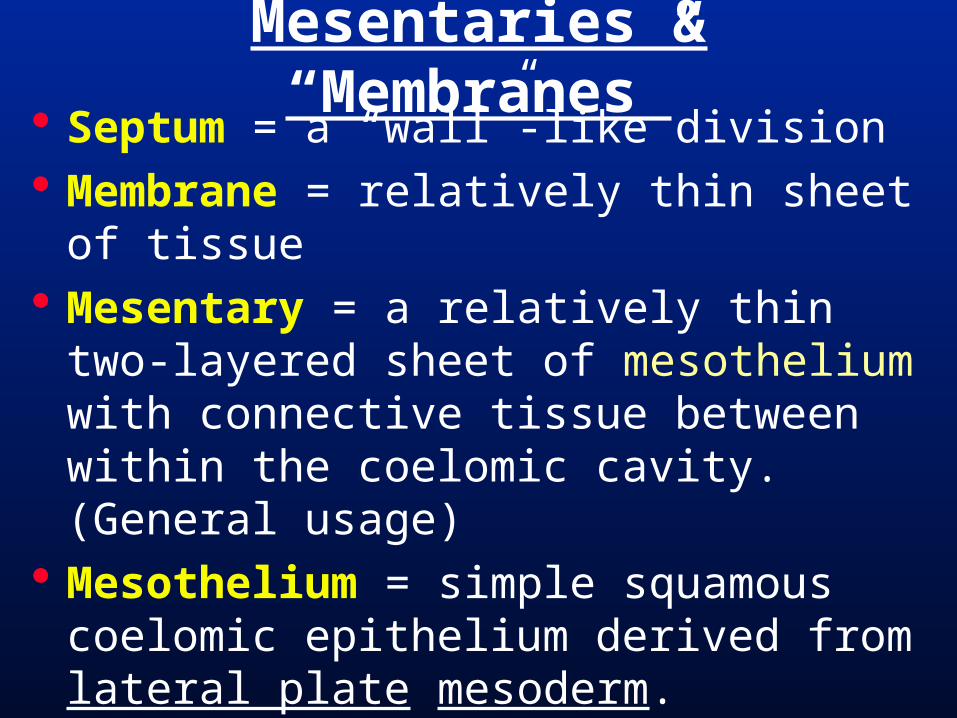
Septum = a “wall”-like division Membrane = relatively thin sheet of
tissue Mesentary = a relatively thin two-
layered sheet of mesothelium with connective tissue between within the coelomic cavity. (General usage)
Mesothelium = simple squamous coelomic epithelium derived from lateral plate mesoderm.
Mesentaries & “Membranes”
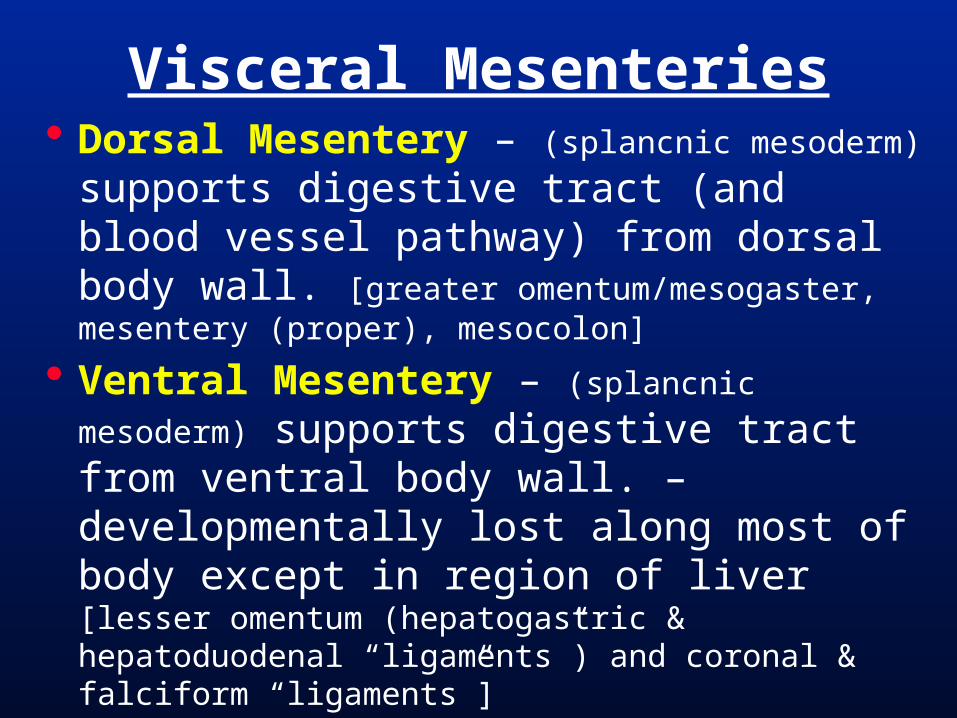
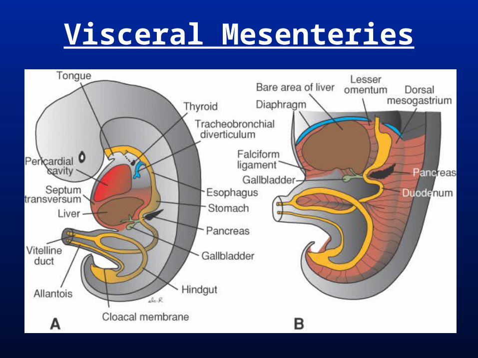
Visceral Mesenteries Dorsal Mesentery – (splancnic mesoderm)
supports digestive tract (and blood vessel pathway) from dorsal body wall. [greater omentum/mesogaster, mesentery (proper), mesocolon]
Ventral Mesentery – (splancnic mesoderm) supports digestive tract from ventral body wall. – developmentally lost along most of body except in region of liver [lesser omentum (hepatogastric & hepatoduodenal “ligaments”) and coronal & falciform “ligaments”]
Vertebrate Neurula (section)
notochordsomite
inter-mediatemeso-derm
lateralplate mesoderm
gut
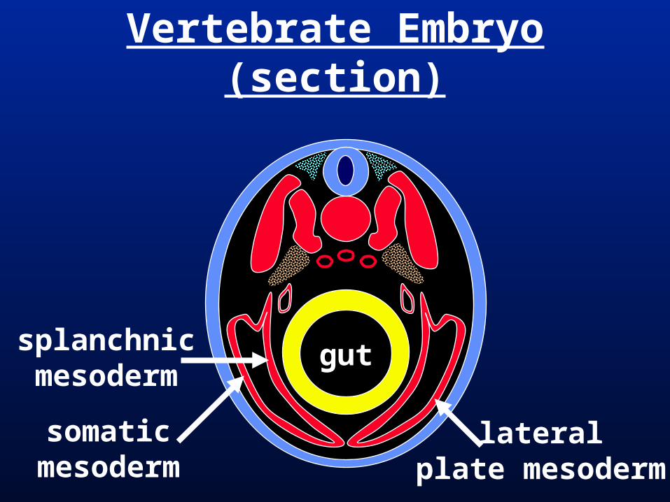
Vertebrate Embryo (section)
splanchnicmesoderm
somaticmesoderm
gut
lateralplate mesoderm
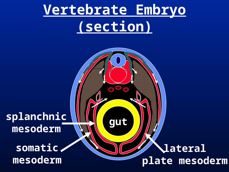
Vertebrate Embryo (section)
gut
lateralplate mesoderm
splanchnicmesoderm
somaticmesoderm
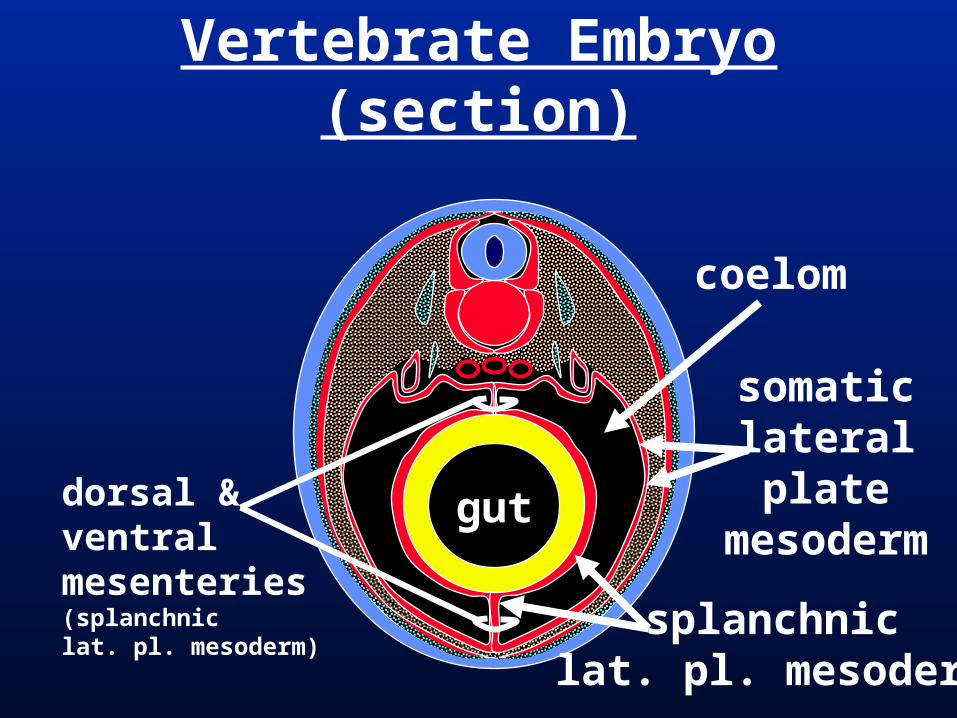
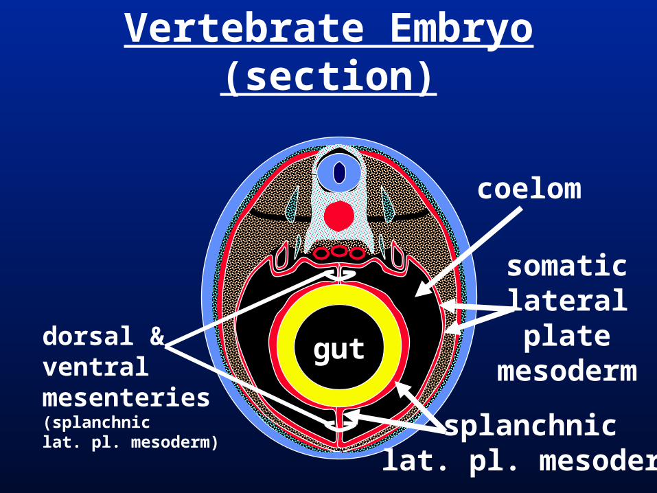
Vertebrate Embryo (section)
gut
coelom
dorsal & ventralmesenteries(splanchnic lat. pl. mesoderm)
splanchniclat. pl. mesoderm
somaticlateralplate
mesoderm
Vertebrate Embryo (section)
gutdorsal & ventralmesenteries(splanchnic lat. pl. mesoderm)
coelom
splanchniclat. pl. mesoderm
somaticlateralplate
mesoderm
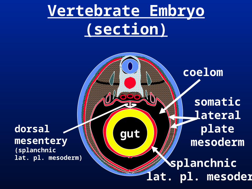
Vertebrate Embryo (section)
gutdorsalmesentery(splanchnic lat. pl. mesoderm)
coelom
splanchniclat. pl. mesoderm
somaticlateralplate
mesoderm
Visceral Mesenteries
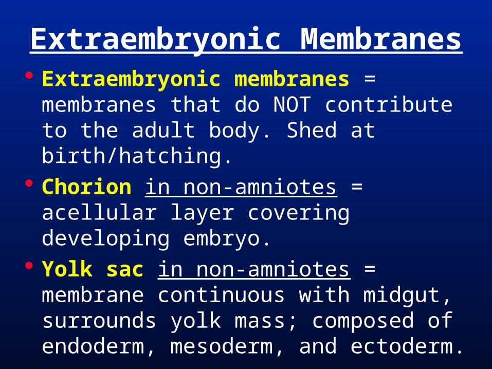
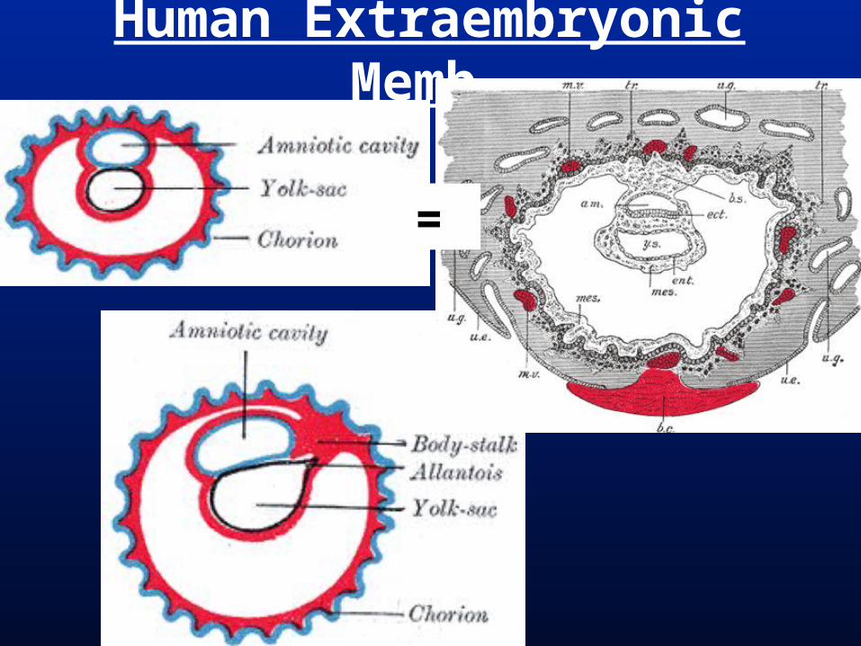
Extraembryonic membranes = membranes that do NOT contribute to the adult body. Shed at birth/hatching.
Chorion in non-amniotes = acellular layer covering developing embryo.
Yolk sac in non-amniotes = membrane continuous with midgut, surrounds yolk mass; composed of endoderm, mesoderm, and ectoderm.
Extraembryonic Membranes
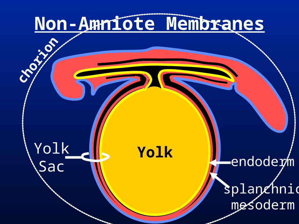
Non-Amniote Membranes
YolkYolkSac endoderm
splanchnicmesoderm
chor
ion
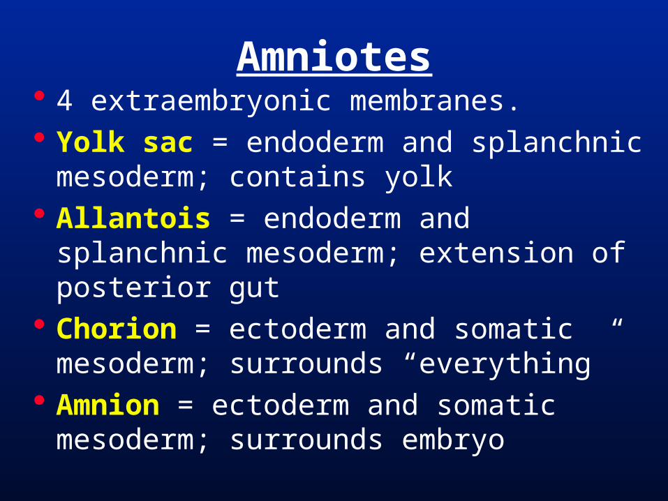
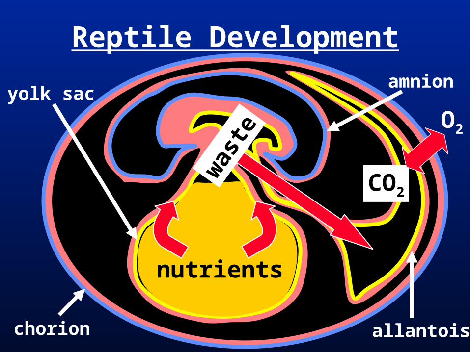
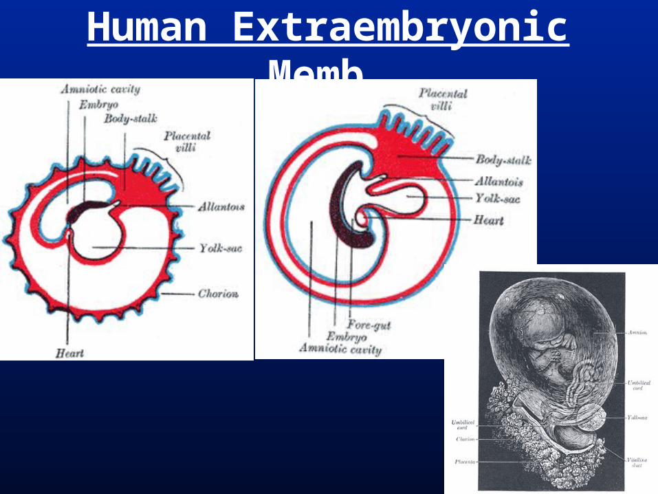
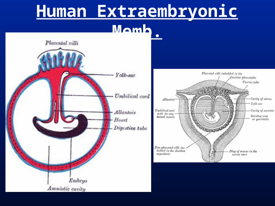
4 extraembryonic membranes. Yolk sac = endoderm and splanchnic
mesoderm; contains yolk Allantois = endoderm and splanchnic
mesoderm; extension of posterior gut Chorion = ectoderm and somatic
mesoderm; surrounds “everything” Amnion = ectoderm and somatic
mesoderm; surrounds embryo
Amniotes
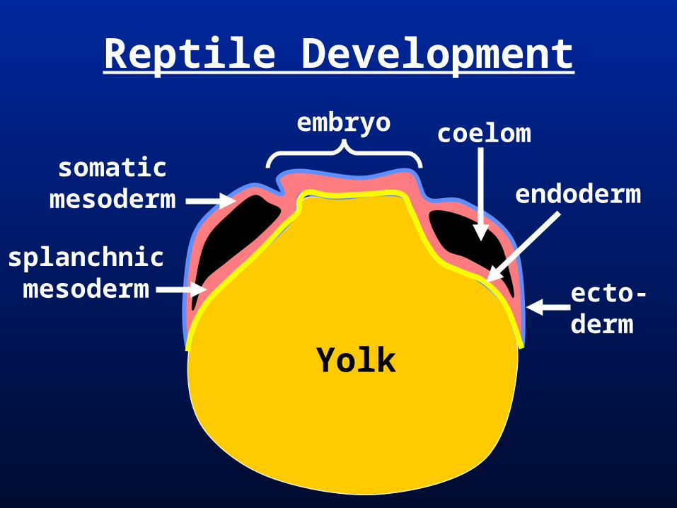
Reptile Development
coelom
endoderm
ecto-derm
somaticmesoderm
splanchnicmesoderm
embryo
Yolk
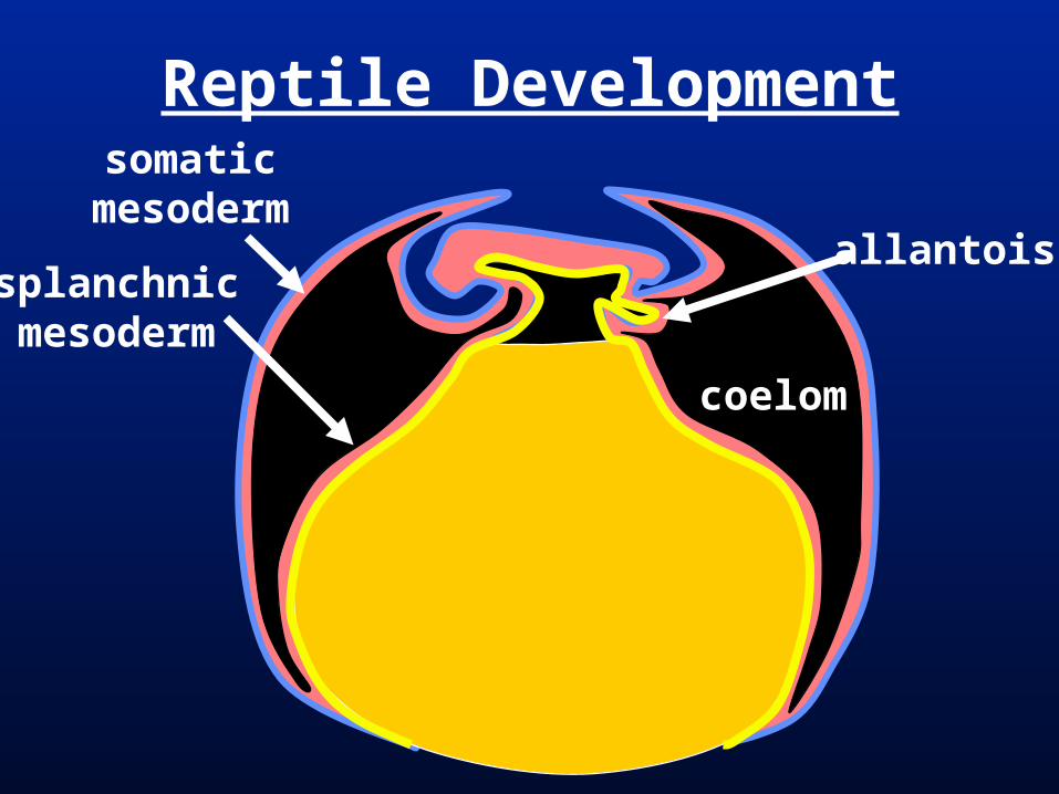
Reptile Development
coelom
allantois
somaticmesoderm
splanchnicmesoderm
Reptile Development
allantois
amnionyolk sac
chorion
nutrients
was
te
O2
CO2
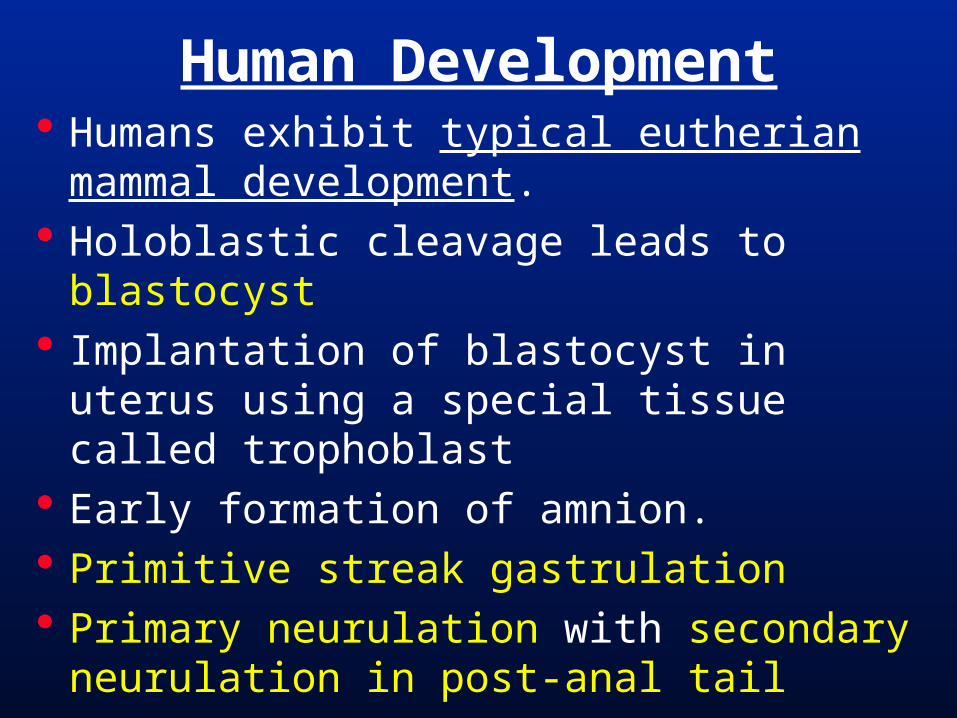
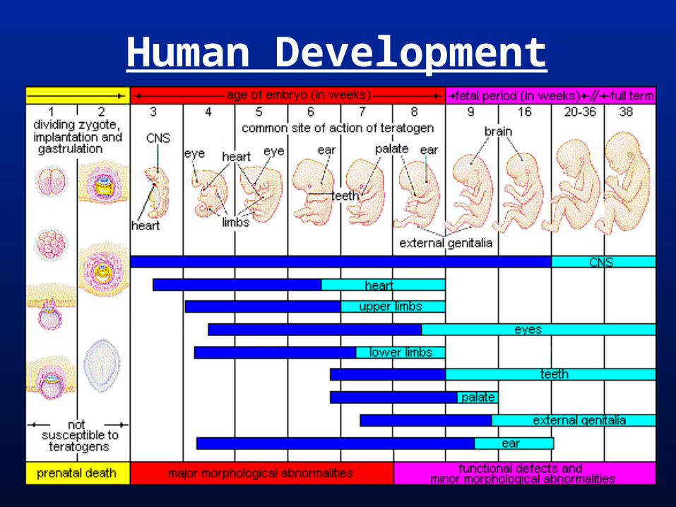
Human Development Humans exhibit typical eutherian
mammal development. Holoblastic cleavage leads to blastocyst Implantation of blastocyst in uterus
using a special tissue called trophoblast Early formation of amnion. Primitive streak gastrulation Primary neurulation with secondary
neurulation in post-anal tail
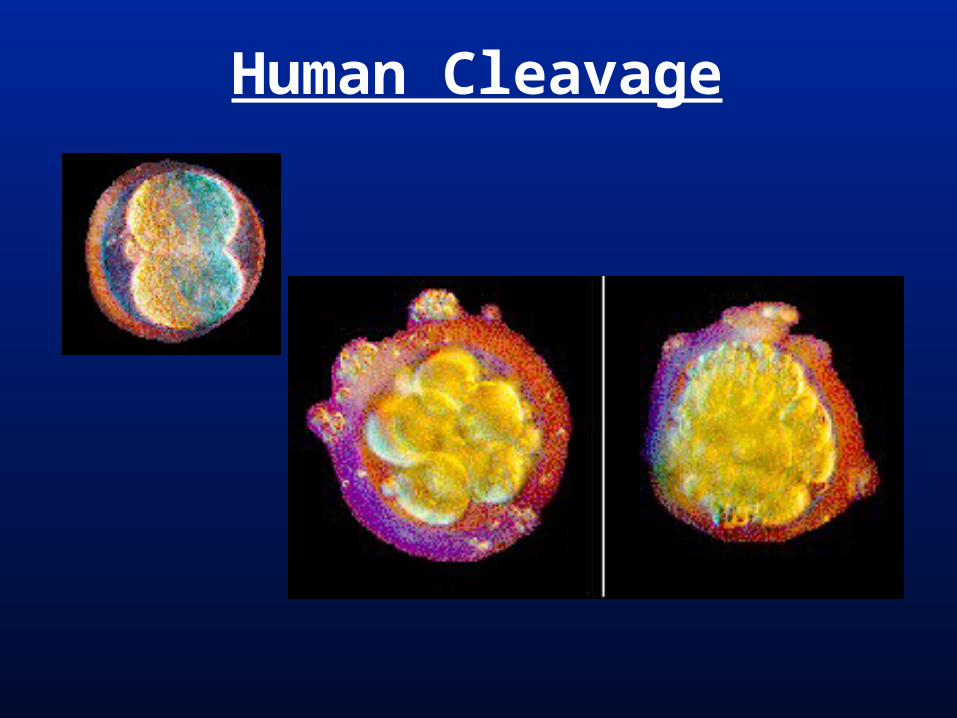
Human Cleavage
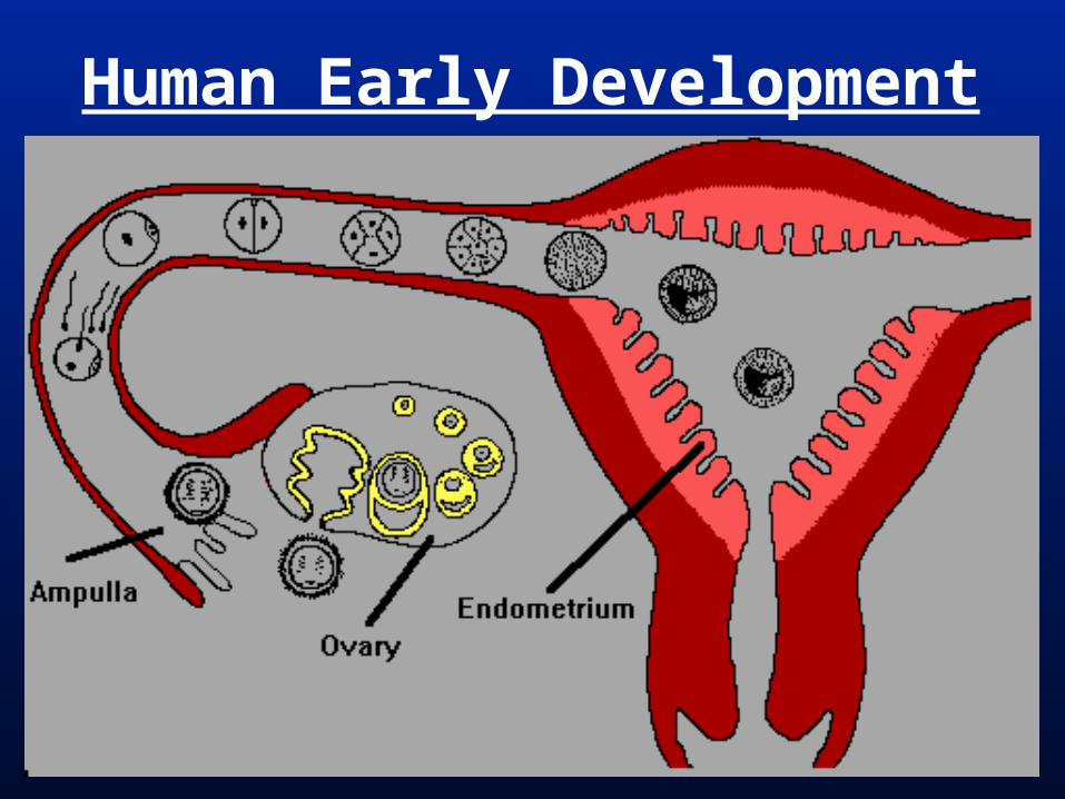
Human Early Development
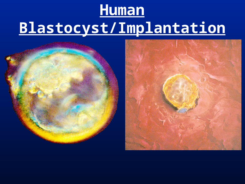
Human Blastocyst/Implantation
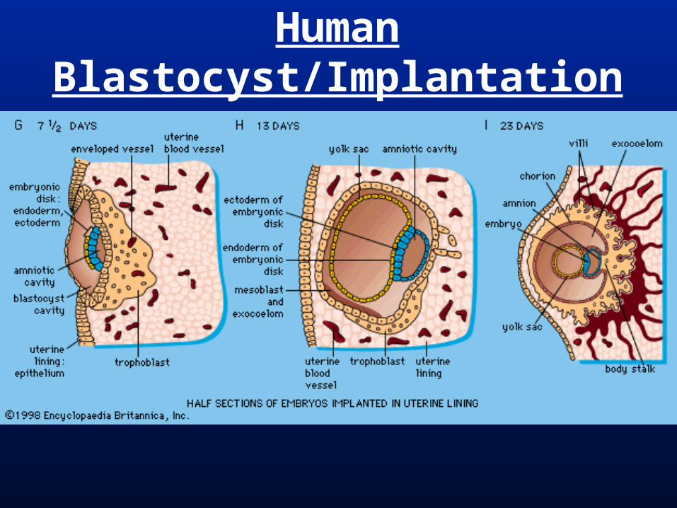
Human Blastocyst/Implantation
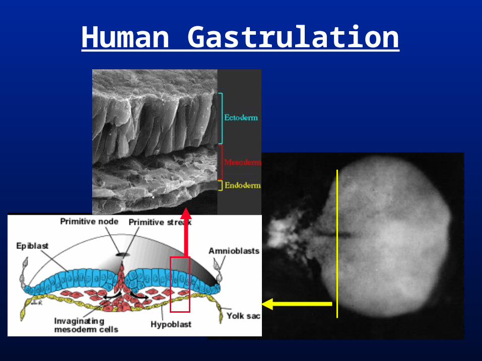
Human Gastrulation
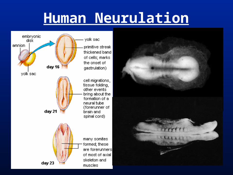
Human Neurulation
Human Development
Human Extraembryonic Memb.
=
Human Extraembryonic Memb.
Human Extraembryonic Memb.

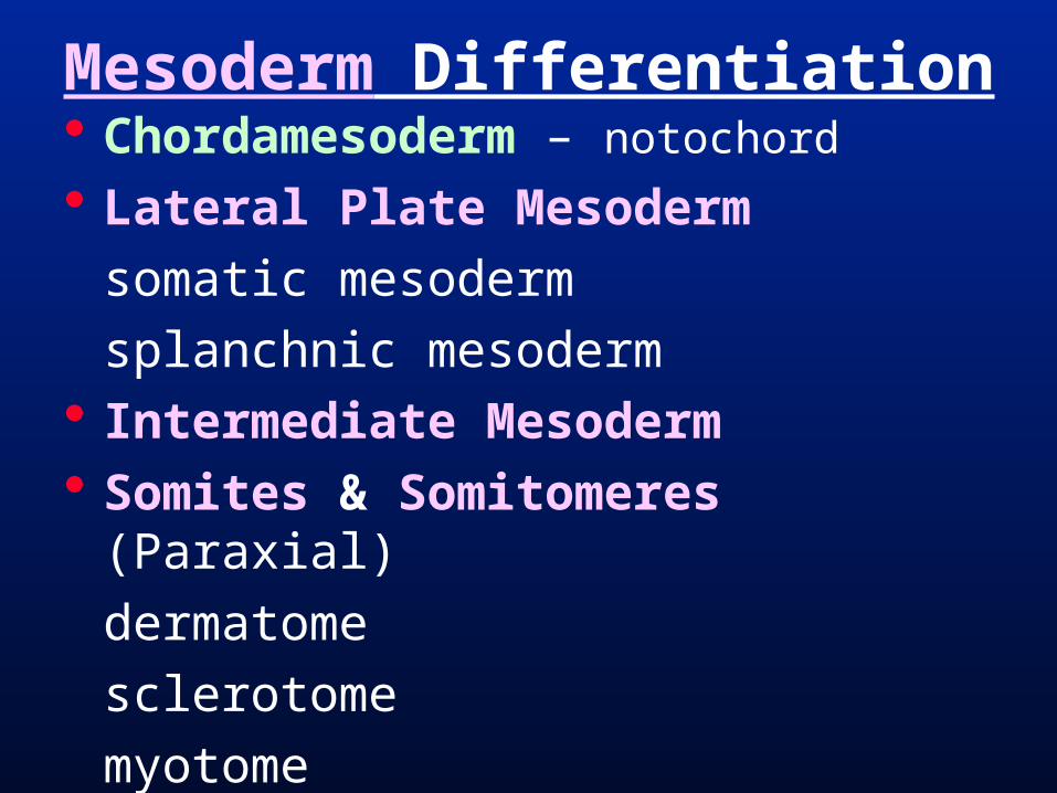
Mesoderm Differentiation Chordamesoderm – notochord
Lateral Plate Mesoderm
somatic mesoderm
splanchnic mesoderm Intermediate Mesoderm Somites & Somitomeres (Paraxial)
dermatome
sclerotome
myotome
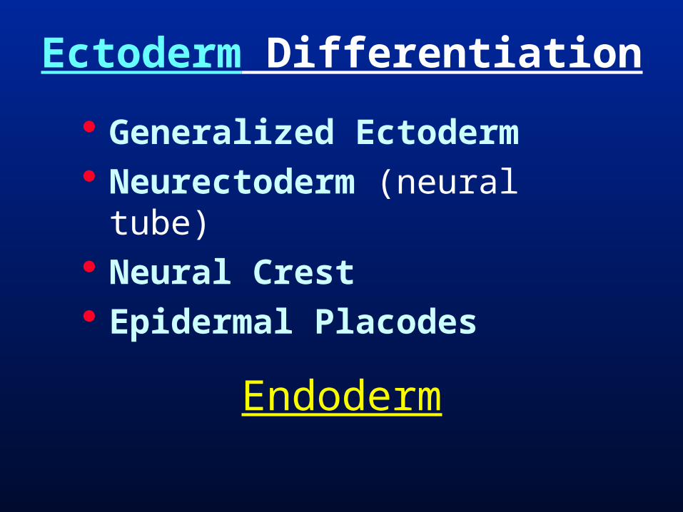
Ectoderm Differentiation
Generalized Ectoderm Neurectoderm (neural tube) Neural Crest Epidermal Placodes
Endoderm