Embed Size (px)
Citation preview
DEVELOPMENTAL BIOLOGY 42, 391-400 (1975)
Normal Stages of Development of the Axolotl,
Ambystoma mexicanum
G. M. SCHRECKENBERG
Department of Biology, Fairleigh Dickenson Uniuersity, Rutherford, New Jersey 07070
AND
A. G. JACOBSON
Department of Zoology, University of Texas, Austin, Texas 78712
Accepted October 29, 1974
Illustrations and descriptions of normal stages of the axolotl Ambystoma mexicanum are given. The time to reach each morphological stage is compared with similar stages in two other species, Ambystoma maculatum and Taricha torosa.
INTRODUCTION 14-19 follow one another rapidly while days The Mexican axolotl, Ambystoma may separate successive larval stages.
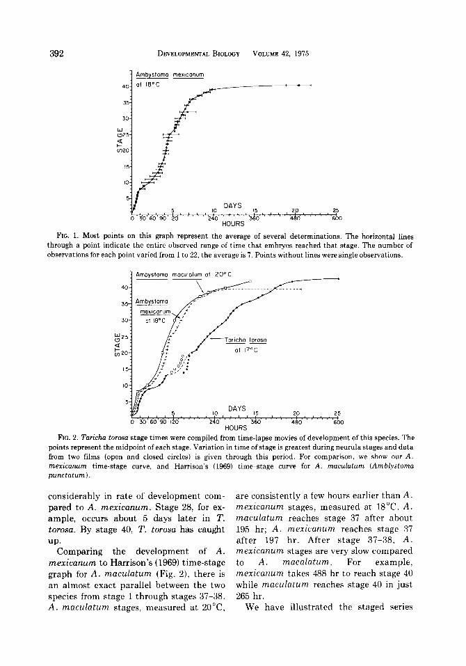
mexicanum (also called Siredon The time between the stages is illus- mericanum), has long been used in labora- trated in Fig. 1. The data for Fig. 1 were tories around the world. This convenient compiled by rearing embryos in a constant- laboratory animal probably will be used temperature box (18” f 0,5”C), inspecting increasingly for experimental studies of frequently, and recording the time since development. We give here a series of stage 1 that each stage first appears. Even normal stages for this species and data on at constant temperature and using eggs the timing of the stages. from the same mating, there is variation in
We have tried to make these normal timing of stages. Differences in egg size stages of the axolotl as comparable as may contribute to this timing variation. possible to Harrison’s (1969) staged series We have timing data for the California of Ambystoma maculatum (Amblystoma newt Taricha torosa (staged by Twitty and punctatum). The Twitty and Bodenstein Bodenstein, 1962), which we present here (1962) staged series of Taricha torosa (Tri- for comparison (Fig. 2). The T. torosa data turus torosus), which was also modeled is taken from time-lapse movies. The mov- after Harrison’s series, served as a useful ies were made at an exposure interval of 1 supplementary guide. Gastrulation and min. The movies were analyzed using a neurulation of the axolotl looks more like stop-motion projector equipped with a that of T. torosa than of A. maculatum, frame counter. A movie was run forward but the timing of stages is most similar in and backward until a decision could be the two Ambystoma species. reached as to what frame was the midpoint
Development is, of course, continuous of each stage. The time since stage 1 could and the designated stages gradually grade then be read directly in minutes from the into one another. The stages are based on frame counter. The timing in different changing external morphological features. movies was similar except through neurula The amount of time between different stages. stages varies. For example, neurula stages After cleavage stages, T. torosa lags
391 Copyright 0 1975 by Academic Press, Inc. All rights of reproduction in any form reserved.
392 DEVELOPMENTAL BIOLOGY VOLUME 42. 1975
1 Ambystomo mexiconum
at 18'C
FIG. 1. Most points on this graph represent the average of several determinations. The horizontal lines through a point indicate the entire observed range of time that embryos reached that stage. The number of observations for each point varied from 1 to 22, the average is 7. Points without lines were single observations.
1 Ambvstoma I
DAYS 5 IO ,5 20 25
0 30 60 90 120 240 360 480 600 HOURS
FIG. 2. Taricha torosa stage times were compiled from time-lapse movies of development of this species. The points represent the midpoint of each stage. Variation in time of stage is greatest during neurula stages and data from two films (open and closed circles) is given through this period. For comparison, we show our A. mexicanurn time-stage curve, and Harrison’s (1969) time-stage curve for A. maculatum (Anblystoma punctatum).
considerably in rate of development com- pared to A. mexicanum. Stage 28, for ex- ample, occurs about 5 days later in T. torosa. By stage 40, T. torosa has caught
up. Comparing the development of A.
mexicanum to Harrison’s (1969) time-stage graph for A. maculatum (Fig. Z), there is an almost exact parallel between the two species from stage 1 through stages 37-38. A. maculatum stages, measured at ZO”C,
are consistently a few hours earlier than A. mexicanum stages, measured at 18°C. A. maculatum reaches stage 37 after about 195 hr; A. mexicanum reaches stage 37 after 197 hr. After stage 37-38, A. mexicanum stages are very slow compared to A. macalatum. For example, mexicanum takes 488 hr to reach stage 40 while maculatum reaches stage 40 in just 265 hr.
We have illustrated the staged series
BRIEF NOTES 393
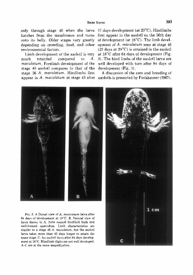
only through stage 40 when the larva 17 days development (at 2O’C). Hindlimbs hatches from the membranes and turns first appear in the axolotl on the 56th day onto its belly. Older stages vary greatly of development (at 18°C). The limb devel- depending on crowding, food, and other opment of A. maculatum seen at stage 46 environmental factors. (22 days at 20°C) is attained in the axolotl
Limb development of the axolotl is very at 18°C after 64 days of development (Fig. much retarded compared to A. 3). The hind limbs of the axolotl larva are maculatum. Forelimb development of the well developed with toes after 84 days of stage 40 axolotl compares to that of the development (Fig. 3). stage 36 A. maculatum. Hindlimbs first A discussion of the care and breeding of appear in A. macdatum at stage 43 after axolotls is presented by Fankhauser (1967).
FIG. 3. A Dorsal view of A. mexicanurn larva after 64 days of development at 18°C. B. Ventral view of larva shown in A. Note conical hindlimb buds and well-formed operculum. Limb characteristics are similar to a stage 46 A. maculatum, but the axolotl larva takes more than 40 days longer to attain the same stage. C. An axolotl larva after 84 days develop- ment at 18°C. Hindlimb digits are not well developed. A-C are at the same magnification.
394 DEVELOPMENTAL BIOLOGY VOLUME 42, 1975
The cleaving axolotl egg has been studied this stage. Polar bodies may often be seen in detail (Skoblina, 1965; Meinertz, 1970; near the animal pole. Signoret and Lesfresne, 1971; Hara, 1971; Stage 2. Two cells. The cleavage furrow Rott, 1973). Ignat’eva (1968) described ax- extends from pole to pole. olotl gastrulation and compares it to the Stage 3. Four cells. The second cleavage sturgeon. C.-O. Jacobson (1959, 1962) de- furrow also extends from pole to pole at scribed and mapped axolotl neurulation. right angles to the first.
The illustrations of stages that follow Stage 4. Eight cells. The third cleavage (Fig. 4A-C) are photographs of living em- furrow forms along a latitude above the bryos, all at the same magnification. Ex- equator. The animal hemisphere quartet of cept as noted below, all embryos are cells is thus smaller than the quartet be- viewed from above as they naturally orient low. themselves when they are removed from Stage 5. Sixteen cells. Stage 5 animal the jelly capsules and placed unrestrained hemisphere cells appear about half the size in a dish of water. The only exceptions are of stage 4 cells. stages 9, 10, and 11 whose diagnostic fea- Stage 6. About 32 cells around a central ture, the forming blastopore, is visible only cavity. from beneath. These three embryos were Stage 7. The upper cells appear about photographed through an inverted micro- half the size of stage 6. Cleavage is becom- scope. The view of them is of their vegetal ing asynchronous even among the animal hemispheres. hemisphere cells. Stage 7 may be consid-
Through cleavage stages (l-7), blastula ered the end of cleavage stages and the stages (7-g), and early gastrula stages beginning of blastula stages. (g-11), the embryos rest with their animal Stage 8. Blastula stage. The upper cells poles uppermost. Between the end of stage are about half the size of those at stage 7 at 11 and the beginning of late gastrula stage the beginning of stage 8. Stage 8 lasts 9-16 12, the embryo rotates, turning the future hr and the cells continue to divide, becom- dorsal side uppermost. This change in ing too small to distinguish individually orientation coincides with the formation of without magnification (Stage 8+). the archenteron and the disappearance of Stage 9. Vegetal pole view. The first the blastocoel. The embryo retains this signs of gastrulation are pigment concen- orientation from gastrula stage 12 through tration (indicative of cell elongation per- neurula stages 13-19. As neural tube for- pendicular to the surface) and invagination mation approaches completion at stage 19, at the midline of the future dorsal lip of the the embryo begins to lie on its side, is blastopore. An animal pole view does not partially on its side at stage 20, and corn- appear much different from stage 8+. pletely on its side from stage 21 through 39. Stage 10. Vegetal pole view. The dorsal Stage 39 lasts for many days and gradually lip of the blastopore is fully formed as a grades into stage 40. At stage 40, larvae crescent and involution has begun. have turned onto their bellies. They hatch Stage 11. Vegetal pole view. The lateral from the membranes about this time. lips have formed making the blastopore
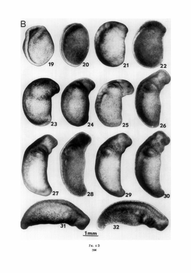
In the list that follows, each stage is into a semicircle. briefly characterized to call attention to Stage 12. Dorsal view. As the archen- diagnostic features in staging. Note the teron forms and the blastocoel recedes, the progressive increase in length of the em- embryo rotates until it rests dorsal side up. bryo from stages 25-40. This brings the blastopore into view from
Stage 1. Uncleaved, single-celled egg. above. The ventral lip has formed to make Cytoplasmic rearrangements occur during the blastopore a complete circle surround-
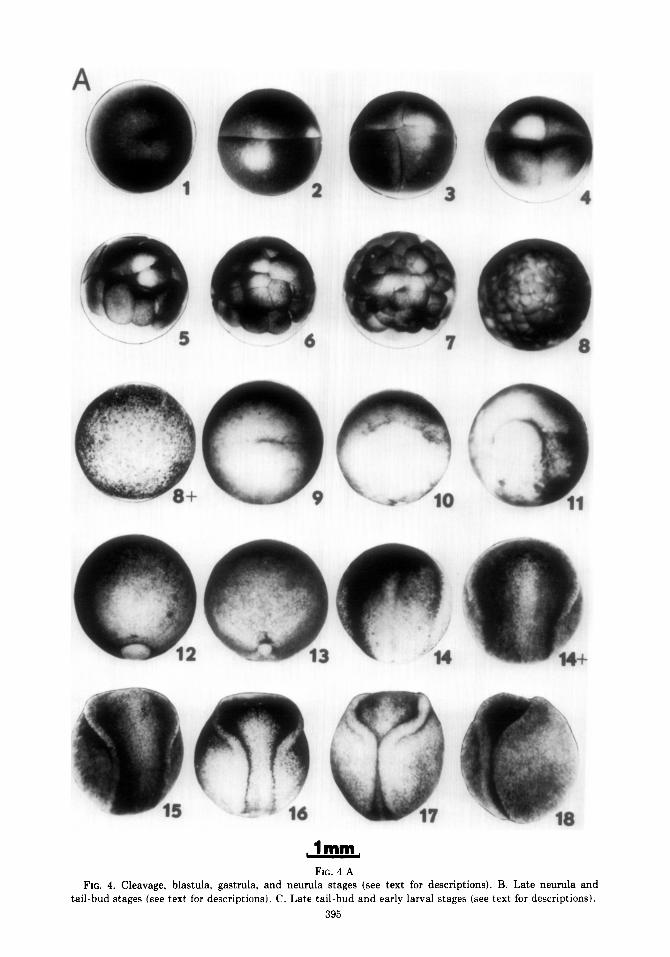
FIG. 4 A FIG. 4. Cleavage, blastula, gastrula, and neurula stages (see text for descriptions). B. Late neurula and
tail-bud stages (see text for descriptions). C. Late tail-bud and early larval stages (see text for descriptions).
395
- --
FIG. 4 B
396

397
BRIEF NoTEs
4”
FIG. 4 c
398 DEVELOPMENTAL BIOLOGY VOLUME 42, 1975
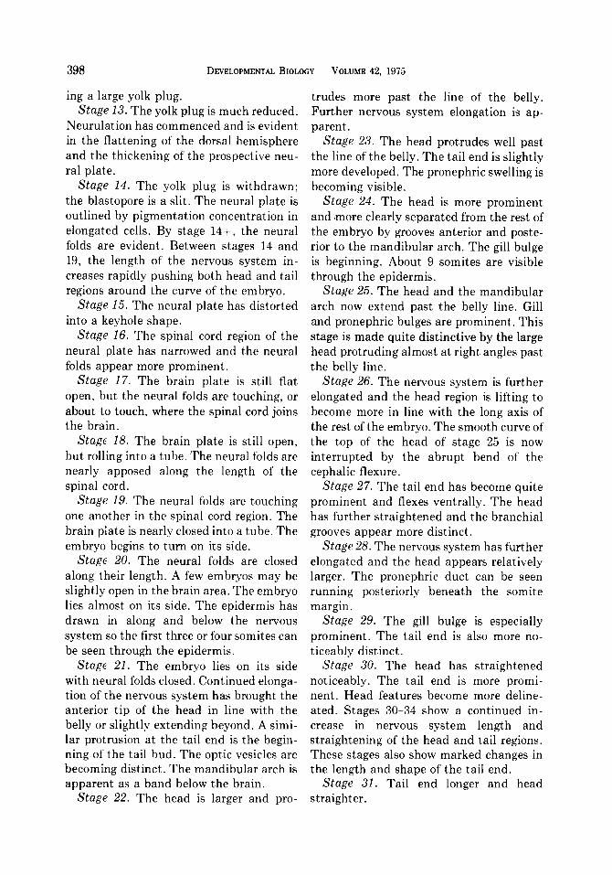
ing a large yolk plug. Stage 13. The yolk plug is much reduced.
Neurulation has commenced and is evident in the flattening of the dorsal hemisphere and the thickening of the prospective neu- ral plate.
Stage 14. The yolk plug is withdrawn; the blastopore is a slit. The neural plate is outlined by pigmentation concentration in elongated cells. By stage 14-t, the neural folds are evident. Between stages 14 and 19, the length of the nervous system in- creases rapidly pushing both head and tail regions around the curve of the embryo.
Stage 15. The neural plate has distorted into a keyhole shape.
Stage 16. The spinal cord region of the neural plate has narrowed and the neural folds appear more prominent.
Stuge 17. The brain plate is still flat open, but the neural folds are touching, or about to touch, where the spinal cord joins the brain.
Stage 18. The brain plate is still open, but rolling into a tube. The neural folds are nearly apposed along the length of the spinal cord.
Stage 19. The neural folds are touching one another in the spinal cord region. The brain piate is nearly closed into a tube. The embryo begins to turn on its side.
Stage 20. The neural folds are closed along their length. A few embryos may be slightly open in the brain area. The embryo lies almost on its side. The epidermis has drawn in along and below the nervous system so the first three or four somites can be seen through the epidermis.
Stage 21. The embryo lies on its side with neural folds closed. Continued elonga- tion of the nervous system has brought the anterior tip of the head in line with the belly or slightly extending beyond. A simi- lar protrusion at the tail end is the begin- ning of the tail bud. The optic vesicles are becoming distinct. The mandibular arch is apparent as a band below the brain.
Stage 22. The head is larger and pro-
trudes more past the line of the belly. Further nervous system elongation is ap- parent.
Stage 23. The head protrudes well past the line of the belly. The tail end is slightly more developed. The pronephric swelling is becoming visible.
Stage 24. The head is more prominent and ,more clearly separated from the rest of the embryo by grooves anterior and poste- rior to the mandibular arch. The gill bulge is beginning. About 9 somites are visible through the epidermis.
Stage 25. The head and the mandibular arch now extend past the belly line. Gill and pronephric bulges are prominent. This stage is made quite distinctive by the large head protruding almost at right angles past the belly line.
Stage 26. The nervous system is further elongated and the head region is lifting to become more in line with the long axis of the rest of the embryo. The smooth curve of the top of the head of stage 25 is now interrupted by the abrupt bend of the cephalic flexure.
Stage 27. The tail end has become quite prominent and flexes ventrally. The head has further straightened and the branchial grooves appear more distinct.
Stage 28. The nervous system has further elongated and the head appears relatively larger. The pronephric duct can be seen running posteriorly beneath the somite margin.
Stage 29. The gill bulge is especially prominent. The tail end is also more no- ticeably distinct.
Stage 30. The head has straightened noticeably. The tail end is more promi- nent. Head features become more deline- ated. Stages 30-34 show a continued in- crease in nervous system length and straightening of the head and tail regions. These stages also show marked changes in the length and shape of the tail end.
Stage 31. Tail end longer and head straighter.
BRIEF NOTES 399
Stage 32. Tail end is longer, head is straighter. The gill bulge is more circum- scribed.
Stage 33. Tail and head both straighter. Groove posterior to gill bulge more promi- nent. At this stage the body muscles be- come responsive. If pricked in the side, the animal bends toward the tormentor.
Stage 34. The nervous system is a straight line from tip of the tail to cephalic flexure. The heart begins to beat. Three divisions of the gills are first apparent.
Stage 35. The three gills are more promi- nent. Fins appear on tail and back. A few pigment cells make their appearance on the flank.
Stage 36. The three gill buds and the nasal pit lie in an almost straight line. Fins and tail have enlarged.
Stage 37. Pigmentation is more exten- sive. The gills protrude farther. Tail and fins are larger.
Stage 38. Gills are longer, finger-like, and project ventrally.
Stage 39. Gills are longer, point more horizontally and posteriorly, and begin to branch. Flanks and eye are much more pigmented. Fins are very extensive. The cloaca is more distinct. This stage lasts a long time and during it all these features change, especially the size and elaboration of the gills.
Stage 40. The distinguishing feature is the turning of the larva onto its belly. Hatching from the membranes occurs at this time. The gills are long and branched. Pigmentation is very extensive. Forelimbs
are just beginning to become visible as bulges behind the gills.
REFERENCES
FRANKHAUSER, G. (1967). Urodeles. In “Methods in Developmental Biology” (F. H. Wilt and N. K. Wessells, eds.), pp. 95-97. Crowell, New York.
HARA, K. (1971). Cinematographic observation of “surface contraction waves” (SCW) during the early cleavage of axolotl eggs. Wilhelm Roui Arch. Entwichlungsmech. Organismen 167, 183-186.
HARRISON, R. G. (1969). “Organization and Develop- ment of the Embryo” (S. Wilens, ed.), pp. 44-66. Yale Univ. Press, New Haven, CT.
IGNAT’EVA, G. M. (1968) (In Russian) Dynamics of morphogenetic movements at the period of gastru- lation in axolotl embryos. Dokl. Akad. Nauk SSSR 179, 1005-1008.
JACOBSON, C.-O. (1959) The localization of the pre- sumptive cerebral regions in the neural plate of the axolotl larva. J. Enbtyol. Exp. Morphol. 7, l-21.
JACOBSON, C.-O. (1962) Cell migration in the neural plate and the process of neurulation in the axolotl larva. zool. &drag Uppsala 35, 433-499.
MEINERTZ, T. (1970) Eine Untersuchung tiber die Dauer der Zellteilung und die Teilungfrequenz in axolotlen-Eiern. 2. Mikrosk. Anat. Forsch. 81, 405-412.
Ron, N. N. (1973) (In Russian) Correlation between karyokinesis and cytokinesis during the first cleav- age divisions in the axolotl (Ambystoma mer- icanum, Cope). Ontogenez 4, 190-192.
SIGNORET, J., and LESFRESNE, J. (1971). Contribution a l’etude de la segmentation de l’oeuf d’axolotl: I. Definition de la transition blastuleenne. Ann. En- bryol. Morphogen. 4, 113-123.
SKOBLINO, M. N. (1965) (In Russian) Dimensionless characterization of the duration of the mitotic phases of the first cleavage divisions in the axolotl. Dokl. Akad. Nuuk SSSR 160, 700-703.
Twrrrv, V. C., and BODENSTEIN, D. (1962). In “Experi-
mental Embryology” (R. Rugh ed.), p. 90, Burgess, Minneapolis, MN.