Embed Size (px)
Citation preview

Personalized Medicine and Imaging
Dual Targeting of Tissue Factor and CD105 forPreclinical PET Imaging of Pancreatic CancerHaiming Luo1, Christopher G. England2, Sixiang Shi3, Stephen A. Graves2,Reinier Hernandez2, Bai Liu4, Charles P. Theuer5, Hing C.Wong4,Robert J. Nickles2, and Weibo Cai1,2,3,6
Abstract
Purpose: Pancreatic adenocarcinoma is a highly aggressivecancer, currently treated with limited success and dismal out-comes.Newdiagnostic and treatment strategies offer the potentialto reduce cancer mortality. Developing highly specific noninva-sive imaging probes for pancreatic cancer is essential to improvingdiagnostic accuracy and monitoring therapeutic intervention.
Experimental Design: A bispecific heterodimer was synthe-sized by conjugating an anti-tissue factor (TF) Fab with an anti-CD105 Fab, via the bio-orthogonal "click" reaction betweentetrazine (Tz) and trans-cyclooctene (TCO). The heterodimer waslabeled with 64Cu for PET imaging of nude mice bearing BXPC-3xenograft and orthotopic pancreatic tumors.
Results: PET imaging of BXPC-3 (TF/CD105þ/þ) xenografttumors with 64Cu-labeled heterodimer displayed significantlyenhanced tumor uptake (28.8� 3.2%ID/g; n¼ 4; SD) at 30 hours
postinjection, as compared with each of their monospecific Fabtracers (12.5 � 1.4 and 7.1 � 2.6 %ID/g; n ¼ 3; SD). In addition,the activity–concentration ratio allowed for effective tumor visu-alization (tumor/muscle ratio 75.2 � 9.4 at 30 hours postinjec-tion.; n¼ 4; SD). Furthermore, 64Cu-NOTA-heterodimer enabledsensitive detection of orthotopic pancreatic tumor lesions with anuptake of 17.1� 4.9%ID/g at 30 hours postinjection and tumor/muscle ratio of 72.3 � 46.7.
Conclusions: This study demonstrates that dual targeting ofTF and CD105 provided synergistic improvements in bindingaffinity and tumor localization of the heterodimer. Dual-targeted imaging agents of pancreatic and other cancersmay assist in diagnosing pancreatic malignancies as well asreliable monitoring of therapeutic response. Clin Cancer Res; 22(15);3821–30. �2016 AACR.
IntroductionPancreatic adenocarcinoma is one of the most lethal cancers
worldwide, with an overall 5-year survival rate of approximately7% (1). Despite aggressive treatment protocols, including surgicalintervention, chemotherapy, and radiotherapy, 5-year survivalrates have remained unchanged over the last decade. These dismaloutcomes have been linked, in part, to ineffective diagnostic toolsdetecting pancreatic malignancies and monitoring therapeuticresponse (2). Currently, there are no clinically reliable serumbiomarkers available for the recognition of early symptoms ofpancreatic cancer in patients (3). Initial diagnoses and diseasestaging are commonly accomplished using CT, MRI, and endo-scopic ultrasonography (4, 5). As over 80% of patients arediagnosed with unresectable disease, there is an urgent need forsensitive and specific imaging agents for pancreatic cancer (6).
Molecular imaging allows for the noninvasive assessment ofphysiologic and pathologic processes at the cellular or molecularlevel in living organisms (7). PET imaging using 18F-FDG caneffectively detect primary pancreatic tumors and hepatic metas-tases commonlymissed by CT andMRI (8).While 18F-FDGPET isa sensitive diagnostic modality for detecting pancreatic malig-nancies, this radioactive sugar molecule is limited by poor spec-ificity and high off-target uptake in inflammatory diseases (9, 10).To improve upon the limitations of 18F-FDG PET, immunoPETemploys radioactive mAbs or antibody fragments for selectivetargeting of malignancies. Antibody-based imaging agents offerhigh specificity for epitopes known to be differentially expressedon cancer cells (11).
Furthermore, bispecific antibody fragments are promisingalternatives to conventional antibody therapeutics, as they allowfor simultaneous recognition of two antigens. The bispecificproperties of heterodimers promote enhanced tumor uptakethrough improved specificity (12, 13). Although bispecific anti-bodies have been proposed for combination therapy in clinicaltrials, their potential utilization as molecular imaging agentsremains largely unexplored (14, 15).
Concurrent targeting of tumor vasculature and cancercells can provide additional benefits for molecular imaging.A majority of pancreatic adenocarcinomas are associatedwith arterial thrombosis, migratory thrombophlebitis, tumorangiogenesis, and rapid metastasis (16, 17). Tissue factor (TF),which serves as the primary initiator of the extrinsic pathwayof blood coagulation, is overexpressed in most solid tumors,including pancreatic cancer (18). In general, it is thought thatoverexpression of TF contributes to higher incidence rates of
1Department of Radiology, University of Wisconsin-Madison,Wiscon-sin. 2DepartmentofMedicalPhysics,UniversityofWisconsin-Madison,Wisconsin. 3Materials ScienceProgram,UniversityofWisconsin-Madi-son,Wisconsin. 4Altor BioSciences, Miramar, Florida. 5TRACON Phar-maceuticals, Inc., San Diego, California. 6University of Wisconsin Car-bone Cancer Center, Madison,Wisconsin.
Note: Supplementary data for this article are available at Clinical CancerResearch Online (http://clincancerres.aacrjournals.org/).
Corresponding Author:Weibo Cai, University of Wisconsin–Madison, 1111 High-land Ave, Madison, WI 53705-2275. Phone: 608-262-1749; Fax: 608-265-0614;E-mail: [email protected]
doi: 10.1158/1078-0432.CCR-15-2054
�2016 American Association for Cancer Research.
ClinicalCancerResearch
www.aacrjournals.org 3821
on July 8, 2018. © 2016 American Association for Cancer Research. clincancerres.aacrjournals.org Downloaded from
Published OnlineFirst March 29, 2016; DOI: 10.1158/1078-0432.CCR-15-2054

thrombotic complications in cancer patients (19). Similarly,CD105 (also called endoglin) is a proliferation-associated cell-surface protein highly expressed on activated endothelial cells.Overexpression of CD105 is known to be associated withdecreased patient survival for most cancers (20). Given the het-erogeneous nature and complex stromal microenvironment ofpancreatic cancer, targeting of individual tumor-specific antigensmay result in suboptimal imaging agent accumulation (21).
In the current article, we describe the development andevaluation of a novel bispecific heterodimer through conjuga-tion of two mAb Fab fragments, respectively, targeting TF andCD105. Antibody fragments were generated through enzymaticdigestion of ALT-836, an anti-TF chimeric mAb, and TRC105, amAb recognizing both human and murine CD105. The con-jugation of two mAb fragments was accomplished through theinverse-electron-demand Diels–Alder reaction between elec-tron-deficient tetrazine (Tz) and strained trans-cyclooctene(TCO), taking advantage of the emerging TCO/Tz "click chem-istry" platform. In addition, we have shown that the biologicactivity of the heterodimer was maintained after the conjuga-tion reaction. To date, there are no investigations into thesimultaneous targeting of TF and CD105 for molecular imag-ing. We hypothesized that this TF/CD105 heterodimer wouldsynergistically harness the targeting capabilities of ALT-836-Faband TRC105-Fab. To test this hypothesis, we examined theadvantages of dual TF/CD105 targeting in terms of tumor-binding affinity and specificity in a human pancreatic cancermouse model.
Materials and MethodsChemicals
TRC105 and ALT-836 were, respectively, provided by TRACONPharmaceuticals Inc. and Altor Bioscience Corporation. 2-S-(4-isothiocyanatobenzyl)-1,4,7-triazacyclononane-1,4,7-triaceticacid (p-SCN-Bn-NOTA) was obtained from Marocyclics, Inc..TCO-PEG4-NHS ester and Tz-PEG5-NHS ester were purchasedfrom Click Chemistry Tools. Pierce immobilized papain, proteinA column, and all other reaction buffers and chemicals werepurchased from Thermo Fisher Scientific.
Fab generation and characterizationALT-836 (2mg/mL) and TRC105 (2mg/mL) were individually
digested in reaction buffer [20mmol/L sodiumphosphatemono-basic, 10 mmol/L disodium ethylenediaminetetraacetic acid(EDTA), and 80 mmol/L cysteine, HCl] for 4 hours at 37�C, withimmobilized papain/total volume at a ratio of 1:10. The reactionmixture was centrifuged at 13,200 rpm for 5 minutes to removeimmobilized papain. The reaction was filtered through a Milli-pore 0.22-mm syringe filter (EMD Millipore) before the superna-tant was purified by size exclusion chromatography using aHiPrep 16/60 Sephacryl S-100 HR (GEHealthcare). The collectedfraction (�50 kDa) was concentrated using a 10-kDa molecularweight cut-off spin filter (Amicon Ultra-4) and purified with aprotein A column (NAb Protein A Spin Kit; Thermo FisherScientific). The purity of ALT-836-Fab and TRC105-Fab wasevaluated by SDS–PAGE with Coomassie brilliant blue R-250staining (Thermo Fisher Scientific).
Heterodimer synthesis and purificationALT-836-Fab (10 nmol/L) was mixed with 20 nmol/L TCO-
PEG4-NHS ester in PBS at pH 8.5 and incubated for 2 hours at25�C. Concurrently, 10 nmol/L of TRC105-Fab was incubatedwith 20 nmol/L of Tz-PEG5-NHS ester using the same conditionsdescribed for TCO-PEG4-NHS ester. Reaction mixtures wereindividually passed through PD-10 columns for collection ofactivated ALT-836-Fab and TRC105-Fab, respectively. ActivatedALT-836-Fab and TRC105-Fab weremixed at an equimolar ratioin PBS buffer and incubated at 25�C for 2 hours. The hetero-dimers were separated by gel filtration on a HiPrep 16/60Sephacryl S-100 HR and purity was evaluated by 10% SDS–PAGE gel.
NOTA conjugation and 64Cu labelingChelation of Fab conjugates was accomplished with a reaction
ratio of p-SCN-Bn-NOTA: Fab of 10:1 at pH 9.0. NOTA-hetero-dimer was purified using a PD-10 column (GE Healthcare LifeSciences) with PBS as themobile phase. High-specific activity (> 5Ci/mmol) 64Cu was produced in a CTI RDS 112 cyclotron via 64Ni(p,n)64Cu reaction. For radiolabeling, 50 to 100 mg of NOTA-Fabwas reacted with 74-148-MBq (2–4 mCi) of 64CuCl2 in 300 mL ofsodium acetate buffer (0.1 mol/L, pH 4.5) at 37�C for 30minutesunder constant agitation (400 rpm). 64Cu-NOTA-Fab was puri-fied from free activity using a PD-10 column with PBS as themobile phase.
Cell lines and animal modelThe human pancreatic adenocarcinoma cell line (BXPC-3) was
obtained from ATCC and cultured according to the supplier'sprotocol. All animal studies were conducted under a protocolapproved by the University of Wisconsin Institutional AnimalCare and Use Committee. For implantation, 5 � 106 tumor cellsweremixed withMatrigel (BD Biosciences) at a ratio of 1:1 beforesubcutaneously injected into the forelimb axillary of 4- to 5-week-old female athymic nude mice (Envigo). Over the subsequent 3to 4 weeks postinoculation, tumors were monitored and allowedto grow to 5 to 8 mm in diameter.
For orthotopic tumor models, the splenic portion of the pan-creas was carefully exposed and 2 � 106 tumor cells in Matrigel:PBS (ratio 1:1, total volume of 30 mL) were injected into thetail of the pancreas. The incision was closed in two layers (peri-toneum and abdominal wall) with interrupted sutures. After
Translational Relevance
The high mortality rate of pancreatic cancer can be attrib-uted to insufficient diagnostic tools for early disease detection.Several novel imaging agents have beenpreclinically evaluatedfor detecting pancreatic cancer, yet many of these agents aretargeted to single receptors with limited specificity. In thisstudy, we investigate the advantages of using a novel hetero-dimeric imaging construct, cotargeted to the tumor vasculature(CD105) and the surface of malignant cells (tissue factor), formolecular imaging. The heterodimer was shown to be a moreeffective PET imaging agent, in comparison with single-tar-geted CD105 or tissue factor antibody tracers, in both xeno-graft and orthotopic mouse models. Currently, the clinicalbenefits of heterodimeric imaging agents for the recognition ofearly disease symptoms and monitoring of therapeuticsresponse in patients have been unexplored.
Luo et al.
Clin Cancer Res; 22(15) August 1, 2016 Clinical Cancer Research3822
on July 8, 2018. © 2016 American Association for Cancer Research. clincancerres.aacrjournals.org Downloaded from
Published OnlineFirst March 29, 2016; DOI: 10.1158/1078-0432.CCR-15-2054

implantation, nude mice were inspected daily for bleeding orwound complications. After 2 weeks, mice were examined fortumor formation twice a week by palpation. After 5 to 12 weeks,tumors were confirmed by PET imaging and histology to bepancreatic adenocarcinoma.
Flow cytometryIn vitro TF and CD105 binding affinity/specificity of the
heterodimer was evaluated by flow cytometry in BXPC-3 cells.Briefly, cells were harvested and suspended in PBS supplemen-ted with 2% BSA at a concentration of 5 � 106 cells/mL. Cellswere incubated with 100 nmol FITC-labeled heterodimer andFab conjugates for 30 minutes at 25�C. After washing, sampleswere analyzed with a FACSCalibur 4-color cytometer (BDBiosciences). FlowJo software was employed for analysis offlow cytometry data (Tree Star Inc.).
PET imaging and biodistribution studiesPET and PET/CT scans were performed using an Inveon micro-
PET/microCT rodent scanner (Siemens Healthcare). Image recon-struction and region of interest (ROI) analysis of each PET imagewas carried out as described previously (22). BXPC-3 tumor-bearing mice were intravenously injected with 5 to 10 MBq
(�150–300 mCi) of either 64Cu-NOTA-heterodimer, 64Cu-NOTA-ALT-836-Fab, or 64Cu-NOTA-TRC105-Fab. Sequentialstatic PET scans were acquired at 3, 15, 24, and 30 hours post-injection of tracers. Twentymillion coincidence events permousewere acquired for each static PET emission scan. CT-based atten-uation correction was performed, and the registered CT image setfused with the PET image set for anatomic orientation. After thelast PET scan at 30 hours postinjection, mice were euthanized forbiodistribution analysis. Tissues were collected and wet-weighedbefore the activity was assayed using an automated g-counter(2470 WIZARD 2; Perkin Elmer). The activity concentration wasdecay corrected to the time of injection, and the results wereexpressed as apercentage of injected radioactivity dose per gramoftissue (%ID/g; mean � SD; � 3 mice per group).
HistologySlices of tissue at 5-mm thickness were fixed with cold acetone
for 10minutes and air-dried for 30minutes. After rinsingwith PBSand blocking with 10%donkey serum for 30minutes at 25�C, theslides were incubated with 20 nmol/L FITC-labeled Fab conju-gates for TF or CD105 staining. Then, rat anti-mouse CD31antibody (Thermo Fisher Scientific) and Cy3-labeled donkeyanti-rat IgG (Thermo Fisher Scientific) were used for CD31
Figure 1.Synthesis and characterization of the heterodimer. A, schematic representation of the synthesis of the heterodimer. B, SDS–PAGE gel confirming the identity andpurity of heterodimer. C, flow cytometry analysis in BXPC-3 after 1-hour incubation of FITC-Fab conjugates (50 nmol/L) confirmed the dual targeting of TF andCD105 and specificity of the heterodimer. D, competitive binding assay comparing heterodimer (circles), ALT-836-Fab (squares), and TRC105-Fab (triangles)binding affinities. IC50 values were markedly lower for the heterodimer (11.35 � 1.04 nmol/L) compared with ALT-836-Fab (288.9 � 18 nmol/L) and TRC105-Fab(583.9 � 36 nmol/L).
PET Imaging of Pancreatic Cancer
www.aacrjournals.org Clin Cancer Res; 22(15) August 1, 2016 3823
on July 8, 2018. © 2016 American Association for Cancer Research. clincancerres.aacrjournals.org Downloaded from
Published OnlineFirst March 29, 2016; DOI: 10.1158/1078-0432.CCR-15-2054
staining (red). 4',6-diamidino-2-phenylindole (DAPI; ThermoFisher Scientific) was used to stain cell nuclei and confocalfluorescence images were acquired with an Eclipse Ti microscope(Nikon).
Statistical analysesQuantitative data were expressed as mean � SD. Means were
compared using the unpaired Student t test. P values of less than0.05 were considered statistically significant.
ResultsSynthesis and characterization of heterodimer
Monovalent Fab antibody fragments were created by papaindigestion of intact ALT-836 or TRC105 mAb and separated bysize exclusion chromatography. Purification of fragments wasaccomplished with protein A affinity columns. Derived Fabfragments were reacted with Diels–Alder orthogonal reactivepair Tz/TCO for the generation of bispecific heterodimer(Fig. 1A). The reaction was monitored by SDS–PAGE and aband at approximately 100 kDa was observed, correspondingto the Fab dimer molecular weight. Bispecific heterodimerantibody fragments were purified by size exclusion chroma-tography from unreacted Fabs, and the reaction efficiency wascalculated to be 38% (Supplementary Fig. S1). Purity andidentity of the heterodimer (M[H]þ:103.50 kDa) were con-
firmed by SDS–PAGE and MALDI-TOF mass spectra (Fig. 1Band Supplementary Fig. S2).
In vitro bispecificity of the heterodimerTo evaluate the binding affinity and bispecificity of the
heterodimer, FITC-labeled Fab conjugates were used to sepa-rately visualize the binding to human pancreatic cancer cells(BXPC-3), which express high levels of both TF and CD105.Compared with TRC105-Fab and ALT-Fab, the heterodimerrevealed significantly stronger binding to BXPC-3 cells (Fig.1C and Supplementary Fig. S3), which was effectively blockedwith a saturating dose of ALT-836 or TRC105 antibody or both.Together, these results demonstrate that dual targeting of TFand CD105 resulted in enhanced binding affinity and speci-ficity of the heterodimer.
Finally, a competitive binding assay was performed to deter-mine and compare the binding affinities of the heterodimer,ALT-836-Fab, and TRC105-Fab to BXPC-3 cells (Fig. 1D). Theresults of the binding isotherm showed a concentration-depen-dent displacement of bound 64Cu-NOTA-heterodimer withIC50 values of 11.35 � 1.04, 288.9 � 18, and 583.9 � 36nmol/L for heterodimer, ALT-836-Fab, and TRC105-Fab,respectively. Only a partial displacement of the bound radi-oligand at high concentrations (mmol/L) of the competingantibody fragments demonstrated the ambivalent nature ofheterodimer binding.
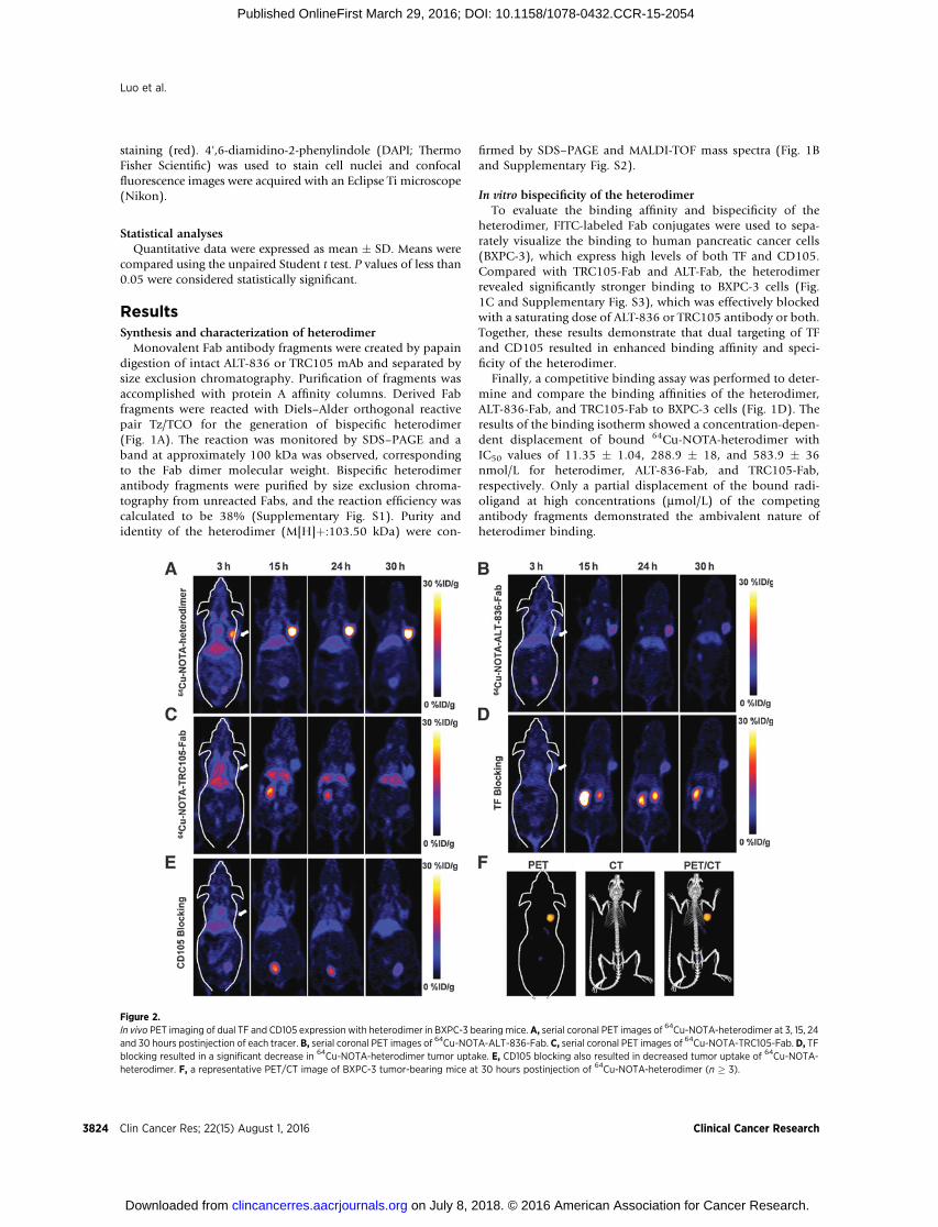
Figure 2.In vivo PET imaging of dual TF and CD105 expression with heterodimer in BXPC-3 bearing mice. A, serial coronal PET images of 64Cu-NOTA-heterodimer at 3, 15, 24and 30 hours postinjection of each tracer. B, serial coronal PET images of 64Cu-NOTA-ALT-836-Fab. C, serial coronal PET images of 64Cu-NOTA-TRC105-Fab. D, TFblocking resulted in a significant decrease in 64Cu-NOTA-heterodimer tumor uptake. E, CD105 blocking also resulted in decreased tumor uptake of 64Cu-NOTA-heterodimer. F, a representative PET/CT image of BXPC-3 tumor-bearing mice at 30 hours postinjection of 64Cu-NOTA-heterodimer (n � 3).
Luo et al.
Clin Cancer Res; 22(15) August 1, 2016 Clinical Cancer Research3824
on July 8, 2018. © 2016 American Association for Cancer Research. clincancerres.aacrjournals.org Downloaded from
Published OnlineFirst March 29, 2016; DOI: 10.1158/1078-0432.CCR-15-2054

Heterodimer shows enhanced tumor-specific targeting in vivoTo evaluate the dual-targeting properties and high specificity of
the heterodimer in vivo, each antibody fragmentwas conjugated tothe chelator NOTA and radiolabeled with 64Cu. The averagenumber of NOTA per antibody fragment was 3.6 � 0.4 (Supple-mentary Fig. S4), and radiolabeling yield was 91.5% after 15-minute incubationwith 64Cu, as determined by radio-TLC. A totalof 200–300 mCi of 64Cu-NOTA-heterodimer, 64Cu-NOTA-ALT-836-Fab, or 64Cu-NOTA-TRC105-Fab were administered toBXPC-3 (TF/CD105þ/þ) tumor-bearing athymic nude mice andtime points of 3, 15, 24, and 30 hours postinjection were chosenfor serial static PET scans imaging, based upon previous PETimaging results using monovalent and divalent antibody frag-ments (23, 24). Coronal PET images of BXPC-3 tumor-bearingmice showed rapid tumor accumulation of all three tracers withsharp delineation of tumor xenografts. The 64Cu-NOTA-hetero-
dimer displayed significantly higher tumor accumulation, incomparison with the two monovalent fragments (Fig. 2A; Sup-plementary Movie S1). Also, a representative PET/CT image of aBXPC-3 tumor-bearing mouse at 30 hours postinjection with64Cu-NOTA-heterodimer is shown in Fig. 2F. ROI analyses ofthe images were performed for quantification of tracer uptake andexpressed as%ID/g in BXPC-3 tumors as well as nontarget tissues,including blood pool, liver, kidney, and muscle. As clearly indi-cated in PET images (n ¼ 4; Figs. 2A and 3A), 64Cu-NOTA-heterodimer displayed high tumor accumulation at early timepoints (18.7� 4.6%ID/g at 3 hours postinjection) that peaked at29.7 � 3.3 %ID/g, 24 hour postinjection of tracer. Maximumtumor uptake values for 64Cu-NOTA-ALT-836-Fab and 64Cu-NOTA-TRC105-Fab were significantly lower (P < 0.01) at 13.3� 1.7%ID/g and 8.7� 1.8%ID/g, respectively (n¼ 3; Fig. 2B andC and Fig. 3B and C). In contrast, the peak tumor uptake of free
Figure 3.QuantitativeROI analysis of in vivo imaging data.A, time-activity curves of theBXPC-3 tumor, blood, liver, kidney, andmuscle following intravenous administration of64Cu-NOTA-heterodimer, 64Cu-NOTA-ALT-836-Fab (B), or 64Cu-NOTA-TRC105-Fab (C). D, time-activity curves of BXPC-3 tumor and tissues after intravenousadministration of 64Cu-NOTA-heterodimer after TF or E, 64Cu-NOTA-heterodimer after CD105 blocking. F, comparison of tumor uptake in above all groupsby quantitative analysis of the PET data (n � 3).
PET Imaging of Pancreatic Cancer
www.aacrjournals.org Clin Cancer Res; 22(15) August 1, 2016 3825
on July 8, 2018. © 2016 American Association for Cancer Research. clincancerres.aacrjournals.org Downloaded from
Published OnlineFirst March 29, 2016; DOI: 10.1158/1078-0432.CCR-15-2054

copper was 2.0 � 1.8 %ID/g, which was significantly lower thanthat of this Fab conjugate (n ¼ 3 and Supplementary Fig. S5).Consistent with its higher molecular weight, 64Cu-NOTA-hetero-dimer displayed a longer blood half-life, evidenced by the initiallyelevated heart activity concentrations of 11.5� 1.2%ID/g and 2.1� 0.5 %ID/g at 3 and 30 hours postinjection, respectively (n ¼4; Fig. 3A). Kidney uptake of 64Cu-NOTA-ALT-836-Fab and 64Cu-NOTA-TRC105-Fab was higher than that of 64Cu-NOTA-hetero-dimer, demonstrating renal clearance as the major excretionpathway for Fab fragments (Fig. 3B and C). Liver uptake wascomparable between 64Cu-NOTA-heterodimer and 64Cu-NOTA-ALT-836-Fab; however, twofold lower values were registered for64Cu-NOTA-TRC105-Fab, indicating less dominant hepatic clear-ance of this Fab conjugate.
To demonstrate that 64Cu-NOTA-heterodimer retained its invivo specificity toward both TF and CD105, a large (40 mg/kg)dose of either ALT-836 or TRC105 or both ALT-836 and CD105intact antibody was administered in mice 12 hours prior toinjection of the bispecific tracer. Peak tumor uptake values of64Cu-NOTA-heterodimer significantly diminished to 6.0 � 1.3%ID/g, 8.9 � 3.8 %ID/g, and 2.8 � 1.0 %ID/g after TF, CD105,and TF plus CD105 blockade (n ¼ 3; Fig. 2D, E, 3D, E andSupplementary Fig. S6), respectively. Tracer uptake in all major
organs was similar between 64Cu-NOTA-heterodimer and 64Cu-NOTA-heterodimerwith TForCD105blockade, except the BXPC-3 tumor (significantly higher in the former), further confirmingthe TF and CD105 dual specificity of 64Cu-NOTA-heterodimer(Fig. 3F). PET data demonstrated that dual-targeting using theheterodimer tracer offers a significant advantage in terms ofabsolute tumor uptake, target specificity, and off-target uptakecompared with monospecific Fab fragments.
After terminal scans at 30 hours postinjection, ex vivo biodis-tribution and histology studies were performed to validate in vivoPETdata and confirm thebiodistributionprofile of tracers (Fig. 4Aand B). 64Cu-NOTA-heterodimer showed prominent tumor andlow background uptake, with an excellent tumor/muscle ratio of75.2 � 9.4. This was superior to those of 64Cu-NOTA-ALT-836-Fab (42.4 � 17.6) and 64Cu-NOTA-TRC105-Fab (29.0 � 19.7),achieved at 30 hours postinjection (Fig. 4C). Figure 4D shows nostatistically significant difference between PET-derived and bio-distribution data, suggesting that ROI analyses of the PET imagesaccurately reflected tracer distribution in vivo as well as the dualspecificity of 64Cu-NOTA-heterodimer.
Immunofluorescence staining of resected BXPC-3 tumor sec-tions was carried out to correlate PET-tracer uptake with in situ TF/CD105 expression (Fig. 5). FITC-labeled Fab conjugates were
Figure 4.Ex vivobiodistribution validates the results of PET imaging.A,biodistribution of 64Cu-NOTA-heterodimer,64Cu-NOTA-ALT-836-Fab, and 64Cu-NOTA-TRC105-Fab inBXPC-3 bearing mice, 30 hours postinjection (n � 3). B, biodistribution of 64Cu-NOTA-heterodimer after TF or CD105 blocking (n � 3). C, tumor-to-normaltissue comparison of 64Cu-NOTA-heterodimer and 64Cu-labeled Fab fragments. D, comparison of tumor uptake of 64Cu-NOTA-heterodimer and Fab conjugatetracers between microPET and biodistribution data, 30 hours postinjection Differences were not statistically significant (P > 0.05). n.s., not significant.
Luo et al.
Clin Cancer Res; 22(15) August 1, 2016 Clinical Cancer Research3826
on July 8, 2018. © 2016 American Association for Cancer Research. clincancerres.aacrjournals.org Downloaded from
Published OnlineFirst March 29, 2016; DOI: 10.1158/1078-0432.CCR-15-2054

visualized using fluorescence microscopy, which showed locali-zation consistent with TF and CD105 in situ expression profiles.FITC-TRC105-Fab uptakewas localizedwithin tumor vasculature,confirmed by colocalization with CD31, and on the cells, whereasFITC-ALT-836-Fab was found on the membrane of TF-expressingBXPC-3 cells. In agreement with its TF/CD105 bivalent property,FITC-heterodimer staining showed a stronger fluorescent signalthat was distributed in both BXPC-3 cells and tumor-associatedvasculature in the tissue. Blocking with ALT-836 or TRC105resulted in low-intensity signals of TF/CD105 staining, confirm-ing the TF/CD105 bispecificity of 64Cu-NOTA-heterodimer.
Heterodimer shows enhanced detection of orthotopicpancreatic tumors
To investigate the potential of 64Cu-NOTA-heterodimer forsensitive detection of pancreatic adenocarcinomas, an orthotopictumor model was established by stereotactically seeding BXPC-3cells in the pancreas. The 64Cu-NOTA-heterodimer was examinedfor its ability to detect in vivo pancreatic malignancies. Growthkinetics of BXPC-3 orthotopic tumors was monitored using theheterodimer imaging agent, and the tumors were shown tosteadily increase in size from day 30 to 75 (Fig. 6A). Sequentialcoronal and transverse images of slices containing BXPC-3 ortho-
topic tumors showed a sharp definition of tumor contours,whereas negligible radioactivity was observed surrounding thekidney position (Fig. 6B; Supplementary Movie S2). Immuno-PET/CT imaging was performed on mice with confirmed tumors,approximately 2 to 3 mm in diameter (Fig. 6C). Posterior quan-titative analysis of the images in orthotopic mice revealed apersistently high tumor uptake of 64Cu-NOTA-heterodimer from9.5� 1.8%ID/g at 3 hours postinjection to the peak value of 17.8� 1.5 %ID/g at 24 hours postinjection. This is in contrast with no64Cu-NOTA-heterodimer accumulation at the pancreas of normalmice (Supplementary Fig. S7, S8 andMovie S3). Altogether, thesedata suggest that 64Cu-NOTA-heterodimer offers sufficient sensi-tivity for the detection of TF/CD105-positive orthotopic pancre-atic tumors.
After the last imaging time point (30 hours postinjection),ex vivo imaging in mice with orthotopic pancreatic tumorsand biodistribution studies were performed. Primary tumorsin the pancreas were clearly visible and displayed high activityfurther validating in vivo PET data (Fig. 6D). Ex vivo64Cu-NOTA-heterodimer biodistribution studies at 30 hours post-injection showed a tissue distribution of tracer similar tothat of the subcutaneous xenograft models (Fig. 6E). Hema-toxylin and eosin (H&E) staining of pancreas sections
Figure 5.Immunofluorescence staining ofBXPC-3 tumors sections. FITC-heterodimer, FITC-ALT-836-Fab,FITC-TRC105-Fab, and FITC-heterodimer with TF or CD105blocking were used for TF/CD105staining (green). Rat anti-mouse CD31antibody and Cy3-labeled donkeyanti-rat IgG were used for CD31staining (red). DAPI was used to staincell nuclei (bar, 30 mm).
PET Imaging of Pancreatic Cancer
www.aacrjournals.org Clin Cancer Res; 22(15) August 1, 2016 3827
on July 8, 2018. © 2016 American Association for Cancer Research. clincancerres.aacrjournals.org Downloaded from
Published OnlineFirst March 29, 2016; DOI: 10.1158/1078-0432.CCR-15-2054

confirmed the presence of a localized tumor mass, consistentwith the observation of a sharply defined tumor in PET/CTimages (Fig. 6F). In addition, TF/CD105 immunofluorescencerevealed a strong fluorescent signal that distributed in bothtumor cell and tumor-associated vasculature (colocalized withCD31). Overall, small animal PET/CT using 64Cu-NOTA-het-erodimer enabled highly specific and sensitive detection ofaggressive BXPC-3 pancreatic tumors.
DiscussionMost patients with pancreatic cancer are diagnosed with met-
astatic disease, and only a small fraction of patients are suitablecandidates for surgical resection (6). Although a recent random-ized phase III trial showed that gemcitabine, in combination withnab-paclitaxel, significantly improved overall survival of patientswith metastatic pancreatic cancer, advanced pancreatic canceroften becomes highly resistant to chemotherapy (25). To enhancepatient survival, there is an urgent need for improved diagnostictools for simplified detection of pancreatic malignancies. Molec-ular imaging not only has the potential to improve the diagnosticimaging and staging of malignancies but can also provide valu-able intraoperative guidance to enhance patient survival duringsurgical intervention (7). The specificity of molecular imaging
probes is imperative to accurately diagnose and stage pancreaticcancer.
Imaging disease-related biomarkers enables more accuratepatient classification and better prognostication of treatmentsuccess. A link between pancreatic cancer and venous throm-boembolisms has been well established and is thought to beassociatedwith tumor growth and angiogenesis (26). Overexpres-sion of TF in pancreatic cancer plays a critical role in the patho-physiology of cancer-related thrombosis and angiogenesis (27).Because of this correlation, combined targeting of TF and angio-genic pathways is an appealing strategy to potentially circumventtreatment resistance and improve patient survival. In pancreaticcancer, overexpression of angiogenic factors, such as VEGF,CD105, and integrin correlates with disease progression (20,28). However, treatments targeting known oncogenes or growthfactors such as K-Ras, VEGF, and EGF/EGFR in pancreatic cancerhave resulted in suboptimal therapeutic response (29). Upregula-tion of CD105 is common inmost solid tumors, so the concurrenttargeting of TF andCD105mayoffer newpossibilities for effectivetreatment of pancreatic malignancies.
In this study, we sought to investigate the benefits of simulta-neous targeting of TF and CD105 for potential imaging andtherapy of pancreatic cancer. By fusing two Fab fragments frommAbs against TF and CD105, respectively, we created a
Figure 6.Heterodimer-based PET imaging in orthotopic BXPC-3 tumors. A, serial coronal PET images of 64Cu-NOTA-heterodimer in mice at 30, 60, and 75 days of seedingBXPC-3 cells into the tail of the pancreas.B, coronal and transverse PET images ofmice-bearing orthotopic BXPC-3 tumor at 3, 15, 24, and 30 hours following injectionof 64Cu-NOTA-heterodimer. C, coregistered PET/CT images of mice-bearing orthotopic BXPC-3 tumor at 30 hours postinjection of 64Cu-NOTA-heterodimer.D, ex vivo PET imaging of excised pancreas from the mouse-bearing orthotopic BXPC-3 tumor at 30 hours postinjection E, ex vivo biodistribution of 64Cu-NOTA-heterodimerin orthotopic BXPC-3 tumor-bearing mice and normal mice at 30 hours postinjection (n ¼ 3); F, H&E and immunofluorescence staining of normalpancreatic tissue and pancreatic tumors (bar, 30 mm).
Clin Cancer Res; 22(15) August 1, 2016 Clinical Cancer Research3828
Luo et al.
on July 8, 2018. © 2016 American Association for Cancer Research. clincancerres.aacrjournals.org Downloaded from
Published OnlineFirst March 29, 2016; DOI: 10.1158/1078-0432.CCR-15-2054

heterobifunctional construct possessing excellent in vivo tumor-homing capabilities. Our results from noninvasive PET imagingwith 64Cu-NOTA-heterodimer demonstrated a significantlyhigher tumor uptake of the heterodimer compared with that ofeither the Fab fragment orwhole antibody (24, 30). This indicatedthat dual-TF/CD105 targeting provides a synergistic tumor-target-ing advantage in BXPC-3 tumors (Figs. 3 and 4). More impor-tantly, this targeting advantage did not occur at the expense ofincreased nonspecific tracer accumulation in normal organs, asindicated by a high tumor/muscle ratio of 75.2� 9.4 at 30 hoursafter 64Cu-NOTA-heterodimer administration. BXPC-3 orthoto-pic tumor nodules were easily identifiable, owing to high traceruptake (17.1 � 4.9 %ID/g at 30 hours postinjection) and tumor/muscle ratio (72.3 � 46.7). These findings may have significantimplications for cancer therapy, in which combined TF andantiangiogenic inhibition therapies may effectively address cur-rent therapeutic limitations, including TF inhibitor resistance andthe heterogeneous expression of TF in pancreatic cancer.
An emerging trend in the clinical implementation of multi-functional pharmaceutics is theranostics, a combined diagnosticand therapeutic approach designed to eliminate multistep pro-cedures, reduce delays in treatment, and facilitate patient careoverall (31). Recently, a bispecific antibody known as Blincyto(blinatumomab, AMGEN) was approved by the FDA for treatingB-cell acute lymphoblastic leukemia (32). This has much interestin the application of bispecific antibodies for cancer diagnosticsand treatment (14). The simplified molecular engineering plat-form we presented in this study is not limited to antibodies, yet iswidely applicable to other disease targeting ligands, includingpeptides, small-molecular-weight proteins, aptamers, and manynanoplatforms.
In conclusion, our novel heterodimer demonstrated that dual-targeting enhanced tumor accumulation, augmented targetingspecificity, and improved diagnostic sensitivity. This enabled the
detection of 2- to 3-mm pancreatic malignant lesions in ortho-topic tumor-bearing mice. In the future, this paradigm may beexpanded for the construction of novel heterodimers using pre-viously failed clinical antibody candidates. In turn, those failedantibodies may be revived for improving therapeutic outcomes.
Disclosure of Potential Conflicts of InterestC.P. Theuer is a CEO at TRACON Pharmaceuticals. No potential conflicts
of interest were disclosed by the other authors.
Authors' ContributionsConception and design: H. Luo, C.G. England, H.C. Wong, W. CaiDevelopment of methodology: H. Luo, S. Shi, R. HernandezAcquisition of data (provided animals, acquired and managed patients,provided facilities, etc.): H. Luo, C.G. England, S. Shi, S.A. GravesAnalysis and interpretation of data (e.g., statistical analysis, biostatistics,computational analysis): H. Luo, C.G. England, W. CaiWriting, review, and/or revision of the manuscript: H. Luo, C.G. England,R. Hernandez, C.P. Theuer, H.C. Wong, W. CaiAdministrative, technical, or material support (i.e., reporting or organizingdata, constructing databases): H. Luo, S. Shi, S.A. Graves, B. Liu, W. CaiStudy supervision: H. Luo, C.G. England, W. CaiOther (provide radioactive precursors): R.J. Nickles
Grant SupportThis work is supported, in part, by the University ofWisconsin–Madison, the
NIH (NIBIB/NCI 1R01CA169365, P30CA014520, 5T32GM08349, andT32CA009206), the Department of Defense (W81XWH-11-1-0644 andW81XWH-11-1-0648), the National Science Foundation (DGE-1256259), andthe American Cancer Society (125246-RSG-13-099-01-CCE).
The costs of publication of this articlewere defrayed inpart by the payment ofpage charges. This article must therefore be hereby marked advertisement inaccordance with 18 U.S.C. Section 1734 solely to indicate this fact.
Received August 22, 2015; revised February 1, 2016; acceptedMarch 1, 2016;published OnlineFirst March 29, 2016.
References1. Siegel RL, Miller KD, Jemal A. Cancer statistics, 2015. CA Cancer J Clin
2015;65:5–29.2. Zamboni G, Hirabayashi K, Castelli P, Lennon AM. Precancerous lesions of
the pancreas. Best Pract Res Clin Gastroenterol 2013;27:299–322.3. Goggins M. Markers of pancreatic cancer: working toward early detection.
Clin Cancer Res 2011;17:635–7.4. Holzapfel K, Reiser-Erkan C, Fingerle AA, ErkanM, Eiber MJ, Rummeny EJ,
et al. Comparison of diffusion-weighted MR imaging and multidetector-row CT in the detection of liver metastases in patients operated forpancreatic cancer. Abdom Imaging 2011;36:179–84.
5. Canto MI, Hruban RH, Fishman EK, Kamel IR, Schulick R, Zhang Z, et al.Frequent detection of pancreatic lesions in asymptomatic high-risk indi-viduals. Gastroenterology 2012;142:796–804.
6. Vincent A,Herman J, SchulickR,HrubanRH,GogginsM. Pancreatic cancer.Lancet 2011;378:607–20.
7. Hussain T, Nguyen QT. Molecular imaging for cancer diagnosis andsurgery. Adv Drug Deliv Rev 2014;66:90–100.
8. Pakzad F, Groves AM, Ell PJ. The role of positron emission tomography inthe management of pancreatic cancer. Semin Nucl Med 2006;36:248–56.
9. Nguyen NQ, Bartholomeusz DF. 18F-FDG-PET/CT in the assessment ofpancreatic cancer: is the contrast or a better-designed trial needed? JGastroenterol Hepatol 2011;26:613–5.
10. Kramer-MarekG,Gore J, KorcM.Molecular imaging in pancreatic cancer–aroadmap for therapeutic decisions. Cancer Lett 2013;341:132–8.
11. Knowles SM, Wu AM. Advances in immuno-positron emission tomogra-phy: antibodies for molecular imaging in oncology. J Clin Oncol2012;30:3884–92.
12. Kontermann RE. Dual targeting strategies with bispecific antibodies. MAbs2012;4:182–97.
13. Luo H, Hong H, Yang SP, Cai W. Design and applications of bispecificheterodimers: molecular imaging and beyond. Mol Pharm 2014;11:1750–61.
14. Sheridan C. Amgen's bispecific antibody puffs across finish line. NatBiotechnol 2015;33:219–21.
15. Garber K. Bispecific antibodies rise again. Nat Rev Drug Discov 2014;13:799–801.
16. Hidalgo M. Pancreatic cancer. N Engl J Med 2010;362:1605–17.17. Khorana AA, Fine RL. Pancreatic cancer and thromboembolic disease.
Lancet Oncol 2004;5:655–63.18. Nitori N, Ino Y, Nakanishi Y, Yamada T, Honda K, Yanagihara K, et al.
Prognostic significance of tissue factor in pancreatic ductal adenocarcino-ma. Clin Cancer Res 2005;11:2531–9.
19. Khorana AA, Ahrendt SA, Ryan CK, Francis CW, Hruban RH, Hu YC, et al.Tissue factor expression, angiogenesis, and thrombosis in pancreatic can-cer. Clin Cancer Res 2007;13:2870–5.
20. Hong H, Chen F, Zhang Y, Cai W. New radiotracers for imaging ofvascular targets in angiogenesis-related diseases. Adv Drug Deliv Rev2014;76:2–20.
21. Junttila MR, de Sauvage FJ. Influence of tumour micro-environ-ment heterogeneity on therapeutic response. Nature 2013;501:346–54.
22. Luo H, Hong H, Slater MR, Graves SA, Shi S, Yang Y, et al. PET of c-Met inCancer with (6)(4)Cu-Labeled Hepatocyte Growth Factor. J Nucl Med2015;56:758–63.
www.aacrjournals.org Clin Cancer Res; 22(15) August 1, 2016 3829
PET Imaging of Pancreatic Cancer
on July 8, 2018. © 2016 American Association for Cancer Research. clincancerres.aacrjournals.org Downloaded from
Published OnlineFirst March 29, 2016; DOI: 10.1158/1078-0432.CCR-15-2054

23. Shi S, OrbayH, Yang Y, Graves SA, Nayak TR,HongH, et al. PET Imaging ofAbdominal Aortic Aneurysm with 64Cu-Labeled Anti-CD105 AntibodyFab Fragment. J Nucl Med 2015;56:927–32.
24. HongH, Zhang Y,Nayak TR, Engle JW,WongHC, Liu B, et al. Immuno-PETof tissue factor in pancreatic cancer. J Nucl Med 2012;53:1748–54.
25. Thota R, Pauff JM, Berlin JD. Treatment of metastatic pancreatic adeno-carcinoma: a review. Oncology 2014;28:70–4.
26. Sorensen HT, Mellemkjaer L, Olsen JH, Baron JA. Prognosis of cancersassociated with venous thromboembolism. N Engl J Med 2000;343:1846–50.
27. Kasthuri RS, Taubman MB, Mackman N. Role of tissue factor in cancer.J Clin Oncol 2009;27:4834–8.
28. Clarke JM, Hurwitz HI. Understanding and targeting resistance to anti-angiogenic therapies. J Gastrointest Oncol 2013;4:253–63.
29. Ni X, Yang J, Li M. Imaging-guided curative surgical resection of pancreaticcancer in a xenograft mouse model. Cancer Lett 2012;324:179–85.
30. Zhang Y, Hong H, Orbay H, Valdovinos HF, Nayak TR, Theuer CP, et al.PET imaging of CD105/endoglin expression with a (6)(1)/(6)(4)Cu-labeled Fab antibody fragment. Eur J Nucl Med Mol Imaging 2013;40:759–67.
31. Palekar-Shanbhag P, Jog SV, Chogale MM, Gaikwad SS. Theranostics forcancer therapy. Curr Drug Deliv 2013;10:357–62.
32. Chames P, Baty D. Bispecific antibodies for cancer therapy: the light at theend of the tunnel? MAbs 2009;1:539–47.
Clin Cancer Res; 22(15) August 1, 2016 Clinical Cancer Research3830
Luo et al.
on July 8, 2018. © 2016 American Association for Cancer Research. clincancerres.aacrjournals.org Downloaded from
Published OnlineFirst March 29, 2016; DOI: 10.1158/1078-0432.CCR-15-2054
2016;22:3821-3830. Published OnlineFirst March 29, 2016.Clin Cancer Res Haiming Luo, Christopher G. England, Sixiang Shi, et al. Imaging of Pancreatic CancerDual Targeting of Tissue Factor and CD105 for Preclinical PET
Updated version
10.1158/1078-0432.CCR-15-2054doi:
Access the most recent version of this article at:
Material
Supplementary
http://clincancerres.aacrjournals.org/content/suppl/2016/03/30/1078-0432.CCR-15-2054.DC1
Access the most recent supplemental material at:
Cited articles
http://clincancerres.aacrjournals.org/content/22/15/3821.full#ref-list-1
This article cites 32 articles, 8 of which you can access for free at:
E-mail alerts related to this article or journal.Sign up to receive free email-alerts
Subscriptions
Reprints and
To order reprints of this article or to subscribe to the journal, contact the AACR Publications Department at
Permissions
Rightslink site. Click on "Request Permissions" which will take you to the Copyright Clearance Center's (CCC)
.http://clincancerres.aacrjournals.org/content/22/15/3821To request permission to re-use all or part of this article, use this link
on July 8, 2018. © 2016 American Association for Cancer Research. clincancerres.aacrjournals.org Downloaded from
Published OnlineFirst March 29, 2016; DOI: 10.1158/1078-0432.CCR-15-2054