Embed Size (px)
Citation preview

Preclinical PET/CT is a well-established imaging tool not only in oncology research, but also neurology research, cardiac imaging, and metabolic applications. The CT component of PET/CT offers an anatomical reference and attenuation map for functional PET images. PET/CT will continue to be a valuable tool for a range of functions due to high-throughput, ease of use, and high-resolution for bone and pulmonary applications. As with clinical PET technologies, preclinical PET has evolved from standalone PET, to integrated PET/CT, and now to integrated PET/MR. PET/MR technology offers distinctive features of MRI: (1) superior soft tissue contrast, (2) imaging without ionizing radiation, and (3) multiparametric imaging. Preclinical oncology researchers, who have traditionally used PET/CT methods, are now weighing the possible benefits of using PET/MR methods in their laboratories. Our goal with this report is to provide an overview of the general considerations and potential application benefits of adopting PET/MR in preclinical oncology research.
Preclinical PET/CT & PET/MR: General Considerations
When comparing the potential benefits of preclinical PET/CT versus PET/MR, researchers need to evaluate a range of factors. This includes considerations such as site preparation,
overhead costs, operator expertise and general functional capabilities. A comparative overview of these factors is shown in Table 1 (page 2).
Site preparation for both PET/MR and PET/CT typically includes common considerations of the PET modality such as radionuclide storage, shielding and operator safety (Sasser et al., 2015). In general, modern PET/CT systems are self-shielded for X-ray photons and have minimal loading and power requirements. PET/MR systems require relatively more site preparation owing to RF shielding considerations, facility requirements and loading demands of the MR modality (Sasser et al., 2015). However, these constraints are lessened with the development of more compact and cryogen-free PET/MR systems. The Bruker BioSpec PET/MR 3T system has adopted this approach and can typically be installed with less site preparation and reduced long term overhead without compromise in performance (Table 2 page 3). Additionally, Bruker PET Insert and Bruker Inline systems can be integrated with existing Bruker MR systems, where the initial investment for MR modality site preparations has already been made, limiting the need to invest in a costly secondary PET/MR site preparation (Table 2).
General Considerations and Applications Strengths of Preclinical PET/MR in Oncology ResearchAuthors: T Sasser*, A Attarwala**, E Frederick*, C Molinos***, M Heidenreich**
Authors Affiliation: Bruker BioSpin Preclinical Imaging Division, Nuclear Molecular Imaging, USA*, Germany**, Spain***
Corresponding author: T Sasser [email protected]
PET/CT PET/MR
Overhead/siterequirements
Relatively lower initial investment and maintenance costs.
Relatively higher initial investmentand maintenance costs.
System complexity and operator expertise
Relatively lower system complexity often accessible to non-specialists.
Relatively higher system complexity requiring a high level of expertise for some methods.
Anatomical contrast Innate contrast or measurements for skeletal, lung, and adipose tissues with limited soft tissue contrast. CT modality may be used in standalone mode for studies of bone and pulmonary disease.
Excellent contrast for soft tissue including organs and diseased tissues often without contrast agents.
Functional Largely limited to PET functional imaging with CT providing primarily anatomical information or limited targeted imaging using contrast enhancement.
Multiplex and multiparametric functional MR imaging (e.g. spectroscopy, CEST, and MRA techniques) with functional PET tracers for enhanced sensitivity and specificity.
Ionizing radiation Yes. X-ray may impact sensitive animal strains and disease models (Jackson et al., 2010, Kersemans et al., 2011, Osborne et al., 2013). With strategies to minimize dose levels the effects can be reduced or negligible.
Ionizing radiation due to PET tracers only.
Throughput potential Relatively higher throughput due to shorter CT scan times that require little preparation. Multiple animals may be imaged at a time.
Relatively lower throughput due to necessary MR scan preparations. Some aspect of system design and workflow can significanlty increase throughput. Multiple animal scans are possible, but this is uncommon due to complexity of such experiments.
Attenuation correction Provides excellent attenuation map for dense tissue (e.g. bone) that attenuate the PET signal. Established methods for attenuation corrections can be used.
Soft tissue attenuation maps provide reliable corrections, but may require advanced considerations for skeletal structures.
Attenuation maps must be calculated separately for MR transparent materials (i.e. for cradles, coils, etc.) (Eldib et al., 2016).
ECG/respiratory triggering and motion correction
Standard ECG/respiratory monitoring are required for PET cardiac/motion gated imaging.
For simultaneous systems, PET cardiac/motion may be retrospectively gated using MR image reconstruction algorithms.
For sequential systems, standard ECG/respiratory monitoring are still required for PET cardiac/motion gated imaging.
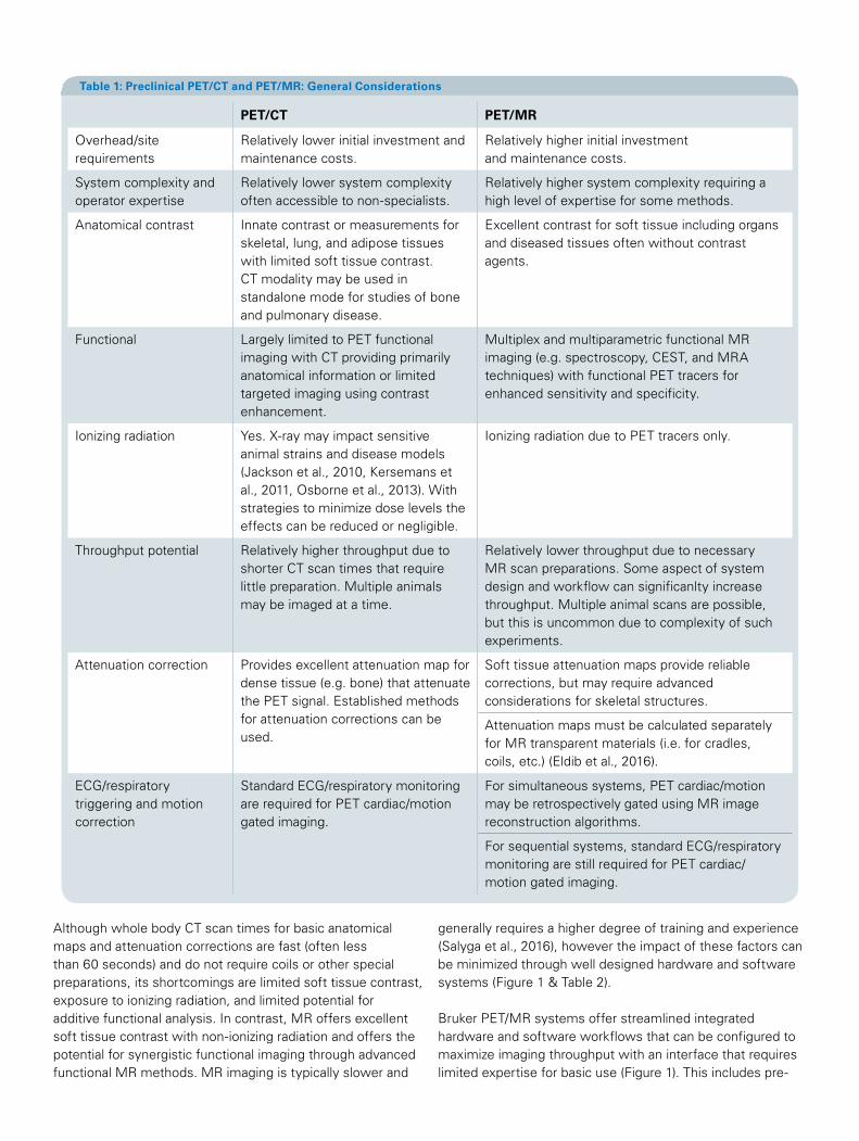
Table 1: Preclinical PET/CT and PET/MR: General Considerations
Although whole body CT scan times for basic anatomical maps and attenuation corrections are fast (often less than 60 seconds) and do not require coils or other special preparations, its shortcomings are limited soft tissue contrast, exposure to ionizing radiation, and limited potential for additive functional analysis. In contrast, MR offers excellent soft tissue contrast with non-ionizing radiation and offers the potential for synergistic functional imaging through advanced functional MR methods. MR imaging is typically slower and
generally requires a higher degree of training and experience (Salyga et al., 2016), however the impact of these factors can be minimized through well designed hardware and software systems (Figure 1 & Table 2).
Bruker PET/MR systems offer streamlined integrated hardware and software workflows that can be configured to maximize imaging throughput with an interface that requires limited expertise for basic use (Figure 1). This includes pre-

Insert Inline
Bruker PET Insert Bruker PET Inline Bruker PET/MR 3T
PET ring is mounted inside the magnet bore
PET ring mounted in front of high-field magnet
PET ring mounted in front of magnet
Simultaneous scanning with spatially and temporally registered data
Sequential scanning with spatially registered data
Sequential scanning with spatially registered data
Integrated ParaVision PET/MR workflow
Integrated ParaVision PET/MR workflow
Integrated ParaVision PET/MR workflow
Low attenuation cradles available Low attenuation cradles available Low attenuation cradles available
Low attenuation PET coils, with extended FOV designed for optimized simultaneous imaging, are available
Standard bore mounted volume coils can be used
Standard bore mounted volume coils can be used
Table 2: PET/MR Configurations
Figure 1
Streamlined PET/MR workflows can facilitate higher throughput imaging with limited expertise. Top) Intuitive multimodal study registration interface. Bottom) Workflow allows user to select preloaded attenuation maps for PET optimized cradles and coils.
defined protocols for PET/MR with automated registration and bed and coil attenuation maps preloaded for streamlined processing. Further, system configurations can be optimized for individual application and throughput needs. For example, the Bruker PET Insert (with PET detector positioned in the magnet bore) provides simultaneous high sensitivity PET and MR scans (with extended FOVs), reducing the overall scan time to ranges that would be no longer than a typical MR only acquisition.
If certain design aspects are not considered, PET/MR integrations can have significantly compromised MR imaging capability (such as limited methods and flexibility), and/or compromises in PET performance resolution and sensitivity as well as field-of-view, limiting the scan speed, application potential, or potential for imaging larger species (e.g. rat). A detailed review of PET/MR hardware integrations and technologies, including special design features to reduce cross modality interference is beyond the scope of this article. There are several excellent reviews of this topic (Wehrl et al., 2009, Vandenberghe et al., 2015, Eldib et al., 2016).

Advantages of PET/MR in Preclinical Oncology
The considerations for PET/MR discussed above are common among most molecular imaging applications. With the broader acceptance of PET/MR instrumentation, the relative merits of PET/MR and PET/CT for individual research applications, including oncology research, are beginning to emerge (Werhl et al., 2016). PET/CT imaging has been used widely in studies of cancer therapeutics (Zhang et al., 2017, Vilchis-Juárez et al., 2014), tracer development (Wang et al., 2017), and tumor biology studies (Rossenfeldt et al., 2013). However, PET/MR has the potential to reduce workload, improve data quality, and even allow for more complex experimental objectives (Table 3).
As mentioned above, one of the key benefits of PET/MR is the excellent anatomical soft-tissue contrast. In preclinical oncology this offers the unique ability to detect tumor margins/volumes in a broad range of models which can improve the functional analysis of complementary PET data. MR has been shown to detect early stage Xenograft tumors as well as orthotropic and spontaneous tumors in most organs at very early stages (Figure 2A). Barring a few exceptions, CT imaging does not provide a precise definition of tumor location or margins. CT data may reveal the margins/volume of some late stage subcutaneous Xenograft tumors
Figure 2:
MRI
MRI RARE MRI DWI (fire) FDG-PET (rainbow)
(A) (B)
(C)
Unique capabilities of PET/MR in preclinical oncology. (A) Early stage orthotopic CT-2A glioma mouse imaged at 8 days by 18F-FDG/PET-MR. PET/MR can provide tumor margin detections in a much broader range of xenograft, orthotopic, and spontaneous tumor models, and at early stages of tumor progression. (B) Xenograft SKOV3 tumor mouse imaged by 18F-FDG/PET-MR. PET/MR can provide exquisite soft tissue details particularly relevant in studies of tumor biology. (C) Late stage mouse CT-2A glioma imaged by 18F-FDG/PET and DWI MR. Multiplex and multiparametric detections with intersecting functional PET and functional MR can reveal biological processes not ascertainable with an individual functional MR or individual PET. Images acquired using Bruker PET Insert with BioSpec 70/20. Courtesy: Dr. Uwe Himmelreich, Dr. Willy Gsell, Dr. Cindy Casteels and Dr. Matteo Riva, Molecular Small Animal Imaging Center (MoSAIC), University hospital of Leuven, Belgium.
and lung tumors (owing to the tumor density relative to surrounding low density lung tissue, though typically this requires high resolution (high radiation dose) scans with gating).
The ability to obtain an anatomical detection of tumors can improve preclinical PET oncology studies in a several ways. Orthotopic and spontaneous tumor models are believed by many to be more representative models of disease progression and treatment efficacies. However, the location and timing of tumor development is typically not predictable, and these tumors are usually not detected by CT, which can limit confidences for distinguishing true tumor uptake versus spurious background PET signal. As a result, MR contrast may better inform researchers evaluating novel tracers and therapeutics using orthotopic models. With PET/MR tracer accumulation measurements can be made even from the earliest stages of tumor development when signal to background differences may be relatively low.
Further most researchers using PET/CT to validate novel PET tracers for oncology will perform secondary methods for accurate tumor volume or mass measurements for tracer uptake calculations (commonly %ID/ml or SUV calculations) in part due to limitations in methods for determining tumor
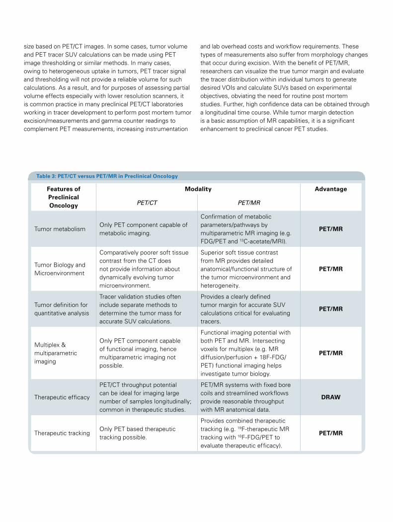
size based on PET/CT images. In some cases, tumor volume and PET tracer SUV calculations can be made using PET image thresholding or similar methods. In many cases, owing to heterogeneous uptake in tumors, PET tracer signal and thresholding will not provide a reliable volume for such calculations. As a result, and for purposes of assessing partial volume effects especially with lower resolution scanners, it is common practice in many preclinical PET/CT laboratories working in tracer development to perform post mortem tumor excision/measurements and gamma counter readings to complement PET measurements, increasing instrumentation
and lab overhead costs and workflow requirements. These types of measurements also suffer from morphology changes that occur during excision. With the benefit of PET/MR, researchers can visualize the true tumor margin and evaluate the tracer distribution within individual tumors to generate desired VOIs and calculate SUVs based on experimental objectives, obviating the need for routine post mortem studies. Further, high confidence data can be obtained through a longitudinal time course. While tumor margin detection is a basic assumption of MR capabilities, it is a significant enhancement to preclinical cancer PET studies.
Features ofPreclinical Oncology
Modality Advantage
PET/CT PET/MR
Tumor metabolismOnly PET component capable of metabolic imaging.
Confirmation of metabolic parameters/pathways by multiparametric MR imaging (e.g. FDG/PET and 13C-acetate/MRI).
PET/MR
Tumor Biology and Microenvironment
Comparatively poorer soft tissue contrast from the CT does not provide information about dynamically evolving tumor microenvironment.
Superior soft tissue contrast from MR provides detailed anatomical/functional structure of the tumor microenvironment and heterogeneity.
PET/MR
Tumor definition for quantitative analysis
Tracer validation studies often include separate methods to determine the tumor mass for accurate SUV calculations.
Provides a clearly defined tumor margin for accurate SUV calculations critical for evaluating tracers.
PET/MR
Multiplex & multiparametric imaging
Only PET component capable of functional imaging, hence multiparametric imaging not possible.
Functional imaging potential with both PET and MR. Intersecting voxels for multiplex (e.g. MR diffusion/perfusion + 18F-FDG/PET) functional imaging helps investigate tumor biology.
PET/MR
Therapeutic efficacy
PET/CT throughput potential can be ideal for imaging large number of samples longitudinally; common in therapeutic studies.
PET/MR systems with fixed bore coils and streamlined workflows provide reasonable throughput with MR anatomical data.
DRAW
Therapeutic trackingOnly PET based therapeutic tracking possible.
Provides combined therapeutic tracking (e.g. 19F-therapeutic MR tracking with 18F-FDG/PET to evaluate therapeutic efficacy).
PET/MR
Table 3: PET/CT versus PET/MR in Preclinical Oncology

Beyond the benefits of anatomical tumor detections, PET and MR can be combined to reveal intricacies of tumor biology including tumor metastasis, tumor progression, and tumor metabolism through multiplex detections inherently synchronized in space (using both Bruker PET Inline and Insert systems) and potentially even synchronized in time (using a simultaneous system such as the Bruker PET Insert. Multiplex detections using both PET and functional MR techniques for a range of relevant molecular processes can allow for more complex study objectives (Wehrl et al., 2014). This capability could be applied to a range of molecular processes in tumor biology. For example, the role of tumor microenvironment, through stromal remodeling via a range of molecular factors, and its role in tumor progressions is better appreciated (Hu et al., 2018, Kouidhi et al., 2018). PET/MR provides a unique tool for interrogating the intricacies of tumor microenvironment owing to the achievable anatomical resolution of the tumor environment (Figure 2B) and may be enhanced by combined detections of factors such as perfusion/diffusion, protease activity, hypoxia, metabolites and metabolism (Figure 2C). Even individual variables (e.g. metabolism) can be interrogated with more refined precision using multiplex capabilities. Tumor glucose, fat, and amino acid metabolism is relatively and differentially skewed relevant to normal tissues and typically differs between cancers and is the target of some cancer therapeutics (Kouidhi et al., 2018). Combined, PET and functional MR can simultaneously interrogate upstream, downstream, and parallel pathways of metabolism to fully characterize these changes and underlying biology (Wehrl et al., 2014, 2016).
Conclusion
Some general considerations for adopting PET/MR will need to be made related to facilities and system capabilities. For most researchers, the potential benefits of PET/MR in preclinical oncology studies of candidate tracer or candidate therapeutics as well as studies of tumor biology will justify investments in these technologies and methods. Researchers should consider both performance of specific integrated PET/MR technologies as well as specific hardware and software workflow implementations to account for unique aspects of PET/MR multimodal detections. Such features can significantly influence the accessibility and application potential for these technologies.

References
Jackson IL, Vujaskovic Z, Down JD. Revisiting strain-related differences in radiation sensitivity of the mouse lung: rec-ognizing and avoiding the confounding effects of pleural effusions. (2010) Radiat. Res. 172, 10-20.
Kersemans V, Thompson J, Cornelissen B, Woodcock M, Allen PD, Buls N, Muschel RJ, Hill MA, Smart SC. (2011) Micro-CT for anatomic referencing in PET and SPECT: radiation dose, biological damage, and image quality. J. Nucl. Med. 52, 1827-1833.
Osborne DR, Yan S, Stuckey A, Pryer L, Richey T, Wall JS. Characterization of X-ray dose in murine animals using microCT, a new low-dose detector and nanoDot dosimeters. (2012) PLoS One 7, e49936.
Eldib M, Bini J, Faul DD, Oesingmann N, Tsoumpas C, Fayad ZA. Attenuation Correction for MR Coils in Combined PET/MR Imaging: A Review. (2016) PET Clin. 11(2): 151–160.
Sasser TA, Bahadur A, Frederick E, Leevy MW. Considerations When Setting Up a Preclinical In Vivo Imaging Labora-tory (2015) Bruker Preclinical Imaging Applications. https://www.bruker.com/fileadmin/user_upload/8-PDF-Docs/Pre-clinicalImaging/Brochures/Considerations_Preclinical_Lab_T158053.pdf
Wehrl HF, Judenhofer MS, Wiehr S, Pichler BJ. Pre- clinical PET/MR: technological advances and new perspectives in biomedical research. (2009) Eur J Nucl Med Mol Imaging. 36 pl 1:S56-68. doi: 10.1007/s00259-009-1078-0
Sałyga A, Guzikowska-Ruszkowska I, Czepczynski R1, Ruchała M. PET/MR - a rapidly growing technique of imaging in oncology and neurology. (2016) Nucl Med Rev Cent East Eur. 2016;19(1):37-41. doi: 10.5603/NMR.2016.0007.
Vandenberghe S, Marsden PK. PET-MRI: a review of challenges and solutions in the development of integrated multi-modality imaging. (2015) Phys Med Biol. 21;60(4):R115-54. doi: 10.1088/0031-9155/60/4/R115.
Wehrl HF, Sauter AW, Divine MR, Pichler BJ (2016)Combined PET/MR: a technology becomes mature. Wehrl HF, Sauter AW, Divine MR, Pichler BJ3. J Nucl Med. 2015 Feb;56(2):165-8. doi: 10.2967/jnumed.114.150318.
Zhang L, Nomie K, Zhang H, Bell T, Pham L, Kadri S, Segal J, Li S, Zhou S, Santos D5 B-Cell Lymphoma Patient-Derived Xenograft Models Enable Drug Discovery and Are a Platform for Personalized Therapy. (2017) Clin Cancer Res. 1;23(15):4212-4223. doi: 10.1158/1078-0432.CCR-16-2703.
Vilchis-Juárez A, Ferro-Flores G, Santos-Cuevas C, Morales-Avila E, Ocampo-García B, Díaz-Nieto L, Luna-Gutiérrez M, Jiménez-Mancilla N, Pedraza-López M, Gómez-Oliván L. Molecular targeting radiotherapy with cyclo-RGDFK(C) pep-tides conjugated to 177Lu-labeledgold nanoparticles in tumor-bearing mice. (2014) J Biomed Nanotechnol. 10(3):393-404.
Wang W, Zhao J, Wen X, Lin CC, Li J, Huang Q, Yu Y, Lin SY, Li C. MicroPET/CT Imaging of AXL Downregula-tion by HSP90 Inhibition in Triple-Negative Breast Cancer. (2017) Contrast Media Mol Imaging. 17:1686525. doi: 10.1155/2017/1686525.
Rosenfeldt MT, O’Prey J, Morton JP, Nixon C, MacKay G, Mrowinska A, Au A, Rai TS, Zheng L, Ridgway R, Adams PD, Anderson KI, Gottlieb E, Sansom OJ, Ryan KM. p53 status determines the role of autophagy in pancreatic tumour development. (2013) Nature. 12;504(7479):296-300. doi: 10.1038/nature12865.
Wehrl HF, Wiehr S, Divine MR, Gatidis S, Gullberg GT, Maier FC, Rolle AM, Schwenck J, Thaiss WM, Pichler BJ. Pre-clinical and Translational PET/MR Imaging. (2014) J Nucl Med. 2014 Jun 1;55(Supplement 2):11S-18S.
Hu Z, Wang X, Cui Y, Li C, Wang S. LASP1 in Tumor and Tumor Microenvironment. (2017) Hu Z, Wang X, Cui Y, Li C, Wang S. Curr Mol Med. 2017;17(8):541-548. doi: 10.2174/1566524018666180222115103.
Kouidhi S, Ben Ayed F, Benammar Elgaaied A. Targeting Tumor Metabolism: A New Challenge to Improve Immunother-apy. (2018) Front Immunol. 9:353. doi: 10.3389/fimmu.2018.00353. eCollection 2018.