Embed Size (px)
Citation preview
Dual Laser Illuminator
for TIRF Microscopy and
Simultaneous Targeted
Laser Action
iLas²

The iLas² system is a unique multi-application device that offers
complete control over any other laser illumination. It provides
researchers the ability to manage and modify the position and
focalization of laser light in real time.
❚ Uniform illumination TIRFwith multi-wavelength controls and penetration depths
❚ Uniform wide-fieldlaser illumination with limited background signal: Dark field laser illumination
❚ Close to coverslip optical sectioning (Oblique illumination)
❚ FRAP/Photoactivation/Photoablation iLas is known for providing the ability to combine the fastest full field of view laser action with the fastest acquisition routines.
❚ Any combination of the previous capabilities
iLas² platform set up menu. Choose from a large range of applications that can be combined to carry out simple to very complex experiments. iLas² is known for its ability to develop imaging platforms that meet the criteria for a multiple user model.
iLas² application GUI. All settings perfectly interact with MetamorphTM acquisition windows, macro capabilities and region tools.
Intellectual property: FR 359 479, FR 356 727, patent pending
2www.roperscientific.fr
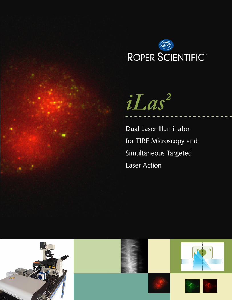
Two optical fibers for laser inputs
Towards microscope illumination port
iLas² Hardware Specifications ❚ iLas² double laser illuminator system permits simultaneous
FRAP/FLIP/PA applications and TIRF/wide-field/PALM applications (300x210x100mm)
❚ Fastest motorized TIRF angle ( <1ms response time) ❚ Patent pending azimuthal averaging provides perfect TIRF/
wide-field illumination uniformity ❚ 20000 laser positions a second in vectorial control mode ❚ Diffraction limited laser spot ❚ Superimpose all optical paths (no commutation delays and
positioning issue) ❚ 350-650nm light range
Software ❚ Standalone software
❚ Fully compatible and interacting with MetamorphTM
❚ Independent laser control
❚ Independent adjustments of penetration depths
❚ Fast TIRF angle motorization
❚ Streaming capabilities between TIRF/FRAP/WF
Compatibility ❚ Leïca DMI series ❚ Olympus IX series ❚ Nikon Ti ❚ Zeiss Axio observer and Axiovert 200 ❚ Autofocus devices ❚ Works with any TIRF objective ❚ iLas² FRAP/FLIP/PA perfectly integers Yokogawa
spinning disk systems
Laser Launch System ❚ Up to five laser lines (diode lasers) ❚ 50 kHz AOTF modulation ❚ Two outputs with a 100% commutation with a switching
time <2ms ❚ Single mode polarization maintaining fibers ❚ Low noise and low heat release ❚ Various wavelength choice:
405,445,457,473,491,515,532,561,594,635,647nm
3www.roperscientific.fr
Roper Scientific has been developing microscopy systems since 2006. In particular, our FRAP3D and its successor iLas in wide-field and spinning disk microscopy have been widely disseminated and are highly recognized.
As a commercial product, we can propose very complex acquisition protocols to meet a wide variety of imaging time regimes.
Our commitment has always been to match biological challenges and applications with the most sophisticated and advanced technologies available.
Because our goal is to continually improve our efforts in this direction, we closely collaborate with academic institutions such as the Institut Curie (France/Paris) and the Imaging Core Facility (PICT-IBiSA), that hosts part of our development structure.
Thanks to this scientific environment, we are achieving our goal of providing expertise and continually meeting scientist expectations.
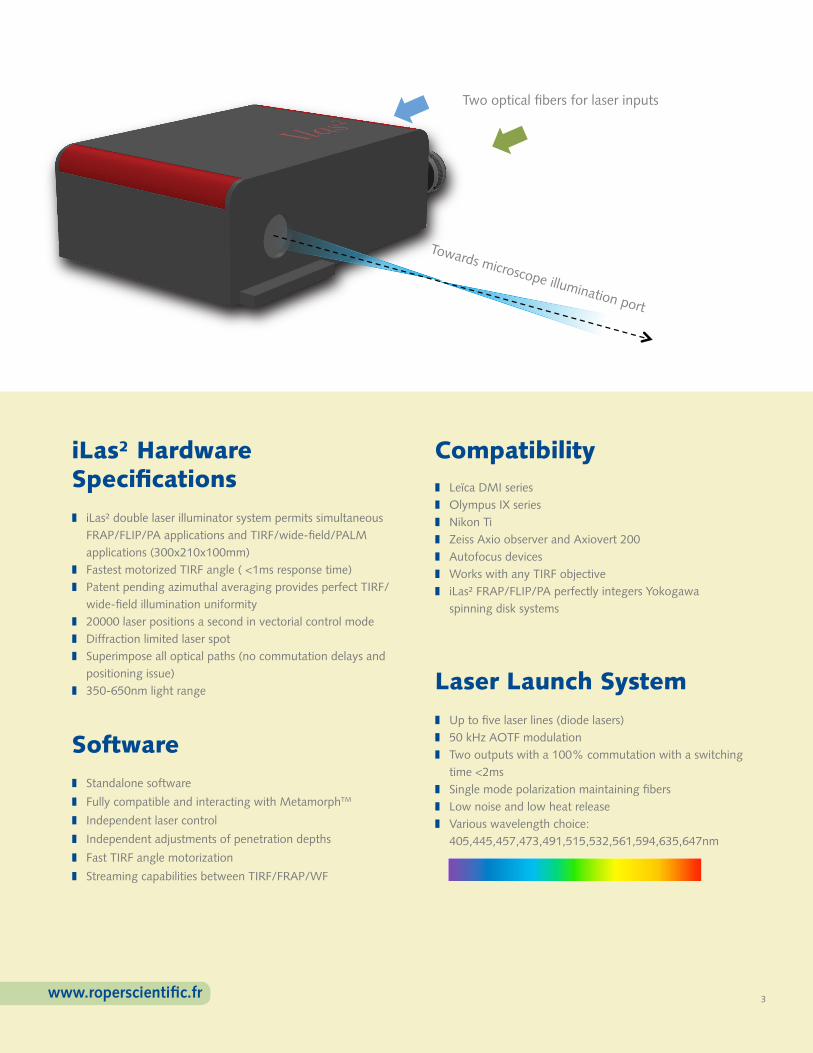
Total Internal Reflection Fluorescence (TIRF) microscopy is the ideal technique for observations close the coverslip surface as it provides the highest axial resolution possible (between 60 to 300nm depending on the angle of incidence). This technique covers a large field of applications such as single molecule tracking, imaging secretion processes, interaction of cell membrane with matrix components or actin filament behavior.
Double transfected M10 stable cell line (Langerine-YFP in green; mCherry-Rab11A in red).
Images were acquired at 10fps, 100ms exp in stream mode using an image splitter (dualview,dv2) to get simultaneous detections of the two fluorescences in TIRF. Image taken with PICT-IBiSA team @ Mifobio 2010, fr.
Single transfected M10 stable cells (mCherry-Rab11A) in Ultra Fast TIRF/WF.
Images were acquired at 10fps/100ms (for 2 minutes), streaming both time and penetration depths (TIRF/wide-field). Here is shown the overlay of Maximum Intensity projections for TIRF illumination (green; 600 frames) , while red color represents wide-field illumination (600 frames). Our Ultra fast dual imaging modality allows to rely plasma membrane appearance of single vesicles (TIRF) with their movements within the cell body ( note “trajectories” in red that end up in yellow when entering the evanescent field). Image taken with B. Cinquin and J. Salamero @ Institut Curie, Paris.
In-vitro actin polymerization. The actin filaments growth starts from a longitudinal micro-pattern functionalized with an activator of nucleation. Images were acquired at 1 frame every 10s in TIRF illumination. TIRF is necessary in order to remove the high background of actin monomers in solution.
FRAP experiments have been realized to investigate the filaments polarity and growth mechanism from the imposed nucleation geometry. Image courtesy of L. Blanchoin, iRTSV/LPCV, CEA Grenoble.
Multi-wavelengthSmooth illumination
TIRF ❚ Fastest angle motorized TIRF ❚ Simultaneous multi-wavelength TIRF with penetration depth adaptation ❚ Unmatched illumination uniformity (no fringes patterns) ❚ Angle scan
4www.roperscientific.fr
0s 100s 320s 370s
For more information on iLas²:
HEAD OFFICERoper Scientific, SASZ.I. Petite Montagne Sud8, rue du Forez91017 Evry Cedex, FranceTel: +33 160860365Fax: +33 160860709
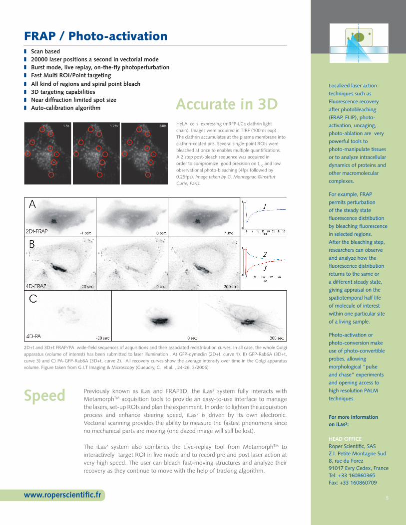
FRAP / Photo-activation
HeLA cells expressing (mRFP-LCa clathrin light chain). Images were acquired in TIRF (100ms exp). The clathrin accumulates at the plasma membrane into clathrin-coated pits. Several single-point ROIs were bleached at once to enables multiple quantifications. A 2 step post-bleach sequence was acquired in order to compromize good precision on t1/2 and low observational photo-bleaching (4fps followed by 0.25fps). Image taken by G. Montagnac @Institut Curie, Paris.
2D+t and 3D+t FRAP/PA wide-field sequences of acquisitions and their associated redistribution curves. In all case, the whole Golgi apparatus (volume of interest) has been submitted to laser illumination . A) GFP-dymeclin (2D+t, curve 1). B) GFP-Rab6A (3D+t, curve 3) and C) PA-GFP-Rab6A (3D+t, curve 2). All recovery curves show the average intensity over time in the Golgi apparatus volume. Figure taken from G.I.T Imaging & Microscopy (Gueudry, C. et al. , 24-26, 3/2006)
Previously known as iLas and FRAP3D, the iLas² system fully interacts with MetamorphTM acquisition tools to provide an easy-to-use interface to manage the lasers, set-up ROIs and plan the experiment. In order to lighten the acquisition process and enhance steering speed, iLas² is driven by its own electronic. Vectorial scanning provides the ability to measure the fastest phenomena since no mechanical parts are moving (one dazed image will still be lost).
The iLas² system also combines the Live-replay tool from MetamorphTM to interactively target ROI in live mode and to record pre and post laser action at very high speed. The user can bleach fast-moving structures and analyze their recovery as they continue to move with the help of tracking algorithm.
Localized laser action
techniques such as
Fluorescence recovery
after photobleaching
(FRAP, FLIP), photo-
activation, uncaging,
photo-ablation are very
powerful tools to
photo-manipulate tissues
or to analyze intracellular
dynamics of proteins and
other macromolecular
complexes.
For example, FRAP
permits perturbation
of the steady state
fluorescence distribution
by bleaching fluorescence
in selected regions.
After the bleaching step,
researchers can observe
and analyze how the
fluorescence distribution
returns to the same or
a different steady state,
giving appraisal on the
spatiotemporal half life
of molecule of interest
within one particular site
of a living sample.
Photo-activation or
photo-conversion make
use of photo-convertible
probes, allowing
morphological “pulse
and chase” experiments
and opening access to
high resolution PALM
techniques.
Accurate in 3D
Speed
❚ Scan based ❚ 20000 laser positions a second in vectorial mode ❚ Burst mode, live replay, on-the-fly photoperturbation ❚ Fast Multi ROI/Point targeting ❚ All kind of regions and spiral point bleach ❚ 3D targeting capabilities ❚ Near diffraction limited spot size ❚ Auto-calibration algorithm
5www.roperscientific.fr
1.5s 1.75s 240s
For more information on iLas²:
HEAD OFFICERoper Scientific, SASZ.I. Petite Montagne Sud8, rue du Forez91017 Evry Cedex, FranceTel: +33 160860365Fax: +33 160860709

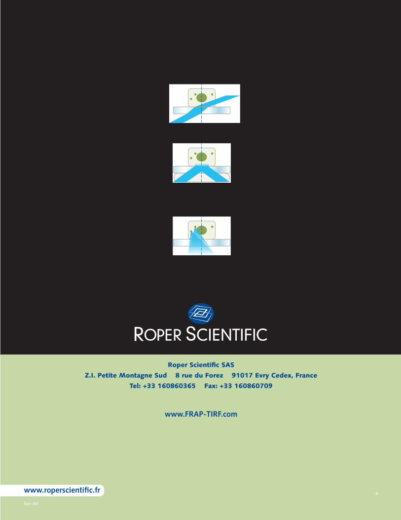
Dark Field Laser Illumination / Oblique Illumination Sectioning
In addition to other capabilities, iLas² enables users to conduct wide-field acquisition taking advantage of a tilted illumination to lower background and enhance the excitation illumination (Dark field laser illumination). As a result, users maintain image quality and achieve less excitation power with less observational bleaching or faster acquisition rates.
The oblique illumination sectioning is the extension of the dark field laser illumination. For high incident angles but smaller than the critical angle, starting the TIRF domain, the angle of the excitation beam going through the sample is so high that the illuminated thickness is very thin ( around 2 µm) over the FOV, as shown on the following schematic.
❚ Combine illumination power of lasers with illumination uniformity ❚ Lower background for better event detection ❚ TIRF/PALM capabilities
Single molecule detection and tracking are very demanding techniques. Both require high performance imaging capabilities and the premium optical quality at the excitation and at the emission.
iLas² provides the ability to produce wide-field laser illumination (either wide-field, oblique or TIRF) while it significantly improves the illumination uniformity. Thus, event detection probability isn’t modulated by random fringe patterns and tracks receive better continuities.
TIRF images of a thin fluorescent layer. Left image was acquired with a regular commercial TIRF setup. Right image has been acquired using the iLas².
Common limitations of
wide-field illumination
systems are S/N ratio
and background blur that
results from out-of-focus
planes in the sample.
One very simple way to
decrease the background
signal is available through
iLas². The same way
as SPIM (Single Plane
Illumination Microscopy)
the background is
lowered because the
out-of-range planes
above the field of view
(FOV) are not illuminated
and thus do not add blur
to the final image.
Single Molecule (ie PALM, STORM…)
Wide-field images of living M10 cells, expressing YFP-Langerine (B. Cinquin & J.Salamero, Institut Curie, Paris). Left image has been illuminated with perpendicular laser illumination. Right image has been illuminated with 50° tilted illumination using the same power and acquisition settings. Background went down from 157 to 76 gray levels (white square region).
❚ Lower background ❚ Lower illumination needed ❚ Close to coverslip optical sectioning ❚ No need for fast/high power wide-field light source
6www.roperscientific.fr
For more information on iLas²:
HEAD OFFICERoper Scientific, SASZ.I. Petite Montagne Sud8, rue du Forez91017 Evry Cedex, FranceTel: +33 160860365Fax: +33 160860709

Evolve 128™ EMCCD24 x 24-µm pixels
Smallest, most powerful scientific EMCCD camera on the market
Most advanced feature set available for low-light applications
Lowest dark current available for an EMCCD camera
Lowest read noise available for an EMCCD camera
Superb electron multiplication (EM) gain and bias stability
Most accurate EM calibration technique in the industry
Available with exclusive eXcelon™ technology
Backed by Photometrics’ worldwide support team
Ideal for sophisticated researcher and multi-user labs
Available with Exclusive
Technology
Primary applications:
In Vivo Imaging
Calcium Imaging
Cell Physiology
Live Cell Microscopy
Single Molecule Fluorescence
Available with Exclusive
Technology
Evolve 128™ EMCCD512 x 512 imaging array, 16 x 16-µm pixels
Primary applications:
Quantitative FRET
Multiprobe experiments
Ratiometric ion imaging
Confocal microscopy
Live-cell fluorescence imaging
Smallest, most powerful scientific EMCCD camera on the market
Most advanced feature set available for low-light applications
Lowest dark current available for an EMCCD camera
Lowest read noise available for an EMCCD camera
Ideal for sophisticated researcher and multi-user labs
Superb electron multiplication (EM) gain and bias stability
Most accurate EM calibration technique in the industry
Available with exclusive eXcelon™ technology
Backed by Photometrics’ worldwide support team
Primary applications:
Live-cell imaging
High-speed emission
ratio imaging
Low-copy gene analysis and
gene expression profiling
Quantitative FRET, FRAP, FISH
Luminescence
CoolSNAP™ HQ21392 x 1040 imaging array, 6.45 x 6.45-µm pixels
The CoolSNAP HQ2 Monochrome camera from Photometrics® delivers
fast, high-resolution imaging for quantitative fluorescence microscopy
applications. This cooled CCD camera provides a large dynamic range
with very low noise at both 10 MHz and 20 MHz. The fine pitch of
the pixels is ideally matched to the resolution of optical microscopes.
Megapixel resolution and small pixels allow imaging of very fine detail,
yet the pixels can be easily binned to improve sensitivity. Advanced
interline-transfer CCD technology provides high quantum efficiency,
most notably in the near-infrared (NIR) portion of the spectrum.
7www.roperscientific.fr
H I G H P E R F O R M A N C E E M C C D & C C D C A M E R A S F O R L I F E S C I E N C E S
Rev A0
8www.roperscientific.fr
Roper Scientific SAS
Z.I. Petite Montagne Sud 8 rue du Forez 91017 Evry Cedex, France
Tel: +33 160860365 Fax: +33 160860709
www.FRAP-TIRF.com