Embed Size (px)
Citation preview
Drug Controlled Release From Structured Bioresorbable FilmsUsed in Medical Devices—A Mathematical Model
Meital Zilberman, Alon Malka
Department of Biomedical Engineering, Faculty of Engineering, Tel-Aviv University, Tel-Aviv 69978, Israel
Received 13 December 2007; revised 1 April 2008; accepted 17 June 2008Published online 5 September 2008 in Wiley InterScience (www.interscience.wiley.com). DOI: 10.1002/jbm.b.31200
Abstract: A mathematical model for predicting drug release profiles from structured
bioresorbable films was developed and studied. These films, which combine good mechanical
properties with desired drug release profiles, are designed for use in various biomedical
applications. Our structured polymer/drug films are prepared using a promising technique for
controlling the drug location/dispersion in the film. The present model was used for predicting
drug release profiles from two film types that is films in which the drug is located on the
surface (A-type) and films in which the drug is located in the bulk (B-type). The model is based
on Fick’s 2nd law of diffusion and assumes that the drug release profile from the films is
affected by the host polymer’s characteristics, the drug location/dispersion in the film and the
drug’s characteristics. This semiempirical model uses the weight loss profile of the host
polymers as well as the change in their degree of crystallinity with degradation. Our study
indicates that the model correlates well with in vitro release results, exhibiting a mean error of
less than 7% for most studied cases. It also shows that the host polymer’s degradation has a
greater effect on the drug release profile than the degree of crystallinity. This new model
exhibits a potential for simulating the release profile of bioactive agents from structured films
for a wide variety of biomedical applications. ' 2008 Wiley Periodicals, Inc. J Biomed Mater Res Part
B: Appl Biomater 89B: 155–164, 2009
Keywords: drug release; bioresorbable films; mathematical model; medical device;
dexamethasone
INTRODUCTION
Bioresorbable drug-eluting films can be used as basic ele-
ments in various biomedical implants, such as tracheal
stents1 and coatings for orthopaedic implants.2 Poly(a-hydroxy acid) films loaded with water-soluble and water-
insoluble drugs have been developed and studied for vari-
ous applications.3–8 For example, tetracycline was released
from poly(L-lactic acid) (PLLA) barrier films, and the sys-
tem was studied for potential use in periodontal therapy,3
gentamicin was released from poly(a-hydroxy acid) films
for potential use in local treatment of bone infection,2,4,5
dexamethasone (DM) was released from various poly(a-hydroxy acid) films used as basic elements in tracheal
films,1 and sirolimus was released from a bilayer PLLA-
PLGA film in order to simulate its release from a stent.7
Bioactive agents with a relatively high molecular weight,
such as albumin and the malaria vaccine (synthetic poly-
peptide), were released from PLGA films.6,8 New combina-
tions of materials, other than the traditional poly(a-hydroxy
acid), were tried as host polymers9–13 in order to exhibit
proper mechanical properties with the desired release pro-
file from polymeric bioresorbable films. For example, a
combination of alginate and polyethylene glycol and a graft
copolymer of vinyl alcohol and lactic/glycolic acid were
tried in paclitaxel eluting films for stent applications,9,10
and segmented poly(ether-ester-amide) films and cross-
linked gelatin films were loaded with bioactive agents such
as metronidazole for dental applications.11,12
Solution casting of polymers is a well-known method
for preparing polymer films. To incorporate drugs by this
method, the polymer is dissolved in a solvent and mixed
with the drug prior to casting. The solvent is then evapo-
rated and the polymer/drug film is created. We have previ-
ously reported a method for controlling drug location/
dispersion in the film.14,15 To review briefly, bioresorbable
polymeric films containing drugs were prepared using the
solution casting technique, accompanied by a postprepara-
tion isothermal heat treatment. In this process, the solvent
evaporation rate determines the kinetics of drug and poly-
mer solidification and thus, the drug dispersion/location
within the film. Solubility effects in the starting solution
also contribute to the postcasting diffusion processes and
occur concomitantly to the drying step. In general, two
Correspondence to: M. Zilberman (e-mail: [email protected])
' 2008 Wiley Periodicals, Inc.
155
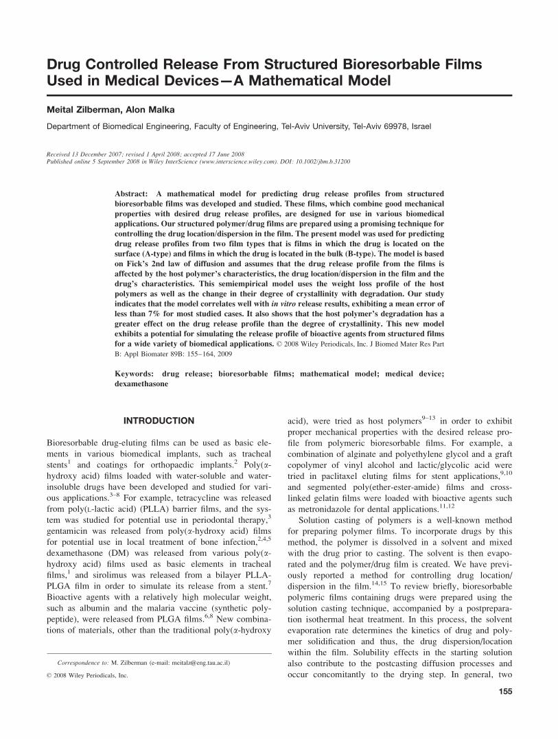
types of polymer/drug film structures were created and
studied for all matrix polymer types, as presented in Figure
1 for PLLA and poly(DL-lactic acid) (PDLLA) films loaded
with DM:
a. A polymer film with large crystalline drug particles
located on its surface, as presented in the upper micro-
graphs of Figure 1. This structure, derived from a
dilute solution of both polymer and drug, was obtained
using the slow solvent evaporation rate, which enables
prior drug nucleation and growth on the polymer solu-
tion surface. This skin formation is accompanied by a
later polymer core formation/solidification. This struc-
ture was named the ‘‘A-type.’’
b. A polymer film with small drug particles and crystals
distributed within the bulk, as presented in the lower
micrographs of Figure 1. This structure, derived from a
concentrated solution, was obtained using the fast sol-
vent evaporation rate and resulted from drug nucleation
and segregation within a dense polymer solution. Solid-
ification of drug and polymer occurred concomitantly.
In semicrystalline-based films, such as PLLA/DM, the
drug is located in amorphous domains of a semicrystal-
line matrix, around the spherulites.14 This structure
was named the ‘‘B-type.’’
We have studied the morphology and formation process
of these structured films extensively, and a detailed model
describing the structuring of these films is described else-
where.14 Our study indicates that both A-type and B-type
structures can be developed for both amorphous and semi-
crystalline polymeric films loaded with DM and also with
other drugs, such as cortisone14 and gentamicin.2 The type
of polymer affects the film morphology but has almost no
effect on the drug location/dispersion within the polymeric
film. The latter is determined mainly by the kinetics of film
formation.2,14,15
Mathematical models for controlled release of bioactive
agents from bioresorbable matrices are based on diffusion
aspects, on the structural characteristics of the matrix poly-
mer and its degradation and swelling, and on the micro-
environmental pH changes inside the polymer matrix pores
that are due to the degradation products. Prediction of the
drug release profile using a model can obviously be very
useful in the device’s design phase, as the model enables
fast evaluation and tuning of the various parameters for
achieving an optimal release profile, while reducing labora-
tory tasks to a minimum.16,17
Early drug delivery models described either systems
based on nondegradable matrices or surface-eroding sys-
tems.18 Several more recent models described bulk-eroding
systems, in which the drug is physically immobilized. For
example, Siepman and Gopferich19 quantified drug release
from slab-shaped PLLA and poly(DL-lactic-co-glycolicacid) (PDLGA) matrices. Their model was based on Higu-
chi’s classical pseudo steady-state equation for oversatu-
rated, planar, nondegrading polymeric films, where the
permeability of the drug within the polymer matrix was
assumed to increase with time due to cleavage of the poly-
mer’s bonds. Another possibility for simulating the effect
of erosion on the diffusion process is to use a diffusion
coefficient which increases with time. Various theories,
therefore related the drug diffusion coefficient inside a
degradable polymer directly to its molecular weight, as
Figure 1. Polarized light micrographs of DM-loaded PLLA (semicrystalline) and PDLLA (amorphous)
films: upper micrographs, A-type; lower micrographs, B-type.
156 ZILBERMAN AND MALKA
Journal of Biomedical Materials Research Part B: Applied Biomaterials
short chains offer less restriction to drug diffusion than
long chains. This was one of the main assumptions in the
models of Charlier et al.20 for predicting the release rate of
Mifepristone from 50/50 PDLGA bulk-eroding films of var-
ious molecular weights, and Faisant et al.21 who examined
the release rate of 5-fluorouracil from 50/50 PDLGA
microspheres. Both used an additional assumption regard-
ing the polymer’s first order degradation kinetics in order
to calculate an appropriate time-dependent diffusion coeffi-
cient, which they used in Fick’s laws of diffusion. Zhang
et al.22 addressed three mechanisms for drug release in a
microspheric matrix, namely dissolution of the drug from
the polymer matrix, diffusion of the dissolved drug, and
erosion of the matrix.
However, our promising technique for controlling the
drug location/dispersion in the film raised the need for a
suitable model which could predict the release profile of var-
ious drugs from these structured films, and therefore, shorten
the design process of biomedical implants based on these
drug-eluting films. This need was the driving force for the
current research, in which a suitable mathematical model
was developed. The hypotheses of our study were: (1) A
model based on Fick’s laws will be able to provide good
prediction of the drug release profile from our structured
drug-eluting films, and (2) The drug release profile from the
films is affected by the host polymer’s characteristics (degra-
dation profile and degree of crystallinity), by the drug loca-
tion/dispersion in the film, and by the drug characteristics.
MATERIALS AND METHODS
Case Definition and Model Assumptions
A mathematical model for predicting drug release from our
structured drug-eluting films was developed in this study,
using Matlab 6.1. The release profiles that were predicted
using this model were compared with experimental results
for certain films loaded with DM.
The Experimental System. Polymer films (0.12–0.15
mm thick) consisting of poly(a-hydroxy acid) and DM (5%
w/w in each film) were prepared by a three-step solution
processing method as follows: The components were mixed
in a solvent at room temperature until polymer dissolution.
Both dilute and concentrated solutions were prepared for
the various polymer/DM systems. The solutions were then
cast into Petri dishes, and solvent drying was performed
under atmospheric pressure at room temperature. A slow
solvent evaporation rate was used for dilute solutions in
order to obtain A-type films, whereas a fast solvent evapo-
ration rate was used for concentrated solutions in order to
obtain B-type films. Finally, an isothermal heat treatment at
a temperature higher than the glass transition temperature
of the polymer (60–908C) was performed for 1 h in a
vacuum oven. This heat treatment enabled the disposal of
residual solvent. The process of film formation is described
in greater detail elsewhere.15
The films were immersed in sterile water at 378C for 20
weeks in order to determine the DM release kinetics. Sam-
ples of 1.5 mL were collected every week and were
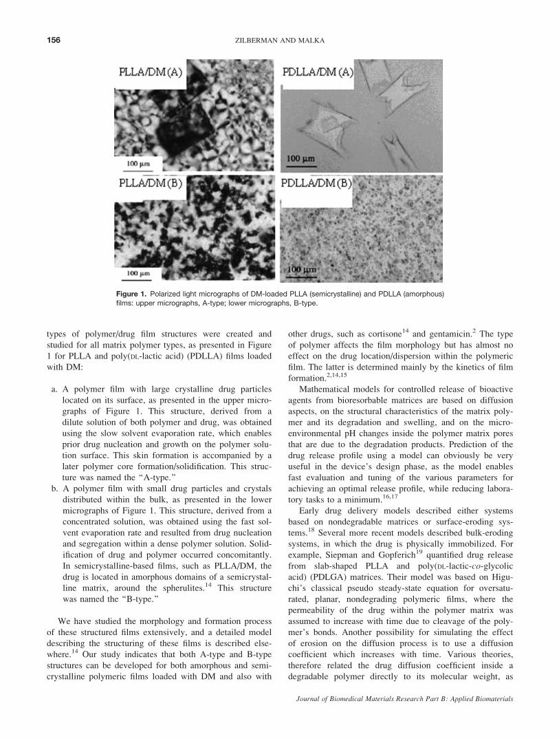
replaced with sterile water. A schematic representation of
the experimental system is presented in Figure 2. The DM
content in each sample was measured using high perform-
ance liquid chromatography (HPLC), DX 500 (Dionex
Corp., Sunnyvale, CA). An AD-20 ultraviolet detector
(Dionex Corp., Sunnyvale, CA) was used to monitor ab-
sorbance at 254 nm. Five samples were examined for each
film type. The examined films are as follows:
Films based on amorphous host polymers:
1. 50/50 PDLGA, A and B types.
2. 85/15 PDLGA, A and B types.
Figure 2. The experimental system at the initial point: A-type film and B-type film floated on water
in a Petri dish. The diffusion occurs in the Z direction. [Color figure can be viewed in the online
issue, which is available at www.interscience.wiley.com.]
157DRUG CONTROLLED RELEASE FROM STRUCTURED BIORESORBABLE FILMS
Journal of Biomedical Materials Research Part B: Applied Biomaterials
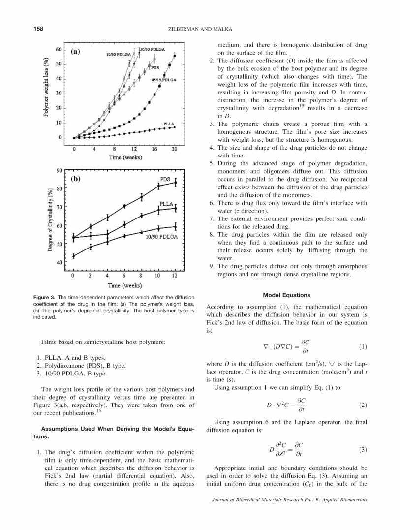
Films based on semicrystalline host polymers:
1. PLLA, A and B types.
2. Polydioxanone (PDS), B type.
3. 10/90 PDLGA, B type.
The weight loss profile of the various host polymers and
their degree of crystallinity versus time are presented in
Figure 3(a,b, respectively). They were taken from one of
our recent publications.15
Assumptions Used When Deriving the Model’s Equa-
tions.
1. The drug’s diffusion coefficient within the polymeric
film is only time-dependent, and the basic mathemati-
cal equation which describes the diffusion behavior is
Fick’s 2nd law (partial differential equation). Also,
there is no drug concentration profile in the aqueous
medium, and there is homogenic distribution of drug
on the surface of the film.
2. The diffusion coefficient (D) inside the film is affected
by the bulk erosion of the host polymer and its degree
of crystallinity (which also changes with time). The
weight loss of the polymeric film increases with time,
resulting in increasing film porosity and D. In contra-
distinction, the increase in the polymer’s degree of
crystallinity with degradation15 results in a decrease
in D.3. The polymeric chains create a porous film with a
homogenous structure. The film’s pore size increases
with weight loss, but the structure is homogenous.
4. The size and shape of the drug particles do not change
with time.
5. During the advanced stage of polymer degradation,
monomers, and oligomers diffuse out. This diffusion
occurs in parallel to the drug diffusion. No reciprocal
effect exists between the diffusion of the drug particles
and the diffusion of the monomers.
6. There is drug flux only toward the film’s interface with
water (z direction).7. The external environment provides perfect sink condi-
tions for the released drug.
8. The drug particles within the film are released only
when they find a continuous path to the surface and
their release occurs solely by diffusing through the
water.
9. The drug particles diffuse out only through amorphous
regions and not through dense crystalline regions.
Model Equations
According to assumption (1), the mathematical equation
which describes the diffusion behavior in our system is
Fick’s 2nd law of diffusion. The basic form of the equation
is:
r � ðDrCÞ ¼ @C
@tð1Þ
where D is the diffusion coefficient (cm2/s), ! is the Lap-
lace operator, C is the drug concentration (mole/cm3) and tis time (s).
Using assumption 1 we can simplify Eq. (1) to:
D � r2C ¼ @C
@tð2Þ
Using assumption 6 and the Laplace operator, the final
diffusion equation is:
D@2C
@Z2¼ @C
@tð3Þ
Appropriate initial and boundary conditions should be
used in order to solve the diffusion Eq. (3). Assuming an
initial uniform drug concentration (C0) in the bulk of the
Figure 3. The time-dependent parameters which affect the diffusion
coefficient of the drug in the film: (a) The polymer’s weight loss,
(b) The polymer’s degree of crystallinity. The host polymer type is
indicated.
158 ZILBERMAN AND MALKA
Journal of Biomedical Materials Research Part B: Applied Biomaterials
polymeric film or on its surface leads to the following ini-
tial condition:
C ¼ C0 at t ¼ 0; Z ¼ Zfilm for A-type films ð4aÞC ¼ C0 at t ¼ 0; 0 � Z < Zfilm for B-type films ð4bÞ
Second initial condition: there is no drug in the medium
(water) at the starting point:
C ¼ 0 at t ¼ 0; Zfilm < Z � L ð5Þ
This is a closed system, and therefore, drug particles
cannot leave the system. Hence, the flux at the boundaries
is equal to 0, and the boundary conditions are:
@C
@Zðt; Z ¼ 0Þ ¼ 0 ð6Þ
@C
@Zðt; Z ¼ LÞ ¼ 0 ð7Þ
Using the above initial and boundary conditions, the
differential Eq. (3) was resolved via the ‘‘pdepe’’ Matlab
function, yielding C(Z,t) which describes how the drug con-
centration changes with time, and how it changes along the
Z-axis.The released drug particles reach the surface of the film
(Z 5 Zfilm), and the cumulative drug particles can therefore
be ‘‘counted’’ as follows:
mðtÞ ¼Z t
0
d
dt0CðZ ¼ Zfilm; t
0ÞAdt0 ð8Þ
where m(t) is the cumulative drug released (moles), t, t0—Time (s), CðZ ¼ Zfilm; t
0Þ—area concentration of drug
(mole/cm2), and A is the film area (cm2).
Estimation of the Diffusion Coefficient. The diffusion
coefficient (D) in our model is divided into two regions,
corresponding to the Z-axis division. One region is inside
the film at 0 � Z � Zfilm, where the diffusion coefficient is
time depended during the film degradation and can be writ-
ten as Df(t). The second region is in the water at Zfilm \ Z� L, where the diffusion coefficient is constant and can be
written as Dw. Hence, D is expressed as follows:
D ¼ DfðtÞ at 0 � Z � Zfilm ð9Þ
D ¼ Dw at Zfilm < Z � L ð10Þ
Because the diffusion medium in both cases is water,
the following theoretical equation for diffusion in water
can be used23:
Dw ¼ KBT
2lR0
ð11Þ
where Dw is the theoretical diffusion coefficient of a spher-
ical particle in water (cm2/s), KB is Boltzmann’s constant
5 1.38 3 10216 (g cm2/s2 K), T is the absolute tempera-
ture (3108K 5 378C), l is the water’s viscosity 5 0.01 (g/
cm s), and R0 is the radius of a spherical particle (cm).
In our study, R0 was estimated using light and electron
microscopy.
In our model we had to slightly change Eq. (11)
because of physical restriction on the drug particles’
motion. In A-type films the drug particles sense mainly
the water and not the film in the surrounding and the non-
ideal (nonspherical) shape of the drug particles is there-
fore the dominant restriction. Equation (13) actually
shows how far are the particles from spherical shape. In
B-type films, the drug particles sense mainly the polymer
chains of the film, which create paths through which the
drug particles move. In B-type films, Eq. (14) elucidates
the ability of the drug particles to pass through the porous
structure.
A restriction factor (Fr) was therefore added to Eq. (14)
as follows:
Dw ¼ KBT
2lR0Fr
ð12Þ
where:
Fr ¼ ðsurface area of particleÞ=ðsurface area of equivalentsphereÞ in A-type films
ð13ÞFr ¼ average pore size=R0 in B-type films ð14Þ
The initial diffusion coefficient inside the film (Df0) is
based on Eq. (12), but we should consider the effects of
the film characteristics as follows:
Df0 ¼ DwP0FsFt ð15Þ
where Df0 is the initial diffusion coefficient inside the film
(cm2/s), and P0 is the initial porosity of the film (%). P0
indicates the volume of water inside the film, which was
evaluated experimentally and used in our model. Fs is a
surface factor ([1) that we used in A-type films. It gives
an indication for the initial binding (physical interactions
such as hydrogen bonds) between the drug and the film sur-
face. The value of Fs is higher for smaller (weaker) interac-
tions. When there are relatively weak interactions between
the host polymer and the drug molecules (in A-type films),
the diffusion coefficient is closer to that of the drug in water.
Ft is a tortuosity factor (\1) used for B-type films. It indi-
cates the initial tortuosity of the film, which can indicate the
ratio of shortest and the actual path of particles. Fs and Ft
values were evaluated using our experimental data.15
Ft ¼ L1=L2 ð16Þ
159DRUG CONTROLLED RELEASE FROM STRUCTURED BIORESORBABLE FILMS
Journal of Biomedical Materials Research Part B: Applied Biomaterials
where L1 is the distance between particle location and film
surface (Z 5 Zfilm), and L2 is the length of the actual parti-
cle path inside the film. The factors Fr, Fs, and Ft for the
various films used in this study are presented in Table I.
The time-dependent diffusion coefficient in the bulk
[Df(t)] starts from its initial value Df0 [calculated in Eq.
(15)] and changes with time due to the polymer’s weight
loss (bulk erosion) and changes in its degree of crystallin-
ity, as stated in assumption 2. Df(t) is evaluated as
follows:
DfðtÞ ¼ Df0 þ fðDw � Df0Þ3WðtÞ3ð100%� CRSTLðtÞÞgð17Þ
where W(t) is the film’s weight loss (%), and CRSTL(t) isthe polymer’s degree of crystallinity (%). Both parameters
were measured for all polymers used in this study, and the
results are presented in Figure 3. Because diffusion of drug
particles occurs through the amorphous regions of the semi-
crystalline polymer rather than through the dense crystal-
line regions (assumption 9), we used [100% 2 CRSTL(t)]in Eq. (17).
The degree of crystallinity was measured as follows: the
heat of fusion (DHm) was determined by differential scan-
ning calorimetry using an indium-calibrated TA Instru-
ments DSC 2010 differential scanning calorimeter (DSC).
The measurements were carried out on 10 mg samples
under N2 atmosphere, heating the samples from 308C to
2508C (above their melting points), using a heating rate of
108C/min. The analysis was performed using TA Universal
TABLE I. Film Types Used for Obtaining the Experimental Dataand Their Calculated Factors Used in the Model
The Polymer Film Type Fr Fs Ft
50/50 PDLGA A 2 30 –
B 40 – 0.33
85/15 PDLGA A 9 80 –
B 30 – 0.67
PDS B 20 – 0.67
10/90 PDLGA B 50 – 0.67
PLLA A 7 50 –
B 20 – 0.33
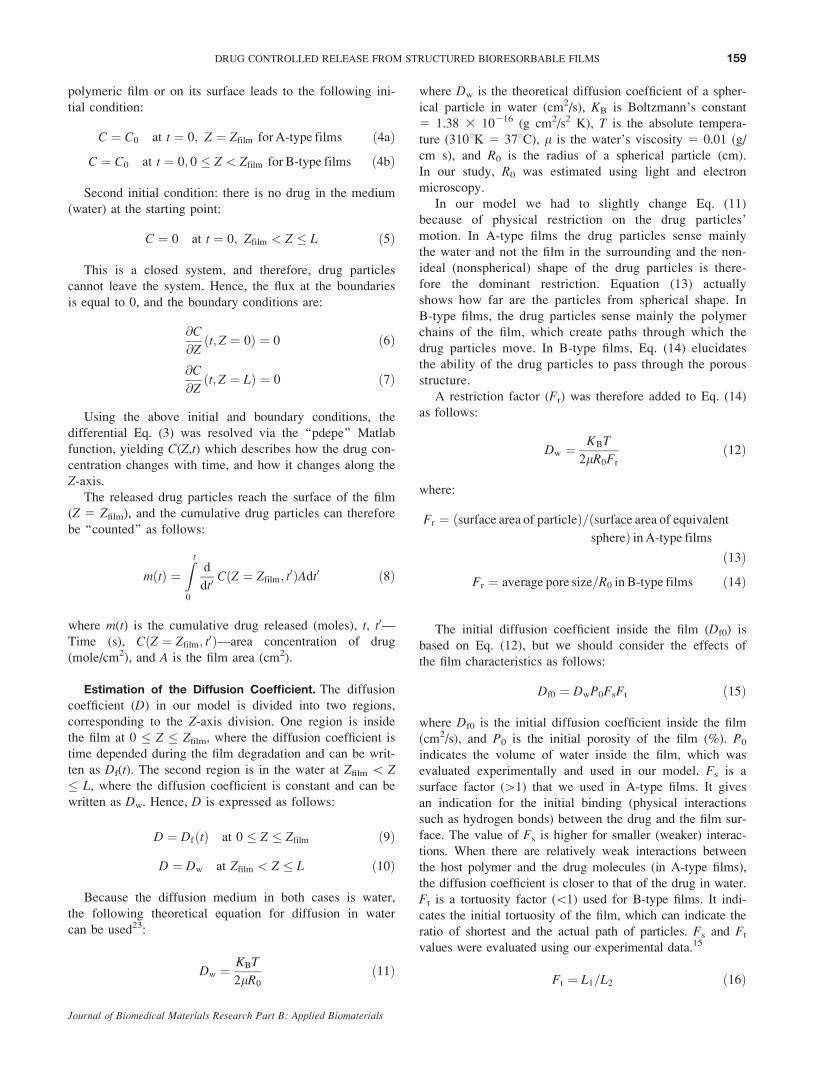
Figure 4. DM release profile from amorphous polymer films containing 5% (w/w) DM. The experi-
mental results (mean —— and upper and lower limits -------) are compared with the predicted
results (—l—): (a) A-type (85/15 PDLGA)/DM, (b) B-type (85/15 PDLGA)/DM, (c) A-type (50/50
PDLGA)/DM, (d) B-type (50/50 PDLGA)/DM. [Color figure can be viewed in the online issue, which isavailable at www.interscience.wiley.com.]
160 ZILBERMAN AND MALKA
Journal of Biomedical Materials Research Part B: Applied Biomaterials
Analysis Software. The degree of crystallinity, CRST(t),was calculated by the relationship:
%CRST ¼DHm
DHF
3100 ð18Þ
where DHm and DHF are the heats of fusion of the sample
(a semicrystalline material) and the perfect crystal, respec-
tively. DHF (PLLA) 5 93.6 J/g,24 DHF (PGA) 5 191.2 J/g
and DHF (PDS) 5 141.2 J/g.25
Mean errors for the predicted release profiles were eval-
uated as follows: for each data point, when the model’s
prediction is within the error of the experimental results,
the model’s error is considered as 0. When the model’s
prediction is not in the range of the experimental’s error,
there appears an error (in %) for this point. The average
error is the average of the various errors of all points.
RESULTS AND DISCUSSION
As mentioned earlier, the experimental data used to vali-
date this model were taken from one of our recent stud-
ies.15 Two types of films were used, A-type with drug
located on the surface of the polymer film and B-type with
drug located in the bulk. Amorphous and semicrystalline
polymers were used for both types of drugs (Table I). The
predicted DM release profile was compared with the exper-
imental release profile for each type of structured film. The
results for the amorphous based films are presented in Fig-
ure 4 and the results for the semicrystalline-based films are
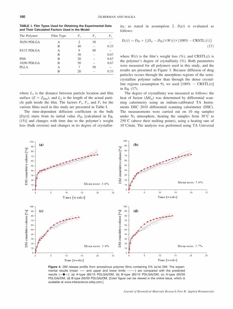
presented in Figure 5. A very good fit was obtained for all
studied films (less than 7% error), except for the 10/90
PDLGA/DM based film, where the predicted DM release
during the first 7 weeks was higher than the experimental
DM release [Figure 5(b)]. In A-type films the release pro-
file exhibited a burst release of �30% followed by a
release profile, which was determined by the degradation
rate of the host polymer, whereas the B-type films exhib-
ited a release profile governed by the degradation rate of
the host polymer with a low burst release. Our model dem-
onstrated a good fit for both types of films. These results
thus support our first hypothesis about the good predictabil-
ity of a model based on Fick’s laws.
The advantage of building a model for drug delivery
systems is the ability to elucidate the effect of the system’s
Figure 5. DM release profile from semicrystalline polymer films containing 5% (w/w) DM. The ex-
perimental results (mean —— and upper and lower limits -------) are compared with the predicted
results (—l—): (a) B-type PDS / DM, (b) B-type (10/90 PDLGA)/DM, (c) A-type PLLA/DM, (d) B-
type PLLA/DM. [Color figure can be viewed in the online issue, which is available at www.interscience.wiley.com.]
161DRUG CONTROLLED RELEASE FROM STRUCTURED BIORESORBABLE FILMS
Journal of Biomedical Materials Research Part B: Applied Biomaterials
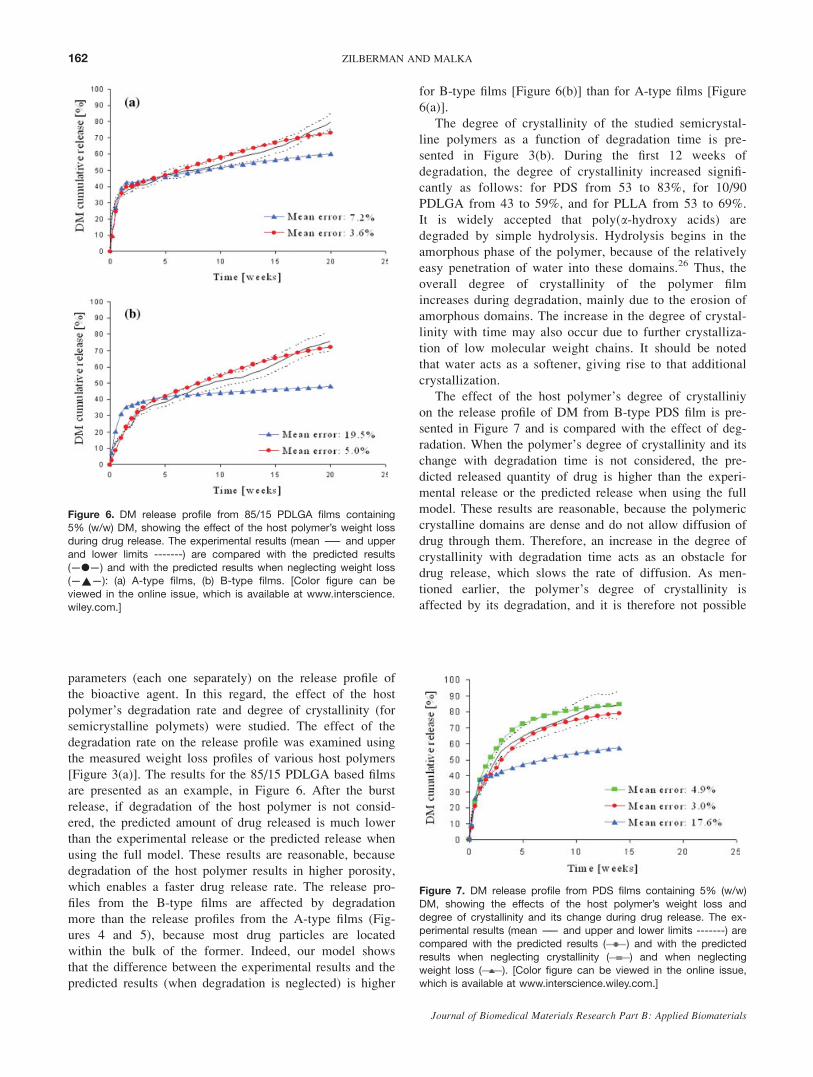
parameters (each one separately) on the release profile of
the bioactive agent. In this regard, the effect of the host
polymer’s degradation rate and degree of crystallinity (for
semicrystalline polymets) were studied. The effect of the
degradation rate on the release profile was examined using
the measured weight loss profiles of various host polymers
[Figure 3(a)]. The results for the 85/15 PDLGA based films
are presented as an example, in Figure 6. After the burst
release, if degradation of the host polymer is not consid-
ered, the predicted amount of drug released is much lower
than the experimental release or the predicted release when
using the full model. These results are reasonable, because
degradation of the host polymer results in higher porosity,
which enables a faster drug release rate. The release pro-
files from the B-type films are affected by degradation
more than the release profiles from the A-type films (Fig-
ures 4 and 5), because most drug particles are located
within the bulk of the former. Indeed, our model shows
that the difference between the experimental results and the
predicted results (when degradation is neglected) is higher
for B-type films [Figure 6(b)] than for A-type films [Figure
6(a)].
The degree of crystallinity of the studied semicrystal-
line polymers as a function of degradation time is pre-
sented in Figure 3(b). During the first 12 weeks of
degradation, the degree of crystallinity increased signifi-
cantly as follows: for PDS from 53 to 83%, for 10/90
PDLGA from 43 to 59%, and for PLLA from 53 to 69%.
It is widely accepted that poly(a-hydroxy acids) are
degraded by simple hydrolysis. Hydrolysis begins in the
amorphous phase of the polymer, because of the relatively
easy penetration of water into these domains.26 Thus, the
overall degree of crystallinity of the polymer film
increases during degradation, mainly due to the erosion of
amorphous domains. The increase in the degree of crystal-
linity with time may also occur due to further crystalliza-
tion of low molecular weight chains. It should be noted
that water acts as a softener, giving rise to that additional
crystallization.
The effect of the host polymer’s degree of crystalliniy
on the release profile of DM from B-type PDS film is pre-
sented in Figure 7 and is compared with the effect of deg-
radation. When the polymer’s degree of crystallinity and its
change with degradation time is not considered, the pre-
dicted released quantity of drug is higher than the experi-
mental release or the predicted release when using the full
model. These results are reasonable, because the polymeric
crystalline domains are dense and do not allow diffusion of
drug through them. Therefore, an increase in the degree of
crystallinity with degradation time acts as an obstacle for
drug release, which slows the rate of diffusion. As men-
tioned earlier, the polymer’s degree of crystallinity is
affected by its degradation, and it is therefore not possible
Figure 6. DM release profile from 85/15 PDLGA films containing5% (w/w) DM, showing the effect of the host polymer’s weight loss
during drug release. The experimental results (mean —— and upper
and lower limits -------) are compared with the predicted results
(—l—) and with the predicted results when neglecting weight loss(—~—): (a) A-type films, (b) B-type films. [Color figure can be
viewed in the online issue, which is available at www.interscience.
wiley.com.]
Figure 7. DM release profile from PDS films containing 5% (w/w)
DM, showing the effects of the host polymer’s weight loss and
degree of crystallinity and its change during drug release. The ex-perimental results (mean —— and upper and lower limits -------) are
compared with the predicted results ( ) and with the predicted
results when neglecting crystallinity ( ) and when neglecting
weight loss ( ). [Color figure can be viewed in the online issue,which is available at www.interscience.wiley.com.]
162 ZILBERMAN AND MALKA
Journal of Biomedical Materials Research Part B: Applied Biomaterials
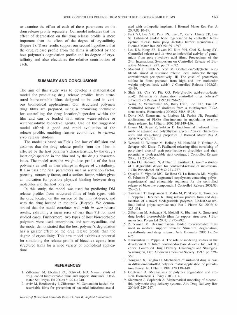
to examine the effect of each of these parameters on the
drug release profile separately. Our model indicates that the
effect of degradation on the drug release profile is more
important than the effect of the degree of crystallinity
(Figure 7). These results support our second hypothesis that
the drug release profile from the films is affected by the
host polymer’s degradation profile and its degree of crys-
tallinity and also elucidates the relative contribution of
each.
SUMMARY AND CONCLUSIONS
The aim of this study was to develop a mathematical
model for predicting drug release profiles from struc-
tured bioresorbable films designed to be used in vari-
ous biomedical applications. Our structured polymer/
drug films are prepared using a promising technique
for controlling the drug location/dispersion within the
film and can be loaded with either water-soluble or
water-insoluble bioactive agents. Use of the suggested
model affords a good and rapid evaluation of the
release profile, enabling further economical in vitro/invivo release studies.
The model is based on Fick’s 2nd law of diffusion and
assumes that the drug release profile from the films is
affected by the host polymer’s characteristics, by the drug’s
location/dispersion in the film and by the drug’s character-
istics. The model uses the weight loss profile of the host
polymers as well as their change in degree of crystallinity.
It also uses empirical parameters such as restriction factor,
porosity, tortuosity factor, and a surface factor, which gives
an indication for possible physical binding between drug
molecules and the host polymer.
In this study, the model was used for predicting DM
release profiles from structured films of both types, with
the drug located on the surface of the film (A-type), and
with the drug located in the bulk (B-type). We demon-
strated that the model correlates well with in vitro release
results, exhibiting a mean error of less than 7% for most
studied cases. Furthermore, two types of host bioresorbable
polymers were used, amorphous, and semicrystalline, and
the model demonstrated that the host polymer’s degradation
has a greater effect on the drug release profile than the
degree of crystallinity. This new model exhibits a potential
for simulating the release profile of bioactive agents from
structured films for a wide variety of biomedical applica-
tions.
REFERENCES
1. Zilberman M, Eberhart RC, Schwade ND. In-vitro study ofdrug loaded bioresorbable films and support structures. J Bio-mater Sci Polym Ed 2002;13:1221–1240.
2. Aviv M, Berdicevsky I, Zilberman M. Gentamicin-loaded bio-resorbable films for prevention of bacterial infections associ-
ated with orthopedic implants. J Biomed Mater Res Part A2007;83:10–19.
3. Park YJ, Lee YM, Park SN, Lee JY, Ku Y, Chung CP, LeeSJ. Enhanced guided bone regeneration by controlled tetra-cycline release from poly(L-lactide) barrier membranes. JBiomed Mater Res 2000;51:391–397.
4. Lee KB, Kang SB, Kwon IC, Kim YH, Choi K, Jeong SY.Controlled release and in vitro antimicrobial activity of genta-micin from poly-a-hydroxy acid films. Proceedings of the24th International Symposium on Controlled Release of Bio-active Materials 1997. pp 571–572.
5. Mauduit J, Bulkh N, Vert M. Gentamicin/poly(lactic acid)blends aimed at sustained release local antibiotic therapyadministrated per-operatively. III The case of gentamicinsulfate in films prepared from high and low molecularweight poly(DL-lactic acids). J Controlled Release 1993;25:43–49.
6. Shah SS, Cha Y, Pitt CG. Poly(glycolic acid-co-DL-lacticacid): Diffusion or degradation controlled drug delivery?J Controlled Release 1992;18:261–270.
7. Wang X, Venkatraman SS, Boey FYC, Loo JSC, Tan LP.Controlled release of sirolimus from a multilayered PLGAstent matrix. Biomaterials 2006;27:5588–5595.
8. Dorta MJ, Santovena A, Liabres M, Farina JB. Potentialapplications of PLGA film-implants in modulating in-vitrodrugs release. Int J Pharm 2002;248:149–156.
9. Livnat M, Beyar R, Seliktar D. Endoluminal hydrogel filmsmade of alginate and polyethylene glycol: Physical characteri-atics and drug-eluting properties. J Biomed Mater Res A2005;75A:710–722.
10. Westedt U, Wittmar M, Hellwig M, Hanefeld P, Greiner A,Schaper AK, Kissel T. Paclitaxel releasing films consisting ofpoly(vinyl alcohol)-graft-poly(lactide-co-glycolide) and theirpotential as biodegradable stent coatings. J Controlled Release2006;111:235–246.
11. Cetin EO, Buduneli N, Athhan E, Kurilmaz L. In-vitro studiesof a degradable device for controlled-release of meloxicam.J Clin Periodontol 2005;32:773–777.
12. Quaglia F, Vignole MC, De Rosa G, La Rotonda MI, MaglioG, Palumbo R. New segmented copolymers containing poly(e-caprolactone) and etheramide segments for the controlledrelease of bioactive compounds. J Controlled Release 2002;83:263–271.
13. Tarvainen T, Karjalainen T, Malin M, Perakorpi K, TuominenJ, Seppala J, Jarvinen K. Drug release profiles from and deg-radation of a novel biodegradable polymer, 2,2-bis(2-oxazo-line) linked poly(e-caprolactone). Eur J Pharm Sci 2002;16:323–331.
14. Zilberman M, Schwade N, Meidell R, Eberhart R. Structureddrug loaded bioresorbable films for support structures. J Bio-mater Sci: Polym Ed 2001;12:875–892.
15. Zilberman M. Dexamethasone loaded bioresorbable filmsused in medical support devices: Structure, degradation,crystallinity and drug release. Acta Biomater 2005;1:615–625.
16. Narasimhan B, Peppas A. The role of modeling studies in thedevelopment of future controlled-release devices. In: Park K,editor. Controlled Drug Delivery: Challenges and Strategies.Washington, DC: American Chemical Society; 1997. pp 529–558.
17. Tongwen X, Binglin H. Mechanism of sustained drug releasein diffusion-controlled polymer matrix-application of percola-tion theory. Int J Pharm 1998;170:139–149.
18. Gopferich A. Mechanisms of polymer degradation and ero-sion. Biomaterials 1996;17:103–114.
19. Siepmann J, Gopferich A. Mathematical modeling of bioerod-ible polymeric drug delivery systems. Adv Drug Delivery Rev2001;48:229–247.
163DRUG CONTROLLED RELEASE FROM STRUCTURED BIORESORBABLE FILMS
Journal of Biomedical Materials Research Part B: Applied Biomaterials
20. Charlier A, Leclerc B, Couarraze G. Release of mifepristonefrom biodegradable matrices: Experimental and theoreticalevaluations. Int J Pharm 2000;200:115–120.
21. Faisant N, Siepmann J, Benoit JP. PLGA-based microparticles:Education of mechanisms and a new, simple mathematicalmodel quantifying drug release. Eur J Pharm Sci 2002;15:355–366.
22. Zhang M, Yang Z, Chow L, Wang C. Simulation of drugrelease from biodegradable polymeric microspheres withbulk and surface erosions. J Pharm Sci 2003;92:2040–2056.
23. Cussler EL. Diffusion Mass Transfer in Fluid Systems, 2ndEd. Cambridge, United Kingdom: Cambridge UniversityPress; 1977.
24. Celli MA, Scandola M. Thermal properties and physical agingof Poly-L-lactic acid. Polymer 1992;33:2699–2710.
25. Ishikiriyama K, Pyda M, Zhang G, Forschner T, Grebowicz J,Wunderlich B. Heat capacity of poly-p-dioxanone. J Macro-mol Sci Phys Ed 1998;B37:27–44.
26. Pistner H, Bendix DR, Muhling J, Reuther JF. Poly(L-lactide):A long-term degradation study in vivo. III. Analytical Charac-terization. Biomaterials 1993;14:291–298.
164 ZILBERMAN AND MALKA
Journal of Biomedical Materials Research Part B: Applied Biomaterials


![Pulsed Laser Deposition of YSZ and Al2O3 Thin Films: Part 1 ......thin films [16-26]. Pulsed laser deposition has also been used for the development of nano-structured thin films [27,](https://img.pdfslide.us/doc/110x75/60f688b3c8026a3be761a2f6/pulsed-laser-deposition-of-ysz-and-al2o3-thin-films-part-1-thin-films-16-26.jpg)