Embed Size (px)
Citation preview
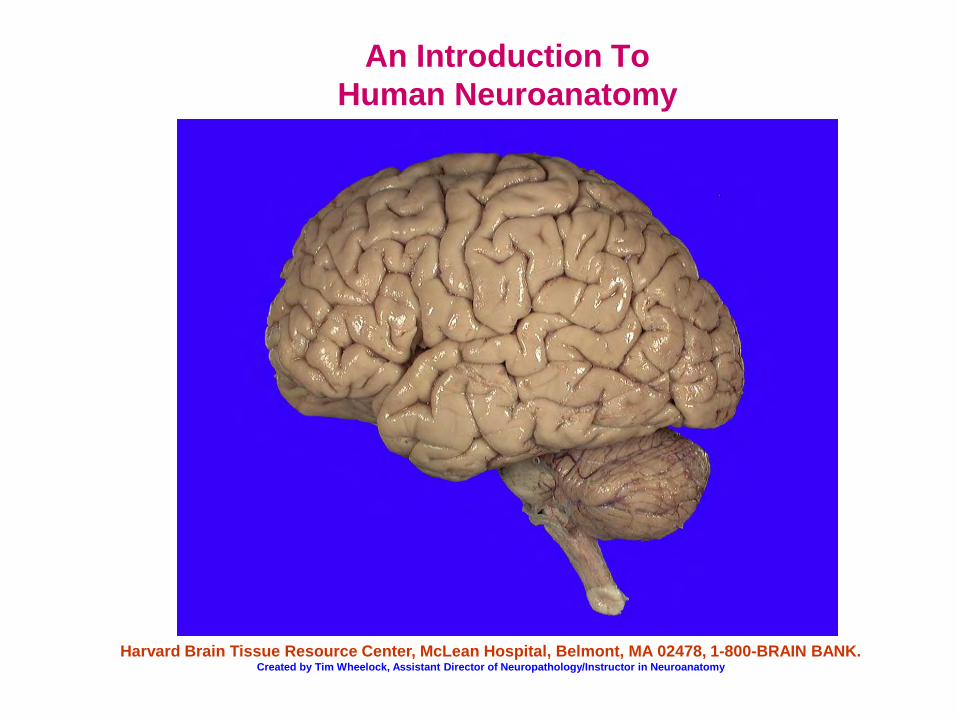
An Introduction To Human Neuroanatomy
Harvard Brain Tissue Resource Center, McLean Hospital, Belmont, MA 02478, 1-800-BRAIN BANK. Created by Tim Wheelock, Assistant Director of Neuropathology/Instructor in Neuroanatomy

Welcome
This Introduction to Human Neuroanatomy provides a look at the structure of the human brain. In it, we will explore each major region of the brain, as well as the brain’s coverings, blood supply, and ventricular system. We hope that this presentation proves interesting and informative. Please feel free to relay any questions, comments, or suggestions you may have. Thank you.
Subjects and brain regions covered in this presentation
• The brain’s surfaces • Directional terminology and planes of section • The divisions of the brain • The meninges: the brain’s coverings • The cerebral cortex • Neurons and glia (support cells) • The brain’s blood supply • The ventricular system and cerebrospinal fluid. • The hippocampus • The amygdala • The striatum • The thalamus • The hypothalamus • The cerebellum • The brainstem
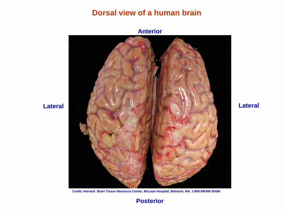
Dorsal view of a human brain
Lateral
Anterior
Credit: Harvard Brain Tissue Resource Center, McLean Hospital, Belmont, MA. 1-800-BRAIN BANK
Posterior
Lateral
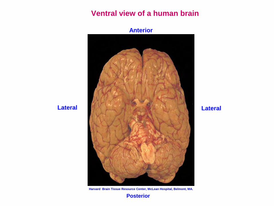
Ventral view of a human brain
Anterior
Posterior
Lateral Lateral
Harvard Brain Tissue Resource Center, McLean Hospital, Belmont, MA.
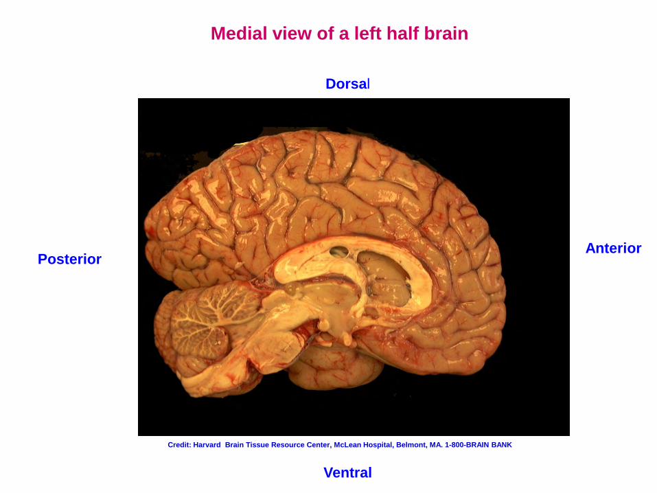
Medial view of a left half brain
Anterior Posterior
Ventral
Dorsal
Credit: Harvard Brain Tissue Resource Center, McLean Hospital, Belmont, MA. 1-800-BRAIN BANK
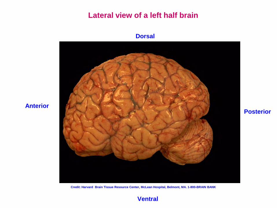
Lateral view of a left half brain
Ventral
Posterior Anterior
Dorsal
Credit: Harvard Brain Tissue Resource Center, McLean Hospital, Belmont, MA. 1-800-BRAIN BANK
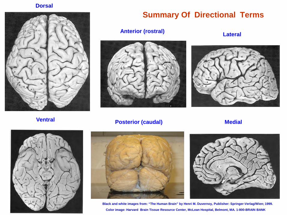
Summary Of Directional Terms Dorsal
Ventral
Anterior (rostral)
Posterior (caudal)
Lateral
Medial
Black and white images from: “The Human Brain” by Henri M. Duvernoy, Publisher: Springer-Verlag/Wien; 1999.
Color image: Harvard Brain Tissue Resource Center, McLean Hospital, Belmont, MA. 1-800-BRAIN BANK
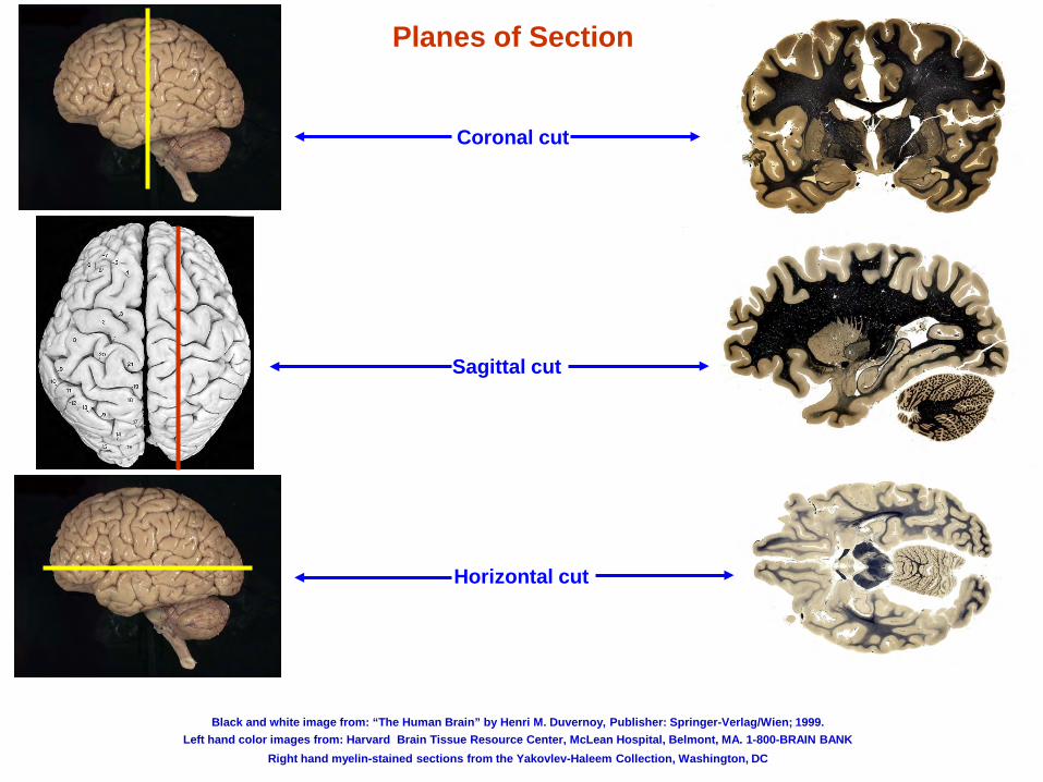
Coronal cut
Sagittal cut
Horizontal cut
Planes of Section
Black and white image from: “The Human Brain” by Henri M. Duvernoy, Publisher: Springer-Verlag/Wien; 1999. Left hand color images from: Harvard Brain Tissue Resource Center, McLean Hospital, Belmont, MA. 1-800-BRAIN BANK
Right hand myelin-stained sections from the Yakovlev-Haleem Collection, Washington, DC
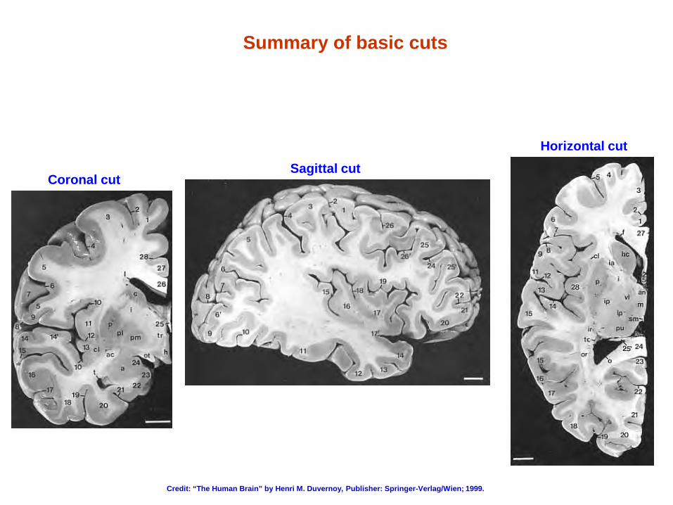
Summary of basic cuts
Coronal cut Sagittal cut
Horizontal cut
Credit: “The Human Brain” by Henri M. Duvernoy, Publisher: Springer-Verlag/Wien; 1999.
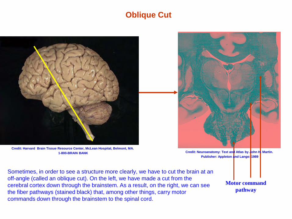
Oblique Cut
Sometimes, in order to see a structure more clearly, we have to cut the brain at an off-angle (called an oblique cut). On the left, we have made a cut from the cerebral cortex down through the brainstem. As a result, on the right, we can see the fiber pathways (stained black) that, among other things, carry motor commands down through the brainstem to the spinal cord.
Motor command pathway
Credit: Harvard Brain Tissue Resource Center, McLean Hospital, Belmont, MA. 1-800-BRAIN BANK Credit: Neuroanatomy: Text and Atlas by John H. Martin.
Publisher: Appleton and Lange: 1989
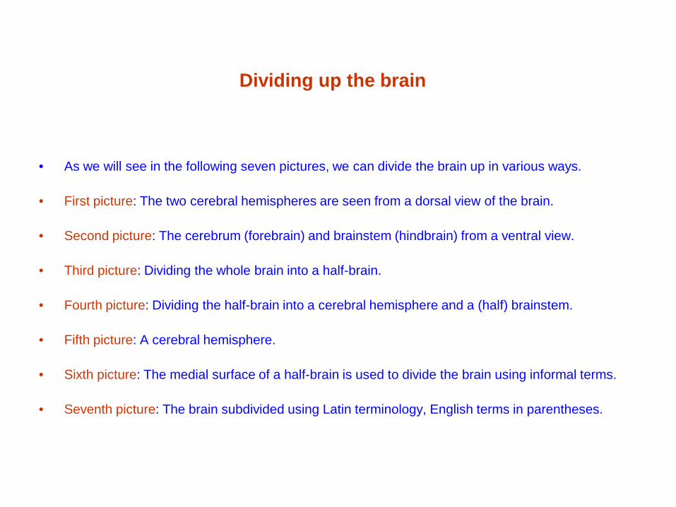
Dividing up the brain
• As we will see in the following seven pictures, we can divide the brain up in various ways.
• First picture: The two cerebral hemispheres are seen from a dorsal view of the brain.
• Second picture: The cerebrum (forebrain) and brainstem (hindbrain) from a ventral view.
• Third picture: Dividing the whole brain into a half-brain.
• Fourth picture: Dividing the half-brain into a cerebral hemisphere and a (half) brainstem.
• Fifth picture: A cerebral hemisphere.
• Sixth picture: The medial surface of a half-brain is used to divide the brain using informal terms.
• Seventh picture: The brain subdivided using Latin terminology, English terms in parentheses.
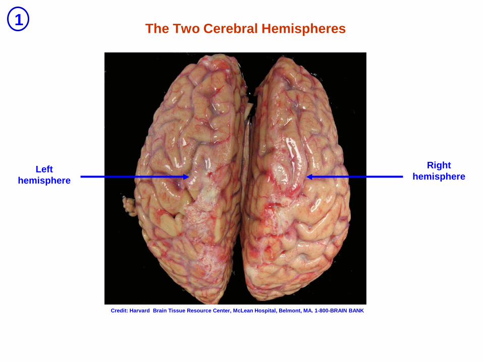
The Two Cerebral Hemispheres
Left hemisphere
Right hemisphere
1
Credit: Harvard Brain Tissue Resource Center, McLean Hospital, Belmont, MA. 1-800-BRAIN BANK
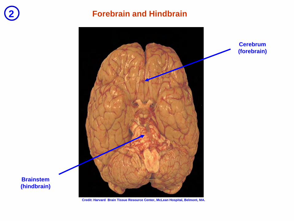
Forebrain and Hindbrain
Cerebrum (forebrain)
Brainstem (hindbrain)
2
Credit: Harvard Brain Tissue Resource Center, McLean Hospital, Belmont, MA.
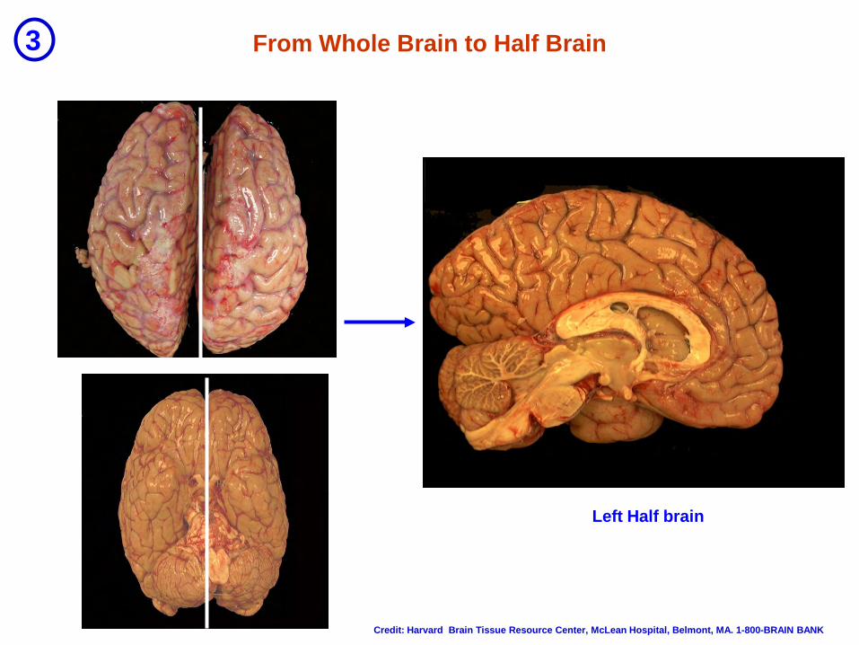
From Whole Brain to Half Brain
Left Half brain
3
Credit: Harvard Brain Tissue Resource Center, McLean Hospital, Belmont, MA. 1-800-BRAIN BANK
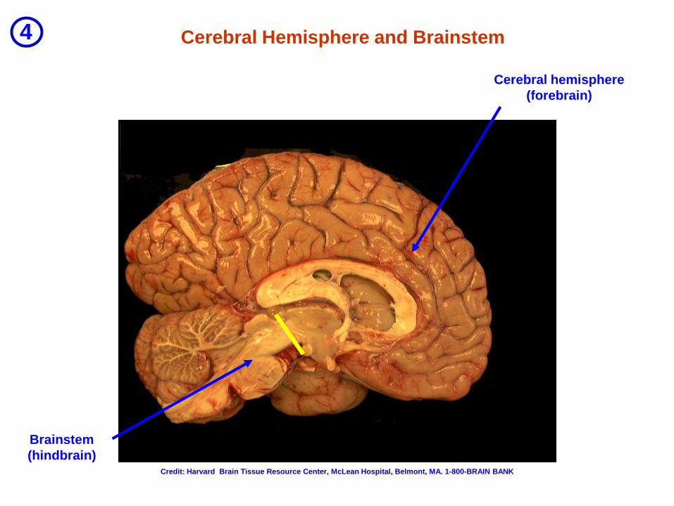
Cerebral Hemisphere and Brainstem
Cerebral hemisphere (forebrain)
Brainstem (hindbrain)
4
Credit: Harvard Brain Tissue Resource Center, McLean Hospital, Belmont, MA. 1-800-BRAIN BANK
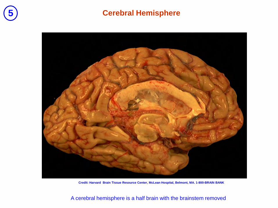
Cerebral Hemisphere
A cerebral hemisphere is a half brain with the brainstem removed
5
Credit: Harvard Brain Tissue Resource Center, McLean Hospital, Belmont, MA. 1-800-BRAIN BANK
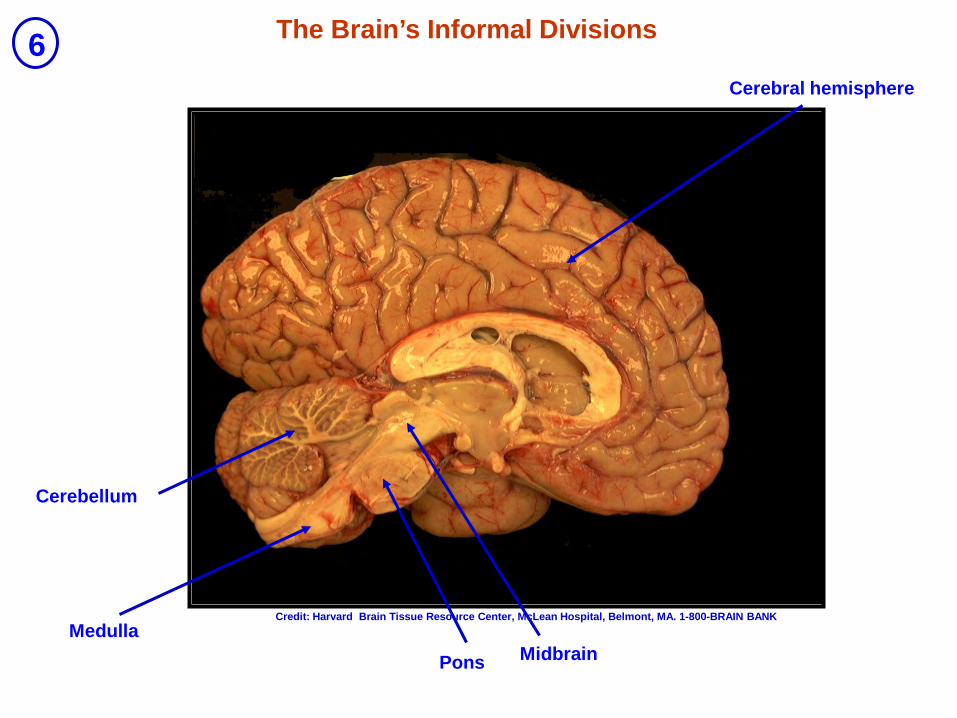
Cerebral hemisphere
Midbrain
Pons
Medulla
The Brain’s Informal Divisions
Cerebellum
6
Credit: Harvard Brain Tissue Resource Center, McLean Hospital, Belmont, MA. 1-800-BRAIN BANK

Telencephalon (end-brain)
Diencephalon (inter-brain)
Mesencephalon (midbrain)
Metencephalon (after-brain)
Myelencephalon (medulla)
The Brain’s Formal Divisions 7
Credit: Harvard Brain Tissue Resource Center, McLean Hospital, Belmont, MA. 1-800-BRAIN BANK
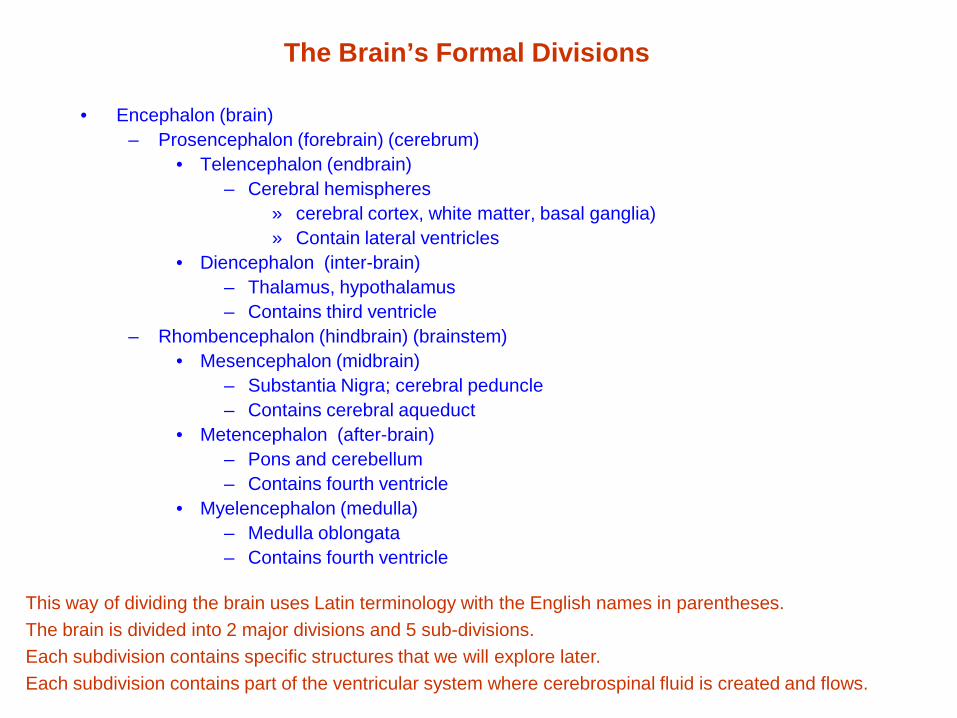
The Brain’s Formal Divisions
• Encephalon (brain) – Prosencephalon (forebrain) (cerebrum)
• Telencephalon (endbrain) – Cerebral hemispheres
» cerebral cortex, white matter, basal ganglia) » Contain lateral ventricles
• Diencephalon (inter-brain) – Thalamus, hypothalamus – Contains third ventricle
– Rhombencephalon (hindbrain) (brainstem) • Mesencephalon (midbrain)
– Substantia Nigra; cerebral peduncle – Contains cerebral aqueduct
• Metencephalon (after-brain) – Pons and cerebellum – Contains fourth ventricle
• Myelencephalon (medulla) – Medulla oblongata – Contains fourth ventricle
This way of dividing the brain uses Latin terminology with the English names in parentheses. The brain is divided into 2 major divisions and 5 sub-divisions. Each subdivision contains specific structures that we will explore later. Each subdivision contains part of the ventricular system where cerebrospinal fluid is created and flows.
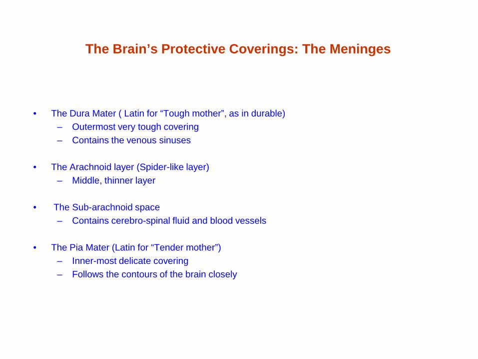
The Brain’s Protective Coverings: The Meninges
• The Dura Mater ( Latin for “Tough mother”, as in durable) – Outermost very tough covering – Contains the venous sinuses
• The Arachnoid layer (Spider-like layer)
– Middle, thinner layer
• The Sub-arachnoid space – Contains cerebro-spinal fluid and blood vessels
• The Pia Mater (Latin for “Tender mother”)
– Inner-most delicate covering – Follows the contours of the brain closely
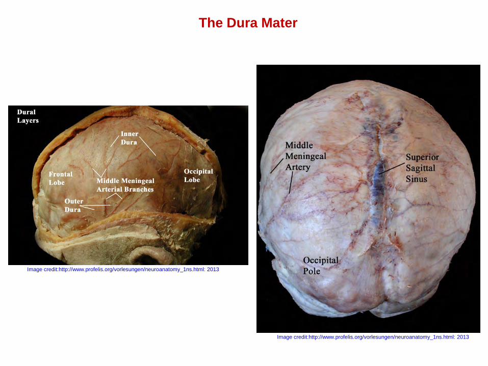
The Dura Mater
Image credit:http://www.profelis.org/vorlesungen/neuroanatomy_1ns.html: 2013
Image credit:http://www.profelis.org/vorlesungen/neuroanatomy_1ns.html: 2013
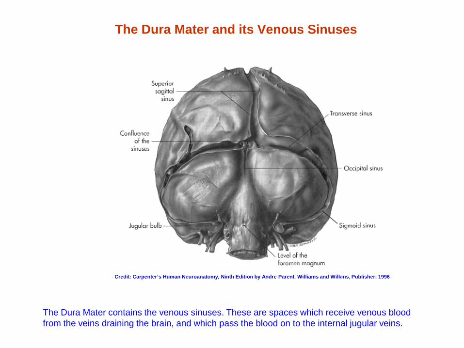
The Dura Mater and its Venous Sinuses
The Dura Mater contains the venous sinuses. These are spaces which receive venous blood from the veins draining the brain, and which pass the blood on to the internal jugular veins.
Credit: Carpenter’s Human Neuroanatomy, Ninth Edition by Andre Parent. Williams and Wilkins, Publisher: 1996
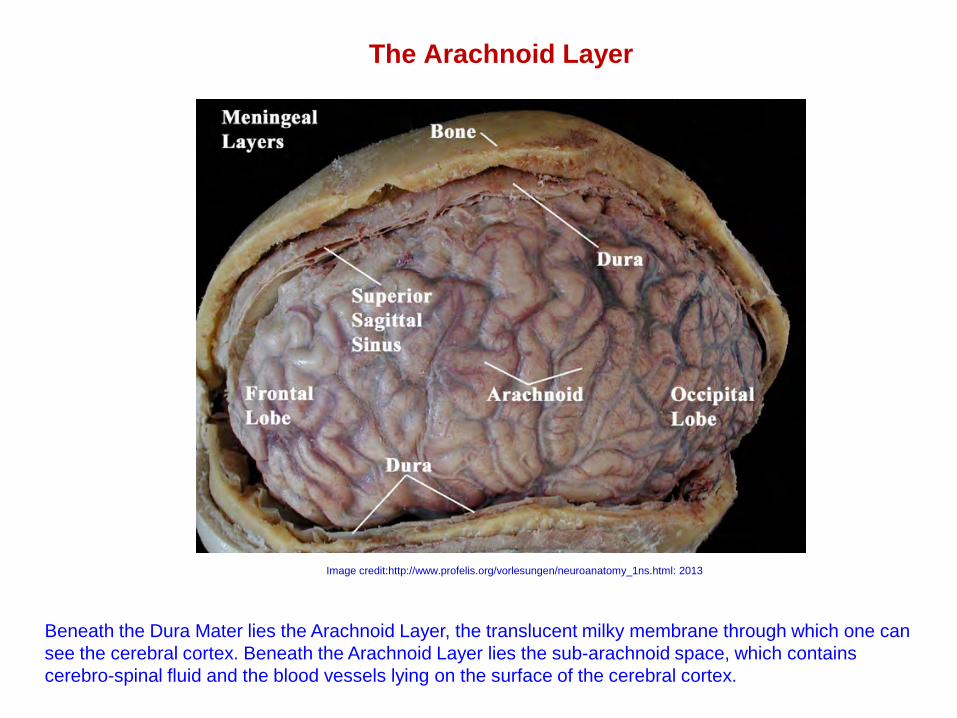
The Arachnoid Layer
Image credit:http://www.profelis.org/vorlesungen/neuroanatomy_1ns.html: 2013
Beneath the Dura Mater lies the Arachnoid Layer, the translucent milky membrane through which one can see the cerebral cortex. Beneath the Arachnoid Layer lies the sub-arachnoid space, which contains cerebro-spinal fluid and the blood vessels lying on the surface of the cerebral cortex.
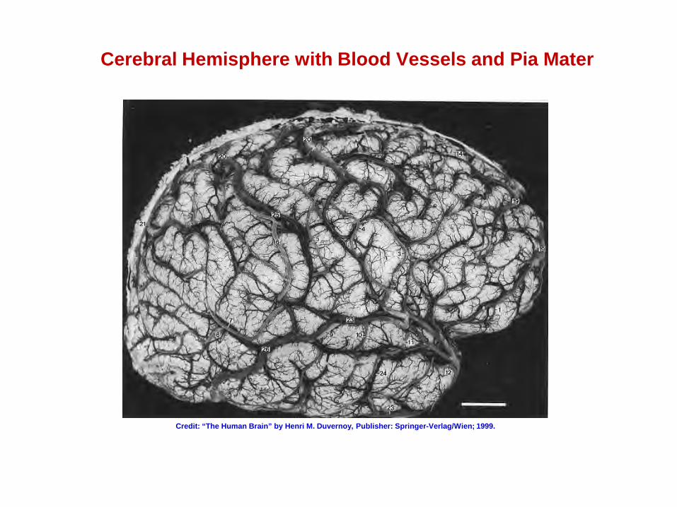
Cerebral Hemisphere with Blood Vessels and Pia Mater
Credit: “The Human Brain” by Henri M. Duvernoy, Publisher: Springer-Verlag/Wien; 1999.
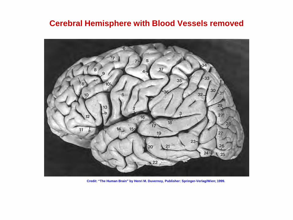
Cerebral Hemisphere with Blood Vessels removed
Credit: “The Human Brain” by Henri M. Duvernoy, Publisher: Springer-Verlag/Wien; 1999.
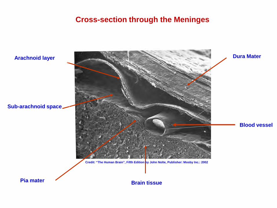
Cross-section through the Meninges
Dura Mater Arachnoid layer
Sub-arachnoid space
Blood vessel
Pia mater Brain tissue
Credit: “The Human Brain”, Fifth Edition by John Nolte, Publisher: Mosby Inc.: 2002
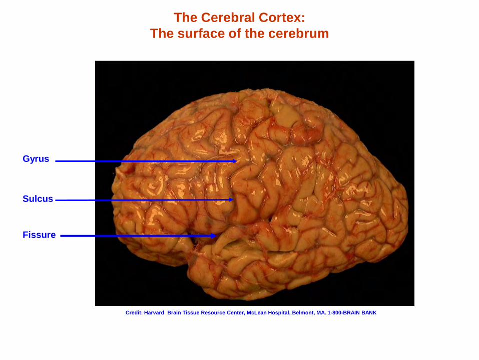
The Cerebral Cortex: The surface of the cerebrum
Gyrus
Sulcus
Fissure
Credit: Harvard Brain Tissue Resource Center, McLean Hospital, Belmont, MA. 1-800-BRAIN BANK
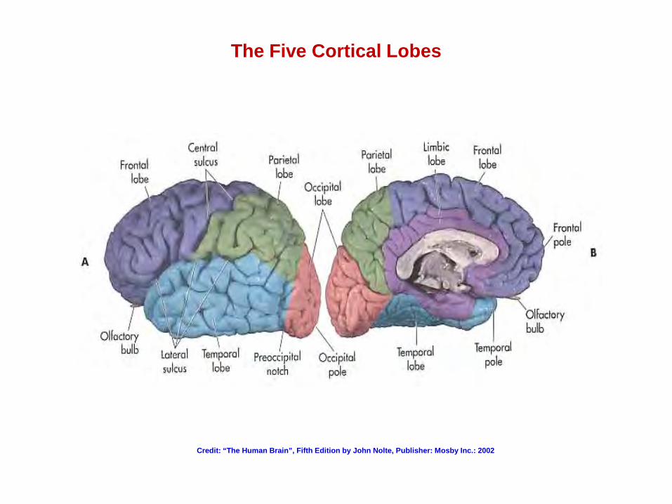
The Five Cortical Lobes
Credit: “The Human Brain”, Fifth Edition by John Nolte, Publisher: Mosby Inc.: 2002
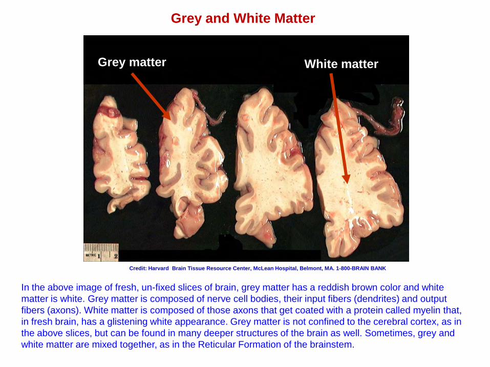
Grey and White Matter
Grey matter White matter
In the above image of fresh, un-fixed slices of brain, grey matter has a reddish brown color and white matter is white. Grey matter is composed of nerve cell bodies, their input fibers (dendrites) and output fibers (axons). White matter is composed of those axons that get coated with a protein called myelin that, in fresh brain, has a glistening white appearance. Grey matter is not confined to the cerebral cortex, as in the above slices, but can be found in many deeper structures of the brain as well. Sometimes, grey and white matter are mixed together, as in the Reticular Formation of the brainstem.
Credit: Harvard Brain Tissue Resource Center, McLean Hospital, Belmont, MA. 1-800-BRAIN BANK
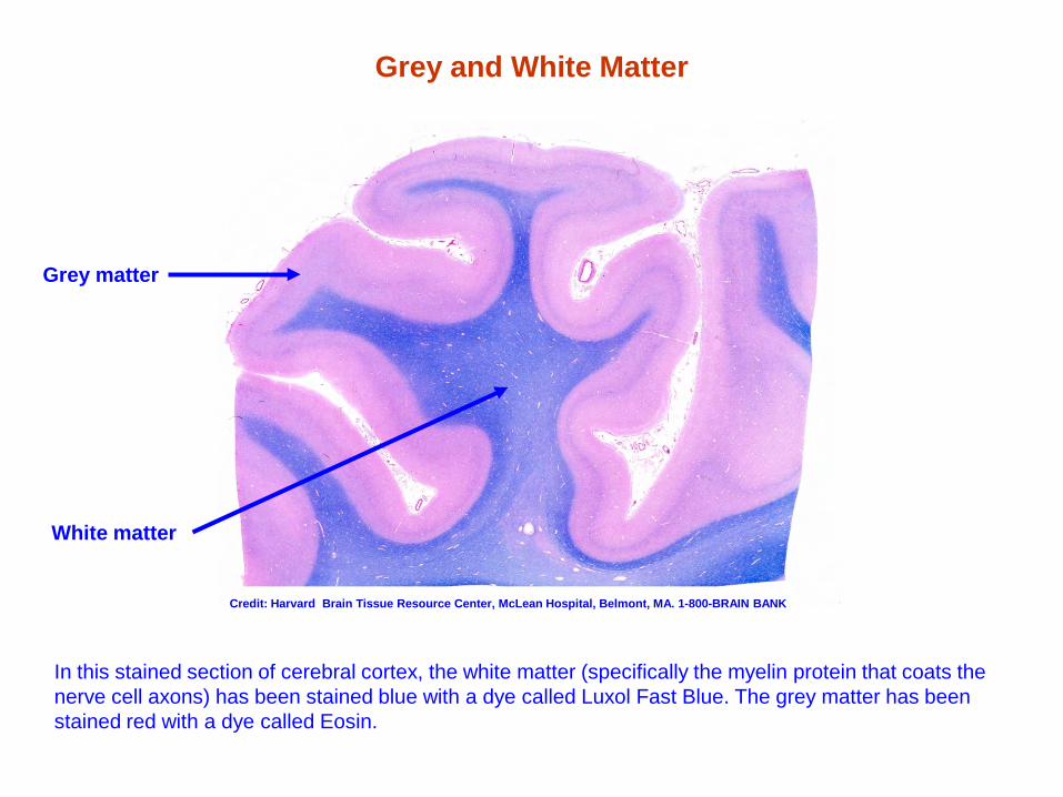
Grey and White Matter
Luxol Fast Blue-Hematoxylin-Eosin stain
Grey matter
White matter
In this stained section of cerebral cortex, the white matter (specifically the myelin protein that coats the nerve cell axons) has been stained blue with a dye called Luxol Fast Blue. The grey matter has been stained red with a dye called Eosin.
Credit: Harvard Brain Tissue Resource Center, McLean Hospital, Belmont, MA. 1-800-BRAIN BANK
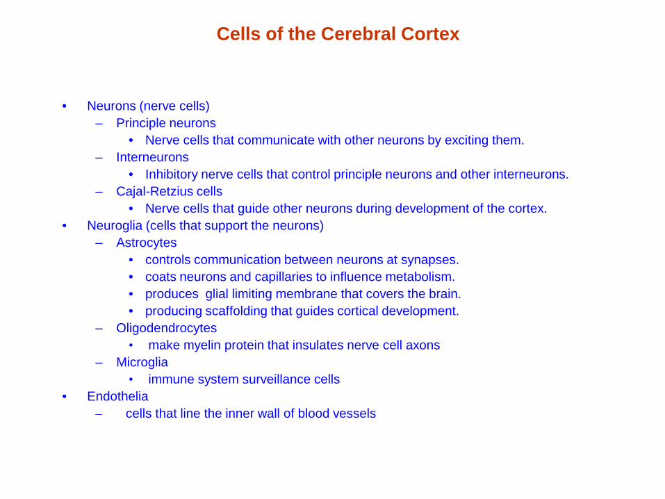
Cells of the Cerebral Cortex
• Neurons (nerve cells) – Principle neurons
• Nerve cells that communicate with other neurons by exciting them. – Interneurons
• Inhibitory nerve cells that control principle neurons and other interneurons. – Cajal-Retzius cells
• Nerve cells that guide other neurons during development of the cortex. • Neuroglia (cells that support the neurons)
– Astrocytes • controls communication between neurons at synapses. • coats neurons and capillaries to influence metabolism. • produces glial limiting membrane that covers the brain. • producing scaffolding that guides cortical development.
– Oligodendrocytes • make myelin protein that insulates nerve cell axons
– Microglia • immune system surveillance cells
• Endothelia – cells that line the inner wall of blood vessels
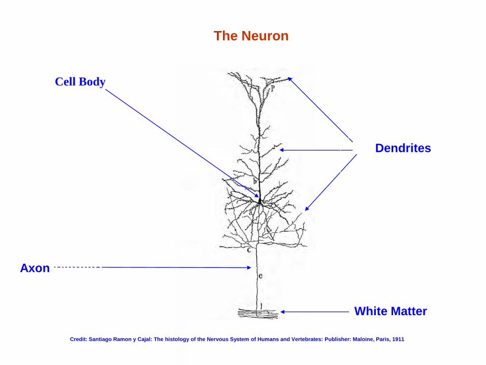
White Matter (axons coated with myelin)
Axon (output)
Dendrites
(input)
Cell Body (soma)
The Neuron
Credit: Santiago Ramon y Cajal: The histology of the Nervous System of Humans and Vertebrates: Publisher: Maloine, Paris, 1911
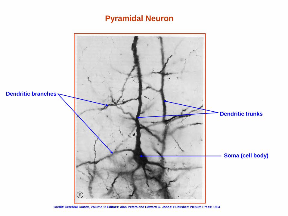
Pyramidal Neuron
Soma (cell body)
Dendritic trunks
Dendritic branches
Credit: Cerebral Cortex, Volume 1: Editors: Alan Peters and Edward G. Jones: Publisher: Plenum Press: 1984
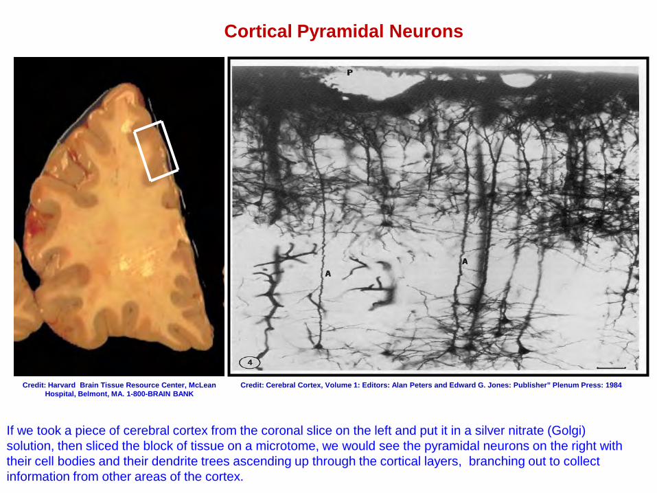
Cortical Pyramidal Neurons
If we took a piece of cerebral cortex from the coronal slice on the left and put it in a silver nitrate (Golgi) solution, then sliced the block of tissue on a microtome, we would see the pyramidal neurons on the right with their cell bodies and their dendrite trees ascending up through the cortical layers, branching out to collect information from other areas of the cortex.
Credit: Cerebral Cortex, Volume 1: Editors: Alan Peters and Edward G. Jones: Publisher” Plenum Press: 1984 Credit: Harvard Brain Tissue Resource Center, McLean Hospital, Belmont, MA. 1-800-BRAIN BANK
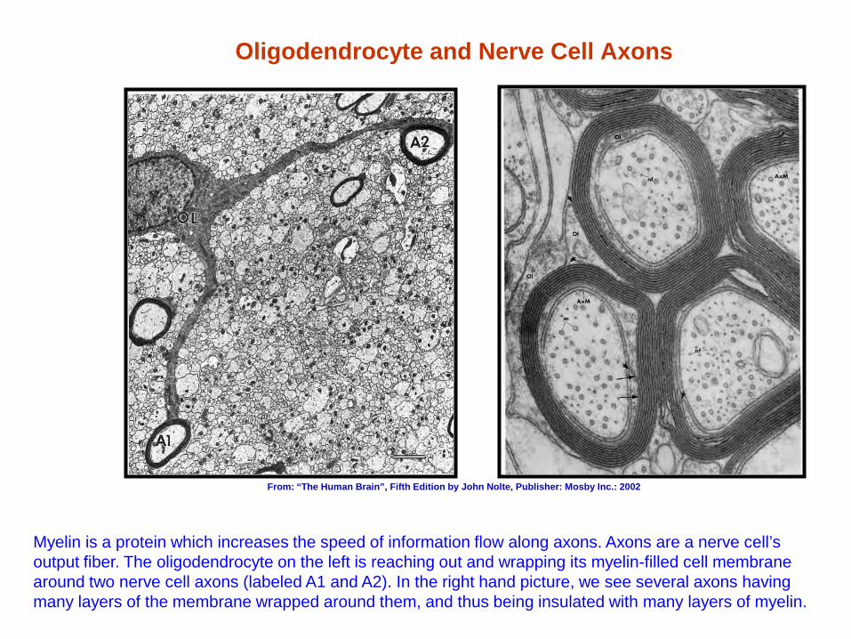
Oligodendrocyte and Nerve Cell Axons
Myelin is a protein which increases the speed of information flow along axons. Axons are a nerve cell’s output fiber. The oligodendrocyte on the left is reaching out and wrapping its myelin-filled cell membrane around two nerve cell axons (labeled A1 and A2). In the right hand picture, we see several axons having many layers of the membrane wrapped around them, and thus being insulated with many layers of myelin.
From: “The Human Brain”, Fifth Edition by John Nolte, Publisher: Mosby Inc.: 2002
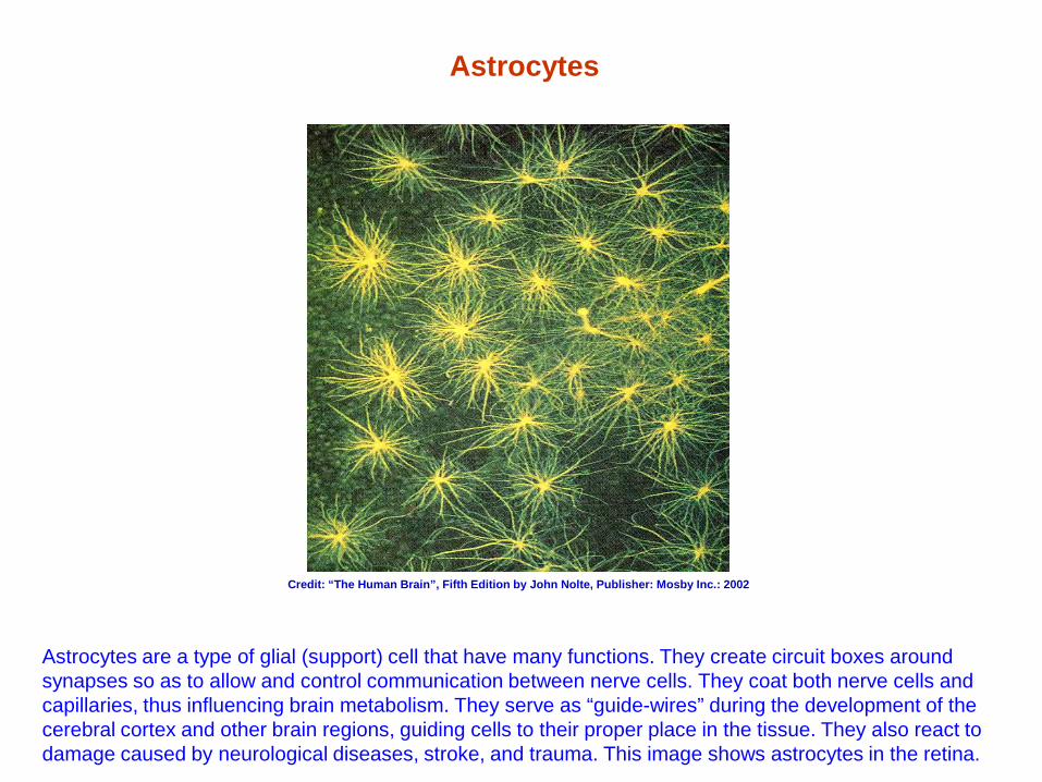
Astrocytes
Astrocytes are a type of glial (support) cell that have many functions. They create circuit boxes around synapses so as to allow and control communication between nerve cells. They coat both nerve cells and capillaries, thus influencing brain metabolism. They serve as “guide-wires” during the development of the cerebral cortex and other brain regions, guiding cells to their proper place in the tissue. They also react to damage caused by neurological diseases, stroke, and trauma. This image shows astrocytes in the retina.
Credit: “The Human Brain”, Fifth Edition by John Nolte, Publisher: Mosby Inc.: 2002
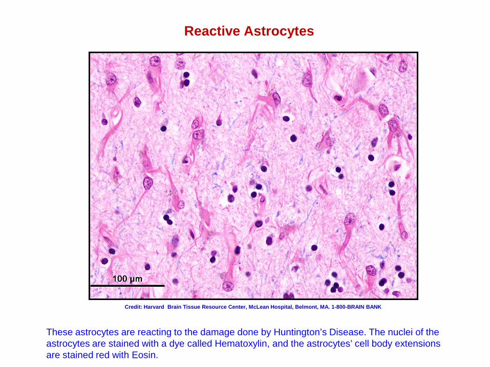
Reactive Astrocytes
These astrocytes are reacting to the damage done by Huntington’s Disease. The nuclei of the astrocytes are stained with a dye called Hematoxylin, and the astrocytes’ cell body extensions are stained red with Eosin.
Credit: Harvard Brain Tissue Resource Center, McLean Hospital, Belmont, MA. 1-800-BRAIN BANK
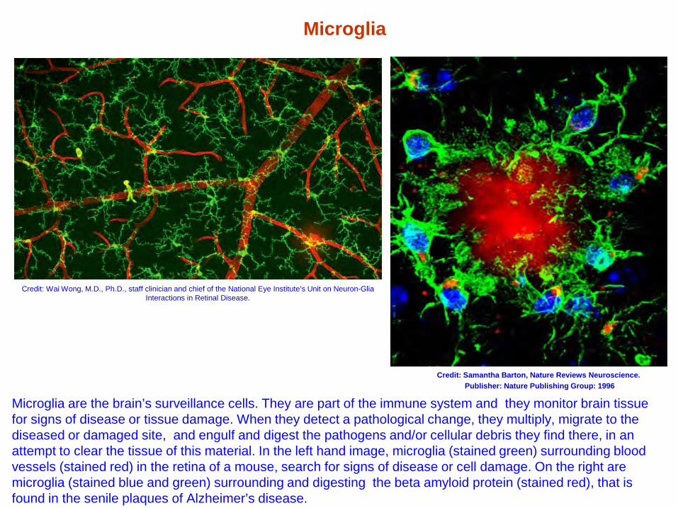
Microglia
Microglia are the brain’s surveillance cells. They are part of the immune system and they monitor brain tissue for signs of disease or tissue damage. When they detect a pathological change, they multiply, migrate to the diseased or damaged site, and engulf and digest the pathogens and/or cellular debris they find there, in an attempt to clear the tissue of this material. In the left hand image, microglia (stained green) surrounding blood vessels (stained red) in the retina of a mouse, search for signs of disease or cell damage. On the right are microglia (stained blue and green) surrounding and digesting the beta amyloid protein (stained red), that is found in the senile plaques of Alzheimer’s disease.
Credit: Samantha Barton, Nature Reviews Neuroscience. Publisher: Nature Publishing Group: 1996
Credit: Wai Wong, M.D., Ph.D., staff clinician and chief of the National Eye Institute’s Unit on Neuron-Glia Interactions in Retinal Disease.
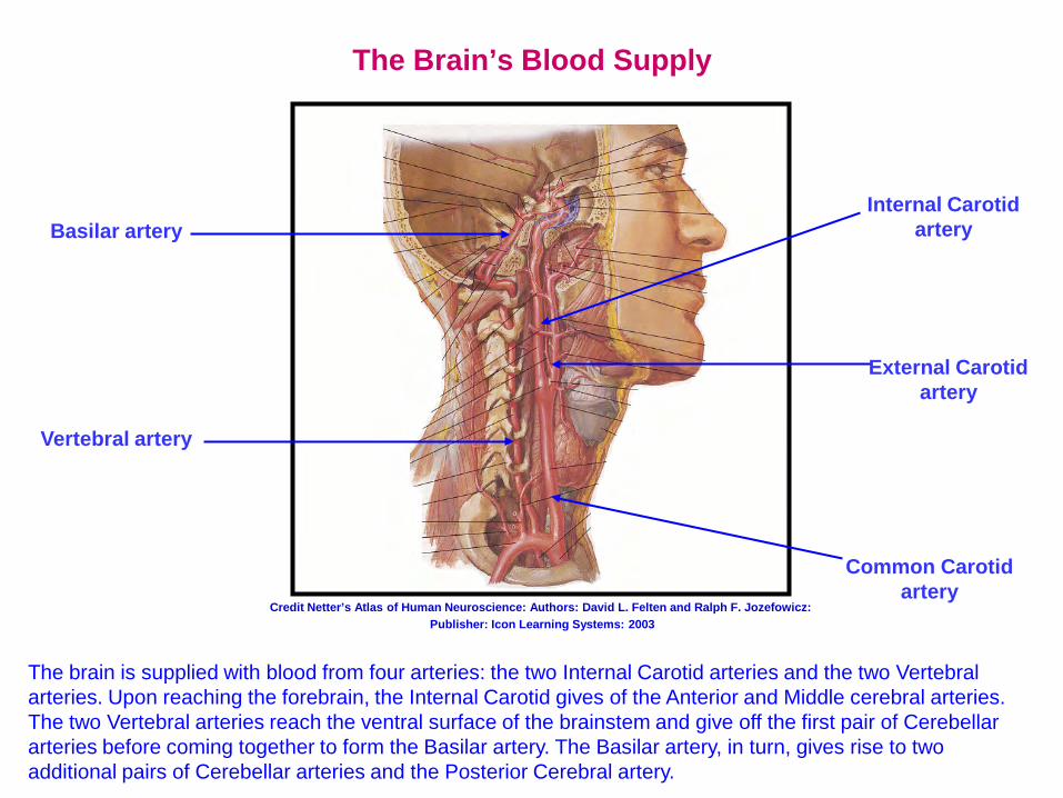
The Brain’s Blood Supply
Internal Carotid artery
Vertebral artery
Basilar artery
External Carotid artery
Common Carotid artery
Credit Netter’s Atlas of Human Neuroscience: Authors: David L. Felten and Ralph F. Jozefowicz: Publisher: Icon Learning Systems: 2003
The brain is supplied with blood from four arteries: the two Internal Carotid arteries and the two Vertebral arteries. Upon reaching the forebrain, the Internal Carotid gives of the Anterior and Middle cerebral arteries. The two Vertebral arteries reach the ventral surface of the brainstem and give off the first pair of Cerebellar arteries before coming together to form the Basilar artery. The Basilar artery, in turn, gives rise to two additional pairs of Cerebellar arteries and the Posterior Cerebral artery.
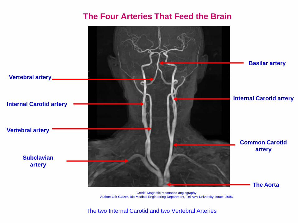
The Four Arteries That Feed the Brain
The two Internal Carotid and two Vertebral Arteries
Common Carotid artery
Internal Carotid artery
Vertebral artery
Vertebral artery
Subclavian artery
The Aorta
Basilar artery
Internal Carotid artery
Credit: Magnetic resonance angiography Author: Ofir Glazer, Bio-Medical Engineering Department, Tel-Aviv University, Israel: 2006
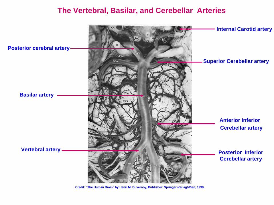
The Vertebral, Basilar, and Cerebellar Arteries
Vertebral artery
Superior Cerebellar artery
Basilar artery
Internal Carotid artery
Posterior cerebral artery
Anterior Inferior Cerebellar artery
Posterior Inferior Cerebellar artery
Credit: “The Human Brain” by Henri M. Duvernoy, Publisher: Springer-Verlag/Wien; 1999.
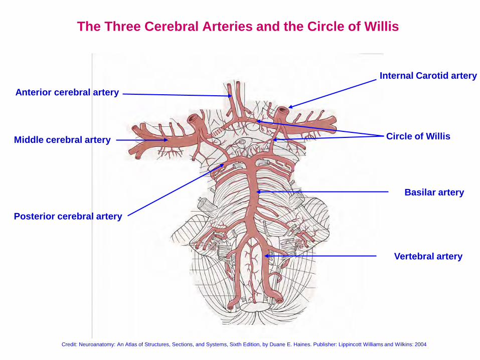
The Three Cerebral Arteries and the Circle of Willis
Circle of Willis
Vertebral artery
Basilar artery
Anterior cerebral artery
Middle cerebral artery
Posterior cerebral artery
Internal Carotid artery
Credit: Neuroanatomy: An Atlas of Structures, Sections, and Systems, Sixth Edition, by Duane E. Haines. Publisher: Lippincott Williams and Wilkins: 2004
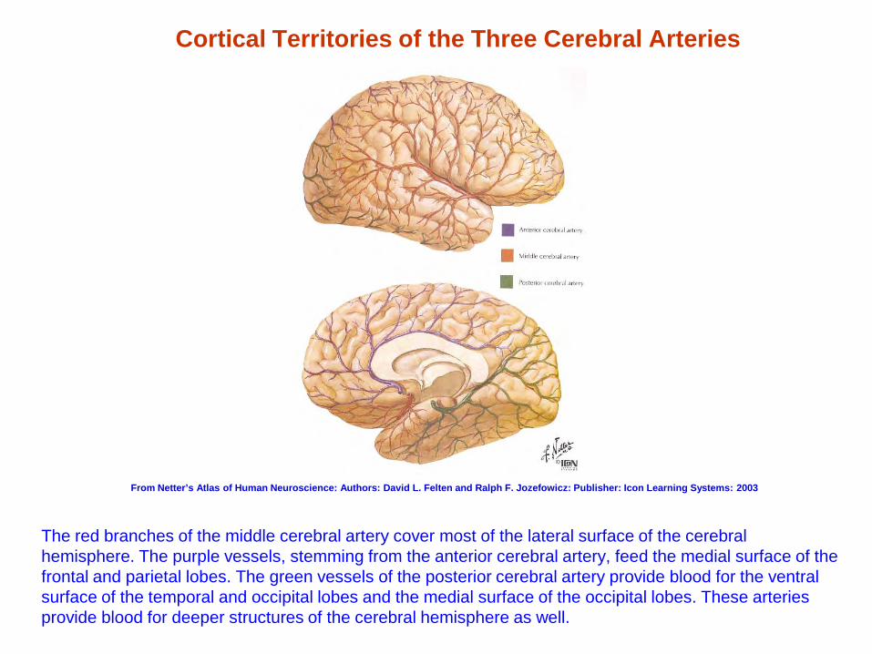
Cortical Territories of the Three Cerebral Arteries
The red branches of the middle cerebral artery cover most of the lateral surface of the cerebral hemisphere. The purple vessels, stemming from the anterior cerebral artery, feed the medial surface of the frontal and parietal lobes. The green vessels of the posterior cerebral artery provide blood for the ventral surface of the temporal and occipital lobes and the medial surface of the occipital lobes. These arteries provide blood for deeper structures of the cerebral hemisphere as well.
From Netter’s Atlas of Human Neuroscience: Authors: David L. Felten and Ralph F. Jozefowicz: Publisher: Icon Learning Systems: 2003
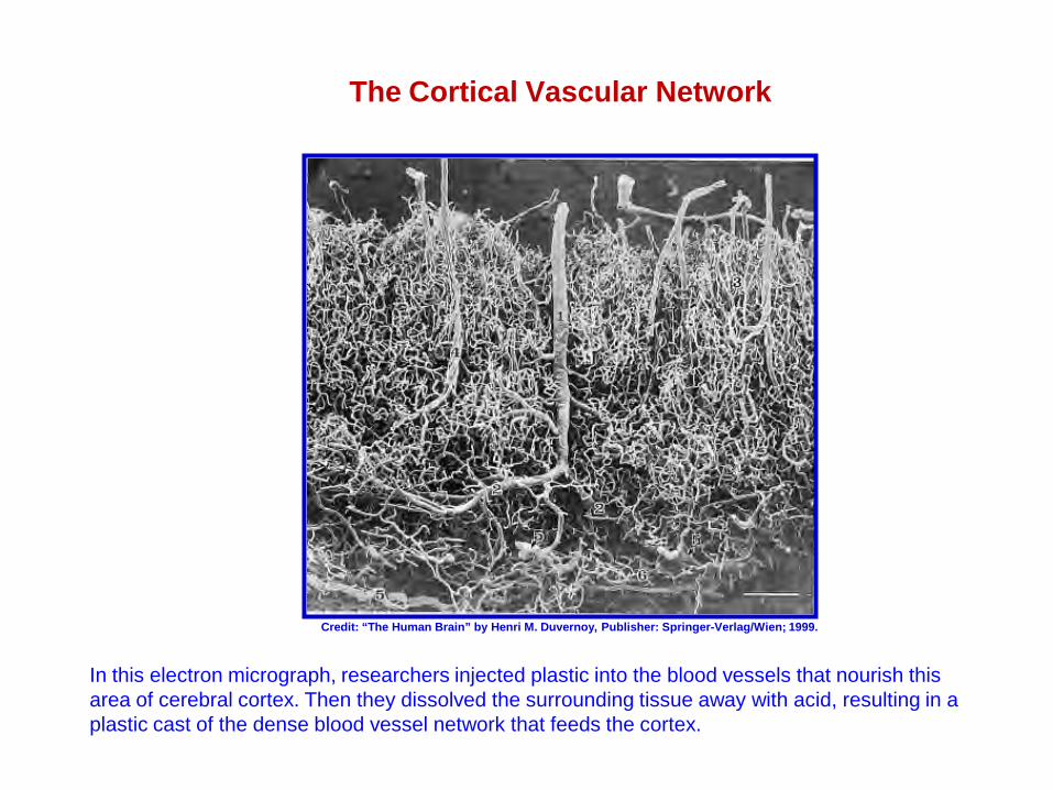
The Cortical Vascular Network
In this electron micrograph, researchers injected plastic into the blood vessels that nourish this area of cerebral cortex. Then they dissolved the surrounding tissue away with acid, resulting in a plastic cast of the dense blood vessel network that feeds the cortex.
Credit: “The Human Brain” by Henri M. Duvernoy, Publisher: Springer-Verlag/Wien; 1999.
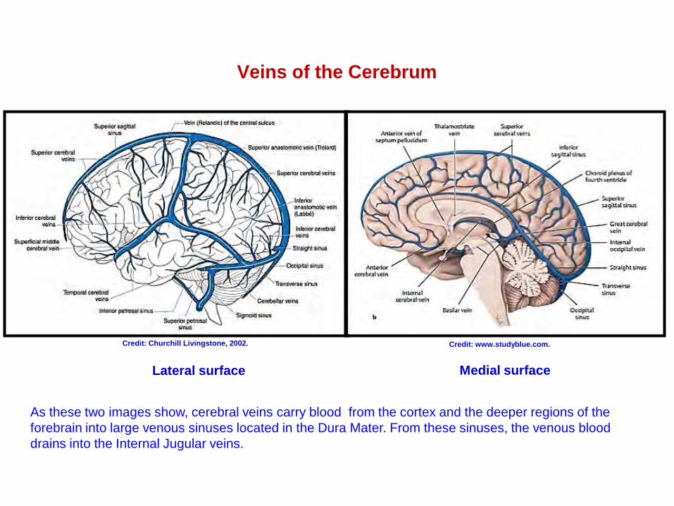
Veins of the Cerebrum
As these two images show, cerebral veins carry blood from the cortex and the deeper regions of the forebrain into large venous sinuses located in the Dura Mater. From these sinuses, the venous blood drains into the Internal Jugular veins.
Credit: Churchill Livingstone, 2002. Credit: www.studyblue.com.
Lateral surface Medial surface
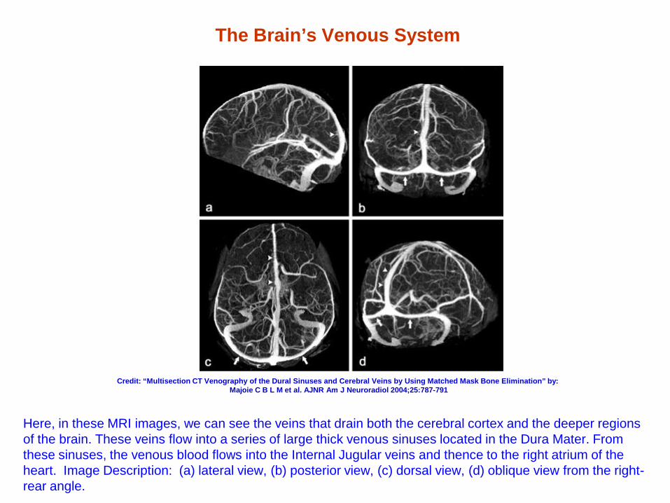
The Brain’s Venous System
Here, in these MRI images, we can see the veins that drain both the cerebral cortex and the deeper regions of the brain. These veins flow into a series of large thick venous sinuses located in the Dura Mater. From these sinuses, the venous blood flows into the Internal Jugular veins and thence to the right atrium of the heart. Image Description: (a) lateral view, (b) posterior view, (c) dorsal view, (d) oblique view from the right-rear angle.
Credit: “Multisection CT Venography of the Dural Sinuses and Cerebral Veins by Using Matched Mask Bone Elimination” by: Majoie C B L M et al. AJNR Am J Neuroradiol 2004;25:787-791
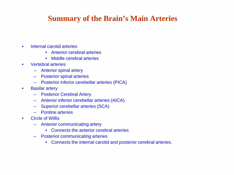
Summary of the Brain’s Main Arteries
• Internal carotid arteries • Anterior cerebral arteries • Middle cerebral arteries
• Vertebral arteries – Anterior spinal artery – Posterior spinal arteries – Posterior inferior cerebellar arteries (PICA)
• Basilar artery – Posterior Cerebral Artery – Anterior inferior cerebellar arteries (AICA) – Superior cerebellar arteries (SCA) – Pontine arteries
• Circle of Willis – Anterior communicating artery
• Connects the anterior cerebral arteries – Posterior communicating arteries
• Connects the internal carotid and posterior cerebral arteries.
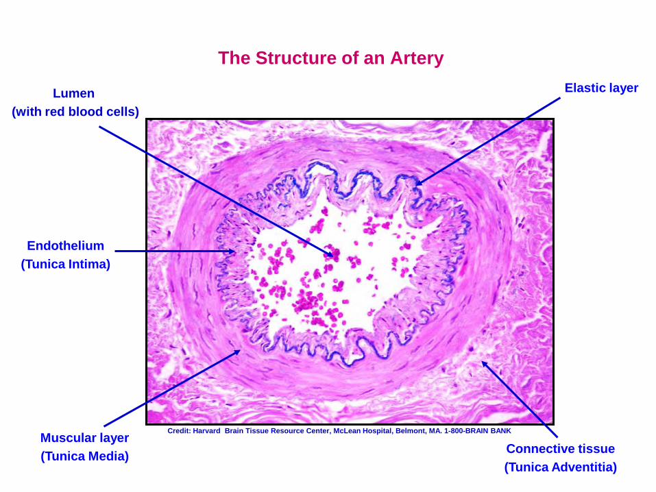
The Structure of an Artery
Muscular layer (Tunica Media)
Lumen (with red blood cells)
Elastic layer
Connective tissue (Tunica Adventitia)
Endothelium (Tunica Intima)
Credit: Harvard Brain Tissue Resource Center, McLean Hospital, Belmont, MA. 1-800-BRAIN BANK
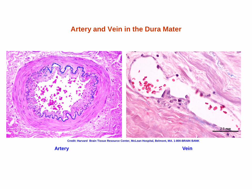
Artery and Vein in the Dura Mater
Artery Vein Credit: Harvard Brain Tissue Resource Center, McLean Hospital, Belmont, MA. 1-800-BRAIN BANK
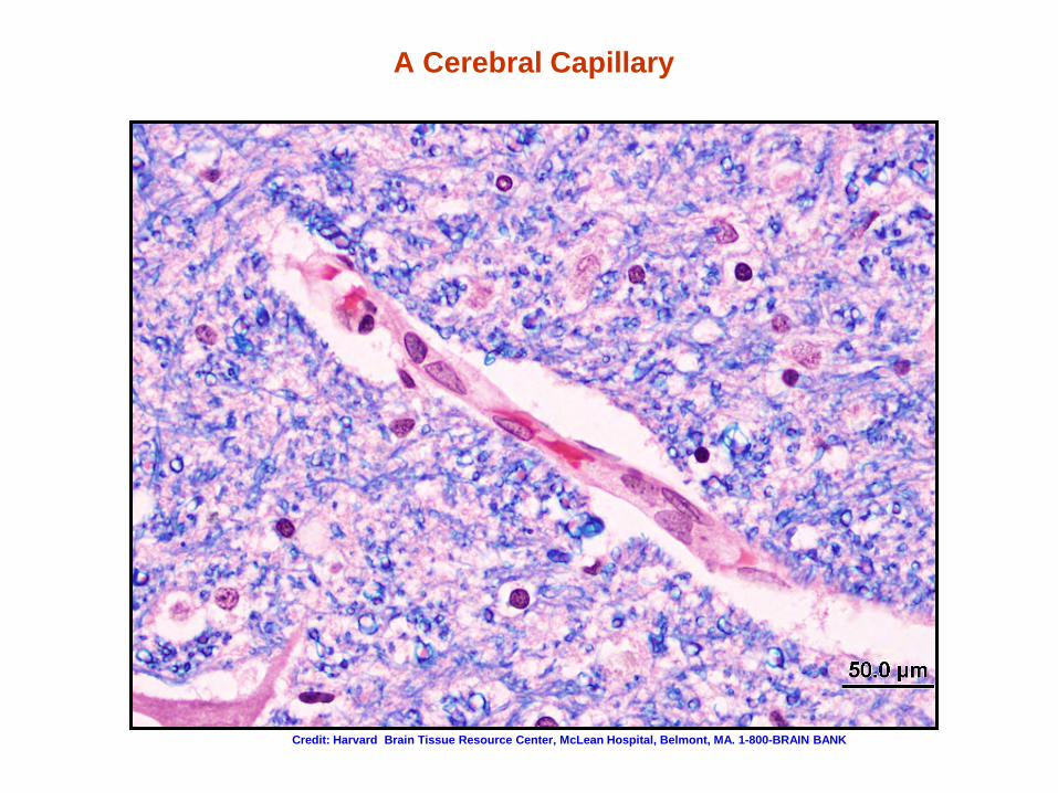
A Cerebral Capillary
Credit: Harvard Brain Tissue Resource Center, McLean Hospital, Belmont, MA. 1-800-BRAIN BANK
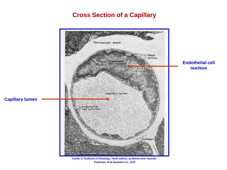
Cross Section of a Capillary
Capillary lumen
Endothelial cell nucleus
Credit: A Textbook of Histology, Tenth edition, by Bloom and Faucett: Publisher: W.B.Saunders Co. 1975
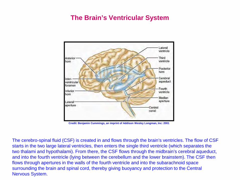
The Brain’s Ventricular System
The cerebro-spinal fluid (CSF) is created in and flows through the brain’s ventricles. The flow of CSF starts in the two large lateral ventricles, then enters the single third ventricle (which separates the two thalami and hypothalami). From there, the CSF flows through the midbrain’s cerebral aqueduct, and into the fourth ventricle (lying between the cerebellum and the lower brainstem). The CSF then flows through apertures in the walls of the fourth ventricle and into the subarachnoid space surrounding the brain and spinal cord, thereby giving buoyancy and protection to the Central Nervous System.
Credit: Benjamin Cummings, an imprint of Addison Wesley Longman, Inc: 2001
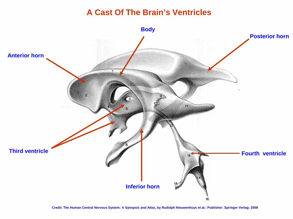
A Cast Of The Brain’s Ventricles
Fourth ventricle Third ventricle
Posterior horn
Inferior horn
Anterior horn
Body
Credit: The Human Central Nervous System: A Synopsis and Atlas, by Rudolph Nieuwenhuys et al.: Publisher: Springer Verlag: 2008
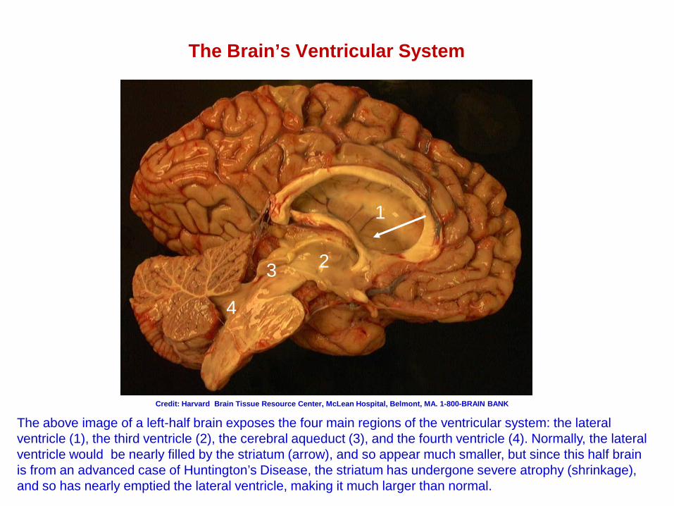
The Brain’s Ventricular System
The above image of a left-half brain exposes the four main regions of the ventricular system: the lateral ventricle (1), the third ventricle (2), the cerebral aqueduct (3), and the fourth ventricle (4). Normally, the lateral ventricle would be nearly filled by the striatum (arrow), and so appear much smaller, but since this half brain is from an advanced case of Huntington’s Disease, the striatum has undergone severe atrophy (shrinkage), and so has nearly emptied the lateral ventricle, making it much larger than normal.
Credit: Harvard Brain Tissue Resource Center, McLean Hospital, Belmont, MA. 1-800-BRAIN BANK
1
3 2
4
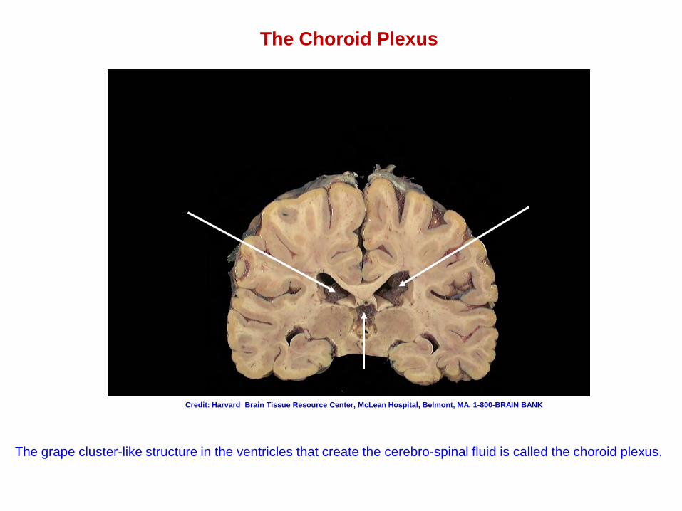
The grape cluster-like structure in the ventricles that create the cerebro-spinal fluid is called the choroid plexus.
The Choroid Plexus
Credit: Harvard Brain Tissue Resource Center, McLean Hospital, Belmont, MA. 1-800-BRAIN BANK
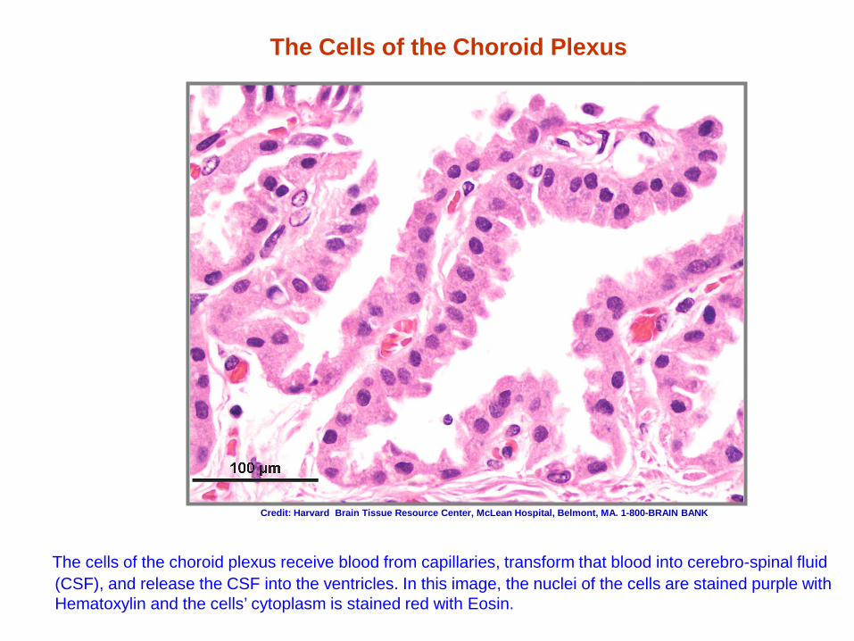
The Cells of the Choroid Plexus
The cells of the choroid plexus receive blood from capillaries, transform that blood into cerebro-spinal fluid (CSF), and release the CSF into the ventricles. In this image, the nuclei of the cells are stained purple with Hematoxylin and the cells’ cytoplasm is stained red with Eosin.
Credit: Harvard Brain Tissue Resource Center, McLean Hospital, Belmont, MA. 1-800-BRAIN BANK

The Hippocampus
Memory formation
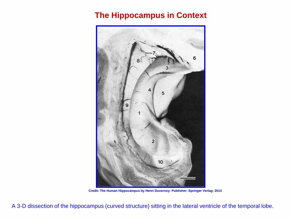
The Hippocampus in Context
A 3-D dissection of the hippocampus (curved structure) sitting in the lateral ventricle of the temporal lobe.
Credit: The Human Hippocampus by Henri Duvernoy: Publisher: Springer Verlag: 2013
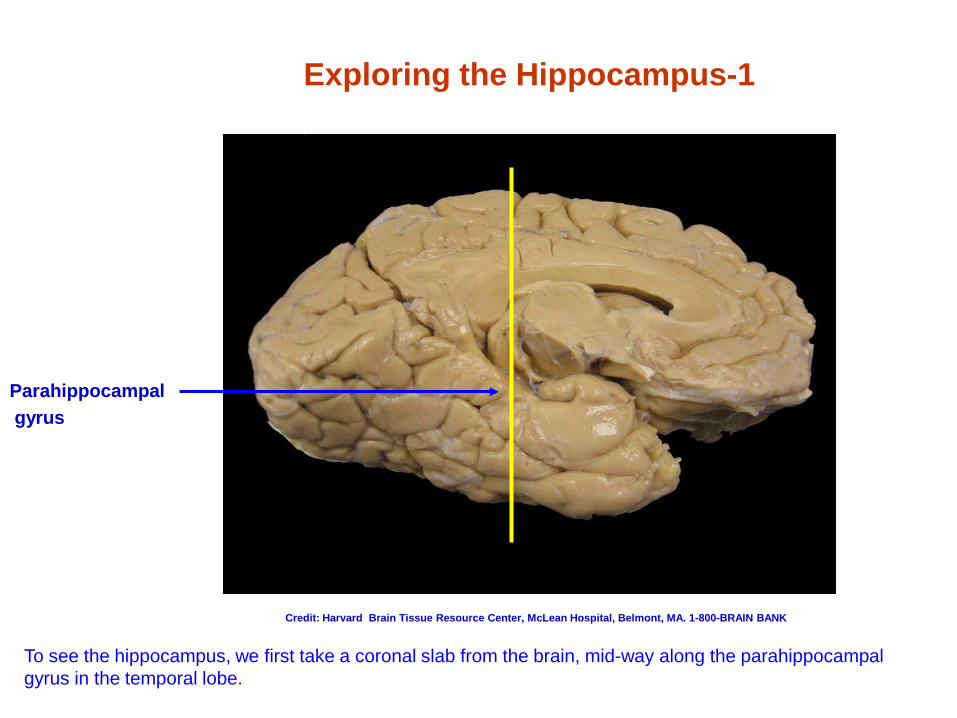
Exploring the Hippocampus-1
To see the hippocampus, we first take a coronal slab from the brain, mid-way along the parahippocampal gyrus in the temporal lobe.
Parahippocampal gyrus
Credit: Harvard Brain Tissue Resource Center, McLean Hospital, Belmont, MA. 1-800-BRAIN BANK
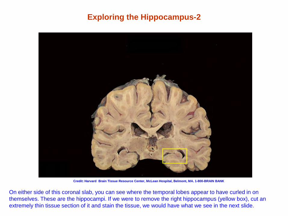
Exploring the Hippocampus-2
On either side of this coronal slab, you can see where the temporal lobes appear to have curled in on themselves. These are the hippocampi. If we were to remove the right hippocampus (yellow box), cut an extremely thin tissue section of it and stain the tissue, we would have what we see in the next slide.
Credit: Harvard Brain Tissue Resource Center, McLean Hospital, Belmont, MA. 1-800-BRAIN BANK
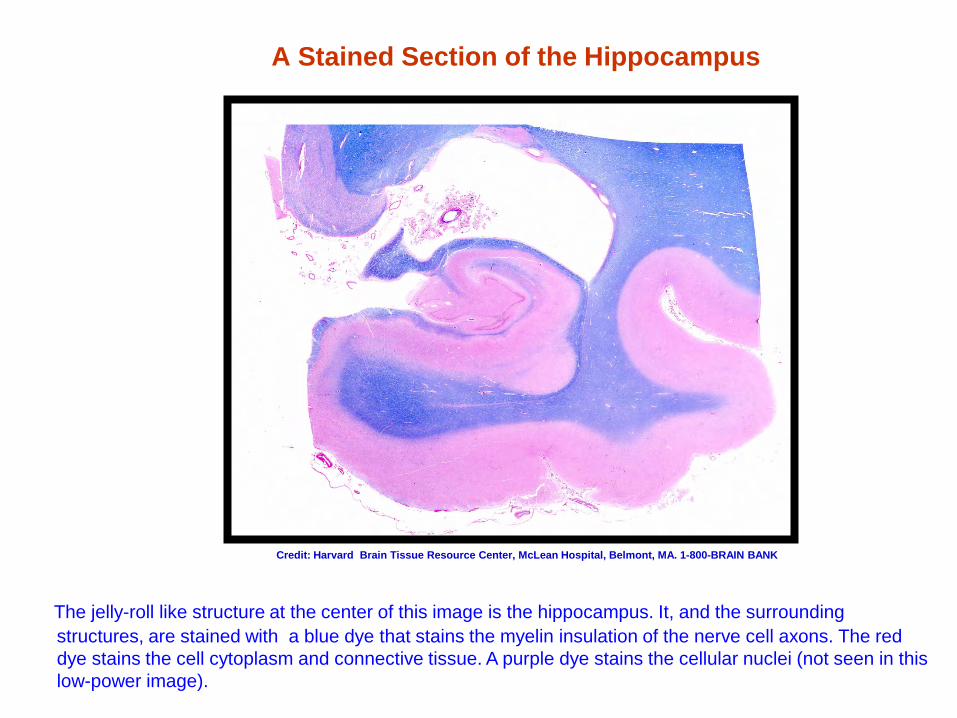
A Stained Section of the Hippocampus
The jelly-roll like structure at the center of this image is the hippocampus. It, and the surrounding structures, are stained with a blue dye that stains the myelin insulation of the nerve cell axons. The red dye stains the cell cytoplasm and connective tissue. A purple dye stains the cellular nuclei (not seen in this low-power image).
Credit: Harvard Brain Tissue Resource Center, McLean Hospital, Belmont, MA. 1-800-BRAIN BANK
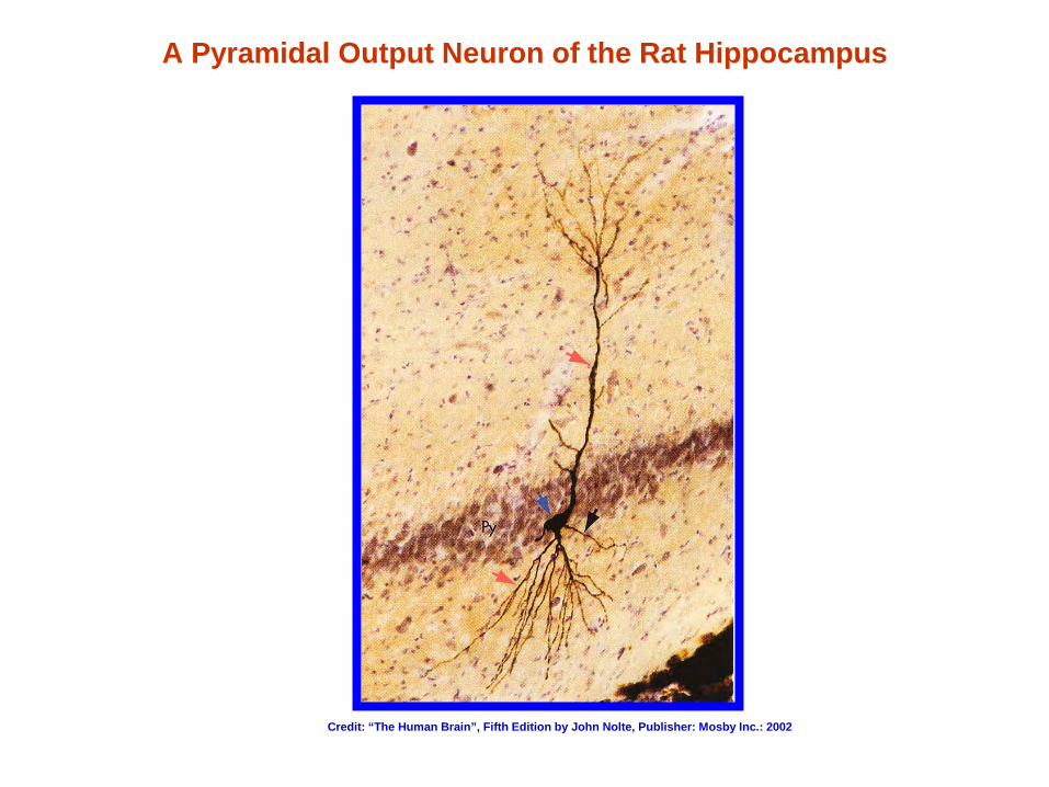
A Pyramidal Output Neuron of the Rat Hippocampus
Credit: “The Human Brain”, Fifth Edition by John Nolte, Publisher: Mosby Inc.: 2002

The Amygdala
Recognition and evaluation Fear conditioning
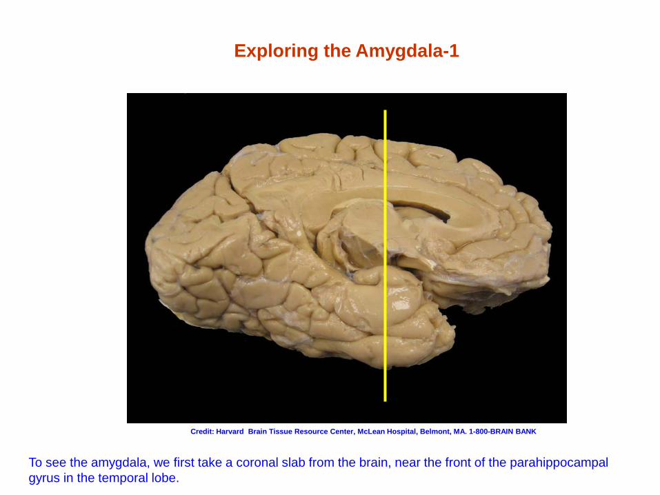
To see the amygdala, we first take a coronal slab from the brain, near the front of the parahippocampal gyrus in the temporal lobe.
Exploring the Amygdala-1
Credit: Harvard Brain Tissue Resource Center, McLean Hospital, Belmont, MA. 1-800-BRAIN BANK
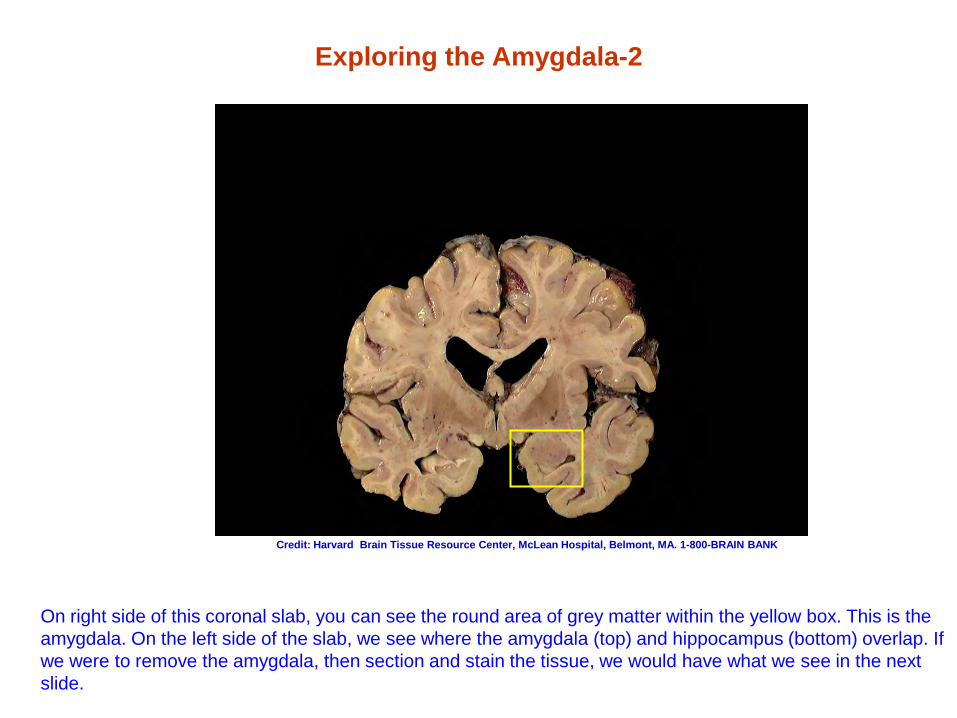
Exploring the Amygdala-2
On right side of this coronal slab, you can see the round area of grey matter within the yellow box. This is the amygdala. On the left side of the slab, we see where the amygdala (top) and hippocampus (bottom) overlap. If we were to remove the amygdala, then section and stain the tissue, we would have what we see in the next slide.
Credit: Harvard Brain Tissue Resource Center, McLean Hospital, Belmont, MA. 1-800-BRAIN BANK
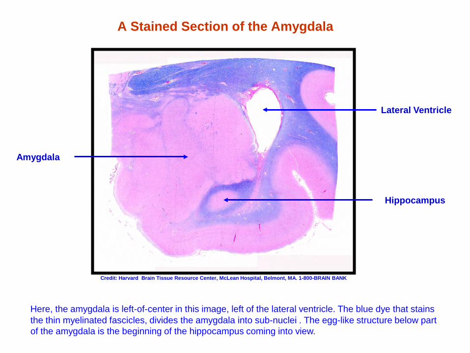
A Stained Section of the Amygdala
Here, the amygdala is left-of-center in this image, left of the lateral ventricle. The blue dye that stains
the thin myelinated fascicles, divides the amygdala into sub-nuclei . The egg-like structure below part of the amygdala is the beginning of the hippocampus coming into view.
Amygdala
Lateral Ventricle
Hippocampus
Credit: Harvard Brain Tissue Resource Center, McLean Hospital, Belmont, MA. 1-800-BRAIN BANK
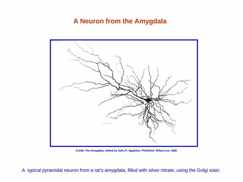
A Neuron from the Amygdala
A typical pyramidal neuron from a rat’s amygdala, filled with silver nitrate, using the Golgi stain.
Credit: The Amygdala, edited by John P. Aggleton: Publisher: Wiley-Liss: 1992
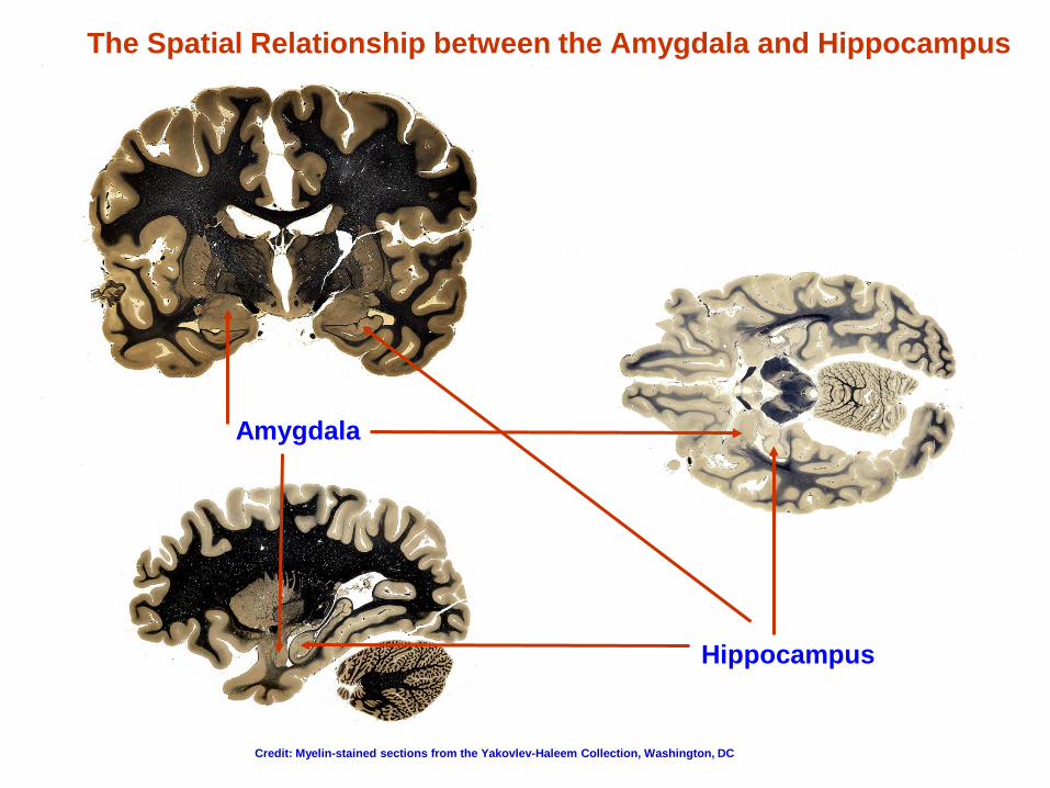
The Spatial Relationship between the Amygdala and Hippocampus
Amygdala
Hippocampus
Credit: Myelin-stained sections from the Yakovlev-Haleem Collection, Washington, DC
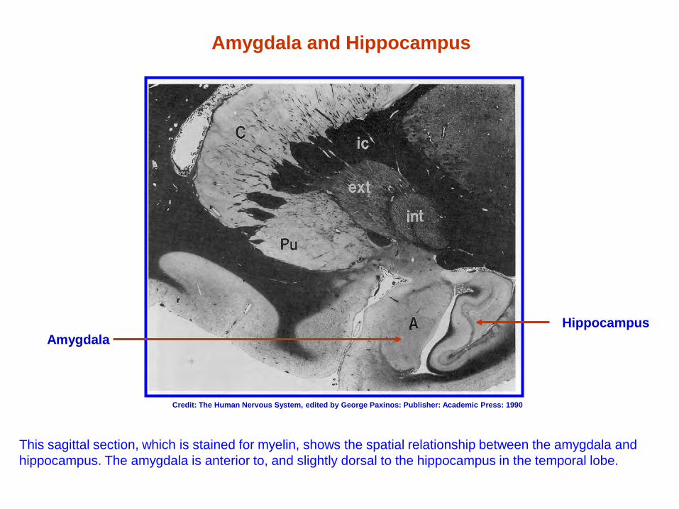
Amygdala and Hippocampus
This sagittal section, which is stained for myelin, shows the spatial relationship between the amygdala and hippocampus. The amygdala is anterior to, and slightly dorsal to the hippocampus in the temporal lobe.
Hippocampus Amygdala
Credit: The Human Nervous System, edited by George Paxinos: Publisher: Academic Press: 1990
The Striatum
The choreography of context-dependent movement Motivation, habit, and addiction formation
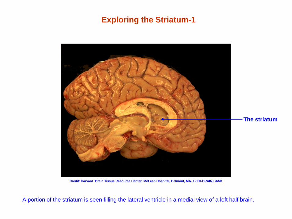
Exploring the Striatum-1
A portion of the striatum is seen filling the lateral ventricle in a medial view of a left half brain.
The striatum
Credit: Harvard Brain Tissue Resource Center, McLean Hospital, Belmont, MA. 1-800-BRAIN BANK
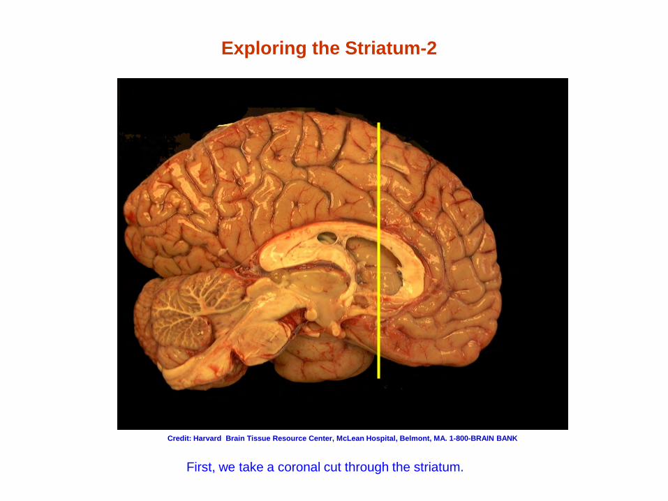
Exploring the Striatum-2
First, we take a coronal cut through the striatum.
Credit: Harvard Brain Tissue Resource Center, McLean Hospital, Belmont, MA. 1-800-BRAIN BANK
Exploring the Striatum-3
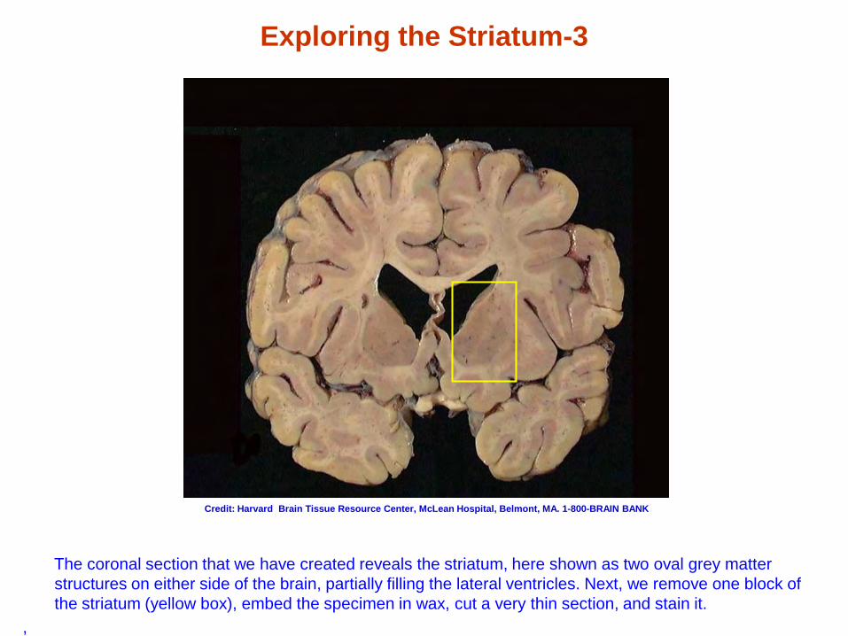
The coronal section that we have created reveals the striatum, here shown as two oval grey matter structures on either side of the brain, partially filling the lateral ventricles. Next, we remove one block of the striatum (yellow box), embed the specimen in wax, cut a very thin section, and stain it.
,
Credit: Harvard Brain Tissue Resource Center, McLean Hospital, Belmont, MA. 1-800-BRAIN BANK
Divisions of the Striatum
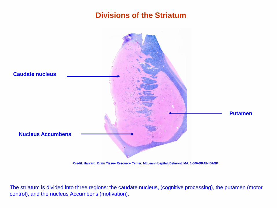
The striatum is divided into three regions: the caudate nucleus, (cognitive processing), the putamen (motor control), and the nucleus Accumbens (motivation).
Putamen
Caudate nucleus
Nucleus Accumbens
Credit: Harvard Brain Tissue Resource Center, McLean Hospital, Belmont, MA. 1-800-BRAIN BANK
The Principle Nerve Cell of the Striatum: The Medium Spiny Neuron
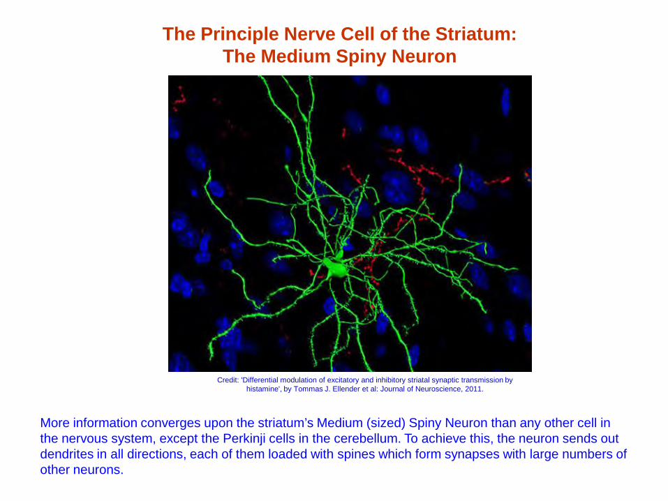
More information converges upon the striatum’s Medium (sized) Spiny Neuron than any other cell in the nervous system, except the Perkinji cells in the cerebellum. To achieve this, the neuron sends out dendrites in all directions, each of them loaded with spines which form synapses with large numbers of other neurons.
Credit: 'Differential modulation of excitatory and inhibitory striatal synaptic transmission by histamine', by Tommas J. Ellender et al: Journal of Neuroscience, 2011.
The Striatum in Context
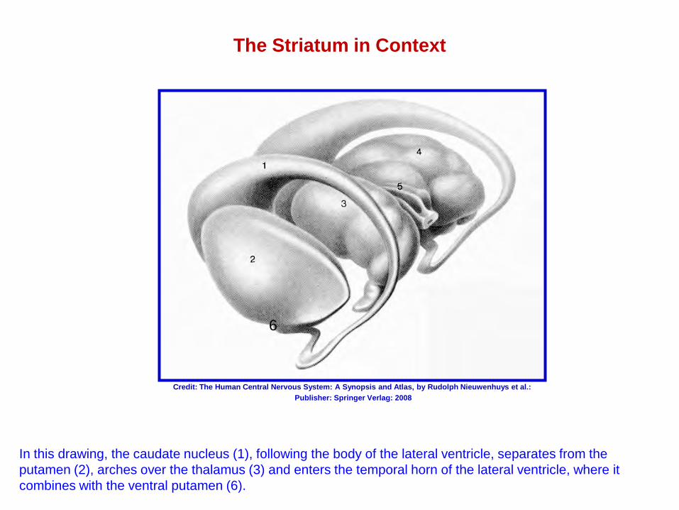
In this drawing, the caudate nucleus (1), following the body of the lateral ventricle, separates from the putamen (2), arches over the thalamus (3) and enters the temporal horn of the lateral ventricle, where it combines with the ventral putamen (6).
Credit: The Human Central Nervous System: A Synopsis and Atlas, by Rudolph Nieuwenhuys et al.: Publisher: Springer Verlag: 2008
6
The Cerebellum
Posture and balance The coordination of movement
Skill formation Implicit memory formation
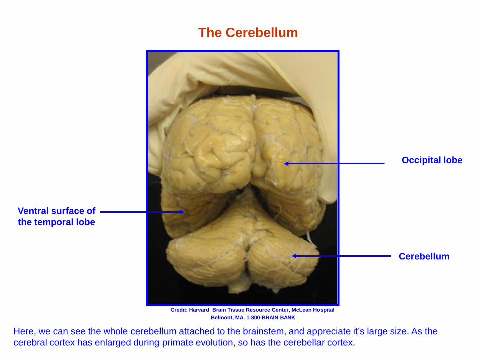
The Cerebellum
Occipital lobe
Ventral surface of the temporal lobe
Cerebellum
Here, we can see the whole cerebellum attached to the brainstem, and appreciate it’s large size. As the cerebral cortex has enlarged during primate evolution, so has the cerebellar cortex.
Credit: Harvard Brain Tissue Resource Center, McLean Hospital Belmont, MA. 1-800-BRAIN BANK
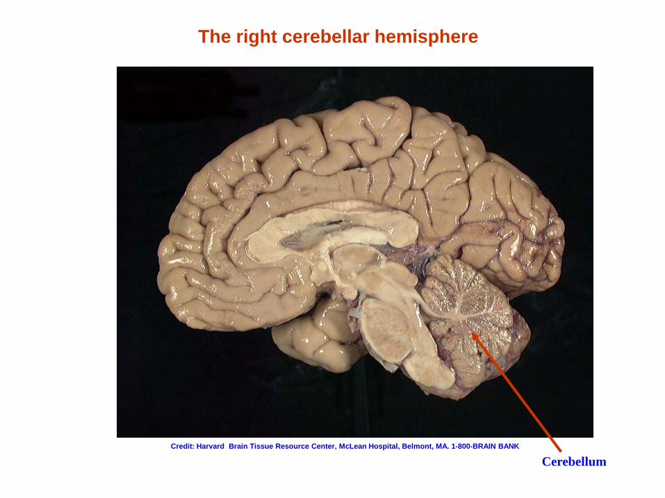
The right cerebellar hemisphere
Cerebellum Credit: Harvard Brain Tissue Resource Center, McLean Hospital, Belmont, MA. 1-800-BRAIN BANK
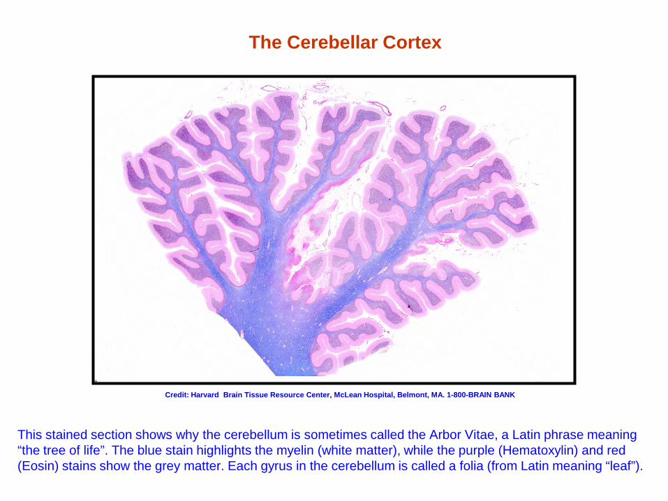
The Cerebellar Cortex
This stained section shows why the cerebellum is sometimes called the Arbor Vitae, a Latin phrase meaning “the tree of life”. The blue stain highlights the myelin (white matter), while the purple (Hematoxylin) and red (Eosin) stains show the grey matter. Each gyrus in the cerebellum is called a folia (from Latin meaning “leaf”).
Credit: Harvard Brain Tissue Resource Center, McLean Hospital, Belmont, MA. 1-800-BRAIN BANK
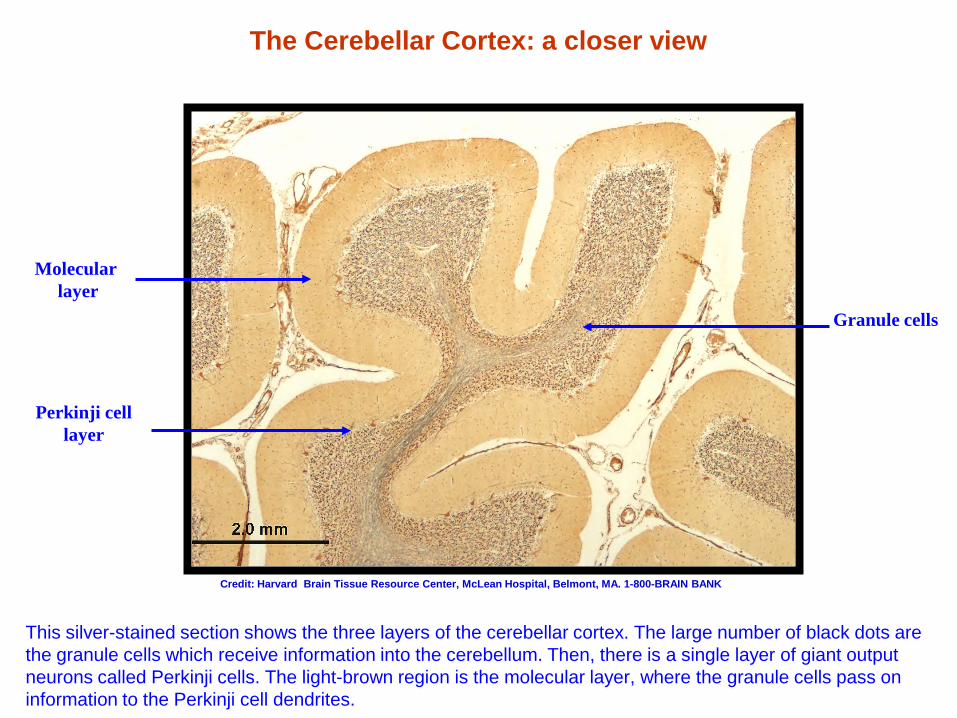
The Cerebellar Cortex: a closer view
This silver-stained section shows the three layers of the cerebellar cortex. The large number of black dots are the granule cells which receive information into the cerebellum. Then, there is a single layer of giant output neurons called Perkinji cells. The light-brown region is the molecular layer, where the granule cells pass on information to the Perkinji cell dendrites.
Granule cells
Perkinji cell layer
Molecular layer
Credit: Harvard Brain Tissue Resource Center, McLean Hospital, Belmont, MA. 1-800-BRAIN BANK
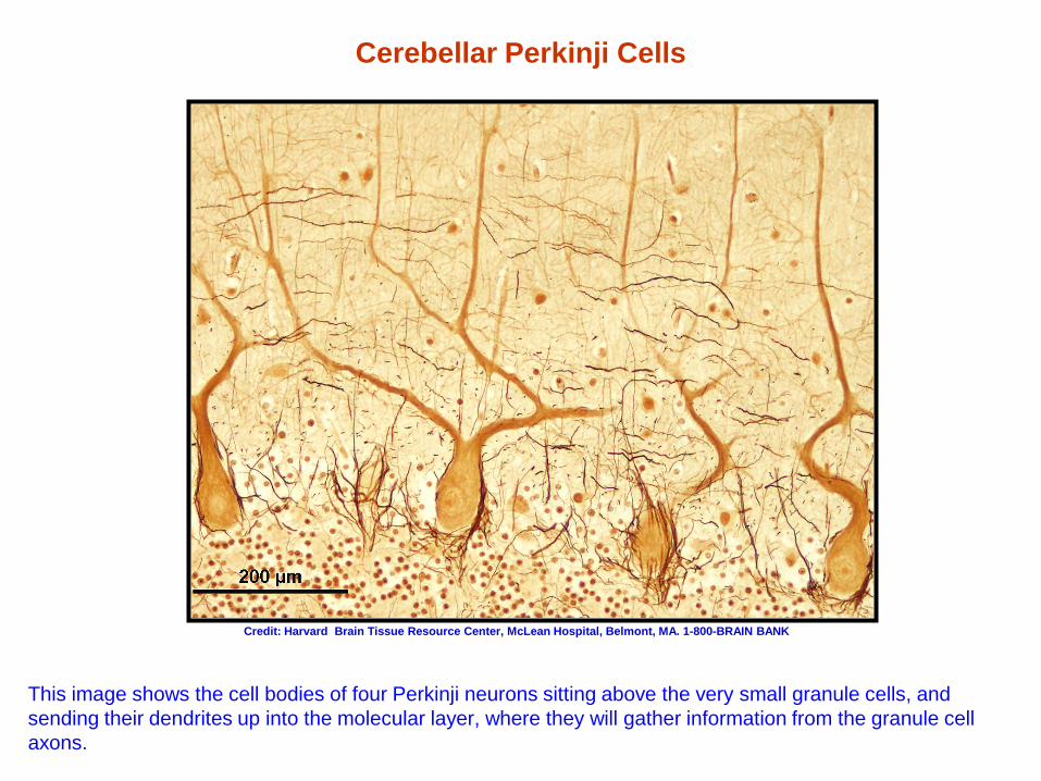
Cerebellar Perkinji Cells
This image shows the cell bodies of four Perkinji neurons sitting above the very small granule cells, and sending their dendrites up into the molecular layer, where they will gather information from the granule cell axons.
Credit: Harvard Brain Tissue Resource Center, McLean Hospital, Belmont, MA. 1-800-BRAIN BANK
The Thalamus
The The gateway to the cerebral cortex The cerebrum’s central relay station
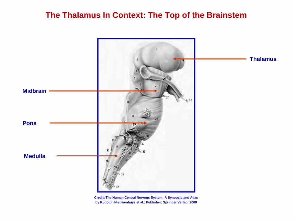
The Thalamus In Context: The Top of the Brainstem
Thalamus
Midbrain
Pons
Medulla
Credit: The Human Central Nervous System: A Synopsis and Atlas by Rudolph Nieuwenhuys et al.: Publisher: Springer Verlag: 2008
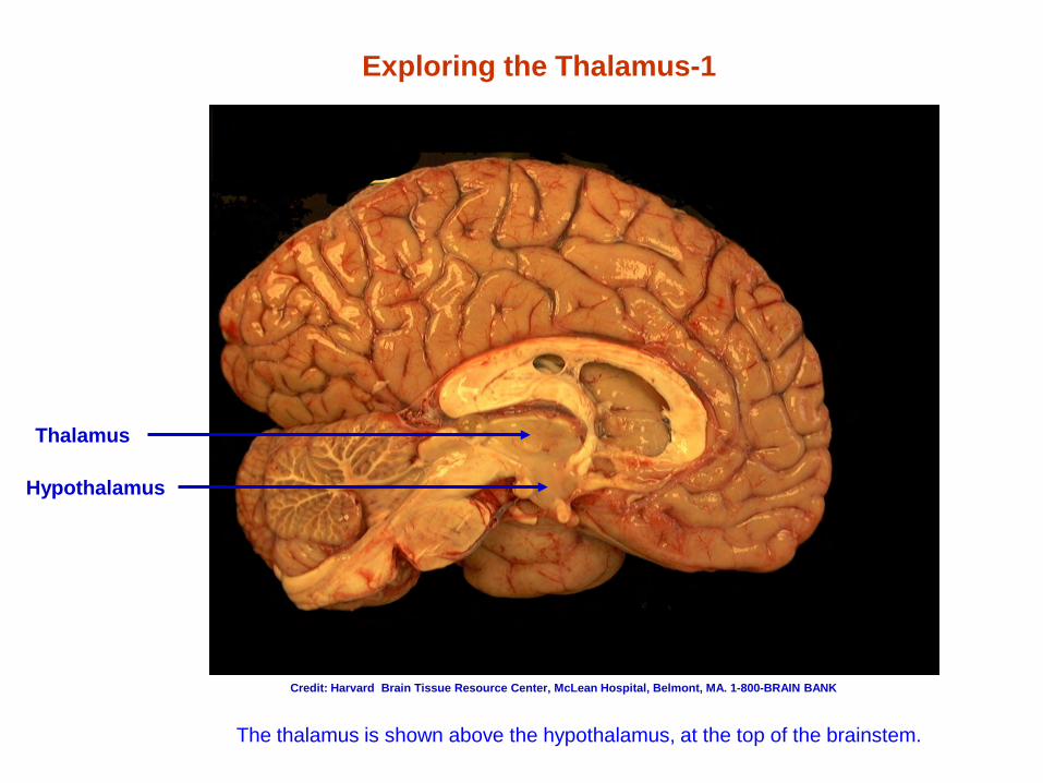
Exploring the Thalamus-1
Thalamus
The thalamus is shown above the hypothalamus, at the top of the brainstem.
Hypothalamus
Credit: Harvard Brain Tissue Resource Center, McLean Hospital, Belmont, MA. 1-800-BRAIN BANK
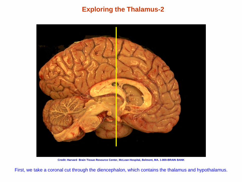
Exploring the Thalamus-2
First, we take a coronal cut through the diencephalon, which contains the thalamus and hypothalamus.
Credit: Harvard Brain Tissue Resource Center, McLean Hospital, Belmont, MA. 1-800-BRAIN BANK
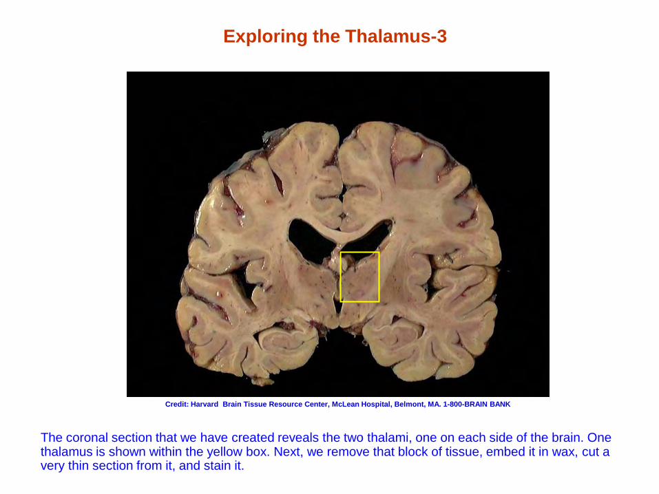
Exploring the Thalamus-3
The coronal section that we have created reveals the two thalami, one on each side of the brain. One thalamus is shown within the yellow box. Next, we remove that block of tissue, embed it in wax, cut a very thin section from it, and stain it.
Credit: Harvard Brain Tissue Resource Center, McLean Hospital, Belmont, MA. 1-800-BRAIN BANK
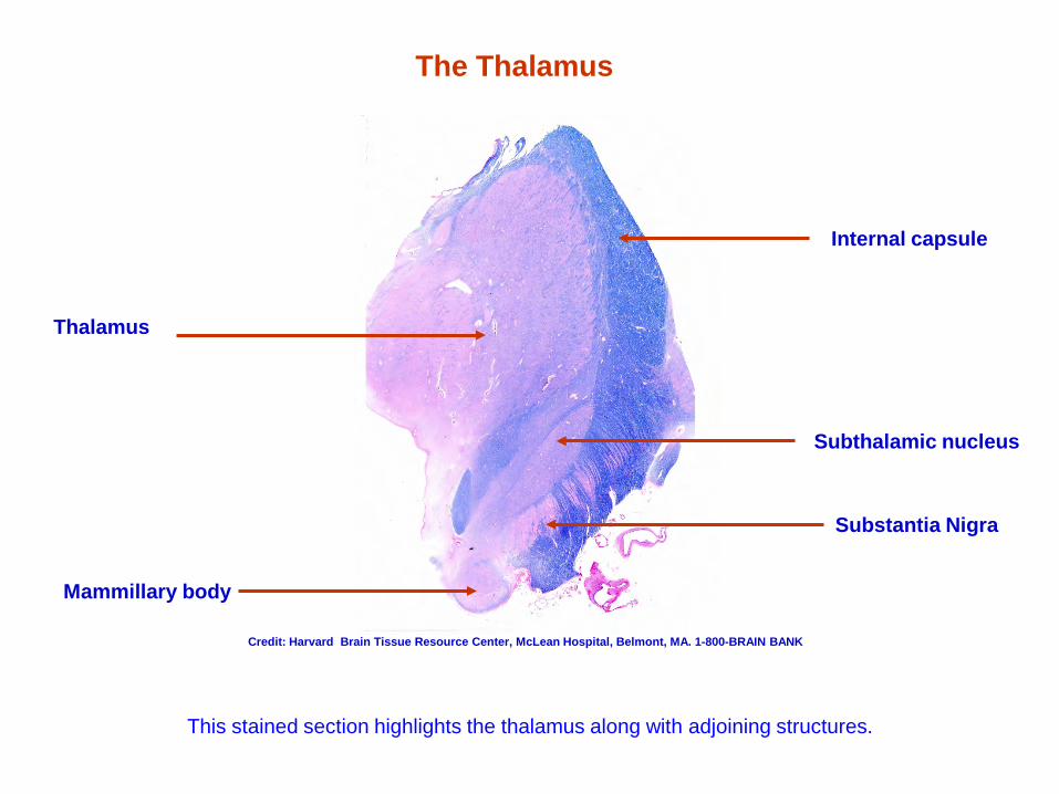
The Thalamus
This stained section highlights the thalamus along with adjoining structures.
Thalamus
Subthalamic nucleus
Mammillary body
Internal capsule
Substantia Nigra
Credit: Harvard Brain Tissue Resource Center, McLean Hospital, Belmont, MA. 1-800-BRAIN BANK
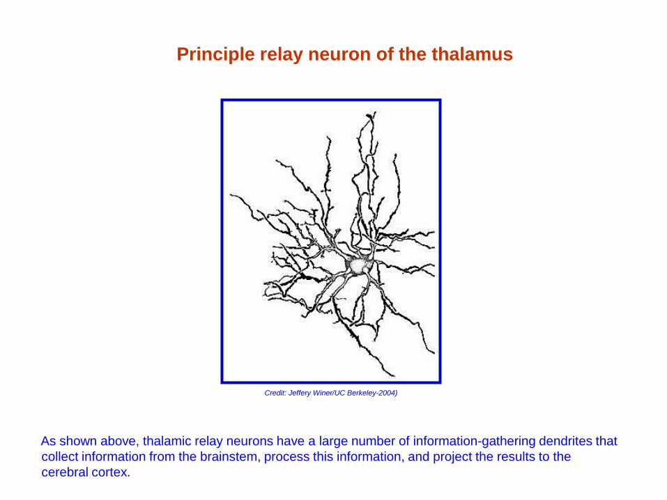
Principle relay neuron of the thalamus
As shown above, thalamic relay neurons have a large number of information-gathering dendrites that collect information from the brainstem, process this information, and project the results to the cerebral cortex.
Credit: Jeffery Winer/UC Berkeley-2004)
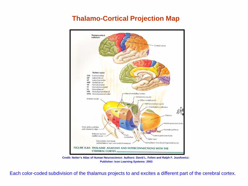
Thalamo-Cortical Projection Map
Each color-coded subdivision of the thalamus projects to and excites a different part of the cerebral cortex.
Credit: Netter’s Atlas of Human Neuroscience: Authors: David L. Felten and Ralph F. Jozefowicz: Publisher: Icon Learning Systems: 2003
The Hypothalamus
Homeostasis Hormonal regulation Autonomic control Instinctual drives
Pituitary gland control
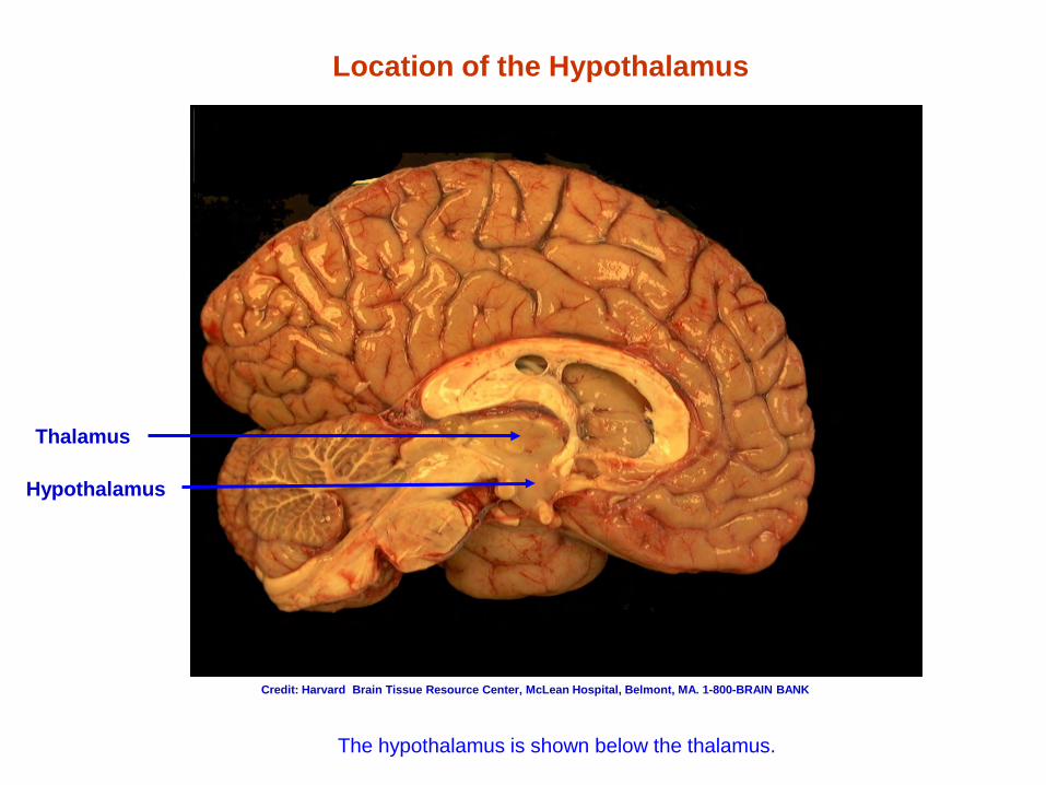
Location of the Hypothalamus
Thalamus
The hypothalamus is shown below the thalamus.
Hypothalamus
Credit: Harvard Brain Tissue Resource Center, McLean Hospital, Belmont, MA. 1-800-BRAIN BANK
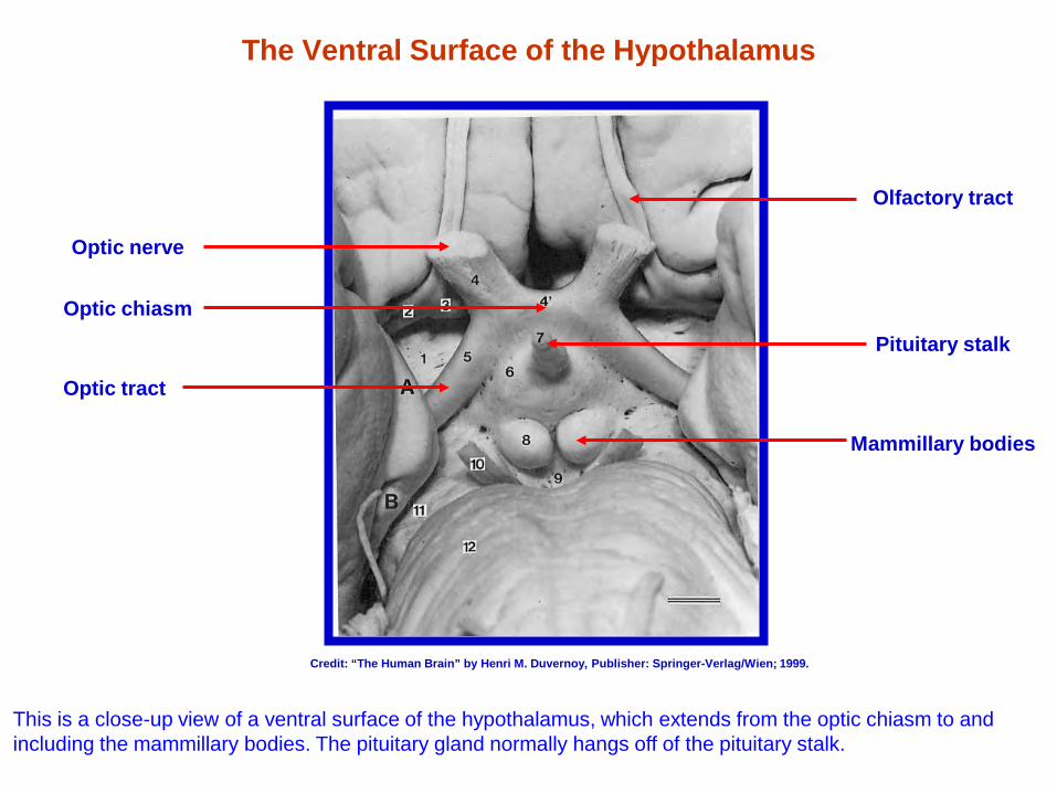
The Ventral Surface of the Hypothalamus
Optic nerve
Optic tract
Pituitary stalk
Mammillary bodies
Olfactory tract
Optic chiasm
This is a close-up view of a ventral surface of the hypothalamus, which extends from the optic chiasm to and including the mammillary bodies. The pituitary gland normally hangs off of the pituitary stalk.
Credit: “The Human Brain” by Henri M. Duvernoy, Publisher: Springer-Verlag/Wien; 1999.
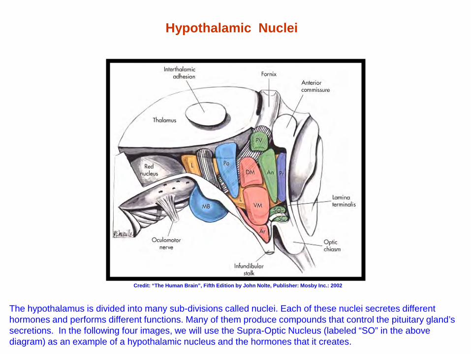
Hypothalamic Nuclei
The hypothalamus is divided into many sub-divisions called nuclei. Each of these nuclei secretes different hormones and performs different functions. Many of them produce compounds that control the pituitary gland’s secretions. In the following four images, we will use the Supra-Optic Nucleus (labeled “SO” in the above diagram) as an example of a hypothalamic nucleus and the hormones that it creates.
Credit: “The Human Brain”, Fifth Edition by John Nolte, Publisher: Mosby Inc.: 2002
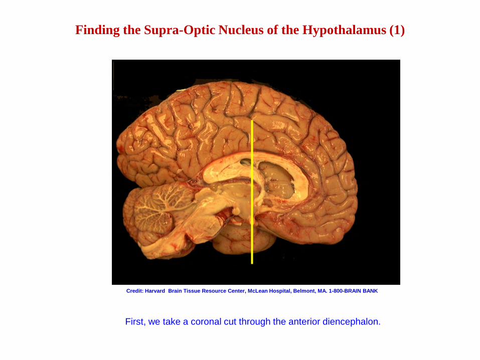
Finding the Supra-Optic Nucleus of the Hypothalamus (1)
First, we take a coronal cut through the anterior diencephalon.
Credit: Harvard Brain Tissue Resource Center, McLean Hospital, Belmont, MA. 1-800-BRAIN BANK
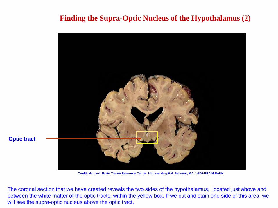
Finding the Supra-Optic Nucleus of the Hypothalamus (2)
Optic tract
The coronal section that we have created reveals the two sides of the hypothalamus, located just above and between the white matter of the optic tracts, within the yellow box. If we cut and stain one side of this area, we will see the supra-optic nucleus above the optic tract.
Credit: Harvard Brain Tissue Resource Center, McLean Hospital, Belmont, MA. 1-800-BRAIN BANK
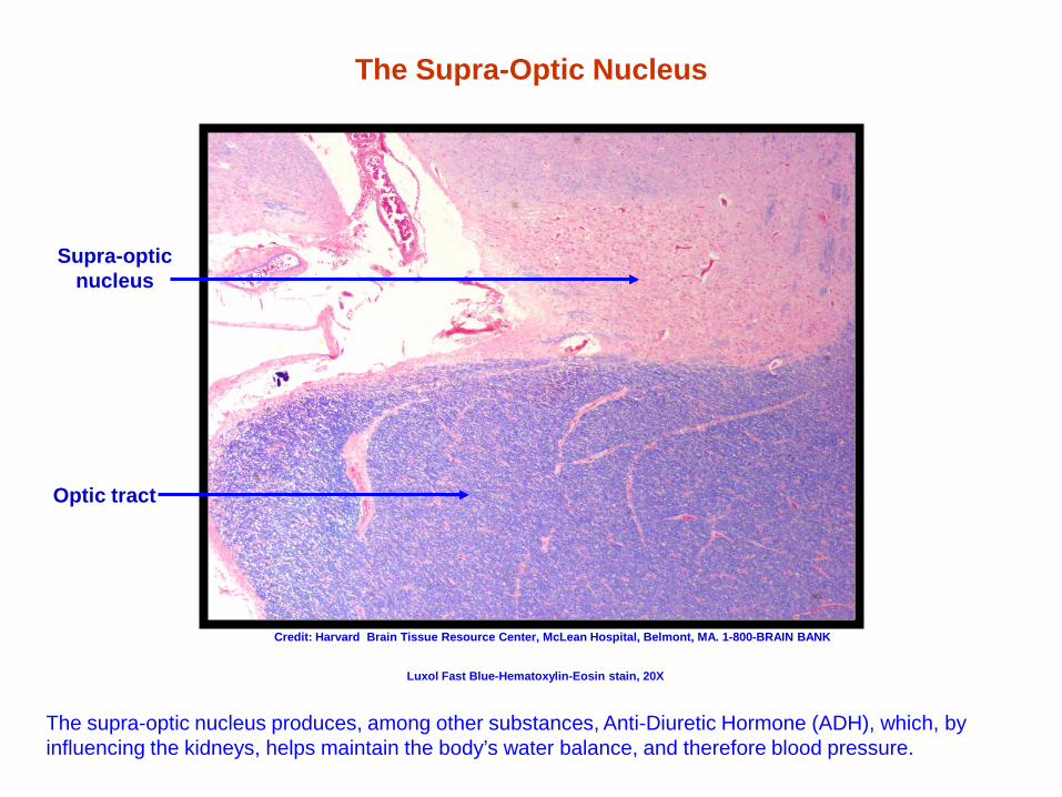
The Supra-Optic Nucleus
Supra-optic nucleus
Optic tract
The supra-optic nucleus produces, among other substances, Anti-Diuretic Hormone (ADH), which, by influencing the kidneys, helps maintain the body’s water balance, and therefore blood pressure.
Credit: Harvard Brain Tissue Resource Center, McLean Hospital, Belmont, MA. 1-800-BRAIN BANK
Luxol Fast Blue-Hematoxylin-Eosin stain, 20X
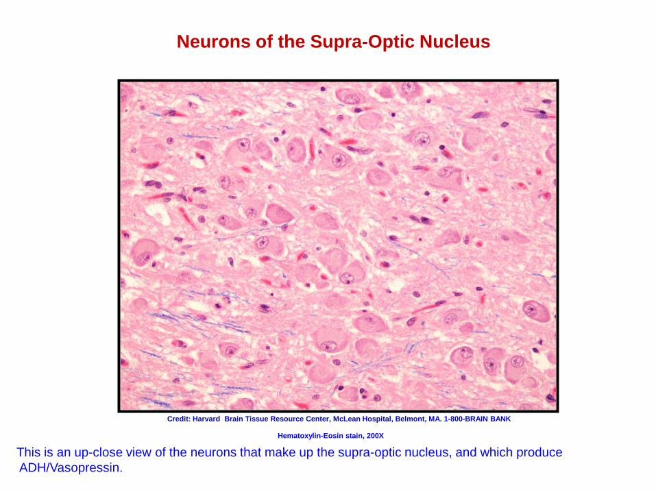
Neurons of the Supra-Optic Nucleus
This is an up-close view of the neurons that make up the supra-optic nucleus, and which produce ADH/Vasopressin.
Credit: Harvard Brain Tissue Resource Center, McLean Hospital, Belmont, MA. 1-800-BRAIN BANK
Hematoxylin-Eosin stain, 200X
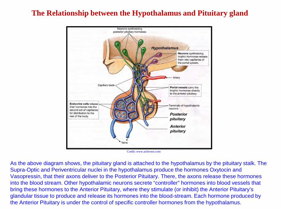
The Relationship between the Hypothalamus and Pituitary gland
As the above diagram shows, the pituitary gland is attached to the hypothalamus by the pituitary stalk. The Supra-Optic and Periventricular nuclei in the hypothalamus produce the hormones Oxytocin and Vasopressin, that their axons deliver to the Posterior Pituitary. There, the axons release these hormones into the blood stream. Other hypothalamic neurons secrete “controller” hormones into blood vessels that bring these hormones to the Anterior Pituitary, where they stimulate (or inhibit) the Anterior Pituitary’s glandular tissue to produce and release its hormones into the blood-stream. Each hormone produced by the Anterior Pituitary is under the control of specific controller hormones from the hypothalamus.
Credit: www.acbrown.com
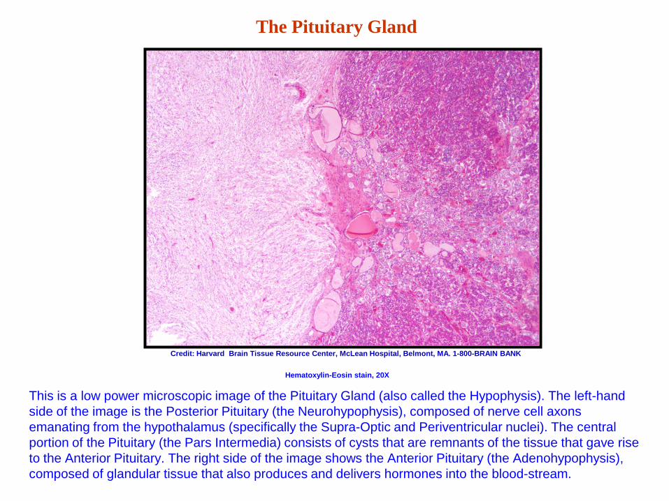
The Pituitary Gland
This is a low power microscopic image of the Pituitary Gland (also called the Hypophysis). The left-hand side of the image is the Posterior Pituitary (the Neurohypophysis), composed of nerve cell axons emanating from the hypothalamus (specifically the Supra-Optic and Periventricular nuclei). The central portion of the Pituitary (the Pars Intermedia) consists of cysts that are remnants of the tissue that gave rise to the Anterior Pituitary. The right side of the image shows the Anterior Pituitary (the Adenohypophysis), composed of glandular tissue that also produces and delivers hormones into the blood-stream.
Credit: Harvard Brain Tissue Resource Center, McLean Hospital, Belmont, MA. 1-800-BRAIN BANK
Hematoxylin-Eosin stain, 20X
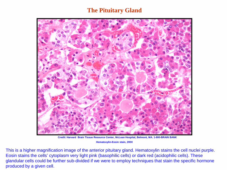
The Pituitary Gland
This is a higher magnification image of the anterior pituitary gland. Hematoxylin stains the cell nuclei purple. Eosin stains the cells’ cytoplasm very light pink (basophilic cells) or dark red (acidophilic cells). These glandular cells could be further sub-divided if we were to employ techniques that stain the specific hormone produced by a given cell.
Credit: Harvard Brain Tissue Resource Center, McLean Hospital, Belmont, MA. 1-800-BRAIN BANK
Hematoxylin-Eosin stain, 200X
The Brainstem
• The brainstem is, in many ways, the most fascinating part of the entire nervous system. It is
packed with nerve cell groups (nuclei), fiber pathways, and functional regions. The brainstem serves as an information conduit between the brain and spinal cord, contains most of the cranial nerves and their nuclei, produces many of the brain’s chemical messengers (neurotransmitters), such as dopamine, noradrenalin, and serotonin, and has control centers for basic bodily functions such as respiration, cardiac rhythms, urination, bowel movements, and sexual functions. It also produces fibers that reach up into the forebrain, bringing the cerebral cortex into a state of arousal, and therefore allowing consciousness, sensory experience, memory, attention, learning and motor activity to exist. Finally, it is the central reflexive and integration center of the brain.
• Although a complete examination of the brainstem is well beyond the scope of this introduction, we will, in the following images, look at selected brainstem structures that will give us an idea of how much the brainstem accomplishes.
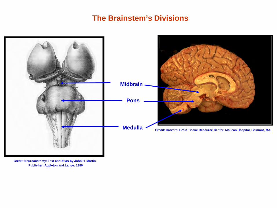
The Brainstem’s Divisions
Midbrain
Pons
Medulla Credit: Harvard Brain Tissue Resource Center, McLean Hospital, Belmont, MA.
Credit: Neuroanatomy: Text and Atlas by John H. Martin. Publisher: Appleton and Lange: 1989
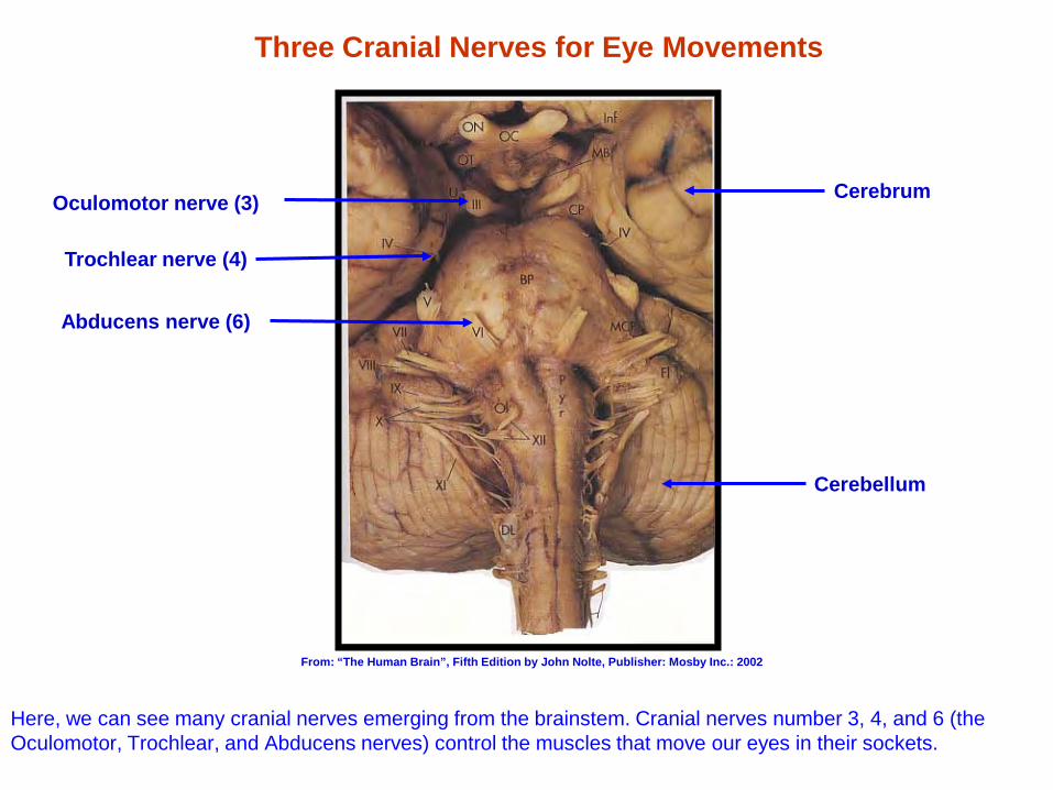
Three Cranial Nerves for Eye Movements
Here, we can see many cranial nerves emerging from the brainstem. Cranial nerves number 3, 4, and 6 (the Oculomotor, Trochlear, and Abducens nerves) control the muscles that move our eyes in their sockets.
Oculomotor nerve (3)
Trochlear nerve (4)
Abducens nerve (6)
Cerebellum
Cerebrum
From: “The Human Brain”, Fifth Edition by John Nolte, Publisher: Mosby Inc.: 2002
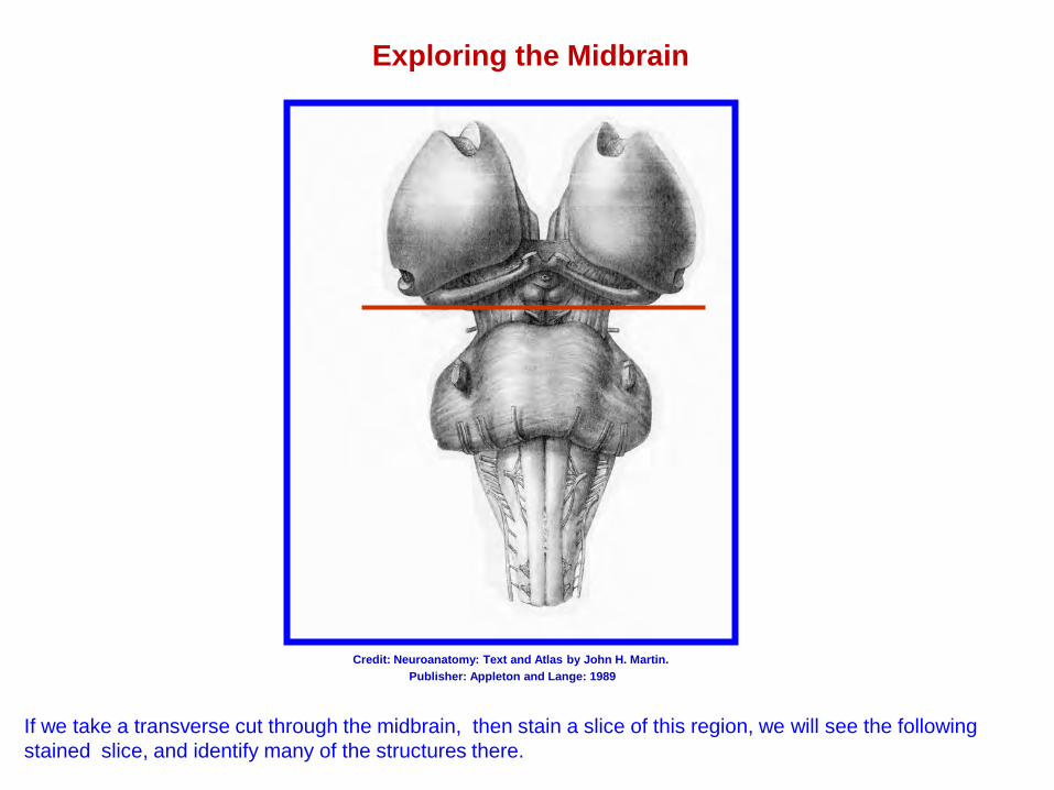
Exploring the Midbrain
If we take a transverse cut through the midbrain, then stain a slice of this region, we will see the following stained slice, and identify many of the structures there.
Credit: Neuroanatomy: Text and Atlas by John H. Martin. Publisher: Appleton and Lange: 1989
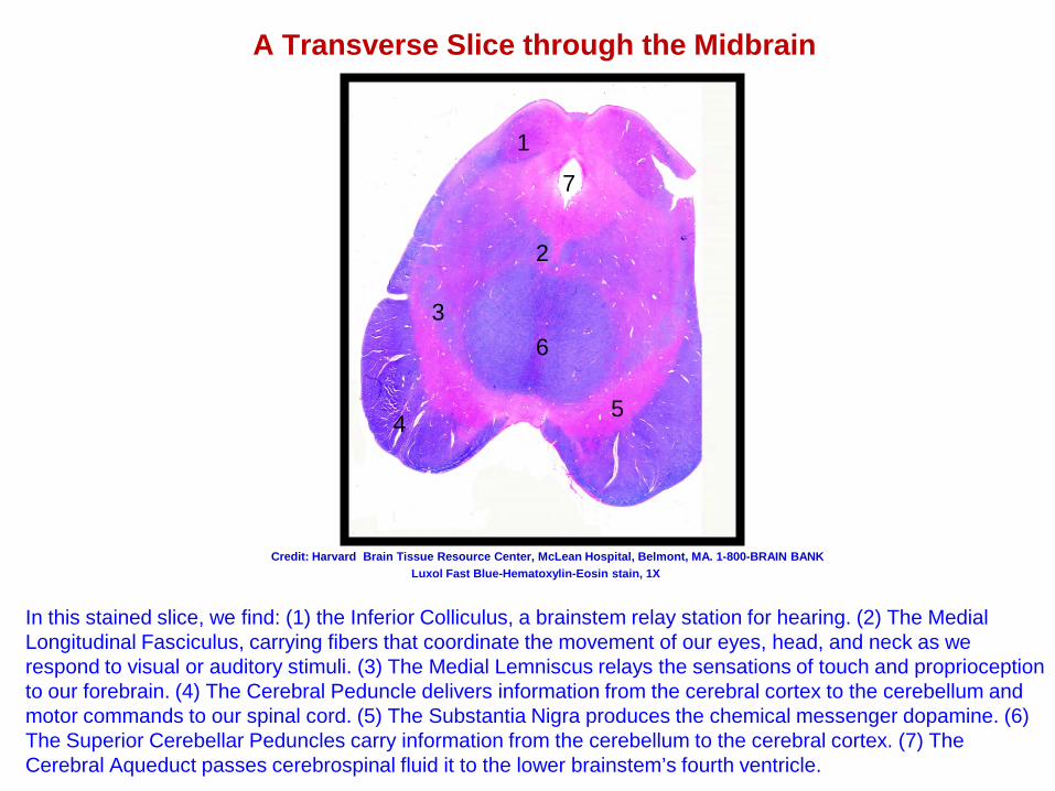
A Transverse Slice through the Midbrain
In this stained slice, we find: (1) the Inferior Colliculus, a brainstem relay station for hearing. (2) The Medial Longitudinal Fasciculus, carrying fibers that coordinate the movement of our eyes, head, and neck as we respond to visual or auditory stimuli. (3) The Medial Lemniscus relays the sensations of touch and proprioception to our forebrain. (4) The Cerebral Peduncle delivers information from the cerebral cortex to the cerebellum and motor commands to our spinal cord. (5) The Substantia Nigra produces the chemical messenger dopamine. (6) The Superior Cerebellar Peduncles carry information from the cerebellum to the cerebral cortex. (7) The Cerebral Aqueduct passes cerebrospinal fluid it to the lower brainstem’s fourth ventricle.
Credit: Harvard Brain Tissue Resource Center, McLean Hospital, Belmont, MA. 1-800-BRAIN BANK
1
2
3
7
4 5
6
Luxol Fast Blue-Hematoxylin-Eosin stain, 1X
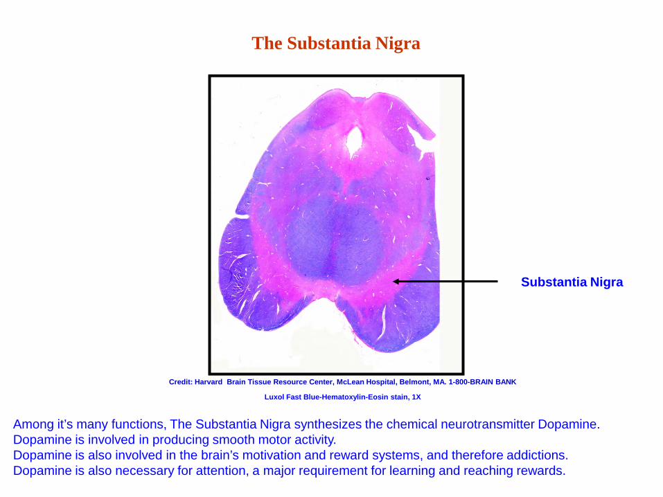
The Substantia Nigra
Substantia Nigra
Among it’s many functions, The Substantia Nigra synthesizes the chemical neurotransmitter Dopamine. Dopamine is involved in producing smooth motor activity. Dopamine is also involved in the brain’s motivation and reward systems, and therefore addictions. Dopamine is also necessary for attention, a major requirement for learning and reaching rewards.
Credit: Harvard Brain Tissue Resource Center, McLean Hospital, Belmont, MA. 1-800-BRAIN BANK
Luxol Fast Blue-Hematoxylin-Eosin stain, 1X
The Substantia Nigra and Parkinson’s Disease
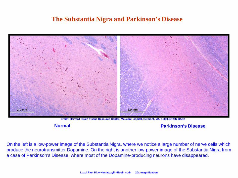
Normal Parkinson’s Disease
Luxol Fast Blue-Hematoxylin-Eosin stain 20x magnification
Credit: Harvard Brain Tissue Resource Center, McLean Hospital, Belmont, MA. 1-800-BRAIN BANK
On the left is a low-power image of the Substantia Nigra, where we notice a large number of nerve cells which produce the neurotransmitter Dopamine. On the right is another low-power image of the Substantia Nigra from a case of Parkinson’s Disease, where most of the Dopamine-producing neurons have disappeared.
The Substantia Nigra and Parkinson’s Disease
Normal Parkinson’s Luxol Fast Blue-Hematoxylin-Eosin stain 400x magnification
Credit: Harvard Brain Tissue Resource Center, McLean Hospital, Belmont, MA. 1-800-BRAIN BANK
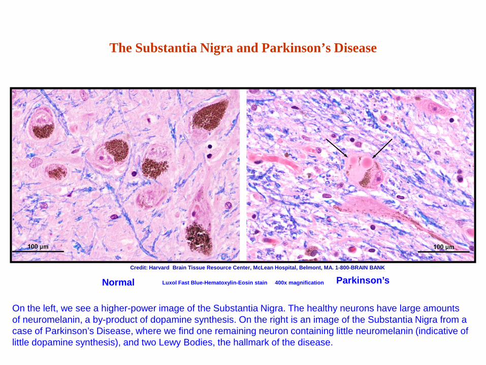
On the left, we see a higher-power image of the Substantia Nigra. The healthy neurons have large amounts of neuromelanin, a by-product of dopamine synthesis. On the right is an image of the Substantia Nigra from a case of Parkinson’s Disease, where we find one remaining neuron containing little neuromelanin (indicative of little dopamine synthesis), and two Lewy Bodies, the hallmark of the disease.
Locating the Dorsal Raphe Nucleus
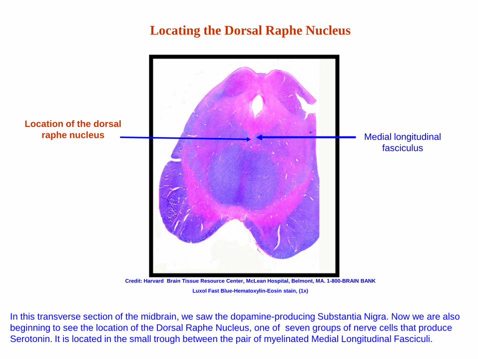
In this transverse section of the midbrain, we saw the dopamine-producing Substantia Nigra. Now we are also beginning to see the location of the Dorsal Raphe Nucleus, one of seven groups of nerve cells that produce Serotonin. It is located in the small trough between the pair of myelinated Medial Longitudinal Fasciculi.
Location of the dorsal raphe nucleus
Credit: Harvard Brain Tissue Resource Center, McLean Hospital, Belmont, MA. 1-800-BRAIN BANK
Medial longitudinal fasciculus
Luxol Fast Blue-Hematoxylin-Eosin stain, (1x)
Location of the Dorsal Raphe Nucleus
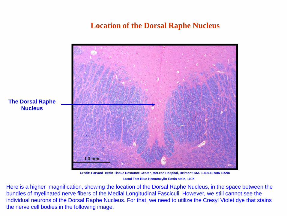
Here is a higher magnification, showing the location of the Dorsal Raphe Nucleus, in the space between the bundles of myelinated nerve fibers of the Medial Longitudinal Fasciculi. However, we still cannot see the individual neurons of the Dorsal Raphe Nucleus. For that, we need to utilize the Cresyl Violet dye that stains the nerve cell bodies in the following image.
The Dorsal Raphe Nucleus
Credit: Harvard Brain Tissue Resource Center, McLean Hospital, Belmont, MA. 1-800-BRAIN BANK
Luxol Fast Blue-Hematoxylin-Eosin stain, 100X
Dorsal Raphe neurons
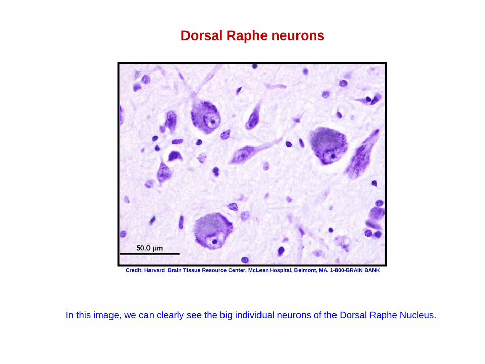
In this image, we can clearly see the big individual neurons of the Dorsal Raphe Nucleus.
Credit: Harvard Brain Tissue Resource Center, McLean Hospital, Belmont, MA. 1-800-BRAIN BANK
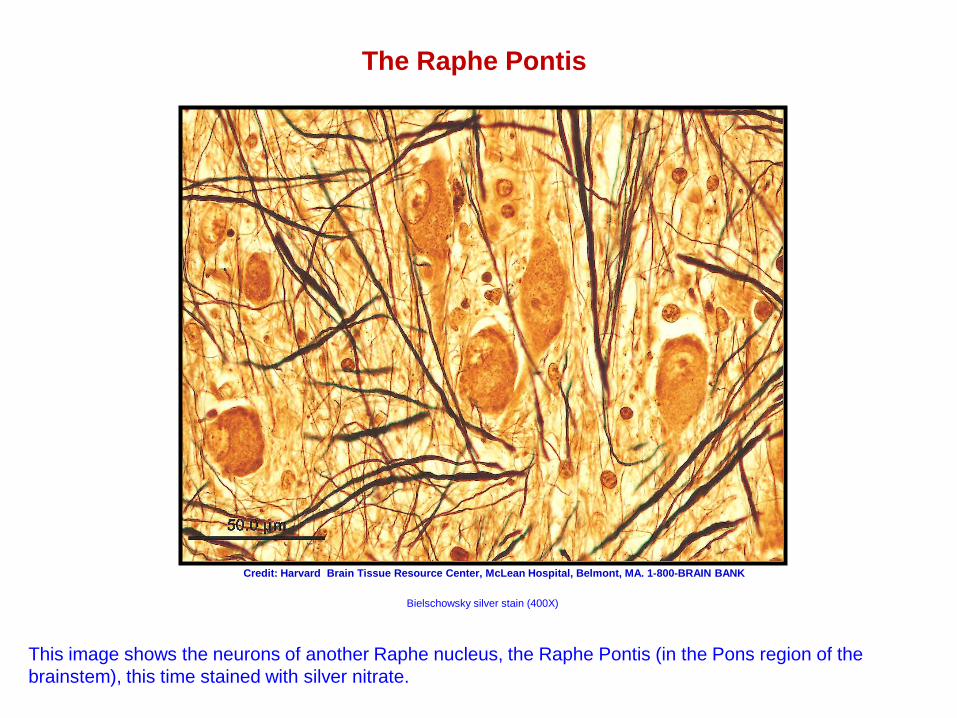
The Raphe Pontis
Bielschowsky silver stain (400X)
Credit: Harvard Brain Tissue Resource Center, McLean Hospital, Belmont, MA. 1-800-BRAIN BANK
This image shows the neurons of another Raphe nucleus, the Raphe Pontis (in the Pons region of the brainstem), this time stained with silver nitrate.
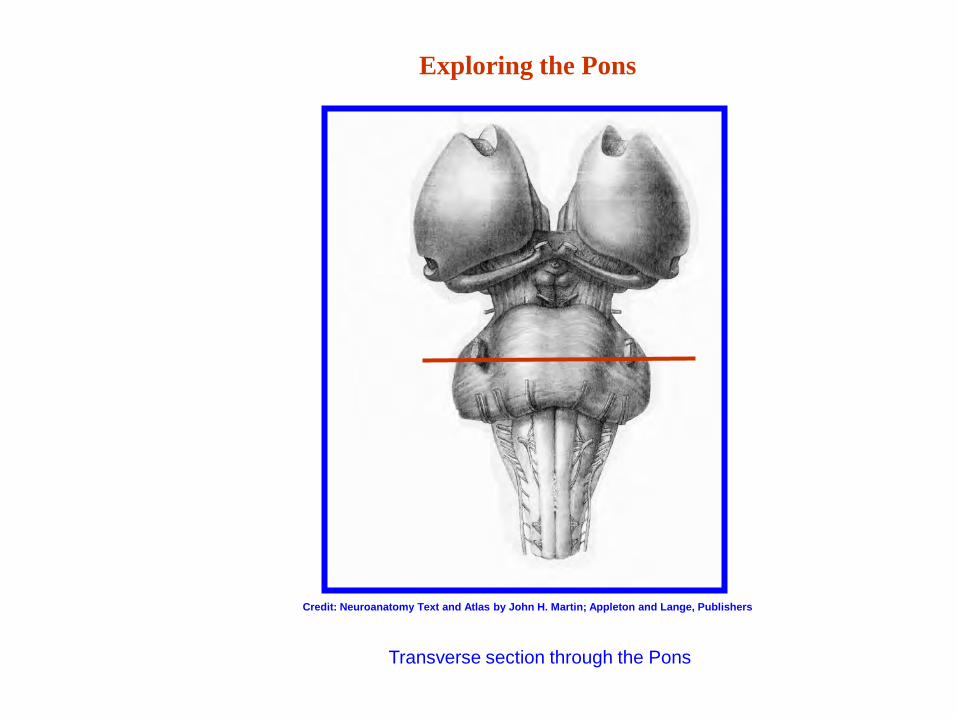
Exploring the Pons
Credit: Neuroanatomy Text and Atlas by John H. Martin; Appleton and Lange, Publishers
Transverse section through the Pons
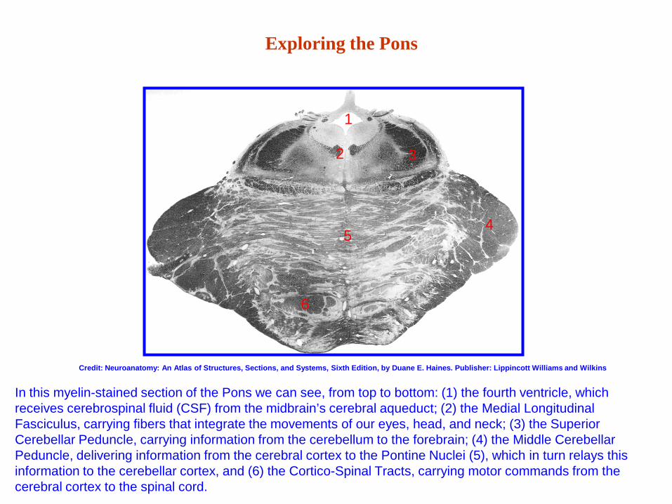
Exploring the Pons
In this myelin-stained section of the Pons we can see, from top to bottom: (1) the fourth ventricle, which receives cerebrospinal fluid (CSF) from the midbrain’s cerebral aqueduct; (2) the Medial Longitudinal Fasciculus, carrying fibers that integrate the movements of our eyes, head, and neck; (3) the Superior Cerebellar Peduncle, carrying information from the cerebellum to the forebrain; (4) the Middle Cerebellar Peduncle, delivering information from the cerebral cortex to the Pontine Nuclei (5), which in turn relays this information to the cerebellar cortex, and (6) the Cortico-Spinal Tracts, carrying motor commands from the cerebral cortex to the spinal cord.
1
2 3
4 5
6
Credit: Neuroanatomy: An Atlas of Structures, Sections, and Systems, Sixth Edition, by Duane E. Haines. Publisher: Lippincott Williams and Wilkins
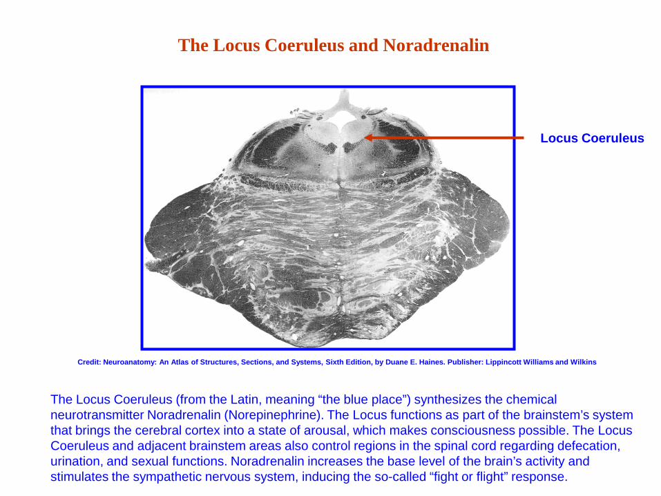
The Locus Coeruleus and Noradrenalin
Locus Coeruleus
The Locus Coeruleus (from the Latin, meaning “the blue place”) synthesizes the chemical neurotransmitter Noradrenalin (Norepinephrine). The Locus functions as part of the brainstem’s system that brings the cerebral cortex into a state of arousal, which makes consciousness possible. The Locus Coeruleus and adjacent brainstem areas also control regions in the spinal cord regarding defecation, urination, and sexual functions. Noradrenalin increases the base level of the brain’s activity and stimulates the sympathetic nervous system, inducing the so-called “fight or flight” response.
Credit: Neuroanatomy: An Atlas of Structures, Sections, and Systems, Sixth Edition, by Duane E. Haines. Publisher: Lippincott Williams and Wilkins
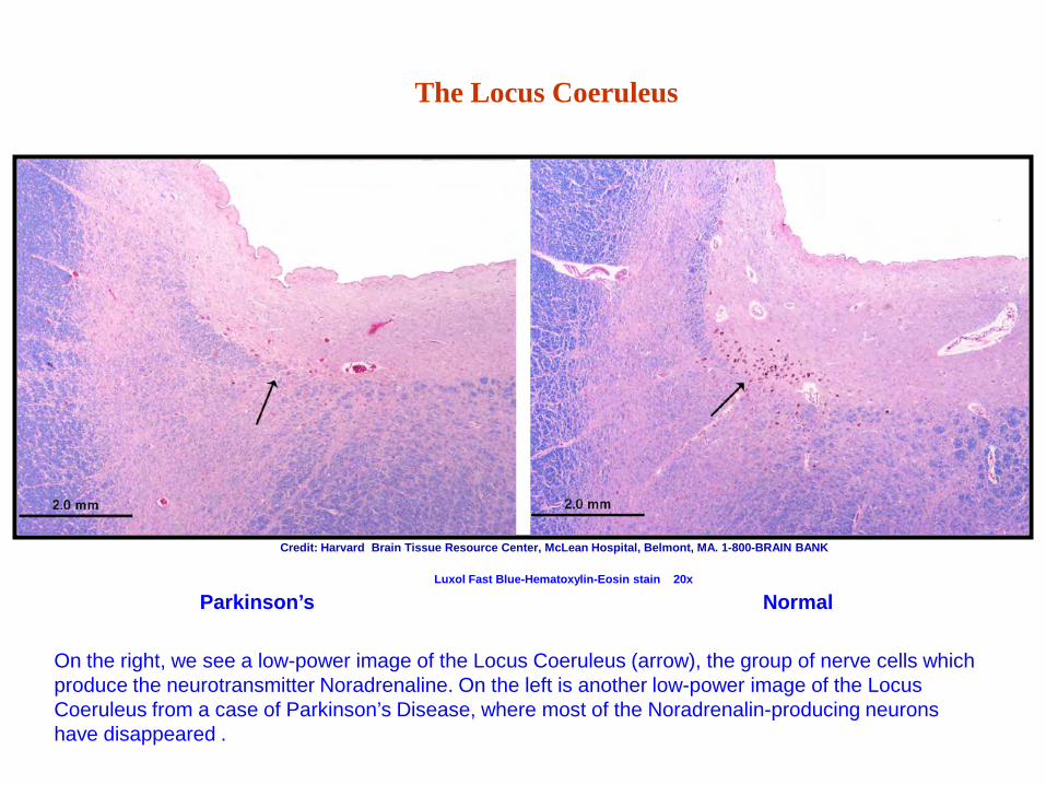
The Locus Coeruleus
Normal Parkinson’s Luxol Fast Blue-Hematoxylin-Eosin stain 20x
Credit: Harvard Brain Tissue Resource Center, McLean Hospital, Belmont, MA. 1-800-BRAIN BANK
On the right, we see a low-power image of the Locus Coeruleus (arrow), the group of nerve cells which produce the neurotransmitter Noradrenaline. On the left is another low-power image of the Locus Coeruleus from a case of Parkinson’s Disease, where most of the Noradrenalin-producing neurons have disappeared .

The Locus Coeruleus
Normal Parkinson’s Disease Luxol Fast Blue-Hematoxylin-Eosin stain 400x
Credit: Harvard Brain Tissue Resource Center, McLean Hospital, Belmont, MA. 1-800-BRAIN BANK
On the right, we see a higher power image of the Locus Coeruleus, with nerve cells containing plenty of neuromelanin, a by-product of noradrenalin synthesis. On the left is another image of the Locus Coeruleus from a case of Parkinson’s Disease, where most of the neurons are pale, having lost most of their neuromelanin or have Lewy Bodies (arrows), the pathological hallmark of the disease.
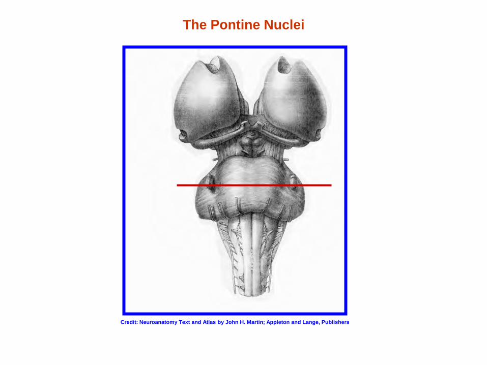
The Pontine Nuclei
Credit: Neuroanatomy Text and Atlas by John H. Martin; Appleton and Lange, Publishers
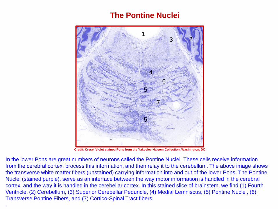
The Pontine Nuclei
In the lower Pons are great numbers of neurons called the Pontine Nuclei. These cells receive information from the cerebral cortex, process this information, and then relay it to the cerebellum. The above image shows the transverse white matter fibers (unstained) carrying information into and out of the lower Pons. The Pontine Nuclei (stained purple), serve as an interface between the way motor information is handled in the cerebral cortex, and the way it is handled in the cerebellar cortex. In this stained slice of brainstem, we find (1) Fourth Ventricle, (2) Cerebellum, (3) Superior Cerebellar Peduncle, (4) Medial Lemniscus, (5) Pontine Nuclei, (6) Transverse Pontine Fibers, and (7) Cortico-Spinal Tract fibers. .
Credit: Cresyl Violet stained Pons from the Yakovlev-Haleem Collection, Washington, DC
1 2 3
4
5
5 6
7
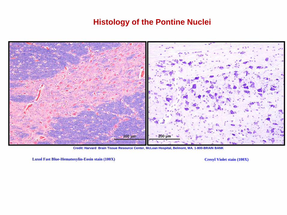
Histology of the Pontine Nuclei
Cresyl Violet stain (100X) Luxol Fast Blue-Hematoxylin-Eosin stain (100X)
Credit: Harvard Brain Tissue Resource Center, McLean Hospital, Belmont, MA. 1-800-BRAIN BANK
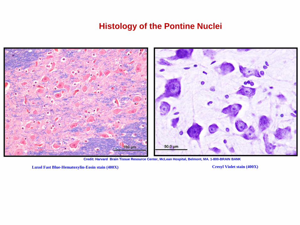
Histology of the Pontine Nuclei
Credit: Harvard Brain Tissue Resource Center, McLean Hospital, Belmont, MA. 1-800-BRAIN BANK
Luxol Fast Blue-Hematoxylin-Eosin stain (400X) Cresyl Violet stain (400X)
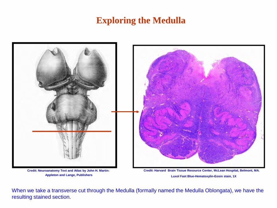
Exploring the Medulla
Credit: Harvard Brain Tissue Resource Center, McLean Hospital, Belmont, MA. Credit: Neuroanatomy Text and Atlas by John H. Martin: Appleton and Lange, Publishers
When we take a transverse cut through the Medulla (formally named the Medulla Oblongata), we have the resulting stained section.
Luxol Fast Blue-Hematoxylin-Eosin stain, 1X
Exploring the Medulla
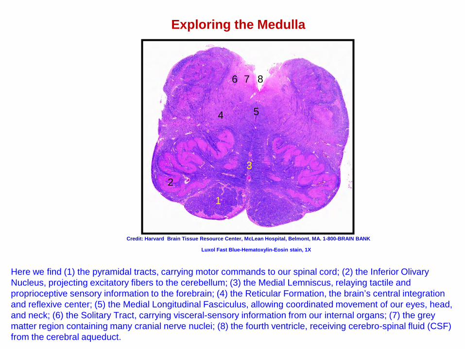
Here we find (1) the pyramidal tracts, carrying motor commands to our spinal cord; (2) the Inferior Olivary Nucleus, projecting excitatory fibers to the cerebellum; (3) the Medial Lemniscus, relaying tactile and proprioceptive sensory information to the forebrain; (4) the Reticular Formation, the brain’s central integration and reflexive center; (5) the Medial Longitudinal Fasciculus, allowing coordinated movement of our eyes, head, and neck; (6) the Solitary Tract, carrying visceral-sensory information from our internal organs; (7) the grey matter region containing many cranial nerve nuclei; (8) the fourth ventricle, receiving cerebro-spinal fluid (CSF) from the cerebral aqueduct.
Credit: Harvard Brain Tissue Resource Center, McLean Hospital, Belmont, MA. 1-800-BRAIN BANK
2
1
5
7 8 6
4
3
Luxol Fast Blue-Hematoxylin-Eosin stain, 1X
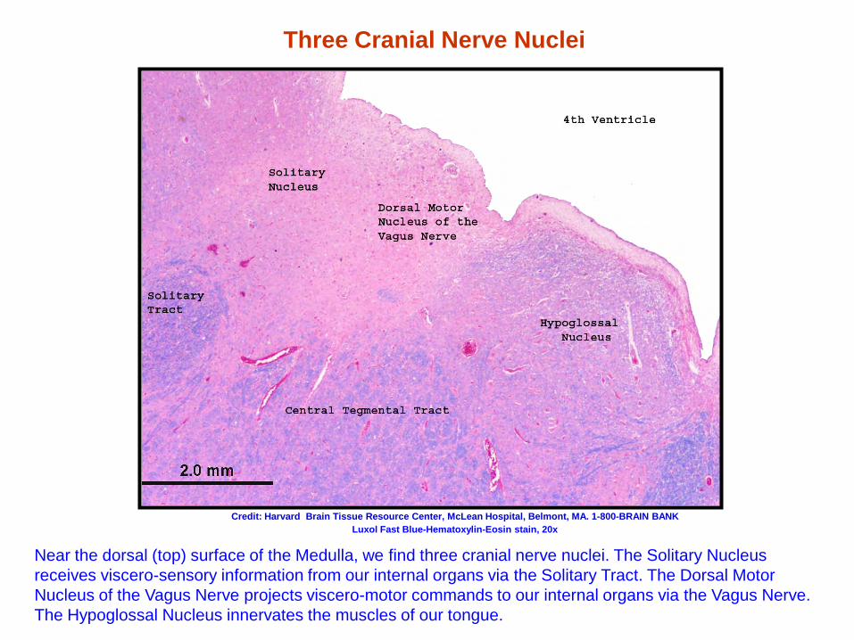
Three Cranial Nerve Nuclei
Luxol Fast Blue-Hematoxylin-Eosin stain, 20x Credit: Harvard Brain Tissue Resource Center, McLean Hospital, Belmont, MA. 1-800-BRAIN BANK
Near the dorsal (top) surface of the Medulla, we find three cranial nerve nuclei. The Solitary Nucleus receives viscero-sensory information from our internal organs via the Solitary Tract. The Dorsal Motor Nucleus of the Vagus Nerve projects viscero-motor commands to our internal organs via the Vagus Nerve. The Hypoglossal Nucleus innervates the muscles of our tongue.
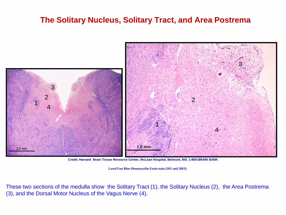
The Solitary Nucleus, Solitary Tract, and Area Postrema
Credit: Harvard Brain Tissue Resource Center, McLean Hospital, Belmont, MA. 1-800-BRAIN BANK
These two sections of the medulla show the Solitary Tract (1), the Solitary Nucleus (2), the Area Postrema (3), and the Dorsal Motor Nucleus of the Vagus Nerve (4).
1 2
3
1
2
3
4
4
Luxol Fast Blue-Hematoxylin-Eosin stain (20X and 200X)
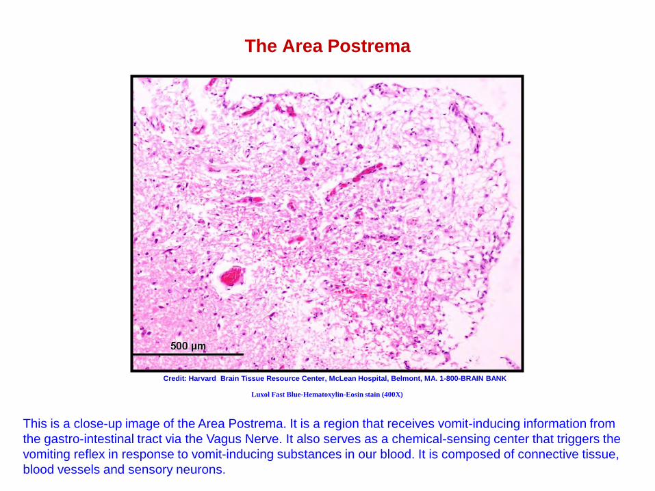
The Area Postrema
Credit: Harvard Brain Tissue Resource Center, McLean Hospital, Belmont, MA. 1-800-BRAIN BANK
This is a close-up image of the Area Postrema. It is a region that receives vomit-inducing information from the gastro-intestinal tract via the Vagus Nerve. It also serves as a chemical-sensing center that triggers the vomiting reflex in response to vomit-inducing substances in our blood. It is composed of connective tissue, blood vessels and sensory neurons.
Luxol Fast Blue-Hematoxylin-Eosin stain (400X)
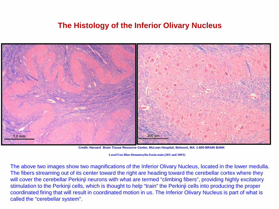
The Histology of the Inferior Olivary Nucleus
Luxol Fast Blue-Hematoxylin-Eosin stain (20X and 200X)
Credit: Harvard Brain Tissue Resource Center, McLean Hospital, Belmont, MA. 1-800-BRAIN BANK
The above two images show two magnifications of the Inferior Olivary Nucleus, located in the lower medulla. The fibers streaming out of its center toward the right are heading toward the cerebellar cortex where they will cover the cerebellar Perkinji neurons with what are termed “climbing fibers”, providing highly excitatory stimulation to the Perkinji cells, which is thought to help “train” the Perkinji cells into producing the proper coordinated firing that will result in coordinated motion in us. The Inferior Olivary Nucleus is part of what is called the “cerebellar system”.
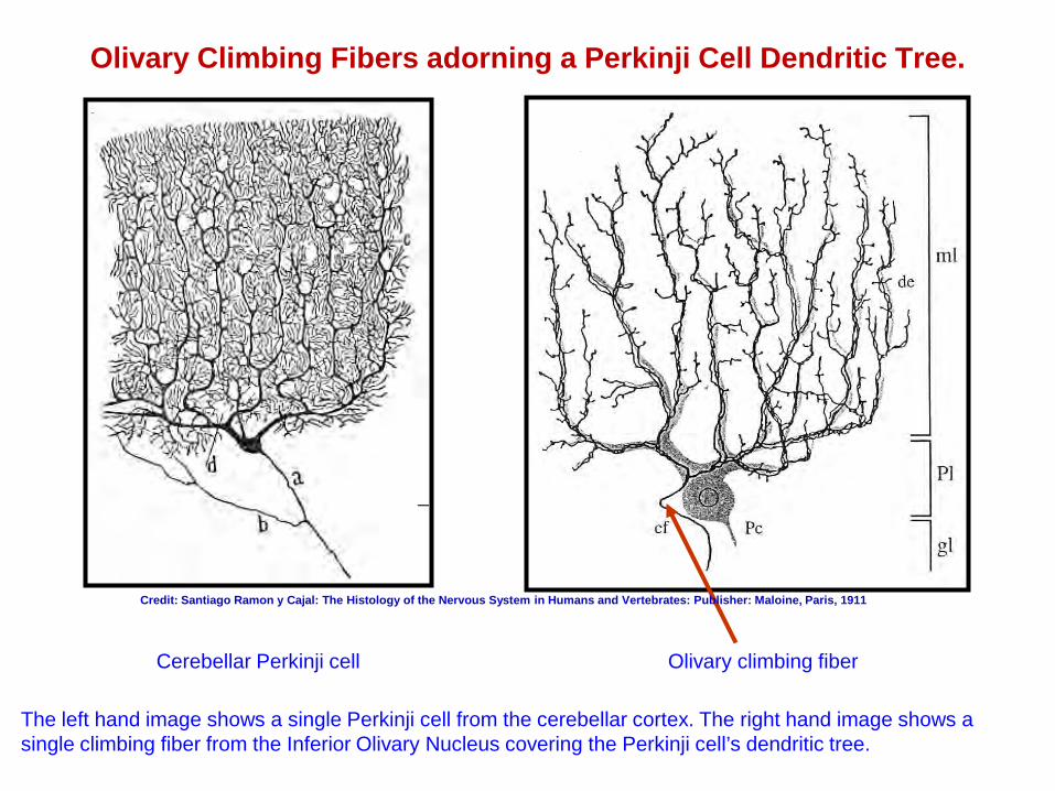
Olivary Climbing Fibers adorning a Perkinji Cell Dendritic Tree.
Cerebellar Perkinji cell Olivary climbing fiber
The left hand image shows a single Perkinji cell from the cerebellar cortex. The right hand image shows a single climbing fiber from the Inferior Olivary Nucleus covering the Perkinji cell’s dendritic tree.
Credit: Santiago Ramon y Cajal: The Histology of the Nervous System in Humans and Vertebrates: Publisher: Maloine, Paris, 1911
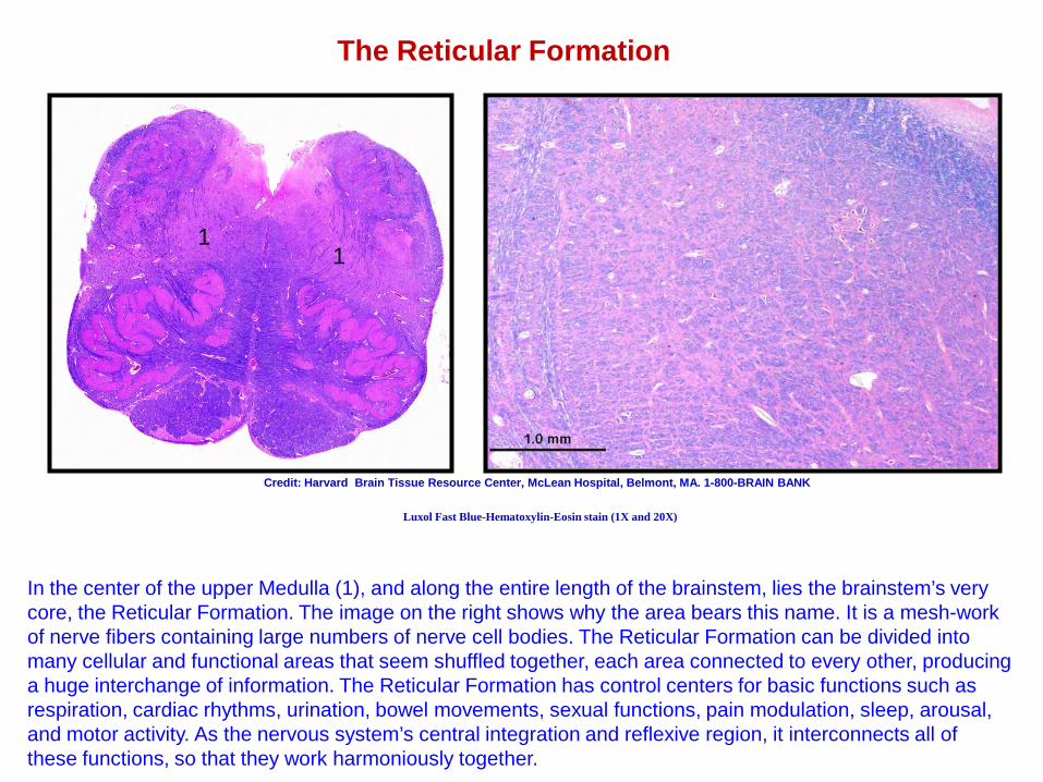
The Reticular Formation
Luxol Fast Blue-Hematoxylin-Eosin stain (1X and 20X)
Credit: Harvard Brain Tissue Resource Center, McLean Hospital, Belmont, MA. 1-800-BRAIN BANK
In the center of the upper Medulla (1), and along the entire length of the brainstem, lies the brainstem’s very core, the Reticular Formation. The image on the right shows why the area bears this name. It is a mesh-work of nerve fibers containing large numbers of nerve cell bodies. The Reticular Formation can be divided into many cellular and functional areas that seem shuffled together, each area connected to every other, producing a huge interchange of information. The Reticular Formation has control centers for basic functions such as respiration, cardiac rhythms, urination, bowel movements, sexual functions, pain modulation, sleep, arousal, and motor activity. As the nervous system’s central integration and reflexive region, it interconnects all of these functions, so that they work harmoniously together.
1 1
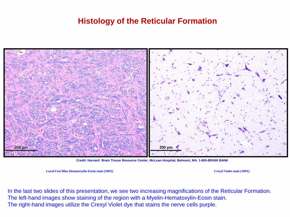
Histology of the Reticular Formation
Luxol Fast Blue-Hematoxylin-Eosin stain (100X) Cresyl Violet stain (100X)
Credit: Harvard Brain Tissue Resource Center, McLean Hospital, Belmont, MA. 1-800-BRAIN BANK
In the last two slides of this presentation, we see two increasing magnifications of the Reticular Formation. The left-hand images show staining of the region with a Myelin-Hematoxylin-Eosin stain. The right-hand images utilize the Cresyl Violet dye that stains the nerve cells purple.
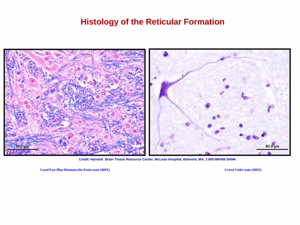
Histology of the Reticular Formation
Luxol Fast Blue-Hematoxylin-Eosin stain (400X) Cresyl Violet stain (400X)
Credit: Harvard Brain Tissue Resource Center, McLean Hospital, Belmont, MA. 1-800-BRAIN BANK
Acknowledgements
In creating this Introduction to Neuroanatomy as a completely non-profit, educational-only experience, we at the Harvard Brain Tissue Resource Center have supplemented the images generated by our own department with those from the atlases and papers of other anatomists and researchers. Please find these acknowledgments under their respective images. Thank you.
THE END
We at the Harvard Brain Tissue Resource Center hope that you have enjoyed this introduction to human neuroanatomy, and that it has proven informative and useful. Please feel free to relay any questions, comments, or suggestions that you may have. Thank you.