Embed Size (px)
Citation preview
8/6/2019 Neuroanatomy Presentation
http://slidepdf.com/reader/full/neuroanatomy-presentation 1/19
Neuroanatomy: Morphologyof the brain
Chapter 2
Majority of illustrations in this presentation are f rom Biological Psychology
4th edition (© Sinuer Publications)
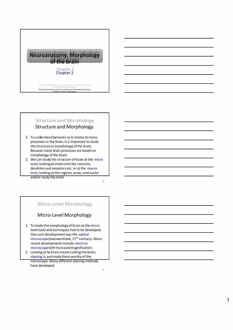
Structure and Morphology
1. To understand behavior as it relates to many
processes in the brain, it is important to study
the structure or morphology of the brain.
Because many brain processes are based on
morphology of the brain.
2. We can study the structure of brain at the micro
level, looking at small units like neurons,
dendrites and receptors etc. or at the macro
level, looking at the regions, areas, and nucleiand/or study the brain
2
Micro-Level Morphology
1. To study the morphology of brain at the micro
level tools and techniques had to be developed.One such development was the optical
microscope(Leeuwenhoek, 17th century). More
recent developments include electron
microscopewith increased magnification.
2. Looking at he brain meant cutting the brain,
staining it, and make them worthy of the
microscope. Many different staining methods
have developed.3

8/6/2019 Neuroanatomy Presentation
http://slidepdf.com/reader/full/neuroanatomy-presentation 2/19

8/6/2019 Neuroanatomy Presentation
http://slidepdf.com/reader/full/neuroanatomy-presentation 3/19
Neurons
7
There are 100
billion neurons in
the human brain.Packed with 10
times more glial
cells. Each neuron
is divided into
three parts;
dendrites, cell
body and axon.
Histology
1. How do we know the parts of a neuron? Or
what are the shapes and sizes of different
neurons in the brain?
2. To answer these questions neuroanatomists use
histological methods to stain neurons and
assess their different shapes and sizes.
8
Immunocytochemistry
Staining processes have improved to include use of
labeled antibodies to stain targeted proteins in thebrain.
9
GAP-43 staining in the retina
8/6/2019 Neuroanatomy Presentation
http://slidepdf.com/reader/full/neuroanatomy-presentation 4/19
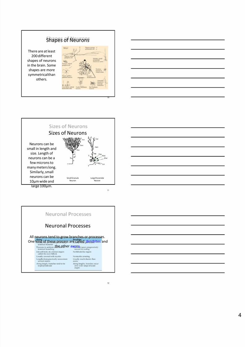
Shapes of Neurons
10
There are at least
200 different
shapes of neuronsin the brain. Some
shapes are more
symmetrical than
others.
Sizes of Neurons
11
Neurons can be
small in length and
size. Length of
neurons can be a
few microns to
many meters long.
Similarly, small
neurons can be
10µm wide andlarge 100µm.
Large Pyramidal
Neuron
Small Granule
Neuron
Neuronal Processes
All neurons tend to grow branches or processes.
One kind of these process are called dendrites andthe other axons.
12
8/6/2019 Neuroanatomy Presentation
http://slidepdf.com/reader/full/neuroanatomy-presentation 5/19
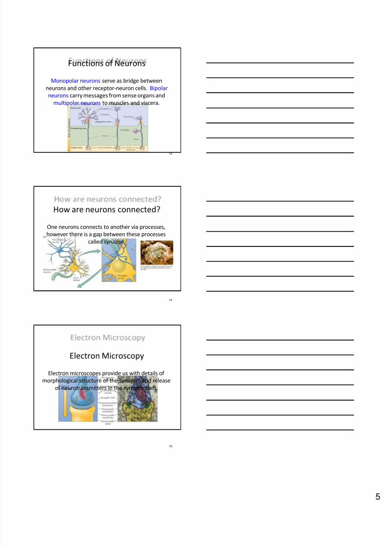
Functions of Neurons
Monopolar neurons serve as bridge between
neurons and other receptor-neuron cells. Bipolar
neurons carry messages from sense organs andmultipolar neurons to muscles and viscera.
13
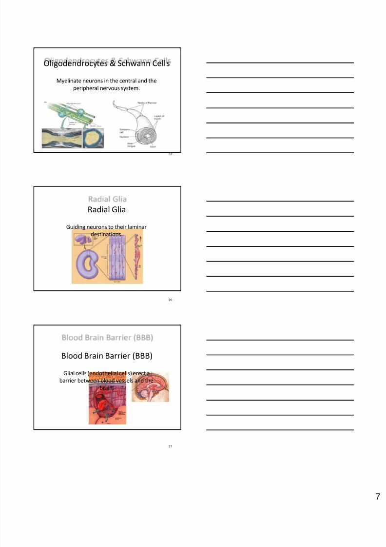
How are neurons connected?
One neurons connects to another via processes,
however there is a gap between these processes
called synapse.
14
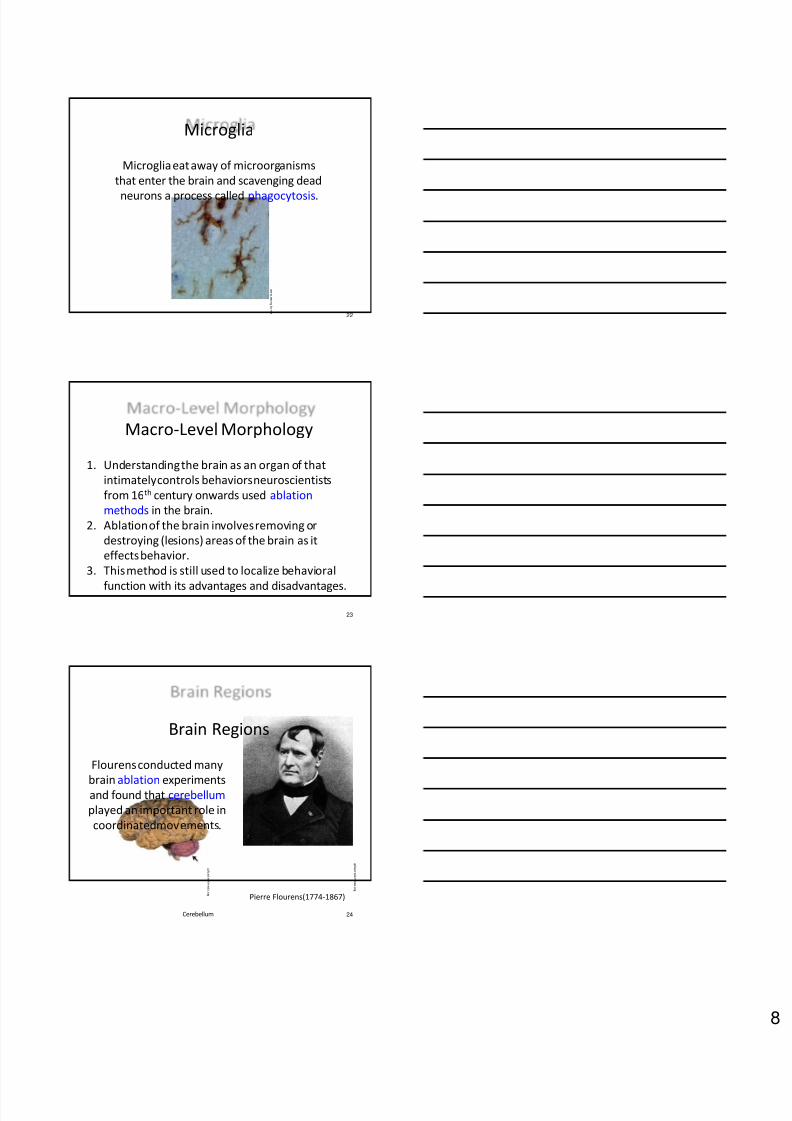
Electron Microscopy
Electron microscopes provide us with details of
morphological structure of the synapse, and releaseof neurotransmitters in the synaptic cleft.
15

8/6/2019 Neuroanatomy Presentation
http://slidepdf.com/reader/full/neuroanatomy-presentation 6/19
Chemical Transport
Most chemicals in the neurons are synthesized and
recycled from and to the nucleus of the cell, they
need to be transported to the synaptic sites.
16
Glial Cells
Glial cells in the brain are non-communicative cells
and engage in other functions, some of which are
listed below:
17
1. Provide scaffolding to neurons
2. Provide nutrition to neurons
3. Myelinate (insulate) neurons
4. Guide neuronal development
5. Make blood-brain barrier (BBB)
6. Phagocytosis (eat microorganisms & deadneurons)
18
Provide support and Nutrition to
neurons in the and extracellularenvironment
m e m b e r s . t r i p o d . c o m
Astrocytes in the cortex
Arstrocytes
8/6/2019 Neuroanatomy Presentation
http://slidepdf.com/reader/full/neuroanatomy-presentation 7/19
19
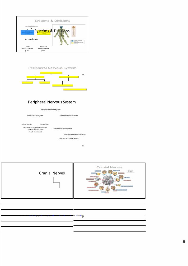
Oligodendrocytes & Schwann Cells
Myelinate neurons in the central and the
peripheral nervous system.
20
Radial Glia
Guiding neurons to their laminar
destinations.
21
Blood Brain Barrier (BBB)
Glial cells (endothelial cells) erect a
barrier between blood vessels and thebrain.
8/6/2019 Neuroanatomy Presentation
http://slidepdf.com/reader/full/neuroanatomy-presentation 8/19
22
Microglia
Microglia eat away of microorganisms
that enter the brain and scavenging dead
neurons a process called phagocytosis.
w w w . m i c r o g l i a . n e t
Macro-Level Morphology
1. Understanding the brain as an organ of that
intimately controls behaviors neuroscientists
from 16th century onwards used ablation
methods in the brain.
2. Ablation of the brain involves removing or
destroying (lesions) areas of the brain as it
effects behavior.
3. This method is still used to localize behavioral
function with its advantages and disadvantages.
23
Brain Regions
Flourensconducted many
brain ablation experimentsand found that cerebellum
played an important role in
coordinated movements.
24
Pierre Flourens(1774-1867)
u p l o a d . w i k i m e d i a . o r g
Cerebellum
u p l o a d . w i k i m e d i a . o r g
8/6/2019 Neuroanatomy Presentation
http://slidepdf.com/reader/full/neuroanatomy-presentation 9/19
25
Systems & Divisions
Nervous System
CentralNervous System
(CNS)
PeripheralNervous System
(PNS)
26
Peripheral Nervous System
Peripheral Nervous System
Somatic Nervous System Autonomic Nervous System
Cranial Nerves Spinal Nerves
Sympathetic Nervous System
Parasympathetic Nervous System
Controls the viscera (organs)
Process sensory information and
controls the voluntary
muscle movements
27
Cranial Nerves
Mnemonic: On Old Olympic Towering Tops A Famous Vocal German Viewed Some Hops
8/6/2019 Neuroanatomy Presentation
http://slidepdf.com/reader/full/neuroanatomy-presentation 10/19
28
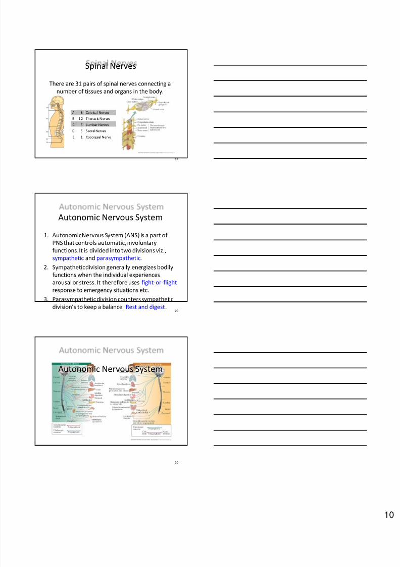
Spinal Nerves
There are 31 pairs of spinal nerves connecting a
number of tissues and organs in the body.
A 8 Cervical Nerves
B 1 2 Th orac ic N erves
C 5 Lumbar Nerves
D 5 Sacral Nerves
E 1 Coccygeal Nerve
29
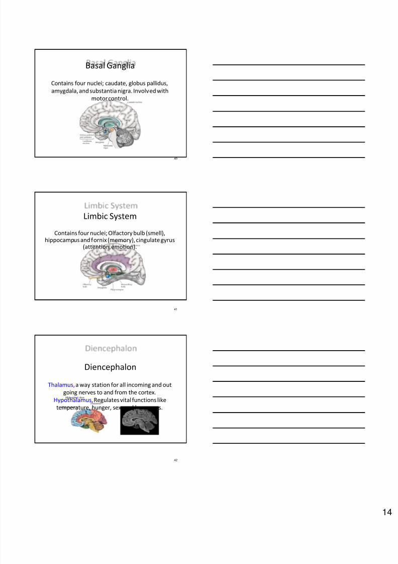
Autonomic Nervous System
1. Autonomic Nervous System (ANS) is a part of
PNS that controls automatic, involuntary
functions. It is divided into two divisions viz.,
sympathetic and parasympathetic.
2. Sympathetic division generally energizes bodily
functions when the individual experiences
arousal or stress. It therefore uses fight-or-flight
response to emergency situations etc.
3. Parasympathetic division counters sympatheticdivision’s to keep a balance. Rest and digest.
30
Autonomic Nervous System

8/6/2019 Neuroanatomy Presentation
http://slidepdf.com/reader/full/neuroanatomy-presentation 11/19
31
Central Nervous System
Central Nervous System
Forebrain(Procencephalon)
Midbrain(Mesencephalon)
Hindbrain(Rhombencephalon)
Cerebrum
(Telencephalon)
Thalamus
(Diencephalon)
Reticular
Activation
System
CerebellumPonsMedulla
Superior
Colliculus
Inferior
ColliculusLimbic
System
Isocortex Basal
Ganglia
32
Orientation of Nervous System
To orient ourselves about the nervous system in
3D, we need to understand three planes in which
brain can be sectioned.
33
Development of Nervous System

8/6/2019 Neuroanatomy Presentation
http://slidepdf.com/reader/full/neuroanatomy-presentation 12/19
34
Telencephalon (Cerebrum)
Telencephalon or cerebrum or the neo-cortex can
be divided into four lobes, with specific functions.
35
Frontal Lobe
The frontal lobe contains the motor strip, and
represents the body map (homunculus).
36
Parietal Lobe
The parietal lobe contains the somatosensory strip,
and represents the body map (homunculus).

8/6/2019 Neuroanatomy Presentation
http://slidepdf.com/reader/full/neuroanatomy-presentation 13/19
37
Occipital Lobe
The occipital lobe houses visual areas in the brain.
It contains 30 some areas that process visual
information.
j o u r n a l o f c o s m o l o g y . c o m
38
Temporal Lobe
The temporal lobe houses sensory functions as
hearing, taste, and smell. Also in this area are
memory functions and nuclei for emotions.
e n . w i k i p e d i a . o r g
Temporal Lobe
39
Medial Cerebrum
If you cut the cerebral hemispheres in the middle, a
set of new structures come in view. Many of theseperform arrays of different functions.
8/6/2019 Neuroanatomy Presentation
http://slidepdf.com/reader/full/neuroanatomy-presentation 14/19
40
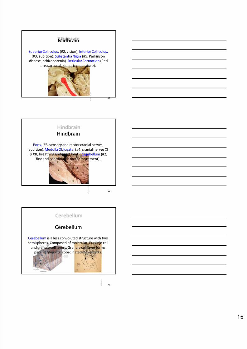
Basal Ganglia
Contains four nuclei; caudate, globus pallidus,
amygdala, and substantia nigra. Involved with
motor control.
41
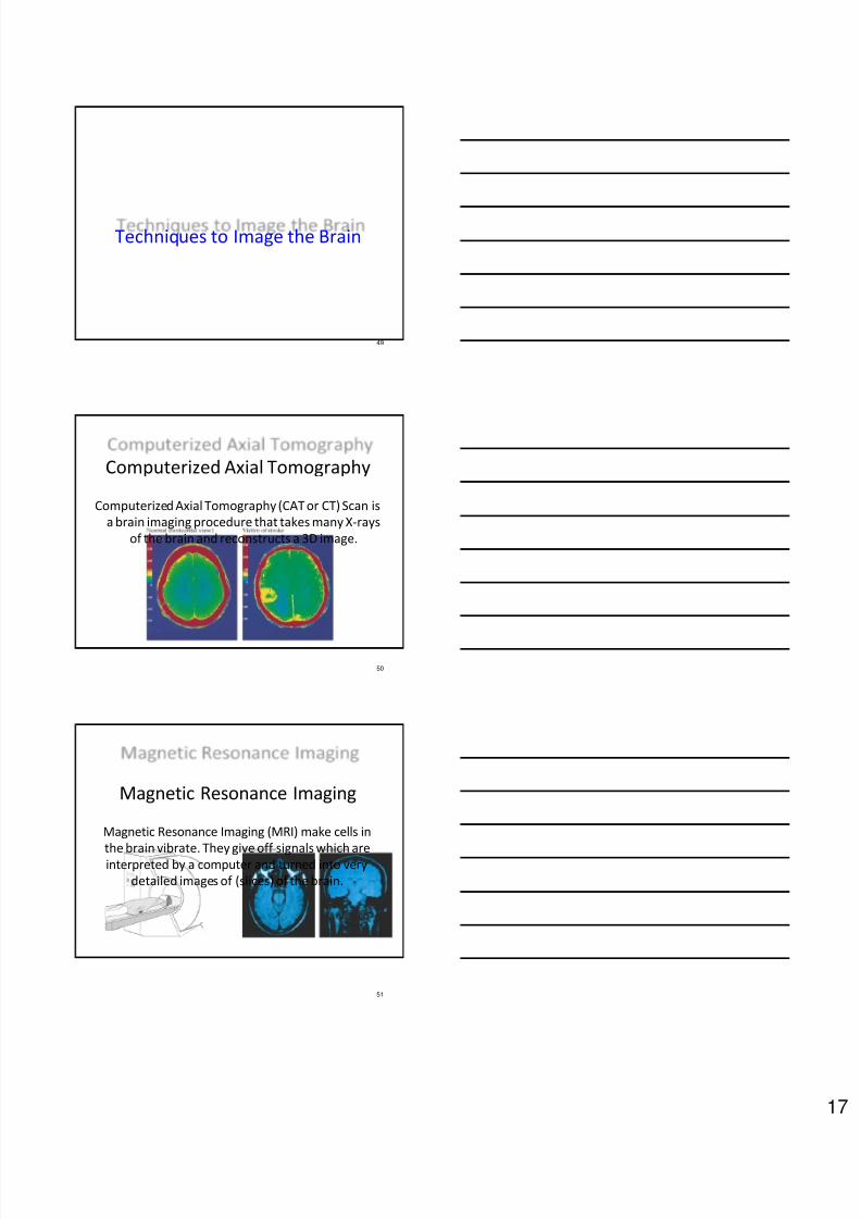
Limbic System
Contains four nuclei; Olfactory bulb (smell),hippocampus and fornix (memory), cingulate gyrus
(attention, emotion).
42
Diencephalon
Thalamus,a way station for all incoming and out
going nerves to and from the cortex.Hypothalamus,Regulates vital functions like
temperature, hunger, sex, and hormones.
8/6/2019 Neuroanatomy Presentation
http://slidepdf.com/reader/full/neuroanatomy-presentation 15/19
43
Midbrain
Superior Colliculus, (#2, vision), Inferior Colliculus,
(#3, audition). Substantia Nigra (#5, Parkinson
disease, schizophrenia). Reticular Formation (Redarea, arousal, sleep, temperature).
w w w . m e d . u m i c h . e d u
44
Hindbrain
Pons, (#3, sensory and motor cranial nerves,
audition).Medulla Oblogata, (#4, cranial nerves XI
& XII, breathing and heart beat). Cerebellum (#2,
fine and coordinated motor movement).
w w w . m e d . u m i c h . e d u
45
Cerebellum
Cerebellum is a less convoluted structure with two
hemispheres. Composed of molecular, Purkinje celland granule cell layers. Granule cell layer forms
parallel fibers for coordinated movements.
e n . w i k i p e d i a . o r g

8/6/2019 Neuroanatomy Presentation
http://slidepdf.com/reader/full/neuroanatomy-presentation 16/19
46
Layers of the Cerebrum
Cerebrum compared to cerebellum contain six
layers. Some layer receive inputs while others send
outputs.
47
Ventricles
Cerebrospinal Fluid (CSF) runs through the
ventricles, absorbing shock and providing important
minerals and electrolytes for the brain.
48
Meninges and BBB
Large volumes of blood
(20%) swaddle the brainand provide
nourishment. Meninges
consist of dura mater,
pia mater and arachnoid
space. Blood Brain
Barrier (BBB) keeps
bacteria and other toxic
agents outside the brain.
8/6/2019 Neuroanatomy Presentation
http://slidepdf.com/reader/full/neuroanatomy-presentation 17/19
8/6/2019 Neuroanatomy Presentation
http://slidepdf.com/reader/full/neuroanatomy-presentation 18/19
52
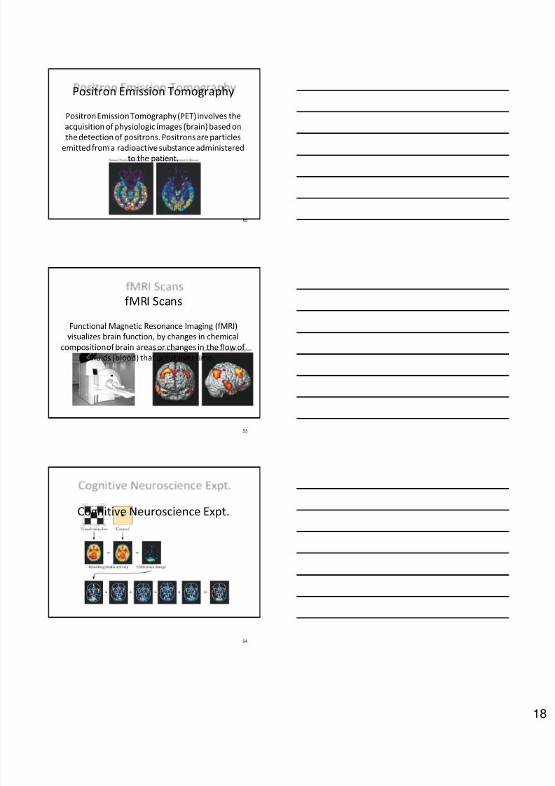
Positron Emission Tomography
Positron Emission Tomography (PET) involves the
acquisition of physiologic images (brain) based on
the detection of positrons. Positrons are particlesemitted from a radioactive substance administered
to the patient.
53
fMRI Scans
Functional Magnetic Resonance Imaging (fMRI)
visualizes brain function, by changes in chemical
composition of brain areas or changes in the flow of
fluids (blood) that occur over time.
54
Cognitive Neuroscience Expt.

8/6/2019 Neuroanatomy Presentation
http://slidepdf.com/reader/full/neuroanatomy-presentation 19/19
55
Optical Imaging & TMS
Optical imaging uses near infrared light to measure
responses by the cortex. Transcranial Magnetic
Stimulation (TMS) stimulates the brain of an alertsubject mapped by optical imaging.
56
Event-related Potential
Event-related potentials
measures the brain's
electrical activity as it
responds to impinging
stimuli (events). Excellent
temporal resolution
(faster response)
compared to PET or
fMRI.