Embed Size (px)
Citation preview
1
Università degli Studi di Cagliari
Dottorato di Ricerca in
Tossicologia – Indirizzo Farmacologia e Farmacoterapia delle
Tossicodipendenze
Ciclo XXVI
Vulnerability to cognitive, neurotoxic and neuroinflammatory effects of
toxins that induce Parkinson's disease after administration of
amphetamine-related drugs in mice
Settore/i scientifico disciplinari di afferenza
BIO/14
Presentata da Dott. ssa Giulia Costa
Coordinatore Dottorato Prof. Gaetano Di Chiara
Relatore Prof.ssa Micaela Morelli
Tutor Dott.ssa Annalisa Pinna
Esame finale anno accademico 2012 – 2013

2
3
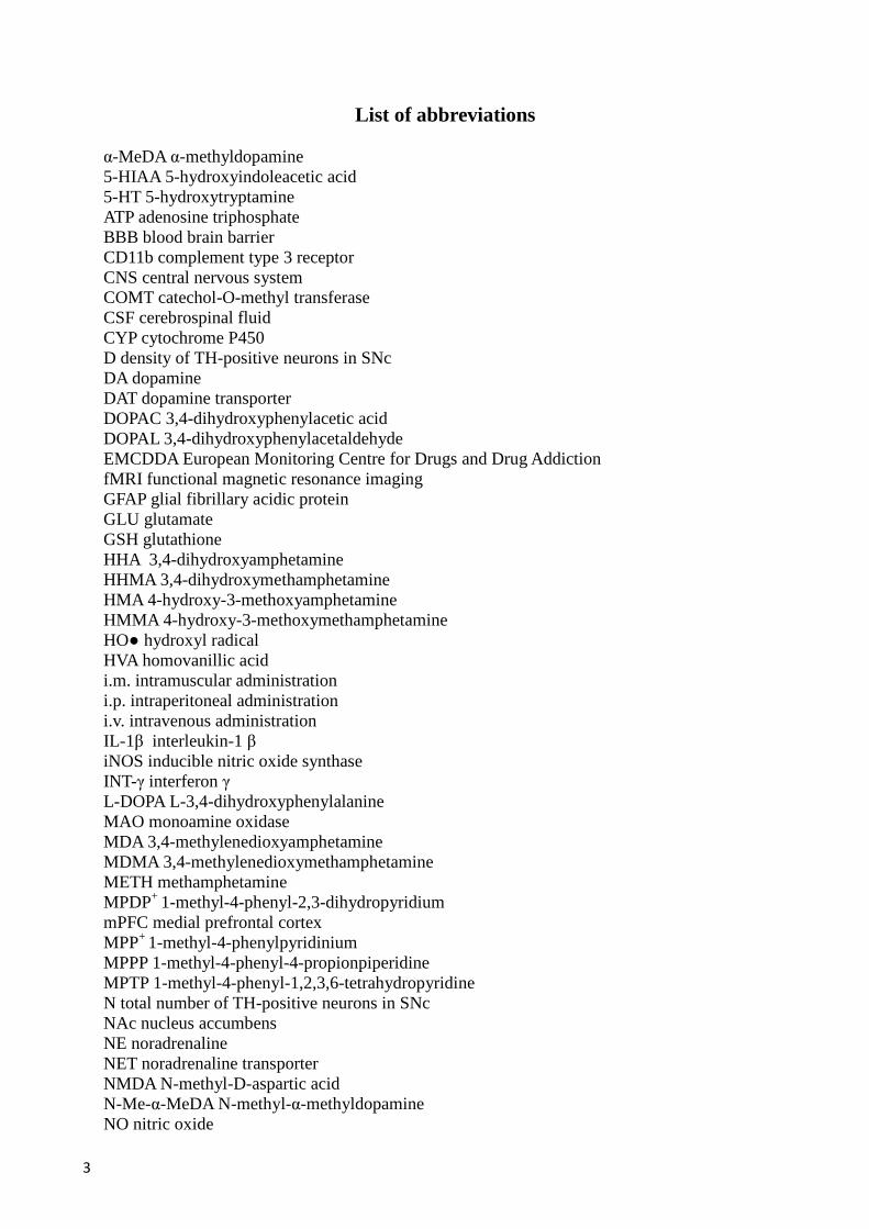
List of abbreviations
α-MeDA α-methyldopamine 5-HIAA 5-hydroxyindoleacetic acid 5-HT 5-hydroxytryptamine ATP adenosine triphosphate BBB blood brain barrier CD11b complement type 3 receptor CNS central nervous system COMT catechol-O-methyl transferase CSF cerebrospinal fluid CYP cytochrome P450 D density of TH-positive neurons in SNc DA dopamine DAT dopamine transporter DOPAC 3,4-dihydroxyphenylacetic acid DOPAL 3,4-dihydroxyphenylacetaldehyde EMCDDA European Monitoring Centre for Drugs and Drug Addiction fMRI functional magnetic resonance imaging GFAP glial fibrillary acidic protein GLU glutamate GSH glutathione HHA 3,4-dihydroxyamphetamine HHMA 3,4-dihydroxymethamphetamine HMA 4-hydroxy-3-methoxyamphetamine HMMA 4-hydroxy-3-methoxymethamphetamine HO● hydroxyl radical HVA homovanillic acid i.m. intramuscular administration i.p. intraperitoneal administration i.v. intravenous administration IL-1β interleukin-1 β iNOS inducible nitric oxide synthase INT-γ interferon γ L-DOPA L-3,4-dihydroxyphenylalanine MAO monoamine oxidase MDA 3,4-methylenedioxyamphetamine MDMA 3,4-methylenedioxymethamphetamine METH methamphetamine MPDP+ 1-methyl-4-phenyl-2,3-dihydropyridium mPFC medial prefrontal cortex MPP+ 1-methyl-4-phenylpyridinium MPPP 1-methyl-4-phenyl-4-propionpiperidine MPTP 1-methyl-4-phenyl-1,2,3,6-tetrahydropyridine N total number of TH-positive neurons in SNc NAc nucleus accumbens NE noradrenaline NET noradrenaline transporter NMDA N-methyl-D-aspartic acid N-Me-α-MeDA N-methyl-α-methyldopamine NO nitric oxide
4
NOR novel object recognition task ONOO− peroxynitrite nNOS neuronal nitric oxide synthase O2
- superoxide
PD Parkinson’s disease PET positron emission tomography PFC prefrontal cortex RNS reactive nitrogen species ROS reactive oxygen species s.c. subcutaneous administration SERT 5-hydroxytryptamine transporter SN substantia nigra SNc substantia nigra pars compacta SOD superoxide dismutase T-4,5-D tryptamine-4,5-dione TH tyrosine hydroxylase TNF-α tumour necrosis factor α TPH tryptophan hydroxylase VMAT 2 vesicular monoamine transporter

5
Table of contents
Abstract ........................................................................................................................................................ 7
Introduction ................................................................................................................................................. 8
1. MDMA ...................................................................................................................................................... 8
1.1. Pharmacokinetics ........................................................................................................................... 10
1.2 Pharmacology and toxicology ......................................................................................................... 11
1.2.1 Toxicology in humans ............................................................................................................. 12
Abuse properties of drugs during adolescence ............................................................................ 15
1.2.2 Toxicology in experimental animals ....................................................................................... 16
Non-human primates ................................................................................................................... 16
Rodents ....................................................................................................................................... 17
1.2.3 Mechanisms involved in MDMA toxicology .......................................................................... 19
Hyperthermia ............................................................................................................................... 19
ROS production and oxidative stress .......................................................................................... 20
Neuroinflammation ..................................................................................................................... 21
1.3 MDMA and Parkinson’s disease ...................................................................................................... 23
1.4 MDMA and nigrostriatal system in humans and experimental animals ........................................... 24
1.5 MDMA and limbic system in humans and experimental animals .................................................... 25
1.6 MDMA and the PFC in humans and experimental animals ............................................................. 26
2. MPTP ...................................................................................................................................................... 27
2.1. Toxicology ...................................................................................................................................... 27
2.1.1 Toxicology in humans ............................................................................................................. 27
2.1.2 Toxicology in experimental animals ........................................................................................ 28
Non-human primates .................................................................................................................... 28
Mice ............................................................................................................................................. 29
2.2 Pharmacological and toxicological mechanisms ............................................................................. 29
Decline in ATP production and increase in ROS levels ................................................................... 31
Nitration and oxidative stress ............................................................................................................ 32
Neuroinflammation ........................................................................................................................... 32
2.3 MPTP and the limbic system in humans and mice ........................................................................... 33
2.4 MPTP and the PFC in humans and experimental animals ............................................................... 34
Aims of the study ....................................................................................................................................... 36
Materials and methods ............................................................................................................................. 37
4.1 Animals .................................................................................................................................................. 37
4.2 Drugs ..................................................................................................................................................... 37
6
4.3 Treatment .............................................................................................................................................. 37
4.4 Immunohistochemistry ......................................................................................................................... 38
Analysis of CD11b immunoreactivity .............................................................................................. 38
Analysis of GFAP immunoreactivity ................................................................................................ 39
Stereological counting of TH-positive neurons ............................................................................... 39
Analysis of TH-positive fibers ......................................................................................................... 40
4.5 Memory tasks ....................................................................................................................................... 40
NOR task .......................................................................................................................................... 40
Spontaneous alternation in a Y-maze ................................................................................................ 41
4.6 Statistics ................................................................................................................................................ 41
Results ....................................................................................................................................................... 42
5.1 MDMA increases the vulnerability of mice to the neuroinflammation and neurotoxicity induced by MPTP in the SNc and striatum .................................................................................................................... 42
5.1.1 Immunohistochemistry ...................................................................................................................... 42
CD11b immunoreactivity in SNc and striatum ................................................................................. 42
GFAP immunoreactivity in SNc and striatum ................................................................................... 43
TH immunoreactivity in SNc and striatum ....................................................................................... 45
5.2 MDMA increases the vulnerability of mice to the neuroinflammation induced by MPTP in hippocampus and mPFC and is associated with cognitive deficits ............................................................. 46
5.2.1 Immunohistochemistry ...................................................................................................................... 46
CD11b immunoreactivity in hippocampus and mPFC ...................................................................... 46
GFAP immunoreactivity in hippocampus and mPFC ....................................................................... 48
5.2.2 Memory tasks .................................................................................................................................... 49
NOR task ........................................................................................................................................... 49
Spontaneous alternation behavior in a Y-maze ................................................................................. 50
Discussion .................................................................................................................................................. 52
Conclusions ............................................................................................................................................... 57
Acknowledgments ..................................................................................................................................... 57
Bibliografy .................................................................................................................................................. 58

7
Abstract
Clinical observations report a higher propensity to develop Parkinson’s disease (PD) in
amphetamine users. 3,4-Methylenedioxymethamphetamine (MDMA) is an amphetamine-related
drug which may have neuroinflammatory and neurotoxic effects. The present study was aimed at
evaluating in mice whether administration of MDMA during adolescence might influence
neurotoxicity towards dopaminergic neurons and neuroinflammatory effects of 1-methyl-4-
phenyl-1,2,3,6-tetrahydropyridine (MPTP), a toxin known to induce PD in humans, in motor,
limbic and cortical areas, and consequently affects cognitive performance.
Mice received MDMA (10 mg/kg, twice a day/a week) for 9 weeks, followed by MPTP (20
mg/kg × 4 administrations), starting 2 weeks after MDMA discontinuation. Activation of
astroglia and microglia by GFAP and CD11b immunohistochemistry in motor areas, as
substantia nigra compacta (SNc) and striatum, limbic and cortical areas, as hippocampus and
medial prefrontal cortex (mPFC), was assessed. Degeneration of dopaminergic neurons by
tyrosine hydroxylase (TH) immunohistochemistry in SNc and striatum was also evaluated.
Neurochemical evaluations were paired with assessment of cognitive performance by means of
the novel object recognition (NOR) and spontaneous alternation in a Y-maze tests.
MPTP administration to MDMA-pretreated mice elicited a stronger increase in CD11b and
GFAP levels in motor, limbic and cortical areas, and a stronger decrease of TH-positive neurons
and fibers in motor areas, compared with either substance administered alone. Furthermore,
NOR performance in the same group was lower, compared with mice that received either
substance alone.
Results demonstrate that MDMA administration during adolescence influence negatively MPTP
effects on motor, limbic and cortical areas and result in cognitive impairment.
8
Introduction
1. MDMA
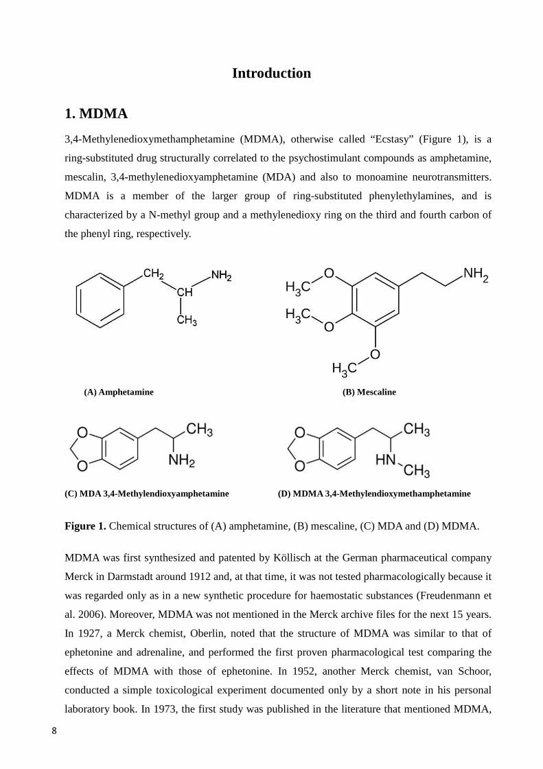
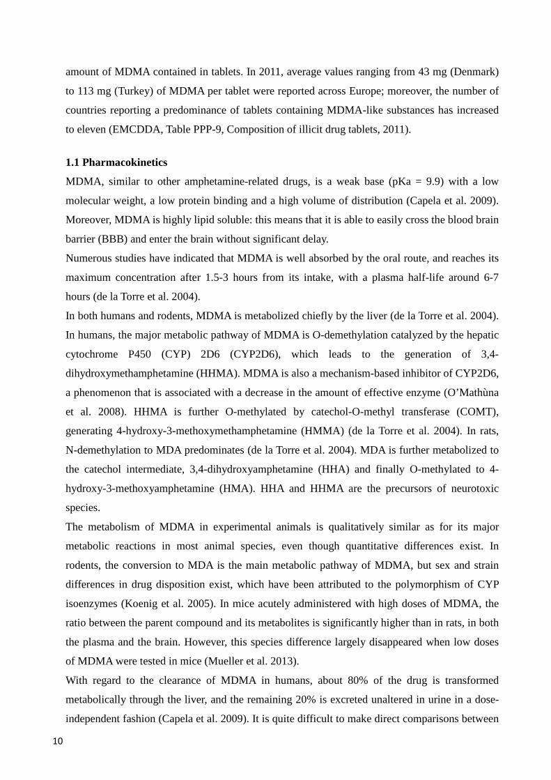
3,4-Methylenedioxymethamphetamine (MDMA), otherwise called “Ecstasy” (Figure 1), is a
ring-substituted drug structurally correlated to the psychostimulant compounds as amphetamine,
mescalin, 3,4-methylenedioxyamphetamine (MDA) and also to monoamine neurotransmitters.
MDMA is a member of the larger group of ring-substituted phenylethylamines, and is
characterized by a N-methyl group and a methylenedioxy ring on the third and fourth carbon of
the phenyl ring, respectively.
(A) Amphetamine (B) Mescaline
(C) MDA 3,4-Methylendioxyamphetamine (D) MDMA 3,4-Methylendioxymethamphetamine
Figure 1. Chemical structures of (A) amphetamine, (B) mescaline, (C) MDA and (D) MDMA.
MDMA was first synthesized and patented by Köllisch at the German pharmaceutical company
Merck in Darmstadt around 1912 and, at that time, it was not tested pharmacologically because it
was regarded only as in a new synthetic procedure for haemostatic substances (Freudenmann et
al. 2006). Moreover, MDMA was not mentioned in the Merck archive files for the next 15 years.
In 1927, a Merck chemist, Oberlin, noted that the structure of MDMA was similar to that of
ephetonine and adrenaline, and performed the first proven pharmacological test comparing the
effects of MDMA with those of ephetonine. In 1952, another Merck chemist, van Schoor,
conducted a simple toxicological experiment documented only by a short note in his personal
laboratory book. In 1973, the first study was published in the literature that mentioned MDMA,
9
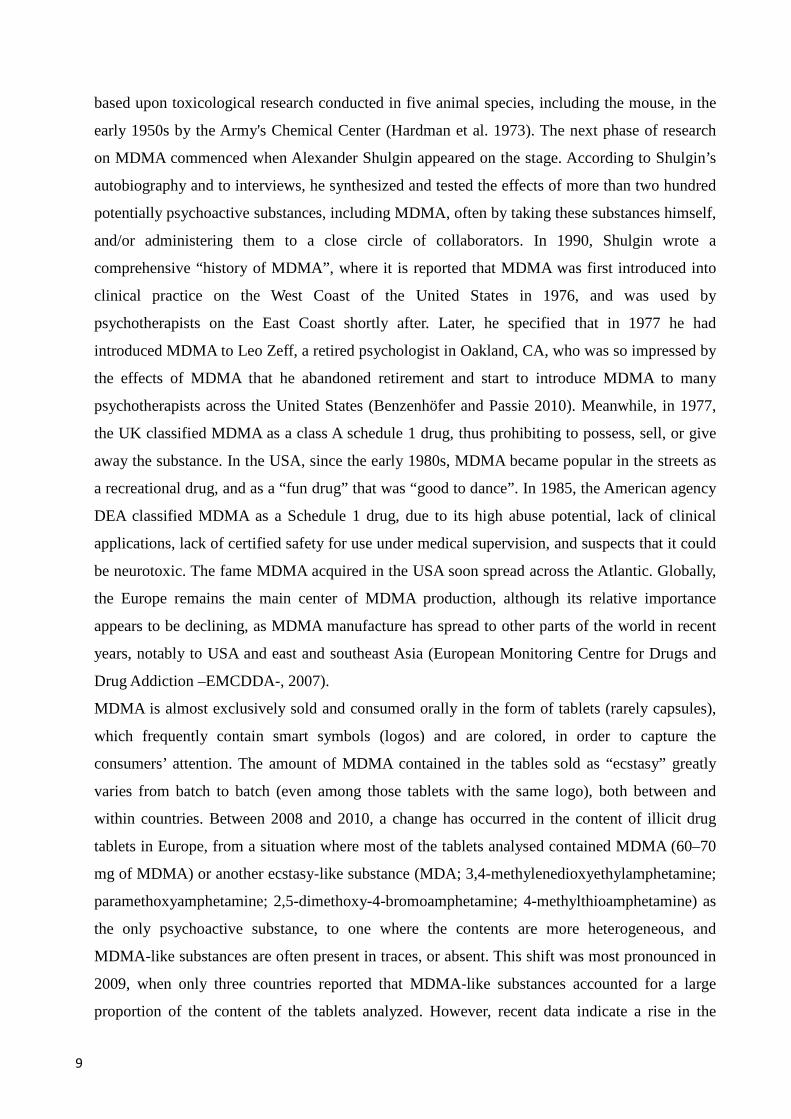
based upon toxicological research conducted in five animal species, including the mouse, in the
early 1950s by the Army's Chemical Center (Hardman et al. 1973). The next phase of research
on MDMA commenced when Alexander Shulgin appeared on the stage. According to Shulgin’s
autobiography and to interviews, he synthesized and tested the effects of more than two hundred
potentially psychoactive substances, including MDMA, often by taking these substances himself,
and/or administering them to a close circle of collaborators. In 1990, Shulgin wrote a
comprehensive “history of MDMA”, where it is reported that MDMA was first introduced into
clinical practice on the West Coast of the United States in 1976, and was used by
psychotherapists on the East Coast shortly after. Later, he specified that in 1977 he had
introduced MDMA to Leo Zeff, a retired psychologist in Oakland, CA, who was so impressed by
the effects of MDMA that he abandoned retirement and start to introduce MDMA to many
psychotherapists across the United States (Benzenhöfer and Passie 2010). Meanwhile, in 1977,
the UK classified MDMA as a class A schedule 1 drug, thus prohibiting to possess, sell, or give
away the substance. In the USA, since the early 1980s, MDMA became popular in the streets as
a recreational drug, and as a “fun drug” that was “good to dance”. In 1985, the American agency
DEA classified MDMA as a Schedule 1 drug, due to its high abuse potential, lack of clinical
applications, lack of certified safety for use under medical supervision, and suspects that it could
be neurotoxic. The fame MDMA acquired in the USA soon spread across the Atlantic. Globally,
the Europe remains the main center of MDMA production, although its relative importance
appears to be declining, as MDMA manufacture has spread to other parts of the world in recent
years, notably to USA and east and southeast Asia (European Monitoring Centre for Drugs and
Drug Addiction –EMCDDA-, 2007).
MDMA is almost exclusively sold and consumed orally in the form of tablets (rarely capsules),
which frequently contain smart symbols (logos) and are colored, in order to capture the
consumers’ attention. The amount of MDMA contained in the tables sold as “ecstasy” greatly
varies from batch to batch (even among those tablets with the same logo), both between and
within countries. Between 2008 and 2010, a change has occurred in the content of illicit drug
tablets in Europe, from a situation where most of the tablets analysed contained MDMA (60–70
mg of MDMA) or another ecstasy-like substance (MDA; 3,4-methylenedioxyethylamphetamine;
paramethoxyamphetamine; 2,5-dimethoxy-4-bromoamphetamine; 4-methylthioamphetamine) as
the only psychoactive substance, to one where the contents are more heterogeneous, and
MDMA-like substances are often present in traces, or absent. This shift was most pronounced in
2009, when only three countries reported that MDMA-like substances accounted for a large
proportion of the content of the tablets analyzed. However, recent data indicate a rise in the
10
amount of MDMA contained in tablets. In 2011, average values ranging from 43 mg (Denmark)
to 113 mg (Turkey) of MDMA per tablet were reported across Europe; moreover, the number of
countries reporting a predominance of tablets containing MDMA-like substances has increased
to eleven (EMCDDA, Table PPP-9, Composition of illicit drug tablets, 2011).
1.1 Pharmacokinetics
MDMA, similar to other amphetamine-related drugs, is a weak base (pKa = 9.9) with a low
molecular weight, a low protein binding and a high volume of distribution (Capela et al. 2009).
Moreover, MDMA is highly lipid soluble: this means that it is able to easily cross the blood brain
barrier (BBB) and enter the brain without significant delay.
Numerous studies have indicated that MDMA is well absorbed by the oral route, and reaches its
maximum concentration after 1.5-3 hours from its intake, with a plasma half-life around 6-7
hours (de la Torre et al. 2004).
In both humans and rodents, MDMA is metabolized chiefly by the liver (de la Torre et al. 2004).
In humans, the major metabolic pathway of MDMA is O-demethylation catalyzed by the hepatic
cytochrome P450 (CYP) 2D6 (CYP2D6), which leads to the generation of 3,4-
dihydroxymethamphetamine (HHMA). MDMA is also a mechanism-based inhibitor of CYP2D6,
a phenomenon that is associated with a decrease in the amount of effective enzyme (O’Mathùna
et al. 2008). HHMA is further O-methylated by catechol-O-methyl transferase (COMT),
generating 4-hydroxy-3-methoxymethamphetamine (HMMA) (de la Torre et al. 2004). In rats,
N-demethylation to MDA predominates (de la Torre et al. 2004). MDA is further metabolized to
the catechol intermediate, 3,4-dihydroxyamphetamine (HHA) and finally O-methylated to 4-
hydroxy-3-methoxyamphetamine (HMA). HHA and HHMA are the precursors of neurotoxic
species.
The metabolism of MDMA in experimental animals is qualitatively similar as for its major
metabolic reactions in most animal species, even though quantitative differences exist. In
rodents, the conversion to MDA is the main metabolic pathway of MDMA, but sex and strain
differences in drug disposition exist, which have been attributed to the polymorphism of CYP
isoenzymes (Koenig et al. 2005). In mice acutely administered with high doses of MDMA, the
ratio between the parent compound and its metabolites is significantly higher than in rats, in both
the plasma and the brain. However, this species difference largely disappeared when low doses
of MDMA were tested in mice (Mueller et al. 2013).
With regard to the clearance of MDMA in humans, about 80% of the drug is transformed
metabolically through the liver, and the remaining 20% is excreted unaltered in urine in a dose-
independent fashion (Capela et al. 2009). It is quite difficult to make direct comparisons between

11
pharmacokinetics results obtained in humans and the few pharmacokinetics results from animal
studies, since large mammals tend to dispose drugs at a lower rate than small mammals. In spite
of that, it has been published that the clearance of MDMA and its metabolites was significantly
faster in the mouse compared with the rat (Mueller et al. 2013).
1.2 Pharmacology and toxicology
Preclinical data gathered from research performed in laboratory animals, indicate that the
systemic administration of MDMA affects the functions of both peripheral and central nervous
system (CNS), mainly by acting on monoaminergic systems. Thus, initial studies showed that
MDMA and its main metabolite, MDA, stimulate the efflux of 5-hydroxytryptamine (5-HT) (de
la Torre et al. 2004) and dopamine (DA) from preloaded synaptosomes (Johnson et al. 1986;
Schmidt et al. 1987). Subsequent reports have demonstrated that MDMA binds all the three
presynaptic monoamine transporters with differences between species, exhibiting, in rodents, the
highest affinity for the 5-hydroxytryptamine transporter (SERT), whilst the affinities for the
noradrenaline (NET) and dopamine (DAT) transporters are lower (Rudnick and Wall 1992;
Steele et al. 1987). MDMA also enhances the release of acetylcholine both in vitro in striatal
slices and in vivo in prefrontal cortex (PFC) and striatum of rats, an effect that appears to derive
from the activation of serotonergic, dopaminergic and/or histaminergic receptors (Gudelsky and
Yamamoto 2008). At variance with data obtained in studies concerned with rodent transporters, it
has been reported that, in humans, MDMA displays higher affinity for NET and lower, but
similar, affinities for SERT and DAT (Verrico et al. 2007).
Once translocated to the cytoplasm, MDMA increases the extracellular levels of 5-HT, DA and
noradrenaline (NA) in multiple brain regions (Gudelsky and Yamamoto 2008). MDMA causes
the dissipation of the proton gradient between the vesicles and the cytosol that is necessary for
the proper functioning of the vesicular monoamine transporter (VMAT2), also inhibiting VMAT2-
mediated influx and proper storage of 5-HT in the terminal (Rudnick and Wall 1992). This event
is boosted by the block of reuptake on presynaptic terminals, and by the inhibition of the
monoamine oxidase (MAO) type B (Leonardi and Azmitia 1994). MDMA also blocks the
activity of tryptophan hydroxylase (TPH, the rate-limiting enzyme in the synthesis of 5-HT), an
effect that occurs as soon as fifteen min after MDMA intake, and persists for up to two weeks
(Schmidt and Taylor 1987). Furthermore, MDMA causes a global increase in extracellular 5-HT
throughout the brain regions bearing raphe afferents (Rudnick and Wall 1992).
12
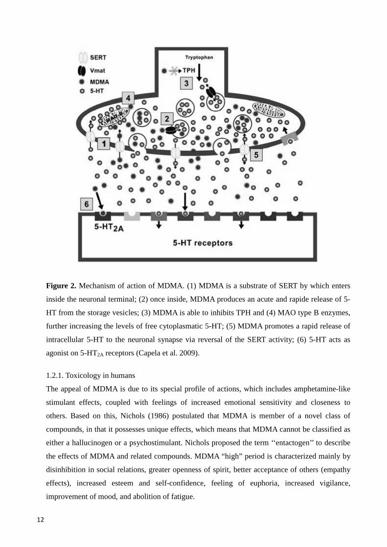
Figure 2. Mechanism of action of MDMA. (1) MDMA is a substrate of SERT by which enters
inside the neuronal terminal; (2) once inside, MDMA produces an acute and rapide release of 5-
HT from the storage vesicles; (3) MDMA is able to inhibits TPH and (4) MAO type B enzymes,
further increasing the levels of free cytoplasmatic 5-HT; (5) MDMA promotes a rapid release of
intracellular 5-HT to the neuronal synapse via reversal of the SERT activity; (6) 5-HT acts as
agonist on 5-HT2A receptors (Capela et al. 2009).
1.2.1. Toxicology in humans
The appeal of MDMA is due to its special profile of actions, which includes amphetamine-like
stimulant effects, coupled with feelings of increased emotional sensitivity and closeness to
others. Based on this, Nichols (1986) postulated that MDMA is member of a novel class of
compounds, in that it possesses unique effects, which means that MDMA cannot be classified as
either a hallucinogen or a psychostimulant. Nichols proposed the term ‘‘entactogen’’ to describe
the effects of MDMA and related compounds. MDMA “high” period is characterized mainly by
disinhibition in social relations, greater openness of spirit, better acceptance of others (empathy
effects), increased esteem and self-confidence, feeling of euphoria, increased vigilance,
improvement of mood, and abolition of fatigue.
13
However, not every MDMA experience is positive, since up to the 25% of users report having
had at least one adverse reaction, with prevalence of unpleasant feelings and bodily sensations
(Davison and Parrott 1997). However, the possibility exists that the positive effects of MDMA
may subside with repeated use, as suggested by Peroutka et al. (1988). Peripheral adverse effects
of MDMA in humans include cardiac arrhythmias, hypertension, hyperthermia, hyponatremia,
liver complications, seizures, coma, and death (Schifano 2004). Hyperthermia is a very relevant
clinical problem in MDMA users, since body temperature elevation produced by the drug may
reach up to 43 °C (Capela et al. 2009), and around 85–90% of recreational MDMA users report
an increase in body temperature and sweating, accompanied by dehydration (Davison and Parrott
1997). The issue of MDMA-induced hyperthermia is complex, since the biological mechanisms
involved in heat production and progression to hyperthermia after exposure to the drug are not
clearly understood. It is conceivable that the administration of a large dose of MDMA evokes 5-
HT release that activates sufficient 5-HT1B and 5-HT2A receptors within vasculature (Gudelsky et
al. 1986) to induce constriction, interfering with the normal thermoregulatory mechanisms of the
body (Sprague et al. 2003). In this regard, it is worth to mention the study of Blessing and
colleagues (2003) where it is demonstrated that clozapine and olanzapine, atypical antipsychotic
agents that act as antagonists respectively at the 5-HT1A and 5-HT2 receptors, reverse severe and
potentially fatal hyperthermia elicited by MDMA acute administration in rats and rabbits.
Several reports in humans and in laboratory animals indicate that the rises in body temperature
induced by MDMA are strictly influenced by the external environment (Carvalho et al. 2002;
Huether et al. 1997), with an important factor for the toxicity of the drug being the social
gathering (also called “aggregation toxicity”). Thus, the typical conditions encountered in
“raves” and clubs parties, where the music is deafening, room temperatures are high due to
crowding, and people usually assume few water and lots of alcohol are crucial to amplify
MDMA-induced hyperthermia, and also all the other acute toxic effects of MDMA described
above (Green et al. 2003).
Another problem related to the MDMA use is the so-called “serotonin syndrome”, which is
caused by drug-induced excess of intersinaptic 5-HT (Gillman 1999; Hall and Henry 2006;
Huether et al. 1997). The symptoms include behavioral hyperactivity, mental confusion,
agitation, hyperreflexia, hyperpyrexia, tachycardia, shivering, clonus, myoclonus, ocular
oscillations, and tremor (Gillman 1999; Hall and Henry 2006; Huether et al. 1997). The
“serotonin syndrome” is often conceptualized as an unusual, or atypical, severe adverse MDMA
reaction. However, Gillman (1999) argued that this syndrome was neither rare nor idiosyncratic,
but represented a continuum of responses from mild to severe. The mild “serotonin syndrome”
14
(which is limited to three symptoms from above list) generally requires no direct medical
intervention. Stronger responses (including four or more symptoms) would often necessitate
medical supervision, and while severe reactions (including most of the symptoms from the list)
could prove fatal. Nevertheless, in the light of the widespread use of MDMA, fatal intoxications
still remain rare events (EMCDDA, Annual report on the State of the Drugs Problem in Europe,
2010). Exposure to MDMA could be regarded as a “chemical stressor” on the immune system, as
it induces immunosuppression (Connor 2004; Connor et al. 2005), which may have a significant
impact on the health of abusers. These effects may involve alterations of neutrophil
phagocytosis, reduction of the production of inflammatory cytokines, suppression of the
production of interferon γ (INT-γ), and reduction of the expression of MCH-II molecules at the
level of neuronal dendrites and macrophages. Furthermore, MDMA reduces the number of
circulating lymphocytes, especially of TCD4+, suppresses the proliferation of the T cell line, and
diverts the production of cytokines by favouring the specialization of Th0 into Th2. The
immunosuppressive effects of MDMA are likely not a result of a direct action of the substance
on immune cells, but rather stem from the release of immunomodulatory endogenous substances
(Boyle and Connor 2010).
MDMA can also cause a series of neurobehavioural disturbances that can appear during the first
few hours from drug intake, and include depression, mania, psychosis, panic attacks, irritability,
hallucinations, insomnia, tiredness, fatigue, and paranoid ideas (Cole and Sumnall 2003; Davison
and Parrott 1997; Hall and Henry 2006; McCann et al. 1996). Hallucinations and paranoia can
persist for days, or even weeks, after the intake of MDMA. In addition, cases of potentially fatal
neurological effects, such as subarachnoid and intracranial hemorrhage and thrombosis, have
been reported in MDMA consumers (Green et al. 2003).
In contrast to the acute effects described so far, the evaluation of the long term effects of
MDMA, including neurotoxicity, is complex, and these effects should not be underestimated.
Adverse neuropsychiatric effects have been described after chronic MDMA use and, most
notably, some recreational MDMA users display cognitive deficits (Fox et al. 2001; Parrott and
Lasky 1998) that become more marked in heavy drug users (Morgan 2000).
The existence of cognitive dysfunctions contributes to support the evidence of neurotoxicity
induced by MDMA in the human brain. Indeed, functional magnetic resonance imaging (fMRI)
studies have demonstrated that MDMA may induce serotonergic toxicity in brain areas involved
in the regulation of cognition, mood and memory, such as the hippocampus and the PFC. A
recent fMRI investigation that involved heavy abstinent MDMA users showed a significantly
greater spatial extent of activation than controls in both the primary and secondary visual cortex,
15
suggesting that MDMA use may be associated with a long-lasting increase in cortical
excitability, possibly through the loss of 5-HT input to cortical and subcortical regions
(Bauernfeind et al. 2011). Positron emission tomography (PET) studies reported significant
reductions in SERT binding compared to control subjects (McCann et al. 1998). In another PET
study, Reneman et al. (2001) found an indication that women who heavy abused MDMA might
be more susceptible to neurotoxic effects on serotonergic neurons than men who were heavy
MDMA users, and that MDMA-induced neurotoxic changes in several brain regions of women
who formerly abused MDMA are reversible.
Even though in humans the neurotoxicity of MDMA on the serotonergic system has been
suggested by several studies on the one hand, on the other hand the effects of MDMA on
dopaminergic neurons are less certain (Tai et al. 2011). In this regard, it is worth mentioning a
recent study that analyzed the neurotoxicity of MDMA and its catechol metabolites, α-
methyldopamine (α-MeDA) and N-methyl-α-methyldopamine (N-Me-α-MeDA) in human
dopaminergic SH-SY5Y cells. In this investigation, α-MeDA and N-Me-α-MeDA were found to
be neurotoxic to SH-SY5Y cells, leading to caspase 3-independent cell death in a concentration-
and time-dependent manner, whereas MDMA did not show a concentration and time-dependent
induction of cell death (Ferreira et al. 2013).
Abuse properties of drugs during adolescence
It must be considered that an increased risk of developing drug abuse and drug-related problems
is often associated with the age of the consumers (Hawkins et al. 1992). Adolescents are likely to
participate at “raves” and clubs parties, and it is during adolescence that most drug use and abuse
patterns are initiated. In both humans and experimental animals, adolescence is a critical period
during development (Casey et al. 2000), which is generally associated with the acquisition of
mature survival skills that allow independence from parental care. Novelty-seeking, a
temperamental/behavioral trait that is typical of this age period, might substantially contribute to
both psychological and psychobiological vulnerability. In the light of preclinical studies
suggesting that the behavioral responses to psychostimulants are the result of neurobiological
adaptations that occur primarily in the mesolimbic dopaminergic system (Pierce and Kumaresan
2006), it is expected that exposure to MDMA during adolescence, as happen with other
substances, would produce long-term changes in these systems that perpetuate in adulthood
(Chambers et al. 2003; Morley-Fletcher et al. 2002; Sisk and Zehr 2005; Wahlstrom et al. 2009),
and that the adverse peripheral reactions observed in adult would be more pronounced (Spear
2000).
16
Regarding cognitive deficits, little is known of brain functional modifications of MDMA use
during adolescence. In the study of Jacobsen and co-workers (2004), adolescent MDMA users
displayed significant delays in reaction time during simple, selective and divided attention tasks
and abnormal function of the left hippocampus during high working memory load. Moreover, the
study of Weinborn and collaborators (2011) support a deficit in prospective memory functioning
amongst MDMA young users (18–30 years) for longer (15 min) ongoing task delays, as
compared with shorter (2 min) delays.
1.2.2. Toxicology in experimental animals
The most important acute effects of MDMA depend on several factors, such as the dose
administered, the age at administration, the species, the environmental temperature, the thermal
conductivity of the animal housing, and the hydration status.
Non-human primates MDMA suppresses locomotor activity in non-human primates following both intramuscular
(i.m.) (Von Huben et al. 2007) and oral (Crean et al. 2007) administration. Differently from what
observed in rats, the environmental temperature seems not to affect MDMA-induced
hyperthermia in non-human primates, as suggested by data in rhesus macaques administered
with doses ranging between 0.56 and 2.4 mg/kg that displayed a similar degree of hyperthermia
across a wide range of environmental conditions (18 °C to 30 °C) (Von Huben et al. 2007).
Nevertheless, MDMA induces acute hyperthermia in unrestrained rhesus monkeys, an effect that
is much more pronounced than in rats, mice, pigs, rabbits and humans. Maximum and average
temperature in the four hours interval post-dosing was elevated by 0.7–0.9 °C after racemic
MDMA and either enantiomer (Taffe et al. 2006).
Non-human primates administered MDMA seem to be very susceptible towards the neurotoxic
effects of this substance. A dose-dependent reduction in the 5-HT content in several brain areas
has been observed in squirrel monkeys after the subcutaneous (s.c.) administration of MDMA at
doses ranging between 2.5 and 5.0 mg/kg (Ricaurte et al. 1988a). In another study, MDMA was
administered at the same doses, and significant decreases in cerebrospinal fluid (CSF) levels of
5-hydroxyindoleacetic acid (5-HIAA) and brain 5-HT and 5-HIAA concentrations were observed
(Insel et al. 1989). In addition, the long-lasting deficits characteristic of MDMA-induced
neurotoxicity were evidenced by the fact that squirrel monkeys presented reduced serotonergic
innervations and reduced 5-HT levels seven years following exposure to the drug. The route of
drug administration also seems to affect the degree of 5-HT depletion, as oral administration has
been reported to be less toxic than s.c. injection. It is noteworthy that repeated MDMA

17
administrations at a dose of 5 mg/kg have been reported to produce an 86% depletion of frontal
cortex 5-HT when given s.c., compared with a 42% depletion elicited by the same dose given
orally (Ricaurte et al. 1988b). Furthermore, since a single 5 mg/kg oral dose in non-human
primates has been suggested to be equivalent to a 1.4 mg/kg dose in a 70 kg human, based on
interspecies dose scaling, these data may indicate a possible risk of serotonergic damage in
humans even after a single dose (Ricaurte et al. 1988b).
There are few preclinical studies in non-human primates focused on MDMA ability to induce
cognitive deficits. Nevertheless, Taffe and collaborators (2001) have demonstrated that rhesus
monkeys, treated with a MDMA regimen able to induce a 50% reduction of 5-HIAA in CSF and
persistent reductions of 5-HT content in several neocortical regions and hippocampus, and
display overt deficits in a range of cognitive domains, like self-ordered spatial search, five-
choice reaction time task, progressive ratio responding, and bimanual motor skill task that do not
persist when animals are retested few months later.
Rodents
Administration of MDMA has profound effects not only on the cardiovascular and
neuroendocrine systems, but also on the thermoregulatory system and the basal metabolism of
rats (Gordon et al. 1991). Several studies have reported that rats housed at room temperature
conditions (20–24 °C) display an acute hyperthermic response following administration of
MDMA (Colado et al. 1999; Dafters 1994; Mechan et al. 2002; O’Loinsigh et al. 2001).
Exposing rats to higher ambient room temperature conditions results in rats having a higher
hyperthermic response to MDMA, while exposure to lower ambient temperatures is associated
with a hypothermic response to MDMA (Malberg and Seiden 1998).
Another interesting point in MDMA toxicity is that MDMA tablets are often taken, in the “raves”
and clubs parties, together with caffeinated beverages or with beverages contaminated with
caffeine in varying amounts. Rat studies have shown that caffeine enhances hyperthermic and
tachycardic responses induced by MDMA (Vanattou-Saïfoudine et al. 2010).
There is considerable evidence that MDMA administration produces a major release of both 5-
HT and DA from their respective terminals in the forebrain (Johnson et al. 1986; Schmidt et al.
1987), and that MDMA-induced hyperthermia may be associated with an increased 5-HT
(Shankaran and Gudelsky 1999). Several studies have demonstrated that MDMA causes long-
term neurodegeneration in the rat brain, particularly in the neocortex, striatum, thalamus,
hippocampus, septum, and amygdala, even though strain differences in this effect exist, with the
Dark Agouti strain being more sensitive than other commonly used strains (Baumann et al. 2007;
Colado et al. 1995; Logan et al. 1988). The terminal portions of axons have been shown to be

18
selectively vulnerable to MDMA-induced damage, as indicated by the reduced density of fine,
arborized 5-HT axons and sparing of smooth, straight preterminal fibers (Molliver et al. 1990). A
very relevant point in MDMA toxicity observed in rats is that this effect appeared particularly
marked during adolescence. Thus, adolescent rats, repeatedly treated with MDMA at several
time points, displayed a significantly reduced SERT-immunoreactive fibers density in the
hippocampus, a deficit in the novel object-recognition task (NOR), an increased impulsivity in
the elevated plus-maze, and a reduced sensitivity to a 5-HT1A agonist challenge (Meyer et al.
2008). Paralleling to serotonergic fibers damage, other research groups displayed that astrocyte
hypertrophy can occur as a result of neuronal injury after single or repeated MDMA treatment,
and can lead to the enhanced expression of glial fibrillary acidic protein (GFAP) (Adori et al.
2006; Aguirre at al. 1999), the major protein of astrocyte intermediate filaments. In addition, it
has been shown that acute MDMA treatment induces learning deficits in rats. Adolescent
Sprague–Dawley rats treated with MDMA displayed, five days after the last dose, an impairment
in NOR and increased open arm exploration in the elevated plus maze (Piper and Meyer 2004).
This suggests that MDMA exposure during the adolescence may influence cognitive and
affective functioning in the absence of severe serotonergic damage (Piper and Meyer 2004).
Similar to what observed in rats, MDMA administration produces a hyperthermic response in
mice. The mice strain, the MDMA dose (Mann et al. 1997) and the housing conditions appear to
influence the magnitude and features of the response detected. Fantegrossi and colleagues (2003)
have demonstrated that racemic MDMA and the S(+)-MDMA enantiomer were approximately
equipotent in terms of their lethal effects across singly housed mice or mice housed in crowded
cages (twelve per cage), while the R(–)-MDMA enantiomer was approximately half as potent.
MDMA lethality was reversed with the temperature decrease, as a cold environment significantly
attenuated the lethal effects of racemic MDMA in singly housed mice, and completely abolished
the lethal effects of racemic MDMA and S(+)-MDMA in mice housed in crowded cages. These
effects could likely be explained by an inhibition of MDMA metabolism (Mueller et al. 2013).
It is now well established that MDMA produces in mice a neurotoxic profile different from the
serotonergic one observed in rats or non-human primates, eliciting a significant dopaminergic
neurotoxicity (O’Callaghan and Miller 1994). The basis for the variations in the profile of
MDMA neurotoxicity across different species is unknown, but it has recently been suggested that
differences in MDMA disposition and CYP metabolism may play a key role (Green et al. 2012).
Nevertheless, MDMA can elicit dopaminergic neurotoxicity depending on its dosage (Itzhak et
al. 2003).
19
Several studies have described MDMA-induced cognitive deficits in mice, but very few is
known about cognitive deficits elicited by MDMA in adolescent mice (Ros-Simò et al. 2013;
Vidal-Infer et al. 2012), indicating that further investigations are necessary to clarify whether
neurochemical deficits induced by MDMA during adolescence correlate with behavioral and
cognitive abnormalities.
1.2.3 Mechanisms involved in MDMA toxicology
Even though a large number of studies have been carried out to address the mechanisms involved
in MDMA-induced neurotoxicity, these remain to be fully elucidated. Several lines of evidence
indicate that multiple mechanisms can be involved, and that these mechanisms may differ
depending on the specific toxic effect considered.
Hyperthermia
Several in vivo studies indicate that hyperthermia plays an important role in MDMA-induced
neurotoxicity. As mentioned above, MDMA toxicity to 5-HT terminals during hyperthermic and
hypothermic conditions can be enhanced and attenuated, respectively. The mechanism by means
of which MDMA interferes with the regulation of body temperature is not fully understood. A
study by Mechan et al. (2002) has demonstrated that the temperature of the tail was unaltered in
rats following a dose of MDMA that produced a significant rise in rectal temperature. Since
vasodilation of tail vessels is a major mechanism by which rats lose temperature (Grant 1963)
these results suggest that MDMA could interfere with heat loss mechanisms. In this regard, it is
worth mentioning the work by Gordon et al. (1991) who examined modifications in
thermoregulatory mechanisms by MDMA by measuring metabolic rate, evaporative water loss
and rectal temperature of rats housed at 10°, 20° and 30 °C. MDMA was able to produce a
temperature-dependent increase in the metabolic rate and evaporative water loss. Hyperthermia
by itself is not able to decrease the striatal levels of DA in rodents (Granado et al. 2011), but
might interact with other known mediators of MDMA neurotoxicity, such as increased glutamate
(GLU) neurotransmission and ROS production. A role for GLU in this effect is suggested by
results showing that rats treated with the neuroprotective NMDA receptor antagonists
memantine, MK-801, or CGS 19755 displayed a significantly decreased hyperthermia in
response to MDMA (Nisijima et al. 2012). Moreover, it is noteworthy that MDMA-induced
hyperthermia is associated with an increase in the formation of ROS and reactive nitrogen
species (RNS) in Hep G2 cells (da Silva et al. 2013).

20
ROS production and oxidative stress
The hypothesis that substituted amphetamines may induce toxicity by inducing ROS production
was proposed as early as 1989 by Stone and collaborators, after the observation that MDMA-
induced inactivation of TPH was reversed by sulfhydryl-reducing compounds. This hypothesis
was later supported by the findings that the neurotoxic effects of MDMA can be attenuated by
either ROS scavengers and antioxidants or the over-expression of antioxidant enzymes (Cadet et
al. 1994 and 1995a; Jahanthi et al. 1999).
Consistent evidence from several reports supports the idea that ROS are produced from four
different pathways.
The first takes into consideration that MDMA elicits DA release both in vitro and in vivo (Cadoni
et al. 2005), which may undergo over auto-enzymatic oxidation, resulting in the production of
ROS catalyzed by Fe+++ via the Fenton reaction, mainly hydroxyl radical (HO●), superoxide
(O2-) and hydrogen peroxide (H2O2) (Siraki and O'Brien 2002), cytotoxic quinone (Stokes et al.
1999), and reactive aldehyde intermediates, such as 3,4-dihydroxyphenylacetaldehyde (DOPAL)
(Marchitti et al. 2007). The role of elevation in DA release in MDMA-induced neurotoxicity has
been substantiated by studies showing that the blockade of DAT (Kanthasamy et al. 2002)
protect against neuronal damage induced MDMA.
The second pathway that lead to the production of ROS involve the GLU, that may contribute to
MDMA-induced neurotoxic effets in three different ways. In the first, GLU activates N-methyl-
D-aspartic acid (NMDA) receptor, leading to the increase in intracellular Ca++ levels, an effect
that causes the activation of a variety of proteases and kinases and results in the breakdown of
cytoskeletal proteins and the formation of ROS (Sattler and Tymianski 2000 and 2001). The
second occur via a Ca++-mediated activation of a synthase that produces nitric oxide (NO).
Under physiological conditions, both the neuronal form of nitric oxide synthase (nNOS) and
inducible form of nitric oxide synthase (iNOS), locateted in glial cells, produce significant
amounts of NO, while levels of the HO● radical are kept in check by the abundance of
superoxide dismutase (SOD). In pathological conditions, NO can either react with O2- to form
peroxynitrite (ONOO−), or with Fe++ and Cu++ to generate ROS (Lafon-Cazal et al. 1993; Radi
et al. 1991). The mechanism through which NO and ONOO− mediate the toxicity of substituted
amphetamines is not completely understood, however, the inhibition of nNOS, and the decrease
of ONOO− levels, attenuate MDMA-induced depletions in striatal 5-HT (Darvesh et al. 2005).
Moreover, it has been suggested that NO and ONOO− may oxidaze tyrosine residues in proteins,
producing nitro-tyrosyne (Stamler and Hausladen 1998). Moreover, as mentioned before,
MDMA-induced hyperthermia may interact with GLU, contributing to the neurotoxic effect.
21
The third pathway involves the demethylated metabolites of MDMA, such as MDA, that can
promote the generation of cytotoxic species, as ROS and quinone intermediates.
The last mechanism involve the mitochondrial complex I. It has been demonstrated that MDMA
administration determines the inhibition of mitochondrial complex I in the striatum of mouse,
leading to ROS overproduction (Puerta et al. 2010)
In addition to these four pathways, the 5-HT released from MDMA may reacts with ROS and
RNS, leading to the formation of a 5-HT-toxic metabolite, tryptamine-4,5-dione (T-4,5-D), which
rapidly conjugates with glutathione (GSH) and reacts with other –SH-containing groups (Wrona
and Dryhurst 2001). The role of elevation in 5-HT release in MDMA-induced neurotoxicity has
been substantied by the finding that blockade of 5-HT uptake by fluoxetine or citalopram
facilitates the recovery of TPH activity following MDMA administration (Schmidt and Taylor
1987).
Taken together ROS, produced from the oxidation of DA, from GLU and from MDMA
metabolites may contribute to mitochondria inactivation and oxidation of macromolecules such
as lipids, DNA, and proteins, leading to neuronal death (Quinton and Yamamoto 2006).
Neuroinflammation
The role of neuroinflammation in the toxicity of amphetamine-related drugs is not as well
defined as that of hyperthermia and oxidative stress; nevertheless, its importance in the toxicity
of these drugs is highlighted by the fact that only neurotoxic amphetamines produce microglial
activation (Thomas et al. 2004).
Microglial cells are the immune cells of the CNS (Kim and de Vellis 2005). They respond to
insult with a reaction known as “microglial activation”. Many molecules and conditions can
trigger a transformation of resting (or surveying) microglia to activated (alerted or reactive)
states, included MDMA. There are few preclinical studies that have investigated the mechanisms
by which microglial activation may contribute to MDMA-induced neurotoxicity, and all of them
suggest that an increase in the expression of cytokines, such as interleukin-1 beta (IL-1β), may
promote neuroinflammation (Orio et al. 2004; O’Shea et al. 2005). Conversely, activation of
microglial cells by MDMA seems not to depend on hyperthermia (Orio et al. 2004).
Astroglial cells hold neurons in place, get nutrients to them, and digest pats of dead neurons.
Moreover, astrocytes can generate chemical signals in order to communicate with neurons: once
activated, the levels of Ca++ in the citoplasma of astocytes are increased, and gliotransmitters are
released. Gliotransmitters are able to deliver their message in a process very similar to that used
by neurotransmitters (Agulhon et al. 2012).
22
Activation of microglia and astroglia occurs at different stages, and, tipically, activated microglia
secretes pro-inflammatory cytokines (Allan and Rothwell 2001), which promote astrocytic
activation. Among the various cytokines, IL-1β is a pivotal mediator also in the neurotoxicity
induced by activated astrocytes. IL-1β is fast expressed in these pathological conditions, and
determines the up-regulation of other inflammatory cytokines, such as IL-6 and tumour necrosis
factor alpha (TNF-α) (Johnstone et al. 1999; Merril and Benveniste 1996; Smith et al. 2012).
MDMA administration to rodents elicits astrocytic hypertrophy in several brain areas, the
hallmark of which is an enhanced expression of GFAP, that accompanies the rapid and persistent
decline in DA and TH (O’Callaghan and Miller 1994; Granado et al. 2008a). Sharma and Ali
(2008) have reported that MDMA administration induces the most massive activation of
astrocytes, that is located in the edematous areas of the cortex, and that the magnitude and
intensity of GFAP immunoreaction in the brain is more pronounced in mice than in rats.
Several studies have reported a dual effect of MDMA, on both microglial and astroglial
activation. A research from Granado and collaborators (2008a) suggested for the first time that
MDMA administration induces in mice DA toxicity in SNc and striatum, paired with an increase
in neuroinflammation, by showing that MDMA produces a significant decrease in the number of
TH-positive neurons in the SNc, and in TH and DAT-immunoreactivity in the striatum.
Moreover, a recent study from our laboratory has demonstrated that administration of MDMA to
mice, elicits a significant stereoselective activation of CD11b and GFAP immunoreactivity in the
striatum, nucleus accumbens, motor cortex, and SNc, and correlates with an increase in body
temperature and motility (Frau et al. 2013). The glial response is further potentiated by the co-
administration of MDMA with caffeine (Khairnar et al. 2010).
23
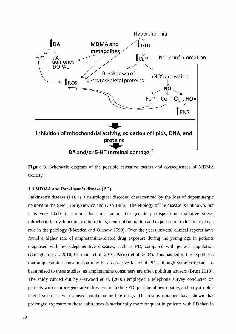
Figure 3. Schematic diagram of the possible causative factors and consequences of MDMA
toxicity.
1.3 MDMA and Parkinson’s disease (PD)
Parkinson's disease (PD) is a neurological disorder, characterized by the loss of dopaminergic
neurons in the SNc (Hornykiewicz and Kish 1986). The etiology of the disease is unknown, but
it is very likely that more than one factor, like genetic predisposition, oxidative stress,
mitochondrial dysfunction, excitotoxicity, neuroinflammation and exposure to toxins, may play a
role in the patology (Marsden and Olanow 1998). Over the years, several clinical reports have
found a higher rate of amphetamine-related drug exposure during the young age in patients
diagnosed with neurodegenerative diseases, such as PD, compared with general population
(Callaghan et al. 2010; Christine et al. 2010; Parrott et al. 2004). This has led to the hypothesis
that amphetamine consumption may be a causative factor of PD, although some criticism has
been raised to these studies, as amphetamine consumers are often polidrug abusers (Brust 2010).
The study carried out by Garwood et al. (2006) employed a telephone survey conducted on
patients with neurodegenerative diseases, including PD, peripheral neuropathy, and amyotrophic
lateral sclerosis, who abused amphetamine-like drugs. The results obtained have shown that
prolonged exposure to these substances is statistically more frequent in patients with PD than in
24
patients suffering from other neurodegenerative diseases; moreover, exposure to amphetamine
occurred long before diagnosis in a significant percentage of individuals.
The causes of the dopaminergic cell loss in the SNc that underlies PD patients are not clear, but
in the last decade solid evidence has linked it to intense neuroinflammation. PET studies
revealed microglial activation in pons, basal ganglia including substantia nigra (SN), frontal and
temporal cortical regions of individuals with PD, starting early in the disease process without
significant longitudinal changes (Gerhard et al. 2006; McGeer et al. 1988; Ouchi et al. 2005).
Further supporting a pathogenic role of neuroinflammation, post-mortem samples taken from PD
patients have also shown infiltration of CD4+ and CD8+ T cells in the brain (Brochard et al.
2009). However, despite the progress made so far, the fundamental question remains whether
immune-associated mechanisms are the main cause of the progressive loss of dopaminergic
neurons, or are rather a consequence of the dopaminergic neurons.
Even though the typical symptoms of PD are motor (Fearnley and Lees 1991), non motor
symptoms, including cognitive deficits, are increasingly being recognized in PD patients
(Foltynie et al. 2004), and no studies have evaluated the influence of the exposure to exogenous
substances (including amphetamine-related drugs) early in life on the onset of these symptoms.
1.4 MDMA and the nigrostriatal system in humans and experimental animals
The nigrostriatal system connects the SN to the striatum. It is one of the four DA pathways in the
brain, and is usually more envisioned as involved in the production of movement, although fMRI
and PET studies indicate that this pathway in the healthy modulates non-motor behavior and
cognition (Cropley et al. 2006; Landau et al. 2009).
Despite the continuously increasing number of studies that are focused on MDMA-induced
toxicity in the mice nigrostriatal system, few studies have addressed this issue in humans (Tai et
al. 2011). As mentioned before, MDMA mostly affects the dopaminergic system in mice, rather
than the serotonergic system (O’Callaghan and Miller 1994). MDMA administration depletes DA
and its metabolites in several brain regions, but in particular produces long-term degeneration of
striatal DA nerve terminals (Brodkin et al. 1993; Colado et al. 2001; Izco et al. 2007). A
significant contribute to the elucidation of the effects of MDMA in the nigrostriatal system of
mice comes from a recent study by Granado and co-workers (2008a), which shows that not all
the dopaminergic systems are affected, because they found no TH or DAT fiber loss in the NAc,
indicating that the dopaminergic neurotoxicity of MDMA is selective for the nigrostriatal
pathway. In a later study, the same group demonstrated that the decrease in TH levels expression
in mice treated with MDMA is accompanied by a reduction in DAT, which is considered an
important marker of functional dopaminergic nerve terminals, suggesting that MDMA does not

25
reduce TH synthesis, but rather damages dopaminergic terminals, with an effect that appears
more pronounced in the striosomal compartment than in the matrix (Granado et al. 2008b).
Remarkably, a similar pattern of striosomal damage in the striatum has been observed following
the administration of dopaminergic neurotoxins such as 1-methyl-4-phenyl-1,2,3,6-
tetrahydropyridine (MPTP) (Iravani et al. 2005), and confirms earlier studies which described
dopaminergic terminal loss in the mouse striatum following MDMA administration (Fornai et al.
2004).
1.5 MDMA and the limbic system in humans and experimental animals
The limbic system includes several brain areas such as the limbic cortex, hippocampus,
parahippocampal gyrus, limbic midbrain areas, amygdala, anterior thalamic nuclei, fornix,
column of fornix, mamillary body, septum pellucidum, habenular commisure, cingulate gyrus,
and the olfactory bulbs. The hippocampus, which plays a prominent role in the consolidation of
information from short-term memory to long-term memory and spatial navigation (Eichenbaum
et al. 1996; Squire 1992), contains two main interlocking parts, where can be identified two
major neuronal types: in the Cornu Ammonis the predominant neuronal type has a pyramidal
morphology, whereas in the dentate gyrus, the predominant neuronal type is the granular cells.
Moreover, in both the two areas, it has been identified tyrosine hydroxylase (TH)-labeled
terminals and GABA-containing terminals (Bentivoglio and Morelli 2005; Milner and Bacon
1989).
As mentioned earlier, cognitive deficits are among the typical long-term effects observed in
heavy MDMA users. These deficits depend on the effects of MDMA on the hippocampus, seem
to be dose-related, and may vary according to complexity of the cognitive task.
Fox and colleagues (2001) have demonstrated that both medium and high MDMA users display
cognitive deficits that depend on drug dosage but are not related to the extent of drug use.
Previous studies have also associated consumption of MDMA, considered as a whole, with
cognitive problems (Bolla et al. 1998; Parrott and Laskey 1998), whilst other have shown that
impaired cognitive performance may depend on both the amount of MDMA consumed per
session and the duration of MDMA use (Morgan 1999). Moreover, Brown and collaborators
(2010) have demonstrated that memory tasks with high complexity reveal more marked deficits
in MDMA users than memory tasks with a low complexity. Finally, PET studies have displayed
differences in the activation of the hippocampus between MDMA users and healthy controls
during working memory tasks (Becker et al. 2013; Daumann 2004 and 2005; Moeller et al.
2004).

26
Several studies have also described MDMA-induced learning and cognitive deficits in mice.
Thus, acute and repeated pretreatment with MDMA modifies the acquisition and execution of an
active avoidance task (Trigo et al. 2008). Moreover, repeated administration of MDMA at
different doses is associated with deficits in the Morris water maze that persist even long after
last MDMA administration (Busceti et al. 2008). Interestingly, the same doses of MDMA that
induced spatial learning deficits in the Morris water maze were able to induce tau protein
phosphorylation in the hippocampus, a biochemical hallmark of Alzheimer’s disease,
frontotemporal dementia, and other chronic neurodegenerative disorders characterized by a
progressive cognitive decline (Alonso et al. 2001; Goedert 1993).
1.6 MDMA and the PFC in humans and experimental animals
The ascending mesostriatal and mesocortical dopaminergic systems widely modulate the
cognitive functions supported by the PFC (Dalley et al. 2004; Ragozzino et al. 1999), a brain
area enriched with dopaminergic terminals (Bentivoglio and Morelli 2005), and the striatal
structures associated with it (Passingham and Sakai 2004). Thus, a wealth of evidence collected
in drug abusers and individuals affected by various diseases, including PD patients (Bowen and
Davison 1975; Downes et al. 1989) indicates that an altered dopaminergic function is associated
with some of the cognitive impairments typically seen after PFC damage (Manes et al. 2002;
Rogers et al. 1999). Moreover, even though most of the dopaminergic afferents to the PFC arise
from the ventral tegmental area, some of them originate in the central area of the SNc (Albanese
et al. 1986; Maurice et al. 1999; Middleton and Strick 2002) which can be very relevant for the
changes in cognitive function associated with PD.
A PET study by McCann et al. (2008) showed that memory performance of recreational MDMA
users was inversely associated with SERT binding levels, in the dorso-lateral PFC, orbitofrontal
cortex, and parietal cortex. Moreover, a recent PET study has revealed glucose hypometabolism
in the PFC and parietal cortex of chronic MDMA and other drugs users, which correlates with
verbal learning and recall deficits (Bosch et al. 2013).
Consistent with these data are findings in rodents showing that chronic administration of
amphetamine-related drugs can induce enduring reductions in monoamine levels in the striatum
and PFC, and that, under at least some schedules of administration, neurotoxic and
neuroinflammatory effects can occur (Atkins et al. 2009; Ball and Slane 2012; Ramos et al.
2005).

27
2. MPTP
In the late 1970s, an analogue of the synthetic opioid meperidine (Demerol ®), with the chemical
name of 1-methyl-4-phenyl-4-propionpiperidine (MPPP), and with an effect comparable to that
induced by heroin, was discovered. A twenty-three years old graduate student, Barry Kidstone,
set up a home laboratory to synthesize MPPP, but after four injections of what he thought to be
MPPP, he experienced severe bradykinesia (Langston and Ballard 1983). Furthermore, similar to
patients with idiopathic PD, he responded to treatment with L-3,4-dihydroxyphenylalanine (L-
DOPA) and developed the same complications associated with L-DOPA therapy, including motor
fluctuations and dyskinesias (Langston and Ballard 1983). Investigations were then conducted to
clarify the etiology of his condition which found that MPPP was contaminated with MPTP, a
byproduct of the synthetic reaction, which was later identified as the responsible of the
degeneration of dopaminergic neurons in the SNc (Langston et al. 1983).
After this first documented episode, others young drug addicts developed an idiopathic
parkinsonian syndrome after intravenous (i.v.) self-administration of MPPP (Ballard et al. 1985).
Since its discovery, MPTP has been widely used to create animal models of PD in a variety of
species (Jakowec and Petzinger 2004; Kopin 1987; Kurosaki et al. 2004), though the most used
species are currently the non-human primates and the mouse. Susceptibility to MPTP varies
across species, strain and age of animals (Giovanni et al. 1994a and b). Non-human primates are
the species most sensitive and rats the lowest sensitive to MPTP neurotoxicity, whereas mouse
strains widely vary in their sensitivity to the toxin, with the C57BL/6J being the most susceptible
(Hamre et al. 1999; Sedelis et al. 2000). MPTP neurotoxicity has been found to be strongly age-
dependent in all the species (Date et al. 1990; Irwin et al. 1992; Ovadia et al. 1995; Ricaurte et
al. 1986). Rats have been generally not used for modeling PD with systemic MPTP
administration like in other species, since the dosage required to induce a significant
dopaminergic degeneration is associated with a high mortality rate (Giovanni et al. 1994a and b).
However, stereotaxic injection of the toxic metabolite 1-methyl-4-phenylpyridinium (MPP+) has
been used to model PD in rats (Heikkila et al. 1985a).
2.1 Toxicology
2.1.1 Toxicology in humans
Administration of MPTP to humans and experimental animals causes the degeneration of
dopaminergic neurons in SNc and the depletion of DA in striatum. The severity of MPTP-
induced lesion depends on the regimen and route of administration, and on the species
considered.
28
With the exception of a single report (Ballard et al. 1985), all human cases of MPTP intoxication
were caused by one, or few repeated administrations of the toxin (Langston 1987). A
neuropathologic study of the brains of three MPTP-exposed addicts has revealed another
important similarity with idiopathic PD (Langston et al. 1999), that is the loss of dopaminergic
neurons restricted to the SNc, though not accompanied by Lewy bodies. The absence of Lewy
bodies may be due to the young age at the onset of MPTP-induced parkinsonism, since age may
be an important factor for development of the Lewy bodies (Gibb and Lees 1988). Moreover,
PET studies using [18F]-DOPA have revealed that MPTP-intoxicated individuals display a
severely reduced DA uptake similar to that of late-stage idiopathic PD (Calne et al. 1985; Snow
et al. 2000; Vingerhoets et al. 1994). Finally, the depletion of nigral dopaminergic neurons was
found to be consistently present together with gliosis and clustering of microglia around nerve
cells (Langston et al. 1999).
2.1.2 Toxicology in experimental animals
Non-human primates
Non-human primate models closely resemble the behavioral and neuroanatomical features of
human PD and this species may be useful for exploring the neurological and pathological
mechanisms of the disease (Fox and Brotchie 2010). Moreover, and similar to humans, non-
human primates are susceptible to doses of MPTP lower than those that cause a nigrostriatal
lesion in other species (Johannessen et al. 1985). Similar to humans, PET studies in cynomolgus
and rhesus monkeys treated with MPTP have revealed a severe reduction in DA uptake (Pate et
al. 1993). Thus, Moratalla and co-workers (1992), have reported that MPTP treatment in squirrel
monkeys induces a higher loss of dopaminergic uptake-site binding in the putamen than in the
caudatus, especially posteriorly. Moreover, Iravani and collaborators (2005) showed that
subacute MPTP treatement is associated with a greater damage in striosomes than in matrix
within the caudate nucleus. Administration of MPTP to non-human primates causes several
parkinsonian-like symptoms, including bradykinesia, postural instability, freezing, stooped
posture, and rigidity (Porras et al. 2012). These symptoms may be also accompanied by
cognitive impairment manifested in several tasks and reflecting a general impairment of
attentional and executive functions (Decamp et al. 2004; Decamp and Schneider 2004 and 2006;
Schneider and Kovelowski 1990; Schneider and Roeltgen 1993). Interestingly, MPTP-treated
primates also suffer a dramatic disruption of sleep–wake architecture, with reduced sleep
efficacy that persisted years after MPTP administration (Porras et al. 2012), reminiscent of what
observed in idiopatic PD patients (Comella 2008). Similar to idiopatic PD patients, MPTP-
29
lesioned non-human primates respond to anti-parkinsonian therapies such as L-DOPA and DA
receptor agonists. The degree of clinical response to L-DOPA is dependent on the severity of the
lesion and parkinsonian state.
Mice
To overcome the economic and moral limitations of experiments involving MPTP use in non-
human primates, a MPTP model has been developed in mice. The C57BL/6J strain, in particular,
is very susceptible to MPTP neurotoxicity, making an excellent conventional preclinical model
for PD. In general, MPTP is administered to mice in either an acute or subchronic regimen
(Heikkila et al. 1984; Sonsalla and Heikkila 1986). In these models, MPTP can produce death of
dopaminergic neurons in SNc by at least the 40% in C57BL/6J mice and significant depletions in
the striatal level of DA and its metabolites, 3,4-dihydroxyphenylacetic acid (DOPAC) and
homovanillic acid (HVA), along with a reduction in the striatal synaptosomal DA uptake
(Ricaurte et al. 1986). However, if the survival time of mice is extended, the neurotoxic effects
of MPTP may be reversible (Hallman et al. 1985). MPTP administration in mice induce a
neuroinflammatory effect in the SNc, striatum (Czlonkowska et al. 1996; Kohutnicka et al. 1998;
Kurkowska-Jastrzebska et al. 1999; Kurosaki et al. 2003), and hippocampus (Luellen et al.
2003). Bradykinesia, akinesia, altered balance and other motor features can be observed in
MPTP-treated mice through various behavioral analyses (Fleming et al. 2013; Sedelis et al.
2001; Tillerson et al. 2002) although, despite the evidence of DA reductions, mice that receive
MPTP acutely do not always exhibit motor dysfunctions or motor abnormalities (Heikkila et al.
1989; Meredith and Rademacher 2011). Whole-body tremor and postural abnormalities also have
been reported, but chiefly in the first day after lesioning (Sedelis et al. 2001). Cognitive deficits
after MPTP exposure in mice parallel the findings in non-human primates, even though with a
much less effect (Tanila et al. 1998). In general, all these behavioral alterations tend to be highly
variable, with some mice developing severe deficits whereas others exhibit little or no behavioral
changes (Sedelis et al. 2001). This variability may be due to a number of factors, including the
dose of MPTP, the mouse strain, the test sensitivity, and the period elapsed between lesioning
and evaluation, when recovery may occur.
2.2 Pharmacological and toxicological mechanisms
The mechanism of MPTP toxicity is quite similar among humans, non-human primates and
mice. MPTP is a highly lipophilic protoxin which rapidly crosses the BBB after systemic
administration. Subsequently, MPTP is bioactivated by MAO type B enzymes to the unstable
intermediate 1-methyl-4-phenyl-2,3-dihydropyridium (MPDP+), exclusively in non-
30
dopaminergic cells, especially in astrocytes (Ekblom et al. 1993). MPDP+ spontaneously
oxidizes to MPP+ at least in vitro (Chiba et al. 1985; Fritz et al. 1985), whereas it is not clear if
this reaction may occur in vivo. Another mechanism for MPDP+ oxidation to MPP+ involves
HO● radicals (Castagnoli et al. 1985), which appears in line with the evidence showing that
transgenic mice expressing high levels of superoxide dismutase are resistant to MPTP
(Przedborski et al. 1992), and that dopaminergic neurons are very susceptible to the toxin. Once
generated, MPP+ is released into the extracellular space by a mechanism that is still unknown.
MPP+ is not able to enter dopaminergic neurons freely, and its uptake depends on active plasma
membrane carrier systems, such as the DAT, for which MPP+ bears a high affinity (Chiba et al.
1985; Heikkila et al. 1985b). Interestingly, DAT is an absolute requirement for MPTP-induced
neurotoxicity, since this effect is abolished in transgenic mice lacking this transporter (Bezard et
al. 1999; Gainetdinov et al. 1997).
Once inside the cell, MPP+ can either be sequestered into synaptic vesicles via VMAT2 (Del
Zompo et al. 1993; Wimalasena et al. 2008) or enter into the mitochondria where it interferes
with the mitochondrial complex I (Mizuno et al. 1987; Richardson et al. 2007). Both of these
steps have been implicated in processes that either protect or kill the dopaminergic neurons. Staal
and co-workers (2000) have demonstrated that rat striatal vesicles have a higher density of
VMAT 2 and a greater ability to sequester MPP+, compared with those isolated from mice, thus
justifying why rats are less sensitive to MPP+-induced toxicity. The primary reason for the
neuroprotective effect of vesicular MPP+ sequestration is that less MPP+ can be accumulated into
mitochondria, thus reducing mitochondrial damage.
Regarding the metabolism of MPTP, three major specie-related differences have been identified:
1) non-human primates, but not rodents, show a persistently high concentration of MPTP
metabolites in the striatum, compared to other brain regions; 2) the rodent brain clears MPTP and
its metabolites much more rapidly than what observed in non-human primates; 3) the
predominant metabolite retained by the non-human primates brain is MPP+, while MPP+ cannot
be detected in rodent brain for more than a few hours after injection (Johannessen et al. 1985).
The presence of CYP2D6 in human and nonhuman primates and of the similar isoform CYP2D1
in rat brain indicates that this subfamily of CYP enzymes may be responsible for MPTP
metabolism (Herraiz et al. 2006; Mann and Tyndale 2010). The persistence of MPP+ in the non-
human primate brain may explain the heightened toxicity of MPTP in this species. The principal
route of elimination of MPTP is through the urine (Lau et al. 1988).
The ability to interfere with mitochondrial respiration at the level of complex I is a key
mechanism in the toxic effects of MPP+ (Nicklas et al. 1985; Suzuki et al. 1990). Importantly, the
31
cytotoxic effects of MPP+ are more marked in cells that are particularly sensitive to a deficiency
in aerobic energy metabolism, a condition that applies to dopaminergic neurons, which have
been found to be more vulnerable to inhibition of oxidative phosphorylation than other types of
neurons (Marey-Semper et al. 1993 and 1995). However, the alterations in energy metabolism
and generation of ROS peak within hours from MPTP administration, long before the occurrence
of overt neuronal damage (Jackson-Lewis et al. 1995). Therefore, these events appear not to be a
primary cause of neuronal death, but to initiate a cascade of events that eventually kill
dopaminergic neurons (Gibson et al. 2010; Serra et al. 2002). One controversial aspect is
whether MPTP induces neuronal death via apoptosis or necrosis. Przedborski and Jackson-Lewis
(1998) have proposed that subchronic administration of MPTP is most likely to cause apoptosis,
whereas necrotic cell death would be triggered by acute dosing. In this regard, it has however to
be definitively clarified if during neurodegenerative conditions, such as PD, neurons degenerate
by apoptosis or necrosis (Anglade et al. 1997; Kösel et al. 1997).
Further contributions to clarify the mechanisms of MPTP-mediated neurotoxicity apparently
come from NO (Przedborski et al. 1996), the induction of GLU-mediated excitotoxicity, and the
stimulation of neuroinflammation. Some studies have also suggested that MPP+ may act
independently of inhibition of mitochondrial complex I (Choi et al. 2008; Gerlach et al. 1996).
Decline in ATP production and increase in ROS levels
As mentioned before, MPP+ blocks mitochondrial complex I (Nicklas et al. 1985; Suzuki et al.
1990), leading to a decline in adenosine triphosphate (ATP) levels. There is evidence of a good
correlation between MPP+ concentrations, decline in ATP synthesis and cell death in hepatoma
cells (Sandy et al. 1988). Furthermore, MPP+ has been shown to affect complex III (Mizuno et
al. 1988), which leads to an increased production of ROS, HO● and O2- in particular.
In vitro experiments have shown that the interaction between MPP+ and the complex I may also
induce the formation of ROS, and in particular HO● and O2- (Cleeter et al. 1992; Sriram et al.
1997). Interestingly, investigations in humans have found a decrease in complex I activity in
various diseases, including PD (Lin and Beal 2006; Mizuno et al. 1989). Thus, low activity of
complex I may translate to increased production of ROS, a depletion of SOD, and eventually an
increased oxidative stress within the DA neuron. This idea is supported by experiments showing
that, as long as sufficient stores of SOD are present, mice are protected against the effects of
MPTP (Klivenyi et al. 1998). Moreover, neuronal stress could arise from a perturbation of DA
homeostasis, as MPTP-induced DA release from citoplasmatic vesicular storage may trigger the
formation of oxidizing metabolites and by-products, such as DA quinones and ROS (Graham
1978). In line with this, protection of neuronal damage by MPP+ has been achieved in cell
32
cultures treated with antioxidants (Perry et al. 1985), and resistance to MPP+ neurotoxicity has
been demonstrated in SOD transgenic mice (Cadet et al. 1995; Przedborski et al. 1992).
Nitration and oxidative stress
Immunohistochemical studies (Dehmer et al. 2000; Liberatore et al. 1999) show that iNOS up-
regulation occurs in microglia 24 hours after the administration of MPTP, an effect that lead to
an increase in the production of NO and, subsequently, of OONO-. Both NO and OONO- may
interfere with mitochondrial function (Cleeter et al. 1994; Radi et al. 1994), thus adding to the
toxic effect of MPP+ on cellular energy supplies. Evidence for a role of NO in these effects
comes from studies showing that inhibition of NOS can attenuate MPTP neurotoxicity in non-
human primates (Hantraye et al. 1996) and mice (Przedborski et al. 1996; Smith et al. 1994).
An excitotoxic mechanism for MPTP neurotoxicity has also been proposed, based initially on the
observation that intrastriatal administration of MPP+ to rats can lead to a marked increase in
extracellular GLU and aspartate levels (Carboni et al. 1990). Subsequently, the neuroprotective
effects of excitatory amino acid receptor antagonists have been described (Turski et al. 1991;
Zuddas et al. 1992). Furthermore, overstimulation of GLU receptors can alter local Ca++
homeostasis, and Ca++ is known to up-regulate enzymes like phospholipase A2, nNOS and
xanthine oxidase, all of which are found in mitochondria and can elevate ROS production,
eventually promoting mitochondrial dysfunction.
Neuroinflammation
The idea that glia may participate in MPTP-induced neurotoxicity is supported by findings
showing that toxins activate microglia and promote the production of toxic compounds, and that
astroglia is the major site where MPTP is metabolized to MPP+.
After the discovery of the presence of gliosis in post-mortem brains from MPTP-lesioned
addicts, similar findings were later observed in the SNc of non-human primates (Barcia et al.
2004; McGeer et al. 2003) and mice (Czlonkowska et al. 1996; Kohutnicka et al. 1998;
Kurkowska-Jastrzebska et al. 1999). The inflammation discovered in humans and experimental
animals exposed to MPTP may be related to activated microglial cells, that express inflammatory
mediators such NO and cytokines (IL-1β and IL-6), as well as ROS, TNF-α, GLU, and
arachidonic acid metabolites, which are toxic to dopaminergic neurons (Hanisch 2002). In line
with this, an increase in both glial cells expressing iNOS and levels of proteins that contain nitro-
tyrosine have been detected in autoptic samples from the SNc of patients with idiopatic PD
(Hunot et al. 1996). In this regard, Hirsch and Hunot (2000) suggested that MPTP acts directly
on the induction of cytokines that activate iNOS, although elevation in striatal IL-1β and IL-6
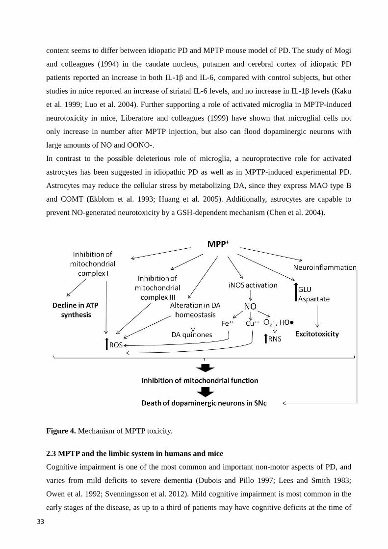
33
content seems to differ between idiopatic PD and MPTP mouse model of PD. The study of Mogi
and colleagues (1994) in the caudate nucleus, putamen and cerebral cortex of idiopatic PD
patients reported an increase in both IL-1β and IL-6, compared with control subjects, but other
studies in mice reported an increase of striatal IL-6 levels, and no increase in IL-1β levels (Kaku
et al. 1999; Luo et al. 2004). Further supporting a role of activated microglia in MPTP-induced
neurotoxicity in mice, Liberatore and colleagues (1999) have shown that microglial cells not
only increase in number after MPTP injection, but also can flood dopaminergic neurons with
large amounts of NO and OONO-.
In contrast to the possible deleterious role of microglia, a neuroprotective role for activated
astrocytes has been suggested in idiopathic PD as well as in MPTP-induced experimental PD.
Astrocytes may reduce the cellular stress by metabolizing DA, since they express MAO type B
and COMT (Ekblom et al. 1993; Huang et al. 2005). Additionally, astrocytes are capable to
prevent NO-generated neurotoxicity by a GSH-dependent mechanism (Chen et al. 2004).
Figure 4. Mechanism of MPTP toxicity.
2.3 MPTP and the limbic system in humans and mice
Cognitive impairment is one of the most common and important non-motor aspects of PD, and
varies from mild deficits to severe dementia (Dubois and Pillo 1997; Lees and Smith 1983;
Owen et al. 1992; Svenningsson et al. 2012). Mild cognitive impairment is most common in the
early stages of the disease, as up to a third of patients may have cognitive deficits at the time of
34
their diagnosis (Foltynie et al. 2004). Nigrostriatal dysfunction alone is probably not sufficient
for the development of cognitive deficits in PD patients, and it is interesting to remark that
activated microglia have been observed not only in the SNc and putamen, where DA loss is
prominent, but also in the hippocampus of PD patients (McGeer et al. 1988 and 2001; McGeer
and McGeer 2004). As mentioned earlier, activated microglia produce a variety of inflammatory
cytokines (Allan and Rothwell 2001), which have been implicated in cognitive deficits in PD
(Menza et al. 2010; Wilson et al. 2002). In addition, supporting a link between IL and cognitive
performance, it is noteworthy that IL-2 immunotherapy of cancer patients is associated with
pronounced cognitive disturbances (Caraceni et al. 1993; Denicoff et al. 1987), particularly in
tests involving spatial learning and memory (Caraceni et al. 1993).
Data suggesting a link between neuroinflammation and cognitive deficits have also been
obtained in mice, where MPTP treatment induces dopaminergic neurons loss in the SNc paired
with a great inflammatory response (Frau et al. 2011; Miller et al. 2011) and cognitive deficits
(Dluzen and Kreutzberg 1993; Deguil et al. 2010; Tanila et al. 1998). The increase of IL-6 in
areas such as the hippocampus and hypothalamus induced by MPTP may be related to
parkinsonian-like cognitive deficits (Wang et al. 2009), however, may be not the only
responsible. Since the mechanism of cognitive deficits in PD has not been elucidated yet, the
degree of striatal DA loss, the extent of neuroinflammation, as well as modifications in other
brain areas related to cognitive processes may all potentially be involved and combine to elicit
cognitive deficits observed in MPTP-treated mice.
2.4 MPTP and the PFC in humans and experimental animals
In idiopathic PD patients, abnormal cortical metabolic activity occurs in both the motor and non-
motor regions (Eidelberg et al. 1994; Huang et al. 2007; Ridding et al. 1995). An hypothesis is
that such changes could index the non-motor symptoms of PD thought to precede the more
classic motor manifestations, such as depression, cognitive deficits and apathy (Cooper et al.
1991; Shen et al. 2013), and which involve brain areas, including the PFC, where was found a
reduced DA content following treatment with dopaminergic neurotoxicant (Tadaiesky et al.
2008). Supporting a role of PFC in MPTP-induced cognitive deficits in experimental animals, a
study by Storvik and co-workers (2010) has shown that specific transcriptomic changes occurr in
the PFC of parkinsonian MPTP-treated macaques, and that a large part of these changes may also
be observed in human post-mortem samples from patients with neurodegenerative diseases, such
as PD and Alzheimer disease, analysed by quantitative PCR. Similar results were obtained with a
single intranasal infusion of MPTP in rats and mice, which produces progressive signs of PD
35
such as impairments in olfactory, cognitive, and motor functions, associated with a decrease in
the levels of DA in the olfactory bulb, striatum, and PFC (Prediger et al. 2010).
36
Aims of the study
On the basis of epidemiological studies showing a higher incidence of PD in amphetamine-
related drugs consumers, the present study evaluated in mice treated as adolescent with MDMA,
and administered after MDMA washout with MPTP, the neuroinflammatory and neurotoxic
effects in motor brain areas and the neuroinflammatory effects in non-motor brain areas.
Moreover, cognitive deficits were evaluated by memory tasks.
The first aim of this study was to assess whether administration of MDMA to late adolescent
mice may facilitate the neuroinflammatory and neurotoxic effects of 1-methyl-4-phenyl-1,2,3,6-
tetrahydropyridine (MPTP), in the nigrostriatal system of adult mice. Studies on the effects of
amphetamine-related drugs in an experimental model of PD may, in fact, not only help to
understand whether there is an increased risk of developing PD associated with the use of these
substances during adolescence, but also clarify some of the mechanisms that promote the
degeneration of DA neurons.
To pursue this first aim, the following experiments were carried out:
•••• Assessment of inflammatory response in SNc and striatum studied through
immunohistochemical analysis of GFAP and CD11b, as markers of astroglia and
microglia.
•••• Assessment of dopaminergic neuronal damage in SNc and striatum studied by TH
immunohistochemistry.
The second aim of this study was to elucidate whether MDMA may amplify the
neuroinflammatory effect elicited by MPTP in the hippocampus and in the mPFC. In the attempt
to link neurochemical changes with behavioral modifications, this study investigated whether
cognitive impairment took place in mice treated with MDMA, MPTP, and their combination.
To pursue this second aim, the following experiments were carried out:
•••• Assessment of inflammatory response in hippocampus and mPFC studied through
immunohistochemical analysis of GFAP and CD11b.
•••• Assessment of NOR task and spontaneous alternation behavior in a Y-maze, which were
performed both throughout the MDMA treatment and after MPTP administration.

37
Materials and methods
4.1 Animals
Male C57BL/6J mice (Charles River, Calco, Italy) weighing 20–23 g at the beginning of the
experiments, were used. Mice were maintained at constant temperature (21±1°C) under a 12-h
light/dark cycle with food and water ad libitum. Experimental procedures were approved by the
Ethical Committee of the University of Cagliari, in compliance with Italian guidelines for care
and use of experimental animals (DL 116/92) and European Communities Council Directive
(2010/63/EEC). Efforts were made to minimize the number of animals used and maximize
humane treatment.
4.2 Drugs
MDMA was synthesized at the Department of Environmental and Life Sciences of the University
of Cagliari (Frau et al. 2013). MPTP was purchased from Santa Cruz Biotechnology, CA, USA.
MDMA was dissolved in saline, whereas MPTP was dissolved in distilled water. Both drugs
were administered intraperitoneally (i.p.) in a volume of 10 ml/kg.
4.3 Treatment
Late adolescent mice (8 weeks old) were treated with either MDMA (10 mg/kg i.p.) or vehicle
according to a 9-week administration schedule (Fig. 5). In detail, each mouse received two
administrations a day, separated by a 4–6-hour interval, twice a week, on the second and fifth
day of each week, for a total of 36 MDMA, or vehicle, administrations. Experimental groups
were composed as follows: MDMA-treated mice, n=18-19; vehicle-treated mice n=17-18.
During MDMA treatment, mice performance in the NOR was evaluated at the following time
points: week 1, week 4, and week 9 and then re-assessed two weeks after MDMA
discontinuation. NOR performance was always measured on the sixth day of the week. The same
mice were also evaluated for spontaneous alternation behavior in a Y-maze, 24 h after NOR
(seventh day of the week) at the same time points indicated above. Two weeks after MDMA
discontinuation, mice (19 weeks old) were distributed into four groups and challenged with
either MPTP (20 mg/kg, i.p.) or vehicle, once a day for 4 consecutive days. Experimental groups
were composed as follows: vehicle+vehicle, vehicle+MPTP, MDMA+vehicle, MDMA+MPTP
(n=5-10). Mice were evaluated again for their NOR performance and spontaneous alternation in
a Y-maze, respectively after 24 or 48 hours from the last MPTP or vehicle injection. The day
after the completion of behavioral experiments, mice were anesthetized with chloral hydrate,
transcardially perfused with 4% paraformaldehyde in phosphate buffer (0.1 M, pH 7.4), and their
brains were removed and processed for immunohistochemistry.
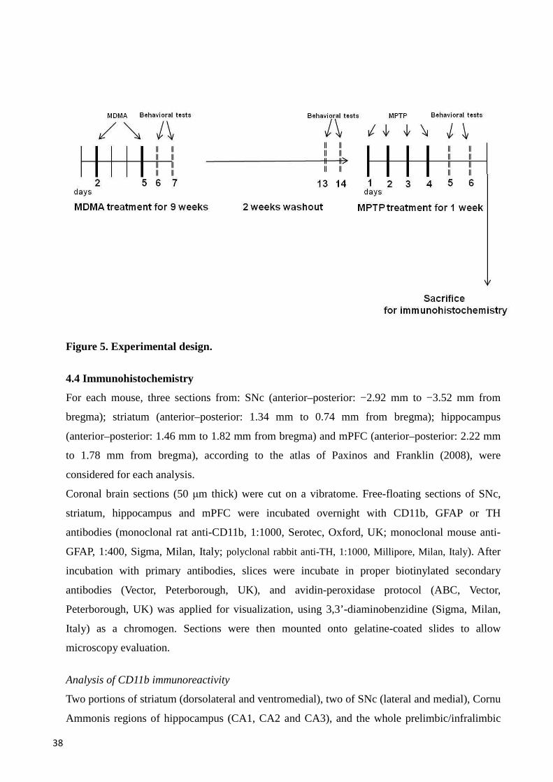
38
Figure 5. Experimental design.
4.4 Immunohistochemistry
For each mouse, three sections from: SNc (anterior–posterior: −2.92 mm to −3.52 mm from
bregma); striatum (anterior–posterior: 1.34 mm to 0.74 mm from bregma); hippocampus
(anterior–posterior: 1.46 mm to 1.82 mm from bregma) and mPFC (anterior–posterior: 2.22 mm
to 1.78 mm from bregma), according to the atlas of Paxinos and Franklin (2008), were
considered for each analysis.
Coronal brain sections (50 µm thick) were cut on a vibratome. Free-floating sections of SNc,
striatum, hippocampus and mPFC were incubated overnight with CD11b, GFAP or TH
antibodies (monoclonal rat anti-CD11b, 1:1000, Serotec, Oxford, UK; monoclonal mouse anti-
GFAP, 1:400, Sigma, Milan, Italy; polyclonal rabbit anti-TH, 1:1000, Millipore, Milan, Italy). After
incubation with primary antibodies, slices were incubate in proper biotinylated secondary
antibodies (Vector, Peterborough, UK), and avidin-peroxidase protocol (ABC, Vector,
Peterborough, UK) was applied for visualization, using 3,3’-diaminobenzidine (Sigma, Milan,
Italy) as a chromogen. Sections were then mounted onto gelatine-coated slides to allow
microscopy evaluation.
Analysis of CD11b immunoreactivity
Two portions of striatum (dorsolateral and ventromedial), two of SNc (lateral and medial), Cornu
Ammonis regions of hippocampus (CA1, CA2 and CA3), and the whole prelimbic/infralimbic

39
area of mPFC were analyzed for both hemispheres. Images were digitized with a video camera
(Pixelink® PL-A686, Ottawa, Canada), captured at ×20 real magnification in gray scale, and then
evaluated with the Scion Image analysis software (Scion Corp. Frederick, MD, USA). A
threshold, set above the mean value ± S.E.M. of the background, was applied for background
correction. The area occupied by values above the threshold was automatically calculated inside
each frame. No significant differences in CD11b levels were obtained between the two portions
of SNc, the two portions of striatum or the three regions of the hippocampus, therefore the data
obtained were pooled together. For each level of these areas, the obtained value was first
normalized with respect to vehicle, and values from different levels were averaged thereafter.
Analysis of GFAP immunoreactivity
For the striatum, the hippocampus and the mPFC, GFAP analysis was performed similarly to
what described for CD11b, whereas, for the SNc, the whole left and right area were analyzed.
Images were captured and evaluated with the Pixelink® software. Absolute number of GFAP-
positive cells was obtained separately for each level within each brain area, and no significant
differences in the number of GFAP-positive cells were observed among the two hemispheres of
SNc, the two portions of striatum or among the three regions of the hippocampus, therefore the
data obtained were pooled together. Thereafter, the number of cells × level from each mouse was
normalized with respect to vehicle, and individual values from the levels were averaged to
generate a mean value.
Stereological counting of TH-positive neurons
The total number (N) and density (D) of TH-positive neurons in SNc were measured
stereologically on both hemispheres using a microscope (Leica, DMLB, Ballerup, Denmark)
equipped with a projecting camera (Basler Vision Technologies, Ahrensburg, Germany) and a
microscope stage connected to an xyz stepper (PRIOR ProScan) controlled by a computer
equipped with new CAST Visiopharm software (Denmark).
Systemic uniform, random sampling was used to choose sections for analysis. The SNc region
was carefully outlined under lower magnification (×5) according to the atlas Paxinos and
Franklin (2008).
The number of labelled neurons was calculated under ×63 magnification using randomized
meander sampling and optical dissector methods. The cut thickness of sections was 50 µm and
the optical dissector height was 12 µm. The sampling area covered 20% of the region of interest.
The counting frame (8183, 4 µm2) applied the exclusion and inclusion lines. In each sampled

40
area, the dissector position was adjusted. For further details see Frau et al. (2011) and Sterio
(1984).
Analysis of TH-positive fibers
Images were digitized in gray scale, then TH-immunoreacted striata were captured at 20× real
magnification in gray scale and evaluated using the image analysis software Scion Image. The
average gray values from white matter were subtracted from each section to correct for
background immunoreactivity.
For each level of striatum, the obtained value was first normalized with respect to vehicle, and
values from different levels were averaged thereafter.
4.5 Memory tasks
NOR task
Evaluation of NOR performance is widely used for assessing non-spatial working memory in
rodents (Ennaceur and Delacour 1988). NOR experiments were performed in a Plexiglas cage
(length 25.5 cm, width 19 cm, height 14 cm) with the floor covered with sawdust. Objects to be
discriminated were made of plastic, and differed as to their shape and color. Moreover, objects
had no genuine significance, and had not been previously associated with either rewarding or
aversive stimuli. Experimental procedure consisted of three phases: habituation (S0), acquisition
(S1), and testing (S2). Habituation to the test cage (single 5 min trial) took place on the fifth day
of the week during MDMA treatment, and on the fourth day during MPTP treatment, 6 hours
after drug administration. Acquisition was performed the day after S0, by placing each mouse in
the test cage together with two copies of the same object (familiar objects). Mice were left to
freely explore the objects for 3 min. Testing phase took place 60 min after S1. Mice were
exposed to a copy of the objects already presented in S1, plus a copy of an object that they had
never experienced before (novel object).
Objects exploration was defined as the mouse sniffing, gnawing or touching the objects with the
nose, whereas sitting on and/or turning around the objects were not considered exploratory
behaviors. To avoid the presence of olfactory cues, objects were thoroughly cleaned after each
session. Moreover, the combination of objects (novel vs. familiar) and their respective locations
in the cage (right vs. left) were counterbalanced, to prevent biased preferences for particular
objects and/or locations. Mice performance was videotaped, and the following parameters were
evaluated: total amount of time spent by each mouse in exploring the objects during S1 and S2,
41
and percentage of time spent exploring the novel object over the total amount of time spent in
exploring both objects (novel and familiar) during S2 (Sawyer et al. 2012; Simola et al. 2008).
Spontaneous alternation behavior in a Y-maze
Evaluation of spontaneous alternation behavior in a Y-maze is widely utilized to investigate
spatial short-term memory and general cognitive function in rodents (Hughes 2004; Maurice et
al. 1994; Yamada et al, 1996). The apparatus used was made of black PVC, and had three equal
arms (length 40 cm, width 11 cm, height 20.5 cm). Arms converged onto a central triangular
area, and the maze had his floor covered with sawdust. To avoid the presence of olfactory cues,
the maze was thoroughly cleaned and the sawdust was replaced in between each trial. Mice were
individually placed in the central area, and left free to explore the whole apparatus for a single 8
min trial, during which their performance was videotaped. Percentage of spontaneous alternation
was calculated based on the sequence of arm entries, as reported elsewhere (Simola et al. 2008;
Yamada et al. 1996).
4.6 Statistics
Statistical analysis was performed with Statistica for Windows (StatSoft, Tulsa, OK, USA). The
statistical significance was assessed by Student’s t-test, one, or two-way analysis of variance
(ANOVA), followed by Newman–Keuls post-hoc test when appropriate. Three animals were
excluded from the analysis of behavioral tests, because they did not perform the tasks assigned.
Significance was set at p<0.05. Results are expressed as mean±S.E.M. for every analysis
performed.

42
Results
5.1 MDMA increases the vulnerability of mice to neuroinflammation and neurotoxicity
induced by MPTP in the SNc and striatum
5.1.1 Immunohistochemistry
CD11b immunoreactivity in SNc and striatum
In vehicle+vehicle treated mice, resting microglia and very low levels of CD11b were observed,
in both the SNc and the striatum. Similarly, mice exposed to chronic MDMA (10 mg/kg twice a
day/twice a week) followed by vehicle administration did not show significant microglia
activation, in both the SNc and the striatum. On the other hand, MPTP administration (20 mg/kg
× 4 administrations) to vehicle-pretreated mice elicited an elevation in microglia in the SNc,
compared to vehicle+vehicle, and a higher elevation in microglia in striatum, compared to
vehicle+vehicle and MDMA+vehicle treated mice. Finally, MPTP administration to MDMA-
pretreated mice elicited a higher microglia activation in both the SNc and striatum, compared to
all the other groups (Fig. 6).
In the SNc, one-way ANOVA showed a significant effect of treatment (F3,19=4.82, p<0.01), and
subsequent Newman-Keuls post-hoc test revealed that the levels of CD11b were higher in
vehicle+MPTP mice compared with vehicle+vehicle treated mice. Moreover, MPTP
administration in MDMA pretreated mice elicited a significantly higher microglia activation
compared with all the other experimental groups (Fig.6A).
In the striatum, one-way ANOVA showed a significant effect of treatment (F3,18=20.05, p<0.01),
and subsequent Newman-Keuls post-hoc test revealed that the levels of CD11b were higher in
vehicle+MPTP mice compared with vehicle+vehicle and MDMA+vehicle treated mice.
Moreover, MPTP administration in MDMA pretreated mice elicited a significantly higher
microglia activation compared with all the other experimental groups (Fig.6B).
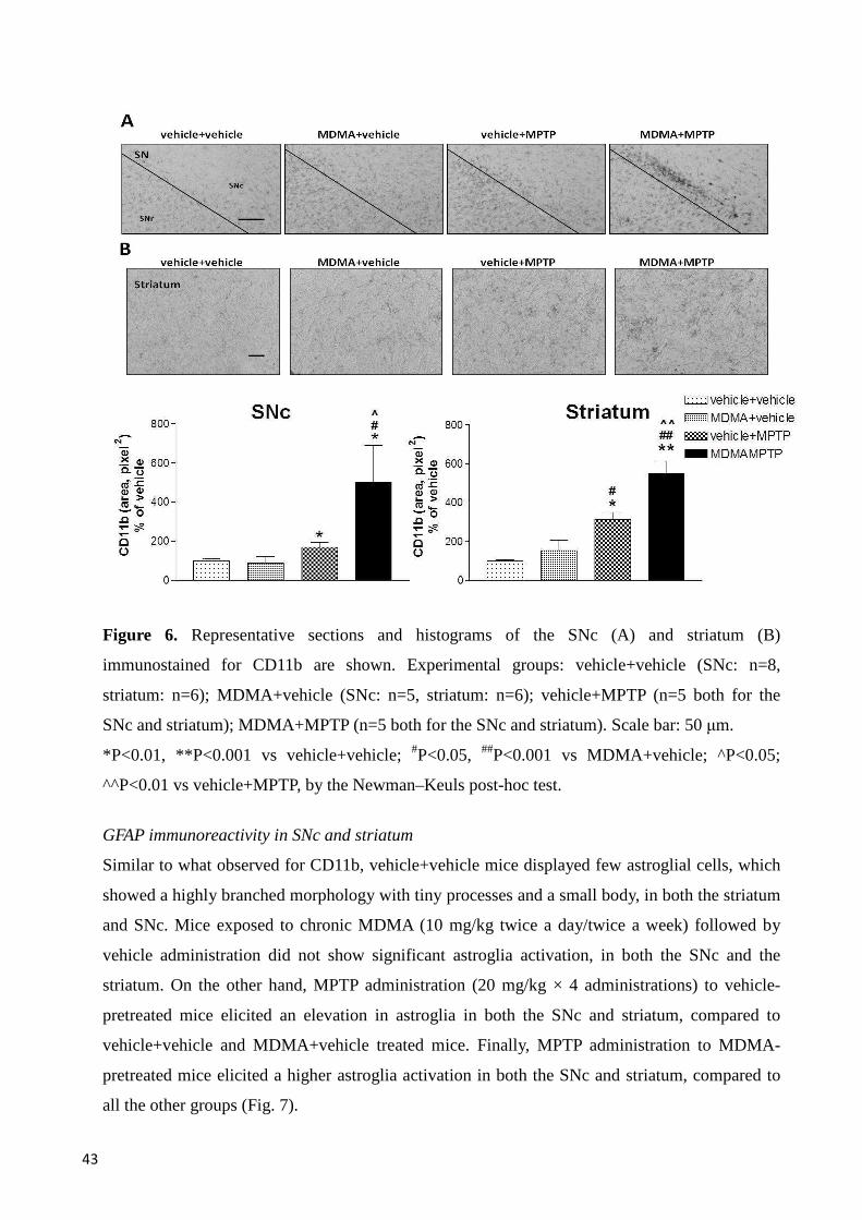
43
Figure 6. Representative sections and histograms of the SNc (A) and striatum (B)
immunostained for CD11b are shown. Experimental groups: vehicle+vehicle (SNc: n=8,
striatum: n=6); MDMA+vehicle (SNc: n=5, striatum: n=6); vehicle+MPTP (n=5 both for the
SNc and striatum); MDMA+MPTP (n=5 both for the SNc and striatum). Scale bar: 50 µm.
*P<0.01, **P<0.001 vs vehicle+vehicle; #P<0.05, ##P<0.001 vs MDMA+vehicle; ^P<0.05;
^^P<0.01 vs vehicle+MPTP, by the Newman–Keuls post-hoc test.
GFAP immunoreactivity in SNc and striatum
Similar to what observed for CD11b, vehicle+vehicle mice displayed few astroglial cells, which
showed a highly branched morphology with tiny processes and a small body, in both the striatum
and SNc. Mice exposed to chronic MDMA (10 mg/kg twice a day/twice a week) followed by
vehicle administration did not show significant astroglia activation, in both the SNc and the
striatum. On the other hand, MPTP administration (20 mg/kg × 4 administrations) to vehicle-
pretreated mice elicited an elevation in astroglia in both the SNc and striatum, compared to
vehicle+vehicle and MDMA+vehicle treated mice. Finally, MPTP administration to MDMA-
pretreated mice elicited a higher astroglia activation in both the SNc and striatum, compared to
all the other groups (Fig. 7).
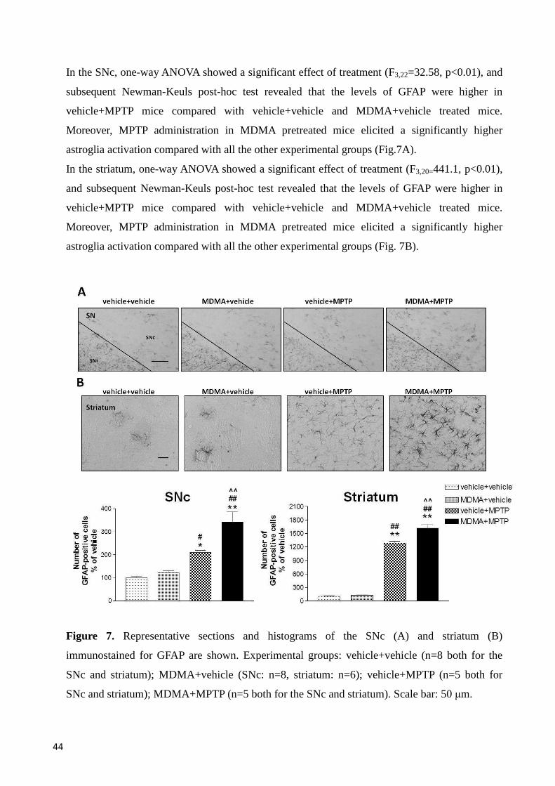
44
In the SNc, one-way ANOVA showed a significant effect of treatment (F3,22=32.58, p<0.01), and
subsequent Newman-Keuls post-hoc test revealed that the levels of GFAP were higher in
vehicle+MPTP mice compared with vehicle+vehicle and MDMA+vehicle treated mice.
Moreover, MPTP administration in MDMA pretreated mice elicited a significantly higher
astroglia activation compared with all the other experimental groups (Fig.7A).
In the striatum, one-way ANOVA showed a significant effect of treatment (F3,20=441.1, p<0.01),
and subsequent Newman-Keuls post-hoc test revealed that the levels of GFAP were higher in
vehicle+MPTP mice compared with vehicle+vehicle and MDMA+vehicle treated mice.
Moreover, MPTP administration in MDMA pretreated mice elicited a significantly higher
astroglia activation compared with all the other experimental groups (Fig. 7B).
Figure 7. Representative sections and histograms of the SNc (A) and striatum (B)
immunostained for GFAP are shown. Experimental groups: vehicle+vehicle (n=8 both for the
SNc and striatum); MDMA+vehicle (SNc: n=8, striatum: n=6); vehicle+MPTP (n=5 both for
SNc and striatum); MDMA+MPTP (n=5 both for the SNc and striatum). Scale bar: 50 µm.
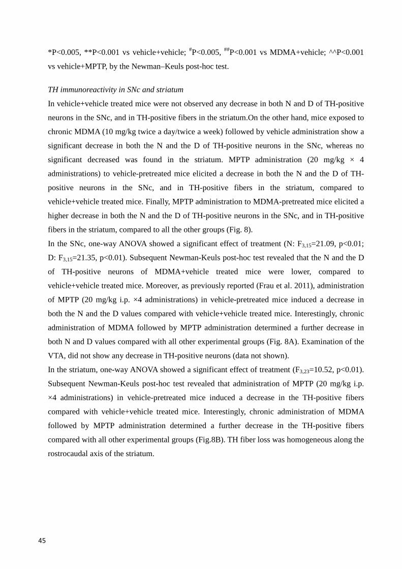
45
*P<0.005, **P<0.001 vs vehicle+vehicle; #P<0.005, ##P<0.001 vs MDMA+vehicle; ^^P<0.001
vs vehicle+MPTP, by the Newman–Keuls post-hoc test.
TH immunoreactivity in SNc and striatum
In vehicle+vehicle treated mice were not observed any decrease in both N and D of TH-positive
neurons in the SNc, and in TH-positive fibers in the striatum.On the other hand, mice exposed to
chronic MDMA (10 mg/kg twice a day/twice a week) followed by vehicle administration show a
significant decrease in both the N and the D of TH-positive neurons in the SNc, whereas no
significant decreased was found in the striatum. MPTP administration (20 mg/kg × 4
administrations) to vehicle-pretreated mice elicited a decrease in both the N and the D of TH-
positive neurons in the SNc, and in TH-positive fibers in the striatum, compared to
vehicle+vehicle treated mice. Finally, MPTP administration to MDMA-pretreated mice elicited a
higher decrease in both the N and the D of TH-positive neurons in the SNc, and in TH-positive
fibers in the striatum, compared to all the other groups (Fig. 8).
In the SNc, one-way ANOVA showed a significant effect of treatment (N: F3,15=21.09, p<0.01;
D: F3,15=21.35, p<0.01). Subsequent Newman-Keuls post-hoc test revealed that the N and the D
of TH-positive neurons of MDMA+vehicle treated mice were lower, compared to
vehicle+vehicle treated mice. Moreover, as previously reported (Frau et al. 2011), administration
of MPTP (20 mg/kg i.p. ×4 administrations) in vehicle-pretreated mice induced a decrease in
both the N and the D values compared with vehicle+vehicle treated mice. Interestingly, chronic
administration of MDMA followed by MPTP administration determined a further decrease in
both N and D values compared with all other experimental groups (Fig. 8A). Examination of the
VTA, did not show any decrease in TH-positive neurons (data not shown).
In the striatum, one-way ANOVA showed a significant effect of treatment (F3,23=10.52, p<0.01).
Subsequent Newman-Keuls post-hoc test revealed that administration of MPTP (20 mg/kg i.p.
×4 administrations) in vehicle-pretreated mice induced a decrease in the TH-positive fibers
compared with vehicle+vehicle treated mice. Interestingly, chronic administration of MDMA
followed by MPTP administration determined a further decrease in the TH-positive fibers
compared with all other experimental groups (Fig.8B). TH fiber loss was homogeneous along the
rostrocaudal axis of the striatum.
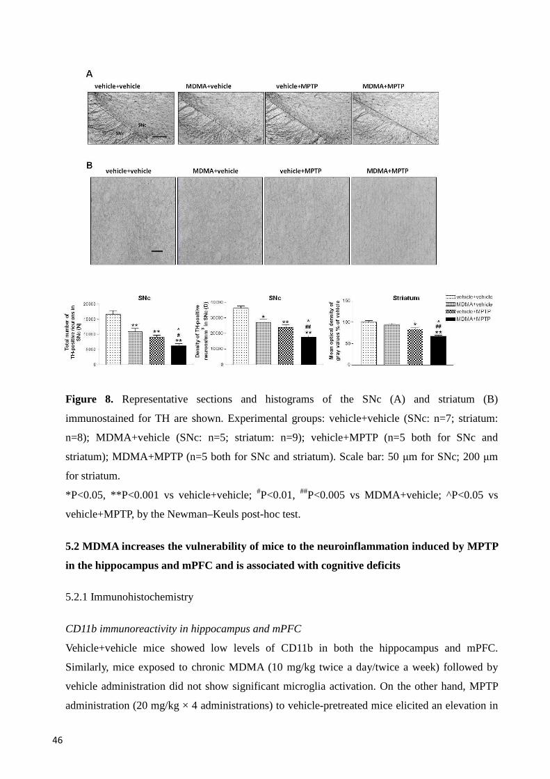
46
Figure 8. Representative sections and histograms of the SNc (A) and striatum (B)
immunostained for TH are shown. Experimental groups: vehicle+vehicle (SNc: n=7; striatum:
n=8); MDMA+vehicle (SNc: n=5; striatum: n=9); vehicle+MPTP (n=5 both for SNc and
striatum); MDMA+MPTP (n=5 both for SNc and striatum). Scale bar: 50 µm for SNc; 200 µm
for striatum.
*P<0.05, **P<0.001 vs vehicle+vehicle; #P<0.01, ##P<0.005 vs MDMA+vehicle; ^P<0.05 vs
vehicle+MPTP, by the Newman–Keuls post-hoc test.
5.2 MDMA increases the vulnerability of mice to the neuroinflammation induced by MPTP
in the hippocampus and mPFC and is associated with cognitive deficits
5.2.1 Immunohistochemistry
CD11b immunoreactivity in hippocampus and mPFC
Vehicle+vehicle mice showed low levels of CD11b in both the hippocampus and mPFC.
Similarly, mice exposed to chronic MDMA (10 mg/kg twice a day/twice a week) followed by
vehicle administration did not show significant microglia activation. On the other hand, MPTP
administration (20 mg/kg × 4 administrations) to vehicle-pretreated mice elicited an elevation in
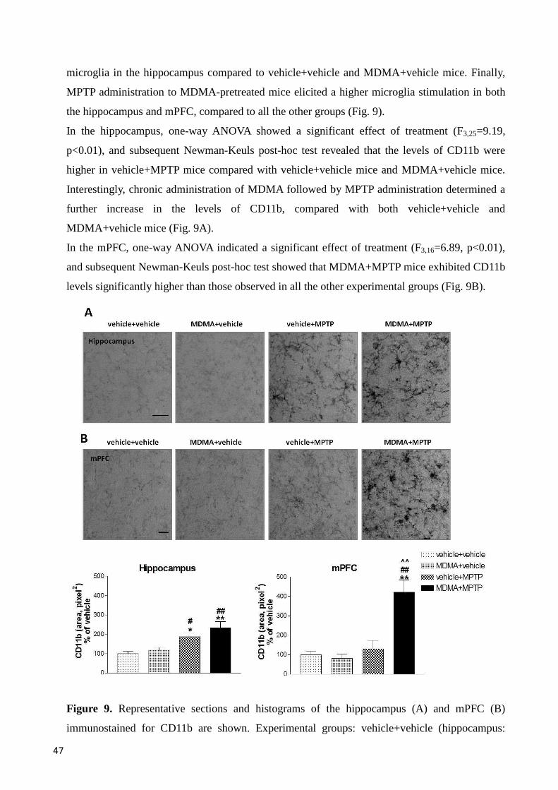
47
microglia in the hippocampus compared to vehicle+vehicle and MDMA+vehicle mice. Finally,
MPTP administration to MDMA-pretreated mice elicited a higher microglia stimulation in both
the hippocampus and mPFC, compared to all the other groups (Fig. 9).
In the hippocampus, one-way ANOVA showed a significant effect of treatment (F3,25=9.19,
p<0.01), and subsequent Newman-Keuls post-hoc test revealed that the levels of CD11b were
higher in vehicle+MPTP mice compared with vehicle+vehicle mice and MDMA+vehicle mice.
Interestingly, chronic administration of MDMA followed by MPTP administration determined a
further increase in the levels of CD11b, compared with both vehicle+vehicle and
MDMA+vehicle mice (Fig. 9A).
In the mPFC, one-way ANOVA indicated a significant effect of treatment (F3,16=6.89, p<0.01),
and subsequent Newman-Keuls post-hoc test showed that MDMA+MPTP mice exhibited CD11b
levels significantly higher than those observed in all the other experimental groups (Fig. 9B).
Figure 9. Representative sections and histograms of the hippocampus (A) and mPFC (B)
immunostained for CD11b are shown. Experimental groups: vehicle+vehicle (hippocampus:
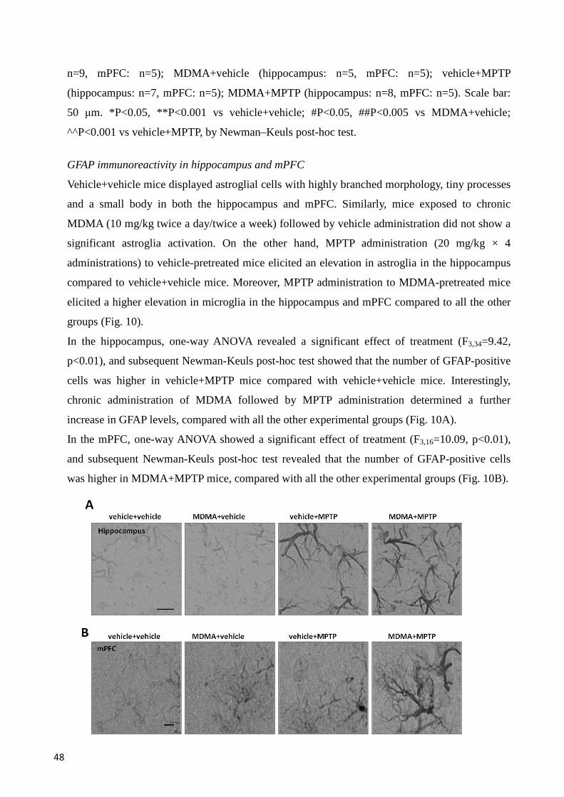
48
n=9, mPFC: n=5); MDMA+vehicle (hippocampus: n=5, mPFC: n=5); vehicle+MPTP
(hippocampus: n=7, mPFC: n=5); MDMA+MPTP (hippocampus: n=8, mPFC: n=5). Scale bar:
50 µm. *P<0.05, **P<0.001 vs vehicle+vehicle; #P<0.05, ##P<0.005 vs MDMA+vehicle;
^^P<0.001 vs vehicle+MPTP, by Newman–Keuls post-hoc test.
GFAP immunoreactivity in hippocampus and mPFC
Vehicle+vehicle mice displayed astroglial cells with highly branched morphology, tiny processes
and a small body in both the hippocampus and mPFC. Similarly, mice exposed to chronic
MDMA (10 mg/kg twice a day/twice a week) followed by vehicle administration did not show a
significant astroglia activation. On the other hand, MPTP administration (20 mg/kg × 4
administrations) to vehicle-pretreated mice elicited an elevation in astroglia in the hippocampus
compared to vehicle+vehicle mice. Moreover, MPTP administration to MDMA-pretreated mice
elicited a higher elevation in microglia in the hippocampus and mPFC compared to all the other
groups (Fig. 10).
In the hippocampus, one-way ANOVA revealed a significant effect of treatment (F3,34=9.42,
p<0.01), and subsequent Newman-Keuls post-hoc test showed that the number of GFAP-positive
cells was higher in vehicle+MPTP mice compared with vehicle+vehicle mice. Interestingly,
chronic administration of MDMA followed by MPTP administration determined a further
increase in GFAP levels, compared with all the other experimental groups (Fig. 10A).
In the mPFC, one-way ANOVA showed a significant effect of treatment (F3,16=10.09, p<0.01),
and subsequent Newman-Keuls post-hoc test revealed that the number of GFAP-positive cells
was higher in MDMA+MPTP mice, compared with all the other experimental groups (Fig. 10B).
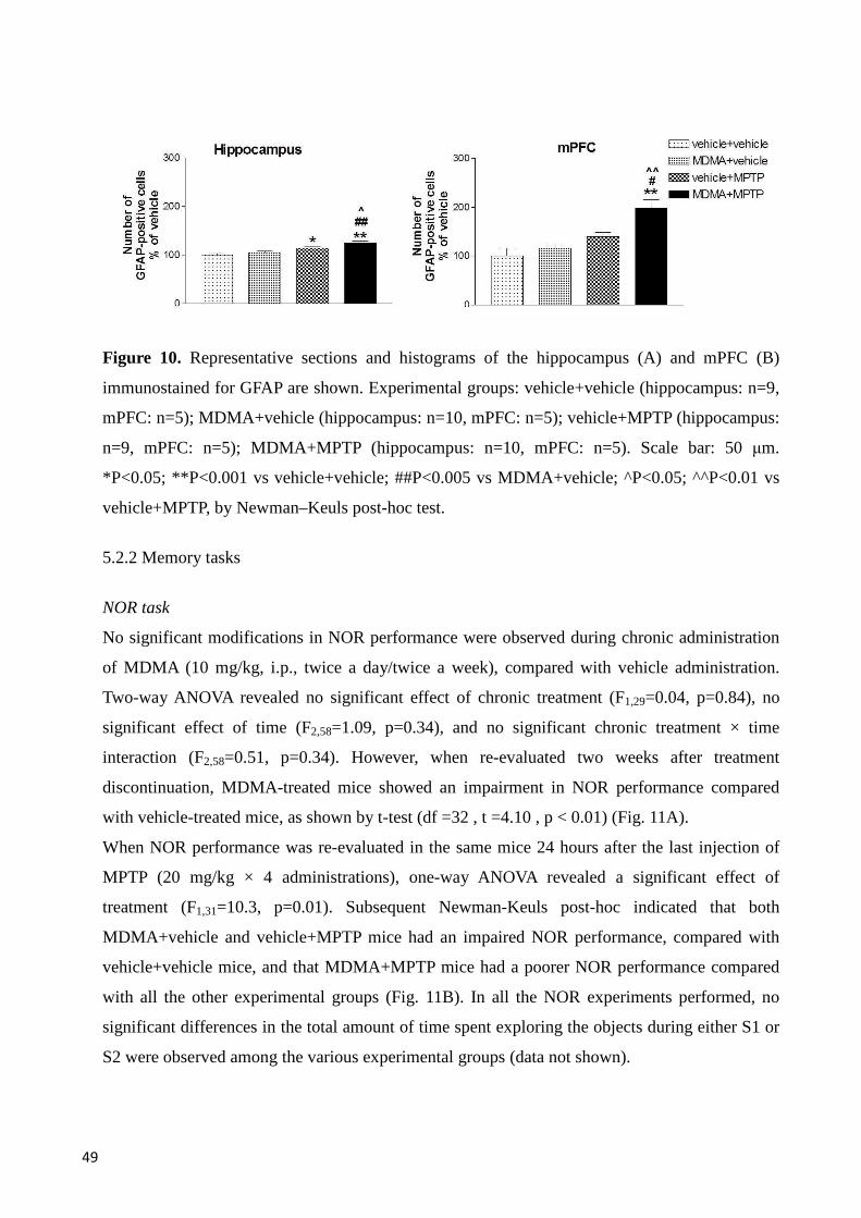
49
Figure 10. Representative sections and histograms of the hippocampus (A) and mPFC (B)
immunostained for GFAP are shown. Experimental groups: vehicle+vehicle (hippocampus: n=9,
mPFC: n=5); MDMA+vehicle (hippocampus: n=10, mPFC: n=5); vehicle+MPTP (hippocampus:
n=9, mPFC: n=5); MDMA+MPTP (hippocampus: n=10, mPFC: n=5). Scale bar: 50 µm.
*P<0.05; **P<0.001 vs vehicle+vehicle; ##P<0.005 vs MDMA+vehicle; ^P<0.05; ^^P<0.01 vs
vehicle+MPTP, by Newman–Keuls post-hoc test.
5.2.2 Memory tasks
NOR task
No significant modifications in NOR performance were observed during chronic administration
of MDMA (10 mg/kg, i.p., twice a day/twice a week), compared with vehicle administration.
Two-way ANOVA revealed no significant effect of chronic treatment (F1,29=0.04, p=0.84), no
significant effect of time (F2,58=1.09, p=0.34), and no significant chronic treatment × time
interaction (F2,58=0.51, p=0.34). However, when re-evaluated two weeks after treatment
discontinuation, MDMA-treated mice showed an impairment in NOR performance compared
with vehicle-treated mice, as shown by t-test (df =32 , t =4.10 , p < 0.01) (Fig. 11A).
When NOR performance was re-evaluated in the same mice 24 hours after the last injection of
MPTP (20 mg/kg × 4 administrations), one-way ANOVA revealed a significant effect of
treatment (F1,31=10.3, p=0.01). Subsequent Newman-Keuls post-hoc indicated that both
MDMA+vehicle and vehicle+MPTP mice had an impaired NOR performance, compared with
vehicle+vehicle mice, and that MDMA+MPTP mice had a poorer NOR performance compared
with all the other experimental groups (Fig. 11B). In all the NOR experiments performed, no
significant differences in the total amount of time spent exploring the objects during either S1 or
S2 were observed among the various experimental groups (data not shown).
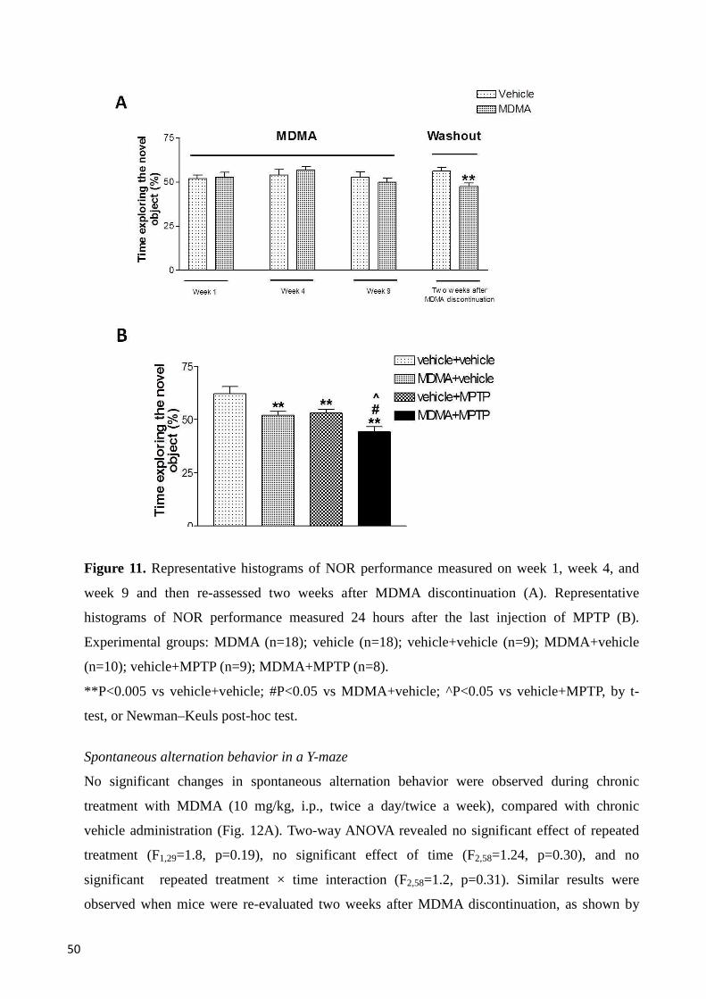
50
Figure 11. Representative histograms of NOR performance measured on week 1, week 4, and
week 9 and then re-assessed two weeks after MDMA discontinuation (A). Representative
histograms of NOR performance measured 24 hours after the last injection of MPTP (B).
Experimental groups: MDMA (n=18); vehicle (n=18); vehicle+vehicle (n=9); MDMA+vehicle
(n=10); vehicle+MPTP (n=9); MDMA+MPTP (n=8).
**P<0.005 vs vehicle+vehicle; #P<0.05 vs MDMA+vehicle; ^P<0.05 vs vehicle+MPTP, by t-
test, or Newman–Keuls post-hoc test.
Spontaneous alternation behavior in a Y-maze
No significant changes in spontaneous alternation behavior were observed during chronic
treatment with MDMA (10 mg/kg, i.p., twice a day/twice a week), compared with chronic
vehicle administration (Fig. 12A). Two-way ANOVA revealed no significant effect of repeated
treatment (F1,29=1.8, p=0.19), no significant effect of time (F2,58=1.24, p=0.30), and no
significant repeated treatment × time interaction (F2,58=1.2, p=0.31). Similar results were
observed when mice were re-evaluated two weeks after MDMA discontinuation, as shown by
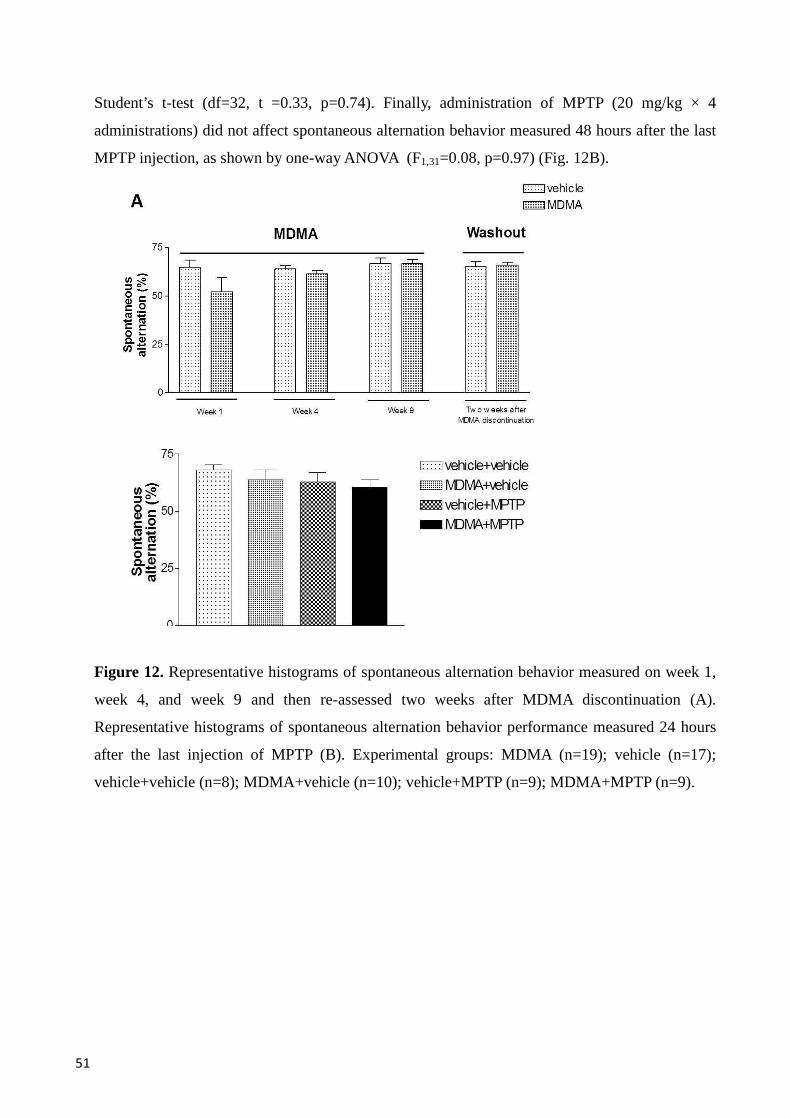
51
Student’s t-test (df=32, t =0.33, p=0.74). Finally, administration of MPTP (20 mg/kg × 4
administrations) did not affect spontaneous alternation behavior measured 48 hours after the last
MPTP injection, as shown by one-way ANOVA (F1,31=0.08, p=0.97) (Fig. 12B).
Figure 12. Representative histograms of spontaneous alternation behavior measured on week 1,
week 4, and week 9 and then re-assessed two weeks after MDMA discontinuation (A).
Representative histograms of spontaneous alternation behavior performance measured 24 hours
after the last injection of MPTP (B). Experimental groups: MDMA (n=19); vehicle (n=17);
vehicle+vehicle (n=8); MDMA+vehicle (n=10); vehicle+MPTP (n=9); MDMA+MPTP (n=9).
52
Discussion
In this work of thesis, late adolescent mice were treated as adolescent with MDMA, and
administered as adults, after MDMA washout, with MPTP, a neurotoxin relevant for humans as it
can induce a syndrome that reproduces the features of PD (Langston and Ballard 1983; Langston
et al. 1983 and 1999), in order to study whether MDMA pre-exposure could modify the
vulnerability to the noxious effects of MPTP. The results of this study demonstrate that the
neuroinflammation in the nigrostriatal, limbic and cortical systems, as well as the neurotoxicity
in the nigrostriatal system, elicited by MPTP, was exacerbated by chronic administration of
MDMA to mice during adolescence. Moreover, MDMA administration during adolescence
induces cognitive impairment, and amplified the detrimental effects of MPTP on cognitive
performance measured at adulthood.
The study of how MDMA modulates the toxic effects of other substances appears a “hot topic”
in neuropharmacology and neurotoxicology because this drug is largely consumed by
adolescents (Strote et al. 2002), and adolescence is a critical period for the development of the
brain (Casey et al. 2000). The adolescent brain appears particularly vulnerable to the long-term
noxious effects of exogenous substances, including drugs of abuse (Cadoni et al. 2013; Daza-
Losada et al. 2009). It has been demonstrated, in rats, that almost all drugs causing drug
addiction increase the release of DA in the mesolimbic pathway (Pierce and Kumaresan 2006),
and it has been demonstrated that amphetamine administration in adolescent rats induces a
prominent sensitization of DA release, whereas no change was found in adult rats (Laviola et al.
2001). Therefore, it is becoming increasingly clear that MDMA should be regarded as harmful
not only because its use may have implications for drug dependence (Fernandez-Serrano et al.
2011; Smirnov et al. 2013), but also because MDMA use could function as a “primer” for later
brain insults.
Besides showing detrimental effects of MDMA itself, studies in experimental animals have
shown that this substance may amplify the neuroinflammation and neurotoxicity elicited by other
psychoactive/toxic substances, eventually leading to the exacerbation of neuronal damage. The
potentiation of MPTP-mediated neuroinflammation in mice pretreated with MDMA during
adolescence appeared very marked in striatum and mPFC, lower in SNc and hippocampus,
involving both microglia and astroglia. It should be noted that MDMA alone did not appear to
produce an increase in neuroinflammation in any brain areas evaluated. In this regard, it has to
be considered that in the present study immunohistochemical evaluation of microglia and
astroglia was performed three weeks after the last MDMA administration, and therefore it is
53
feasible that the acute neuroinflammatory effects of MDMA may have faded over time.
Moreover, it has to be take into account the differences in doses and protocols of MDMA
administration between the present and previous studies (Frau et al. 2013; Granado et al. 2008a
and 2011; Khairnar et al. 2009).
It is noteworthy that MPTP alone elicited both astrogliosis and microgliosis in the SNc, striatum
and hippocampus, compared with vehicle+vehicle treated mice. At variance with these results,
neither MDMA nor MPTP alone significantly stimulated both astrogliosis and microgliosis in the
mPFC. This latter finding deserves consideration, since it suggests that the neurochemical
modifications induced by repeated exposure to MDMA, even if subtle, may prime the brain for
the effects of later noxious stimuli. In addition, MPTP administration in MDMA pretreated mice
significantly stimulates astrogliosis, but not microgliosis, in the hippocampus, compared with
MPTP alone. These results are confirmed by previous evidences in rodents studies reporting
neuroinflammatory effects of MPTP in the SNc, striatum (Czlonkowska et al. 1996; Kohutnicka
et al. 1998; Kurkowska-Jastrzebska et al. 1999; Kurosaki et al. 2003), and hippocampus
(Kokovay and Cunningham 2005; Sy et al. 2010; Wang et al. 2009). The increased microgliosis
and astrogliosis in hippocampus could be explained by the presence of a very high density of
binding sites for MPP+ in this region (Del Zompo et al. 1992). As suggested by Thomas and co-
workers (2004), the amphetamine-related drugs associated with DA or 5-HT nerve terminal
damage also cause microglial activation, whereas non-neurotoxic amphetamine-related drugs do
not have this effect.
MPTP administration in mice chronically pretreated during late adolescence with MDMA
induced higher dopaminergic neuron degeneration than in MPTP or MDMA alone treated mice.
Since, in these mice, the degree of neuronal degeneration induced by MPTP appears additive to
that induced by MDMA, we speculate that dopaminergic degeneration produced by MDMA may
create the basis for a degeneration which could reach the critical threshold to induce PD in
presence of environmental toxins. Moreover, mice chronically treated with MDMA showed
reduced N and the D of TH-positive neurons in the SNc three weeks after the interruption of
MDMA treatment. This result, in line with previous reports showing that acute repeated MDMA
administration produces dopaminergic neuron degeneration (Granado et al. 2008a and 2011),
adds the important indication that also sporadic, but prolonged treatment of mice with this drug
is toxic to dopaminergic neurons. Therefore, MDMA, like other recognized amphetamine-related
drugs, such as methamphetamine (Albers et al. 1996; Sinchai et al. 2011), increases
dopaminergic neuron vulnerability even when administered at low/medium doses and in an
irregular way. Regarding the reduction in N and the D of TH-positive neurons in the SNc but not
54
in TH-positive fibers in striatum it can be hypothesized, as previously suggested (Rosenblad et
al. 1999), that partial DA lesion might be associated to compensatory sprouting in the striatum.
As mentioned in the introduction, MDMA and MPTP have several common mechanisms able to
induce toxicity and this could underlie the exacerbation of MPTP-mediated neuroinflammatory
and neurotoxic effects observed in MDMA pretreated mice. MDMA has been reported to inhibit
the CYP2D6 (O’Mathùna et al. 2008), which has been shown to convert MPTP into non-toxic
metabolites (Herraiz et al. 2006; Mann and Tyndale 2010). In addition, it is possible that
prolonged exposure to MDMA result in an elevation of intracellular oxidative stress, due to
several mechanisms, that would eventually render neurons more sensitive to the effects of MPTP,
which acts itself by promoting the generation of ROS (Cleeter et al. 1992; Sriram et al. 1997).
Thus, it has been demonstrated that MDMA, as MPTP, can inhibit mitochondrial complex I in
mice (Nicklas et al. 1985; Puerta et al, 2010; Suzuki et al. 1990), leading to mitochondria
inactivation and oxidizing macromolecules such as lipids, DNA, and proteins. Moreover,
MDMA, could amplify oxidative stress also by promoting the release of DA (Cadoni et al.
2005), which can undergo auto-oxidation, and in turn generate free radicals and ROS (Marchitti
et al. 2007; Siraki and O'Brien 2002; Stokes et al. 1999), eventually leading to the exacerbation
of neuroinflammatory and neurotoxic phenomenons triggered by a later noxious insult. This
latter mechanism could be particularly relevant to the results of this study, as it might justify why
MDMA pre-exposure was associated with a more marked exacerbation of MPTP-
neuroinflammation in the striatum and mPFC, and neurotoxicity in SNc, which are highly
enriched with dopaminergic terminals and neurons (Bentivoglio and Morelli 2005), than in the
hippocampus, where dopaminergic innervation is less dense (Bentivoglio and Morelli, 2005;
Milner and Bacon 1989).
In addition to neuroinflammatory and neurotoxic changes, mice treated with MDMA during
adolescence displayed a persistent impairment in NOR performance after treatment
discontinuation, and MDMA pretreatment was found to further aggravate cognitive impairment
induced by MPTP. These results appear of great relevance since, despite the ever-increasing use
of MDMA in humans, scarce information is available on the long-term effects on cognition
elicited by the consumption of this drug during adolescence. Importantly, the impairment in
NOR performance observed in this study appears clearly attributable to deficits in cognitive
function. In fact, no differences were found between MDMA- and vehicle-treated mice as for the
total amount of time spent in object exploration during both S1 and S2. This indicates that the
regimen of MDMA used in this study is devoid of non-specific effects on object exploration.
Moreover, MDMA seemed not to alter the ability of mice to discriminate between different
55
objects. In fact, mice could recognize novel objects at different time points during the MDMA
treatment, but eventually loses this ability when re-evaluated at two weeks after treatment
discontinuation, which would suggest the onset of progressive and long-term toxicity of MDMA.
Striatum, hippocampus and mPFC have been shown to regulate item recognition in rodents
(Cropley et al. 2006; Dalley et al. 2004; Eichenbaum et al. 1996; Landau et al. 2009; Ragozzino
et al. 1999; Squire 1992). Therefore, the impairment in NOR performance observed here is likely
attributable to the neuroinflammatory effect caused by MDMA and MPTP in these areas.
Remarkably, a support to the existence of a causal link between neuroinflammation and
cognitive impairment comes from previous studies which have employed the systemical or
intrahippocampal injection of lipopolysaccharide (LPS) in rodents. Thus, LPS has been shown
not only to stimulate the expression of inflammation-related molecules, including GFAP, iNOS,
cycloxygenase (COX)-2, IL-6 and IL-1β (Herber et al. 2006; Jain et al. 2002; Pang et al. 2006;
Sparkman et al. 2006) but also to engender cognitive impairment. Furthermore, METH, the
abuse of which in humans is often associated with neurocognitive impairment (Nestor et al.
2011; North et al. 2013), causes an inflammatory response characterized by an upregulation of
GFAP and CD11b levels,TNF-α, and TNF-receptor 1 protein levels (Gonçalves et al. 2010) in
mice.
Interestingly, the cognitive impairment was narrowed to NOR, whilst no deficits were detectable
in spontaneous alternation behavior. This lack of effect could be observed both during MDMA
treatment and following MPTP administration, and no exacerbation of MPTP effects was present
in MDMA-treated mice. These results could be explained in the light of previous studies that
have shown how spontaneous alternation behavior may be relatively resistant to noxious brain
insults that impair NOR performance, suggesting a different sensitivity of these two tasks to
neuronal damage (Moriguchi et al. 2012; Simola et al. 2008). Moreover, it is interesting to
consider that MPTP has been demonstrated to scarcely affect alternation behavior in mice (Tanila
et al. 1998), which could justify the lack of an impairment in spontaneous alternation behavior in
mice challenged with MPTP.
The present study, encompassing both neurochemical and behavioral findings, may significantly
contribute to the elucidation of the interactions between MDMA and substances which are toxic
for the brain, and appear also of potential relevance for humans. Several clinical and preclinical
studies recognize that PD has multiple origins, from genes to toxins and several concomitant
factors may contribute to its development (Marsden and Olanow 1998). Since one of the
possibilities taken into account in PD pathology is the use of amphetamine-related drugs, the
results of the present work of thesis support the hypothesis these substances might be part of the
56
multiple factors that lead to dopaminergic neuron degeneration and microglial activation
associated with PD. Thus, a major conclusion that can be drawn from the present study is that the
detrimental effects of MDMA, even if subtle, appear to be long-lasting, since they could be
detected after treatment discontinuation. By showing an exacerbation of MPTP effects in several
brain areas as SNc, striatum, hippocampus and mPFC of mice pre-exposed to MDMA, this study
indicates that noxious MDMA-MPTP interactions are widespread in the brain. Moreover, it was
observed that the abuse of MDMA creates the basis for a worsening in cognitive function, in
agreement to data from several studies in adult MDMA consumers (Bolla et al. 1998; Brown et
al. 2010; Fox et al. 2001; Morgan 2000; Parrott and Laskey 1998).
57
Conclusions
In conclusion, by demonstrating that mice repeatedly treated with MDMA during adolescence
display an exacerbation in neuroinflammation, neurotoxicity and cognitive impairment induced
by MPTP, a dopaminergic toxin relevant to humans, the present results underline the importance
of exploring the long-term effects of amphetamine-related drugs, to clarify their potential as risk
factors for the development of neurological and behavioral problems later in life.
Acknowledgments
Dr. Giulia Costa gratefully acknowledges Sardinia Regional Government for the financial
support of her PhD scholarship (P.O.R. Sardegna F.S.E. Operational Programme of the
Autonomous Region of Sardinia, European Social Fund 2007-2013 - Axis IV Human Resources,
Objective l.3, Line of Activity l.3.1
58
Bibliografy
• Adori C, Andó RD, Kovács GG, Bagdy G (2006) Damage of serotonergic axons and immunolocalization of Hsp27,
Hsp72, and Hsp90 molecular chaperones after a single dose of MDMA administration in Dark Agouti rat: temporal,
spatial, and cellular patterns. J Comp Neurol. 497:251-69.
• Aguirre N, Barrionuevo M, Ramírez MJ, Del Río J, Lasheras B (1999) Alpha-lipoic acid prevents 3,4-
methylenedioxy-methamphetamine (MDMA)-induced neurotoxicity. Neuroreport. 10:3675-80.
• Agulhon C, Sun MY, Murphy T, Myers T, Lauderdale K, Fiacco TA (2012) Calcium signaling and gliotransmission
in normal vs. reactive astrocytes. Front Pharmacol. 3:139.
• Albanese A, Altavista MC, Rossi P (1986) Organization of central nervous system dopaminergic pathways. J Neural
Transm Suppl. 22:3-17.
• Albers DS, Zeevalk GD, Sonsalla PK (1996) Damage to dopaminergic nerve terminals in mice by combined
treatment of intrastriatal malonate with systemic methamphetamine or MPTP. Brain Res. 718:217-20.
• Allan SM, Rothwell NJ (2001) Cytokines and acute neurodegeneration. Nat Rev Neurosci. 2:734-44.
• Alonso AD, Zaidi T, Novak M, Barra HS, Grundke-Iqbal I, Iqbal K (2001) Interaction of tau isoforms with
Alzheimer's disease abnormally hyperphosphorylated tau and in vitro phosphorylation into the disease-like protein. J
Biol Chem. 276:37967-73.
• Anglade P, Vyas S, Javoy-Agid F, Herrero MT, Michel PP, Marquez J, Mouatt-Prigent A, Ruberg M, Hirsch EC,
Agid Y (1997) Apoptosis and autophagy in nigral neurons of patients with Parkinson's disease. Histol Histopathol.
12:25-31.
• Atkins K, Burks T, Swann AC, Dafny N (2009) MDMA (ecstasy) modulates locomotor and prefrontal cortex
sensory evoked activity. Brain Res 1302:175-82.
• Ball KT, Slane M (2012) Differential involvement of prelimbic and infralimbic medial prefrontal cortex in discrete
cue-induced reinstatement of 3,4-methylenedioxymethamphetamine (MDMA; ecstasy) seeking in rats.
Psychopharmacology 224:377-85.
• Ballard PA, Tetrud JW, Langston JW (1985) Permanent human parkinsonism due to 1-methyl-4-phenyl-1,2,3,6-
tetrahydropyridine (MPTP): seven cases. Neurology 35:949-56.
• Barcia C, Sánchez Bahillo A, Fernández-Villalba E, Bautista V, Poza Y Poza M, Fernández-Barreiro A, Hirsch EC,
Herrero MT (2004) Evidence of active microglia in substantia nigra pars compacta of parkinsonian monkeys 1 year
after MPTP exposure. Glia 46:402-9.
• Baumann MH, Wang X, Rothman RB (2007) 3,4-Methylenedioxymethamphetamine (MDMA) neurotoxicity in rats:
a reappraisal of past and present findings. Psychopharmacology 189:407-24.
• Bauernfeind AL, Dietrich MS, Blackford JU, Charboneau EJ, Lillevig JG, Cannistraci CJ, Woodward ND, Cao A,
Watkins T, Di Iorio CR, Cascio C, Salomon RM, Cowan RL (2011) Human Ecstasy use is associated with increased
cortical excitability: an fMRI study. Neuropsychopharmacology 36:1127-41.
• Becker B, Wagner D, Koester P, Bender K, Kabbasch C, Gouzoulis-Mayfrank E, Daumann J (2013) Memory-related
hippocampal functioning in ecstasy and amphetamine users: a prospective fMRI study. Psychopharmacology
225:923-34.
• Bentivoglio M, Morelli M (2005) The organization and circuits of mesencephalic dopaminergic neurons and the
distribution of dopamine receptors in the brain. Edited by S.B. Dunnett 1:1-107.
59
• Benzenhöfer U, Passie T (2010) Rediscovering MDMA (ecstasy): the role of the American chemist Alexander T.
Shulgin. Addiction 105:1355-61.
• Bezard E, Gross CE, Fournier MC, Dovero S, Bloch B, Jaber M (1999) Absence of MPTP-induced neuronal death
in mice lacking the dopamine transporter. Exp Neurol. 155:268-73.
• Blessing WW, Seaman B, Pedersen NP, Ootsuka Y (2003) Clozapine reverses hyperthermia and sympathetically
mediated cutaneous vasoconstriction induced by 3,4-methylenedioxymethamphetamine (ecstasy) in rabbits and rats.
J Neurosci. 23:6385-91.
• Bolla KI, McCann UD, Ricaurte GA (1998) Memory impairment in abstinent MDMA ("Ecstasy") users. Neurology
51:1532-7.
• Bosch OG, Wagner M, Jessen F, Kühn KU, Joe A, Seifritz E, Maier W, Biersack HJ, Quednow BB (2013) Verbal
memory deficits are correlated with prefrontal hypometabolism in (18)FDG PET of recreational MDMA users. PLoS
One 8:e61234.
• Bowen DM, Davison AN (1975) Letter: Extrapyramidal diseases and dementia. Lancet 1:1199-200.
• Boyle NT, Connor TJ (2010) Methylenedioxymethamphetamine ('Ecstasy')-induced immunosuppression: a cause for
concern? Br J Pharmacol. 161:17-32.
• Brochard V, Combadière B, Prigent A, Laouar Y, Perrin A, Beray-Berthat V, Bonduelle O, Alvarez-Fischer D,
Callebert J, Launay JM, Duyckaerts C, Flavell RA, Hirsch EC, Hunot S (2009) Infiltration of CD4+ lymphocytes
into the brain contributes to neurodegeneration in a mouse model of Parkinson disease. J Clin Invest. 119:182-92.
• Brodkin J, Malyala A, Nash JF (1993) Effect of acute monoamine depletion on 3,4-
methylenedioxymethamphetamine-induced neurotoxicity. Pharmacol Biochem Behav. 45:647-53.
• Brown J, McKone E, Ward J (2010) Deficits of long-term memory in ecstasy users are related to cognitive
complexity of the task. Psychopharmacology 209:51-67.
• Brust JC (2010) Substance abuse and movement disorders. Mov Disord. 25:2010-20.
• Busceti CL, Biagioni F, Riozzi B, Battaglia G, Storto M, Cinque C, Molinaro G, Gradini R, Caricasole A, Canudas
AM, Bruno V, Nicoletti F, Fornai F (2008) Enhanced tau phosphorylation in the hippocampus of mice treated with
3,4-methylenedioxymethamphetamine ("Ecstasy"). J Neurosci. 28:3234-45.
• Cadet JL, Ladenheim B, Baum I, Carlson E, Epstein C (1994) CuZn-superoxide dismutase (CuZnSOD) transgenic
mice show resistance to the lethal effects of methylenedioxyamphetamine (MDA) and of
methylenedioxymethamphetamine (MDMA). Brain Res. 655:259-62.
• Cadet JL, Ladenheim B, Hirata H, Rothman RB, Ali S, Carlson E, Epstein C, Moran TH (1995a) Superoxide
radicals mediate the biochemical effects of methylenedioxymethamphetamine (MDMA): evidence from using
CuZn-superoxide dismutase transgenic mice. Synapse. 21:169-76.
• Cadet JL, Ali SF, Rothman RB, Epstein CJ (1995b) Neurotoxicity, drugs and abuse, and the CuZn-superoxide
dismutase transgenic mice. Mol Neurobiol. 11:155-63.
• Cadoni C, Simola N, Espa E, Fenu S, Di Chiara G (2013) Strain dependence of adolescent Cannabis influence on
heroin reward and mesolimbic dopamine transmission in adult Lewis and Fischer 344 rats. Addict Biol.
doi:10.1111/adb.12085.
• Cadoni C, Solinas M, Pisanu A, Zernig G, Acquas E, Di Chiara G (2005) Effect of 3,4-
methylendioxymethamphetamine (MDMA, "ecstasy") on dopamine transmission in the nucleus accumbens shell
and core. Brain Res. 1055:143-8.
60
• Callaghan RC, Cunningham JK, Sajeev G, Kish SJ (2010) Incidence of Parkinson's disease among hospital patients
with methamphetamine-use disorders. Mov Disord. 25:2333-9.
• Calne DB, Langston JW, Martin WR, Stoessl AJ, Ruth TJ, Adam MJ, Pate BD, Schulzer M (1985) Positron
emission tomography after MPTP: observations relating to the cause of Parkinson's disease. Nature 317:246-8.
• Capela JP, Carmo H, Remião F, Bastos ML, Meisel A, Carvalho F (2009) Molecular and cellular mechanisms of
ecstasy-induced neurotoxicity: an overview. Mol Neurobiol 39:210-71.
• Caraceni A, Martini C, Belli F, Mascheroni L, Rivoltini L, Arienti F, Cascinelli N (1993) Neuropsychological and
neurophysiological assessment of the central effects of interleukin-2 administration. Eur J Cancer. 29A:1266-9.
• Carboni S, Melis F, Pani L, Hadjiconstantinou M, Rossetti ZL (1990) The non-competitive NMDA-receptor
antagonist MK-801 prevents the massive release of glutamate and aspartate from rat striatum induced by 1-methyl-
4-phenylpyridinium (MPP+). Neurosci Lett. 117:129-33.
• Carvalho M, Carvalho F, Remião F, de Lourdes Pereira M, Pires-das-Neves R, de Lourdes Bastos M. Effect of 3,4-
methylenedioxymethamphetamine ("ecstasy") on body temperature and liver antioxidant status in mice: influence of
ambient temperature. Arch Toxicol. 2002;76(3):166-72.
• Casey BJ, Giedd JN, Thomas KM (2000) Structural and functional brain development and its relation to cognitive
development. Biol Psychol. 54:241-57.
• Castagnoli N Jr, Chiba K, Trevor AJ (1985) Potential bioactivation pathways for the neurotoxin 1-methyl-4-phenyl-
1,2,3,6-tetrahydropyridine (MPTP). Life Sci. 36:225-30.
• Chambers RA, Taylor JR, Potenza MN (2003) Developmental neurocircuitry of motivation in adolescence: a critical
period of addiction vulnerability. Am J Psychiatry. 160:1041-52.
• Chen Y, Vartiainen NE, Ying W, Chan PH, Koistinaho J, Swanson RA (2001) Astrocytes protect neurons from nitric
oxide toxicity by a glutathione-dependent mechanism. Journal of Neurochemistry 77:1601–1610.
• Chiba K, Peterson LA, Castagnoli KP, Trevor AJ, Castagnoli N Jr (1985) Studies on the molecular mechanism of
bioactivation of the selective nigrostriatal toxin 1-methyl-4-phenyl-1,2,3,6-tetrahydropyridine. Drug Metab Dispos.
13:342-7.
• Choi WS, Kruse SE, Palmiter RD, Xia Z (2008) Mitochondrial complex I inhibition is not required for
dopaminergic neuron death induced by rotenone, MPP+, or paraquat. Proc Natl Acad Sci USA 105:15136-41.
• Christine CW, Garwood ER, Schrock LE, Austin DE, McCulloch CE (2010) Parkinsonism in patients with a history
of amphetamine exposure. Mov Disord. 25:228-31.
• Cleeter MW, Cooper JM, Schapira AH (1992) Irreversible inhibition of mitochondrial complex I by 1-methyl-4-
phenylpyridinium: evidence for free radical involvement. J Neurochem. 58:786-9.
• Cleeter MW, Cooper JM, Darley-Usmar VM, Moncada S, Schapira AH (1994) Reversible inhibition of cytochrome
c oxidase, the terminal enzyme of the mitochondrial respiratory chain, by nitric oxide. Implications for
neurodegenerative diseases. FEBS Lett. 345:50-4.
• Colado MI, Camarero J, Mechan AO, Sanchez V, Esteban B, Elliott JM, Green AR (2001) A study of the
mechanisms involved in the neurotoxic action of 3,4-methylenedioxymethamphetamine (MDMA, 'ecstasy') on
dopamine neurones in mouse brain. Br J Pharmacol. 134:1711-23.
• Colado MI, Granados R, O'Shea E, Esteban B, Green AR (1999) The acute effect in rats of 3,4-
methylenedioxyethamphetamine (MDEA, "eve") on body temperature and long term degeneration of 5-HT neurones
in brain: a comparison with MDMA ("ecstasy"). Pharmacol Toxicol. 84:261-6.

61
• Colado MI, Williams JL, Green AR (1995) The hyperthermic and neurotoxic effects of 'Ecstasy' (MDMA) and 3,4
methylenedioxyamphetamine (MDA) in the Dark Agouti (DA) rat, a model of the CYP2D6 poor metabolizer
phenotype. Br J Pharmacol. 115:1281-9.
• Cole JC, Sumnall HR (2003) Altered states: the clinical effects of Ecstasy. Pharmacol Ther. 98:35-58.
• Comella CL (2008) Sleep disorders in Parkinson's disease. Curr Treat Options Neurol. 10:215-21.
• Connor TJ (2004) Methylenedioxymethamphetamine (MDMA, 'Ecstasy'): a stressor on the immune system.
Immunology 111:357-67.
• Connor TJ, Harkin A, Kelly JP (2005) Methylenedioxymethamphetamine suppresses production of the
proinflammatory cytokine tumor necrosis factor-alpha independent of a beta-adrenoceptor-mediated increase in
interleukin-10. J Pharmacol Exp Ther. 312:134-43.
• Cooper JA, Sagar HJ, Jordan N, Harvey NS, Sullivan EV (1991) Cognitive impairment in early, untreated
Parkinson's disease and its relationship to motor disability. Brain 114:2095-122.
• Crean RD, Davis SA, Taffe MA (2007) Oral administration of (+/-)3,4-methylenedioxymethamphetamine and
(+)methamphetamine alters temperature and activity in rhesus macaques. Pharmacol Biochem Behav. 87:11-9.
• Cropley VL, Fujita M, Innis RB, Nathan PJ (2006) Molecular imaging of the dopaminergic system and its
association with human cognitive function. Biol Psychiatry 59:898-907.
• Członkowska A, Kohutnicka M, Kurkowska-Jastrzebska I, Członkowski A (1996) Microglial reaction in MPTP (1-
methyl-4-phenyl-1,2,3,6-tetrahydropyridine) induced Parkinson's disease mice model. Neurodegeneration 5:137-43.
• Dafters RI (1994) Effect of ambient temperature on hyperthermia and hyperkinesis induced by 3,4-
methylenedioxymethamphetamine (MDMA or "ecstasy") in rats. Psychopharmacology 114:505-8.
• Dalley JW, Cardinal RN, Robbins TW (2004) Prefrontal executive and cognitive functions in rodents: neural and
neurochemical substrates. Neurosci Biobehav Rev. 28:771-84.
• Darvesh AS, Yamamoto BK, Gudelsky GA (2005) Evidence for the involvement of nitric oxide in 3,4-
methylenedioxymethamphetamine-induced serotonin depletion in the rat brain. J Pharmacol Exp Ther. 312:694-701.
• da Silva DD, Silva E, Carvalho F, Carmo H (2013) Mixtures of 3,4-methylenedioxymethamphetamine (ecstasy) and
its major human metabolites act additively to induce significant toxicity to liver cells when combined at low, non-
cytotoxic concentrations. J Appl Toxicol. doi: 10.1002/jat.2885.
• Date I, Felten DL, Felten SY (1990) Long-term effect of MPTP in the mouse brain in relation to aging:
neurochemical and immunocytochemical analysis. Brain Res. 519:266-76.
• Daumann J Jr, Fischermann T, Heekeren K, Thron A, Gouzoulis-Mayfrank E (2004) Neural mechanisms of working
memory in ecstasy (MDMA) users who continue or discontinue ecstasy and amphetamine use: evidence from an 18-
month longitudinal functional magnetic resonance imaging study. Biol Psychiatry 56:349-55.
• Daumann J, Fischermann T, Heekeren K, Henke K, Thron A, Gouzoulis-Mayfrank E (2005) Memory-related
hippocampal dysfunction in poly-drug ecstasy (3,4-methylenedioxymethamphetamine) users. Psychopharmacology
180:607-11.
• Davison D, Parrott AC (1997) Ecstasy (MDMA) in Recreational Users: Self-Reported Psychological and
Physiological Effects. Hum. Psychopharmacol. Clin. Exp. 12: 221–226.
• Daza-Losada M, Rodríguez-Arias M, Aguilar MA, Miñarro J (2009) Acquisition and reinstatement of MDMA-
induced conditioned place preference in mice pre-treated with MDMA or cocaine during adolescence. Addict Biol.
14:447-56.
62
• Decamp E, Schneider JS (2004) Attention and executive function deficits in chronic low-dose MPTP-treated non-
human primates. Eur J Neurosci. 20:1371-8.
• Decamp E, Schneider JS (2006) Effects of nicotinic therapies on attention and executive functions in chronic low-
dose MPTP-treated monkeys. Eur J Neurosci. 24:2098-104.
• Decamp E, Tinker JP, Schneider JS (2004) Attentional cueing reverses deficits in spatial working memory task
performance in chronic low dose MPTP-treated monkeys. Behav Brain Res. 152:259-62.
• Deguil J, Chavant F, Lafay-Chebassier C, Pérault-Pochat MC, Fauconneau B, Pain S (2010) Neuroprotective effect
of PACAP on translational control alteration and cognitive decline in MPTP parkinsonian mice. Neurotox Res.
17:142-55.
• Dehmer T, Lindenau J, Haid S, Dichgans J, Schulz JB (2000) Deficiency of inducible nitric oxide synthase protects
against MPTP toxicity in vivo. J Neurochem. 74:2213-6.
• de la Torre R, Farré M, Roset PN, Pizarro N, Abanades S, Segura M, Segura J, Camí J (2004) Human pharmacology
of MDMA: pharmacokinetics, metabolism, and disposition. Ther Drug Monit 26:137-44.
• Del Zompo M, Piccardi MP, Ruiu S, Albanese A, Morelli M (1992) Localization of MPP+ binding sites in the brain
of various mammalian species. J Neural Transm Park Dis Dement Sect. 4:181-90.
• Del Zompo M, Piccardi MP, Ruiu S, Quartu M, Gessa GL, Vaccari A (1993) Selective MPP+ uptake into synaptic
dopamine vesicles: possible involvement in MPTP neurotoxicity. Br J Pharmacol. 109:411-4.
• Denicoff KD, Rubinow DR, Papa MZ, Simpson C, Seipp CA, Lotze MT, Chang AE, Rosenstein D, Rosenberg SA
(1987) The neuropsychiatric effects of treatment with interleukin-2 and lymphokine-activated killer cells. Ann Intern
Med. 107:293-300.
• Dluzen DE, Kreutzberg JD (1993) 1-Methyl-4-phenyl-1,2,3,6-tetrahydropyridine (MPTP) disrupts social
memory/recognition processes in the male mouse. Brain Res. 609:98-102.
• Downes JJ, Roberts AC, Sahakian BJ, Evenden JL, Morris RG, Robbins TW (1989) Impaired extra-dimensional
shift performance in medicated and unmedicated Parkinson's disease: evidence for a specific attentional dysfunction.
Neuropsychologia 27:1329-43.
• Dubois B, Pillon B (1997) Cognitive deficits in Parkinson's disease. J Neurol. 244:2-8.
• Eidelberg D, Moeller JR, Dhawan V, Spetsieris P, Takikawa S, Ishikawa T, Chaly T, Robeson W, Margouleff D,
Przedborski S, Fahn S (1994) The metabolic topography of parkinsonism. J Cereb Blood Flow Metab. 14:783-801.
• Eichenbaum H, Schoenbaum G, Young B, Bunsey M (1996) Functional organization of the hippocampal memory
system. Proc Natl Acad Sci USA 93:13500-7.
• Ekblom J, Jossan SS, Bergstrüm M, Oreland L, Walum E, Aquilonius SM (1993) Monoamine oxidase-B in
astrocytes. Glia 8:122–132.
• EMCDDA (2007 and 2010) Annual report on the State of the Drugs Problem in Europe. European Monitoring
Centre for Drugs and Drug Addiction, Lisbon
• Ennaceur A, Delacour J (1988) A new one-trial test for neurobiological studies of memory in rats. 1: Behavioral
data. Behav Brain Res. 31:47-59.
• Fantegrossi WE, Godlewski T, Karabenick RL, Stephens JM, Ullrich T, Rice KC, Woods JH (2003)
Pharmacological characterization of the effects of 3,4-methylenedioxymethamphetamine ("ecstasy") and its
enantiomers on lethality, core temperature, and locomotor activity in singly housed and crowded mice.
Psychopharmacology 166:202-11.

63
• Fearnley JM, Lees AJ (1991) Ageing and Parkinson's disease: substantia nigra regional selectivity. Brain 114:2283-
301.
• Fernández-Serrano MJ, Pérez-García M, Verdejo-García A (2011) What are the specific vs. generalized effects of
drugs of abuse on neuropsychological performance? Neurosci Biobehav Rev. 35:377-406.
• Ferreira PS, Nogueira TB, Costa VM, Branco PS, Ferreira LM, Fernandes E, Bastos ML, Meisel A, Carvalho F,
Capela JP (2013) Neurotoxicity of "ecstasy" and its metabolites in human dopaminergic differentiated SH-SY5Y
cells. Toxicol Lett. 216:159-70.
• Fleming SM, Ekhator OR, Ghisays V (2013) Assessment of sensorimotor function in mouse models of Parkinson's
disease. J Vis Exp. 76. doi:10.3791/50303.
• Foltynie T, Brayne CE, Robbins TW, Barker RA (2004) The cognitive ability of an incident cohort of Parkinson's
patients in the UK. The CamPaIGN study. Brain 127:550-60.
• Fornai F, Lenzi P, Frenzilli G, Gesi M, Ferrucci M, Lazzeri G, Biagioni F, Nigro M, Falleni A, Giusiani M,
Pellegrini A, Blandini F, Ruggieri S, Paparelli A (2004) DNA damage and ubiquitinated neuronal inclusions in the
substantia nigra and striatum of mice following MDMA (ecstasy). Psychopharmacology 173:353-63.
• Fox SH, Brotchie JM (2010) The MPTP-lesioned non-human primate models of Parkinson's disease. Past, present,
and future. Prog Brain Res. 184:133-57.
• Fox HC, Parrott AC, Turner JJ (2001) Ecstasy use: cognitive deficits related to dosage rather than self-reported
problematic use of the drug. J Psychopharmacol.15:273-81.
• Frau L, Borsini F, Wardas J, Khairnar AS, Schintu N, Morelli M (2011) Neuroprotective and anti-inflammatory
effects of the adenosine A(2A) receptor antagonist ST1535 in a MPTP mouse model of Parkinson's disease. Synapse
65:181-8.
• Frau L, Simola N, Plumitallo A, Morelli M (2013) Microglial and astroglial activation by 3,4-
methylenedioxymethamphetamine (MDMA) in mice depends on S(+) enantiomer and is associated with an increase
in body temperature and motility. J Neurochem. 124:69-78.
• Freudenmann RW, Öxler F, Bernschneider-Reif S (2006) The origin of MDMA (ecstasy) revisited: The true story
reconstructed from the original documents. Addiction 101: 1241-1245.
• Fritz RR, Abell CW, Patel NT, Gessner W, Brossi A (1985) Metabolism of the neurotoxin in MPTP by human liver
monoamine oxidase B. FEBS Lett. 186:224-8.
• Gainetdinov RR, Fumagalli F, Jones SR, Caron MG (1997) Dopamine transporter is required for in vivo MPTP
neurotoxicity: evidence from mice lacking the transporter. J Neurochem. 69:1322-5.
• Garwood ER, Bekele W, McCulloch CE, Christine CW (2006) Amphetamine exposure is elevated in Parkinson's
disease. Neurotoxicology 27:1003-6.
• Gerhard A, Pavese N, Hotton G, Turkheimer F, Es M, Hammers A, Eggert K, Oertel W, Banati RB, Brooks DJ
(2006) In vivo imaging of microglial activation with [11C](R)-PK11195 PET in idiopathic Parkinson's disease.
Neurobiol Dis. 21:404-12.
• Gerlach M, Götz M, Dirr A, Kupsch A, Janetzky B, Oertel W, Sautter J, Schwarz J, Reichmann H, Riederer P (1996)
Acute MPTP treatment produces no changes in mitochondrial complex activities and indices of oxidative damage in
the common marmoset ex vivo one week after exposure to the toxin. Neurochem Int. 28:41-9.
• Gibb WR, Lees AJ (1988) A comparison of clinical and pathological features of young- and old-onset Parkinson's
disease. Neurology 38:1402-6.

64
• Gibson GE, Starkov A, Blass JP, Ratan RR, Beal MF (2010) Cause and consequence: mitochondrial dysfunction
initiates and propagates neuronal dysfunction, neuronal death and behavioral abnormalities in age-associated
neurodegenerative diseases. Biochim Biophys Acta 1802:122-34.
• Gillman PK (1999) The serotonin syndrome and its treatment. J Psychopharmacol. 13:100-9.
• Giovanni A, Sieber BA, Heikkila RE, Sonsalla PK (1994a) Studies on species sensitivity to the dopaminergic
neurotoxin 1-methyl-4-phenyl-1,2,3,6-tetrahydropyridine. Part 1: Systemic administration. J Pharmacol Exp Ther.
270:1000-7.
• Giovanni A, Sonsalla PK, Heikkila RE (1994b) Studies on species sensitivity to the dopaminergic neurotoxin 1-
methyl-4-phenyl-1,2,3,6-tetrahydropyridine. Part 2:Central administration of 1-methyl-4-phenylpyridinium. J
Pharmacol Exp Ther. 270:1008-14.
• Goedert M (1993) Tau protein and the neurofibrillary pathology of Alzheimer's disease. Trends Neurosci. 16:460-5.
• Gonçalves J, Baptista S, Martins T, Milhazes N, Borges F, Ribeiro CF, Malva JO, Silva AP (2010)
Methamphetamine-induced neuroinflammation and neuronal dysfunction in the mice hippocampus: preventive
effect of indomethacin. Eur J Neurosci 31:315-26
• Gordon CJ, Watkinson WP, O'Callaghan JP, Miller DB (1991) Effects of 3,4-methylenedioxymethamphetamine on
autonomic thermoregulatory responses of the rat. Pharmacol Biochem Behav. 38:339-44.
• Graham DG (1978) Oxidative pathways for catecholamines in the genesis of neuromelanin and cytotoxic quinones.
Mol Pharmacol. 14:633-43.
• Granado N, Ares-Santos S, Oliva I, O'Shea E, Martin ED, Colado MI, Moratalla R (2011) Dopamine D2-receptor
knockout mice are protected against dopaminergic neurotoxicity induced by methamphetamine or MDMA.
Neurobiol Dis. 42:391-403.
• Granado N, O'Shea E, Bove J, Vila M, Colado MI, Moratalla R (2008a) Persistent MDMA-induced dopaminergic
neurotoxicity in the striatum and substantia nigra of mice. J Neurochem. 107:1102-12.
• Granado N, Escobedo I, O'Shea E, Colado I, Moratalla R (2008b) Early loss of dopaminergic terminals in
striosomes after MDMA administration to mice. Synapse 62:80-4.
• Grant RT (1963) Vasodilation and body warming in the rat. J Physiol. 167:311-7.
• Green AR, King MV, Shortall SE, Fone KC (2012) Lost in translation: preclinical studies on 3,4-
methylenedioxymethamphetamine provide information on mechanisms of action, but do not allow accurate
prediction of adverse events in humans. Br J Pharmacol. 166:1523-36.
• Green AR, Mechan AO, Elliott JM, O'Shea E, Colado MI (2003) The pharmacology and clinical pharmacology of
3,4-methylenedioxymethamphetamine (MDMA, "ecstasy"). Pharmacol Rev. 55:463-508.
• Gudelsky GA, Yamamoto BK (2008) Actions of 3,4-methylenedioxymethamphetamine (MDMA) on cerebral
dopaminergic, serotonergic and cholinergic neurons. Pharmacol Biochem Behav. 90:198-207
• Gudelsky GA, Koenig JI, Jackman H, Meltzer HY (1986) Suppression of the hypo- and hyperthermic responses to
5-HT agonists following the repeated administration of monoamine oxidase inhibitors. Psychopharmacology
90:403-7.
• Hall AP, Henry JA (2006) Acute toxic effects of 'Ecstasy' (MDMA) and related compounds: overview of
pathophysiology and clinical management. Br J Anaesth. 96:678-85.
65
• Hallman H, Lange J, Olson L, Strömberg I, Jonsson G (1985) Neurochemical and histochemical characterization of
neurotoxic effects of 1-methyl-4-phenyl-1,2,3,6-tetrahydropyridine on brain catecholamine neurones in the mouse. J
Neurochem. 44:117-27.
• Hamre K, Tharp R, Poon K, Xiong X, Smeyne RJ (1999) Differential strain susceptibility following 1-methyl-4-
phenyl-1,2,3,6-tetrahydropyridine (MPTP) administration acts in an autosomal dominant fashion: quantitative
analysis in seven strains of Mus musculus. Brain Res. 828:91-103.
• Hanisch UK (2002) Microglia as a source and target of cytokines. Glia 40:140-55.
• Hantraye P, Brouillet E, Ferrante R, Palfi S, Dolan R, Matthews RT, Beal MF (1996) Inhibition of neuronal nitric
oxide synthase prevents MPTP-induced parkinsonism in baboons. Nat Med. 2:1017-21.
• Hardman HF, Haavik C0, Stephens MH (1973) Relationship of the structure of mescaline and seven analogs to
toxicity and behavior in five species of laboratory animals. Toxicology and Applied Pharmacology 25:229-309.
• Hawkins JD, Catalano RF, Miller JY (1992) Risk and protective factors for alcohol and other drug problems in
adolescence and early adulthood: implications for substance abuse prevention. Psychol Bull. 112:64-105.
• Heikkila RE, Hess A, Duvoisin RC (1984) Dopaminergic neurotoxicity of 1-methyl-4-phenyl-1,2,5,6-
tetrahydropyridine in mice. Science 224:1451-3.
• Heikkila RE, Nicklas WJ, Duvoisin RC (1985a) Dopaminergic toxicity after the stereotaxic administration of the 1-
methyl-4-phenylpyridinium ion (MPP+) to rats. Neurosci Lett. 59:135-40.
• Heikkila RE, Sieber BA, Manzino L, Sonsalla PK (1989) Some features of the nigrostriatal dopaminergic
neurotoxin 1-methyl-4-phenyl-1,2,3,6-tetrahydropyridine (MPTP) in the mouse. Mol Chem Neuropathol. 10:171-83.
• Heikkila RE, Youngster SK, Manzino L, Cabbat FS, Duvoisin RC (1985b) Effects of 1-methyl-4-phenyl-1,2,5,6-
tetrahydropyridine and related compounds on the uptake of [3H]3,4-dihydroxyphenylethylamine and [3H]5-
hydroxytryptamine in neostriatal synaptosomal preparations. J Neurochem. 44:310-3.
• Herber DL, Maloney JL, Roth LM, Freeman MJ, Morgan D, Gordon MN (2006) Diverse microglial responses after
intrahippocampal administration of lipopolysaccharide. Glia 53:382-91
• Herraiz T, Guillén H, Arán VJ, Idle JR, Gonzalez FJ (2006) Comparative aromatic hydroxylation and N-
demethylation of MPTP neurotoxin and its analogs, N-methylated beta-carboline and isoquinoline alkaloids, by
human cytochrome P450 2D6. Toxicol Appl Pharmacol. 216:387-98.
• Hirsch EC, Hunot S (2000) Nitric oxide, glial cells and neuronal degeneration in parkinsonism. Trends Pharmacol
Sci. 21:163-5.
• Hornykiewicz O, Kish SJ (1986) Biochemical pathophysiology of Parkinson’s disease. Adv Neurol. 45:19–34.
• Huang G, Dragan M, Freeman D, Wilson JX (2005) Activation of catechol-O-methyltransferase in astrocytes
stimulates homocysteine synthesis and export to neurons. Glia 51:47–55.
• Huang C, Tang C, Feigin A, Lesser M, Ma Y, Pourfar M, Dhawan V, Eidelberg D (2007) Changes in network
activity with the progression of Parkinson's disease. Brain 130:1834-46.
• Huether G, Zhou D, Rüther E (1997) Causes and consequences of the loss of serotonergic presynapses elicited by
the consumption of 3,4-methylenedioxymethamphetamine (MDMA, "ecstasy") and its congeners. J Neural Transm.
104:771-94.
• Hughes RN (2004) The value of spontaneous alternation behavior (SAB) as a test of retention in pharmacological
investigations of memory. Neurosci Biobehav Rev. 28:497-505.
66
• Hunot S, Bernard V, Faucheux B, Boissière F, Leguern E, Brana C, Gautris PP, Guérin J, Bloch B, Agid Y, Hirsch
EC (1996) Glial cell line-derived neurotrophic factor (GDNF) gene expression in the human brain: a post mortem in
situ hybridization study with special reference to Parkinson's disease. J Neural Transm. 103:1043-52.
• Insel TR, Battaglia G, Johannessen JN, Marra S, De Souza EB (1989) 3,4-Methylenedioxymethamphetamine
("ecstasy") selectively destroys brain serotonin terminals in rhesus monkeys. J Pharmacol Exp Ther. 249:713-20.
• Iravani MM, Syed E, Jackson MJ, Johnston LC, Smith LA, Jenner P (2005) A modified MPTP treatment regime
produces reproducible partial nigrostriatal lesions in common marmosets. Eur J Neurosci. 21:841-54.
• Irwin I, Finnegan KT, Delanney LE, Di Monte D, Langston JW (1992) The relationships between aging, monoamine
oxidase, striatal dopamine and the effects of MPTP in C57BL/6 mice: a critical reassessment. Brain Res. 572:224-
31.
• Itzhak Y, Ali SF, Achat CN, Anderson KL (2003) Relevance of MDMA ("ecstasy")-induced neurotoxicity to long-
lasting psychomotor stimulation in mice. Psychopharmacology 166:241-8.
• Izco M, Marchant I, Escobedo I, Peraile I, Delgado M, Higuera-Matas A, Olias O, Ambrosio E, O'Shea E, Colado
MI (2007) Mice with decreased cerebral dopamine function following a neurotoxic dose of MDMA (3,4-
methylenedioxymethamphetamine, "Ecstasy") exhibit increased ethanol consumption and preference. J Pharmacol
Exp Ther. 322:1003-12.
• Jacobsen LK, Mencl WE, Pugh KR, Skudlarski P, Krystal JH (2004) Preliminary evidence of hippocampal
dysfunction in adolescent MDMA ("ecstasy") users: possible relationship to neurotoxic effects.
Psychopharmacology 173:383-90.
• Jackson-Lewis V, Jakowec M, Burke RE, Przedborski S (1995) Time course and morphology of dopaminergic
neuronal death caused by the neurotoxin 1-methyl-4-phenyl-1,2,3,6-tetrahydropyridine. Neurodegeneration 4:257-
69.
• Jain NK, Patil CS, Kulkarni SK, Singh A (2002) Modulatory role of cyclooxygenase inhibitors in aging- and
scopolamine or lipopolysaccharide-induced cognitive dysfunction in mice. Behav Brain Res 133:369-76
• Jakowec MW, Petzinger GM (2004) 1-methyl-4-phenyl-1,2,3,6-tetrahydropyridine-lesioned model of parkinson's
disease, with emphasis on mice and nonhuman primates. Comp Med. 54:497-513.
• Jayanthi S, Ladenheim B, Andrews AM, Cadet JL (1999) Overexpression of human copper/zinc superoxide
dismutase in transgenic mice attenuates oxidative stress caused by methylenedioxymethamphetamine (Ecstasy).
Neuroscience. 91:1379-87.
• Johannessen JN, Chiueh CC, Burns RS, Markey SP (1985) Differences in the metabolism of MPTP in the rodent and
primate parallel differences in sensitivity to its neurotoxic effects. Life Sci. 36:219-24.
• Johnson MP, Hoffman AJ, Nichols DE (1986) Effects of the enantiomers of MDA, MDMA and related analogues on
[3H]serotonin and [3H]dopamine release from superfused rat brain slices. Eur J Pharmacol. 132:269-76.
• Johnstone M, Gearing AJ, Miller KM (1999) A central role for astrocytes in the inflammatory response to beta-
amyloid; chemokines, cytokines and reactive oxygen species are produced. J Neuroimmunol. 93:182-93.
• Kaku K, Shikimi T, Kamisaki Y, Shinozuka K, Ishino H, Okunishi H, Takaori S (1999) Elevation of striatal
interleukin-6 and serum corticosterone contents in MPTP-treated mice. Clin Exp Pharmacol Physiol. 26:680-3.
• Kanthasamy A, Sprague JE, Shotwell JR, Nichols DE (2002) Unilateral infusion of a dopamine transporter antisense
into the substantia nigra protects against MDMA-induced serotonergic deficits in the ipsilateral striatum.
Neuroscience 114:917-24.
67
• Khairnar A, Plumitallo A, Frau L, Schintu N, Morelli M (2010) Caffeine enhances astroglia and microglia reactivity
induced by 3,4-methylenedioxymethamphetamine ('ecstasy') in mouse brain. Neurotox Res. 17:435-9.
• Kim SU, de Vellis J (2005) Microglia in health and disease. J Neurosci Res. 81:302-13.
• Klivenyi P, St Clair D, Wermer M, Yen HC, Oberley T, Yang L, Flint Beal M (1998) Manganese superoxide
dismutase overexpression attenuates MPTP toxicity. Neurobiol Dis. 5:253-8.
• Koenig J, Lazarus C, Jeltsch H, Ben Hamida S, Riegert C, Kelche C, Jones BC, Cassel JC (2005) MDMA (ecstasy)
effects in pubescent rats: Males are more sensitive than females. Pharmacol Biochem Behav 81:635-44.
• Kopin IJ (1987) Toxins and Parkinson's disease: MPTP parkinsonism in humans and animals. Adv Neurol. 45:137-
44.
• Kösel S, Egensperger R, von Eitzen U, Mehraein P, Graeber MB (1997) On the question of apoptosis in the
parkinsonian substantia nigra. Acta Neuropathol. 93:105-8.
• Kohutnicka M, Lewandowska E, Kurkowska-Jastrzebska I, Członkowski A, Członkowska A (1998) Microglial and
astrocytic involvement in a murine model of Parkinson's disease induced by 1-methyl-4-phenyl-1,2,3,6-
tetrahydropyridine (MPTP). Immunopharmacology 39:167-80.
• Kurkowska-Jastrzebska I, Wrońska A, Kohutnicka M, Członkowski A, Członkowska A (1999) The inflammatory
reaction following 1-methyl-4-phenyl-1,2,3,6-tetrahydropyridine intoxication in mouse. Exp Neurol. 156:50-61.
• Kurosaki R, Muramatsu Y, Kato H, Araki T (2004) Biochemical, behavioral and immunohistochemical alterations in
MPTP-treated mouse model of Parkinson's disease. Pharmacol Biochem Behav. 78:143-53.
• Lafon-Cazal M, Culcasi M, Gaven F, Pietri S, Bockaert J (1993) Nitric oxide, superoxide and peroxynitrite: putative
mediators of NMDA-induced cell death in cerebellar granule cells. Neuropharmacology 32:1259-66.
• Landau SM, Lal R, O'Neil JP, Baker S, Jagust WJ (2009) Striatal dopamine and working memory. Cereb Cortex
19:445-54.
• Langston JW (1987) MPTP: insights into the etiology of Parkinson's disease. Eur Neurol. 26:2-10.
• Langston JW, Ballard PA Jr (1983) Parkinson's disease in a chemist working with 1-methyl-4-phenyl-1,2,5,6-
tetrahydropyridine. N Engl J Med. 309:310.
• Langston JW, Ballard P, Tetrud JW, Irwin I (1983) Chronic Parkinsonism in humans due to a product of meperidine-
analog synthesis. Science 219:979-80.
• Langston JW, Forno LS, Tetrud J, Reeves AG, Kaplan JA, Karluk D (1999) Evidence of active nerve cell
degeneration in the substantia nigra of humans years after 1-methyl-4-phenyl-1,2,3,6-tetrahydropyridine exposure.
Ann Neurol. 46:598-605.
• Lau YS, Crampton JM, Wilson JA (1988) Urinary excretion of MPTP and its primary metabolites in mice. Life Sci.
43:1459-64.
• Laviola G, Pascucci T, Pieretti S (2001) Striatal dopamine sensitization to D-amphetamine in periadolescent but not
in adult rats. Pharmacol Biochem Behav. 68:115-24.
• Lees AJ, Smith E (1983) Cognitive deficits in the early stages of Parkinson's disease. Brain 106:257-70.
• Leonardi ET, Azmitia EC (1994) MDMA (ecstasy) inhibition of MAO type A and type B: comparisons with
fenfluramine and fluoxetine (Prozac). Neuropsychopharmacology 10:231-8.
• Liberatore GT, Jackson-Lewis V, Vukosavic S, Mandir AS, Vila M, McAuliffe WG, Dawson VL, Dawson TM,
Przedborski S (1999) Inducible nitric oxide synthase stimulates dopaminergic neurodegeneration in the MPTP
model of Parkinson disease. Nat Med. 5:1403-9.
68
• Lin MT, Beal MF (2006) Mitochondrial dysfunction and oxidative stress in neurodegenerative diseases. Nature
443:787-95.
• Logan BJ, Laverty R, Sanderson WD, Yee YB (1988) Differences between rats and mice in MDMA
(methylenedioxymethylamphetamine) neurotoxicity. Eur J Pharmacol. 152:227-34.
• Luellen BA, Miller DB, Chisnell AC, Murphy DL, O'Callaghan JP, Andrews AM (2003) Neuronal and astroglial
responses to the serotonin and norepinephrine neurotoxin: 1-methyl-4-(2'-aminophenyl)-1,2,3,6-tetrahydropyridine.
J Pharmacol Exp Ther. 307:923-31.
• Luo Y, Su Y, Shen Y, Zhao L, Li K (2004) The levels of plasma IL-1beta, IL-6 of C57BL/6J mice treated with MPTP
and brain lateralization. Cell Mol Immunol. 1:219-23.
• Malberg JE, Seiden LS (1998) Small changes in ambient temperature cause large changes in 3,4-
methylenedioxymethamphetamine (MDMA)-induced serotonin neurotoxicity and core body temperature in the rat. J
Neurosci. 18:5086-94.
• Manes F, Sahakian B, Clark L, Rogers R, Antoun N, Aitken M, Robbins T (2002) Decision-making processes
following damage to the prefrontal cortex. Brain 125:624-39.
• Mann H, Ladenheim B, Hirata H, Moran TH, Cadet JL (1997) Differential toxic effects of methamphetamine
(METH) and methylenedioxymethamphetamine (MDMA) in multidrug-resistant (mdr1a) knockout mice. Brain Res.
769:340-6.
• Mann A, Tyndale RF (2010) Cytochrome P450 2D6 enzyme neuroprotects against 1-methyl-4-phenylpyridinium
toxicity in SH-SY5Y neuronal cells. Eur J Neurosci. 31:1185-93.
• Marchitti SA, Deitrich RA, Vasiliou V (2007) Neurotoxicity and metabolism of the catecholamine-derived 3,4-
dihydroxyphenylacetaldehyde and 3,4-dihydroxyphenylglycolaldehyde: the role of aldehyde dehydrogenase.
Pharmacol Rev. 59:125-50.
• Marey-Semper I, Gelman M, Lévi-Strauss M (1993) The high sensitivity to rotenone of striatal dopamine uptake
suggests the existence of a constitutive metabolic deficiency in dopaminergic neurons from the substantia nigra. Eur
J Neurosci. 5:1029-34.
• Marey-Semper I, Gelman M, Lévi-Strauss M (1995) A selective toxicity toward cultured mesencephalic
dopaminergic neurons is induced by the synergistic effects ofenergetic metabolism impairment and NMDA receptor
activation. J Neurosci. 15:5912-8.
• Marsden CD, Olanow CW (1998) The causes of Parkinson's disease are being unraveled and rational
neuroprotective therapy is close to reality. Ann Neurol. 44:S189-96.
• Maurice N, Deniau JM, Glowinski J, Thierry AM (1999) Relationships between the prefrontal cortex and the basal
ganglia in the rat: physiology of the cortico-nigral circuits. J Neurosci. 19:4674-81.
• Maurice T, Hiramatsu M, Itoh J, Kameyama T, Hasegawa T, Nabeshima T (1994) Behavioral evidence for a
modulating role of sigma ligands in memory processes. I. Attenuation of dizocilpine (MK-801)-induced amnesia.
Brain Res. 647:44-56.
• McCann UD, Slate SO, Ricaurte GA (1996) Adverse reactions with 3,4-methylenedioxymethamphetamine (MDMA;
'ecstasy'). Drug Saf. 15:107-15.
• McCann UD, Szabo Z, Scheffel U, Dannals RF, Ricaurte GA (1998) Positron emission tomographic evidence of
toxic effect of MDMA ("Ecstasy") on brain serotonin neurons in human beings. Lancet 352:1433-7.
• McCann UD, Szabo Z, Vranesic M, Palermo M, Mathews WB, Ravert HT, Dannals RF, Ricaurte GA (2008)
Positron emission tomographic studies of brain dopamine and serotonin transporters in abstinent (+/-)3,4-
69
methylenedioxymethamphetamine ("ecstasy") users: relationship to cognitive performance. Psychopharmacology
200:439-50.
• McGeer PL, Itagaki S, Boyes BE, McGeer EG (1988) Reactive microglia are positive for HLA-DR in the substantia
nigra of Parkinson's and Alzheimer's disease brains. Neurology 38:1285-91.
• McGeer PL, McGeer EG (2004) Inflammation and the degenerative diseases of aging. Annals of the New York
Academy of Sciences 1035:104–116.
• McGeer PL, Schwab C, Parent A, Doudet D (2003), Presence of reactive microglia in monkey substantia nigra years
after 1-methyl-4-phenyl-1,2,3,6-tetrahydropyridine administration. Ann Neurol. 54:599–604.
• McGeer EG, Yasojima K, McGeer PL (2001) Inflammation in the pathogenesis of Parkinson’s disease. BCMJ
43:138-141.
• Mechan AO, Esteban B, O'Shea E, Elliott JM, Colado MI, Green AR (2002) The pharmacology of the acute
hyperthermic response that follows administration of 3,4-methylenedioxymethamphetamine (MDMA, 'ecstasy') to
rats. Br J Pharmacol. 135:170-80.
• Menza M, Dobkin RD, Marin H, Mark MH, Gara M, Bienfait K, Dicke A, Kusnekov A (2010) The role of
inflammatory cytokines in cognition and other non-motor symptoms of Parkinson's disease. Psychosomatics 51:474-
9.
• Meredith GE, Rademacher DJ (2011) MPTP mouse models of Parkinson's disease: an update. J Parkinsons Dis.
1:19-33.
• Merrill JE, Benveniste EN (1996) Cytokines in inflammatory brain lesions: helpful and harmful. Trends Neurosci.
19:331-8.
• Meyer JS, Piper BJ, Vancollie VE (2008) Development and characterization of a novel animal model of intermittent
MDMA ("Ecstasy") exposure during adolescence. Ann NY Acad Sci. 1139:151-63.
• Middleton FA, Strick PL (2002) Basal-ganglia 'projections' to the prefrontal cortex of the primate. Cereb Cortex.
12:926-35.
• Miller JA, Trout BR, Sullivan KA, Bialecki RA, Roberts RA, Tjalkens RB (2011) Low-dose 1-methyl-4-phenyl-
1,2,3,6-tetrahydropyridine causes inflammatory activation of astrocytes in nuclear factor-κB reporter mice prior to
loss of dopaminergic neurons. J Neurosci Res. 89:406-17.
• Milner TA, Bacon CE (1989) GABAergic neurons in the rat hippocampal formation: ultrastructure and synaptic
relationships with catecholaminergic terminals. J Neurosci. 9:3410-27.
• Mizuno Y, Ohta S, Tanaka M, Takamiya S, Suzuki K, Sato T, Oya H, Ozawa T, Kagawa Y (1989) Deficiencies in
complex I subunits of the respiratory chain in Parkinson's disease. Biochem Biophys Res Commun.163:1450-5.
• Mizuno Y, Sone N, Saitoh T (1987) Effects of 1-methyl-4-phenyl-1,2,3,6-tetrahydropyridine and 1-methyl-4-
phenylpyridinium ion on activities of the enzymes in the electron transport system in mouse brain. J Neurochem.
48:1787-93.
• Mizuno Y, Sone N, Suzuki K, Saitoh T (1988) Studies on the toxicity of 1-methyl-4-phenylpyridinium ion (MPP+)
against mitochondria of mouse brain. J Neurol Sci. 86:97-110.
• Moeller FG, Steinberg JL, Dougherty DM, Narayana PA, Kramer LA, Renshaw PF (2004) Functional MRI study of
working memory in MDMA users. Psychopharmacology 177:185-94.
• Mogi M, Harada M, Kondo T, Riederer P, Inagaki H, Minami M, Nagatsu T (1994) Interleukin-1 beta, interleukin-6,
epidermal growth factor and transforming growth factor-alpha are elevated in the brain from parkinsonian patients.
Neurosci Lett. 180:147-50.
70
• Molliver ME, Berger UV, Mamounas LA, Molliver DC, O'Hearn E, Wilson MA (1990) Neurotoxicity of MDMA
and related compounds: anatomic studies. Ann NY Acad Sci. 600:649-61
• Moratalla R, Quinn B, DeLanney LE, Irwin I, Langston JW, Graybiel AM (1992) Differential vulnerability of
primate caudate-putamen and striosome-matrix dopamine systems to the neurotoxic effects of 1-methyl-4-phenyl-
1,2,3,6-tetrahydropyridine. Proc Natl Acad Sci USA 89:3859-63.
• Morgan MJ (1999) Memory deficits associated with recreational use of "ecstasy" (MDMA). Psychopharmacology
141(1):30-6.
• Morgan MJ (2000) Ecstasy (MDMA): a review of its possible persistent psychological effects. Psychopharmacology
152:230-48.
• Morley-Fletcher S, Bianchi M, Gerra G, Laviola G (2002) Acute and carryover effects in mice of MDMA
("ecstasy") administration during periadolescence. Eur J Pharmacol. 448:31-8.
• Moriguchi S, Yabuki Y, Fukunaga K (2012) Reduced calcium/calmodulin-dependent protein kinase II activity in the
hippocampus is associated with impaired cognitive function in MPTP-treated mice. J Neurochem. 120:541-51.
• Mueller M, Maldonado-Adrian C, Yuan J, McCann UD, Ricaurte GA (2013) Studies of (±)-3,4-
methylenedioxymethamphetamine (MDMA) metabolism and disposition in rats and mice: relationship to
neuroprotection and neurotoxicity profile. J Pharmacol Exp Ther. 344:479-88.
• Nestor LJ, Ghahremani DG, Monterosso J, London ED (2011) Prefrontal hypoactivation during cognitive control in
early abstinent methamphetamine-dependent subjects. Psychiatry Res 194:287-95
• North A, Swant J, Salvatore MF, Gamble-George J, Prins P, Butler B, Mittal MK, Heltsley R, Clark JT, Khoshbouei
H (2013) Chronic methamphetamine exposure produces a delayed, long-lasting memory deficit. Synapse 67:245-57
• Nicklas WJ, Vyas I, Heikkila RE (1985) Inhibition of NADH-linked oxidation in brain mitochondria by 1-methyl-4-
phenyl-pyridine, a metabolite of the neurotoxin, 1-methyl-4-phenyl-1,2,5,6-tetrahydropyridine. Life Sci. 36:2503-8.
• Nichols DE (1986) Differences between the mechanism of action of MDMA, MBDB, and the classic hallucinogens.
Identification of a new therapeutic class: entactogens. J Psychoactive Drugs. 18:305-13.
• Nisijima K, Kuboshima K, Shioda K, Yoshino T, Iwamura T, Kato S (2012) Memantine attenuates 3,4-
methylenedioxymethamphetamine-induced hyperthermia in rats. Neurosci Lett. 531:198-203.
• O'Callaghan JP, Miller DB (1994) Neurotoxicity profiles of substituted amphetamines in the C57BL/6J mouse. J
Pharmacol Exp Ther. 270:741-51.
• O'Loinsigh ED, Boland G, Kelly JP, O'Boyle KM (2001) Behavioural, hyperthermic and neurotoxic effects of 3,4-
methylenedioxymethamphetamine analogues in the Wistar rat. Prog Neuropsychopharmacol Biol Psychiatry 25:621-
38.
• O'Mathúna B, Farré M, Rostami-Hodjegan A, Yang J, Cuyàs E, Torrens M, Pardo R, Abanades S, Maluf S, Tucker
GT, de la Torre R (2008) The consequences of 3,4-methylenedioxymethamphetamine induced CYP2D6 inhibition in
humans. J Clin Psychopharmacol. 28:523-9.
• Orio L, O'Shea E, Sanchez V, Pradillo JM, Escobedo I, Camarero J, Moro MA, Green AR, Colado MI (2004) 3,4-
Methylenedioxymethamphetamine increases interleukin-1beta levels and activates microglia in rat brain: studies on
the relationship with acute hyperthermia and 5-HT depletion. J Neurochem. 89:1445-53.
• O'Shea E, Sanchez V, Orio L, Escobedo I, Green AR, Colado MI (2005) 3,4-Methylenedioxymethamphetamine
increases pro-interleukin-1beta production and caspase-1 protease activity in frontal cortex, but not in
hypothalamus, of Dark Agouti rats: role of interleukin-1beta in neurotoxicity. Neuroscience 135:1095-105.

71
• Ouchi Y, Yoshikawa E, Sekine Y, Futatsubashi M, Kanno T, Ogusu T, Torizuka T (2005) Microglial activation and
dopamine terminal loss in early Parkinson's disease. Ann Neurol. 57:168–175.
• Ovadia A, Zhang Z, Gash DM (1995) Increased susceptibility to MPTP toxicity in middle-aged rhesus monkeys.
Neurobiol Aging 16:931-7.
• Owen AM, James M, Leigh PN, Summers BA, Marsden CD, Quinn NP, Lange KW, Robbins TW (1992) Fronto-
striatal cognitive deficits at different stages of Parkinson's disease. Brain 115:1727-51.
• Pang Y, Fan LW, Zheng B, Cai Z, Rhodes PG (2006) Role of interleukin-6 in lipopolysaccharide-induced brain
injury and behavioral dysfunction in neonatal rats. Neuroscience 141:745-55
• Parrott AC, Rodgers J, Buchanan T, Scholey AB, Heffernan T, Ling J (2004) The reality of psychomotor problems,
and the possibility of Parkinson's disorder, in some recreational ecstasy/MDMA users: a rejoinder to Sumnall et al.
(2003). Psychopharmacology 171:231-3.
• Parrott AC, Lasky J (1998) Ecstasy (MDMA) effects upon mood and cognition: before, during and after a Saturday
night dance. Psychopharmacology 139:261-8.
• Passingham D, Sakai K (2004) The prefrontal cortex and working memory: physiology and brain imaging. Curr
Opin Neurobiol. 14:163-8.
• Pate BD, Kawamata T, Yamada T, McGeer EG, Hewitt KA, Snow BJ, Ruth TJ, Calne DB (1993) Correlation of
striatal fluorodopa uptake in the MPTP monkey with dopaminergic indices. Ann Neurol. 34:331-8.
• Paxinos G, Franklin KBJ (2008) The mouse brain in stereotaxic coordinates, 3nd ed. San Diego, CA: Academic
Press.
• Peroutka SJ, Newman H, Harris H (1988) Subjective effects of 3,4-methylenedioxymethamphetamine in
recreational users. Neuropsychopharmacology 1:273-7.
• Perry TL, Yong VW, Clavier RM, Jones K, Wright JM, Foulks JG, Wall RA (1985) Partial protection from the
dopaminergic neurotoxin N-methyl-4-phenyl-1,2,3,6-tetrahydropyridine by four different antioxidants in the mouse.
Neurosci Lett. 60:109-14.
• Pierce RC, Kumaresan V (2006) The mesolimbic dopamine system: the final common pathway for the reinforcing
effect of drugs of abuse? Neurosci Biobehav Rev. 30:215-38.
• Piper BJ, Meyer JS (2004) Memory deficit and reduced anxiety in young adult rats given repeated intermittent
MDMA treatment during the periadolescent period. Pharmacol Biochem Behav. 79:723-31.
• Porras G, Li Q, Bezard E (2012) Modeling Parkinson's disease in primates: The MPTP model. Cold Spring Harb
Perspect Med. 2:a009308.
• Prediger RD, Aguiar AS Jr, Rojas-Mayorquin AE, Figueiredo CP, Matheus FC, Ginestet L, Chevarin C, Bel ED,
Mongeau R, Hamon M, Lanfumey L, Raisman-Vozari R (2010) Single intranasal administration of 1-methyl-4-
phenyl-1,2,3,6-tetrahydropyridine in C57BL/6 mice models early preclinical phase of Parkinson's disease. Neurotox
Res. 17:114-29.
• Przedborski S, Jackson-Lewis V (1998) Mechanisms of MPTP toxicity. Mov Disord. 13:35-8.
• Przedborski S, Jackson-Lewis V, Yokoyama R, Shibata T, Dawson VL, Dawson TM (1996) Role of neuronal nitric
oxide in 1-methyl-4-phenyl-1,2,3,6-tetrahydropyridine (MPTP)-induced dopaminergic neurotoxicity. Proc Natl Acad
Sci U S A. 93:4565-71.

72
• Przedborski S, Kostic V, Jackson-Lewis V, Naini AB, Simonetti S, Fahn S, Carlson E, Epstein CJ, Cadet JL (1992)
Transgenic mice with increased Cu/Zn-superoxide dismutase activity are resistant to N-methyl-4-phenyl-1,2,3,6-
tetrahydropyridine-induced neurotoxicity. J Neurosci. 12:1658-67.
• Puerta E, Hervias I, Goñi-Allo B, Zhang SF, Jordán J, Starkov AA, Aguirre N (2010)
Methylenedioxymethamphetamine inhibits mitochondrial complex I activity in mice: a possible mechanism
underlying neurotoxicity. Br J Pharmacol. 160:233-45.
• Quinton MS, Yamamoto BK (2006) Causes and consequences of methamphetamine and MDMA toxicity. AAPS J.
8:E337-47.
• Radi R, Beckman JS, Bush KM, Freeman BA (1991) Peroxynitrite-induced membrane lipid peroxidation: the
cytotoxic potential of superoxide and nitric oxide. Arch Biochem Biophys. 288:481-7.
• Radi R, Rodriguez M, Castro L, Telleri R (1994) Inhibition of mitochondrial electron transport by peroxynitrite.
Arch Biochem Biophys. 308:89-95.
• Ramos M, Goñi-Allo B, Aguirre N (2005) Administration of SCH 23390 into the medial prefrontal cortex blocks the
expression of MDMA-induced behavioral sensitization in rats: an effect mediated by 5-HT2C receptor stimulation
and not by D1 receptor blockade. Neuropsychopharmacology 30:2180-91.
• Ragozzino ME, Detrick S, Kesner RP (1999) Involvement of the prelimbic-infralimbic areas of the rodent prefrontal
cortex in behavioral flexibility for place and response learning. J Neurosci. 19:4585-94.
• Reneman L, Booij J, de Bruin K, Reitsma JB, de Wolff FA, Gunning WB, den Heeten GJ, van den Brink W (2001)
Effects of dose, sex, and long-term abstention from use on toxic effects of MDMA (ecstasy) on brain serotonin
neurons. Lancet 358:1864-9.
• Ricaurte GA, Forno LS, Wilson MA, DeLanney LE, Irwin I, Molliver ME, Langston JW (1988a) (+/-)3,4-
Methylenedioxymethamphetamine selectively damages central serotonergic neurons in nonhuman primates. JAMA
260:51-5.
• Ricaurte GA, DeLanney LE, Irwin I, Langston JW (1988b) Toxic effects of MDMA on central serotonergic neurons
in the primate: importance of route and frequency of drug administration. Brain Res. 446:165-8.
• Ricaurte GA, Langston JW, Delanney LE, Irwin I, Peroutka SJ, Forno LS (1986) Fate of nigrostriatal neurons in
young mature mice given 1-methyl-4-phenyl-1,2,3,6-tetrahydropyridine: a neurochemical and morphological
reassessment. Brain Res. 376:117-24.
• Richardson JR, Caudle WM, Guillot TS, Watson JL, Nakamaru-Ogiso E, Seo BB, Sherer TB, Greenamyre JT, Yagi
T, Matsuno-Yagi A, Miller GW (2007) Obligatory role for complex I inhibition in the dopaminergic neurotoxicity of
1-methyl-4-phenyl-1,2,3,6-tetrahydropyridine (MPTP). Toxicol Sci. 95:196-204.
• Ridding MC, Rothwell JC, Inzelberg R (1995) Changes in excitability of motor cortical circuitry in patients with
Parkinson's disease. Ann Neurol. 37:181–188.
• Rogers RD, Everitt BJ, Baldacchino A, Blackshaw AJ, Swainson R, Wynne K, Baker NB, Hunter J, Carthy T,
Booker E, London M, Deakin JF, Sahakian BJ, Robbins TW (1999) Dissociable deficits in the decision-making
cognition of chronic amphetamine abusers, opiate abusers, patients with focal damage to prefrontal cortex, and
tryptophan-depleted normal volunteers: evidence for monoaminergic mechanisms. Neuropsychopharmacology
20:322-39.
• Rosenblad C, Kirik D, Devaux B, Moffat B, Phillips HS, Björklund A (1999) Protection and regeneration of nigral
dopaminergic neurons by neurturin or GDNF in a partial lesion model of Parkinson's disease after administration
into the striatum or the lateral ventricle. Eur J Neurosci. 11:1554-66.
73
• Ros-Simó C, Moscoso-Castro M, Ruiz-Medina J, Ros J, Valverde O (2013) Memory impairment and hippocampus
specific protein oxidation induced by ethanol intake and 3, 4-methylenedioxymethamphetamine (MDMA) in mice. J
Neurochem. 125:736-46.
• Rudnick G and Wall SC. (1992) "p-chloroamphetamine induces serotonin release through serotonin transporters."
Biochemistry 31(29): 6710-6718.
• Sandy MS, Di Monte D, Cohen P, Smith MT (1988) Role of active oxygen in paraquat and 1-methyl-4-phenyl-
1,2,3,6-tetrahydropyridine (MPTP) cytotoxicity. Basic Life Sci. 49:795-801.
• Sattler R, Tymianski M (2000) Molecular mechanisms of calcium-dependent excitotoxicity. J Mol Med 78:3-13.
• Sattler R, Tymianski M (2001) Molecular mechanisms of glutamate receptor-mediated excitotoxic neuronal cell
death. Mol Neurobiol. 24:107-29.
• Sawyer J, Eaves EL, Heyser CJ, Maswood S (2012) Tropisetron, a 5-HT(3) receptor antagonist, enhances object
exploration in intact female rats. Behav Pharmacol. 23:806-9.
• Schifano F (2004) A bitter pill. Overview of ecstasy (MDMA, MDA) related fatalities. Psychopharmacology
173:242-8.
• Schmidt CJ, Levin JA, Lovenberg W (1987) In vitro and in vivo neurochemical effects of
methylenedioxymethamphetamine on striatal monoaminergic systems in the rat brain. Biochem Pharmacol. 36:747-
55.
• Schmidt CJ, Taylor VL (1987) Depression of rat brain tryptophan hydroxylase activity following the acute
administration of methylenedioxymethamphetamine. Biochem Pharmacol. 36:4095-102.
• Schneider JS, Kovelowski CJ 2nd (1990) Chronic exposure to low doses of MPTP. I. Cognitive deficits in motor
asymptomatic monkeys. Brain Res. 519:122-8.
• Schneider JS, Roeltgen DP (1993) Delayed matching-to-sample, object retrieval, and discrimination reversal deficits
in chronic low dose MPTP-treated monkeys. Brain Res. 615:351-4.
• Sedelis M, Hofele K, Auburger GW, Morgan S, Huston JP, Schwarting RK (2000) MPTP susceptibility in the
mouse: behavioral, neurochemical, and histological analysis of gender and strain differences. Behav Genet. 30:171-
82.
• Sedelis M, Schwarting RK, Huston JP (2001) Behavioral phenotyping of the MPTP mouse model of Parkinson's
disease. Behav Brain Res. 125:109-25.
• Serra PA, Sciola L, Delogu MR, Spano A, Monaco G, Miele E, Rocchitta G, Miele M, Migheli R, Desole MS (2002)
The neurotoxin 1-methyl-4-phenyl-1,2,3,6-tetrahydropyridine induces apoptosis in mouse nigrostriatal glia.
Relevance to nigral neuronal death and striatal neurochemical changes. J Biol Chem. 277:34451-61
• Shankaran M, Gudelsky GA (1999) A neurotoxic regimen of MDMA suppresses behavioral, thermal and
neurochemical responses to subsequent MDMA administration. Psychopharmacology 147:66-72.
• Sharma HS, Ali SF (2008) Acute administration of 3,4-methylenedioxymethamphetamine induces profound
hyperthermia, blood-brain barrier disruption, brain edema formation, and cell injury. Ann N Y Acad Sci. 1139:242-
58.
• Shen CC, Tsai SJ, Perng CL, Kuo BI, Yang AC (2013) Risk of Parkinson disease after depression: a nationwide
population-based study. Neurology 81:1538-44.
• Simola N, Bustamante D, Pinna A, Pontis S, Morales P, Morelli M, Herrera-Marschitz M (2008) Acute perinatal
asphyxia impairs non-spatial memory and alters motor coordination in adult male rats. Exp Brain Res. 185:595-601.
74
• Sinchai T, Plasen S, Sanvarinda Y, Jaisin Y, Govitrapong P, Morales NP, Ratanachamnong P, Plasen D (2011)
Caffeine potentiates methamphetamine-induced toxicity both in vitro and in vivo. Neurosci Lett. 502:65-9.
• Siraki AG, O'Brien PJ (2002) Prooxidant activity of free radicals derived from phenol-containing neurotransmitters.
Toxicology 177:81-90.
• Sisk CL, Zehr JL (2005) Pubertal hormones organize the adolescent brain and behavior. Front Neuroendocrinol.
26:163-74.
• Smirnov A, Najman JM, Hayatbakhsh R, Plotnikova M, Wells H, Legosz M, Kemp R (2013) Young adults'
trajectories of Ecstasy use: a population based study. Addict Behav. 38:2667-74.
• Smith JA, Das A, Ray SK, Banik NL (2012) Role of pro-inflammatory cytokines released from microglia in
neurodegenerative diseases. Brain Res Bull. 87:10-20.
• Smith TS, Swerdlow RH, Parker WD Jr, Bennett JP Jr (1994) Reduction of MPP(+)-induced hydroxyl radical
formation and nigrostriatal MPTP toxicity by inhibiting nitric oxide synthase. Neuroreport. 5:2598-600.
• Snow BJ, Vingerhoets FJ, Langston JW, Tetrud JW, Sossi V, Calne DB (2000) Pattern of dopaminergic loss in the
striatum of humans with MPTP induced parkinsonism. J Neurol Neurosurg Psychiatry 68:313-6.
• Sonsalla PK, Heikkila RE (1986) The influence of dose and dosing interval on MPTP-induced dopaminergic
neurotoxicity in mice. Eur J Pharmacol. 129:339-45.
• Sparkman NL, Buchanan JB, Heyen JR, Chen J, Beverly JL, Johnson RW (2006) Interleukin-6 facilitates
lipopolysaccharide-induced disruption in working memory and expression of other proinflammatory cytokines in
hippocampal neuronal cell layers. J Neurosci 26:10709-16
• Spear LP (2000) The adolescent brain and age-related behavioral manifestations. Neurosci Biobehav Rev. 24:417-
63.
• Sprague JE, Banks ML, Cook VJ, Mills EM (2003) Hypothalamic-pituitary-thyroid axis and sympathetic nervous
system involvement in hyperthermia induced by 3,4-methylenedioxymethamphetamine (Ecstasy). J Pharmacol Exp
Ther. 305:159-66.
• Squire LR (1992) Memory and the hippocampus: a synthesis from findings with rats, monkeys, and humans.
Psychol Rev. 99:195-231.
• Sriram K, Pai KS, Boyd MR, Ravindranath V (1997) Evidence for generation of oxidative stress in brain by MPTP:
in vitro and in vivo studies in mice. Brain Res. 749:44-52.
• Staal RG, Hogan KA, Liang CL, German DC, Sonsalla PK (2000) In vitro studies of striatal vesicles containing the
vesicular monoamine transporter (VMAT2): rat versus mouse differences in sequestration of 1-methyl-4-
phenylpyridinium. J Pharmacol Exp Ther. 293:329-35.
• Stamler JS, Hausladen A (1998) Oxidative modifications in nitrosative stress. Nat Struct Biol. 5:247-9.
• Steele TD, Nichols DE, Yim GK (1987) Stereochemical effects of 3,4-methylenedioxymethamphetamine (MDMA)
and related amphetamine derivatives on inhibition of uptake of [3H]monoamines into synaptosomes from different
regions of rat brain. Biochem Pharmacol. 36:2297-303.
• Sterio DC (1984) The unbiased estimation of number and sizes of arbitrary particles using the disector. J Microsc.
134:127-36.
• Stokes AH, Hastings TG, Vrana KE (1999) Cytotoxic and genotoxic potential of dopamine. J Neurosci Res. 55:659-
65.
75
• Stone DM, Johnson M, Hanson GR, Gibb JW (1989) Acute inactivation of tryptophan hydroxylase by amphetamine
analogs involves the oxidation of sulfhydryl sites. Eur J Pharmacol. 172:93-7.
• Storvik M, Arguel MJ, Schmieder S, Delerue-Audegond A, Li Q, Qin C, Vital A, Bioulac B, Gross CE, Wong G,
Nahon JL, Bezard E (2010) Genes regulated in MPTP-treated macaques and human Parkinson's disease suggest a
common signature in prefrontal cortex. Neurobiol Dis. 38:386-94.
• Strote J, Lee JE, Wechsler H (2002) Increasing MDMA use among college students: results of a national survey. J
Adolesc Health. 30:64-72.
• Suzuki K, Mizuno Y, Yoshida M (1990) Effects of 1-methyl-4-phenyl-1,2,3,6-tetrahydropyridine (MPTP)-like
compounds on mitochondrial respiration. Adv Neurol. 53:215-8.
• Svenningsson P, Westman E, Ballard C, Aarsland D (2012) Cognitive impairment in patients with Parkinson's
disease: diagnosis, biomarkers, and treatment. Lancet Neurol. 11:697-707.
• Sy HN, Wu SL, Wang WF, Chen CH, Huang YT, Liou YM, Chiou CS, Pawlak CR, Ho J (2010) MPTP-induced
dopaminergic degeneration and deficits in object recognition in rats are accompanied by neuroinflammation in the
hippocampus. Pharmacol Biochem Behav. 95:158-65.
• Tadaiesky MT, Dombrowski PA, Figueiredo CP, Cargnin-Ferreira E, Da Cunha C, Takahashi RN (2008) Emotional,
cognitive and neurochemical alterations in a premotor stage model of Parkinson's disease. Neuroscience 156:830-40.
• Taffe MA, Lay CC, Von Huben SN, Davis SA, Crean RD, Katner SN (2006) Hyperthermia induced by 3,4-
methylenedioxymethamphetamine in unrestrained rhesus monkeys. Drug Alcohol Depend. 82:276-81.
• Taffe MA, Weed MR, Davis S, Huitrón-Resendiz S, Schroeder R, Parsons LH, Henriksen SJ, Gold LH (2001)
Functional consequences of repeated (+/-)3,4-methylenedioxymethamphetamine (MDMA) treatment in rhesus
monkeys. Neuropsychopharmacology 24:230-9.
• Tai YF, Hoshi R, Brignell CM, Cohen L, Brooks DJ, Curran HV, Piccini P (2011) Persistent nigrostriatal
dopaminergic abnormalities in ex-users of MDMA ('Ecstasy'): an 18F-dopa PET study. Neuropsychopharmacology
36:735-43.
• Tanila H, Björklund M, Riekkinen P Jr (1998) Cognitive changes in mice following moderate MPTP exposure.
Brain Res Bull. 45:577-82.
• Thomas DM, Dowgiert J, Geddes TJ, Francescutti-Verbeem D, Liu X, Kuhn DM (2004) Microglial activation is a
pharmacologically specific marker for the neurotoxic amphetamines. Neurosci Lett. 367:349-54.
• Tillerson JL, Caudle WM, Reverón ME, Miller GW (2002) Detection of behavioral impairments correlated to
neurochemical deficits in mice treated with moderate doses of 1-methyl-4-phenyl-1,2,3,6-tetrahydropyridine. Exp
Neurol. 178:80-90.
• Trigo JM, Cabrero-Castel A, Berrendero F, Maldonado R, Robledo P (2008) MDMA modifies active avoidance
learning and recall in mice. Psychopharmacology 197:391-400.
• Turski L, Bressler K, Rettig KJ, Löschmann PA, Wachtel H (1991) Protection of substantia nigra from MPP+
neurotoxicity by N-methyl-D-aspartate antagonists. Nature 349:414-8.
• Vanattou-Saïfoudine N, McNamara R, Harkin A (2010) Caffeine promotes dopamine D1 receptor-mediated body
temperature, heart rate and behavioural responses to MDMA ('ecstasy'). Psychopharmacology 211:15-25.
• Verrico CD, Miller GM, Madras BK (2007) MDMA (Ecstasy) and human dopamine, norepinephrine, and serotonin
transporters: implications for MDMA-induced neurotoxicity and treatment. Psychopharmacology 189:489-503.
76
• Vingerhoets FJ, Snow BJ, Tetrud JW, Langston JW, Schulzer M, Calne DB (1994) Positron emission tomographic
evidence for progression of human MPTP-induced dopaminergic lesions. Ann Neurol. 36:765-70.
• Vidal-Infer A, Roger-Sánchez C, Daza-Losada M, Aguilar MA, Miñarro J, Rodríguez-Arias M (2012) Role of the
dopaminergic system in the acquisition, expression and reinstatement of MDMA-induced conditioned place
preference in adolescent mice. PLoS One 7:e43107.
• Von Huben SN, Lay CC, Crean RD, Davis SA, Katner SN, Taffe MA (2007) Impact of ambient temperature on
hyperthermia induced by (+/-)3,4-methylenedioxymethamphetamine in rhesus macaques.
Neuropsychopharmacology 32:673-81.
• Wahlstrom D, Collins P, White T, Luciana M (2009) Developmental changes in dopamine neurotransmission in
adolescence: behavioral implications and issues in assessment. Brain Cogn. 72:146-59.
• Wang WF, Wu SL, Liou YM, Wang AL, Pawlak CR, Ho YJ (2009) MPTP lesion causes neuroinflammation and
deficits in object recognition in Wistar rats. Behav Neurosci. 123:1261-70.
• Weinborn M, Woods SP, Nulsen C, Park K (2011) Prospective memory deficits in Ecstasy users: effects of longer
ongoing task delay interval. J Clin Exp Neuropsychol. 33:1119-28.
• Wilson CJ, Finch CE, Cohen HJ (2002) Cytokines and cognition: the case for a head-to-toe inflammatory paradigm.
J Am Geriatr Soc. 50:2041-56.
• Wimalasena DS, Perera RP, Heyen BJ, Balasooriya IS, Wimalasena K (2008) Vesicular monoamine transporter
substrate/inhibitor activity of MPTP/MPP+ derivatives: a structure-activity study. J Med Chem. 51:760-8.
• Wrona MZ, Dryhurst G (2001) A putative metabolite of serotonin, tryptamine-4,5-dione, is an irreversible inhibitor
of tryptophan hydroxylase: possible relevance to the serotonergic neurotoxicity of methamphetamine. Chem Res
Toxicol. 14:1184-92.
• Yamada K, Noda Y, Hasegawa T, Komori Y, Nikai T, Sugihara H, Nabeshima T (1996) The role of nitric oxide in
dizocilpine-induced impairment of spontaneous alternation behavior in mice. J Pharmacol Exp Ther. 276:460-6.
• Zuddas A, Vaglini F, Fornai F, Fascetti F, Saginario A, Corsini GU (1992) Pharmacologic modulation of MPTP
toxicity: MK 801 in prevention of dopaminergic cell death in monkeys and mice. Ann NY Acad Sci. 648:268-71.