Embed Size (px)
Citation preview
UNIVERSITÀ DEGLI STUDI DI CAGLIARI
DIPARTIMENTO DI SCIENZE BIOMEDICHE
SEZIONE DI CITOMORFOLOGIA
DOTTORATO DI RICERCA
IN
SCIENZE MORFOLOGICHE E FUNZIONALI
Coordinatore: Prof.ssa Valeria Sogos
CURRICULUM: NEUROANATOMIA E NEUROCITOLOGIA
SETTORE SCIENTIFICO-DISCIPLINARE BIO/16
NEUROCHEMICAL CHARACTERIZATION OF PRIMARY SENSORY
NEURONS IN A RAT MODEL OF BORTEZOMIB-INDUCED PERIPHERAL
NEUROPATHY
Tesi della Dott.ssa LAURA PODDIGHE Tutore: Prof.ssa MARINA QUARTU
ESAME FINALE ANNO ACCADEMICO 2012-2013

2
Acknowledgements
There are many people who have contributed toward the completion of this PhD degree.
This dissertation is the result of the work done under the expert guidance of Prof. Marina
Quartu, to which I am sincerely and deeply grateful.
This work has been possible thanks to the collaboration with the research group of Prof.
Guido Cavaletti of “Milan-Bicocca” University, which has provided the model and
conducted the behavior experiments. In this regard, I wish to thank Prof. Cavaletti and all
his staff with a special attention to the brilliant Dr Valentina Carozzi.
At beginning of my PhD I started working on translational medicine and focused my
research activity on the reaction of the nervous system to injury. I had the opportunity to
explore by different points of view, from animal models to humans, an extremely ample and
complex theme. For that reason, I had the immense fortune to be involved in several
projects and what is reported in this thesis is a part of my "journey", which involved me in
a wide and deep scientific experience. Inter alia, during these three years, I have had the
valuable chance to appreciate the importance of international experience, by joining the
Neuroscience and Trauma group at Barts and The London School of Medicine and
Dentistry, Queen Mary University of London. Therefore, I would like to sincerely warmly
thank Prof. Adina Michael-Titus and Prof. John Priestley for welcoming me and giving the
precious opportunity to enrol their research group. Also, last but not least in importance,
my heartfelt thanks to Dr Patrick Pallier for being a great and careful guidance during my
time abroad and for all his teachings.
Laura Poddighe gratefully acknowledges Sardinia Regional Government for the financial
support of her PhD scholarship, P.O.R. Sardegna F.S.E. Operational Programme of the
Autonomous Region of Sardinia, European Social Fund 2007-2013-Axis IV Human
Resources, Objective l.3, Line of Activity l.3.1.
3
List of Pubblications
Quartu M, Carozzi VA, Dorsey SG, Serra MP, Poddighe L, Picci C, Boi M, Melis T, Del Fiacco M,
Meregalli C, Chiorazzi A, Renn CL, Cavaletti G, Marmiroli P. Bortezomib treatment produces
nocifensive behavior and changes in the expression of TRPV1, CGRP and Substance P in the rat DRG,
spinal cord and sciatic nerve. Bio Med Res Int, 2014, in press.
Del Fiacco M, Quartu M, Serra MP, Boi M, Demontis R, Poddighe L, Picci C, Melis T. The human
cuneate nucleus contains discrete subregions whose neurochemical features match those of the relay
nuclei for nociceptive information. Brain Struct Funct. 2013.
Quartu M, Serra MP, Boi M, Demontis R, Melis T, Poddighe L, Picci C, Del Fiacco M.
Polysialylated-neural cell adhesion molecule (PSA-NCAM) in the human nervous system at prenatal,
postnatal and adult ages. In: Recent Advances in Adhesion Research, Series: Human Anatomy and
Physiology-Materials Science and Technologies, Nova Science Publishers, Inc., pp 27-58, 2013.
Quartu M, Serra MP, Boi M, Pillolla G, Melis T, Poddighe L, Del Fiacco M, Falconieri D, Carta G,
Murru E, Cordeddu L, Piras A, Collu M, Banni S. Effect of acute administration of Pistacia lentiscus
L. essential oil on rat cerebral cortex following transient bilateral common carotid artery occlusion.
Lipids Health Dis. 2012;11(1):8.
Congress comunications
Poddighe L, Quartu M, Serra MP, Boi M, Melis T, Picci C, Del Fiacco M, Meregalli C, Canta A,
Chiorazzi A, Sala B, Oggioni N, Cavaletti G, Carozzi VA. Expression of TRPV1 and CGRP in
spinal primary afferent neurons in a rat model of bortezomib-induced peripheral neuropathy treated
with analgesics. Abstract in Eur J Histochem, ISSN 1121-760X volume 57/supplement 3 2013.
Boi M, Quartu M, Serra MP, Poddighe L, Melis T, Picci C, Del Fiacco M, Meregalli C, Canta A,
Chiorazzi A, Sala B, Oggioni N, Cavaletti G, Carozzi V.A Boi M. Study of the calcitonin gene-
related peptidepositive intraepidermal nerve fibers in a rat model of bortezomib-induced peripheral
neuropathy. Abstract in Eur J Histochem, ISSN 1121-760X volume 57/supplement 3 2013.
Serra M.P, Quartu M, Poddighe L, Picci C, Melis T, Del Fiacco M. Locus K: a novel territory of the
human dorsal column nuclei. Abstract in Eur J Histochem, ISSN 1121-760X volume 57/supplement
3 2013.
Melis T, Serra MP, Boi M, Poddighe L, Picci C, Del Fiacco M, Carta G, Murru E, Lisai S, Sirigu
AR, Collu M. Banni S, Quartu M. Dietary essential oil components in the prevention of
ischemia/reperfusion-induced tissue damage in the rat cerebral cortex. Abstract in Eur J Histochem,
ISSN 1121-760X volume 57/supplement 3 2013.
Carozzi VA, Meregalli C, Canta A, Chiorazzi A, Sala1 B, Oggioni N, Lanza M, Quartu M, Serra
M.P, Poddighe L, Picci C, Boi M, Melis T, Del Fiacco M, Caselli G, Cavaletti G. Imidazoline
receptor 2 is an effective target for neuropathic pain in a murine model of bortezomib-induced
peripheral neuropathy. Abstract in Eur J Histochem, ISSN 1121-760X volume 57/supplement 3
2013.
Pallier PN, Poddighe L, Choudhury R, Priestley JV and Michael-Titus AT. A new injectable form
of docosahexaenoic acid improves functional outcome in rodents after spinal cord compression
injury. William Harvey Day, October 2013.
4
Del Fiacco M, Quartu M, Boi M, Picci C, Poddighe L, Serra MP., Melis T, Boccaletti R, Shevel E,
Cianchetti C. Neurochemistry of scalp arteries innervation in patients suffering from chronic
migraine. Abstract in Eur J Histochem, ISSN 1121-760X volume 57/supplement 1 2013.
Boi M, Quartu M, Serra MP, Poddighe L, Melis T, Picci C, Del Fiacco M, Meregalli C, Chiorazzi
A, Marmiroli P, Cavaletti G, Carozzi V.Calcitonin Gene-Related Peptide in dorsal root ganglia and
skin of a rat model of bortezomib-induced peripheral neuropathy. Abstract in Eur J Histochem,
ISSN 1121-760X volume 57/supplement 1 2013.
Quartu M, Serra MP, Boi M, Pillolla G, Melis T, Poddighe L, Del Fiacco M, Falconieri D, Carta G,
Murru E, Cordeddu L, Piras A, Collu M & Banni S. Attività dell’olio essenziale di Pistacia
Lentiscus L. in un modello sperimentale di ipoperfusione/riperfusione nell’encefalo di ratto. Piante
Medicinali, num. speciale 2013 “Scienza nella Tradizione”, P16, pag. 58-59. 21° Congresso
Nazionale di Fitoterapia, Abano Terme, 2013.
Picci C, Quartu M, Serra MP, Melis T, Poddighe L, Boccaletti L, Shevel E, Cianchetti C, Del
Fiacco M. TRPV1-, CGRP-and SP-immunoreactive innervation of scalp arteries in patiens suffering
with chronic migraine. In 22th GISN national meeting, 2012.
Quartu M, Serra MP, Poddighe L, Picci C, Boi M, Melis T, Del Fiacco M, Meregalli C, Chiorazzi
A, Marmiroli P, Cavaletti G, Carozzi V “Expression of TRPV1, CGRP and Substance P in spinal
primary afferent neurons in a rat model of bortezomib-induced peripheral neuropathy”. In 22th
GISN national meeting, 2012.
Melis T, Quartu M, Serra MP, Boi M, Picci C, Poddighe L, Del Fiacco M. Further studies on the
hystology and chemical neuroanatomy of the human cuneate nucleus. In 22th GISN national
meeting, 2012.
Poddighe L, Quartu M, Serra MP, Boi M, Del Fiacco M, Meregalli C, Chiorazzi A, Marmiroli P,
Cavaletti G, Carozzi V. Neurochemical Characterization Of Bortezomib-Induced Peripheral
Neuropathy: Expression of TRPV1, CGRP And Substance P In The Rat DRG and Spinal Cord. In:
8th FENS Forum of Neuroscience. FENS Forum abstracts, vol. 6, 2012.
Del Fiacco M, Quartu M, Serra MP, Boi M, Poddighe L & Melis T. Discovery Of Novel
Subregions Of The Human Cuneate Nucleus Whose Neurochemical Features Match Those Of
Nociceptive Sensory Nuclei. In: 8th FENS Forum of Neuroscience. FENS Forum abstracts, vol. 6,
2012.
Quartu M, Serra MP, Poddighe L, Boi M, Del Fiacco M, Meregalli C, Chiorazzi A, Marmiroli P,
Cavaletti G, Carozzi V. Transient receptor potential vanilloid type 1 (TRPV1) and neuropeptides in
the dorsal root ganglia and spinal cord in a rat model of Bortezomib-induced neuropathy. It. J. Anat.
Embryol., 116, S151, 2011.
Serra MP, Quartu M, Boi M, Pillolla G, Melis T, Poddighe L, Del Fiacco M, Falconieri D, Carta G,
Piras A, Murru E, Collu M, Banni S. Effect of acute administration of dietary Pistacia lentiscus L.
essential oil on the ischemia-reperfusion-induced changes in rat frontal cortex and plasma. It. J.
Anat. Embryol., 116, S171, 2011.
5
Index
ABSTRACT ...................................................................................................................................................... 7
1. INTRODUCTION ......................................................................................................................................... 9
1.1 PERIPHERAL NEUROPATHY ....................................................................................................................... 9
1.1.1 Neuropathic pain.............................................................................................................................. 12
1.1.2 Chemotherapy-inducedperipheral neuropathy ................................................................................. 15
1.1.3 Bortezomib-induced peripheral neuropathy .................................................................................... 17
1.2 DORSAL ROOT GANGLIA ........................................................................................................................ 20
1.2.1 Nociceptors and nerve fiber classification ....................................................................................... 22
1.2.2 Cytoarchitecture of the spinal cord dorsal horn ............................................................................... 24
1.3 PAIN SENSORY PATHWAYS ..................................................................................................................... 29
1.3.1 Central and peripheral mechanisms of nociceptive processing ....................................................... 32
1.4 THE IMPORTANCE OF ANIMAL MODELS OF CHEMOTHERAPY-INDUCED PN ............................................. 38
AIM OF THE STUDY .................................................................................................................................... 39
2. RESULTS .................................................................................................................................................... 40
2.1 GENERAL TOXICITY ............................................................................................................................... 40
2.2 NEUROTOXICITY AND PAIN ASSESSMENT ............................................................................................... 41
2.2.1 Mechanical nociceptive threshold ................................................................................................... 41
2.3 IMMUNOHISTOCHEMISTRY ..................................................................................................................... 42
2.3.1 Dorsal Root Ganglia ........................................................................................................................ 42
2.3.2 Morphometry ................................................................................................................................... 44
2.3.3 Immuno-colocalization .................................................................................................................... 46
2.3.4 Spinal cord ....................................................................................................................................... 51
2.3.5 Sciatic nerve .................................................................................................................................... 53
2.4 WESTERN BLOT ...................................................................................................................................... 55
2.5 TRPV1- AND CGRP MRNAS ................................................................................................................. 57
3. DISCUSSION ............................................................................................................................................. 59
3.1 Neurophysiological evaluation ........................................................................................................... 60
3.2 Bortezomib-induced effects on expression of TRPV1 and CGRP mRNAs and proteins ................... 60
3.3 Bortezomib-induced effects on primary afferent neuron histochemical features ............................... 62
3.4 Percent frequency of labeled DRG neurons........................................................................................ 63
3.5 Relative size frequency of labeled DRG neurons ............................................................................... 63
3.6 TRPV1/neuropeptide colocalization ................................................................................................... 64
3.7 Bortezomib-induced effects on spinal cord histochemical features .................................................... 64
3.8 Comparison with neurochemical phenotype changes observed in other chemotherapy-induced PN . 65
CONCLUSIONS ............................................................................................................................................. 66
4. MATERIALS AND METHODS ................................................................................................................ 67
4.1 ANIMALS AND ANIMAL CARE ................................................................................................................. 67
4.1.1 Drug administration ......................................................................................................................... 67
4.1.2 General toxicity ............................................................................................................................... 68
4.2 NEUROTOXICITY AND PAIN ASSESSMENT..................................................................................................... 68
4.2.1 Mechanical nociceptive threshold ................................................................................................... 68
6
4.3 SAMPLING................................................................................................................................................ 69
4.4 IMMUNOHISTOCHEMISTRY ................................................................................................................ 69
4.4.1 ABC Immunohistochemical technique ............................................................................................ 69
4.4.2 Indirect immunofluorescence .......................................................................................................... 70
4.4.3 Morphometry ................................................................................................................................... 70
4.4.4 Image densitometry ......................................................................................................................... 71
4.5 PROTEIN AND RNA EXTRACTION ........................................................................................................... 71
4.5.1 Protein extraction ............................................................................................................................. 71
4.5.2 RNA extraction ................................................................................................................................ 72
4.6 WESTERN BLOT ...................................................................................................................................... 73
4.7 RT-PCR ................................................................................................................................................. 74
4.7.1 PCR amplification ........................................................................................................................... 75
4.8 STATISTICAL ANALYSIS .......................................................................................................................... 76
5. REFERENCES ............................................................................................................................................ 78

7
Abstract
Peripheral neuropathies, as the result of nerve damage, are characterized by pain,
numbness, and tingling in the extremities and slow nerve conduction. Chemotherapy-
induced peripheral neuropathy is a major dose-limiting side effect of many other
commonly used chemotherapeutic agents, including platinum drugs, taxanes, epothilones
and vinca alkaloids, but also newer agents such as bortezomib.
Bortezomib (VELCADE) is a boronic acid dipeptide, which causes a selective blockade of
proteasome activity. Bortezomib-induced peripheral neuropathy is an important clinical
complication, often difficult to manage or reverse, whose treatment usually involves dose
reduction, interruption, or cessation of therapy.
Though many animal models of chemotherapy-induced peripheral neuropathy have been
designed, knowledge concerning the mechanisms that may underlie neurochemical
changes accompanying the onset of bortezomib-induced peripheral neuropathy is still poor.
In this study, to analyze the possible neurochemical changes occurring in primary sensory
neurons, the effects of a single-dose intravenous administration and a well-established
“chronic” schedule (three times/week for 8 weeks) in a rat model of bortezomib-induced
peripheral neuropathy have been examined.
The transient.receptor.potential.vanilloid.type.1 (TRPV1) channel and sensory
neuropeptides calcitonin.gene-related.peptide (CGRP) and substance.P (SP) were studied
in L4-L5 dorsal root ganglia (DRGs), spinal cord and sciatic nerve using western blot
(WB), immunohistochemistry, and reverse transcriptase-polymerase chain reaction (RT-
PCR). Behavioral measures, performed at the end of the chronic bortezomib-treatment,
confirmed a reduction of the mechanical nociceptive threshold, whereas no difference
occurred in the thermal withdrawal latency. In the DRGs, TRPV1-, CGRP- and SP-
immunoreactive neurons were mostly small- and medium-sized and the proportion of
TRPV1- and CGRP-labeled neurons increased after treatment. A bortezomib-induced
increase in density of TRPV1- and CGRP-immunoreactive innervation in the dorsal horn
was also observed. WB analysis showed a relative increase of TRPV1 in DRG and spinal
cord after both acute and chronic bortezomib-administration. Comparative RT-PCR

8
revealed a decrease of TRPV1 and CGRP mRNA relative levels after chronic treatment.
The characterization of this animal model of peripheral neuropathy suggests that the
neurochemical changes occurring in populations of DRG neurons that are likely involved
in pain transmission appear to be an important component of the sensory neuropathy
induced by the bortezomib-treatment and may represent the outcome of the molecular
machine activated by the drug during the onset and persistence of bortezomib-induced
neuropathic pain.
9
1. Introduction
1.1 Peripheral neuropathy
When a patient presents with symptoms of distal numbness, tingling and pain, or
weakness, the first challenge to the physician is to determine whether the symptoms are the
result of peripheral neuropathy (PN) or of a CNS lesion (England et al., 2005). Peripheral
neuropathy is a general term that indicates any disorder of the peripheral nervous system
and affects both sensory and motor fibers (Hughes, 2002; Federici and Boulis, 2009).
Damage to sensory nerves, in the PN early stages, can produce rapidly progressive
symptoms. Among the different settings, distal symmetrical polyneuropathy is the most
common one; it develops by affecting toes and soles of the feet first and often occurs in a
“stocking and glove” distribution. Thus, in diabetic polyneuropathy, the prototype of motor
and sensory polyneuropathy, sensory abnormalities such as numbness, pain, burning,
paraesthesia, or disaesthesia in the toes or feet are among the earliest symptoms of
polyneuropahty (Martyn and Hughes, 1997; England and Aubury, 2004; England et al.,
2005). The severity of nerve fibre impairment depends on a number of variables, among
which the distance from the parent cell body appears to be a common finding: the higher
the distance from the cell soma the higher the nerve fiber damage. A list of typical signs
and symptoms of peripheral neuropathies (Archer et al., 1983; Lindblom, 1985; Ochoa,
1996; Backonja, 2003; Marchettini et al., 2006), based on a combination of clinical
findings, electrophysiological tests, and laboratory investigations is given below:
paresthesia, is a sensation of tingling, tickling, prickling, pricking, or burning of a
person's skin with no apparent long-term physical effect. The manifestation of a
paresthesia may be transient or chronic.The most familiar kind of paresthesia is the
sensation known as "pins and needles" or of a limb "falling asleep".
dysesthesia, is described as spontaneous or evoked burning pain, often with a
superimposed lancinating component; special cases of dysesthesia include:
hyperalgesia, an increased response to a stimulus which is normally painful;
allodynia, pain due to a stimulus which does not normally provoke pain;
10
hypoesthesia, due to a decreased sensitivity to stimulation or, reversely,
hyperesthesia, an increased sensitivity to stimulation.
Lack of sensation (including loss of proprioception, loss of touch and temperature
discrimination), areflexia and pain can cause other complications relating to recurrent
injuries that may go unnoticed (e.g., unawareness of cuts or burns to the skin), and can lead
to ulcers or poor wound healing. The symptoms of sensory PN can be intermittent or
continuous and may significantly interfere with quality of life (Sommer, 2003; England et
al., 2005; Wolf et al., 2012).
Damage to motor fibers results in decreased control of voluntary movements. Symptoms of
damage to peripheral motor nerves usually begin as weakness or heaviness of the hands
and/or feet; over time the numbness may extend proximally, and mild distal muscle
weakness and atrophy may occur. Damage to motor function can also lead to abnormalities
in muscle, bone, skin, and other organs (Peltier and Russell, 2002; Hay, 2002; Visovsky
and Daly, 2004; Hausheer, 2006; Stillman and Cata, 2006; Ripellino et al., 2014).
Diagnosis of PN requires both a complete assessment of patient conditions to determine
the extent of the neurological deficit and a thorough anamnesis, and a physical examination
to determine the possible aetiology (Head, 2006). In fact, a variety of causes such as
diabetes, alcohol, genetic disease, thyroid dysfunction, metabolic and infectious disease,
cancer and chemotherapy can be implicated in the pathogenesis of clinical syndrome
(Kalet and De Angelis, 2009). Chronic polyneuropathy usually evolves in months or years
and a defining branching point in its evaluation depends on whether it is demyelinating or
axonal (England and Aubury, 2004). Actual origins of polyneuropathy remain a mistery in
approximately 50 percent of cases; in the case of chronic polyneuropathy, even after a
meticulous history, no primary cause is found in 20–25% of patients (England and Aubury,
2004). However, on the basis of the complex combination of precise anamnesis,
neurological examination, and electrodiagnostic tests, specific diagnostic possibilities can
be narrowed to the following few features: - the rate of development and pattern of the
disease (gradually progressive or relapsing), - the relative involvement of motor and
sensory fibres, - the relative involvement of large and small sensory fibres (predominantly
large fibre, predominantly small fibre, or both), and - the electrophysiological findings
(mainly demyelinating, mainly axonal, or both). Such a characterisation helps limiting the
probable causes to a controllable degree. For example, a chronic, steadily progressive
11
distal symmetrical sensori–motor polyneuropathy with mainly axonal features is
presumably secondary to systemic or endocrine diseases, metabolic disorders, medications
or toxins. Instead, a longstanding chronic, largely motor distal symmetrical polyneuropathy
with evenly demyelinating features is almost certainly an inherited polyneuropathy (eg,
Charcot-Marie-Tooth disease).
Once the lesion has been localized to peripheral nerves, the next step is to diagnose the
degree of nerve fiber involvement: single nerve root, multiple nerve roots, or a peripheral
nerve plexus (LoMonaco et al., 1992; Kannarkat, 2007; Azhary et al., 2010). Infact,
another very common way to classify disorders of peripheral nerves takes into account the
clinico-pathological pattern of nerve damage and includes:
FOCAL PERIPHERAL NEUROPATHY, that is a discrete lesion to a nerve, be it a spinal
nerve root, a plexus, an individual major nerve trunk, or a branch from such a
nerve. The neurological deficit is restricted to the motor and sensory territories
supplied by the damaged nerve.
MONONEUROPATHY MULTIPLEX, when two or more individual nerves or branches
are involved. Thus, the symptoms and signs are restricted to the territories of these
damaged nerves. Which of these types of peripheral neuropathies produce bladder,
bowel or sexual dysfunction (Quasthoff et al., 2002; England and Asbury 2004;
Armstrong et al., 2005; England et al., 2005).
POLYNEUROPATHY, i.e. a generalized nerve damage. The peripheral nerves are
affected symmetrically and, usually, the longest nerve fibers are damaged first and
maximally. Thus, the symptoms and signs involve both feet first, and as the
disorder progresses, the hands are also both involved.
Taken as a whole, neuropathic pain is a debilitating condition that affects a large sector of
the population, an estimated 50 million Americans, and represents a burden in terms of
health care expenses and lost productivity (Pasnoor et al., 2013).
A best estimate of population prevalence of pain with neuropathic characteristics is likely
to lie between 6.9% and 10% (Van Hecke et al., 2013). One reason for the high prevalence
rate of chronic neuropathic pain is the absence of effective treatments (Smith and Torrance,
2012). It is still unclear why neuropathies of apparently the same aetiology can be painful
or painless, advances in the standardization of assessment of patients with painful
neuropathies are beginning to have an impact on the design of relevant clinical treatments.
12
In late 2000, the US Congress passed into law a provision that declared 2001–2010 as the
Decade of Pain Control and Research. Despite extensive efforts, the mainstay of analgesics
for chronic pain is still morphine and its analogs, whose long term use is limited by
important side effects (such as the development of tolerance, addiction, hyperalgesia,
sedation, respiratory depression, and constipation) leading to premature death (Premkumar,
2010).
Overall, leading progress has been made in the understanding of cellular and molecular
changes underlying the onset of peripheral neuropathy. However, both acute and chronic
pain results from the engagement of highly plastic molecules and circuits, the
neurochemical basis of which in the pathophysiology of a drug-induced painful sensory
neuropathy are the focus of this study (Sommer, 2003; Colleoni and Sacerdote, 2010;
Wolf, 2010; Ripamonti, 2012; Brix et al., 2013).
1.1.1 Neuropathic pain
Neuropathic pain occurs as a result of neuronal plasticity and neuronal rewiring following
traumatic, viral, surgical, metabolic, or drug-induced damage to the neurons (Premkumar,
2010). Its onset still remains an enigma. It differs from the classical senses (vision, hearing,
touch, taste, and smell) because it has both a discriminative component and a graded
motivational or behavioral drive (Craig, 2003).
Advances in molecular biology techniques and the subsequent discovery of specific
molecules involved in pain processing have contributed to a better understanding of pain
(Furst, 1999; Scholz and Woolf, 2002; Ueda and Rashid, 2003; Ueda, 2008).
In 2011, neuropathic pain has been defined by the International Association for the Study
of Pain (IASP) as “pain caused by a lesion or disease of the somatosensory system”
(Jensen, 2011). Neuropathic pain is different from pain messages carried along healthy
nerves from damaged tissue (eg a fall, cut, or arthritic knee) and, clinically, is treated by
different drugs than pain from damaged tissue. To distinguish between nociception and
pain is basic to understand sensory systems. Pain is a survival mechanism that supplies a
warning sign of ongoing or impending tissue damage (Usunoff et al., 2006; Basbaum et
al., 2009). Thus, nociception is the process by which non electrical signals (thermal,
mechanical, or chemical) are converted to electrochemical ones by a subpopulation of
peripheral nerve fibers, called nociceptors. By contrast, neuropathic pain involves direct
13
nerve stimulation. Another factor that distinguishes neuropathic pain from nociceptive pain
is the different prognosis; in fact, most people with nociceptive pain (for example, after
surgery) recover, whereas injury to a major nerve (for example, plexopathy or limb
amputation) often generates persistent pain (Cohen and Mao, 2014).
Physiologically, pain has been broadly categorized into 3 categories (Scholz and Woolf,
2002):
ACUTE or PHYSIOLOGICAL PAIN,
INFLAMMATORY PAIN,
CHRONIC PAIN.
ACUTE PAIN is experienced at the moment of an insult. It is an unpleasant sensory
experience; it is part of the above mentioned rapid warning relay, evoked by stimulation of
nociceptors specialized in responding only to stimuli approaching or exceeding harmful
intensity responses. Lack of the ability to experience pain, as in the rare condition of
congenital insensitivity to pain with anhidrosis (Ferrell, 2000; Axelrod and Hilz, 2003),
can cause very serious health problems such as self-mutilation, auto-amputation, and
corneal scarring.
INFLAMMATORY PAIN is associated with tissue damage and the ensuing inflammatory
process, and is adaptive because it elicits physiological responses that promote healing. In
inflammatory nociceptive pain, inflammation may cause damage to the neurons and
produce neuropathic pain (Medzhitov, 2008).
CHRONIC PAIN is defined as pain lasting longer than 3 months, outlasting the usual healing
process and generally falls into two subtypes:
NOCICEPTIVE PAIN, caused by damage to body tissue and typically described as a
sharp, aching, or throbbing pain. This kind of pain is often due to benign pathology;
NEUROPATHIC PAIN occurs when there is an actual nerve damage. Innumerable
diseases may be the culprits, examples include autoimmune disease (e.g., multiple
sclerosis), metabolic diseases (e.g., diabetic neuropathy), infections (e.g., shingles
and the consequent postherpetic neuralgia; HIV infections), vascular disease
(stroke), trauma, cancer, and anticancer treatments such as chemotherapeutics or
cytotoxic drugs (Woolf and Mannion, 1999; Bridges et al., 2001; Sah et al., 2003;
Chen et al., 2004).
14
The presence of neuropathic pain is often characterized by stimulus-independent persistent
pain or abnormal sensory perception of pain such as allodynia, pain following a normally
non-painful tactile or thermal stimulus, and hyperalgesia, exaggerated pain sensations as a
result of exposure to mildly noxious stimuli (Woolf and Mannion, 1999; Bridges et al.,
2001).
The profound differences between acute and chronic pain emphasize the fact that pain is
not generated by an immutable hardwired system, but rather implies the engagement of
highly plastic molecules and circuits involving peripheral sensitization, spinal and
supraspinal mechanisms, cortical reorganization (Basbaum et al., 2009). Contextually, a
large variety of changes at the level of components such as transduction, conduction,
transmission, modulation and perception intervene in sensory processing in pain pathway
(Gatchel et al., 2007).
15
1.1.2 Chemotherapy-induced peripheral neuropathy
Beside bone marrow suppression and renal toxicity, the neurotoxic side effects of the
chemotherapeutic agents are very often the reason for early cessation of anti-tumour
therapy or change of the dose regimen, all of which compromise the success of cancer
treatment (Quasthoff and Hartung, 2002; Mantyh, 2006). In addition, this can interfere
with key aspects of quality of life including physical, social, and role functioning and
emotional well being (Ostchega and Fox, 1988; Bakitas, 2007).
Chemotherapy-induced PN is a major dose-limiting side effect observed after clinical
treatment with the vinca alkaloids, the taxanes, the platinum-derived compounds, suramin,
thalidomide and the most recently identified proteasome inhibitors. More than 30% of
patients who receive one of the above anticancer drugs will develop PN (Quasthoff et al.,
2002; Cata et al., 2007; Dick and Flaming, 2010).
So far, the mechanisms responsible for the development of chemotherapy-induced PN are
still uncovered. The mechanism of neurotoxicity is not necessarily the same as that of the
anticancer effect and multiple mechanisms can contribute to the neurotoxicity. Moreover,
toxicity can affect either the neuronal bodies, and generally they are represented by the
primary sensory neurons in the dorsal root ganglia, or the axons or both (Krarup-Hansen et
al., 2007; Albers et al., 2011).
Generally chemotherapy-induced PN is a group of neuromuscular symptoms that result
from nerve damage, though several patterns of chemotherapy-induced PN are commonly
recognized (Visovsky, 2003). Sensory symptoms, such as paresthesias and numbness,
occurring usually between the first and third cycles of therapy, are the earliest. Motor
weakness usually develops in a delayed fashion, and this is explained by the fact that
sensory neurons and axons that transmit pain perception are unmyelinated and lightly
myelinated fibers and are more susceptible than motor fibers to damage from exogenous
toxins (Saif and Reardon, 2005). Many chemotherapeutics are believed to cause
destruction or dysfunction of the myelin sheath that, in turn, may cause parasthesias. Loss
of vibratory sense, two-point discrimination, and proprioception may also result from
damage to the myelin sheath (Mantyh, 2006). It is not uncommon for symptoms to persist
or even develop weeks to months after discontinuation of the chemotherapy. This
phenomenon, known as “coasting”, was first recognized with the vinca alkaloid vincristine.
16
Vinca alkaloids, such as vincristine, and taxanes, like paclitaxel and docetaxel are
microtubule stabilizing agents that exert their effects by disrupting mitosis in dividing cells
(Holland et al., 1973; Boyette-Davis et al., 2013). Suppression of microtubule dynamics
may cause activation of the mitotic checkpoint triggering apoptosit or reversion to the G-
point phase of the cell cycle (Quasthoff and Hartung, 2002). Since microtubules are
important for the development and maintenance of neurons, neurotoxicity appears to be
one of the major side effects (Bhutani et al., 2010; Jordan and Wilson, 2004). Sometimes,
as after paclitaxel-tratment and on the basis of patient descriptors of pain, a peculiar
syndrome of subacute aches and pains has been related to nociceptor sensitization
(Loprinzi, 2011). In the case of platinum-based drug-treatment, the coasting effect has
been related to their ability to bind to deoxyribonucleic acid (DNA) and form Pt-DNA
adducts that, in turn, inhibit transcription and induce apoptosis through DNA damage
recognition pathways (Jung and Lippard, 2007; McWhinney et al., 2007). Oxaliplatin,
instead, affects voltage-gated sodium-channel kinetics, leading to consequent
hyperexitability of sensory neurons (Lehky et al., 2004; Binder et al., 2007; Park et al.,
2009; Cavaletti and Marmiroli, 2010; Pachmanet al., 2011). Thus, oxaliplatin-induced
acute neuropathy is characterized by two electrophysiological hallmarks of peripheral
nerve hyperexcitability, namely cold-induced paresthesias and jaw tightness. Although,
from a neurochemical point of view, these symptoms have led to hypothesize that the
sensitization of the Transient Receptor Potential (TRP) channels type M8 and/or A1
receptors in primary afferent neurons underlies the acute oxaliplatin-induced pain (Binder
et al., 2007; Stengel and Baron, 2009), interestingly mouse model of cisplatin and
oxaliplatin-induced painful neuropathy using knock-down TRP type V1 mice, suggests
that TRPV1 also participates in development of mechanical hyperalgesia in platinum drug-
induced pain (Ta et al., 2010).
17
1.1.3 Bortezomib-induced peripheral neuropathy
Bortezomib (VELCADE; formerly PS-341, LDP-341, MLN341) is a boronic acid
dipeptide (cfr Fig. 1a). It is a novel first-in-class proteasome inhibitor, which specifically
blocks the chymotryptic site of the 26S proteasome and therefore acts by disrupting
various cell signaling pathways, thereby leading to cell cycle arrest, apoptosis, and
inhibition of angiogenesis (Delcros et al., 2003; Wu and Shi, 2013). Since numerous
proteins are target of the proteasome-mediated degradation, multiple cellular processes are
affected by proteasome inhibition (Ciechanover, 1998). Therefore, the effectiveness of
bortezomib in arresting cell growth in different cancers probably involves a variety of
molecular mechanisms. Extensive preclinical research has contributed to elucidate the
bortezomib mechanism of action and to examine its activity, both as a single agent and in
combination with other anticancer drugs, in a wide variety of solid and hematologic tumors
and cancer models (Adams, 2004).
In cell cultures, bortezomib has been shown to induce apoptosis in both hematologic and
solid tumor malignancies, including myeloma (Hideshima et al., 2001), mantle cell
lymphoma (Pham et al., 2003), and non-small cell lung (Ling et al., 2003), ovarian
(Frankel et al., 2000), pancreatic (Shah et al., 2001; Bold et al., 2001), prostate (Frankel et
al., 2000; Adams et al., 2002), and head and neck cancer (Sunwoo et al., 2001).
As a whole, proteasome inhibition appears to induce apoptosis or to increase cell
sensitivity to apoptosis by shifting the balance between pro- and antiapoptotic signals
(Adams, 2003). Though the mechanisms by which bortezomib induces apoptosis are not
yet fully unveiled, it has been shown that proteasome inhibition is associated with the
stabilization of pro- and antiapoptotic proteins (cfr Fig. 1b), including cyclin-dependent
kinase inhibitors (e.g., p21 and p27) and tumor suppressors (e.g., p53) (An et al., 1998;
MacLaren et al., 2001; Shah et al., 2001). Proteasome inhibition also interferes with the
unfolded protein response, thereby causing endoplasmatic reticulum stress (ER-stress) and
increased apoptosis (Lee et al., 2003). The involvement of regulatory proteins in apoptosis
differs according to cell type. Certainly, the transcriptional activator NF-κB has been
recognized a central role in mediating many of the effects of proteasome inhibition. Beside
its involvement in inflammation and immune responses, NF-κB and its signaling pathways
were also recently implicated in tumor development (Karin et al., 2002). Under normal

18
conditions, NF-κB is bound to its inhibitor IκB, and transcriptional activation of genes by
NF-κB is suppressed. In response to cellular stresses, IκB is degraded by the proteasome
and NF-κB is released, activating transcription of genes for growth factors, stress response
enzymes, cell adhesion molecules, and apoptosis inhibitors (Karin et al., 2002). By
contrast, bortezomib inhibits NF-κB activation through proteasome inhibition though
inhibition of NF-κB activation does not completely explain its anticancer activity (Cusack
et al., 2001; Hideshima et al., 2001; Hideshima et al., 2002).
Bortezomib monotherapy, approved by the U.S. FDA in 2003 for the treatment of
refractory multiple myeloma (MM), is effective in the treatment of recurrent and newly
diagnosed multiple myeloma (Richardson et al., 2003; San Miguel et al., 2008), and of
recurrent mantle cell lymphoma (Kane et al., 2007).
However, as already stated above, bortezomib-treatment shows a significant dose-limiting
toxicity by inducing a severe painful PN, typically occurring as earliest as the first cycles
of drug administration and reaching a plateau at cycle 5, whose onset mechanisms appear
to be multifactorial (Richardson et al., 2006; Chaudhry et al., 2008). Studies in animal
models demonstrate that bortezomib exerts its effects by causing a significant neuronal
dysfunction characterized by interfering with transcription, nuclear processing and
transport, and cytoplasmic translation of mRNAs in DRG neurons (Casafont et al., 2009).
Neurophysiological and histopathological findings show that the bortezomib-induced
neuronopathy is dose-dependent and mostly due to a reduction of large and C-fibers.
Abnormal vesicular inclusion bodies appear to be a hallmark of bortezomib-affected
unmyelinated axons together with mitochondrial and endoplasmic reticulum damage (Pei
et al., 2004; Landowski et al., 2005; Ravaglia et al., 2008). As for the molecular pathways
actually involved in the bortezomib-induced neuronopathy they are far from being clearly
defined. One of the inferring is that a potential role in the genesis of bortezomib-induced
PN is played by the dysregulation of the neurotrophin machinery, triggered either by the
activation of the mitochondrial-based apoptotic pathway or by the inhibition of the nerve
growth factor-mediated neuron survival (via interference with the NF-κB pathway), plays
(Hacker et al., 2006; Miller et al., 2009; Piperdi et al., 2011).
19
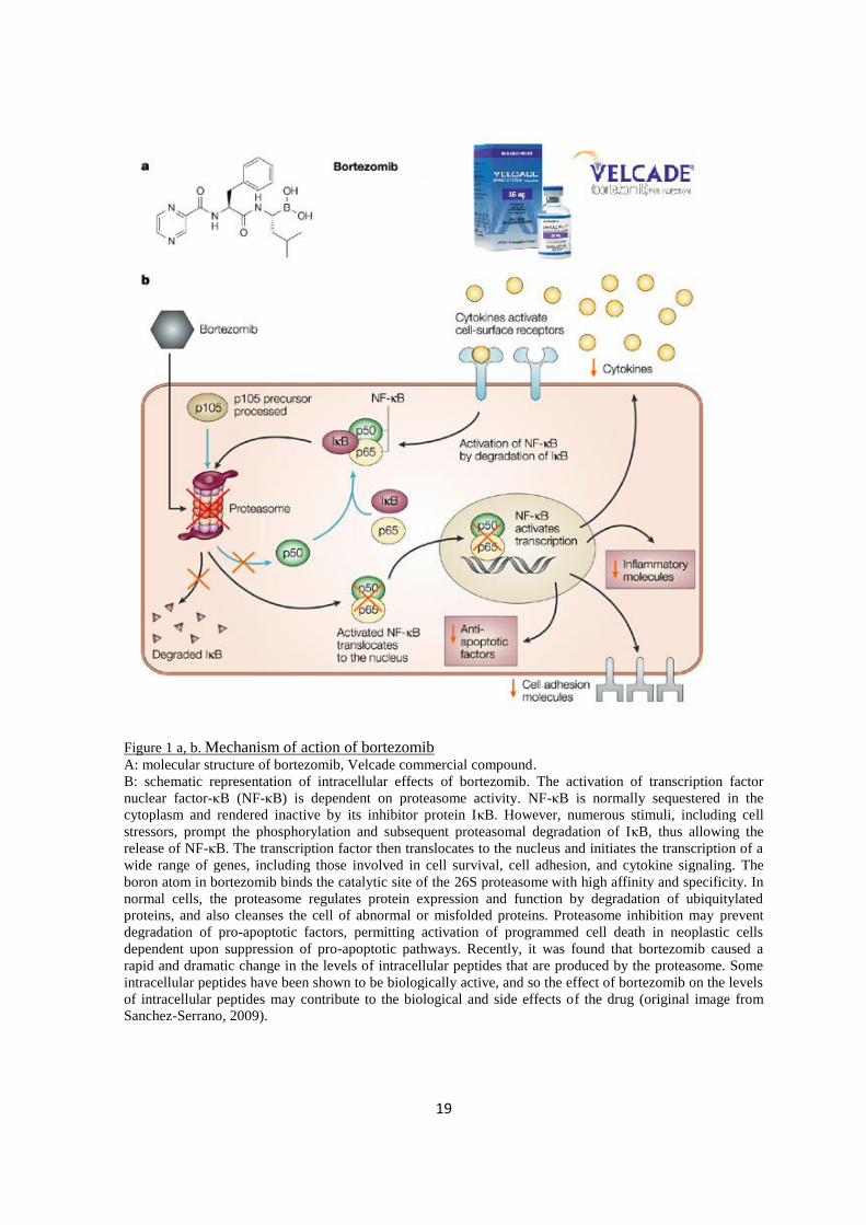
Figure 1 a, b. Mechanism of action of bortezomib
A: molecular structure of bortezomib, Velcade commercial compound.
B: schematic representation of intracellular effects of bortezomib. The activation of transcription factor
nuclear factor-κB (NF-κB) is dependent on proteasome activity. NF-κB is normally sequestered in the
cytoplasm and rendered inactive by its inhibitor protein IκB. However, numerous stimuli, including cell
stressors, prompt the phosphorylation and subsequent proteasomal degradation of IκB, thus allowing the
release of NF-κB. The transcription factor then translocates to the nucleus and initiates the transcription of a
wide range of genes, including those involved in cell survival, cell adhesion, and cytokine signaling. The
boron atom in bortezomib binds the catalytic site of the 26S proteasome with high affinity and specificity. In
normal cells, the proteasome regulates protein expression and function by degradation of ubiquitylated
proteins, and also cleanses the cell of abnormal or misfolded proteins. Proteasome inhibition may prevent
degradation of pro-apoptotic factors, permitting activation of programmed cell death in neoplastic cells
dependent upon suppression of pro-apoptotic pathways. Recently, it was found that bortezomib caused a
rapid and dramatic change in the levels of intracellular peptides that are produced by the proteasome. Some
intracellular peptides have been shown to be biologically active, and so the effect of bortezomib on the levels
of intracellular peptides may contribute to the biological and side effects of the drug (original image from
Sanchez-Serrano, 2009).

20
1.2 Dorsal Root Ganglia
Afferent sensory fibers coalesce together to enter the spinal cord via the dorsal root.
Primary afferent neuronal cell bodies are grouped in a swelling along the dorsal root
extension, just outside the spinal canal and lying near or within the intervertebral foramina,
to form the dorsal root ganglia (DRG). Entering the spinal cord at its dorsal surface in an
area known as the dorsal root entry zone (DREZ), small, medium-size, and large afferent
fibers perform their various functions with glutamate serving as the primary
neurotransmitter (Usunoff et al., 2006). The DRG contain the primary afferent neuronal
perikarya, each surrounded by the satellite glial cells, nerve fibers and Schwann cells
(Lieberman, 1976; Hanani, 2005). DRG neurons are pseudo-unipolar, i.e. they emit a
single axon that, a few tens or hundreds µm from the soma, divides in two branches that
act as a single axon, often referred to as the distal process and the proximal process. The
action potential in DRG neurons may initiate in the distal process in the periphery, bypass
the cell body, and continue to propagate along the proximal process until enters the dorsal
root and reaches the synaptic terminal in the dorsal horn of the spinal cord (Lawson et al.,
1987; Lawson, 1992; Devor, 1999). A capsule composed of connective tissue and a
perineurium, similar to that of peripheral nerves, surrounds the entire DRG and keeps the
microenvironment separate from surrounding extracellular fluid (Olsson, 1990).
DRG neurons can be classified into various subpopulations based on their anatomy,
neurochemistry and physiology (Lawson, 1992; Snider and McMahon, 1998; Hunt and
Mantyh, 2001). The Nissl staining allows to distinguish the DRG neurons into large-light
and small-dark cells. Large cells show a rather pale and unevenly stained cytoplasm, due to
aggregations of Nissl substance interspersed with lightly stained regions, contain
microtubules and a large amount of neurofilaments; they comprise approximately 40% of
lumbar DRG cells (Usunoff et al., 2006). Large DRG neurons have mostly myelinated
axons that conduct in the Aα/β range and receive input from peripheral mechanoreceptors
(Harper and Lawson, 1985; Sommer et al., 1985; LaMotte et al., 1991; Lawson and
Waddell, 1991; Willis and Coggeshall 1991; LaMotte et al., 1992; Truong et al., 2004).
The small-dark cells represent the second major group of DRG neurons; they can be
further subdivided into neuropeptidergic and non neuropeptidergic neurons. The former
21
constitutively express neuropeptides and the calcitonin gene related peptide (CGRP)
represents the best marker for them.They emit unmyelinated axons, namely C fibres, and
innervate mainly polymodal nociceptors (McCarthy and Lawson, 1990; Lawson et al.,
1997). The latter, identifiable by using a variety of histochemical markers, have been
especially characterized by their binding of the lectin Griffonia simplicifolia IB4 (Streit et
al., 1986) or by their expression of a nonlysosomal fluoride-resistant acid phosphatase
(FRAP) (Silverman and Kruger, 1988). This group of small-diameter cells (which will be
referred to as IB4+) also emits unmyelinated axons (C fibres) that are likely to mediate
nociception (Silverman and Kruger, 1990; Alvarez et al., 1991).
While these categories are largely histochemically distinct, at the same time there is also
some degree of overlap among them. Thus, a group that is intermediate in size between the
large-sized and the small-sized neuropeptidergic ones is represented by CGRP- and
substance P (SP) -containing cells that are medium-sized and emit finely myelinated, Aδ
axons. Most of these cells are nociceptors of the high-threshold mechanoreceptor type
(McCarthy and Lawson, 1990; Lawson et al., 1996, 1997). Further overlap occurs between
the IB4 binding and CGRP-containing populations (Bennett et al., 1996).
An additional characterization allows to differentiate DRG neurons on the basis of their
responsiveness to selective target-derived neurotrophic proteins. Availability of
neurotrophic proteins, such as nerve growth factor (NGF) and glial cell line-derived
neurotrophic factor (GDNF), has been directly related to the maintenance of mature
neuronal phenotype and neuronal ability to react to injury. In this context, non-peptidergic
cells appear to be regulated by GDNF since they bear ret, the high affinity GDNF receptor,
whereas peptidergic neurons and large-sized ones, bearing the high affinity trkA receptor,
are regulated by NGF (Priestley et al., 2002).
Subsets of both types of small-sized neurons show a range of expression levels of TRPV1
capsaicin receptor, most likely making them responsive to similar types of noxious stimuli
(Bennett et al., 1996; Priestley et al., 2002).
22
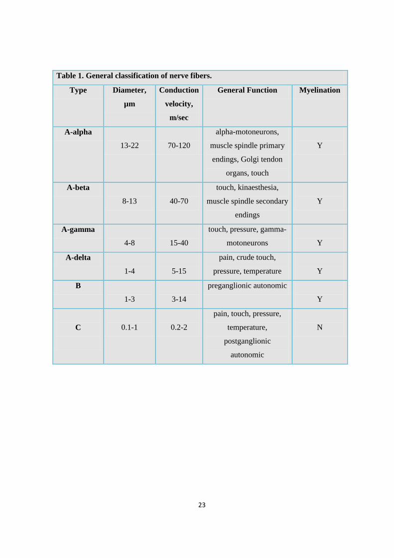
1.2.1 Nociceptors and nerve fiber classification
The term nociception indicates the reception of input delivered to the central nervous
system by sensory receptors known as nociceptors, i.e. the peripheral endings of primary
afferent neurons whose cell bodies are located in the DRG and cranial sensory ganglia.
Nociceptors provide information about tissue injury, though not all information delivered
through this system will be perceived as painful (Usunoff et al., 2006; Julius and Basbaum,
2001).
Afferent fibers involved in pain transmission can be classified in three types (cfr Table 1),
according to their structure, degree of myelination, and function:
A-beta fibers, or mechanoreceptors, transmit sensory information regarding touch,
vibration and hair deflection; they are myelinated large-diameter low-threshold
fibers; they transmit with a conduction velocity of 40-70 m/sec and may contribute
to the development of pathological pain sensation (Ren, 1996) (cfr Table 1);
A-delta fibers respond to noxious mechanical stimulation. They are small-diameter,
myelinated fibers with slower conduction velocity (5-15 m/sec) compared to A-beta
fibers; among them are the temperature-sensitive nociceptors which are sensitive to
intense heat and cold (Cook et al., 1987; Ren, 1996) (cfr Table 1).
C-type polymodal nociceptors originate in the epidermis and deep receptors located
in ligaments, muscles, and connective tissues. They respond to specific chemical
compounds such as histamine, bradykinin, serotonin, prostaglandins, proteolytic
enzymes, potassium and acids. These fibers are the smallest and slowest of the
nociceptors, conducting impulses at approximately 0,5 m/s: C fibers are
unmyelinated and require intense mechanical, thermal, or chemical stimulation to
transmit pain impulses. Physiologically, the C-nociceptor responds directly to
increasing intensity of noxious stimulation -the greater the stimulus strength, the
more vigorous the response (Besson and Chaouch, 1987). Repeated stimulation,
however, enhances C-nociceptor responsivity to a given stimulus strength
(LaMotte, 1984; reviewed by Campbell et al., 1989) (cfr Table 1).
23
Table 1. General classification of nerve fibers.
Type Diameter,
µm
Conduction
velocity,
m/sec
General Function Myelination
A-alpha
13-22
70-120
alpha-motoneurons,
muscle spindle primary
endings, Golgi tendon
organs, touch
Y
A-beta
8-13
40-70
touch, kinaesthesia,
muscle spindle secondary
endings
Y
A-gamma
4-8
15-40
touch, pressure, gamma-
motoneurons
Y
A-delta
1-4
5-15
pain, crude touch,
pressure, temperature
Y
B
1-3
3-14
preganglionic autonomic
Y
C
0.1-1
0.2-2
pain, touch, pressure,
temperature,
postganglionic
autonomic
N
24
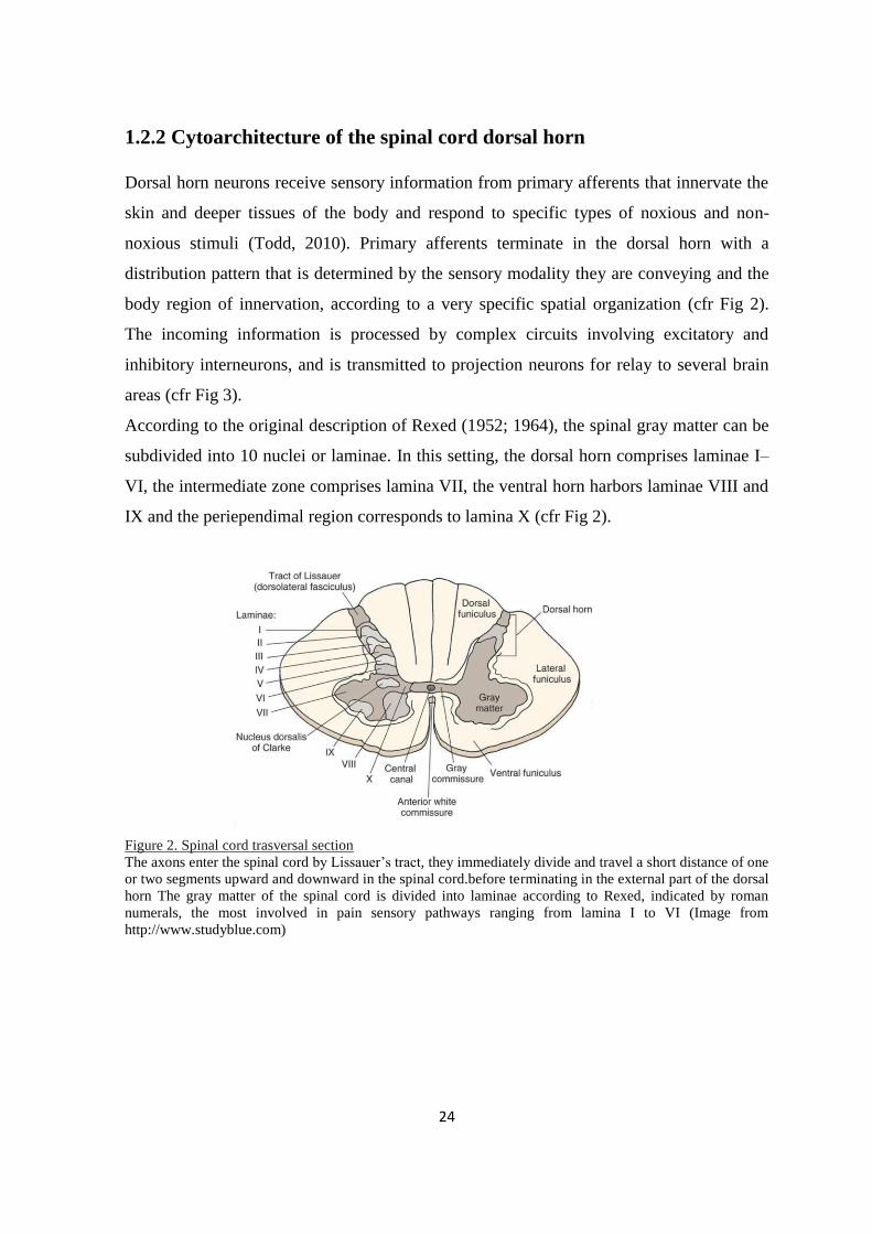
1.2.2 Cytoarchitecture of the spinal cord dorsal horn
Dorsal horn neurons receive sensory information from primary afferents that innervate the
skin and deeper tissues of the body and respond to specific types of noxious and non-
noxious stimuli (Todd, 2010). Primary afferents terminate in the dorsal horn with a
distribution pattern that is determined by the sensory modality they are conveying and the
body region of innervation, according to a very specific spatial organization (cfr Fig 2).
The incoming information is processed by complex circuits involving excitatory and
inhibitory interneurons, and is transmitted to projection neurons for relay to several brain
areas (cfr Fig 3).
According to the original description of Rexed (1952; 1964), the spinal gray matter can be
subdivided into 10 nuclei or laminae. In this setting, the dorsal horn comprises laminae I–
VI, the intermediate zone comprises lamina VII, the ventral horn harbors laminae VIII and
IX and the periependimal region corresponds to lamina X (cfr Fig 2).
Figure 2. Spinal cord trasversal section
The axons enter the spinal cord by Lissauer’s tract, they immediately divide and travel a short distance of one
or two segments upward and downward in the spinal cord.before terminating in the external part of the dorsal
horn The gray matter of the spinal cord is divided into laminae according to Rexed, indicated by roman
numerals, the most involved in pain sensory pathways ranging from lamina I to VI (Image from
http://www.studyblue.com)

25
Laminae I-VI
The dorsomarginal nucleus - Lamina I, capping the dorsal horn, receives many
afferent fibers carrying pain, temperature, and light touch sensations. Many
intersegmental pathways arise from this layer that also contributes fibers for the
lateral and ventral spinothalamic tracts (Patestas and Gartner, 2006; Gaudio, 2012)
The substantia gelatinosa of Rolando - Lamina II, constitute the superficial
dorsal horn and is characterized by the presence of numerous small neurons.
Lamina II can be further divided into outer (IIo) and inner (IIi) parts, with IIi
having a somewhat lower density of neurons. This nucleus extends the entire length
of the cord and is most prominent in the cervical and lumbar levels. C fibers
delivering pain, temperature, and light touch information terminate on neurons in
dorsal horn lamina II. Cells in this lamina also form intersegmental connections.
Descending fibers from higher centers (such as the cerebral cortex) form excitatory
and inhibitory synapses with the cells of the substantia gelatinosa, thus modifying
the incoming pain and temperature sensations (Ralston, 1979; Usunoff et al., 2006;
Todd, 2010)
The nucleus proprius - Laminae III and IV, extends the entire length of the
spinal cord. It is composed of densely clustered large nerve cell bodies, located just
ventral to the substantia gelatinosa, and receives the central processes of the
majority of the DRG neurons. The nucleus proprius receives pain, light touch, and
temperature sensations and provides input to the lateral and ventral spinothalamic
tracts (Beal and Cooper, 1978; Usunoff et al., 2006)
Lamina V extends across the neck of the dorsal horn and in all but the thoracic
region is divided into medial and lateral portions. The lateral portion consists of the
reticular nucleus that is most conspicuous in the cervical neuromeres. The C and
Aβ nociceptive fibers terminate in this layer. Morevoer, corticospinal synapses
have been identified in this lamina.
Lamina VI is a wide zone most prominent in the cervical and lumbar
enlargements. At these levels, it is divided into medial and lateral zones. Terminals
from the posterior roots end in the medial region whereas descending fiber tracts
project to the lateral zone.
26
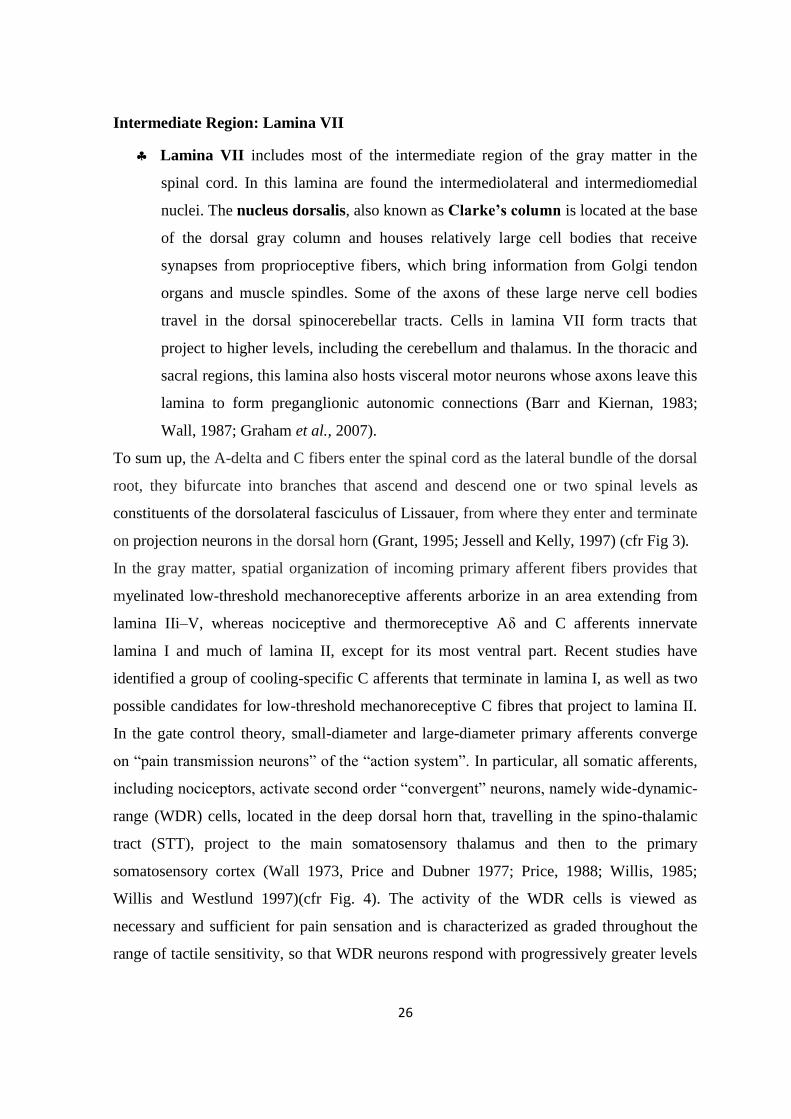
Intermediate Region: Lamina VII
Lamina VII includes most of the intermediate region of the gray matter in the
spinal cord. In this lamina are found the intermediolateral and intermediomedial
nuclei. The nucleus dorsalis, also known as Clarke’s column is located at the base
of the dorsal gray column and houses relatively large cell bodies that receive
synapses from proprioceptive fibers, which bring information from Golgi tendon
organs and muscle spindles. Some of the axons of these large nerve cell bodies
travel in the dorsal spinocerebellar tracts. Cells in lamina VII form tracts that
project to higher levels, including the cerebellum and thalamus. In the thoracic and
sacral regions, this lamina also hosts visceral motor neurons whose axons leave this
lamina to form preganglionic autonomic connections (Barr and Kiernan, 1983;
Wall, 1987; Graham et al., 2007).
To sum up, the A-delta and C fibers enter the spinal cord as the lateral bundle of the dorsal
root, they bifurcate into branches that ascend and descend one or two spinal levels as
constituents of the dorsolateral fasciculus of Lissauer, from where they enter and terminate
on projection neurons in the dorsal horn (Grant, 1995; Jessell and Kelly, 1997) (cfr Fig 3).
In the gray matter, spatial organization of incoming primary afferent fibers provides that
myelinated low-threshold mechanoreceptive afferents arborize in an area extending from
lamina IIi–V, whereas nociceptive and thermoreceptive Aδ and C afferents innervate
lamina I and much of lamina II, except for its most ventral part. Recent studies have
identified a group of cooling-specific C afferents that terminate in lamina I, as well as two
possible candidates for low-threshold mechanoreceptive C fibres that project to lamina II.
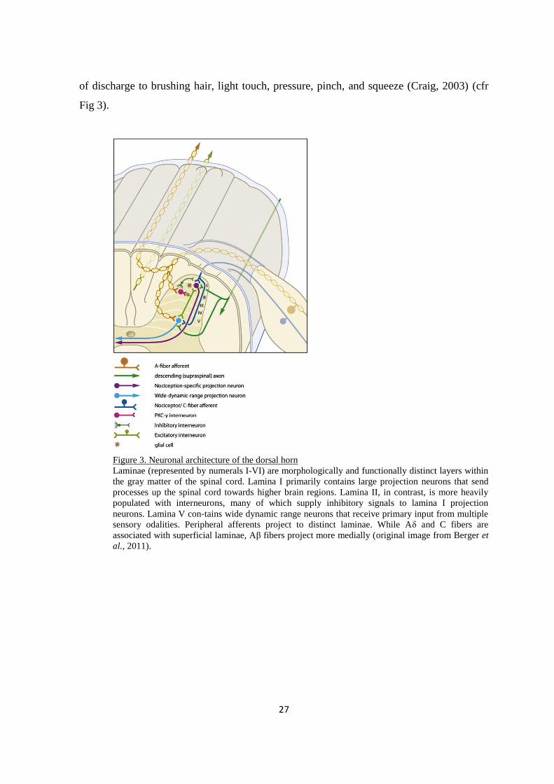
In the gate control theory, small-diameter and large-diameter primary afferents converge
on “pain transmission neurons” of the “action system”. In particular, all somatic afferents,
including nociceptors, activate second order “convergent” neurons, namely wide-dynamic-
range (WDR) cells, located in the deep dorsal horn that, travelling in the spino-thalamic
tract (STT), project to the main somatosensory thalamus and then to the primary
somatosensory cortex (Wall 1973, Price and Dubner 1977; Price, 1988; Willis, 1985;
Willis and Westlund 1997)(cfr Fig. 4). The activity of the WDR cells is viewed as
necessary and sufficient for pain sensation and is characterized as graded throughout the
range of tactile sensitivity, so that WDR neurons respond with progressively greater levels
27
of discharge to brushing hair, light touch, pressure, pinch, and squeeze (Craig, 2003) (cfr
Fig 3).
Figure 3. Neuronal architecture of the dorsal horn
Laminae (represented by numerals I-VI) are morphologically and functionally distinct layers within
the gray matter of the spinal cord. Lamina I primarily contains large projection neurons that send
processes up the spinal cord towards higher brain regions. Lamina II, in contrast, is more heavily
populated with interneurons, many of which supply inhibitory signals to lamina I projection
neurons. Lamina V con‐tains wide dynamic range neurons that receive primary input from multiple
sensory odalities. Peripheral afferents project to distinct laminae. While Aδ and C fibers are
associated with superficial laminae, Aβ fibers project more medially (original image from Berger et
al., 2011).
28
Figure 4. Gate control theory
The theory is based on the understanding that pain is transmitted by two kinds of afferent nerve fibers. One is
the larger myelinated A-delta fiber, which carries quick, intense-pain messages. The other is the smaller,
unmyelinated "C" fiber, which transmits throbbing, chronic pain. A third type of nerve fiber, called A-beta, is
"nonnociceptive," meaning it does not transmit pain stimuli. In this concept, pain can be modulated by the
balance of the interactions among the nociceptive C fibers and non-nociceptive A-alpha (proprioception) and
A-beta afferent (touch) fibers of the peripheral nerves, and the interneurons and projection neurons of the
dorsal horn. The interneuron, which normally inhibits the projection neuron, is spontaneously active and,
thus, reduces (inhibits) the intensity of the noxious input from the C fibers. The influences exerted by the
spontaneous activity of the interneuron on the projection neuron are modulated by excitation from the non-
nociceptive A fibers and inhibition from the nociceptive.
C fibers. In essence, nociceptive C fibers tend to keep the gate open (enhancing perception of pain) by
inhibiting the inhibitory interneuron and exciting the projection neuron. The non-nociceptive A fibers tend to
keep the gate closed (suppression of pain) by exciting the inhibitory interneuron. In addition, the reflected
feedback descending influences from the brain can modulate the excitability of these neurons (original image
from http://www.positivehealth.com).

29
1.3. Pain sensory pathways
Transmission of pain through the chain of primary afferent neurons and dorsal horn cells is
influenced by many factors, some of which originating within the segment, some from
higher centers. The anterolateral system (ALS) has long been known to be a major
pathway for the transmission of general somatic protopathic information to the cerebral
cortex. ALS comprises the spinothalamic tract (STT), the spinomesencephalic tract and the
spinoreticular tract. Fibers carrying pain and temperature ascend in the contralateral
spinothalamic tract. Compression, intrinsic disease, or deliberate section of STT result in
anesthesia of the contralateral body beginning 3 segments below the level of disruption.
STT shows both somatotopic and functional arrangement, as pain fibers are located
anteriorly, temperature ones are located posteriorly. Ascending spinothalamic fibers
terminate in the ventral posterior lateral nucleus and in the intralaminar nuclei (Willis,
1985, Willis and Westlund, 1997; Craig, 2003; Jacobson and Marcus, 2011) (cfr Fig. 5).
Only when nociceptive information reaches the thalamus and cerebral cortex, should we
talk about pain. Pain perception by these higher centers triggers affective responses and
suffering behaviours. The pain experience depends on the circumstances that may alter the
response and varies so much from person to person that it has be also defined as "a
subjective perception with a psychological dimension" (Collins et al., 1960; Hunt and
Mantyh 2001; Scholz and Woolf 2002; Trafton and Basbaum, 2000).
At thalamic level, sensory information from the body, limb and back of the head is
collected by neurons of several thalamic nuclei including the ventral posterolateral (VPL),
posterior (PTh) and intralaminar nuclei, whereas somatosensory information from the head
reaches the ventral posteromedial nucleus (VPM).
As regards the processing of nociceptive information, it seems that the thalamus tells us
about the quality of information (the "what" component) and the somatosensory cortex
tells us about the spatial localition of the sensation (the "where" component). The (lateral)
spinothalamic tract originates from projection neurons of laminae I, V, VI, and VII. That
terminate primarily in VP and PTh thalamic nuclei. Some collateral branches terminate in
the brainstem reticular formation. This tract conveys information perceived with an overlay
of the discriminative aspects associated with various subtleties associated with the
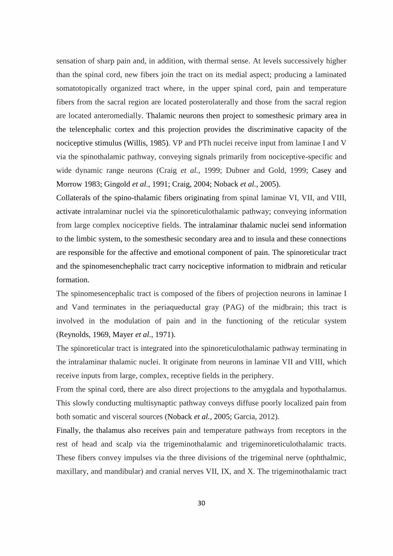
30
sensation of sharp pain and, in addition, with thermal sense. At levels successively higher
than the spinal cord, new fibers join the tract on its medial aspect; producing a laminated
somatotopically organized tract where, in the upper spinal cord, pain and temperature
fibers from the sacral region are located posterolaterally and those from the sacral region
are located anteromedially. Thalamic neurons then project to somesthesic primary area in
the telencephalic cortex and this projection provides the discriminative capacity of the
nociceptive stimulus (Willis, 1985). VP and PTh nuclei receive input from laminae I and V
via the spinothalamic pathway, conveying signals primarily from nociceptive-specific and
wide dynamic range neurons (Craig et al., 1999; Dubner and Gold, 1999; Casey and
Morrow 1983; Gingold et al., 1991; Craig, 2004; Noback et al., 2005).
Collaterals of the spino-thalamic fibers originating from spinal laminae VI, VII, and VIII,
activate intralaminar nuclei via the spinoreticulothalamic pathway; conveying information
from large complex nociceptive fields. The intralaminar thalamic nuclei send information
to the limbic system, to the somesthesic secondary area and to insula and these connections
are responsible for the affective and emotional component of pain. The spinoreticular tract
and the spinomesenchephalic tract carry nociceptive information to midbrain and reticular
formation.
The spinomesencephalic tract is composed of the fibers of projection neurons in laminae I
and Vand terminates in the periaqueductal gray (PAG) of the midbrain; this tract is
involved in the modulation of pain and in the functioning of the reticular system
(Reynolds, 1969, Mayer et al., 1971).
The spinoreticular tract is integrated into the spinoreticulothalamic pathway terminating in
the intralaminar thalamic nuclei. It originate from neurons in laminae VII and VIII, which
receive inputs from large, complex, receptive fields in the periphery.
From the spinal cord, there are also direct projections to the amygdala and hypothalamus.
This slowly conducting multisynaptic pathway conveys diffuse poorly localized pain from
both somatic and visceral sources (Noback et al., 2005; Garcia, 2012).
Finally, the thalamus also receives pain and temperature pathways from receptors in the
rest of head and scalp via the trigeminothalamic and trigeminoreticulothalamic tracts.
These fibers convey impulses via the three divisions of the trigeminal nerve (ophthalmic,
maxillary, and mandibular) and cranial nerves VII, IX, and X. The trigeminothalamic tract
31
is included in the lateral pain system (Patestas and Gartner, 2006; Gaudio, 2012; Noback et
al., 2005) (cfr Fig. 5).
Figure 5. Schematic representation of the anterolateral system
First order neurons reside in the DRG and collect sensory information by means of their centrifugal branches
reaching the periphery. Their centripetal branches enter the spinal cord, ascend 1-2 levels, and terminate in
the substantia gelatinosa; second order neurones carry the sensory information from there to the thalamus.
Fibers arising from second order neurons, before entering the spinothalamic tract, decussate and split: those
carrying crude touch and pressure enter the lateral spinothalamic tract, those carrying pain and temperature
enter the anterior spinothalamic tract. Although functionally distinct, these tracts run alongside each other,
and they can be considered as a single pathway. The fibres travel in their respective pathways, synapsing in
the thalamus. Third order thalamic neurones transfer the sensory signals to the primary sensory cortex. The
descending pathway is represented by the dashed lines (original image from Firestein, Kelley's Textbook of
Rheumatology, 8th ed.).
32
1.3.1 Central and peripheral mechanisms of nociceptive processing
The spinal cord actively amplifies the spinal nociceptive processing because nociceptive
spinal cord neurons change their excitability to inputs from the periphery under painful
conditions. On the other hand, it has to be reminded that the spinal cord is under the
influence of descending influences (Schaible, 2007).
Under normal conditions, the release of excitatory amino acids by afferent fibers induces a
depolarization of dorsal horn neurons. Glutamate, released during tonic activation of C
fibers, is the principal excitatory transmitter acting at the synapse between primary afferent
nociceptors and dorsal horn cells (Alvarez et al., 2010). Glutamatergic receptors on the
second order sensory neurons are mainly ionotropic, i.e. directly coupled to cation
channels, which can be further subdivided into AMPA (amino 3-hydroxy-5-methyl-4-
isoxazolepropionic acid)/kainate and NMDA receptors. Glutamate initially binds to AMPA
receptor that produces a rapid depolarization followed by activation of the NMDA receptor
(Lovinger, 2008; Latremoliere and Woolf, 2009).
Excitatory neuropeptides are co-localized with glutamate. Neuropeptide-mediated
excitatory postsynaptic potentials usually occur after a latency of seconds and are long-
lasting. They may not be sufficient to evoke action potential generation but act
synergistically with glutamate (Urban et al., 1994) Substance P (SP) is released mainly in
the superficial dorsal horn by electrical stimulation of unmyelinated fibres and during
noxious mechanical, thermal or chemical stimulation of the skin and deep tissue (Afrah et
al., 2002; Khasabov et al., 2002; Ma and Woolf, 1995; Mantyh et al., 1997). Neurokinin-1
(NK-1) receptors for SP are mainly located on dendrites and cell bodies of dorsal horn
neurons in laminae I, IV–VI and X (Gauriau and Bernard 2002). Upon strong activation by
SP, NK-1 receptors are internalized. In addition, neurokinin A (NKA) is found in small
DRG cells and in the dorsal horn and spinally released upon noxious stimulation. CGRP,
which is often colocalized with SP in DRG neurons, is released in the spinal cord upon
electrical stimulation of thin fibres and upon noxious mechanical and thermal stimulation,
potentiates the effects of SP (by inhibiting its enzymatic degradation and potentiating its
release). It participates in the phenomenon of central sensitization, i.e. the changes in
synaptic efficacy between afferent neurons and central neurons that increase the perception
of pain. Once released by primary afferent terminals, it acts through postsynaptic CGRP

33
receptors, which activate PKA and PKC (Woolf and Wiesenfeld-Hallin, 1976). CGRP
binding sites are located in lamina I and in the deep dorsal horn. Blockade of CGRP effects
reduces nociceptive responses (Schaible, 2006).
Under pathological conditions, the second and third order neurons undergo widespread
neuronal plasticity and neuronal rewiring to become hyperactive. The result of these
processes may lead to the transformation of an initially adaptive reactivity in a series of
counterproductive, often maladaptive responses. Observed changes include modulation of
synaptic transmission and induction of LTP (Ueda, 2006; Premkumar, 2010). Peripheral
sensitization involves an increase in the responsiveness of nociceptive ion channels and a
reduction in activation threshold of nociceptive neurons (Julius and Basbaum, 2009; Woolf
and Salter, 2008). Sensitization of nociceptive ion channels can result in ectopic or
spontaneous discharges, alteration in ion channel expression, and increased neuronal
sprouting (Nordin et al., 1997; Premkumar, 2010).
Prolonged activation of C fibers by noxious stimuli depolarizes dorsal horn
neurons and induces direct activation of NMDA receptors. Activation of NMDA
receptors subsequently allows a massive influx of calcium within the cell, which
provokes a cascade of intracellular events leading to the long-lasting modification
of the properties of dorsal horn neurons. These events, including the activation of
the calcium-dependent protein kinase C (PKC) and of the nitrous oxide (NO)
synthase, may in turn lead to phosphorylation of the NMDA receptor and thereby
enhance the calcium current generated by a given glutamate release (Kampa et
al., 2004; Van Dongen, 2009; Premkumar, 2010)(cfr Fig. 6).
34
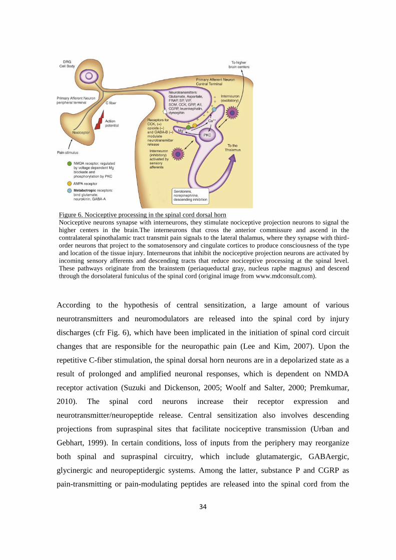
Figure 6. Nociceptive processing in the spinal cord dorsal horn
Nociceptive neurons synapse with interneurons, they stimulate nociceptive projection neurons to signal the
higher centers in the brain.The interneurons that cross the anterior commissure and ascend in the
contralateral spinothalamic tract transmit pain signals to the lateral thalamus, where they synapse with third-
order neurons that project to the somatosensory and cingulate cortices to produce consciousness of the type
and location of the tissue injury. Interneurons that inhibit the nociceptive projection neurons are activated by
incoming sensory afferents and descending tracts that reduce nociceptive processing at the spinal level.
These pathways originate from the brainstem (periaqueductal gray, nucleus raphe magnus) and descend
through the dorsolateral funiculus of the spinal cord (original image from www.mdconsult.com).
According to the hypothesis of central sensitization, a large amount of various
neurotransmitters and neuromodulators are released into the spinal cord by injury
discharges (cfr Fig. 6), which have been implicated in the initiation of spinal cord circuit
changes that are responsible for the neuropathic pain (Lee and Kim, 2007). Upon the
repetitive C-fiber stimulation, the spinal dorsal horn neurons are in a depolarized state as a
result of prolonged and amplified neuronal responses, which is dependent on NMDA
receptor activation (Suzuki and Dickenson, 2005; Woolf and Salter, 2000; Premkumar,
2010). The spinal cord neurons increase their receptor expression and
neurotransmitter/neuropeptide release. Central sensitization also involves descending
projections from supraspinal sites that facilitate nociceptive transmission (Urban and
Gebhart, 1999). In certain conditions, loss of inputs from the periphery may reorganize
both spinal and supraspinal circuitry, which include glutamatergic, GABAergic,
glycinergic and neuropeptidergic systems. Among the latter, substance P and CGRP as
pain-transmitting or pain-modulating peptides are released into the spinal cord from the
35
central terminals of thinly myelinated and unmyelinated primary sensory neurons and are
involved in central sensitization (Dubin and Patapoutian, 2010).
In addition, nociceptive ion channels contribute to the specificity of primary afferent fibers
carrying a selective modality of pain. Among these, the identification of the transient
receptor potential type 1 receptor -TRPV1- was the major catalyst that launched the fields
of somatosensory and pain transduction research to the molecular level (Stucky et al.,
2009).
TRPV1 is a non selective cation channel which likely consists of four subunits, with each
containing 6 transmembrane domains (Voets et al., 2005), it is mainly expressed in
peptidergic neurons, and to a lesser extent in the non-peptidergic nociceptors, the
expression also occurs in various other brain regions and in several non-neuronal tissues.
Particularly TRPV1 is expresed in a subset of small-sized DRG, trigeminal and nodose
ganglia nociceptive neurons bearing C and Aδ fibers (Caterina et al, 1997; Tominaga et al.,
1998) where mediates sensory perception especially in nociception. (Caterina et al., 1997)
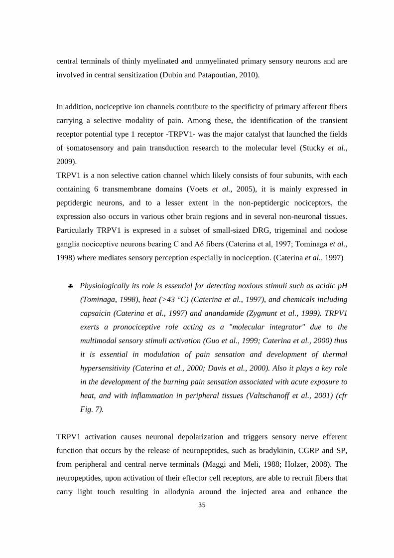
Physiologically its role is essential for detecting noxious stimuli such as acidic pH
(Tominaga, 1998), heat (>43 °C) (Caterina et al., 1997), and chemicals including
capsaicin (Caterina et al., 1997) and anandamide (Zygmunt et al., 1999). TRPV1
exerts a pronociceptive role acting as a "molecular integrator" due to the
multimodal sensory stimuli activation (Guo et al., 1999; Caterina et al., 2000) thus
it is essential in modulation of pain sensation and development of thermal
hypersensitivity (Caterina et al., 2000; Davis et al., 2000). Also it plays a key role
in the development of the burning pain sensation associated with acute exposure to
heat, and with inflammation in peripheral tissues (Valtschanoff et al., 2001) (cfr
Fig. 7).
TRPV1 activation causes neuronal depolarization and triggers sensory nerve efferent
function that occurs by the release of neuropeptides, such as bradykinin, CGRP and SP,
from peripheral and central nerve terminals (Maggi and Meli, 1988; Holzer, 2008). The
neuropeptides, upon activation of their effector cell receptors, are able to recruit fibers that
carry light touch resulting in allodynia around the injected area and enhance the
36
sensitization of nociceptors (Holzer, 2008; Szallasi and Blumberg, 2009). Repeated
activation of the TRPV1 receptor causes the overload of intracellular Ca2+, resulting in
oxidative stress and apoptotic cell injury (Shin et al., 2003; Kim et al., 2005). For these
reasons the receptor has also been shown to play a role in certain chronic pain conditions,
such as neuropathic pain, osteoarthritis, bone cancer pain, inflammatory bowel disease and
migraine (O'Neill et al., 2012). Chronic pain-induced sensitization also intensifies the
expression of TRPV1 receptor in sensory neurons through transcriptional and translational
regulation, post-translational changes and altered trafficking, contributing to the
development of pathological pain states in which increased sensitivity to noxious stimuli
occurs (Hucho and Levine, 2007)
Capsaicin-induced nocifensive behaviour and hyperalgesia in rodents and pain in humans
are well established (Gilchrist et al., 1996; Simone et al., 1987; Szolcsányi, 1977).
TRPV1 antagonists have shown efficacy in animal models of both inflammatory and
neuropathic pain (Patapoutian et al., 2009) but systemic administration of TRPV1
antagonists commonly results in hyperthermia caused by peripheral TRPV1 blockade
(Steiner et al., 2007). Activation of spinal TRPV1 can generate central sensitization and
mechanical allodynia (Patwardhan et al., 2009) and spinal administration of TRPV1
antagonists can attenuate mechanical allodynia induced by nerve injury (Patapoutian et al.,
2009), but the cell types or circuits underlying these effects are unknown (Kim et al.,
2012).
37
Figure 7. TRPV1 activation
The TRPV1 channel can be activated by vanilloids such as capsaicin, as well as endogenous stimulators
including H+, heat, lipoxygenase products and anandamide. TRPV1 channel function is upregulated by
several endogenous mediators present in inflammatory conditions which decrease the threshold for
activation of the channel. Under these conditions, TRPV1 can be activated by physiological body
temperature, slight acidification or lower concentration of TRPV1 agonists. There is evidence that
TRPV1 plays a role in the development of pathophysiological changes and symptoms in chronic pain
sensation Activation of TRPV1 by capsaicin results in sensory neuronal depolarization, and can induce
local sensitization. High concentrations of capsaicin or repeated applications can produce a persistent
local effect on cutaneous nociceptors, which is best described as defunctionalization and constituted by
reduced spontaneous activity and a loss of responsiveness to a wide range of sensory stimuli. When
activated by a combination of heat, acidosis, or endogenous/exogenous agonists, TRPV1 may open
transiently and initiate depolarization mediated by the influx of sodium and calcium ions. In the
nociceptive sensory nerves which selectively express TRPV1 (mostly C- and some Aδ-fibres),
depolarization results in action potentials, which propagate into the spinal cord and brain, and may be
experienced as warming, burning, stinging, or itching sensations. (original Image from
http://www.medscape.com).
38
1.4. The importance of animal models of chemotherapy-induced PN
In the past 20 years, several animal models have been published and used to further
investigate mechanisms of anticancer agent-induced neuropathic pain. Most frequently
experimental models have been developed in rodents (Authier et al., 2009). Many models
were not intended to suggest an immediate translation into clinical use, but rather to
illustrate a concept and to identify a possible novel mechanism that could be exploited to
avoid this important chemotherapy-induced side effect. Collectively, findings achieved by
means of animal models add to the complexity and multiplicity of the mechanisms
underlying the chemotherapy-induced PN.
Primary afferent nerve fibers affected by chemotherapy-induced neurotoxicity exhibit both
positively and negatively regulated nociceptive thresholds (Nahman-Averbuch et al.,
2011). As a whole, nociceptive thresholds are increased when there occurs nerve fiber loss,
whereas are reduced as a result of peripheral and central sensitization (Nahman-Averbuch
et al., 2011). So, thermal hypoalgesia has been associated to chemotherapy-induced PN in
both humans (Dougherty et al., 2004; Cata et al., 2006; Attal et al., 2009; Nahman-
Averbuch et al., 2011) and rodent models (Authier et al., 2000, 2003; Fischer et al., 2001;
Cata et al., 2006, 2008; Garcia et al., 2008; Hori et al., 2010; Xiao et al., 2012; Zheng et
al., 2012). Cold allodynia, a characteristic symptom of painful chemotherapy-induced PN,
also occurs in both patients (Cata et al., 2006) and experimental models (Cata et al., 2006;
Authier et al., 2009; Xiao et al., 2012).
Variations in nociceptive thresholds are accompanied by neurochemical changes. For
example, in the DRGs and hindpaw skin of hyperalgesic mice TRPV1 expression levels
are increased, whereas they decrease in hypoalgesic animals (Pabbidi et al., 2008).
Conversely, thermal hypoalgesia may be underpinned by reduced TRPV1 expression and
function (Pabbidi et al., 2008).
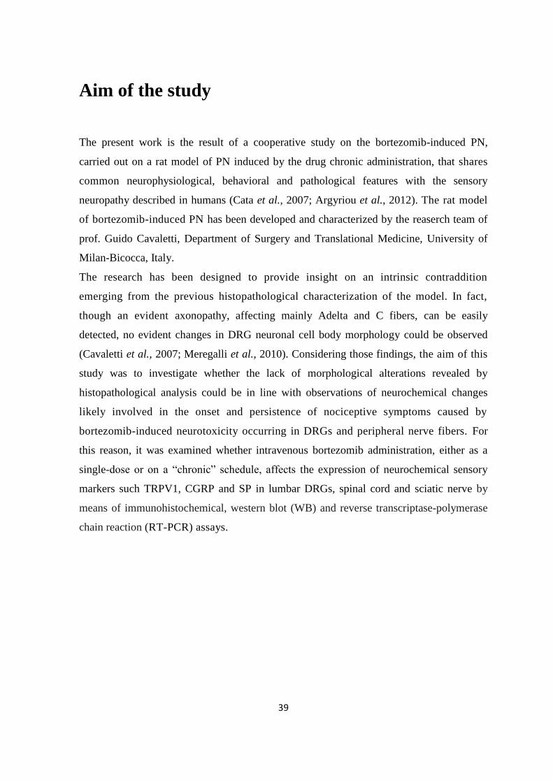
39
Aim of the study
The present work is the result of a cooperative study on the bortezomib-induced PN,
carried out on a rat model of PN induced by the drug chronic administration, that shares
common neurophysiological, behavioral and pathological features with the sensory
neuropathy described in humans (Cata et al., 2007; Argyriou et al., 2012). The rat model
of bortezomib-induced PN has been developed and characterized by the reaserch team of
prof. Guido Cavaletti, Department of Surgery and Translational Medicine, University of
Milan-Bicocca, Italy.
The research has been designed to provide insight on an intrinsic contraddition
emerging from the previous histopathological characterization of the model. In fact,
though an evident axonopathy, affecting mainly Adelta and C fibers, can be easily
detected, no evident changes in DRG neuronal cell body morphology could be observed
(Cavaletti et al., 2007; Meregalli et al., 2010). Considering those findings, the aim of this
study was to investigate whether the lack of morphological alterations revealed by
histopathological analysis could be in line with observations of neurochemical changes
likely involved in the onset and persistence of nociceptive symptoms caused by
bortezomib-induced neurotoxicity occurring in DRGs and peripheral nerve fibers. For
this reason, it was examined whether intravenous bortezomib administration, either as a
single-dose or on a “chronic” schedule, affects the expression of neurochemical sensory
markers such TRPV1, CGRP and SP in lumbar DRGs, spinal cord and sciatic nerve by
means of immunohistochemical, western blot (WB) and reverse transcriptase-polymerase
chain reaction (RT-PCR) assays.
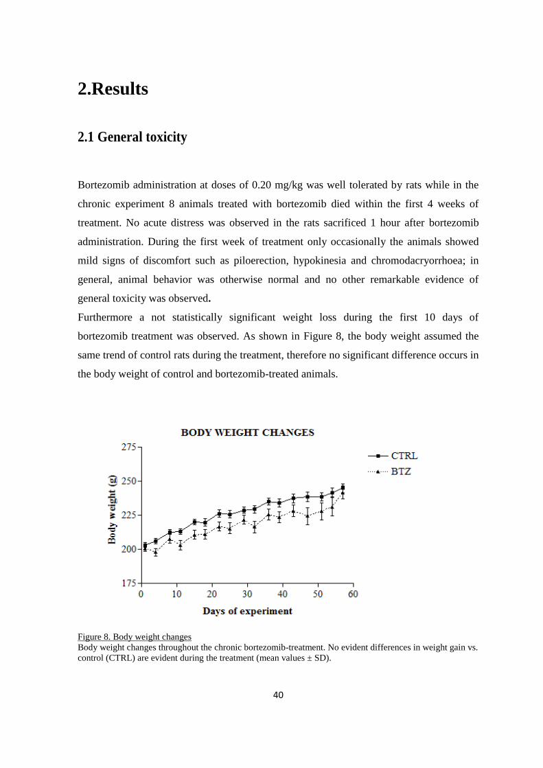
40
2.Results
2.1 General toxicity
Bortezomib administration at doses of 0.20 mg/kg was well tolerated by rats while in the
chronic experiment 8 animals treated with bortezomib died within the first 4 weeks of
treatment. No acute distress was observed in the rats sacrificed 1 hour after bortezomib
administration. During the first week of treatment only occasionally the animals showed
mild signs of discomfort such as piloerection, hypokinesia and chromodacryorrhoea; in
general, animal behavior was otherwise normal and no other remarkable evidence of
general toxicity was observed.
Furthermore a not statistically significant weight loss during the first 10 days of
bortezomib treatment was observed. As shown in Figure 8, the body weight assumed the
same trend of control rats during the treatment, therefore no significant difference occurs in
the body weight of control and bortezomib-treated animals.
Figure 8. Body weight changes
Body weight changes throughout the chronic bortezomib-treatment. No evident differences in weight gain vs.
control (CTRL) are evident during the treatment (mean values ± SD).
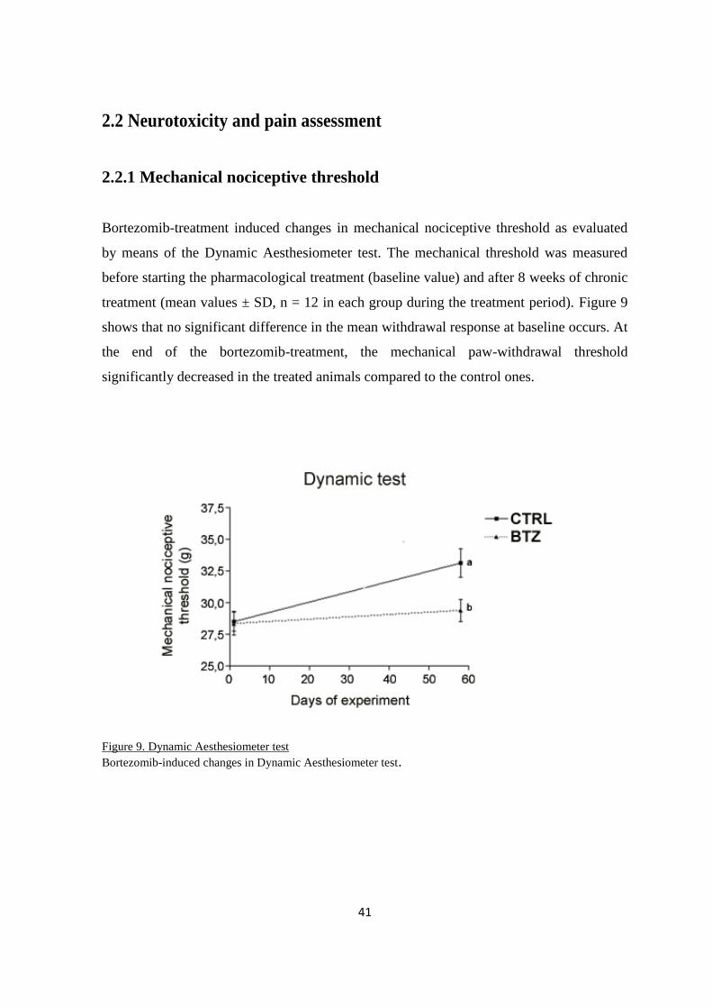
41
2.2 Neurotoxicity and pain assessment
2.2.1 Mechanical nociceptive threshold
Bortezomib-treatment induced changes in mechanical nociceptive threshold as evaluated
by means of the Dynamic Aesthesiometer test. The mechanical threshold was measured
before starting the pharmacological treatment (baseline value) and after 8 weeks of chronic
treatment (mean values ± SD, n = 12 in each group during the treatment period). Figure 9
shows that no significant difference in the mean withdrawal response at baseline occurs. At
the end of the bortezomib-treatment, the mechanical paw-withdrawal threshold
significantly decreased in the treated animals compared to the control ones.
Figure 9. Dynamic Aesthesiometer test
Bortezomib-induced changes in Dynamic Aesthesiometer test.
42
2.3 Immunohistochemistry
Tissue sections were processed for immunohistochemistry for both ABC method and
immunofluorescence to ascertain possible neurochemical changes in TRPV1 receptor and
the neuropeptides SP and CGRP, likely involved in neuropathic pain transmission.
2.3.1 Dorsal Root Ganglia
The immunostaining for TRPV1-, CGRP- and SP-like protein in the DRG was intense in a
subpopulation of small neurons, whereas a more moderate level of staining was observed
in other small and medium neurons (cfr Fig 10, 11).
Figure 10. Bortezomib acute treatment
Immunoreactivity to TRPV1 (A, B), CGRP (C, D) and SP (E-F-G) in representative sections of lumbar DRG
from control (ctrl) (A, C, E) and Acute bortezomib-treated rats (B, D, F, G). Scale bars = 25 μm.
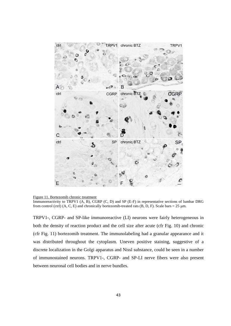
43
Figure 11. Bortezomib chronic treatment
Immunoreactivity to TRPV1 (A, B), CGRP (C, D) and SP (E-F) in representative sections of lumbar DRG
from control (ctrl) (A, C, E) and chronically bortezomib-treated rats (B, D, F). Scale bars = 25 μm.
TRPV1-, CGRP- and SP-like immunoreactive (LI) neurons were fairly heterogeneous in
both the density of reaction product and the cell size after acute (cfr Fig. 10) and chronic
(cfr Fig. 11) bortezomib treatment. The immunolabeling had a granular appearance and it
was distributed throughout the cytoplasm. Uneven positive staining, suggestive of a
discrete localization in the Golgi apparatus and Nissl substance, could be seen in a number
of immunostained neurons. TRPV1-, CGRP- and SP-LI nerve fibers were also present
between neuronal cell bodies and in nerve bundles.
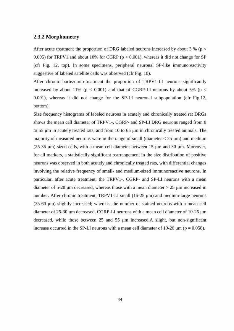
44
2.3.2 Morphometry
After acute treatment the proportion of DRG labeled neurons increased by about 3 % (p <
0.005) for TRPV1 and about 10% for CGRP (p < 0.001), whereas it did not change for SP
(cfr Fig. 12, top). In some specimens, peripheral neuronal SP-like immunoreactivity
suggestive of labeled satellite cells was observed (cfr Fig. 10).
After chronic bortezomib-treatment the proportion of TRPV1-LI neurons significantly
increased by about 11% (p < 0.001) and that of CGRP-LI neurons by about 5% (p <
0.001), whereas it did not change for the SP-LI neuronal subpopulation (cfr Fig.12,
bottom).
Size frequency histograms of labeled neurons in acutely and chronically treated rat DRGs
shows the mean cell diameter of TRPV1-, CGRP- and SP-LI DRG neurons ranged from 8
to 55 μm in acutely treated rats, and from 10 to 65 μm in chronically treated animals. The
majority of measured neurons were in the range of small (diameter < 25 μm) and medium
(25-35 μm)-sized cells, with a mean cell diameter between 15 μm and 30 μm. Moreover,
for all markers, a statistically significant rearrangement in the size distribution of positive
neurons was observed in both acutely and chronically treated rats, with differential changes
involving the relative frequency of small- and medium-sized immunoreactive neurons. In
particular, after acute treatment, the TRPV1-, CGRP- and SP-LI neurons with a mean
diameter of 5-20 μm decreased, whereas those with a mean diameter > 25 μm increased in
number. After chronic treatment, TRPV1-LI small (15-25 μm) and medium-large neurons
(35-60 μm) slightly increased; whereas, the number of stained neurons with a mean cell
diameter of 25-30 μm decreased. CGRP-LI neurons with a mean cell diameter of 10-25 μm
decreased, while those between 25 and 55 μm increased.A slight, but non-significant
increase occurred in the SP-LI neurons with a mean cell diameter of 10-20 μm (p = 0.058).
45
Figure 12. Percentage relative frequency
Size frequency histogram of TRPV1-, CGRP-, and SP-LI DRG eurons from control (ctrl) and acute bortezomib-treated
rats (on top) and from control (ctrl) and chronic bortezomib-treated rats (on bottom). Cells present in at least 6 sections
were measured. X-axis values represent the mean cell diameters expressed in μm; y-axis reports values of relative percent

46
frequency. Curve superimposed on the histogram represents the theoretical normal distribution. N = total number of sized
positive neurons; S.D. = standard deviation.
Table 2. Percentage (± confidence limits) of TRPV1-, CGRP-
and SP-LI DRG neurons in BTZ-treated rats
Acute BTZ-treatment
ctrl BTZ
TRPV1 26.86 ± 0.01% 29.55 ± 0.01%
CGRP 25.32 ± 0.01% 34.89 ± 0.01%
SP 15.86 ± 0.04% 14.73 ± 0.03%
Chronic BTZ-treatment
ctrl BTZ
TRPV1 28.65± 0.02% 39.93 ± 0.03%
CGRP 28.59± 0.02% 34.87± 0.02%
SP 20.60± 0.01% 19.85± 0.01%
2.3.3 Immuno-colocalization
The analysis of double immunofluorescence-stained tissue for TRPV1 and either
neuropeptide revealed that the neuronal co-expression of TRPV1/SP and TRPV1/CGRP is
partial, occurring in approximately 45% of TRPV1-LI neurons in control animals. An
estimation of changes in TRPV1/either neuropeptide co-expressing neurons suggested that,
in the acute treated group (cfr Fig. 13), the TRPV1/CGRP subpopulation did not show any
statistically significant change, whereas the TRPV1/SP one decreased significantly (-12%,
p < 0.05; Table 1). In the chronic treated group (cfr Fig. 14), both TRPV1/CGRP- (-7%; p
< 0.05) and TRPV1/SP-coexpressing neurons (-8%, p < 0.005) showed a statistically
significant decrease (Table 3).
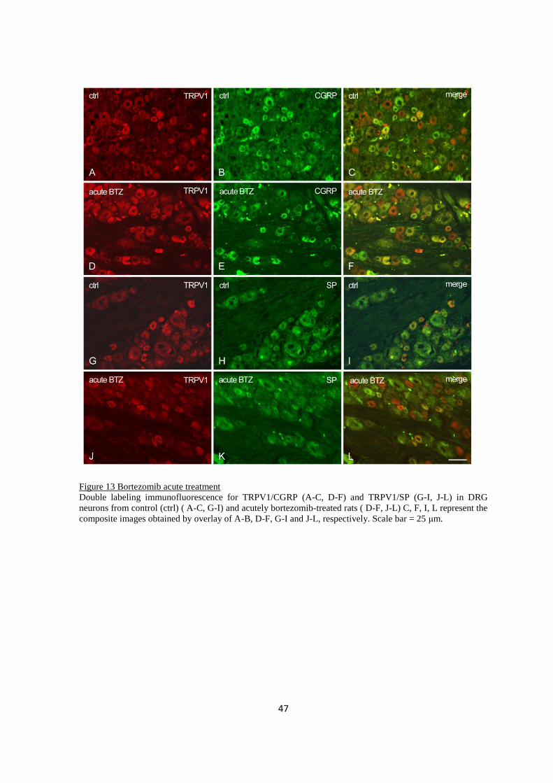
47
Figure 13 Bortezomib acute treatment
Double labeling immunofluorescence for TRPV1/CGRP (A-C, D-F) and TRPV1/SP (G-I, J-L) in DRG
neurons from control (ctrl) ( A-C, G-I) and acutely bortezomib-treated rats ( D-F, J-L) C, F, I, L represent the
composite images obtained by overlay of A-B, D-F, G-I and J-L, respectively. Scale bar = 25 μm.
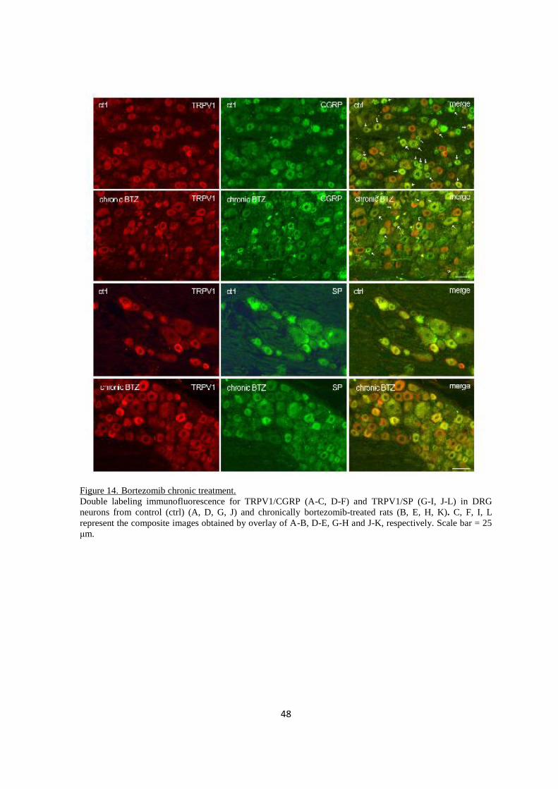
48
Figure 14. Bortezomib chronic treatment.
Double labeling immunofluorescence for TRPV1/CGRP (A-C, D-F) and TRPV1/SP (G-I, J-L) in DRG
neurons from control (ctrl) (A, D, G, J) and chronically bortezomib-treated rats (B, E, H, K). C, F, I, L
represent the composite images obtained by overlay of A-B, D-E, G-H and J-K, respectively. Scale bar = 25
μm.
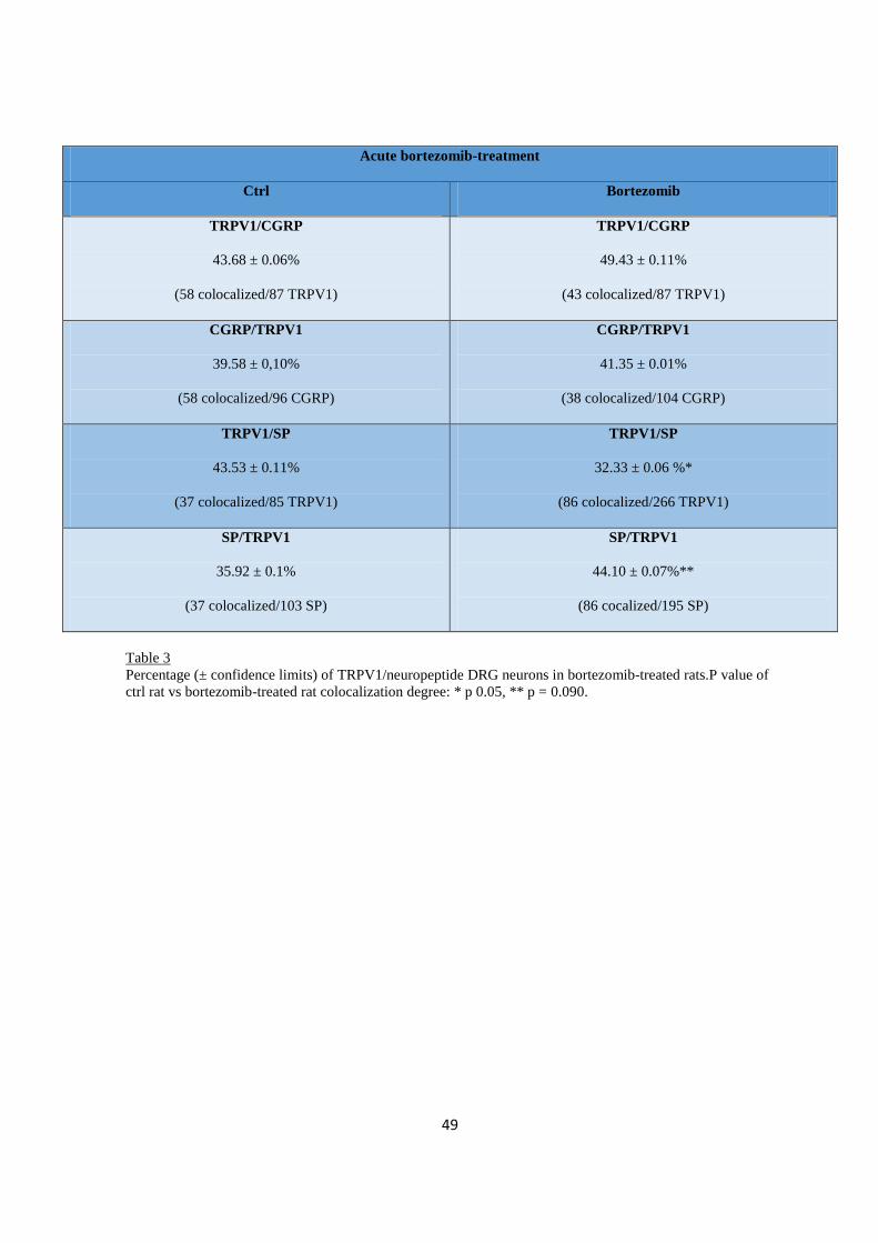
49
Acute bortezomib-treatment
Ctrl Bortezomib
TRPV1/CGRP
43.68 ± 0.06%
(58 colocalized/87 TRPV1)
TRPV1/CGRP
49.43 ± 0.11%
(43 colocalized/87 TRPV1)
CGRP/TRPV1
39.58 ± 0,10%
(58 colocalized/96 CGRP)
CGRP/TRPV1
41.35 ± 0.01%
(38 colocalized/104 CGRP)
TRPV1/SP
43.53 ± 0.11%
(37 colocalized/85 TRPV1)
TRPV1/SP
32.33 ± 0.06 %*
(86 colocalized/266 TRPV1)
SP/TRPV1
35.92 ± 0.1%
(37 colocalized/103 SP)
SP/TRPV1
44.10 ± 0.07%**
(86 cocalized/195 SP)
Table 3
Percentage (± confidence limits) of TRPV1/neuropeptide DRG neurons in bortezomib-treated rats.P value of
ctrl rat vs bortezomib-treated rat colocalization degree: * p 0.05, ** p = 0.090.
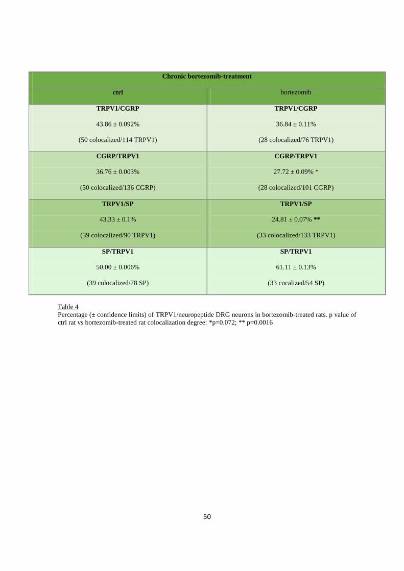
50
Chronic bortezomib-treatment
ctrl bortezomib
TRPV1/CGRP
43.86 ± 0.092%
(50 colocalized/114 TRPV1)
TRPV1/CGRP
36.84 ± 0.11%
(28 colocalized/76 TRPV1)
CGRP/TRPV1
36.76 ± 0.003%
(50 colocalized/136 CGRP)
CGRP/TRPV1
27.72 ± 0.09% *
(28 colocalized/101 CGRP)
TRPV1/SP
43.33 ± 0.1%
(39 colocalized/90 TRPV1)
TRPV1/SP
24.81 ± 0.07% **
(33 colocalized/133 TRPV1)
SP/TRPV1
50.00 ± 0.006%
(39 colocalized/78 SP)
SP/TRPV1
61.11 ± 0.13%
(33 cocalized/54 SP)
Table 4
Percentage (± confidence limits) of TRPV1/neuropeptide DRG neurons in bortezomib-treated rats. p value of
ctrl rat vs bortezomib-treated rat colocalization degree: *p=0.072; ** p=0.0016
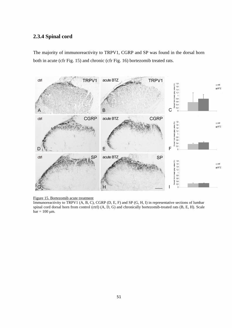
51
2.3.4 Spinal cord
The majority of immunoreactivity to TRPV1, CGRP and SP was found in the dorsal horn
both in acute (cfr Fig. 15) and chronic (cfr Fig. 16) bortezomib treated rats.
Figure 15. Bortezomib acute treatment
Immunoreactivity to TRPV1 (A, B, C), CGRP (D, E, F) and SP (G, H, I) in representative sections of lumbar
spinal cord dorsal horn from control (ctrl) (A, D, G) and chronically bortezomib-treated rats (B, E, H). Scale
bar = 100 μm.
52
Figure 16. Bortezomib chronic treatment
Immunoreactivity to TRPV1 (A, B, C), CGRP (D, E, F) and SP (G, H, I) in representative sections of lumbar
spinal cord dorsal horn from control (ctrl) (A, D, G) and chronically bortezomib-treated rats (B, E, H). Scale
bar = 100 μm.
As can be appreciate from the figures 15 and 16 TRPV1-LI structures were prominent in
Lissauer’s tract and lamina I and occurred with a lighter labeling in inner lamina II. CGRP-
and SP-LI structures showed a wider distribution, being present in Lissauer’s tract, laminae
I-III and lamina V. No changes were found in the ventral horn, where CGRP
immunolabeled motoneurons were detectable.
2.3.5 Image densitometry
Image densitometric analysis of dorsal horn labeling showed a statistically significant
increase in CGRP immunostaining in the acutely treated animals (p < 0.05) (cfr Fig. 15 F)
and a trend to increase in chronically treated ones (p = 0.057) (cfr Fig. 16 C, F, I).
53
2.3.6 Sciatic nerve
TRPV1-, CGRP and SP-LI nerve fibers were easily detectable in longitudinal sections of
sciatic nerves. The results show in the pictures releaved apparent differences between
treated and control animals were found in both acute (cfr Fig.17) and chronic (cfr Fig 18)
treated rats.
Figure 17. Longitudinal sections of sciatic nerves of acute bortezomib treated rats
Double labeling immunofluorescence for TRPV1/CGRP in sciatic nerve from control (ctrl) (A-C) and acute
bortezomib-treated rats (D-F). C, F represent the composite images obtained by overlay of A-B and D-E,
respectively. Scale bar = 10 μm.
Figure 18. Longitudinal sections of sciatic nerves of chronic bortezomib treated rats.
Double labeling immunofluorescence for TRPV1/CGRP in sciatic nerve from control (ctrl) (A-C) and
chronically bortezomib-treated rats (D-F). C, F represent the composite images obtained by overlay of A-B
and D-E, respectively. Scale bar = 10 μm.

54
Analysis of colocalization performed on double immunofluorescence preparations for
TRPV1 and CGRP revealed that, in both acutely (cfr Fig. 17) and chronically treated
animals (cfr Fig. 18), TRPV1-like immunoreactivity occurred mainly in fibers that did not
show CGRP labeling. Due to the paucity of double-labeled structures, no quantitative
evaluation was performed.
55
2.4 Western blot
The western blot was performed only for TRPV1 on homogenate of total protein, we did
not have suitable equipment to recognize CGRP and SP due to their low molecular weight.
Figure 19. Western blot analysis for acute and chronic bortezomib treated rats
Western blot analysis of TRPV1 in DRG (A, E) and spinal cord (C, G) of acutely and chronically
bortezomib-treated rats. B, D, F, H: Relative levels of TRPV1 expression in DRG and spinal cord with
densitometric analysis of the grey levels expressed as a percentage of the optical density (O.D.) ratio of the
TRPV1-positive bands to the GAPDH-positive ones. Ctrl = control rats. Error bars represent standard
deviation. Letters “a” and “b” denote significant differences (p < 0.05).
The antibody against TRPV1 labeled a single protein band at the expected mw of 88.9 kDa
(cfr Fig. 19) in DRGs and spinal cord tissue homogenates. In both acute and chronic
bortezomib treated rats, TRPV1 protein levels were higher than in control animals. After
acute treatment, relative optical density (O.D.) of TRPV1 protein bands increased threefold
in DRG homogenates (cfr Fig. 19 A, B) (p < 0.05) and a mild but not significant increase
56
of TRPV1 protein was also evident in the spinal cord (cfr Fig. 19 C, D). Chronic
bortezomib-treatment produced a statistically significant increase in the relative O.D. of
the TRPV1 protein band in both DRGs (cfr Fig. 19 E, F) and spinal cord (cfr Fig. 19 G, H),
amounting to 75% (p < 0.05) and 100% (p < 0.05), respectively, in bortezomib-treated
versus control animals.
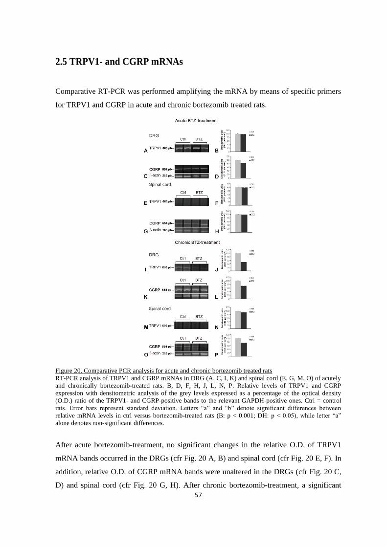
57
2.5 TRPV1- and CGRP mRNAs
Comparative RT-PCR was performed amplifying the mRNA by means of specific primers
for TRPV1 and CGRP in acute and chronic bortezomib treated rats.
Figure 20. Comparative PCR analysis for acute and chronic bortezomib treated rats
RT-PCR analysis of TRPV1 and CGRP mRNAs in DRG (A, C, I, K) and spinal cord (E, G, M, O) of acutely
and chronically bortezomib-treated rats. B, D, F, H, J, L, N, P: Relative levels of TRPV1 and CGRP
expression with densitometric analysis of the grey levels expressed as a percentage of the optical density
(O.D.) ratio of the TRPV1- and CGRP-positive bands to the relevant GAPDH-positive ones. Ctrl = control
rats. Error bars represent standard deviation. Letters “a” and “b” denote significant differences between
relative mRNA levels in ctrl versus bortezomib-treated rats (B: p < 0.001; DH: p < 0.05), while letter “a”
alone denotes non-significant differences.
After acute bortezomib-treatment, no significant changes in the relative O.D. of TRPV1
mRNA bands occurred in the DRGs (cfr Fig. 20 A, B) and spinal cord (cfr Fig. 20 E, F). In
addition, relative O.D. of CGRP mRNA bands were unaltered in the DRGs (cfr Fig. 20 C,
D) and spinal cord (cfr Fig. 20 G, H). After chronic bortezomib-treatment, a significant

58
decrease of the relative O.D. of TRPV1 mRNA band occurred in the DRGs (-50%, p <
0.001; cfr Fig. 20 I, J), but not in the spinal cord (cfr Fig. 20 M, N), while a significant
decrease of the relative O.D. of CGRP mRNA band occurred in the DRGs (- 28%, p <
0.05; cfr Fig. 20 K, L) as well as in the spinal cord (-26%; p < 0.05; cfr Fig. 20 O, P).
59
3. Discussion
The identification of the key effectors of the bortezomib cytotoxicity as well as of the
molecular factors able to contrast or inhibit its action has been hampered, so far, by the fact
that the proteasome controls the half-life of the vast majority of cellular proteins
(Jesenberger and Jentsch, 2004; Adams, 2004). Morevoer, it has been suggested that the
bortezomib molecule per se, compared to other proteasome inhibitors, could be responsible
of a neurotoxic effect, because it induces an hyperaccumulation of not degraded
ubiquitinated proteins (Fernandez et al.,2006). It has been inferred that bortezomib acts
downstream of proteasome inhibition by interfering with stress kinases, survival signals
and/or apoptotic factors (Mitsiades et al., 2002). The dysregulation of mitochondrial
functions and release of reactive oxygen species (ROS) are also common processes
induced by bortezomib (Liu et al., 2003; Jesenberger and Jentsch, 2004; Adams, 2004).
Thus, as early as 3–4 h after bortezomib treatment, epithelial tumor cells show a
dysregulated mitochondrial membrane potential and accumulate ROS, that subsequently
lead to caspase activation (Ling et al., 2003; Fribley et al., 2004).
In this scenario, the present study investigated the effects of bortezomib-treatment given
either as a single acute or a chronic schedule on the neurochemical features of DRG
primary afferent neurons and their relevant peripheral and central processes. Our results, in
agreement with previous observations (Meregalli, 2010), show that prolonged bortezomib
administration induces a PN without appreciable adverse effects on the general condition
of the animals.
As expected based on previous observations (Meregalli et al., 2010; 2012), the
histopathological analysis carried out by colleagues at the University of Milan-Bicocca
revealed that acute bortezomib-treatment did not induce morphological alterations neither
in the sciatic nerve nor in the DRG neuronal perikarya. Histopathological analyssi further
confirmed that, in chronically treated animals, the sciatic nerve showed a mild axonopathy,
affecting mostly the small myelinated and unmyelinated fibers. Similarly, clear
cytoplasmic vacuolization was detected in some DRG satellite cells. No obvious changes
were observed in the dorsal horn of the spinal cord (data not shown).
60
3.1 Neurophysiological evaluation
Neurophysiological and neuropathological assessments were consistent with measures
at baseline and end of treatment time points previously observed (Meregalli et al.,
2010; 2012). As regards the modification of nocifensive behavior, the chronic model of
bortezomib-induced PN was associated with the onset of mechanical allodynia as assessed
after the 8-week treatment. Interestingly, data concerning the weekly outcome of
bortezomib-treatment showed a significant reduction in withdrawal latency after 4 weeks
of treatment; however, at the later time point of 8-weeks (end of treatment period) and the
follow-up 4-week period, the difference was no longer significant (Meregalli et al.,
2010).
Although the bortezomib schedule used in this study differs from that used in clinical
practice with respect to the duration of administration and dosage, calculations to
translate drug doses from animal to human studies (Reagan-Shaw et al., 2008) indicate
that our model of bortezomib-induced neurotoxicity is highly relevant. In fact, it
closely mimics the sensory neuropathy described in human patients by sharing with it
common neurophysiological, behavioral and pathological features, occurring often
within the first cycle of treatment (Cata et al., 2007; Argyriou et al., 2008).
3.2 Bortezomib-induced effects on expression of TRPV1 and CGRP
mRNAs and proteins
Our results provide novel evidence that bortezomib-induced nocifensive behavior is
accompanied by increased TRPV1 protein levels in DRGs and spinal cord and down-
regulated levels of TRPV1 mRNA. Decreased levels of CGRP mRNA were also observed
in both DRGs and spinal cord.
We do not have a clear-cut explanation of the mismatch between levels of TRPV1 mRNA
and protein and details about neuronal mechanisms underlying protein/mRNA reciprocal
changes in this context remain to be elucidated. However, the ubiquitin-proteasome system
has a role in receptor protein catabolism; as an example, NMDA receptor subunits have
61
been shown to be degraded by the proteasome following ubiquitination initiated by several
ligases (Kato et al., 2005). Interestingly, novel evidence has been provided that also
TRPV1 receptor is susceptible to proteasomal degradation, unless it translocates to the
plasma membrane; in fact, blocking TRPV1 trafficking to the plasma membrane markedly
decreases total TRPV1 expression due to proteasomal degradation (Shimizu et al., 2012).
Proteasome-regulated TRPV1turnover appears to be dynamic and has been suggested to be
related to the fine-tuning of nociception (Kato et al., 2005). In bortezomib-induced NP
models, the occurrence of axonal degeneration and Schwann cell endosomal membrane
dilations likely accounts for impaired protein membrane trafficking (Cavaletti et al., 1992;
Shin et al., 2012; Bruna et al., 2010). In our specimens, the enduring bortezomib-induced
proteasome inhibition might have well caused a TRPV1 accumulation in the cytoplasm
which therefore can account for the increased levels of TRPV1 in DRGs.
As for the observed decrease of TRPV1 and CGRP mRNA relative levels, it has to be
highlighted that proteasome blockade causes reduction of transcriptional activity and
nuclear retention of poly(A)RNAs (Voorhees et al., 2003; Cata et al., 2007; Casafont et al.,
2010), though the increase in protein expression itself may act as a retrograde signal to
modulate transcription. Yet, NF-B proteasome-independent activation pathways have
also been reported (Mühlen et al., 2001; Pearson et al., 2011) and, depending on the
targeted cell type, bortezomib may activate rather than inhibit the NF-B canonic
pathway
Plasticity of TRPV1 expression in chronic pain conditions is considered to represent
one of the mechanisms involved in hyperalgesia (Hideshima et al., 2009). The
functional characterization of TRPV1-labeled neurons goes beyond the aim of this
study. However, available experimental studies on the systemic administration of
different TRPV1 antagonists demonstrate a reduction of mechanical allodynia and
hyperalgesia in animal models of neuropathic, inflammatory and postoperative pain and
suggest a role of TRPV1 in the modulation of mechanical hyperalgesia (Pomonis et al.,
2003; McGaraughty et al., 2008; Yu et al., 2008)
Regarding the specific time profile and turnover of TRPV1 protein after the acute
bortezomib delivery, we were not able to find literature data with which to compare our
results, since experimental studies on the effect of a bortezomib single dose, both in rats

62
(Casafont et al., 2010) and mice (Trevisan et al., 2013) were not designed to investigate
the immediate neurotoxicity of its administration. However, pharmacokinetics of
bortezomib are well known because the drug has been shown to be rapidly distributed into
tissues after administration of a single dose, with an initial plasma distribution half-life of
less than 10 minutes. Maximum proteasome inhibition occurs within 1 hour and recovers
close to baseline within 72 to 96 hours after administration (Schwartz and Davidson,
2004). Recently, in the same rat model of bortezomib-induced PN as ours and with the
same dose used in this study, Meregalli et al. (2014) found that a maximal time-dependent
proteasome inhibition can be observed in peripheral blood mononuclear cells and sciatic
nerve specimens asearly as 1 hour after a single dose bortezomib delivery, thus supporting
the possibility of a quick cytoplasmic accumulation of TRPV1 protein, as also suggested
by the low but significant increase of the proportion of TRPV1-LI neurons in the DRG
(present data).
3.3 Bortezomib-induced effects on primary afferent neuron
histochemical features
Bortezomib-treatment also induced an augmented immunoreactivity to TRPV1 and CGRP
in the DRG neurons and spinal dorsal horn, and caused a decrease of
TRPV1/neuropeptide colocalization in DRG neurons. No bortezomib-dependent
changes were observed for SP immunohistochemical detectability. In some specimens,
peripheral neuronal SP-like immunoreactivity suggestive of labeled satellite cells was
observed giving a neurochemical support to our histopathological assessment (see above).
They further suggest that the histological alterations, already visible after a single acute
dose of bortezomib and previously reported also for paclitaxel (Authier et al., 2000), are an
early trait of chemotherapy-induced PN. In addition the preponderance of experimental
evidence and theoretical considerations, support the notion that changes in satellite glial
cells, alone or in concert with changes in neuronal function, do affect nociception (Ohara et
al., 2009).
63
3.4 Percent frequency of labeled DRG neurons
To our knowledge this is the first study reporting that bortezomib treatment in a rat
model causes a general increase in the percent frequency of TRPV1- and CGRP-LI
DRG neurons after both acute and chronic administration. Our immunohistochemical
data are in keeping with those on CGRP-LI neuronal frequency in mouse DRGs after
chronic bortezomib-treatment (Bruna et al., 2010). The increase in TRPV1- and CGRP-
LI in the DRGs occurs with a parallel increase in the spinal cord dorsal horn.
Interestingly, increased immunoreactivity for TRPV1 (Yu et al., 2008; Hudson et al.,
2005; Kim et al., 2008) and CGRP (Zheng et al., 2008) has been previously shown in
uninjured primary afferent neuronal perikarya in different models of nerve lesion.
Moreover, CGRP expression may increase in several pathological conditions, including
partial nerve injury, where pain hypersensitivity occurs (Menard et al., 1996.). In
comparison, cisplatin has been previously reported not alter the DRG expression of SP
(Apfel et al., 1992)
3.5 Relative size frequency of labeled DRG neurons
In our model, bortezomib administration induced a rearrangement in the relative size
frequency of TRPV1-, CGRP- and SP-LI neuronal subgroups, suggesting the
possibility of a drug-induced neuronal phenotype switch that may go with the onset of
bortezomib-induced mechanical hyperalgesia. These observations are in agreement
with those obtained in the mouse model of bortezomib neurotoxicity where medium-
sized neurons slightly decrease, small neurons increase and the relative number of large
CGRP-LI neurons increases (Bruna et al., 2010). Taken together, these data suggest
that the phenotypic switch secondary to bortezomib-treatment may partly represent the
neuronal response to treatment, as also suggested by Bruna et al. (Bruna et al., 2010).
64
3.6 TRPV1/neuropeptide colocalization
Overall, the colocalization data support that a portion of the TRPV1-LI DRG neurons are
neuropeptidergic. The analysis of changes in neuronal colocalization of TRPV1 with
neuropeptides provides a morphological basis to suggest a role for TRPV1 in the release of
these molecules and thus in nociceptor sensitization, and supports the possible implication
of TRPV1-, CGRP- and SP-positive neurons in the persistence of painful neuropathic
symptoms (Holzer, 2007). The fact that the TRPV1/neuropeptide colocalization seen in the
DRGs did not parallel that observed in both central and peripheral nerve processes of
primary afferent neurons is in keeping with previous observations by Guo et al.(1999),
who suggested alternative mechanisms of neuropeptide release in response to capsaicin
application. Future studies will examine how these neurochemical changes affect
neurophysiological function in DRG neurons and the spinal dorsal horn.
3.7 Bortezomib-induced effects on spinal cord histochemical features
The neuronal response to bortezomib neurotoxicity involves not only the DRGs but
extends to the spinal cord dorsal horn, where neurochemical alterations parallel those
observed in the periphery. In particular, following bortezomib treatment, densit ies of
dorsal horn immunolabelling for TRPV1 and CGRP were increased. Importantly, this
response is larger in the chronically bortezomib-treated animals.
The basal expression of TRPV1 mRNA in the spinal cord is in keeping with previous
RT-PCR studies (Sanchez et al., 2001; Kim et al, 2012) and with the recent
demonstration that TRPV1 expression occurs in GABAergic interneurons of the rat
dorsal horn, where it mediates neuropathic mechanical allodynia and disinhibits
nociceptive circuits in the spinal cord (Doly et al., 2004). Interestingly, TRPV1
expression by glial cells has also been described (Chen et al., 2010) and TRPV1 has
been reported to be involved in activating spinal glia in mice with nociceptive and
pathological pain. Moreover, since bidirectional axonal transport of TRPV1 mRNA
along primary afferents has been demonstrated in some pathological conditions such as
experimental acute inflammation (Barajon et al., 1996), it cannot be excluded that part
65
of the TRPV1 mRNA detected in our specimens has a primary afferent origin. If this
was the case, the reduction of TRPV1 mRNA secondary to bortezomib administration
might be explained by the reduction of its production, and hence by the reduction of
centripetal transport, by DRG neurons.
Consistent with changes in TRPV1 transcript and protein levels, CGRP mRNA also
showed a decrease that may be correlated to the increase in density of CGRP-
immunoreactive structures in the bortezomib-treated versus control rats.
3.8 Comparison with neurochemical phenotype changes observed in
other chemotherapy-induced PN
While experimental evidence regarding the effects of bortezomib on the neurochemical
phenotype is limited to data on CGRP and TRP receptors in mouse models, after acute
(Trevisan et al., 2013) and chronic administration (Bruna et al., 2010; Trevisan et al.,
2013), several studies of sensory neuropeptides (Verdú, et al., 1999; Alessandri-Haber et
al., 2008) and TRP receptors in the spinal cord and DRG (Ta et al., 2010; Hudson et
al., 2005) following treatment with different classes of antineoplastic drugs are
available in rodents. It has been demonstrated for instance that platinum-based
compounds, taxanes and vinca alkaloids modulate the expression of TRPV1, as well as
other TRP receptors, in DRG neurons in vitro and in vivo (Zhao et al., 2012; Hudson et al.,
2005). Cisplatin and oxaliplatin affect the expression of TRPV1 and TRPA1 mRNA by
causing a 3 fold mRNA increase as early as 6 hours after treatment in cultured DRG
neurons (Zhao et al., 2012). Similarly, trigeminal ganglia (TG) of cisplatin-treated mice
had significant increases in TRPV1 mRNA expression (Zhao et al., 2012). However,
mRNA changes are not mirrored by immunohistochemistry studies, showing that no
change occurred in the proportion of the TRPV1 immunopositive TG neurons in cisplatin-
and oxaliplatin-treated mice compared to naïve animals (Zhao et al., 2012). In the same
way, TRP receptors have been shown to have a role in the pathogenesis of paclitaxel-
associated pain symptoms that were related to increased TRPV1 mRNA and protein
expression in rat DRG neurons (Hudson et al., 2005). A single injection of paclitaxel is
66
sufficient to increase the expression of TRPV1 mRNA and protein in rat DRG (as seen in
both homogenates and tissue sections by RT-PCR, in situ hybridization and
immunochemistry), and to enhance TRPV1 expression in the paw skin at day 14 after
treatment (Hudson et al., 2005). By contrast, the development of sensory disorders
following vincristine treatment has been related to changes in the expression of
TRPV4 (Zhao et al., 2012).
Conclusions
Our data support the notion that bortezomib treatment selectively affects primary
sensory neurons that for their anatomical and neurochemical characteristics are
likely involved in the processing of painful sensory stimuli.
In DRG neurons and the spinal cord, the bortezomib-induced neurochemical
changes appeared after a single dose of the drug and were enhanced after chronic
treatment.
Taken together, these findings increase our understanding of the nociceptive
symptoms associated with bortezomib-induced sensory neuropathy and may
contribute to the development of new targeted therapies.

67
4.Materials and methods
4.1 Animals and animal care
Animals. A total of 80 adult female Wistar rats (Harlan Italy, Correzzana, Italy), weighing
175–200 g at the beginning of the experiment, were used for the study. Animals were
housed in a limited access animal facility with controlled room temperature (22 ± 2°C) and
relative humidity (55 ± 10%) and under an artificial 12h light/dark cycle (light 7 a.m.–7
p.m.). The care and husbandry of the animals were in conformity with the institutional
guidelines in compliance with national (D.L. n.116, Gazzetta Ufficiale della Repubblica
Italiana, Additional40, Feb 18, 1992 and subsequent modifications) and international laws
and policies (EEC Council Directive 86/609, OJ L 358, 1, Dec.12, 1987; Guide for the
Care and Use of Laboratory Animals, US National Research Council, 8th ed., 2011). The
experimental plan was examined and approved by the Ethics Committee of the University
of Milan Bicocca.
4.1.1 Drug administration
Bortezomib (LC Laboratories, Woburn, MA) was dissolved in 5% Tween 80, 5% ethanol,
90% sterile saline. Animals were divided into two experimental groups. One group of 40
rats was treated with bortezomib by tail vein injection. Eighteen animals received 0.20
mg/kg of bortezomib as single dose for “acute” schedule and 22 animals underwent an 8-
week period of bortezomib administration for “chronic” schedule, 0.20 mg/kg/day three
times per week. The remaining 40 animals were left untreated and used as controls for the
2 time points of examination. Euthanasia for tissue harvesting was done under deep
xylazine/ketamine anesthesia 1 hour after the single bortezomib dose in the “acute”
experimental schedule and one day after the last bortezomib administration in the
“chronic” experimental schedule.
68
4.1.2 General toxicity
The general condition of treated animals was assessed daily and body weight of the
chronically treated rats was measured twice/week to monitor drug toxicity and for drug
dose calculation. Animals with evident distress or remarkable weight loss were carefully
examined by a certified veterinarian experienced with in vivo studies for possible
withdrawal from the study for humane reasons.
4.2 Neurotoxicity and pain assessment
4.2.1 Mechanical nociceptive threshold
The mechanical nociceptive threshold was assessed using a Dynamic Aesthesiometer Test
(model 37450, Ugo Basile Biological Instruments, Comerio, Italy), which generated a
linearly increasing mechanical force. Rats were habituated to the full procedure for 30
min/day on two consecutive days and testing was conducted on the third day. On each day
of testing, animals were placed in a Plexiglas chamber (28 x 40 x 35-cm, wire mesh floor)
in the dynamic aesthesiometer for a 15 min acclimatization period followed by testing. At
each time point, after the acclimatization period, a servo-controlled mechanical stimulus (a
pointed metallic filament, 0.5mm diameter) was applied to the plantar surface of the hind
paw, which exerted a progressively increasing punctuate pressure, reaching up to 50g
within 20 sec. The pressure evoking a clear voluntary hind-paw withdrawal response was
recorded automatically and taken as representing the mechanical nociceptive threshold
index. The mechanical threshold was always assessed alternatively on each side every 2
min on 3 occasions to yield a mean value. The results represent the maximal pressure
(expressed in grams) tolerated by the animals. There was an upper limit cut-off of 30 sec
after which the mechanical stimulus was automatically terminated. If paw movement
subsequent to stimulus initiation appeared to be related to grooming or locomotion, that
trial was repeated after a lapse of 1 min.
69
4.3 Sampling
Immediately after sacrifice, the L4-L6 DRGs with the corresponding spinal cord segments
and the sciatic nerves were rapidly dissected out and either frozen at -80°C for western blot
and RT-PCR analyses or fixed by immersion in freshly prepared 4% phosphate-buffered
paraformaldehyde, pH 7.3, for 4–6 h at 4°C, and then rinsed overnight in 0.1 M phosphate
buffer (PB), pH 7.3, containing 20% sucrose for immunohistochemistry. After sucrose
infiltration, samples were embedded in Optimal Cutting Temperature (OCT) medium for
cryostat sectioning.
4.4 Immunohistochemistry
A total of 36 DRGs and lumbar spinal cord segments were processed: 16 from the acutely
treated group (8 bortezomib-treated, 8 controls) and 20 from the chronically treated ones
(10 bortezomib-treated, 10 controls). Sixteen sciatic nerve segments, 8 from the acutely
treated group (4 bortezomib-treated, 4 controls) and 8 from the chronically treated ones (4
bortezomib-treated, 4 controls), were examined. Cryostat semiconsecutive sections (12 μm
thick) were collected on chrome alum-gelatin (USB Corporation, Cleveland, OH, USA)
coated slides and processed either by the avidin–biotin–peroxidase complex (ABC)
immunohistochemical technique or by immunofluorescence double labeling.
4.4.1. ABC Immunohistochemical technique
For ABC processing, endogenous peroxidase activity was blocked with 0.1%
phenylhydrazine in phosphate buffered saline (PBS) containing 0.2% Triton X-100
(PBS/T) followed by incubation with 20% of either normal goat (NGS) or normal horse
serum (Vector, Burlingame, CA, USA) for 1h at RT and then incubated with primary
antiserum (cfr Table 5).
Biotin-conjugated goat anti-rabbit, donkey anti-goat, and goat anti-guinea-pig serum
(Vector, Burlingame, CA, USA), diluted 1:400, were used as secondary antiserum. The
reaction product was revealed by the ABC complex (BioSpa Div. Milan, Italy), diluted
70
1:250, and followed by incubation with a solution of 0.1 M PB, pH 7.3, containing 0.05%
3-3′-diaminobenzidine (Sigma, Milan, Italy), 0.04% nickel ammonium sulfate and 0.01%
hydrogen peroxide.
4.4.2 Indirect immunofluorescence
For IF the slides were incubated with either 20% normal goat (NGS) or normal horse
serum (Vector, Burlingame, CA, USA) for 1 h at RT then incubated with primary
antiserum overnight at 4C (cfr Table 5).
For double labeling, after a pre-incubation with 20% NGS, sections were double stained
for TRPV1/CGRP, TRPV1/SP by means of subsequent incubations with primary
antiserum, diluted as reported in Table 3. Sections were then incubated with Alexa Fluor
488- or Alexa Fluor 594-conjugated secondary antibodies (Invitrogen, Eugene, OR, USA),
diluted 1:500 and 1:600, respectively. Control immunostaining was obtained substituting
the primary antiserum with normal serum. Slides were examined with an Olympus BX61
microscope, equipped with epifluorescence illumination, and digital images were captured
with a Leica DF 450C camera
4.4.3 Morphometry
Morphometric analysis, carried out by the same examiner blinded to animals’ treatment,
was performed on DRG neuronal cell profiles in digital images captured with a 20x
objective magnification from 10 to 12 animals out of the acutely and chronically treated
groups, respectively. The tissue sections were separated by at least 108 μm and only cells
that obviously showed the nuclear profile were considered. Neuronal mean diameters were
automatically measured by ImageProPlus software. Statistical parameters (mean,
minimum, maximum, S.D.) and histograms of the neuron sizes, processed as a pool, were
obtained by Statistica 6 software. The percentage of DRG immunostained perikarya,
processed as a pool, was calculated as the ratio of the total number of labeled cells found in
four to eight sections to the total number of cells found in the same sections after a
modified Mayer's hematoxylin [certified hematoxylin (1.0 g/l), sodium iodate (0.2 g/l),
71
aluminum ammonium sulfate·12 H2O (50 g/l), chloral hydrate (50 g/l) and citric acid (1
g/l) counterstaining.
4.4.4 Image densitometry
For the quantitative evaluation of SP, CGRP and TRPV1 immunohistochemical labeling in
the dorsal horn of the spinal cord, 6 representative 10x magnification microscopic fields
for each marker (three bortezomib-treated animals and three controls) were blindly
analyzed with ImageJ (http://rsb.info.nih.gov/ij/). Mean gray values from negative controls
were subtracted from the gray values of the immunostained sections to exclude background
staining. Histograms were obtained by calculating the ratio of bortezomib-treated values to
control values.
4.5 Protein and RNA extraction
A total of 32 DRGs and spinal cord segments were analyzed: 12 from the acutely treated
group (6 bortezomib-treated, 6 controls) and 20 from the chronically treated group (10
bortezomib-treated, 10 controls). Total protein and RNA from DRGs and spinal cord tissue
were isolated by the TRIzol® method (Invitrogen, Carlsbad, CA) (1 ml/100 mg of tissue),
the samples were homogenated with TRIzol® using.polytron and followed by addition of
chloroform (200 µl/ml of TRIzol®), the tubes were inverted vigorously by handin then
incubeted for 5 min at room temperature (RT). After centrifugation at 12000*g for 15 min
at 4°C protein and RNA were separated into a lower organic phase and an upper aqueous
phase, respectively.
4.5.1 Protein extraction
The organic phase was incubated in absolute ethanol (EtOH) (300 µl/ml of TRIzol® used
for the initial homogenization) for 2-3 min at RT followed by centrifugation at no more
than 2000 * g for 5 min at 4°C. The supernatant was then combined with isopropyl alcohol
(1.5 ml/ml of TRIzol® used for the initial homogenization), incubated for 10 min at RT,
and then centrifuged at 12000 * g for 10 min at 4°C. The supernatant was withdrawn
72
and the protein pellet was washed 3 times in a solution containing 0.3 M guanidine
hydrocloride (IBI scientific, Kapp Court, Peosta, IA, USA) in 95 % EtOH (2 ml/ml of
TRIzol® used for the initial homogenization) then after 20 min incubation centrifuged at
7500 * g for for 5 min at 4°C, this procedure was repeated for ech wash cycle.
After the final wash the protein pellet was vortexed in asbolute EtOH (2ml) and stored
for 20 min at RT, finally was centrifuged at 7500 * g a for 5 min at 4°C. Samples were
then resuspended in distilled water containing 2% sodium dodecilsulfate (SDS) (300
μl/100 mg of initial tissue). Protein concentrations were determined using the Lowry
protein assay with bovine serum albumin as the standard.
4.5.2 RNA extraction
The aqueous phase was transferred into a fresh tube, the organic phase was saved for
protein extraction. In the fresh tube was added isopropyl alcohol (0,5 ml/ml of TRIzol®
used for the initial homogenization) and incubated for 10 min at RT, then centrifuged at no
more than 12000*g at 4°C for 10 min to allow RNA precipitation. The RNA precipitate
form a gel like pellet on the side and bottom of the vial.
The supernatant was removed and RNA-pellet was washed once with 75% of Ethanol
in RNase-free (RF) diethyl pyrocarbonate (DEPC) water (1ml/ml of TRIzol®used for the
initial homogenization), the mix was vortexed and centrifuged at no more then 7500*g
for 5 min. at 4°C.
At the end of the procedure the RNA-pellet was briefly dryed (air dry) for 5-10 min
then was risuspended in DEPC RF water.
The quantity and purity of the RNA was assessed using both nano-volume
spectrophotometer (Nanophotometer P-360 Implen GmbH, München, Germany) and
electrophoresis technique in 2% agarose gel in TAE.
TAE preparation
For 1 liter of 50x TAE buffer use:
242 g Tris base (2-amino-2-hydroxymethyl-propane-1,3-diol) (= 2 moles)
57.1 mL glacial acetic acid (= 100% acetic acid) (57.19 mL = 1 mole)
100 mL 0.5 M Na2 EDTA (pH 8.0)
Pour H2O to reach a total volume of 1 L.
To prepare 0.5 M Na2EDTA (pH 8.0) add 186.1 g of disodium ethylene
diamino tetraacetate in ddH2O up to 800 ml of H2O. Adjust the pH to 8.0 with
NaOH (20 g approx. NaOH). Solution must be sterilized by autoclaving.
73
4.6 Western blot
Total proteins (40 μg) were separated by SDS-polyacrilamide gel electrophoresis (SDS-
PAGE) using a 10% (w/v) polyacrilamide resolving gel. Internal molecular weight (mw)
standards (Kaleidoscope Prestained Standards, Bio-Rad, Hercules, CA, USA) were run in
parallel. Two gels were run simultaneously for Coomassie staining and immunoblotting.
Proteins for immunoblotting were electrophoretically transferred to a polyvinylidene
fluoride membrane (Bio-Rad, Hercules, CA, USA) using the Mini Trans Blot Cell (Bio-
Rad, Hercules, CA, USA). Blots were blocked by immersion in 20mM Tris base and
137mM sodium chloride (TBS) containing 5% milk powder and 0.1% Tween 20 (TBS-T),
for 60 min at room temperature and incubated overnight at 4 °C with a rabbit polyclonal
primary antibody against TRPV1 (cfr Table 5) (Abcam, Cambridge, UK), diluted 1:1000
in TBS containing 5% milk powder and 0.2% NaN3 (Sigma, Milan, Italy). After a TBS-T
rinse, blots were incubated for 60 min, at room temperature, with peroxidase-conjugated
goat anti-rabbit serum (Sigma Aldrich, St Luis, MO, USA), diluted 1:10000 in TBS/T.
Loading controls were obtained by stripping and immunostaining the membranes as above,
using a monoclonal mouse antibody against glyceraldehyde-3-phosphate dehydrogenase
(GAPDH) (Chemicon, Temecula, CA, USA), diluted 1:1000, as the primary antiserum,
and a peroxidase-conjugated goat anti-mouse serum (Chemicon, Temecula, CA, USA),
diluted 1:5000, as the secondary antiserum. To control for non specific staining, the blots
were stripped and incubated with the relevant secondary antiserum. After a TBS-T rinse,
protein bands were visualized on an autoradiograph (Kodak X-Omat LS, Kodak,
Rochester, NY) using the ECLTM Prime reagents (GE Healthcare, Buckinghamshire, UK).
The approximate mw of immunolabelled protein bands was determined by means of
Molecular Analyst© Software (Version 1.4, Bio-Rad Hercules, CA, USA) by comparing
the position of relevant bands on the autoradiograph film with that of nearby prestained
mw standards. Relative optical density (O.D.) was quantified by means of the GS800™
Calibrated Densitometer and the Quantity One 1 (Bio-Rad, Hercules, CA, USA)
software.
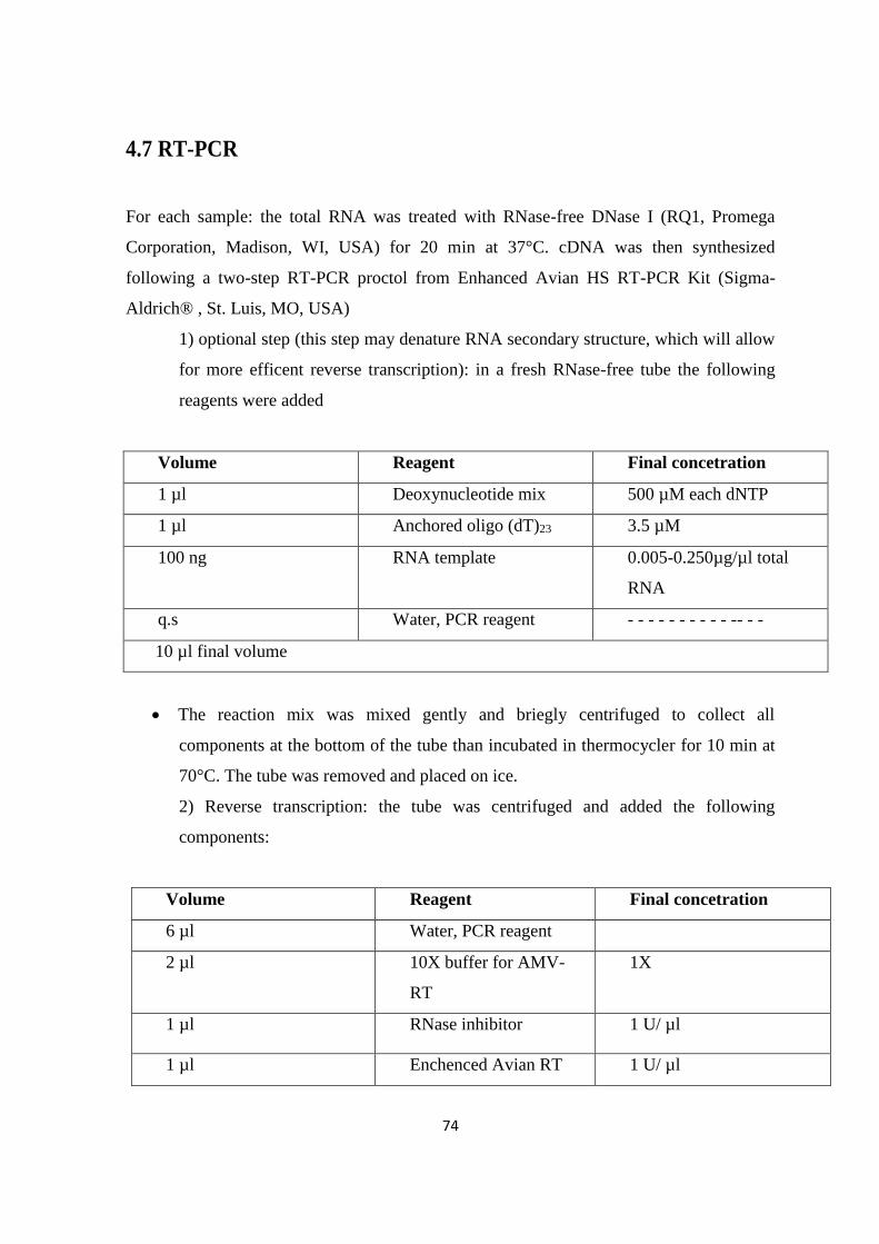
74
4.7 RT-PCR
For each sample: the total RNA was treated with RNase-free DNase I (RQ1, Promega
Corporation, Madison, WI, USA) for 20 min at 37°C. cDNA was then synthesized
following a two-step RT-PCR proctol from Enhanced Avian HS RT-PCR Kit (Sigma-
Aldrich® , St. Luis, MO, USA)
1) optional step (this step may denature RNA secondary structure, which will allow
for more efficent reverse transcription): in a fresh RNase-free tube the following
reagents were added
Volume Reagent Final concetration
1 µl Deoxynucleotide mix 500 µM each dNTP
1 µl Anchored oligo (dT)23 3.5 µM
100 ng RNA template 0.005-0.250µg/µl total
RNA
q.s Water, PCR reagent - - - - - - - - - - -- - -
10 µl final volume
The reaction mix was mixed gently and briegly centrifuged to collect all
components at the bottom of the tube than incubated in thermocycler for 10 min at
70°C. The tube was removed and placed on ice.
2) Reverse transcription: the tube was centrifuged and added the following
components:
Volume Reagent Final concetration
6 µl Water, PCR reagent
2 µl 10X buffer for AMV-
RT
1X
1 µl RNase inhibitor 1 U/ µl
1 µl Enchenced Avian RT 1 U/ µl
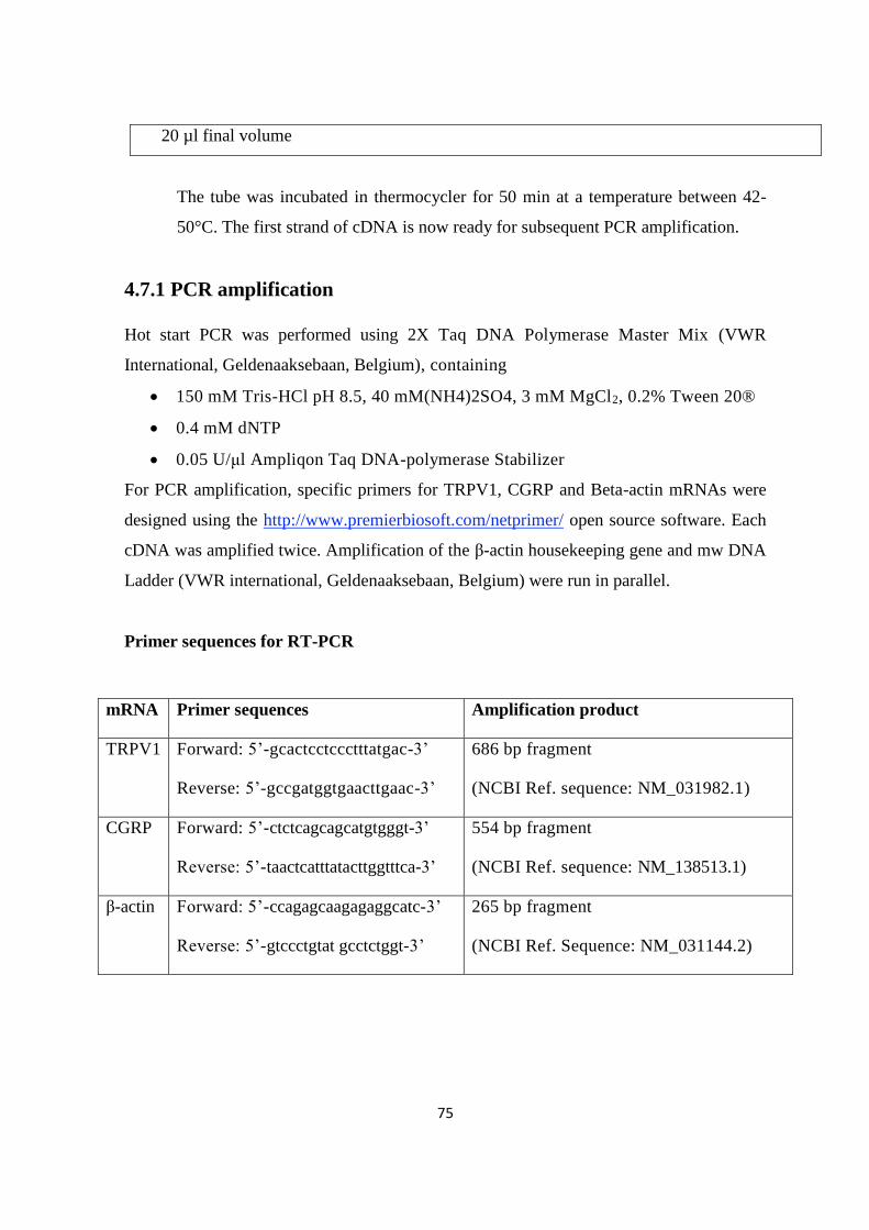
75
20 µl final volume
The tube was incubated in thermocycler for 50 min at a temperature between 42-
50°C. The first strand of cDNA is now ready for subsequent PCR amplification.
4.7.1 PCR amplification
Hot start PCR was performed using 2X Taq DNA Polymerase Master Mix (VWR
International, Geldenaaksebaan, Belgium), containing
150 mM Tris-HCl pH 8.5, 40 mM(NH4)2SO4, 3 mM MgCl2, 0.2% Tween 20®
0.4 mM dNTP
0.05 U/μl Ampliqon Taq DNA-polymerase Stabilizer
For PCR amplification, specific primers for TRPV1, CGRP and Beta-actin mRNAs were
designed using the http://www.premierbiosoft.com/netprimer/ open source software. Each
cDNA was amplified twice. Amplification of the β-actin housekeeping gene and mw DNA
Ladder (VWR international, Geldenaaksebaan, Belgium) were run in parallel.
Primer sequences for RT-PCR
mRNA Primer sequences Amplification product
TRPV1 Forward: 5’-gcactcctccctttatgac-3’
Reverse: 5’-gccgatggtgaacttgaac-3’
686 bp fragment
(NCBI Ref. sequence: NM_031982.1)
CGRP Forward: 5’-ctctcagcagcatgtgggt-3’
Reverse: 5’-taactcatttatacttggtttca-3’
554 bp fragment
(NCBI Ref. sequence: NM_138513.1)
β-actin Forward: 5’-ccagagcaagagaggcatc-3’
Reverse: 5’-gtccctgtat gcctctggt-3’
265 bp fragment
(NCBI Ref. Sequence: NM_031144.2)
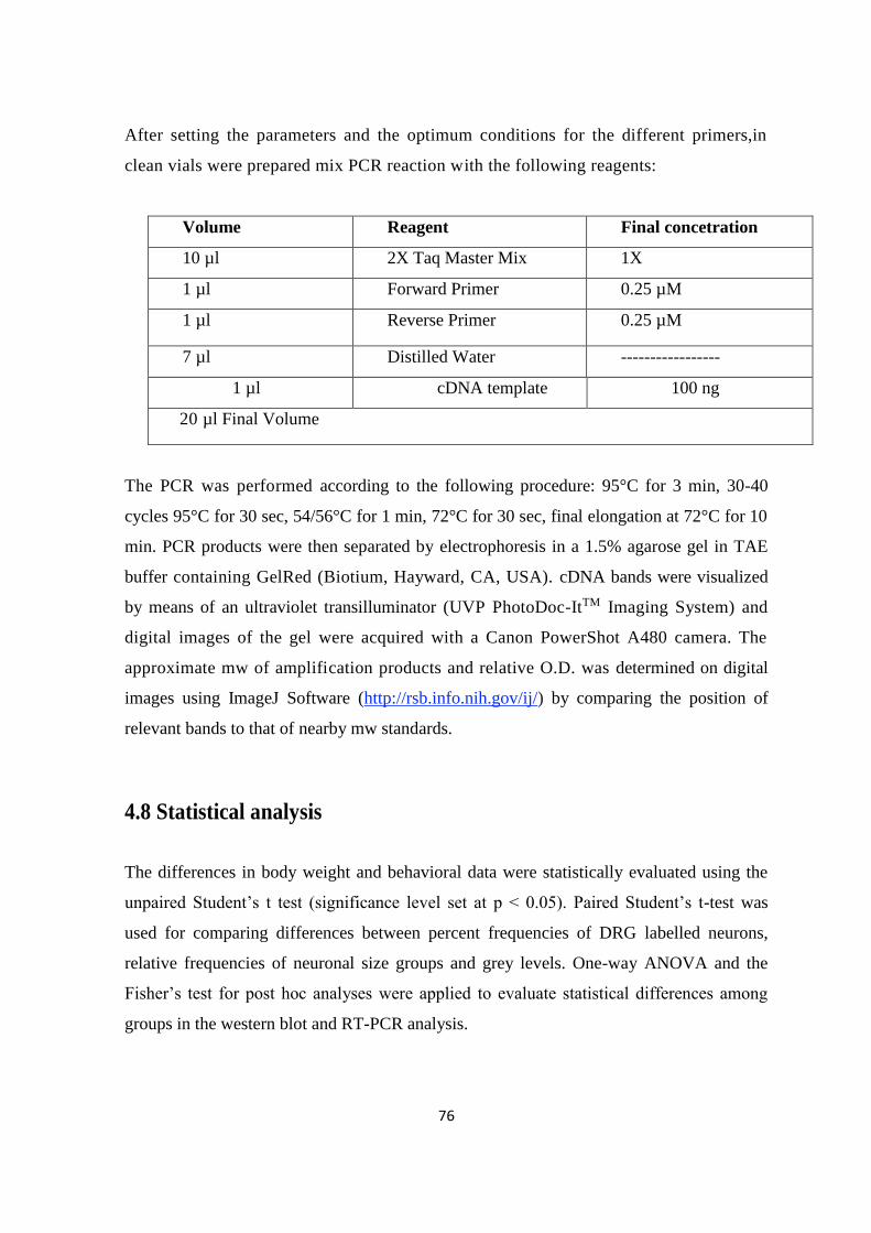
76
After setting the parameters and the optimum conditions for the different primers,in
clean vials were prepared mix PCR reaction with the following reagents:
Volume Reagent Final concetration
10 µl 2X Taq Master Mix 1X
1 µl Forward Primer 0.25 µM
1 µl Reverse Primer 0.25 µM
7 µl Distilled Water -----------------
1 µl cDNA template 100 ng
20 µl Final Volume
The PCR was performed according to the following procedure: 95°C for 3 min, 30-40
cycles 95°C for 30 sec, 54/56°C for 1 min, 72°C for 30 sec, final elongation at 72°C for 10
min. PCR products were then separated by electrophoresis in a 1.5% agarose gel in TAE
buffer containing GelRed (Biotium, Hayward, CA, USA). cDNA bands were visualized
by means of an ultraviolet transilluminator (UVP PhotoDoc-ItTM Imaging System) and
digital images of the gel were acquired with a Canon PowerShot A480 camera. The
approximate mw of amplification products and relative O.D. was determined on digital
images using ImageJ Software (http://rsb.info.nih.gov/ij/) by comparing the position of
relevant bands to that of nearby mw standards.
4.8 Statistical analysis
The differences in body weight and behavioral data were statistically evaluated using the
unpaired Student’s t test (significance level set at p < 0.05). Paired Student’s t-test was
used for comparing differences between percent frequencies of DRG labelled neurons,
relative frequencies of neuronal size groups and grey levels. One-way ANOVA and the
Fisher’s test for post hoc analyses were applied to evaluate statistical differences among
groups in the western blot and RT-PCR analysis.
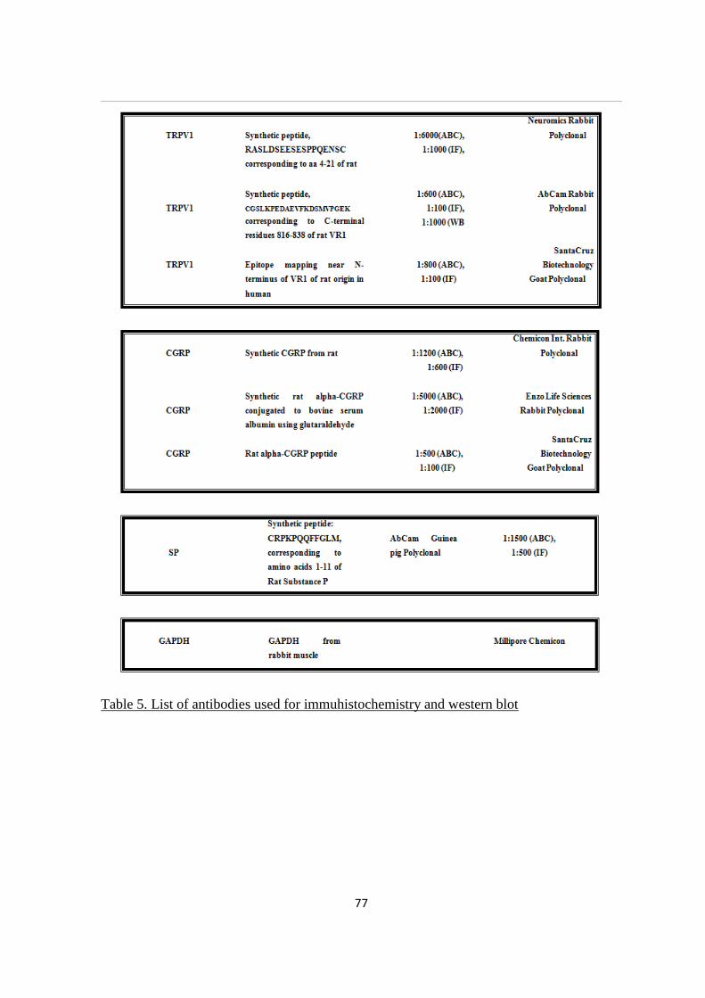
77
Table 5. List of antibodies used for immuhistochemistry and western blot
78
5. REFERENCES
1. Adams J. The development of proteasome inhibitors as anticancer drugs. Cancer
Cell. 2004; 5(5):417-421.
2. Adams J. The proteasome: a suitable antineoplastic target. Nat Rev Cancer.2004
May;4(5):349-360.
3. Adams J. The proteasome: structure, function, and role in the cell. Cancer Treat
Rev. 2003; 29 Suppl 1:3-9.
4. Afrah AW, Fiskå A, Gjerstad J, Gustafsson H, Tjølsen A, Olgart L, Stiller CO,
Hole K, Brodin E. Spinal substance P release in vivo during the induction of long-
term potentiation in dorsal horn neurons. Pain. 2002; 96(1-2):49-55.
5. Albers JW, Chaudhry V, Cavaletti G, Donehower RC. Interventions for preventing
neuropathy caused by cisplatin and related compounds. Cochrane Database Syst
Rev; (2):CD005228.
6. Alessandri-Haber N, Dina OA, Joseph EK, Reichling DB, Levine JD. Interaction of
transient receptor potential vanilloid 4, integrin, and SRC tyrosine kinase in
mechanical hyperalgesia. J Neurosci. 2008; 28(5):1046-1057.
7. Alvarez FJ, Morris HR, Priestley JV. Sub-populations of smaller diameter
trigeminal primary afferent neurons defined by expression of calcitonin gene-
related peptide and the cell surface oligosaccharide recognized by monoclonal
antibody LA4. J Neurocytol. 1991; 20(9):716-731.
8. Alvarez FJ, Priestley JV. Anatomy of somatostatin-immunoreactive fibres and cell
bodies in the rat trigeminal subnucleus caudalis. Neuroscience. 1990; 38(2):343-
357.
9. Alvarez FJ, Villalba RM, Carr PA, Grandes P, Somohano PM. Differential
distribution of metabotropic glutamate receptors 1a, 1b, and 5 in the rat spinal cord.
J Comp Neurol. 2000; 422(3):464-487.
10. An B, Goldfarb RH, Siman R, Dou QP. Novel dipeptidyl proteasome inhibitors
overcome Bcl-2 protective function and selectively accumulate the cyclin-
dependent kinase inhibitor p27 and induce apoptosis in transformed, but not
normal, human fibroblasts. Cell Death Differ. 1998; 5(12):1062-1075.
79
11. Apkarian AV, Bushnell MC, Treede RD, Zubieta JK. Human brain mechanisms of
pain perception and regulation in health and disease. Eur J Pain. 2005; 9(4):463-
484.
12. Archer AG, Watkins PJ, Thomas PK, Sharma AK, Payan J. The natural history of
acute painful neuropathy in diabetes mellitus. J Neurol Neurosurg Psychiatry. 1983;
46(6):491-499.
13. Armstrong T, Almadrones L, Gilbert MR. Chemotherapy-induced peripheral
neuropathy. Oncol Nurs Forum. 2005; 32(2):305-311.
14. Attal N, Bouhassira D, Gautron M, Vaillant JN, Mitry E, Lepère C, Rougier P,
Guirimand F. Thermal hyperalgesia as a marker of oxaliplatin neurotoxicity: a
prospective quantified sensory assessment study. Pain. 2009; 144(3):245-252.
15. Attal N, Bouhassira D, Gautron M, Vaillant JN, Mitry E, Lepère C, Rougier
P,Guirimand F. Thermal hyperalgesia as a marker of oxaliplatin neurotoxicity: a
prospective quantified sensory assessment study. Pain. 2009 ;144(3):245-252.
16. Authier N, Balayssac D, Marchand F, Ling B, Zangarelli A, Descoeur J, Coudore
F, Bourinet E, Eschalier A. Animal models of chemotherapy-evoked painful
peripheral neuropathies. Neurotherapeutics. 2009; 6(4):620-629.
17. Authier N, Fialip J, Eschalier A, Coudoré F. Assessment of allodynia and
hyperalgesia after cisplatin administration to rats. Neurosci Lett. 2000; 291(2):73-6.
18. Authier N, Gillet JP, Fialip J, Eschalier A, Coudore F. An animal model of
nociceptive peripheral neuropathy following repeated cisplatin injections. Exp
Neurol. 2003; 182(1):12-20.
19. Averill S, McMahon SB, Clary DO, Reichardt LF, Priestley JV.
Immunocytochemical localization of trkA receptors in chemically identified
subgroups of adult rat sensory neurons. Eur J Neurosci. 1995; 7(7):1484-1494.
20. Awada A, Albanell J, Canney PA, Dirix LY, Gil T, Cardoso F, Gascon P, Piccart
MJ, Baselga J. Bortezomib/docetaxel combination therapy in patients with
anthracycline-pretreated advanced/metastatic breast cancer: a phase I/II
doseescalation study. Br. J. Cancer. 2008; 98, 1500–1507.
21. Axelrod FB, Hilz MJ. Inherited autonomic neuropathies. Semin Neurol. 2003;
23(4):381-390.
22. Azhary H, Farooq MU, Bhanushali M, Majid A, Kassab MY. Peripheral
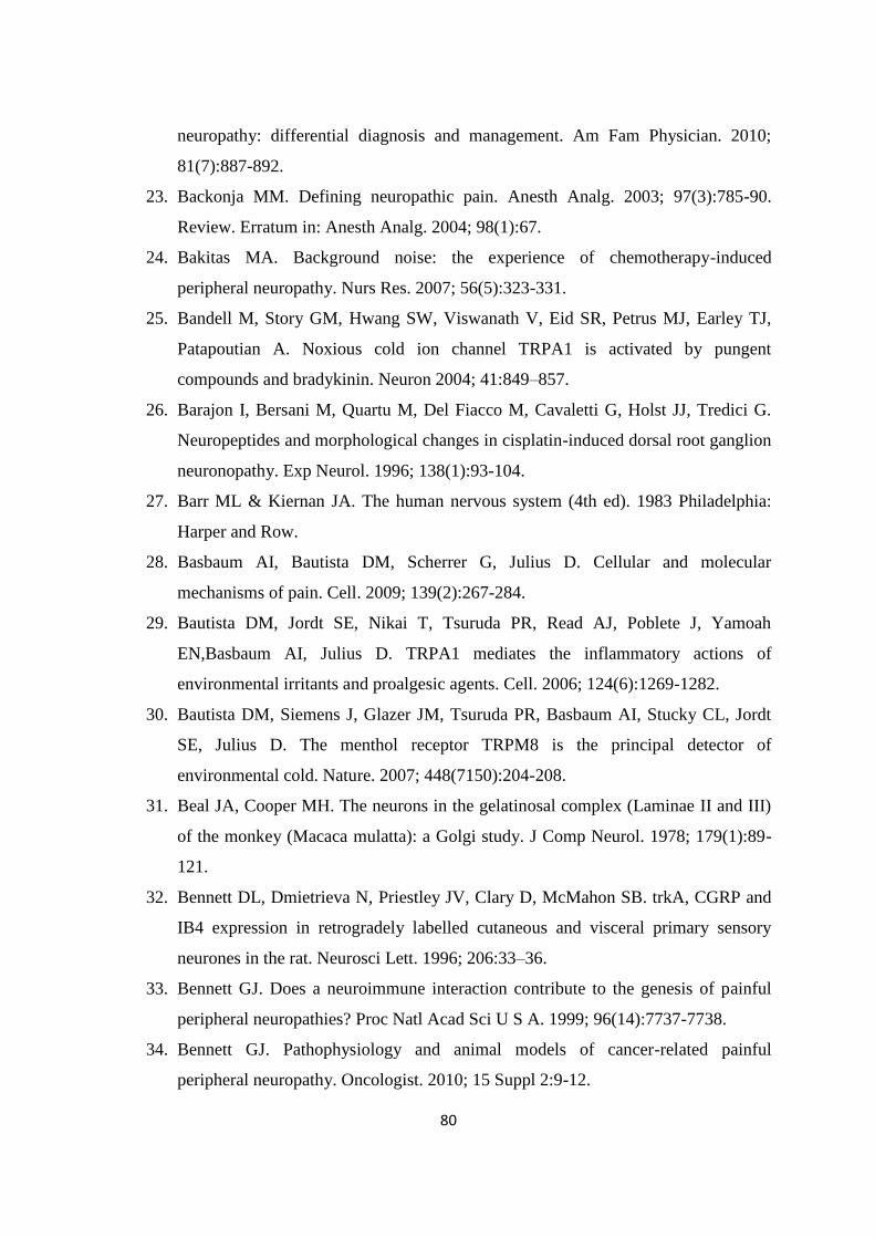
80
neuropathy: differential diagnosis and management. Am Fam Physician. 2010;
81(7):887-892.
23. Backonja MM. Defining neuropathic pain. Anesth Analg. 2003; 97(3):785-90.
Review. Erratum in: Anesth Analg. 2004; 98(1):67.
24. Bakitas MA. Background noise: the experience of chemotherapy-induced
peripheral neuropathy. Nurs Res. 2007; 56(5):323-331.
25. Bandell M, Story GM, Hwang SW, Viswanath V, Eid SR, Petrus MJ, Earley TJ,
Patapoutian A. Noxious cold ion channel TRPA1 is activated by pungent
compounds and bradykinin. Neuron 2004; 41:849–857.
26. Barajon I, Bersani M, Quartu M, Del Fiacco M, Cavaletti G, Holst JJ, Tredici G.
Neuropeptides and morphological changes in cisplatin-induced dorsal root ganglion
neuronopathy. Exp Neurol. 1996; 138(1):93-104.
27. Barr ML & Kiernan JA. The human nervous system (4th ed). 1983 Philadelphia:
Harper and Row.
28. Basbaum AI, Bautista DM, Scherrer G, Julius D. Cellular and molecular
mechanisms of pain. Cell. 2009; 139(2):267-284.
29. Bautista DM, Jordt SE, Nikai T, Tsuruda PR, Read AJ, Poblete J, Yamoah
EN,Basbaum AI, Julius D. TRPA1 mediates the inflammatory actions of
environmental irritants and proalgesic agents. Cell. 2006; 124(6):1269-1282.
30. Bautista DM, Siemens J, Glazer JM, Tsuruda PR, Basbaum AI, Stucky CL, Jordt
SE, Julius D. The menthol receptor TRPM8 is the principal detector of
environmental cold. Nature. 2007; 448(7150):204-208.
31. Beal JA, Cooper MH. The neurons in the gelatinosal complex (Laminae II and III)
of the monkey (Macaca mulatta): a Golgi study. J Comp Neurol. 1978; 179(1):89-
121.
32. Bennett DL, Dmietrieva N, Priestley JV, Clary D, McMahon SB. trkA, CGRP and
IB4 expression in retrogradely labelled cutaneous and visceral primary sensory
neurones in the rat. Neurosci Lett. 1996; 206:33–36.
33. Bennett GJ. Does a neuroimmune interaction contribute to the genesis of painful
peripheral neuropathies? Proc Natl Acad Sci U S A. 1999; 96(14):7737-7738.
34. Bennett GJ. Pathophysiology and animal models of cancer-related painful
peripheral neuropathy. Oncologist. 2010; 15 Suppl 2:9-12.
81
35. Besson JM, Chaouch A. Peripheral and spinal mechanisms of nociception. Physiol
Rev. 1987; 67(1):67-186.
36. Bhave G, Zhu W, Wang H, Brasier DJ, Oxford GS, Gereau RW 4th. cAMP-
dependent protein kinase regulates desensitization of the capsaicin receptor (VR1)
by direct phosphorylation. Neuron. 2002; 35(4):721-731.
37. Binder A, Stengel M, Maag R, Wasner G, Schoch R, Moosig F, Schommer B,
Baron R. Pain in oxaliplatin-induced neuropathy--sensitisation in the peripheral and
central nociceptive system. Eur J Cancer. 2007; 43(18):2658-2663.
38. Bold RJ, Virudachalam S, McConkey DJ. Chemosensitization of pancreatic cancer
by inhibition of the 26S proteasome. J Surg Res. 2001; 100(1):11-7.
39. Boyette-Davis JA, Cata JP, Driver LC, Novy DM, Bruel BM, Mooring DL,
Wendelschafer-Crabb G, Kennedy WR, Dougherty PM. Persistent
chemoneuropathy in patients receiving the plant alkaloids paclitaxel and vincristine.
Cancer Chemother Pharmacol. 2013; 71(3):619-626.
40. Bridges D, Thompson SW, Rice AS. Mechanisms of neuropathic pain. Br J
Anaesth. 2001; 87(1):12-26.
41. Brix Finnerup N, Hein Sindrup S, Staehelin Jensen T. Management of painful
neuropathies. Handb Clin Neurol. 2013; 115:279-290.
42. Campbell IN, Raja SN, Cohen RH, Manning DC, Khan AA and Mayer RA
Peripheral neural mechanisms of nociception. In: Textbook of Pain, 2nd Ed. Eds.
Wall, P.D. and Melzack, R, Churchill Livingstone, Edinburgh. 1989. 22-45.
43. Campbell JN, Meyer RA. Mechanisms of neuropathic pain. Neuron. 2006;
52(1):77-92.
44. Caponigro F, Lacombe D, Twelves C, Bauer J, Govaerts AS, Marréaud S, Milano
A, Anthoney A. An EORTC phase I study of Bortezomib in combination with
oxaliplatin, leucovorin and 5-fluorouracil in patients with advanced colorectal
cancer. Eur J Cancer. 2009; 45(1):48-55.
45. Casafont I, Berciano MT, Lafarga M. Bortezomib induces the formation of nuclear
poly(A) RNA granules enriched in Sam68 and PABPN1 in sensory ganglia
neurons. Neurotox Res. 2010; 17(2):167-178.
46. Casey KL, Morrow TJ. Ventral posterior thalamic neurons differentially responsive
to noxious stimulation of the awake monkey. Science 1983; 221: 675-677.
82
47. Cata JP, Weng HR, Burton AW, Villareal H, Giralt S, Dougherty PM. Quantitative
sensory findings in patients with bortezomib-induced pain. J Pain. 2007; 8(4):296-
306.
48. Cata JP, Weng HR, Burton AW, Villareal H, Giralt S, Dougherty PM. Quantitative
sensory findings in patients with bortezomib-induced pain. J Pain. 2007; 8(4):296-
306.
49. Caterina MJ, Leffler A, Malmberg AB, Martin WJ, Trafton J, Petersen-Zeitz KR,
Koltzenburg M, Basbaum AI, Julius D. Impaired nociception and pain sensation in
mice lacking the capsaicin receptor. Science. 2000; 288(5464):306-313.
50. Caterina MJ, Schumacher MA, Tominaga M, Rosen TA, Levine JD, Julius D. The
capsaicin receptor: a heat-activated ion channel in the pain pathway.Nature.1997;
389:816–824.
51. Cavaletti G, Marmiroli P. Chemotherapy-induced peripheral neurotoxicity. Nat Rev
Neurol. 2010; 6(12):657-666.
52. Cavaletti G, Tredici G, Marmiroli P, Petruccioli MG, Barajon I, Fabbrica D.
Morphometric study of the sensory neuron and peripheral nerve changes induced
by chronic cisplatin (DDP) administration in rats. Acta Neuropathol.
1992;84(4):364-371.
53. Chaudhry V, Cornblath DR, Polydefkis M, Ferguson A, Borrello I. Characteristics
of bortezomib- and thalidomide-induced peripheral neuropathy. J Peripher Nerv
Syst. 2008; 13(4):275-282.
54. Chen H, Lamer TJ, Rho RH, Marshall KA, Sitzman BT, Ghazi SM, Brewer RP.
Contemporary management of neuropathic pain for the primary care physician.
Mayo Clin Proc. 2004; 79(12):1533-1545.
55. Chen Y, Willcockson HH, Valtschanoff JG. Influence of the vanilloid receptor
TRPV1 on the activation of spinal cord glia in mouse models of pain. Exp Neurol.
2009; 220(2):383-390.
56. Ciechanover A. The ubiquitin-proteasome pathway: on protein death and cell life.
EMBO J. 1998; 17(24):7151-7160.
57. Cohen SP, Mao J. Neuropathic pain: mechanisms and their clinical implications.
BMJ. 2014; 348:f7656.
58. Colburn RW, Lubin ML, Stone DJ Jr, Wang Y, Lawrence D, D’Andrea MR,
83
Brandt MR, Liu Y, Flores CM, Qin N. Attenuated cold sensitivity in TRPM8 null
mice. Neuron 2007; 54:379–386.
59. Collins WR Jr, Nulsen FE, Randt CT. Relation of peripheral nerve fiber size and
sensation in man. Arch Neurol. 1960; 3:381-385.
60. Conner JM, Lauterborn JC, Yan Q, Gall CM, Varon S. Distribution of brain-
derived neurotrophic factor (BDNF) protein and mRNA in the normal adult rat
CNS: evidence for anterograde axonal transport. J Neurosci. 1997; 17(7):2295-
2313.
61. Cook AJ, Woolf CJ, Wall PD, McMahon SB. Dynamic receptive field plasticity in
rat spinal cord dorsal horn following C-primary afferent input. Nature. 1987;
325(7000):151-153.
62. Coudoré-Civiale MA, Ling B, Balayssac D. Spinal Substance P and CGRP staining
in vincristine, cisplatin, streptozocin or constriction injury-induced neuropathies
M.A Coudoré-Civiale et al, /J. Pharm. Sci. & Res. Vol.2 (9), 2010, 590-598.
63. Craig AD. Distribution of trigeminothalamic and spinothalamic lamina I
terminations in the macaque monkey. J Comp Neurol 2004a; 477:119 –148.
64. Craig AD. Lamina I, but not lamina V, spinothalamic neurons exhibit responses
that correspond with burning pain. J Neurophysiol. 2004b; 92:2604-2609.
65. Craig AD. Pain mechanisms: labeled lines versus convergence in central
processing. Annu Rev Neurosci. 2003; 26:1-30.
66. Csillik B, Janka Z, Boncz I, Kálmán J, Mihály A, Vécsei L, Knyihár E. Molecular
plasticity of primary nociceptive neurons: relations of the NGF-c-jun system to
neurotomy and chronic pain. Ann Anat. 2003, 185(4):303-314.
67. Cusack JC Jr, Liu R, Houston M, Abendroth K, Elliott PJ, Adams J, Baldwin AS Jr.
Enhanced chemosensitivity to CPT-11 with proteasome inhibitor PS-341:
implications for systemic nuclear factor-kappaB inhibition. Cancer Res. 2001;
61(9):3535-3540.
68. Davis JB, Gray J, Gunthorpe MJ, Hatcher JP, Davey PT, Overend P, Harries MH,
Latcham J, Clapham C, Atkinson K, et al. et al.,Vanilloid receptor-1 is essential for
inflammatory thermal hyperalgesia. Nature 2000; 405:183–187.
69. Decosterd I, Woolf CJ. Spared nerve injury: an animal model of persistent
peripheral neuropathic pain. Pain. 2000; 87(2):149-158.
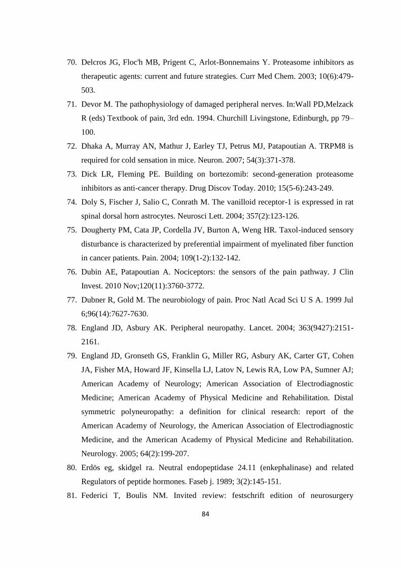
84
70. Delcros JG, Floc'h MB, Prigent C, Arlot-Bonnemains Y. Proteasome inhibitors as
therapeutic agents: current and future strategies. Curr Med Chem. 2003; 10(6):479-
503.
71. Devor M. The pathophysiology of damaged peripheral nerves. In:Wall PD,Melzack
R (eds) Textbook of pain, 3rd edn. 1994. Churchill Livingstone, Edinburgh, pp 79–
100.
72. Dhaka A, Murray AN, Mathur J, Earley TJ, Petrus MJ, Patapoutian A. TRPM8 is
required for cold sensation in mice. Neuron. 2007; 54(3):371-378.
73. Dick LR, Fleming PE. Building on bortezomib: second-generation proteasome
inhibitors as anti-cancer therapy. Drug Discov Today. 2010; 15(5-6):243-249.
74. Doly S, Fischer J, Salio C, Conrath M. The vanilloid receptor-1 is expressed in rat
spinal dorsal horn astrocytes. Neurosci Lett. 2004; 357(2):123-126.
75. Dougherty PM, Cata JP, Cordella JV, Burton A, Weng HR. Taxol-induced sensory
disturbance is characterized by preferential impairment of myelinated fiber function
in cancer patients. Pain. 2004; 109(1-2):132-142.
76. Dubin AE, Patapoutian A. Nociceptors: the sensors of the pain pathway. J Clin
Invest. 2010 Nov;120(11):3760-3772.
77. Dubner R, Gold M. The neurobiology of pain. Proc Natl Acad Sci U S A. 1999 Jul
6;96(14):7627-7630.
78. England JD, Asbury AK. Peripheral neuropathy. Lancet. 2004; 363(9427):2151-
2161.
79. England JD, Gronseth GS, Franklin G, Miller RG, Asbury AK, Carter GT, Cohen
JA, Fisher MA, Howard JF, Kinsella LJ, Latov N, Lewis RA, Low PA, Sumner AJ;
American Academy of Neurology; American Association of Electrodiagnostic
Medicine; American Academy of Physical Medicine and Rehabilitation. Distal
symmetric polyneuropathy: a definition for clinical research: report of the
American Academy of Neurology, the American Association of Electrodiagnostic
Medicine, and the American Academy of Physical Medicine and Rehabilitation.
Neurology. 2005; 64(2):199-207.
80. Erdös eg, skidgel ra. Neutral endopeptidase 24.11 (enkephalinase) and related
Regulators of peptide hormones. Faseb j. 1989; 3(2):145-151.
81. Federici T, Boulis NM. Invited review: festschrift edition of neurosurgery
85
peripheral nervous system as a conduit for delivering therapies for diabetic
neuropathy, amyotrophic lateral sclerosis, and nerve regeneration. Neurosurgery.
2009; 65 (4 Suppl).
82. Fernández-Carvajal A, Fernández-Ballester G, Devesa I, González-Ros JM, Ferrer-
Montiel A. New strategies to develop novel pain therapies: addressing
thermoreceptors from different points of view. Pharmaceuticals (Basel). 2011;
5(1):16-48.
83. Ferrell BA. Pain management. Clin Geriatr Med. 2000; 16(4):853-74.
84. Fischer SJ, McDonald ES, Gross L, Windebank AJ. Alterations in cell cycle
regulation underlie cisplatin induced apoptosis of dorsal root ganglion neurons in
vivo. Neurobiol Dis. 2001; 8(6):1027-1035.
85. Frankel A, Man S, Elliott P, Adams J, Kerbel RS. Lack of multicellular drug
resistance observed in human ovarian and prostate carcinoma treated with the
proteasome inhibitor PS-341. Clin Cancer Res. 2000; 6(9):3719-3728.
86. Fribley A, Zeng Q, Wang CY. Proteasome inhibitor PS-341 induces apoptosis
through induction of endoplasmic reticulum stress-reactive oxygen species in head
and neck squamous cell carcinoma cells. Mol Cell Biol. 2004; 24(22):9695-9704.
87. Fürst S. Transmitters involved in antinociception in the spinal cord. Brain Res Bull.
1999; 48(2):129-141.
88. Garcia JM, Cata JP, Dougherty PM, Smith RG. Ghrelin prevents cisplatin-induced
mechanical hyperalgesia and cachexia. Endocrinology. 2008; 149(2):455-460.
89. Garcia-Larrea L. Insights gained into pain processing from patients with focal brain
lesions. Neurosci Lett. 2012; 520(2):188-191.
90. Gatchel RJ, Peng YB, Peters ML, Fuchs PN, Turk DC. The biopsychosocial
approach to chronic pain: scientific advances and future directions. Psychol Bull.
2007; 133(4):581-624.
91. Gaudio E, Il Sistema Nervoso Centrale, Piccin (nuova libreria S.p.a).2012, Padova.
92. Gauriau C, Bernard JF. Pain pathways and parabrachial circuits in the rat. Exp
Physiol 2002; 87:251–258.
93. Gerke MB, Plenderleith MB. Binding sites for the plant lectin Bandeiraea
simplicifolia I-isolectin B(4) are expressed by nociceptive primary sensory
neurones. Brain Res. 2001; 911(1):101-104.

86
94. Gilchrist HD, Allard BL, Simone DA. Enhanced withdrawal responses to heat and
mechanical stimuli following intraplantar injection of capsaicin in rats. Pain.1996;
67(1):179-188.
95. Giordano J. The neurobiology of nociceptive and anti-nociceptive systems. Pain
Physician. 2005; 8(3):277-290.
96. Graham BA, Brichta AM, Callister RJ. Moving from an averaged to specific view
of spinal cord pain processing circuits. J Neurophysiol. 2007; 98(3):1057-1063.
97. Grant G. Primary afferent projections to the spinal cord. In: “The Rat Nervous
system”, G. Paxinos ed. 2 vol, pp 303-309, Academic Press.1986, Orlando Florida.
98. Guo A, Vulchanova L, Wang J, Li X, Elde R. Immunocytochemical localization of
the vanilloid receptor 1 (VR1): relationship to neuropeptides, the P2X3
purinoceptor and IB4 binding sites. Eur J Neurosci. 1999; 11(3):946-958.
99. Hacker H, Karin M. Regulation and function of IKK and IKK-related kinases. Sci
STKE 2006; 2006:re13.
100. Hanani M. Satellite glial cells in sensory ganglia: from form to function. Brain Res
Brain Res Rev. 2005; 48(3):457-476.
101. Harper AA, Lawson SN. Conduction velocity is related to morphological cell type
in rat dorsal root ganglion neurones. J Physiol. 1985; 359:31-46.
102. Hausheer FH, Schilsky RL, Bain S, Berghorn EJ, Lieberman F. Diagnosis,
management, and evaluation of chemotherapy-induced peripheral neuropathy.
Semin Oncol. 2006; 33(1):15-49.
103. Hay JW. Quality of life effects of chemotherapy-induced neuropathy in ovarian
cancer [abstr 886]. Proc Am Soc Clin Oncol 2002; 21:222a.
104. Head KA. Peripheral neuropathy: pathogenic mechanisms and alternative therapies.
Altern Med Rev. 2006; 11(4):294-329.
105. Henry JL. Effects of substance P on functionally identified units in cat spinal cord.
Brain Res. 1976; 114(3):439-451.
106. Hideshima T, Chauhan D, Podar K, Schlossman RL, Richardson P, Anderson KC.
Novel therapies targeting the myeloma cell and its bone marrow microenvironment.
Semin Oncol. 2001; 28(6):607-612.
107. Hideshima T, Chauhan D, Richardson P, Mitsiades C, Mitsiades N, Hayashi T,
Munshi N, Dang L, Castro A, Palombella V, Adams J, Anderson KC. NF-kappa B
87
as a therapeutic target in multiple myeloma. J Biol Chem. 2002; 277(19):16639-
16647.
108. Hideshima T, Ikeda H, Chauhan D, Okawa Y, Raje N, Podar K, Mitsiades C,
Munshi NC, Richardson PG, Carrasco RD, Anderson KC. Bortezomib induces
canonical nuclear factor-kappaB activation in multiple myeloma cells. Blood. 2009;
114(5):1046-1052.
109. Holland JF, Scharlau C, Gailani S, Krant MJ, Olson KB, Horton J, Shnider BI,
Lynch JJ, Owens A, Carbone PP, Colsky J, Grob D, Miller SP, Hall TC. Vincristine
treatment of advanced cancer: a cooperative study of 392 cases. Cancer Res. 1973;
33(6):1258-1264.
110. Hori K, Ozaki N, Suzuki S, Sugiura Y. Upregulations of P2X(3) and ASIC3
involve in hyperalgesia induced by cisplatin administration in rats. Pain. 2010;
149(2):393-405.
111. Hucho T, Levine JD. Signaling pathways in sensitization: toward a nociceptor cell
biology. Neuron. 2007; 55(3):365-376.
112. Hudson LJ, Bevan S, Wotherspoon G, Gentry C, Fox A, Winter J. VR1 protein
expression increases in undamaged DRG neurons after partial nerve injury. Eur J
Neurosci. 2001 Jun;13(11):2105-2114.
113. Hughes RA. Peripheral neuropathy. BMJ. 2002; 324(7335):466-469.
114. Hunt SP, Mantyh PW and Preastley JV. The organization of biochemically
characterized sensory neurons. In Sensory Neurones: Diversity, Development and
Plasticity, ed. Scott, S. A., pp. 60–76. 1992. Oxford University Press, New York.
115. Hunt SP, Mantyh PW. The molecular dynamics of pain control. Nat Rev Neurosci.
2001; 2(2):83-91.
116. Jacobson S and Marcus EM. Neuroanatomy for the Neuroscientist. 2008 Springer
Science, New York.
117. James SE, Burden H, Burgess R, Xie Y, Yang T, Massa SM, Longo FM, Lu Q.
Anti-cancer drug induced neurotoxicity and identification of Rho pathway signaling
modulators as potential neuroprotectants. Neurotoxicology. 2008; 29(4):605-612.
118. Jesenberger V, Jentsch S. Deadly encounter: ubiquitin meets apoptosis. Nat Rev
Mol Cell Biol. 2002; 3(2):112-121.
119. JesseU, T.M. and Kelly, D.D. Pain and analgesia. In: Principles of Neural Science,

88
3rd Edition, Eds. Kandel, E.R., Schwartz, lH. and Jessell, T.M. 1997. 385-399.
120. Ji RR, Samad TA, Jin SX, Schmoll R, Woolf CJ. p38 MAPK activation by NGF in
primary sensory neurons after inflammation increases TRPV1 levels and maintains
heat hyperalgesia. Neuron. 2002; 36(1):57-68.
121. Jordan MA, Wilson L. Microtubules as a target for anticancer drugs. Nat Rev
Cancer. 2004; 4(4):253-265.
122. Jordt SE, Bautista DM, Chuang HH, McKemy DD, Zygmunt PM, Högestätt ED,
Meng ID, Julius D. Mustard oils and cannabinoids excite sensory nerve fibres
through the TRP channel ANKTM1. Nature. 2004 ;427 (6971):260-265.
123. Julius D, Basbaum AI. Molecular mechanisms of nociception. Nature. 2001;
413(6852):203-210.
124. Julius D. TRP channels and pain. Annu Rev Cell Dev Biol. 2013; 29:355-84.
125. Kaley TJ, Deangelis LM. Therapy of chemotherapy-induced peripheral
neuropathy.Br J Haematol. 2009; 145(1):3-14.
126. Kampa BM, Clements J, Jonas P, Stuart GJ. Kinetics of Mg2+ unblock of NMDA
receptors: implications for spike-timing dependent synaptic plasticity. J Physiol.
2004; 556(Pt 2):337-345.
127. Kane RC, Dagher R, Farrell A, Ko CW, Sridhara R, Justice R, Pazdur R.
Bortezomib for the treatment ofmantle cell lymphoma. Clin.Cancer Res. 2007; 13,
5291–5294.
128. Kannarkat G, Lasher EE, Schiff D. Neurologic complications of chemotherapy
agents. Curr Opin Neurol. 2007; 20(6):719-725.
129. Karin M, Cao Y, Greten FR, Li ZW. NF-kappaB in cancer: from innocent
bystander to major culprit. Nat Rev Cancer. 2002; 2(4):301-310.
130. Kim SR, Lee DY, Chung ES, Oh UT, Kim SU, Jin BK. Transient receptor potential
vanilloid subtype 1 mediates cell death of mesencephalic dopaminergic neurons in
vivo and in vitro. J Neurosci. 2005; 25(3):662-671.
131. Kim YH, Back SK, Davies AJ, Jeong H, Jo HJ, Chung G, Na HS, Bae YC, Kim SJ,
Kim JS, Jung SJ, Oh SB. TRPV1 in GABAergic interneurons mediates neuropathic
mechanical allodynia and disinhibition of the nociceptive circuitry in the spinal
cord. Neuron. 2012; 74(4):640-647.
132. Krarup-Hansen A, Helweg-Larsen S, Schmalbruch H, Rørth M, Krarup C.
89
Neuronal involvement in cisplatin neuropathy: prospective clinical and
neurophysiological studies. Brain. 2007; 130(Pt 4):1076-1088.
133. LaMotte RH, Lundberg LE, Torebjörk HE. Pain, hyperalgesia and activity in
nociceptive C units in humans after intradermal injection of capsaicin. J Physiol.
1992; 448:749-764.
134. LaMotte RH, Shain CN, Simone DA, Tsai EF. Neurogenic hyperalgesia:
psychophysical studies of underlying mechanisms. J Neurophysiol. 1991;
66(1):190-211.
135. Landowski TH, Megli CJ, Nullmeyer KD, Lynch RM, Dorr RT. Mitochondrial-
mediated disregulation of Ca2+ is a critical determinant of Velcade (PS-
41/bortezomib) cytotoxicity in myeloma cell lines. Cancer Res. 2005; 65(9):3828-
3836.
136. Latremoliere A, Woolf CJ. Central sensitization: a generator of pain
hypersensitivity by central neural plasticity. J Pain. 2009; 10(9):895-926.
137. Lawson SN, Crepps BA, Perl ER. Relationship of substance P to afferent
characteristics of dorsal root ganglion neurones in guinea-pig. J Physiol. 1997; 505
(Pt 1):177-191.
138. Lawson SN, Waddell PJ, McCarthy PW. A comparison of the electrophysiological
and immunocytochemical properties of rat dorsal root ganglion neurons with A and
C-fibers. In: Schmidt RF, Schaible HG, Vahle-Hinz C (eds) Fine afferent nerve
fibers and pain. 1987. VCH Publishers,Weinheim and New York, pp 193–203.
139. Lawson SN. Morphological and biochemical cell types of sensory neurons. In:
Scott SA (ed) Sensory neurons: diversity, development and plasticity. 1992. Oxford
University Press, New York, pp 27–59.
140. Lee AH, Iwakoshi NN, Anderson KC, Glimcher LH. Proteasome inhibitors disrupt
the unfolded protein response in myeloma cells. Proc Natl Acad Sci U S A. 2003;
100(17):9946-9951.
141. Lee SE, Kim JH. Involvement of substance P and calcitonin gene-related peptide in
development and maintenance of neuropathic pain from spinal nerve injury model
of rat. Neurosci Res. 2007; 58(3):245-9.
142. Lieberman AR, Sensory ganglia, in: D.N. Landon (Ed.), The Peripheral Nerve,
Chapman and Hall, London, 1976, pp. 188 – 278.

90
143. Lindblom, U. Assessment of abnormal evoked pain in neurological pain patients
and its relation to spontaneous pain: a descriptive and conceptual model with some
analytical results. In: Fields, H.L., Dubner, R., Cervero, F. Eds, Proceeding of the
Fourth World congress on Pain. Advances in Pain Research and Therapy. 1985;
New York, Raven Press. Vol. 9, pp. 409-423.
144. Ling YH, Liebes L, Jiang JD, Holland JF, Elliott PJ, Adams J, Muggia FM, Perez-
Soler R. Mechanisms of proteasome inhibitor PS-341-induced G(2)-M-phase arrest
and apoptosis in human non-small cell lung cancer cell lines. Clin Cancer Res.
2003; 9(3):1145-1154.
145. Ling YH, Liebes L, Zou Y, Perez-Soler R. Reactive oxygen species generation and
mitochondrial dysfunction in the apoptotic response to Bortezomib, a novel
proteasome inhibitor, in human H460 non-small cell lung cancer cells. J Biol
Chem. 2003; 278(36):33714-33723.
146. Liu CW, Corboy MJ, DeMartino GN, Thomas PJ. Endoproteolytic activity of
theproteasome. Science. 2003; 299(5605):408-411.
147. LoMonaco M, Milone M, Batocchi AP, Padua L, Restuccia D, Tonali P. Cisplatin
neuropathy: clinical course and neurophysiological findings. J Neurol. 1992;
239(4):199-204.
148. Lovinger DM. Communication networks in the brain: neurons,
receptors,neurotransmitters, and alcohol. Alcohol Res Health. 2008; 31(3):196-214.
149. Lukacs V, Thyagarajan B, Varnai P, Balla A, Balla T, Rohacs T. Dual regulation
150. Lukacs V, Thyagarajan B, Varnai P, Balla A, Balla T, Rohacs T. Dual regulation of
TRPV1 by phosphoinositides. J Neurosci. 2007; 27(26):7070-7080.
151. Ma QP, Woolf CJ. Involvement of neurokinin receptors in the induction but not the
maintenance of mechanical allodynia in rat flexor motoneurones. J Physiol. 1995
Aug 1; 486 (Pt 3):769-777.
152. Ma QP, Woolf CJ. Noxious stimuli induce an N-methyl-D-aspartate receptor-
dependent hypersensitivity of the flexion withdrawal reflex to touch: implications
for the treatment of mechanical allodynia. Pain. 1995; 61(3):383-390.
153. Maag R, Baron R. Neuropathic pain: translational research and impact for patient
care. Curr Pain Headache Rep. 2006; 10(3):191-198.
154. MacLaren AP, Chapman RS, Wyllie AH, Watson CJ. p53-dependent apoptosis
91
induced by proteasome inhibition in mammary epithelial cells. Cell Death Differ.
2001; 8(3):210-218.
155. Macpherson LJ, Geierstanger BH, Viswanath V, Bandell M, Eid SR, Hwang S,
Patapoutian A. The pungency of garlic: activation of TRPA1 and TRPV1 in
response to allicin. Curr Biol. 2005; 15(10):929-934.
156. Mantyh PW, Rogers SD, Honore P, Allen BJ, Ghilardi JR, Li J, Daughters RS,
Lappi DA, Wiley RG, Simone DA. Inhibition of hyperalgesia by ablation of lamina
I spinal neurons expressing the substance P receptor. Science. 1997;
278(5336):275-279.
157. Mantyh PW. Cancer pain and its impact on diagnosis, survival and quality of life.
Nat Rev Neurosci. 2006; 7(10):797-809.
158. Marchettini P, Lacerenza M, Mauri E, Marangoni C. Painful peripheral
neuropathies. Curr Neuropharmacol. 2006; 4(3):175-181.
159. Martyn CN, Hughes RA. Epidemiology of peripheral neuropathy. J Neurol
Neurosurg Psychiatry. 1997; 62(4):310-318.
160. McCarthy PW, Lawson SN. Cell type and conduction velocity of rat primary
sensory neurons with calcitonin gene-related peptide-like immunoreactivity.
Neuroscience. 1990; 34(3):623-632.
161. McGaraughty S, Chu KL, Brown BS, Zhu CZ, Zhong C, Joshi SK, Honore P,
Faltynek CR, Jarvis MF. Contributions of central and peripheral TRPV1 receptors
to mechanically evoked and spontaneous firing of spinal neurons in inflamed rats. J
Neurophysiol. 2008; 100(6):3158-3166.
162. McKemy DD, Neuhausser WM, Julius D. Identification of a cold receptor reveals a
general role for TRP channels in thermosensation. Nature 2002; 416:52–58.
163. McNamara CR, Mandel-Brehm J, Bautista DM, Siemens J, Deranian KL, Zhao M,
Hayward NJ, Chong JA, Julius D, Moran MM, Fanger CM. TRPA1 mediates
formalin-induced pain. Proc Natl Acad Sci U S A 2007; 104:13525–13530.
164. McWhinney SR, Goldberg RM, McLeod HL. Platinum neurotoxicity
pharmacogenetics. Mol Cancer Ther. 2009 Jan;8(1):10-6.
165. Medzhitov R. Origin and physiological roles of inflammation. Nature. 2008 24;
454(7203):428-435.
166. Menard DP, van Rossum D, Kar S, St Pierre S, Sutak M, Jhamandas K, Quirion R.
92
A calcitonin gene-related peptide receptor antagonist prevents the development of
tolerance to spinal morphine analgesia. J Neurosci. 1996; 16(7):2342-2351.
167. Meregalli C, Canta A, Carozzi VA, Chiorazzi A, Oggioni N, Gilardini A, Ceresa C,
Avezza F, Crippa L, Marmiroli P, Cavaletti G. Bortezomib-induced painful
neuropathy in rats: a behavioral, neurophysiological and pathological study in rats.
Eur J Pain. 2010; 14(4):343-350.
168. Meregalli C, Ceresa C, Canta A, Carozzi VA, Chiorazzi A, Sala B, Oggioni N,
Lanza M, Letari O, Ferrari F, Avezza F, Marmiroli P, Caselli G, Cavaletti G.
CR4056, a new analgesic I2 ligand, is highly effective against bortezomib-induced
painful neuropathy in rats. J Pain Res. 2012; 5:151-167.
169. Michael GJ, Averill S, Nitkunan A, Rattray M, Bennett DL, Yan Q, Priestley JV.
Nerve growth factor treatment increases brain-derived neurotrophic factor
selectively in TrkA-expressing dorsal root ganglion cells and in their central
terminations within the spinal cord. J Neurosci. 1997; 17(21):8476-8490.
170. Millan MJ. The induction of pain: an integrative review. Prog Neurobiol. 1999;
57(1):1-164.
171. Miller SC, Huang R, Sakamuru S, Shukla SJ, Attene-Ramos MS, Shinn P, Van
Leer D, Leister W, Austin CP, Xia M. Identification of known drugs that act as
inhibitors of NF-kappaB signaling and their mechanism of action. Biochem
Pharmacol. 2010; 79(9):1272-1280.
172. Mitsiades N, Mitsiades CS, Poulaki V, Chauhan D, Richardson PG, Hideshima
T,Munshi N, Treon SP, Anderson KC. Biologic sequelae of nuclear factor-kappaB
blockade in multiple myeloma: therapeutic applications. Blood. 2002; 99(11):4079-
4086.
173. Miyano K, Tang HB, Nakamura Y, Morioka N, Inoue A, Nakata Y. Paclitaxel and
vinorelbine, evoked the release of substance P from cultured rat dorsal root
ganglion cells through different PKC isoform-sensitive ion channels.
Neuropharmacology. 2009; 57(1):25-32.
174. Mogil JS, Yu L, Basbaum AI. Pain genes?: natural variation and transgenic
mutants. Annu Rev Neurosci. 2000; 23:777-811.
175. Mühlen S, Ruchaud-Sparagano MH, Kenny B. Proteasome-independent
degradation of canonical NFkappaB complex components by the NleC protein of
93
pathogenic Escherichia coli. J Biol Chem. 2011 Feb 18; 286(7):5100-5107.
176. Nahman-Averbuch H, Yarnitsky D, Granovsky Y, Sprecher E, Steiner M, Tzuk-
ShinaT, Pud D. Pronociceptive pain modulation in patients with painful
chemotherapy-induced polyneuropathy. J Pain Symptom Manage. 2011; 42(2):229-
38.
177. Nakatsuka K, Gupta R, Saito S, Banzawa N, Takahashi K, Tominaga M, Ohta T.
Identification of molecular determinants for a potent mammalian TRPA1 antagonist
by utilizing species differences. J Mol Neurosci. 2013; 51(3):754-762.
178. Ocean AJ, Vahdat LT. Chemotherapy-induced peripheral neuropathy: pathogenesis
and emerging therapies. Support Care Cancer. 2004; 12(9):619-625.
179. Ochoa, J. Management of neuropathic pain patients. In: Current therapy of
Neurologic diseases, eds. RT Johnson, JW Griffin, 5th ed. 1996; New York:
Mosby.
180. Ohara PT, Vit JP, Bhargava A, Romero M, Sundberg C, Charles AC, Jasmin
L.Gliopathic pain: when satellite glial cells go bad. Neuroscientist. 2009;
15(5):450-463.
181. Olsson Y. Microenvironment of the peripheral nervous system under normal and
pathological conditions. Crit Rev Neurobiol. 1990;5 (3):265-311.
182. O'Neill J, Brock C, Olesen AE, Andresen T, Nilsson M, Dickenson AH.
Unravelling the mystery of capsaicin: a tool to understand and treat pain.
Pharmacol Rev. 2012; 64(4):939-971.
183. Ostchega Y, Donohue M, Fox N. High-dose cisplatin-related peripheral
neuropathy. Cancer Nurs. 1988; 11(1):23-32.
184. Pabbidi RM, Yu SQ, Peng S, Khardori R, Pauza ME, Premkumar LS. Influence of
TRPV1 on diabetes-induced alterations in thermal pain sensitivity. Mol Pain.
2008;4:9.
185. Pasnoor M, Dimachkie MM, Barohn RJ. Cryptogenic sensory polyneuropathy.
Neurol Clin. 2013; 31(2):463-476.
186. Patapoutian A. TRP channels and thermosensation. Chem Senses. 2005; 30 Suppl
1:i193-194.
187. Patestas M and Gartner L, A Textbook of Neuroanatomy. 2006, Wiley-Blackwell.
188. Pearson JS, Riedmaier P, Marchès O, Frankel G, Hartland EL. A type III effector
94
protease NleC from enteropathogenic Escherichia coli targets NF-κB for
degradation. Mol Microbiol. 2011; 80(1):219-230.
189. Pei XY, Dai Y, Grant S. Synergistic induction of oxidative injury and apoptosis in
human multiple myeloma cells by the proteasome inhibitor bortezomib and histone
deacetylase inhibitors. Clin Cancer Res. 2004; 10(11):3839-3852.
190. Peier AM, Moqrich A, Hergarden AC, Reeve AJ, Andersson DA, Story GM,
Earley TJ, Dragoni I, McIntyre P, Bevan S, Patapoutian A. A TRP channel that
senses cold stimuli and menthol. Cell. 2002; 108(5):705-715.
191. Peltier AC, Russell JW. Recent advances in drug-induced neuropathies. Curr Opin
Neurol. 2002; 15(5):633-638.
192. Pham LV, Tamayo AT, Yoshimura LC, Lo P, Ford RJ. Inhibition of constitutive
NF-kappa B activation in mantle cell lymphoma B cells leads to induction of cell
cycle arrest and apoptosis. J Immunol. 2003; 171(1):88-95.
193. Piperdi B, Ling YH, Liebes L, Muggia F, Perez-Soler R. Bortezomib:
understanding the mechanism of action. Mol Cancer Ther. 2011; 10(11):2029-
2030.
194. Pomonis JD, Harrison JE, Mark L, Bristol DR, Valenzano KJ, Walker K. N-(4-
Tertiarybutylphenyl)-4-(3-cholorphyridin-2-yl)tetrahydropyrazine -1(2H)-carbox-
amide (BCTC), a novel, orally effective vanilloid receptor 1 antagonist with
analgesic properties: II in vivo characterization in rat models of inflammatory and
neuropathic pain. J Pharmacol Exp Ther. 2003; 306(1):387-393.
195. Premkumar LS. Targeting TRPV1 as an alternative approach to narcotic analgesics
to treat chronic pain conditions. AAPS J. 2010; 12(3):361-370.
196. Price DD, Dubner R. Neurons that subserve the sensory-discriminative aspects of
pain. 1977. Pain 3:307–338.
197. Price DD. Psychological and NeuralMechansims of Pain. 1988. New York: Raven.
241.
198. Priestley JV, Michael GJ, Averill S, Liu M, Willmott N. Regulation of nociceptive
neurons by nerve growth factor and glial cell line derived neurotrophic factor. Can J
Physiol Pharmacol. 2002; 80(5):495-505.
199. Quasthoff S, Hartung HP. Chemotherapy-induced peripheral neuropathy.
JNeurol.2002; 249(1):9-17.
95
200. Ralston HJ 3rd. The fine structure of laminae I, II and III of the macaque spinal
cord. J Comp Neurol. 1979; 184(4):619-642.
201. Ravaglia S, Corso A, Piccolo G, Lozza A, Alfonsi E, Mangiacavalli S, Varettoni
M, Zappasodi P, Moglia A, Lazzarino M, Costa A. Immune-mediated neuropathies
in myeloma patients treated with bortezomib. Clin Neurophysiol. 2008;
119(11):2507-2512.
202. Reagan-Shaw S, Nihal M, Ahmad N. Dose translation from animal to human
studies revisited. FASEB J. 2008; 22(3):659-661.
203. Ren K. Primary afferents and inflammatory hyperexcitability. Pain. 1996; 67(1):1-
2.
204. Rexed B. The cytoarchitectonic organization of the spinal cord in the cat. JComp
Neurol. 1952; 96(3):414-495.
205. Richardson PG, Barlogie B, Berenson J, Singhal S, Jagannath S, Irwin D, Rajkumar
SV, Srkalovic G, Alsina M, Alexanian R, Siegel D, Orlowski RZ, Kuter D,
Limentani SA, Lee S, Hideshima T, Esseltine DL, Kauffman M, Adams J,
Schenkein DP, Anderson KC. A phase 2 study of bortezomib in relapsed, refractory
myeloma. N Engl J Med. 2003; 348(26):2609-2617.
206. Richardson PG, Briemberg H, Jagannath S, Wen PY, Barlogie B, Berenson J, et al.
et al.,Frequency, characteristics, and reversibility of peripheral neuropathy during
treatment of advanced multiple myeloma with bortezomib. J Clin Oncol. 2006;
24(19):3113-3120.
207. Richardson PG, Xie W, Jagannath S, Jakubowiak A, Lonial S, Raje NS, Alsina M,
Ghobrial IM, Schlossman RL, Munshi NC, Mazumder A, Vesole DH, Kaufman JL,
Colson K, McKenney M, Lunde LE, Feather J, Maglio ME, Warren D, Francis D,
Hideshima T, Knight R, Esseltine DL, Mitsiades CS, Weller E, Anderson KC. A
phase II trial of lenalidomide, bortezomib and dexamethasone in patients with
relapsed and relapsed/refractory myeloma. Blood. 2014.
208. Ripamonti CI. Pain management. Ann Oncol. 2012; 23 Suppl 10:x294-301.
209. Ripellino P, Fleetwood T, Cantello R, Comi C. Treatment of chronic inflammatory
demyelinating polyneuropathy: from molecular bases to practical considerations.
Autoimmune Dis. 2014; 2014:201657.
210. Sah DW, Ossipo MH, Porreca F. Neurotrophic factors as novel therapeutics for
96
neuropathic pain. Nat Rev Drug Discov. 2003; 2(6):460-472.
211. Saif MW, Reardon J. Management of oxaliplatin-induced peripheral neuropathy.
Ther Clin Risk Manag. 2005; 1(4):249-258.
212. Sakamoto H, Spike RC, Todd AJ. Neurons in laminae III and IV of the rat spinal
cord with the neurokinin-1 receptor receive few contacts from unmyelinated
primary afferents which do not contain substance P. Neuroscience. 1999;
94(3):903-908.
213. San Miguel JF, Schlag R, Khuageva NK, Dimopoulos MA, Shpilberg O, Kropff M,
Spicka I, Petrucci MT, Palumbo A, Samoilova OS, Dmoszynska A, Abdulkadyrov
KM, Schots R, Jiang B, Mateos MV, Anderson KC, Esseltine DL, Liu K, Cakana
A, van de Velde H, Richardson PG; VISTA Trial Investigators. Bortezomib plus
melphalan and prednisone for initial treatment of multiple myeloma. N Engl J Med.
2008; 359(9):906-917.
214. Sanchez JF, Krause JE, Cortright DN. The distribution and regulation of vanilloid
receptor VR1 and VR1 5' splice variant RNA expression in rat. Neuroscience.
2001;107 (3):373-381.
215. Sauer SK, Bove GM, Averbeck B, Reeh PW. Rat peripheral nerve components
release calcitonin gene-related peptide and prostaglandin E2 in response to noxious
stimuli: evidence that nervi nervorum are nociceptors. Neuroscience. 1999;
92(1):319-325.
216. Schaible HG. Peripheral and central mechanisms of pain generation. Handb Exp
Pharmacol. 2007;(177):3-28.
217. Scholz J, Woolf CJ. Can we conquer pain? Nat Neurosci. 2002; 5 Suppl: 1062-7.
218. Schwartz R, Davidson T. Pharmacology, pharmacokinetics, and practical
applications of bortezomib. Oncology (Williston Park). 2004;18(14 Suppl 11):14-
21.
219. Scott K, Kothari MJ. Evaluating the patient with peripheral nervous system
complaints. J Am Osteopath Assoc. 2005; 105(2):71-83.
220. Shah SA, Potter MW, McDade TP, Ricciardi R, Perugini RA, Elliott PJ, Adams J,
Callery MP. 26S proteasome inhibition induces apoptosis and limits growth of
human pancreatic cancer. J Cell Biochem. 2001; 82(1):110-122.
221. Shin CY, Shin J, Kim BM, Wang MH, Jang JH, Surh YJ, Oh U. Essential role of

97
mitochondrial permeability transition in vanilloid receptor 1-dependent cell death
of sensory neurons. Mol Cell Neurosci. 2003; 24(1):57-68.
222. Silverman JD, Kruger L. Acid phosphatase as a selective marker for a class of small
sensory ganglion cells in several mammals: spinal cord distribution, histochemical
properties, and relation to fluoride-resistant acid phosphatase (FRAP) of rodents.
Somatosens Res. 1988;5 (3):219-246.
223. Silverman JD, Kruger L. Selective neuronal glycoconjugate expression in sensory
and autonomic ganglia: relation of lectin reactivity to peptide and enzyme markers.
J Neurocytol. 1990; 19(5):789-801.
224. Smith BH, Torrance N. Epidemiology of neuropathic pain and its impact on quality
of life. Curr Pain Headache Rep. 2012; 16(3):191-198.
225. Snider WD, McMahon SB. Tackling pain at the source: new ideas about
nociceptors. Neuron. 1998; 20(4):629-32.
226. Sommer C. Painful neuropathies. Curr Opin Neurol. 2003; 16 (5):623-628.
227. Sommer EW, Kazimierczak J, Droz B. Neuronal subpopulations in the dorsal root
ganglion of the mouse as characterized by combination of ultrastructural and
cytochemical features. Brain Res. 1985; 346(2):310-326.
228. Stengel M, Baron R. Oxaliplatin-induced painful neuropathy-flicker of hope or
hopeless pain? Pain. 2009; 144(3):225-256.
229. Stillman M, Cata JP. Management of chemotherapy-induced peripheral neuropathy.
Curr Pain Headache Rep. 2006; 10(4):279-287.
230. Story GM, Peier AM, Reeve AJ, Eid SR, Mosbacher J, Hricik TR, Earley TJ,
Hergarden AC, Andersson DA, Hwang SW, McIntyre P, Jegla T, Bevan S,
Patapoutian A. ANKTM1, a TRP-like channel expressed in nociceptive neurons, is
activated by cold temperatures. Cell 2003; 112:819–829.
231. Streit WJ, Schulte BA, Balentine JD, Spicer SS. Evidence for glycoconjugate in
nociceptive primary sensory neurons and its origin from the Golgi complex. Brain
Res. 1986; 377(1):1-17.
232. Stucky CL, Dubin AE, Jeske NA, Malin SA, McKemy DD, Story GM. Roles of
transient receptor potential channels in pain. Brain Res Rev. 2009; 60(1):2-23.
233. Sunwoo JB, Chen Z, Dong G, Yeh N, Crowl Bancroft C, Sausville E, Adams J,
Elliott P, Van Waes C. Novel proteasome inhibitor PS-341 inhibits activation of
98
nuclear factor-kappa B, cell survival, tumor growth, and angiogenesis in squamous
cell carcinoma. Clin Cancer Res. 2001; 7(5):1419-1428.
234. Szolcsányi J. A pharmacological approach to elucidation of the role of different
nerve fibres and receptor endings in mediation of pain. J Physiol (Paris). 1977;
73(3):251-259.
235. Ta LE, Bieber AJ, Carlton SM, Loprinzi CL, Low PA, Windebank AJ. Transient
Receptor Potential Vanilloid 1 is essential for cisplatin-induced heat hyperalgesia in
mice. Mol Pain. 2010; 6:15.
236. Tatsushima Y, Egashira N, Kawashiri T, Mihara Y, Yano T, Mishima K, Oishi
R.Involvement of substance P in peripheral neuropathy induced by paclitaxel but
not oxaliplatin. J Pharmacol Exp Ther. 2011; 337(1):226-235.
237. Tavee J, Zhou L. Small fiber neuropathy: A burning problem. Cleve Clin J Med.
2009; 76(5):297-305.
238. Todd AJ. Anatomy of primary afferents and projection neurones in the rat spinal
dorsal horn with particular emphasis on substance P and the neurokinin 1 receptor.
Exp Physiol. 2002; 87(2):245-249.
239. Todd AJ. Neuronal circuitry for pain processing in the dorsal horn. Nat Rev
Neurosci. 2010; 11(12):823-836.
240. Tominaga M, Caterina MJ, Malmberg AB, Rosen TA, Gilbert H, Skinner K,
Raumann BE, Basbaum AI, Julius D. The cloned capsaicin receptor integrates
multiple pain-producing stimuli. Neuron. 1998; 21(3):531-543.
241. Trafton JA, Basbaum AI. The contribution of spinal cord neurokinin-1 receptor
signaling to pain. J Pain. 2000; 1 (3 Suppl):57-65.
242. Trevisan G, Materazzi S, Fusi C, Altomare A, Aldini G, Lodovici M, Patacchini R,
Geppetti P, Nassini R. Novel therapeutic strategy to prevent chemotherapy-induced
persistent sensory neuropathy by TRPA1 blockade. Cancer Res.2013; 73(10):3120-
3131.
243. Truong H, McGinnis L, Dindo L, Honda CN, Giesler GJ Jr. Identification of dorsal
root ganglion neurons that innervate the common bile duct of rats. Exp Brain Res.
2004; 155(4):477-484.
244. Ueda H, Rashid MH. Molecular mechanism of neuropathic pain. Drug News
Perspect. 2003; 16(9):605-613.

99
245. Ueda H. Peripheral mechanisms of neuropathic pain - involvement of
lysophosphatidic acid receptor-mediated demyelination. Mol Pain. 2008; 4:11.
246. Urban MO, Gebhart GF. Supraspinal contributions to hyperalgesia. Proc Natl Acad
Sci U S A. 1999 Jul 6;96(14):7687-92.
247. Usunoff KG, Popratiloff A, Schmitt O, Wree A. Functional neuroanatomy of pain.
Adv Anat Embryol Cell Biol. 2006; 184:1-115.
248. Valtschanoff JG, Rustioni A, Guo A, Hwang SJ. Vanilloid receptor VR1 is both
presynaptic and postsynaptic in the superficial laminae of the rat dorsal horn. J
Comp Neurol. 2001; 436(2):225-235.
249. Van Dongen AM, editor. Biology of the NMDA Receptor. Boca Raton (FL): CRC
Press; 2009.
250. Verdú E, Vilches JJ, Rodríguez FJ, Ceballos D, Valero A, Navarro X.Physiological
and immunohistochemical characterization of cisplatin-induced neuropathy in mice.
Muscle Nerve. 1999; 22(3):329-340.
251. Vetter I, Wyse BD, Roberts-Thomson SJ, Monteith GR, Cabot PJ. Mechanisms
involved in potentiation of transient receptor potential vanilloid 1 responses by
ethanol. Eur J Pain. 2008; 12(4):441-454.
252. Visovsky C, Daly BJ. Clinical evaluation and patterns of chemotherapy-induced
peripheral neuropathy. J Am Acad Nurse Pract. 2004; 16(8):353-359.
253. Visovsky C. Chemotherapy-induced peripheral neuropathy. Cancer Invest. 2003;
21(3):439-451.
254. Voets T, Talavera K, Owsianik G, Nilius B. Sensing with TRP channels. Nat Chem
Biol. 2005; 1(2):85-92.
255. Voorhees PM, Dees EC, O'Neil B, Orlowski RZ. The proteasome as a target for
cancer therapy. Clin Cancer Res. 2003 Dec 15;9(17):6316-6325.
256. Wall PD. Dorsal horn electrophysiology. In Handbook of Sensory Physiology—
Somatosensory System, ed. A Iggo, pp. 253–70. 1973 Berlin: Springer-Verlag.
257. Wall PD. The laminar organization of dorsal horn and effects of descending
impulses. J Physiol. 1967; 188(3):403-423.
258. Willis WD. The Pain System. 1985. Basel, Switz.: Karger.
259. WillisWD, Westlund KN. Neuroanatomy of the pain system and of the pathways
that modulate pain. 1997, J. Clin. Neurophysiol. 14:2–31.
100
260. Wolf SL, Barton DL, Qin R, Wos EJ, Sloan JA, Liu H, Aaronson NK, Satele
DV,Mattar BI, Green NB, Loprinzi CL. The relationship between numbness,
tingling, and shooting/burning pain in patients with chemotherapy-induced
peripheral neuropathy (CIPN) as measured by the EORTC QLQ-CIPN20
instrument, N06CA. Support Care Cancer. 2012; 20(3):625-632.
261. Woolf C, Wiesenfeld-Hallin Z. Substance P and calcitonin gene-related peptide
synergistically modulate the gain of the nociceptive flexor withdrawal reflex in the
rat. Neurosci Lett. 1986; 66(2):226-230.
262. Woolf CJ, Mannion RJ. Neuropathic pain: aetiology, symptoms, mechanisms, and
management. Lancet. 1999; 353(9168):1959-1964.
263. Woolf CJ, Salter MW. Neuronal plasticity: increasing the gain in pain. Science.
2000; 288(5472):1765-1769.
264. Wu ZH, Shi Y. When ubiquitin meets NF-κB: a trove for anti-cancer drug
development. Curr Pharm Des. 2013; 19(18):3263-3275.
265. Xiao WH, Zheng H, Bennett GJ. Characterization of oxaliplatin-induced chronic
painful peripheral neuropathy in the rat and comparison with the neuropathy
induced by paclitaxel. Neuroscience. 2012 Feb 17;203:194-206.
266. Xiao WH, Zheng H, Bennett GJ. Characterization of oxaliplatin-induced chronic
painful peripheral neuropathy in the rat and comparison with the neuropathy
induced by paclitaxel. Neuroscience. 2012, 17;203:194-206.
267. Yu L, Yang F, Luo H, Liu FY, Han JS, Xing GG, Wan Y. The role of TRPV1 in
different subtypes of dorsal root ganglion neurons in rat chronic inflammatory
nociception induced by complete Freund's adjuvant. Mol Pain. 2008;4:61.
268. Zhang X, Li L, McNaughton PA. Proinflammatory mediators modulate the heat-
activated ion channel TRPV1 via the scaffolding protein AKAP79/150. Neuron.
2008, 14;59(3):450-461.
269. Zhao M, Isami K, Nakamura S, Shirakawa H, Nakagawa T, Kaneko S. Acute cold
hypersensitivity characteristically induced by oxaliplatin is caused by the enhanced
responsiveness of TRPA1 in mice. Mol Pain. 2012 vol.8; pp 55.
270. Zheng H, Xiao WH, Bennett GJ. Mitotoxicity and bortezomib-induced chronic
painful peripheral neuropathy. Exp Neurol. 2012; 238(2):225-34.

101
271. Zheng LF, Wang R, Xu YZ, Yi XN, Zhang JW, Zeng ZC. Calcitonin gene-related
peptide dynamics in rat dorsal root ganglia and spinal cord following different
sciatic nerve injuries. Brain Res. 2008; 1187:20-32.
272. Zhou XF, Rush RA. Endogenous brain-derived neurotrophic factor is anterogradely
transported in primary sensory neurons.Neuroscience. 1996; 74(4):945-953.
273. Zygmunt PM, Petersson J, Andersson DA, Chuang H, Sørgård M, Di Marzo V,
Julius D, Högestätt ED. Vanilloid receptors on sensory nerves mediate the
vasodilatoraction of anandamide. Nature. 1999, 29;4.