Embed Size (px)
Citation preview
Instructions for use
Title Studies on biological properties and immunosuppressive function of myeloid-derived suppressor cells in tumormicroenvironment
Author(s) 角田, 健太郎
Citation 北海道大学. 博士(医学) 甲第11674号
Issue Date 2015-03-25
DOI 10.14943/doctoral.k11674
Doc URL http://hdl.handle.net/2115/60825
Type theses (doctoral)
Note 配架番号:2156
File Information Kentaro_Sumida.pdf
Hokkaido University Collection of Scholarly and Academic Papers : HUSCAP
学 位 論 文
Studies on biological properties and immunosuppressive
function of myeloid-derived suppressor cells in tumor microenvironment
(がん微小環境におけるミエロイド由来抑制性細胞群の性状と その免疫抑制機能に関する研究)
2015 年 3 月
北 海 道 大 学
角田 健太郎
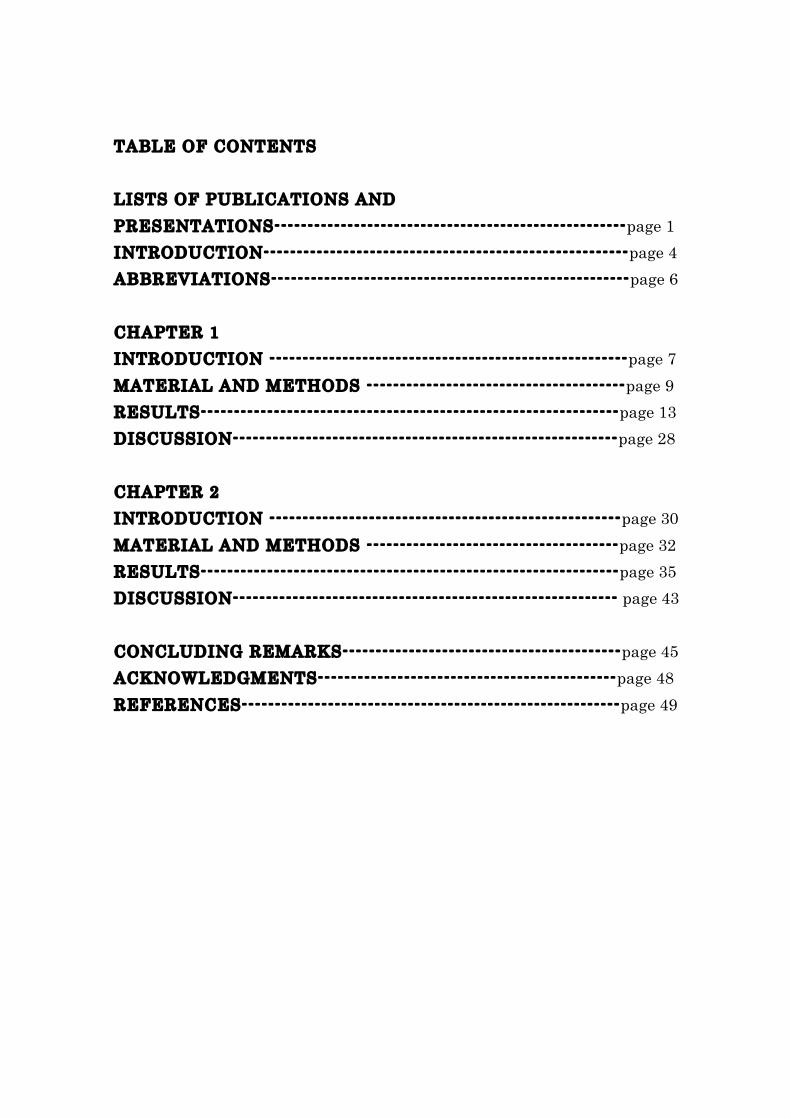
TABLE OF CONTENTS LISTS OF PUBLICATIONS AND PRESENTATIONS-----------------------------------------------------page 1 INTRODUCTION-------------------------------------------------------page 4 ABBREVIATIONS------------------------------------------------------page 6 CHAPTER 1 INTRODUCTION ------------------------------------------------------page 7 MATERIAL AND METHODS ---------------------------------------page 9 RESULTS---------------------------------------------------------------page 13 DISCUSSION----------------------------------------------------------page 28 CHAPTER 2 INTRODUCTION -----------------------------------------------------page 30 MATERIAL AND METHODS --------------------------------------page 32 RESULTS---------------------------------------------------------------page 35 DISCUSSION---------------------------------------------------------- page 43 CONCLUDING REMARKS------------------------------------------page 45 ACKNOWLEDGMENTS---------------------------------------------page 48 REFERENCES---------------------------------------------------------page 49
1
LIST OF PUBLICATIONS 1. IL-11 induces differentiation of myeloid-derived suppressor cells through
activation of STAT3-signaling pathway. Sumida K, Ohno Y, Ohtake J, Kaneumi S, Kishikawa T, Takahashi N, Taketomi A, Kitamura H, Kitamura H. Cancer Res., 2014 in submitting.
2. Anti-IL-6 receptor mAb eliminates myeloid-derived suppressor cells and
inhibits tumor growth by enhancing T-cell responses. Sumida K, Wakita D, Narita Y, Masuko K, Terada S, Watanabe K, Satoh T, Kitamura H, Nishimura T. Eur J Immunol., 42, 2060-72, 2012.
3. Tumor-infiltrating IL-17-producing gammadelta T cells support the
progression of tumor by promoting angiogenesis. Wakita D, Sumida K, Iwakura Y, Nishikawa H, Ohkuri T, Chamoto K, Kitamura H, Nishimura T. Eur J Immunol., 40, 1927-37, 2010.
4. The Key Role of IL-6-Arginase Cascade for Inducing Dendritic
Cell-Dependent CD4+ T Cell Dysfunction in Tumor-Bearing Mice. Narita Y, Kitamura H, Wakita D, Sumida K, Masuko K, Terada S, Nakano K, Nishimura T. J Immunol., 190, 812-820, 2013.
5. IFN-gamma-dependent type 1 immunity is crucial for immunosurveillance
against squamous cell carcinoma in a novel mouse carcinogenesis model. Wakita D, Chamoto K, Ohkuri T, Narita Y, Ashino S, Sumida K, Nishikawa H, Shiku H, Togashi Y, Kitamura H, Nishimura T. Carcinogenesis., 30, 1408-15, 2009.
6. 革新的癌ワクチン, H/K-HELPの開発 —ショートペプチドからヘルパー/キラーロングペプチドへの移行 大竹淳矢、増子和尚、角田健太郎、富樫裕
二、北村秀光、西村孝司 医学のあゆみ 244, 767-778 2013.
2
LIST OF PRESENTATIONS 1. Sumida K, Wakita D, Satoh T, Sakagichi S, Kitamura H, Nishimura T.
Cancer stem cell-induced Treg and MDSC as potent immunosuppressors in tumor-microenvironments. The 19th International Symposium on Molecular Cell Biology of Macrophages 2011. 大阪. 2011.5.
2. Sumida K, Terada S, Kaneumi S, Masuko K, Kishikawa T, Ohtake J,
Ohno Y, Kita T, Kitamura H, and Nishimura T. A mechanism for IL-6- and TGF-β-mediated CD73 expression on myeloid-derived suppressor cells in tumor microenvironments. 第 42回日本免疫学会総会. 千葉. 2013.12.
3. Sumida K, Terada S, Kaneumi S, Masuko K, Kita T, Kitamura H, and
Nishimura T. A role of IL-6 and TGF-β in augmented induction of CD73 essential for MDSC-mediated immunosuppression. 第 72回日本癌学会総会. 横浜. 2013.10.
4.角田健太郎、寺田聖、増子和尚、金海俊, 喜多俊行、北村秀光、西村孝司 IL-6および TGF-βを介したMDSCsの CD73免疫抑制分子の発現増強機構 第 17回がん免疫学会総会. 山口. 2013.7.
5. Sumida K, Daiko W, Terada S, Masuko K, Satoh T, Kitamura H, and
Nishimura T. A role of IL-6 and TGF-β for the functional maturation of myeloid-derived suppressor cells in tumor microenvironments. 第 71回日本癌学会総会. 札幌. 2012.9.
6. 角田健太郎、脇田大功、増子和尚、寺田聖、金海俊、中野基一郎、渡邊絵美,佐藤崇之、北村秀光、西村孝司 担癌生体の未熟ミエロイド細胞から免疫抑制性 MDSCs への機能分化における IL-6、TGF-βの重要性 第 16 回日本がん免疫学会総会. 札幌. 2012.7.
7. 角田健太郎、脇田大功、佐藤崇之、北村秀光、西村孝司. 細胞表面 AMP代

3
謝酵素 CD73を介した担癌生体MDSCの新規免疫抑制機構. 第 15回日本がん免疫学会総会. 大阪. 2011.7.
8. 角田健太郎、脇田大功、佐藤崇之、寺田聖、北村秀光、西村孝司. A novel
immunosuppressive mechanism of myeloid-derived suppressor cells (MDSC) via CD73, ecto-5’-nucleotidase. 第 40 回日本免疫学会総会. 千葉. 2011.11.
4
INTRODUCTION Th1-dependent immunity is critical for induction of tumor-specific cytotoxic T lymphocytes (CTLs) and memory T cells1-4. However, it is difficult to induce tumor-specific T cell responses in tumor-bearing hosts because of the strong immunosuppressive and tumor-evading mechanisms. Activation of antitumor effectors such as tumor-specific CD4+ T and CD8+ T cells are necessary for suppression of tumor growth but are inhibited by various mechanism in tutmor-bearing state. It is well known that immunosuppressive factors such as interleukin (IL)-6, IL-10, and transforming growth factor (TGF)-β impair the functions of T cells and dendritic cells (DCs) in the tumor microenvironment5-8. Regulatory T cells (Tregs) are a major immunosuppressor of T cell activation in tumor-bearing hosts. Indeed, several strategies that control of Treg-mediated immune suppression have been developed to enhance CTL-mediated antitumor activity in tumor-bearing mice9-11. In Capter1, we have revealed that the splenic CD11b+Gr-1+ cells, which were called immature myeloid cell cells (ImC) or myeloid-derived suppressor cell (MDSCs) and abnormally increased in tumor-bearing mice, were classified into three different subsets according to their phenotypic and morphological characteristics; Gr-1low F4/80+ Macrophage (MΦ-ImC), Gr-1mid
stab cells (Neutstab-ImC) and Gr-1high segmented cells (Neutseg-ImC). The splenic MΦ-ImC but not Neutstab-ImC and Neutseg-ImC exhibited a significant immunosuppressive activity in MLR. In contrast, tumor-infiltrating Neutseg-ImC markedly inhibited MLR. Interestingly, we first demonstrated that administration of anti-IL-6R mAb or Gemcitabine (GEM) selectively reduced the MΦ-ImC and Neutstab-ImC populations. The elimination of immunosuppressive ImC enhanced CD8+ T cell responses and inhibited the tumor growth. Moreover, co-administration of anti-IL-6R mAb and GEM exhibited higher antitumor effect in comparison to treatment with single agent. In Capter2, we induced CD11b+ CD14+ monocytic MDSCs from peripheral blood mononuclear cells (PBMCs) of healthy donors under the addition of
5
IL-11. The PBMCs treated with IL-11 were highly expressed various immunosuppressive molecules including IL-10 and Arg-1. T-cell proliferation after the CD3/CD28-stimulation was remarkably reduced when CD11b+
CD14+ cells generated in the presence of IL-11 were co-cultured with CD4+ T or CD8+ T cells, matched autologous healthy donor. Here, we found that IL-11 stimulation of normal PBMCs led to the STAT3 phosphorylation. In addition, we confirmed that neutralization of the IL-11-mediated STAT3 phosphorylation strongly inhibited differentiation of MDSCs. These findings suggest that IL-11 produced under tumor microenvironments may induce monocytic human MDSCs in cancer patients. These results strongly suggested that regulate the immunosuppressive MDSCs populations in tumor-bearing host and would be possible agents for combination therapy with cancer immunotherapy.
6
ABBREVIATIONS anti-IL-6R mAb: anti-IL-6 receptor monoclonal antibody CTL: cytotoxic lymphocyte DC: dendritic cell
ELISA: enzyme-linked immunosorbent assay FACS: fluorescence activated cell sorting GEM: gemcitabine i.d.: intradermally IFN: interferon IL: interleukin ImC: immature myeloid cell iNOS: inducible nitric oxide synthase i.v.: intravenously MDSC: myeloid-derived suppressor cell MLR: mixed leukocyte reaction MΦ: macrophages NK: natural killer
PBMC: peripheral blood mononuclear cell TDF: tumor-derived factor ROS: reactive oxygen species STAT: signal transducer and activator of transcription TGF: transforming growth factor Th: T helper TIL: tumor-infiltrating leukocytes TNF: tumor necrosis factor Treg: regulatory T cells
VEGF: vascular endothelial growth factor WT: wild type
7
CHAPTER 1 Blockade of IL-6 signaling eliminates CD11b+Gr-1+ MDSCs and inhibits the tumor growth by activation of antitumor immunity INTRODUCTION Myeloid-derived suppressor cells (MDSCs) are also an important negative immunoregulator in tumor-bearing hosts. In mice, heterogeneous populations of CD11b+Gr-1+ cells are classified into different subsets according to their phenotypic and morphological characteristics12-23. In human cancer patients, MDSCs are generally defined as monocytic (CD11b+, CD14+, and HLA-DR-/low) or granulocytic (CD11b+ and CD15+) myeloid cells with immunosuppressive activities. These cells have been detected in the blood of patients with glioblastoma, lung cancer, breast cancer, colon cancer, and melanoma24-27. MDSCs strongly inhibit antitumor T cell responses by numerous factors such as arginase-1, S100A8, S100A9, NADPH oxidase, reactive oxygen species, hydrogen peroxide, and peroxynitrite28-33. In addition, MDSCs have been shown to suppress natural killer cell function34. Although MDSCs are considered to be a potential target for tumor immunotherapy, it remains unclear the characteristics of morphological and functional heterogeneity and responsible factors regulating each subpopulations. Therefore, it is important to elucidate the precise immunosuppressive mechanisms of MDSCs in tumor-bearing hosts. In this study, we revealed that splenic CD11b+Gr-1+ cells were classified into three different subsets: Gr-1high; segmented neutrophil (Neutseg-ImC), Gr-1mid; stab neutrophil (Neutstab-ImC), Gr-1low; macrophage (MΦ-ImC), in which Neutseg-ImC and Neutstab-ImC did not suppress T cell responses. In contrast to spleen, tumor-infiltrating CD11b+Gr-1+ cells were consisted in Gr-1high Neutseg-ImC and Gr-1low MΦ-ImC, which had marked immunosuppressive potency. In patients with various types of cancers, systemic levels of inflammatory cytokines, such as IL-1β, TNF-α, and IL-6 are elevated35-39. Recently, Kishimoto’s group has been demonstrated the
8
therapeutic impact of humanized anti-IL-6R mAb on rheumatoid arthritis, Castleman's disease and multiple myeloma40-42. However, antitumor effects on solid tumors and precise mechanisms of antitumor effects were remained unknown. In this study, we first demonstrated that anti-IL-6R mAb treatment decreased the MDSC populations and augment CD8+ T cell-mediated antitumor immunity in tumor-bearing mice.
9
MATERIAL AND METHODS Animals Female BALB/c mice were purchased from Charles River Japan. The mice were maintained in specific pathogen-free conditions and used at 6-8 weeks of age according to the guidelines of animal department at Hokkaido University. Tumor immunotherapy The carcinoma cells, CMC-1 were generated by intradermal injection of methylcholanthrene (MCA) into BALB/c mice43. BALB/c mice were intradermally (i.d.) inoculated with 2x10⁶ CMC-1 cells, and the tumor size was measured by micrometer calipers. The size of the tumor was calculated as following formula; Volume (mm3) = 0.2 × (length (mm) × width (mm)) × (high (mm))2. In the antibody treatment experiments, CMC-1 cells were inoculated i.d. into BALB/c mice, and when the tumor mass reached palpable (7-8 mm), anti-IL-6R mAb (MR16-1) (200 µg) and/or anti-CD8 mAb (53.6.7) (200 µg), control rat IgG was intravenously (i.v.) injected every 3 days. In the chemotherapy experiments, when the tumor mass reached 7-8 mm, CMC-1-bearing mice received i.p. treatment with Gemcitabine (GEM) (120 mg/Kg) weekly, and i.v. treatment with anti-IL-6R mAb and/or anti-CD8 mAb every 3 days. Anti-IL-6R mAb (MR16-1) and control rat IgG was a kind gift from Chugai Pharmaceutical Co. Ltd. Gemcitabine (Gemzar®) was purchased from Eli Lilly. Lymphocytes preparation CMC-1 cells were i.d. inoculated into BALB/c mice, and 30 days after the injection, spleen cells were prepared. The tumor tissues were dissected from the mice and minced. Single-cell suspensions were obtained by digestion with 1 mg/ml collagenase (Sigma) for 30 min at 37 °C. For isolation of CD11b+Gr-1high, CD11b+Gr-1mid and CD11b+Gr-1low cells, the cells prepared from spleens and tumor tissues were stained with PE-conjugated anti-CD11b mAb (M1/70, BD BioScience) and
10
FITC-conjugated anti-Gr-1 mAb (RB6-8C5, BD BioScience) after treatment with purified anti-CD16/CD32 mAb (2.4G2, BD BioScience). The percentages of CD11b+Gr-1+ cells were determined using FACSCanto II (BD Biosciences) and analyzed with FlowJo software (Treestar). Each subpopulation of CD11b+Gr-1+ cells was isolated by FACSAria (BD BioSciences). The purity of the isolated cells was consistently more than 95 %. In vitro differentiation of spleen ImC CMC-1 cells (5x10⁴) were incubated for 72 h with 6-well plate (NUNC). The supernatants were collected and used as conditioned medium containing TDFs. Indicated spleen CD11b+Gr-1+ subpopulations were isolated from CMC-1-bearing mice and cultured in the presence of conditioned medium (50 %), GM-CSF (0.3 ng/ml) or GM-CSF+IL-6 (50 ng/ml)+TGF-β (2 ng/ml) for 2.5 days. Morphological analysis Indicated CD11b+Gr-1+ subpopulations were isolated from spleen and tumor tissues of CMC-1-bearing mice. Cytospins of the isolated cells were prepared with Shandon Cytospin 3 (Thermo Electron) and stained with May-Giemsa stain. Morphological differences were observed using a light microscopy (BX50, OLYMPUS OPTICAL Co, Ltd,.). Generation of CTL in MLR Spleen cells (5x10⁶) from BALB/c mice were cocultured with C57BL/6 spleen cells (1x10⁶), which were pretreated with mitomycin C (60 mg/mL; Kyowa Hakko). Isolated CD11b+Gr-1high, CD11b+Gr-1mid or CD11b+Gr-1low cells (1x10⁵) were added into the culture and these cells were incubated for 4 days in the 5 ml round-bottom tubes (Falcon). After culture, cells were collected and used for analysis. 3H-thymidine incorporation assay Cell proliferation was evaluated as described in the previous paper1. Briefly, 3H-thymidine was added to culture medium for 6 hrs and the radioactivity of
11
the incorporated cell were detected and indicated as proliferation T cells. Cytotoxicity assay Cytotoxicity of cells generated by MLR was measured by 4 hr-51Cr-release assays as described previously44. Antigen-specific cytotoxicity was determined using MBL-2 T-lymphoma cells (H-2b) as target cells. As a negative control, P815 mastocytoma cells (H-2d) were used in the same experiments. The percentage of cytotoxicity was calculated and indicated in the figures as described previously44. In Fig. 6, cytotoxicity was indicated as lytic units (L.U.)/106 effector cells. A L.U. was defined as the numbers of effector cells required for causing 25 % lysis of 5x10³ target cells. Intracellular cytokine staining For detection of cytoplasmic cytokine expression of T cells, spleen cells prepared from CMC-1-bearing mice were stimulated with inactivated-allogeneic spleen cells for 4 days or 2 mg/ml soluble anti-CD3ε mAb (145-2C11, BD Bioscience) for 4 hrs. After the stimulation, cells were restimulated with plate-bound anti-CD3ε mAb in 96-well flat-bottomed plates for 6 hrs. Brefeldin A was added at 4 hrs. Then, the cells were harvested and stained with anti-CD4 and anti-CD8 mAbs (BD Bioscience), and fixed with 4 % paraformaldehyde. After permeabilization, the fixed cells were stained with anti-IFN-γ and anti-IL-4 mAbs (BD bioscience). Data were acquired by FACSCanto ll and analyzed with FlowJo software. PCR analysis Total RNA was extracted from sorted spleen or tumor-infiltrating CD11b+Gr-1high, CD11b+Gr-1mid and CD11b+Gr-1low cells using Isogen RNA extraction kit (Nippongene) according to manufacture’s instructions. cDNA was prepared using Superscript lll RT (Invitrogen). The indicated gene cDNAs were specifically amplified using a GeneAmp 9700 thermalcycler (PE Applied Biosystems) and corresponding primer pairs. The sequences were as follows: mouse β-actin, (sense) 5’-GTGATGGTGGGAATGGGTCAG-3’, (antisense) 5’-TTTGATGTCACGCACGATTTCC-3’, mouse COX-2, (sense)
12
5’-ACACACTCTATCACTGGCACC-3’, (antisense) 5’-TTCAGGGAGAAGCGTTTGC-3’, mouse Arg-1, (sense) 5’-GCAGAGGTCCAGAAGAATGGAA-3’, (antisense), 5’-CTTTTCTTCCTTCCCAGCAGGT-3’, mouse iNOS, (sense) 5’-GGGCTGTCACGGAGATCA-3’, (antisense) 5’-CCATGATGGTCACATTCTGC -3’, mouse IL-6, (sense) 5’-GAGGATACCACTCCCAACAGACC-3’, (antisense) 5’-AAGTGCATCATCGTTGTTCATACA-3’, mouse TGF-β, (sense) 5’-TACGTCAGACATTCGGGAAGCA-3’, (antisense) 5’-GAAGTTGGCATGGTAGCCCTTG-3’ Immunoblotting. Antibodies were purchased from the following sources: Monoclonal Anti-Arginase I antibody and Monoclonal Anti-iNOS/NOS Type II antibody from BD Transduction Laboratories, and polyclonal Anti-COX-2 antibody from cayman, and monoclonal anti-a-tubulin (DM1A) from SIGMA. Cells were lysed in a buffer consisting of 20 mM HEPES, pH 7.5, 100 mM NaCl, 1.5 mM MgCl2, 1 mM EGTA, 10 mM Na4P2O7, 1 % Nonidet P-40, 2 mM dithiothreitol, 1 mM Vanadate, 1 mM phenylmethylsulfonyl fluoride, 2 mg/mL aprotinin and 10 % glycerol. Cell lysates were subjected to immunoblotting with the indicated antibodies. ELISA Whole spleen cells of CMC-1-bearing mice treated with control rat IgG or anti-IL-6R mAb were stimulated with soluble anti-CD3ε mAb (1 mg/ml). After 48 hrs, IFN-γ levels in the culture supernatants were determined by OptEIATM mouse IFN-γ. Statistical analyses Significant differences in the results were determined by using the two-sided Student’s t test. The p<0.05 was considered significant in the present experiments.
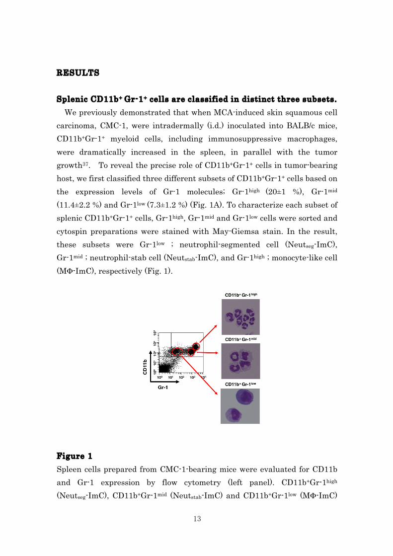
13
RESULTS Splenic CD11b+ Gr-1+ cells are classified in distinct three subsets. We previously demonstrated that when MCA-induced skin squamous cell carcinoma, CMC-1, were intradermally (i.d.) inoculated into BALB/c mice, CD11b+Gr-1+ myeloid cells, including immunosuppressive macrophages, were dramatically increased in the spleen, in parallel with the tumor growth37. To reveal the precise role of CD11b+Gr-1+ cells in tumor-bearing host, we first classified three different subsets of CD11b+Gr-1+ cells based on the expression levels of Gr-1 molecules; Gr-1high (20±1 %), Gr-1mid (11.4±2.2 %) and Gr-1low (7.3±1.2 %) (Fig. 1A). To characterize each subset of splenic CD11b+Gr-1+ cells, Gr-1high, Gr-1mid and Gr-1low cells were sorted and cytospin preparations were stained with May-Giemsa stain. In the result, these subsets were Gr-1low ; neutrophil-segmented cell (Neutseg-ImC), Gr-1mid ; neutrophil-stab cell (Neutstab-ImC), and Gr-1high ; monocyte-like cell (MΦ-ImC), respectively (Fig. 1). Figure 1 Spleen cells prepared from CMC-1-bearing mice were evaluated for CD11b and Gr-1 expression by flow cytometry (left panel). CD11b+Gr-1high (Neutseg-ImC), CD11b+Gr-1mid (Neutstab-ImC) and CD11b+Gr-1low (MΦ-ImC)
14
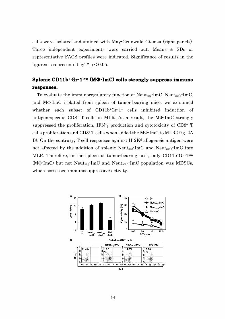
cells were isolated and stained with May-Grunwald Giemsa (right panels). Three independent experiments were carried out. Means ± SDs or representative FACS profiles were indicated. Significance of results in the figures is represented by: * p < 0.05. Splenic CD11b+ Gr-1low (MΦ-ImC) cells strongly suppress immune responses. To evaluate the immunoregulatory function of Neutseg-ImC, Neutstab-ImC, and MΦ-ImC isolated from spleen of tumor-bearing mice, we examined whether each subset of CD11b+Gr-1+ cells inhibited induction of antigen-specific CD8+ T cells in MLR. As a result, the MΦ-ImC strongly suppressed the proliferation, IFN-γ production and cytotoxicity of CD8+ T cells proliferation and CD8+ T cells when added the MΦ-ImC to MLR (Fig. 2A, B). On the contrary, T cell responses against H-2Kd allogeneic antigen were not affected by the addition of splenic Neutseg-ImC and Neutstab-ImC into MLR. Therefore, in the spleen of tumor-bearing host, only CD11b+Gr-1low (MΦ-ImC) but not Neutseg-ImC and Neutstab-ImC population was MDSCs, which possessed immunosuppressive activity.
15
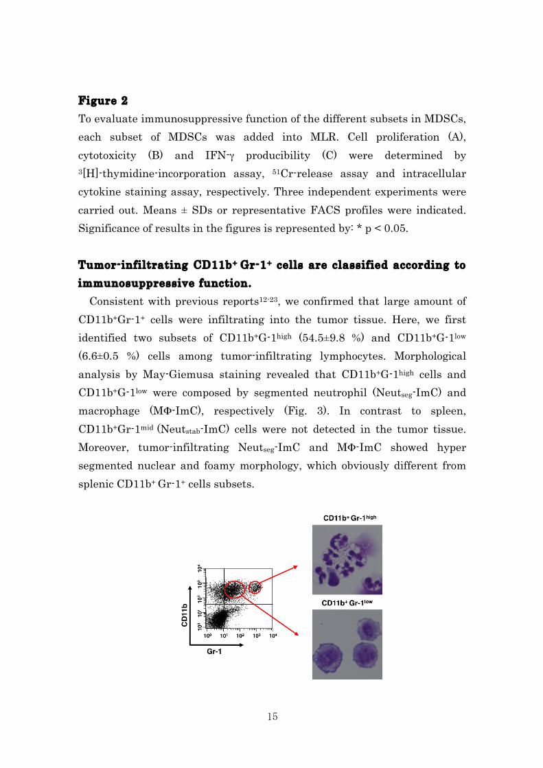
Figure 2 To evaluate immunosuppressive function of the different subsets in MDSCs, each subset of MDSCs was added into MLR. Cell proliferation (A), cytotoxicity (B) and IFN-γ producibility (C) were determined by 3[H]-thymidine-incorporation assay, 51Cr-release assay and intracellular cytokine staining assay, respectively. Three independent experiments were carried out. Means ± SDs or representative FACS profiles were indicated. Significance of results in the figures is represented by: * p < 0.05. Tumor-infiltrating CD11b+ Gr-1+ cells are classified according to immunosuppressive function. Consistent with previous reports12-23, we confirmed that large amount of CD11b+Gr-1+ cells were infiltrating into the tumor tissue. Here, we first identified two subsets of CD11b+G-1high (54.5±9.8 %) and CD11b+G-1low (6.6±0.5 %) cells among tumor-infiltrating lymphocytes. Morphological analysis by May-Giemusa staining revealed that CD11b+G-1high cells and CD11b+G-1low were composed by segmented neutrophil (Neutseg-ImC) and macrophage (MΦ-ImC), respectively (Fig. 3). In contrast to spleen, CD11b+Gr-1mid (Neutstab-ImC) cells were not detected in the tumor tissue. Moreover, tumor-infiltrating Neutseg-ImC and MΦ-ImC showed hyper segmented nuclear and foamy morphology, which obviously different from splenic CD11b+ Gr-1+ cells subsets.
16
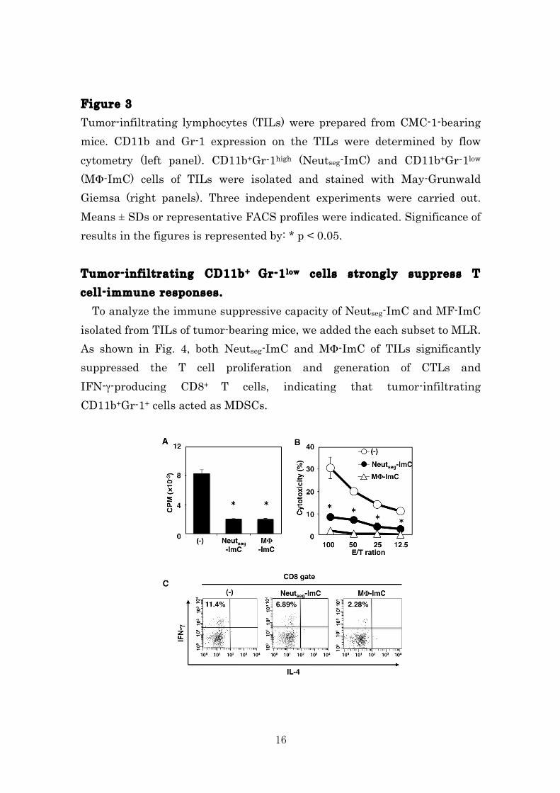
Figure 3 Tumor-infiltrating lymphocytes (TILs) were prepared from CMC-1-bearing mice. CD11b and Gr-1 expression on the TILs were determined by flow cytometry (left panel). CD11b+Gr-1high (Neutseg-ImC) and CD11b+Gr-1low (MΦ-ImC) cells of TILs were isolated and stained with May-Grunwald Giemsa (right panels). Three independent experiments were carried out. Means ± SDs or representative FACS profiles were indicated. Significance of results in the figures is represented by: * p < 0.05. Tumor-infiltrating CD11b+ Gr-1low cells strongly suppress T cell-immune responses. To analyze the immune suppressive capacity of Neutseg-ImC and MF-ImC isolated from TILs of tumor-bearing mice, we added the each subset to MLR. As shown in Fig. 4, both Neutseg-ImC and MΦ-ImC of TILs significantly suppressed the T cell proliferation and generation of CTLs and IFN-γ-producing CD8+ T cells, indicating that tumor-infiltrating CD11b+Gr-1+ cells acted as MDSCs.
17
Figure 4 Each subset of MDSCs in tumor local site were isolated and added into allogeneic MLR. Cell proliferation (A), cytotoxicity (B) and IFN-γ producibility (C) were determined by 3[H]-thymidine-incorporation assay, 51Cr-release assay and intracellular cytokine staining assay, respectively. Three independent experiments were carried out. Means ± SDs or representative FACS profiles were indicated. Significance of results in the figures is represented by: * p < 0.05. Splenic CD11b+Gr-1+ cells differentiate into immunosuppressor by tumor-derived factors. Since Neutseg-ImC isolated from tumor tissues but not that isolated from spleen inhibited immune responses in MLR (Fig. 4), it is possible that tumor microenvironment would influence immune suppressive potency of CD11b+Gr-1+ cells. To evaluate the differentiation potential of the splenic CD11b+Gr-1+ cells, we isolated the three sublets of ImC from spleen cells of CMC-1 tumor-bearing mice and cultured with the conditioned medium containing tumor-derived factors (TDFs) of CMC-1 cells. The morphological analysis by May-Giemsa staining showed that splenic Neutseg-ImC and Neutstab-ImC become hyper segmented cells like the Neutseg-ImC of TILs after incubation with TDFs. In addition the splenic MF-ImC also differentiated to foamy macrophages, similar to MΦ-ImC of TIL (Fig. 5A). The TDF-treated ImC were added in MLR to assess the immunosuppressive activity. Whereas fleshly isolated splenic Neutseg-ImC and Neutstab-ImC did not inhibited the immune responses in MLR (Fig. 2), those cells cultured with TDFs significantly suppressed cell proliferation, cytotoxicity and IFN-γ production of CTL in MLR. Splenic MΦ-ImC also obtained marked suppressive potency by exposing to TDFs (Fig. 5A-C). Therefore, these data clearly indicate that CMC-1-released factor(s) is crucial to provide the immunosuppressive function to the splenic ImC to play a role as MDSCs in the tumor microenvironment.
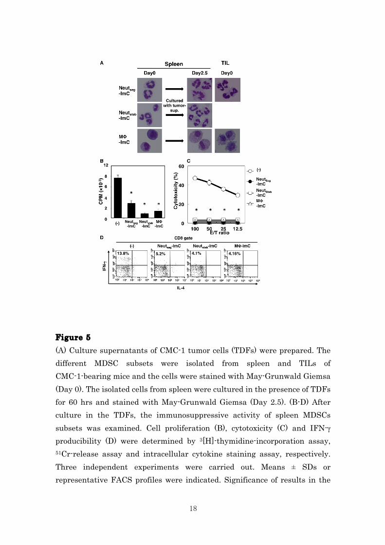
18
Figure 5 (A) Culture supernatants of CMC-1 tumor cells (TDFs) were prepared. The different MDSC subsets were isolated from spleen and TILs of CMC-1-bearing mice and the cells were stained with May-Grunwald Giemsa (Day 0). The isolated cells from spleen were cultured in the presence of TDFs for 60 hrs and stained with May-Grunwald Giemsa (Day 2.5). (B-D) After culture in the TDFs, the immunosuppressive activity of spleen MDSCs subsets was examined. Cell proliferation (B), cytotoxicity (C) and IFN-γ producibility (D) were determined by 3[H]-thymidine-incorporation assay, 51Cr-release assay and intracellular cytokine staining assay, respectively. Three independent experiments were carried out. Means ± SDs or representative FACS profiles were indicated. Significance of results in the
19
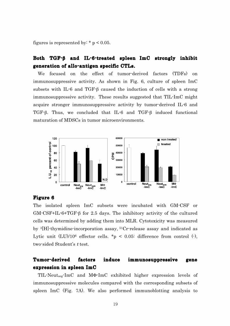
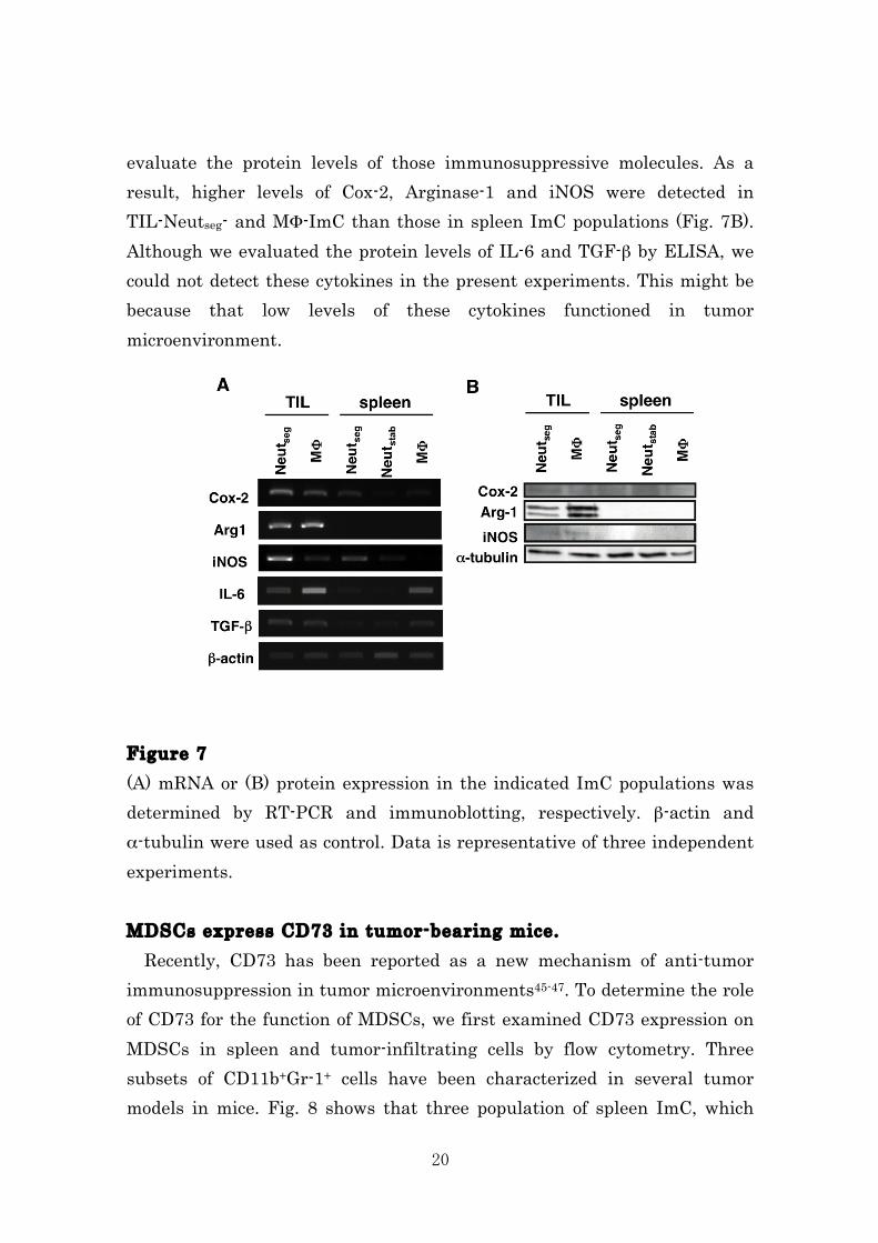
figures is represented by: * p < 0.05. Both TGF-β and IL-6-treated spleen ImC strongly inhibit generation of allo-antigen specific CTLs. We focused on the effect of tumor-derived factors (TDFs) on immunosuppressive activity. As shown in Fig. 6, culture of spleen ImC subsets with IL-6 and TGF-β caused the induction of cells with a strong immunosuppressive activity. These results suggested that TIL-ImC might acquire stronger immunosuppressive activity by tumor-derived IL-6 and TGF-β. Thus, we concluded that IL-6 and TGF-β induced functional maturation of MDSCs in tumor microenvironments. Figure 6 The isolated spleen ImC subsets were incubated with GM-CSF or GM-CSF+IL-6+TGF-β for 2.5 days. The inhibitory activity of the cultured cells was determined by adding them into MLR. Cytotoxicity was measured by 3[H]-thymidine-incorporation assay, 51Cr-release assay and indicated as Lytic unit (LU)/106 effector cells. *p < 0.05: difference from control (-), two-sided Student’s t test. Tumor-derived factors induce immunosuppressive gene expression in spleen ImC TIL-Neutseg-ImC and MΦ-ImC exhibited higher expression levels of immunosuppressive molecules compared with the corresponding subsets of spleen ImC (Fig. 7A). We also performed immunoblotting analysis to
20
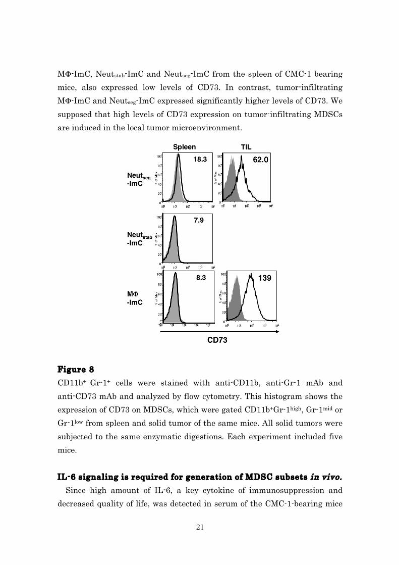
evaluate the protein levels of those immunosuppressive molecules. As a result, higher levels of Cox-2, Arginase-1 and iNOS were detected in TIL-Neutseg- and MΦ-ImC than those in spleen ImC populations (Fig. 7B). Although we evaluated the protein levels of IL-6 and TGF-β by ELISA, we could not detect these cytokines in the present experiments. This might be because that low levels of these cytokines functioned in tumor microenvironment. Figure 7 (A) mRNA or (B) protein expression in the indicated ImC populations was determined by RT-PCR and immunoblotting, respectively. β-actin and α-tubulin were used as control. Data is representative of three independent experiments. MDSCs express CD73 in tumor-bearing mice. Recently, CD73 has been reported as a new mechanism of anti-tumor immunosuppression in tumor microenvironments45-47. To determine the role of CD73 for the function of MDSCs, we first examined CD73 expression on MDSCs in spleen and tumor-infiltrating cells by flow cytometry. Three subsets of CD11b+Gr-1+ cells have been characterized in several tumor models in mice. Fig. 8 shows that three population of spleen ImC, which
21
MΦ-ImC, Neutstab-ImC and Neutseg-ImC from the spleen of CMC-1 bearing mice, also expressed low levels of CD73. In contrast, tumor-infiltrating MΦ-ImC and Neutseg-ImC expressed significantly higher levels of CD73. We supposed that high levels of CD73 expression on tumor-infiltrating MDSCs are induced in the local tumor microenvironment. Figure 8 CD11b+ Gr-1+ cells were stained with anti-CD11b, anti-Gr-1 mAb and anti-CD73 mAb and analyzed by flow cytometry. This histogram shows the expression of CD73 on MDSCs, which were gated CD11b+Gr-1high, Gr-1mid or Gr-1low from spleen and solid tumor of the same mice. All solid tumors were subjected to the same enzymatic digestions. Each experiment included five mice. IL-6 signaling is required for generation of MDSC subsets in vivo. Since high amount of IL-6, a key cytokine of immunosuppression and decreased quality of life, was detected in serum of the CMC-1-bearing mice
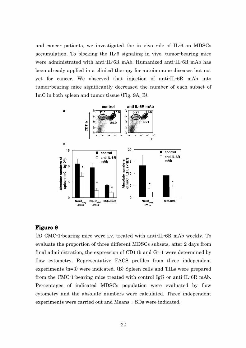
22
and cancer patients, we investigated the in vivo role of IL-6 on MDSCs accumulation. To blocking the IL-6 signaling in vivo, tumor-bearing mice were administrated with anti-IL-6R mAb. Humanized anti-IL-6R mAb has been already applied in a clinical therapy for autoimmune diseases but not yet for cancer. We observed that injection of anti-IL-6R mAb into tumor-bearing mice significantly decreased the number of each subset of ImC in both spleen and tumor tissue (Fig. 9A, B). Figure 9 (A) CMC-1-bearing mice were i.v. treated with anti-IL-6R mAb weekly. To evaluate the proportion of three different MDSCs subsets, after 2 days from final administration, the expression of CD11b and Gr-1 were determined by flow cytometry. Representative FACS profiles from three independent experiments (n=3) were indicated. (B) Spleen cells and TILs were prepared from the CMC-1-bearing mice treated with control IgG or anti-IL-6R mAb. Percentages of indicated MDSCs population were evaluated by flow cytometry and the absolute numbers were calculated. Three independent experiments were carried out and Means ± SDs were indicated.
23
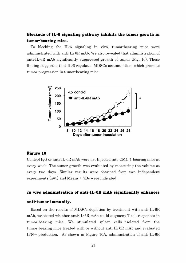
Blockade of IL-6 signaling pathway inhibits the tumor growth in tumor-bearing mice. To blocking the IL-6 signaling in vivo, tumor-bearing mice were administrated with anti-IL-6R mAb. We also revealed that administration of anti-IL-6R mAb significantly suppressed growth of tumor (Fig. 10). These finding suggested that IL-6 regulates MDSCs accumulation, which promote tumor progression in tumor-bearing mice. Figure 10 Control IgG or anti-IL-6R mAb were i.v. Injected into CMC-1-bearing mice at every week. The tumor growth was evaluated by measuring the volume at every two days. Similar results were obtained from two independent experiments (n=5) and Means ± SDs were indicated.
In vivo administration of anti-IL-6R mAb significantly enhances
anti-tumor immunity.
Based on the results of MDSCs depletion by treatment with anti-IL-6R mAb, we tested whether anti-IL-6R mAb could augment T cell responses in tumor-bearing mice. We stimulated spleen cells isolated from the tumor-bearing mice treated with or without anti-IL-6R mAb and evaluated IFN-γ production. As shown in Figure 10A, administration of anti-IL-6R
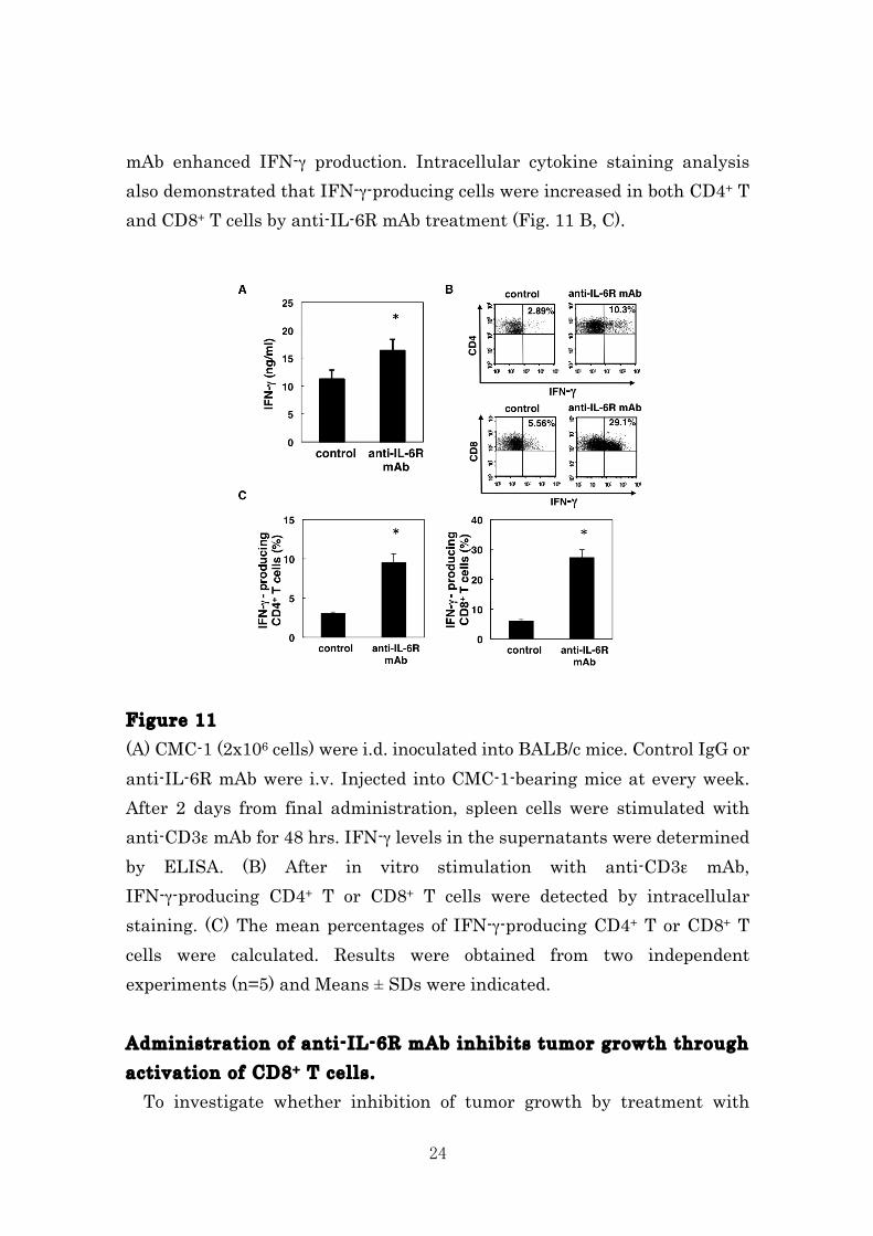
24
mAb enhanced IFN-γ production. Intracellular cytokine staining analysis also demonstrated that IFN-γ-producing cells were increased in both CD4+ T and CD8+ T cells by anti-IL-6R mAb treatment (Fig. 11 B, C). Figure 11 (A) CMC-1 (2x10⁶ cells) were i.d. inoculated into BALB/c mice. Control IgG or anti-IL-6R mAb were i.v. Injected into CMC-1-bearing mice at every week. After 2 days from final administration, spleen cells were stimulated with anti-CD3ε mAb for 48 hrs. IFN-γ levels in the supernatants were determined by ELISA. (B) After in vitro stimulation with anti-CD3ε mAb, IFN-γ-producing CD4+ T or CD8+ T cells were detected by intracellular staining. (C) The mean percentages of IFN-γ-producing CD4+ T or CD8+ T cells were calculated. Results were obtained from two independent experiments (n=5) and Means ± SDs were indicated. Administration of anti-IL-6R mAb inhibits tumor growth through activation of CD8+ T cells. To investigate whether inhibition of tumor growth by treatment with
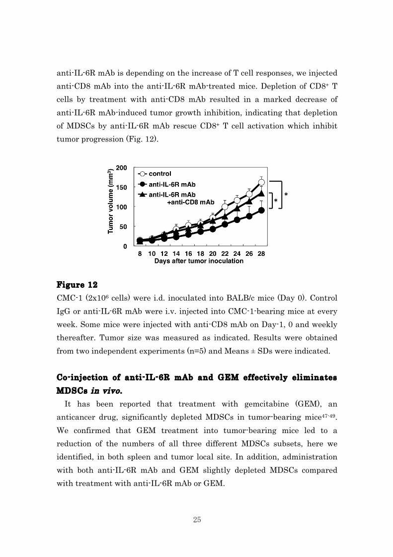
25
anti-IL-6R mAb is depending on the increase of T cell responses, we injected anti-CD8 mAb into the anti-IL-6R mAb-treated mice. Depletion of CD8+ T cells by treatment with anti-CD8 mAb resulted in a marked decrease of anti-IL-6R mAb-induced tumor growth inhibition, indicating that depletion of MDSCs by anti-IL-6R mAb rescue CD8+ T cell activation which inhibit tumor progression (Fig. 12). Figure 12 CMC-1 (2x10⁶ cells) were i.d. inoculated into BALB/c mice (Day 0). Control IgG or anti-IL-6R mAb were i.v. injected into CMC-1-bearing mice at every week. Some mice were injected with anti-CD8 mAb on Day-1, 0 and weekly thereafter. Tumor size was measured as indicated. Results were obtained from two independent experiments (n=5) and Means ± SDs were indicated. Co-injection of anti-IL-6R mAb and GEM effectively eliminates MDSCs in vivo . It has been reported that treatment with gemcitabine (GEM), an anticancer drug, significantly depleted MDSCs in tumor-bearing mice47-49. We confirmed that GEM treatment into tumor-bearing mice led to a reduction of the numbers of all three different MDSCs subsets, here we identified, in both spleen and tumor local site. In addition, administration with both anti-IL-6R mAb and GEM slightly depleted MDSCs compared with treatment with anti-IL-6R mAb or GEM.
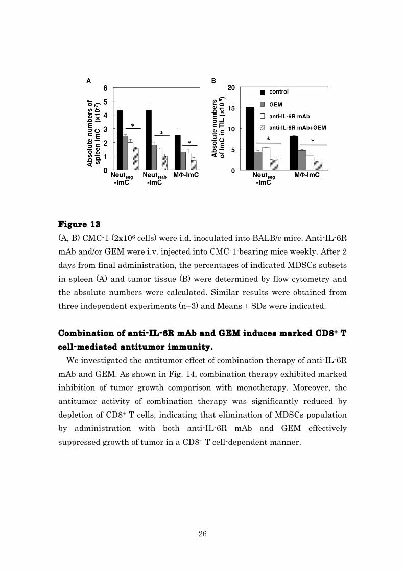
26
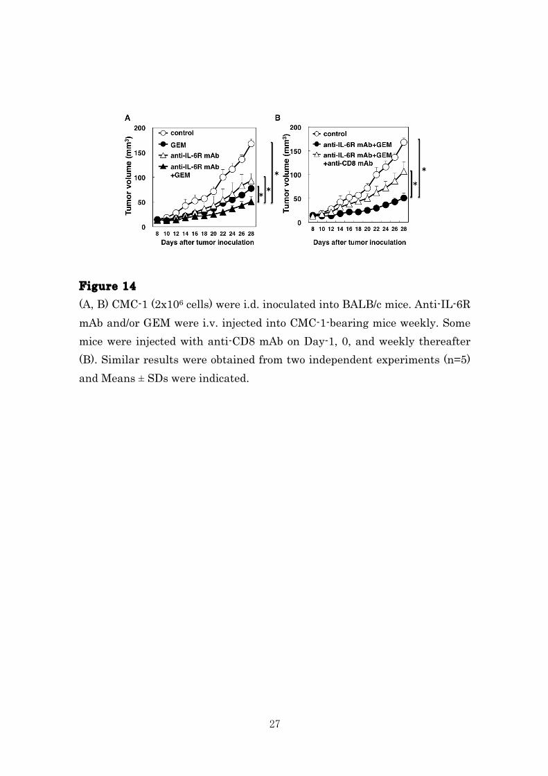
Figure 13 (A, B) CMC-1 (2x10⁶ cells) were i.d. inoculated into BALB/c mice. Anti-IL-6R mAb and/or GEM were i.v. injected into CMC-1-bearing mice weekly. After 2 days from final administration, the percentages of indicated MDSCs subsets in spleen (A) and tumor tissue (B) were determined by flow cytometry and the absolute numbers were calculated. Similar results were obtained from three independent experiments (n=3) and Means ± SDs were indicated. Combination of anti-IL-6R mAb and GEM induces marked CD8+ T cell-mediated antitumor immunity. We investigated the antitumor effect of combination therapy of anti-IL-6R mAb and GEM. As shown in Fig. 14, combination therapy exhibited marked inhibition of tumor growth comparison with monotherapy. Moreover, the antitumor activity of combination therapy was significantly reduced by depletion of CD8+ T cells, indicating that elimination of MDSCs population by administration with both anti-IL-6R mAb and GEM effectively suppressed growth of tumor in a CD8+ T cell-dependent manner.
27
Figure 14 (A, B) CMC-1 (2x10⁶ cells) were i.d. inoculated into BALB/c mice. Anti-IL-6R mAb and/or GEM were i.v. injected into CMC-1-bearing mice weekly. Some mice were injected with anti-CD8 mAb on Day-1, 0, and weekly thereafter (B). Similar results were obtained from two independent experiments (n=5) and Means ± SDs were indicated.
28
DISCUSSION We showed in the present study that CD11b+Gr-1+ cells, accumulating in the tumor tissues and spleen, contain morphologically-distinct three subsets, which also exhibited distinct immunoregulatory function respectively. Importantly, we demonstrated that two granulocytic cell subsets (Neutseg- and Neutstab-ImC) in the spleen did not inhibit T cell responses, whereas tumor-infiltrating Neutseg-ImC had marked suppressive potency, indicating that all CD11b+Gr-1+ cells, commonly-termed MDSCs, were not immunosuppressive myeloid cells and tumor microenvironment was critical condition to obtain inhibitory function. Many investigators have evaluated the phenotype and function of spleen MDSCs and revealed several suppressive mechanisms. However, precise nature of tumor-infiltrating CD11b+Gr-1+ cells was poorly understood. Here we demonstrated the augment inhibitory function of tumor-infiltrating CD11b+Gr-1+ cells, compared with corresponding population in spleen. These data were supported by recent reports that exposure to tumor microenvironment or inflammatory site enhanced suppressive function of MDSCs14, 21, 22. Our findings extended these reports by firstly showing that CD11b+Gr-1+ cells in tumor tissues were classified into Gr-1high Neutseg-ImC and Gr-1low MΦ-ImC, but, in contrast with spleen, Gr-1mid Neutstab-ImC did not exist in the tumor tissue. In addition, importantly, both of Gr-1high Neutseg-ImC and Gr-1low MΦ-ImC exhibited different morphology to spleen CD11b+Gr-1+ cells; tumor-infiltrating Neutseg-ImC have hyper segmented nucleus, tumor-infiltrating MΦ-ImC were foamy macrophages. Spleen Neutstab-ImC immediately differentiated into hyper segmented neutrophils and acquired immunosuppressive potency when cultured in the presence of tumor-derived factors, strongly indicating that less immunosuppressive immature MDSCs in the spleen recruited to tumor tissues and become maturated phenotype and potent suppressive MDSCs. In the future, we must identify the factors, which contributed to acquire immunosuppressive function of CD11b+Gr-1+ cells. In our recent observation, IL-6 and TGF-β would be possible regents to activate MDSCs. We considered that underlying
29
mechanisms of IL-6 and/or TGF-β-mediated immunosuppression were potent targets for cancer immunotherapy to augment tumor-specific T cells responses by removing suppressive populations in tumor-bearing host. We also reveled here that anti-IL-6R mAb administration reduced CD11b+Gr-1+ cells in both spleen and tumor tissue and delayed tumor development in CD8+ T cell dependent manner, indicating that removing immunosuppressive population would be a potent target for cancer immunotherapy to augment tumor-specific T cells responses. Since treatment with anti-IL-6R mAb showed no significant effect on Treg, combination therapy by anti-IL-6R mAb and regents depleting Treg might be effective strategy for cancer immunotherapy. It is important how blockade of IL-6 signaling decrease MDSCs population. We confirmed that when anti-IL-6R mAb was administrated tumor-bearing mice, the numbers of MDSCs were immediately decreased, suggesting that IL-6 may directly maintain MDSCs survival and/or proliferation. Thus, treatment with anti-IL-6R mAb not only eliminates of MDSCs, also modulates MDSCs function in the tumor-bearing host. IL-6 is a multi-functional cytokine. In the tumor-bearing condition, IL-6, produced tumor cells, macrophages and stromal cells, is involved in immunosuppression through reducing DC function50. Moreover, IL-6 directly influences promotion of tumor cell survival and proliferation in a STAT3 dependent manner. These functions of IL-6, coupled with elimination of MDSCs, would also affected antitumor effect of anti-IL-6R mAb treatment.
30
CHAPTER 2 IL-11-mediated STAT3 activation is required for functional differentiation of myeloid-derived suppressor cells in human INTRODUCTION Activation of ant-tumor effectors, which tumor-specific CD4+ T and CD8+ T cells are necessary for the suppression of tumor growth. However, it is also difficult to induce tumor-specific T cell responses in tumor-bearing hosts, because they are suffering from strong immunosuppressive tumor-escape mechanisms. MDSCs are a heterogeneous population of immature myeloid cells that mobilize from the bone marrow and become activated to inhibit tumor-specific immune responses51. Recently, two distinct subpopulations of MDSCs, CD11b+Gr-1high/mid. (Ly-6G+Ly-6Clow) granulocytic MDSCs and CD11b+Gr-1lowLy-6G‒/low Ly-6Chigh monocytic MDSCs, have been described 52-58. MDSCs exhibit the CD11b+Gr-1+ phenotype, and their human counterpart is identified as being CD11b+CD14+HLA-DR‒ or CD11b+CD15+ 59-61. The MDSCs suppress T cell and NK cell responses. MDSCs have been shown to suppress immune responses through a variety of direct mechanisms, including arginase-1, S100A8, S100A9, inducible nitric oxide synthase (iNOS), and production of reactive oxygen species (ROS) 62-70. We reported that the strong immunosuppressive functions of MDSCs, strategies aimed at the elimination or inhibition of MDSCs by administration of anti-IL-6 mAb and/or Gemcitabine might significantly improve anti-tumor responses and the efficacy of cancer immunotherapy58. The IL-11, member of IL-6 family, is defined by the shared use of the gp130 receptor β-subunit. Included within this family is IL-6, recognized for its role as a systemic inflammation, and promotes platelet production 71, 72. More recently, IL-11 was reported to be involved in a mechanism of inflammation, angiogenesis and metastasis in tumor microenvironments 73-75. IL-11 was produced by cancer-associated fibroblasts and myeloid cells in
31
many types of tumor cells such as stomach, liver, pancreas, colon, ovary and breast cancer 76-85. However, there is no report that the relationship between the anti-tumor immune response and IL-11 madiate activation of STAT3. In this study, we found that IL-11-STAT3-mediated regulation of functional differentiation of CD11b+CD14+MDSCs. Indeed, a specific inhibitor of STAT3 significantly blocked the differentiation of induced-MDSCs by IL-11. Moreover, PBMCs from healthy donor cultured with IL-11 acquired immunosuppressive activity. In this paper, we report about a crucial role of IL-11 for inhibitory mechanism of MDSCs, which may be a possible target for cancer immunotherapy.
32
MATERIAL AND METHODS Informed consent Research protocols involving human subjects were approved by the Institutional Review Boards of Hokkaido University Graduate School of Medicine, and the Institute for Genetic Medicine. Written informed consent was obtained from each patient and healthy donor. Isolation of peripheral blood mononuclear cells (PBMCs) and generation of MDSCs PBMCs were isolated from healthy donors by Ficoll-Paque (GE Healthcare, Uppsala, Sweden). PBMCs from healthy donors were cultured in 10% FBS, and 100 mg/mL penicillin/ streptomycin in RPMI 1640 (Wako). To generate functional MDSCs, PBMCs were cultured with of IL-11 (10 ng/ml) and GM-CSF (50 ng/ml) for 7 days. Cultured cells were stained with PE-conjugated anti-CD11b (Beckman, Bear1), FITC-conjugated anti-CD14 (BD Biosciences, M5E2) monoclonal antibodies (mAbs). Dead cells were removed by 7-AAD (Beckman) staining. The percentages of CD11b+CD14+ cells were analyzed by a FACS Canto II (BD Biosciences) and FlowJo software (Treestar). CD11b+CD14+ cells were isolated by FACSAria (BD Biosciences). The purity of the isolated cells was consistently more than 95%. T-cell suppression assay CD3+ T cells were sorted from PBMCs by FACS Aria (BD Biosciences). T cells were labeled with CFSE (Invitrogen) and cultured with CD3/CD28 beads (Invitrogen) for 3 days. Cells were collected, stained for CD4+ T or CD8+ T cell markers and assay for flow cytometric analysis on a FACS Canto II (BD Biosciences). Cells were gated on CD4+ T or CD8+ T cells and the proliferation was determined on the basis of CFSE dilution. ELISA IFN-γ levels in culture supernatants were measured by OptEIA™human IFN-γ, ELISA kits (BD Bioscience), according to the manufacturer's
33
instructions. RT-PCR Total RNA was extracted from cells using an Isogen RNA extraction kit (Qiagen). cDNA was prepared using Superscript III RT (Invitrogen). The indicated cDNAs were specifically amplified using a LightCycler system (Roche Applied Science) and the corresponding primer pairs and probes. The sequences were as follows: IL-11, (sense) 5′-ctgtggggacatgaactgtg-3′, (antisense) 5′-agggtctggggaaactcg-3′, and probe #49, Arg-1, (sense) 5′-tggcagaagtcaagaagaacg-3′, (antisense) 5′-atgcttccaattgccaaact-3′, and probe #64, VEGF, (sense) 5′-ccttgctgctctacctccac-3′, (antisense) 5′-ccacttcgtgatgattctgc-3′, and probe #29, TGF-β, (sense) 5′-actactacgccaaggaggtcac-3′, (antisense) 5′-tgcttgaacttgtcatagatttcg-3′, and probe #21, IL-10, (sense) 5′-gatgccttcagcagagtgaa-3′, (antisense) 5′-gcaacccaggtaacccttaaa-3′, and probe #67, GAPDH, (sense) 5′-agccacatcgctcagacac-3′, (antisense) 5′-gcccaatacgaccaaatcc-3′, and probe #60. Samples were normalized to the housekeeping gene β-actin according to the ΔΔCt method: ΔCt = ΔCtsample – ΔCtreference. Immunoblotting Antibodies were purchased from the following sources: anti-STAT3 (79D7) and anti-p-STAT3 (Tyr705) from Cell Signaling Technology. Cells were lysed in a buffer consisting of 20 mM HEPES, pH 7.5, 100 mM NaCl, 1.5 mM MgCl2, 1 mM EGTA, 10 mM Na4P2O7, 1% Nonidet P-40, 2 mM dithiothreitol, 1 mM vanadate, 1 mM phenylmethylsulfonyl fluoride, 2 mg/ml aprotinin, and 10% glycerol. Cell lysates were subjected to immunoblotting with the indicated antibodies. Immunohistochemistry Cancer tissues were obtained from colorectal surgical specimens (n = 5). Colon specimens were fixed in formalin and embedded in paraffin. After deparaffinization, antigen retrieval was conducted at 95 ℃ for 20-30 min in EDTA buffer (pH 9.0). Endogenous peroxidase activity was blocked with
34
0.3 % H2O2 at room temperature for 10 min. After protein blocking at room temperature for 10 min, slides were incubated with a polyclonal rabbit anti-human IL-11 (RPA057Hu01, Uscn Life Science), or a polyclonal rabbit anti-human phospho-STAT3 (Tyr705) antibody (9145, Cell Signaling Technology) overnight at 4°C. Sections were then incubated at room temperature for 15 min with a horseradish peroxidase (HrP)-labeled anti-rabbit Igs. Positive signals were amplified with CSA II Biotin-free Tyramide Signal Amplification System (Dako Japan) and visualized using 3-3’-diaminobezidine-4HCL (DAB). Hematoxylin-Eosin staining was further performed for the sections.
35
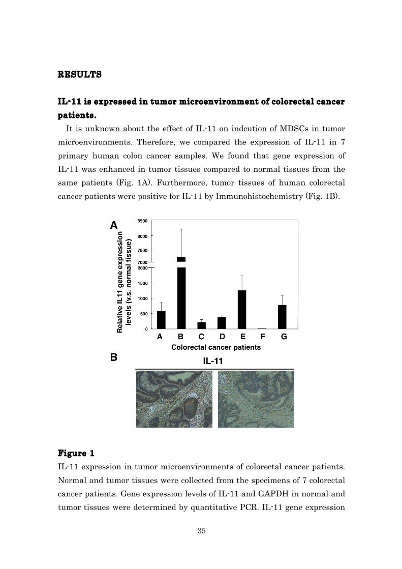
RESULTS IL-11 is expressed in tumor microenvironment of colorectal cancer patients. It is unknown about the effect of IL-11 on indcution of MDSCs in tumor microenvironments. Therefore, we compared the expression of IL-11 in 7 primary human colon cancer samples. We found that gene expression of IL-11 was enhanced in tumor tissues compared to normal tissues from the same patients (Fig. 1A). Furthermore, tumor tissues of human colorectal cancer patients were positive for IL-11 by Immunohistochemistry (Fig. 1B). Figure 1 IL-11 expression in tumor microenvironments of colorectal cancer patients. Normal and tumor tissues were collected from the specimens of 7 colorectal cancer patients. Gene expression levels of IL-11 and GAPDH in normal and tumor tissues were determined by quantitative PCR. IL-11 gene expression
36
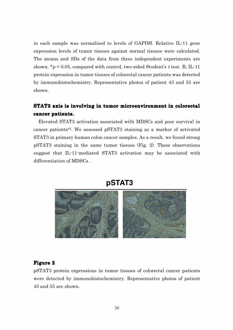
in each sample was normalized to levels of GAPDH. Relative IL-11 gene expression levels of tumor tissues against normal tissues were calculated. The means and SDs of the data from three independent experiments are shown. *p < 0.05, compared with control, two-sided Student’s t test. B, IL-11 protein expression in tumor tissues of colorectal cancer patients was detected by immunohistochemistry. Representative photos of patient 43 and 55 are shown. STAT3 axis is involving in tumor microenvironment in colorectal cancer patients. Elevated STAT3 activation associated with MDSCs and poor survival in cancer patients83. We assessed pSTAT3 staining as a marker of activated STAT3 in primary human colon cancer samples. As a result, we found strong pSTAT3 staining in the same tumor tissues (Fig. 2). These observations suggest that IL-11-mediated STAT3 activation may be associated with differentiation of MDSCs . Figure 2 pSTAT3 protein expressions in tumor tissues of colorectal cancer patients were detected by immunohistochemistry. Representative photos of patient 43 and 55 are shown.
37
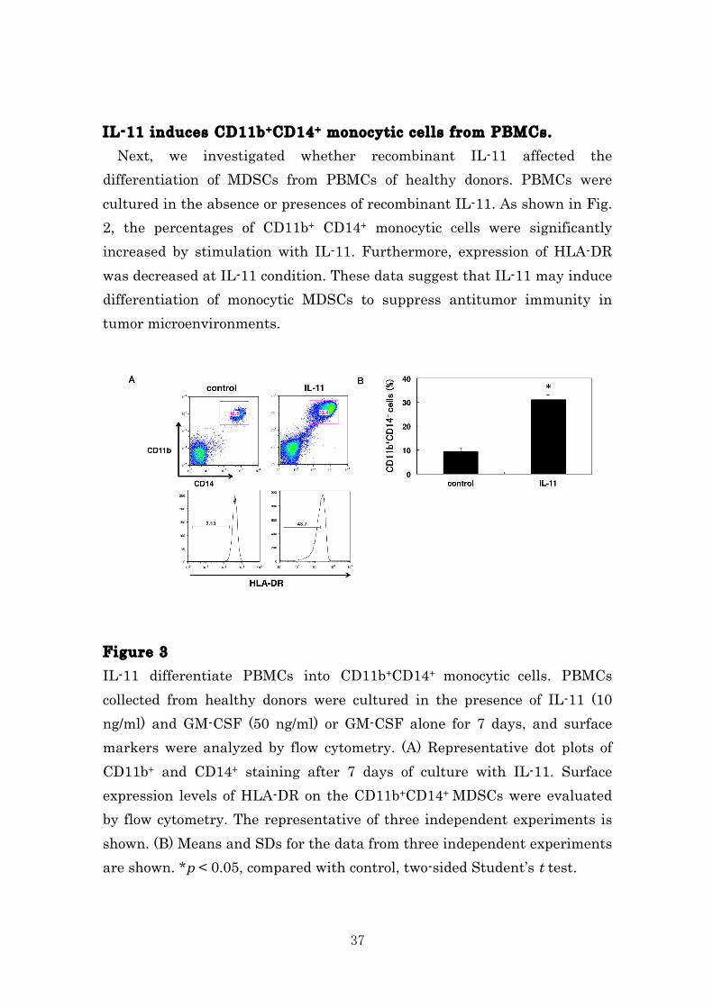
IL-11 induces CD11b+CD14+ monocytic cells from PBMCs. Next, we investigated whether recombinant IL-11 affected the differentiation of MDSCs from PBMCs of healthy donors. PBMCs were cultured in the absence or presences of recombinant IL-11. As shown in Fig. 2, the percentages of CD11b+ CD14+ monocytic cells were significantly increased by stimulation with IL-11. Furthermore, expression of HLA-DR was decreased at IL-11 condition. These data suggest that IL-11 may induce differentiation of monocytic MDSCs to suppress antitumor immunity in tumor microenvironments.
Figure 3 IL-11 differentiate PBMCs into CD11b+CD14+ monocytic cells. PBMCs collected from healthy donors were cultured in the presence of IL-11 (10 ng/ml) and GM-CSF (50 ng/ml) or GM-CSF alone for 7 days, and surface markers were analyzed by flow cytometry. (A) Representative dot plots of CD11b+ and CD14+ staining after 7 days of culture with IL-11. Surface expression levels of HLA-DR on the CD11b+CD14+ MDSCs were evaluated by flow cytometry. The representative of three independent experiments is shown. (B) Means and SDs for the data from three independent experiments are shown. *p < 0.05, compared with control, two-sided Student’s t test.
38
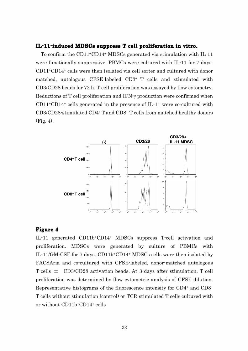
IL-11-induced MDSCs suppress T cell proliferation in vitro. To confirm the CD11+CD14+ MDSCs generated via stimulation with IL-11 were functionally suppressive, PBMCs were cultured with IL-11 for 7 days. CD11+CD14+ cells were then isolated via cell sorter and cultured with donor matched, autologous CFSE-labeled CD3+ T cells and stimulated with CD3/CD28 beads for 72 h. T cell proliferation was assayed by flow cytometry. Reductions of T cell proliferation and IFN-γ production were confirmed when CD11+CD14+ cells generated in the presence of IL-11 were co-cultured with CD3/CD28-stimulated CD4+ T and CD8+ T cells from matched healthy donors (Fig. 4). Figure 4 IL-11 generated CD11b+CD14+ MDSCs suppress T-cell activation and proliferation. MDSCs were generated by culture of PBMCs with IL-11/GM-CSF for 7 days. CD11b+CD14+ MDSCs cells were then isolated by FACSAria and co-cultured with CFSE-labeled, donor-matched autologous T-cells ± CD3/CD28 activation beads. At 3 days after stimulation, T cell proliferation was determined by flow cytometric analysis of CFSE dilution. Representative histograms of the fluorescence intensity for CD4+ and CD8+ T cells without stimulation (control) or TCR-stimulated T cells cultured with or without CD11b+CD14+ cells
39
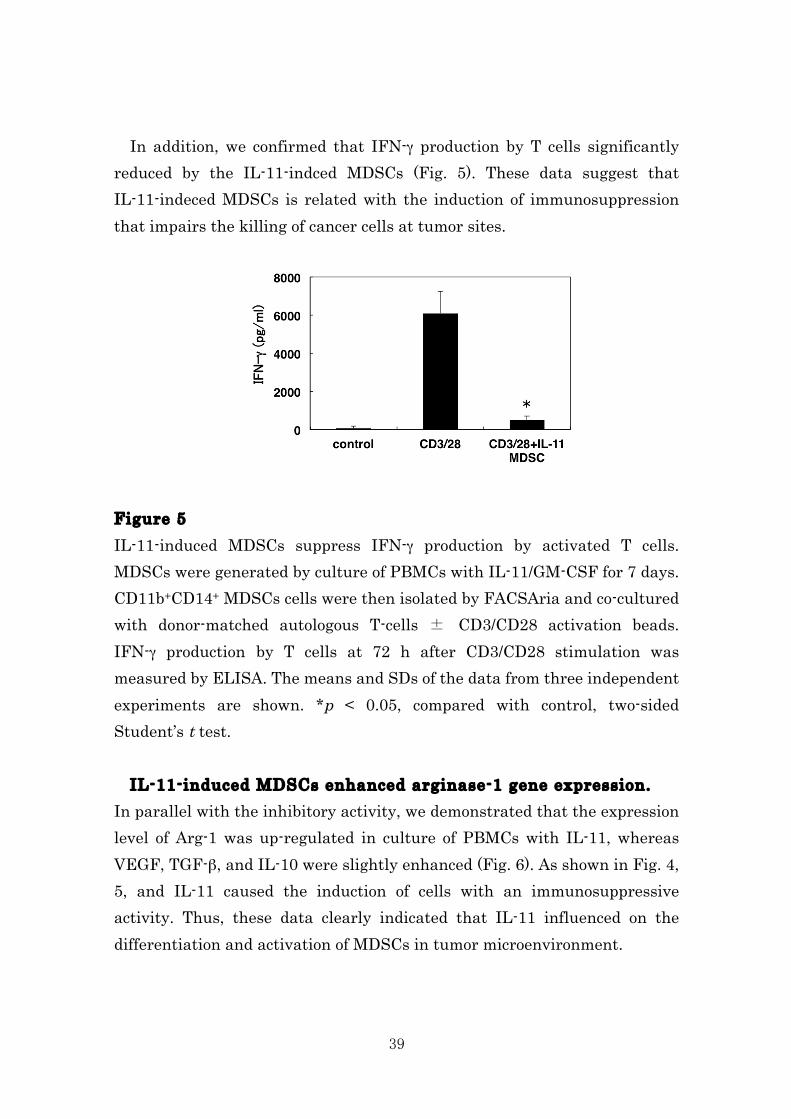
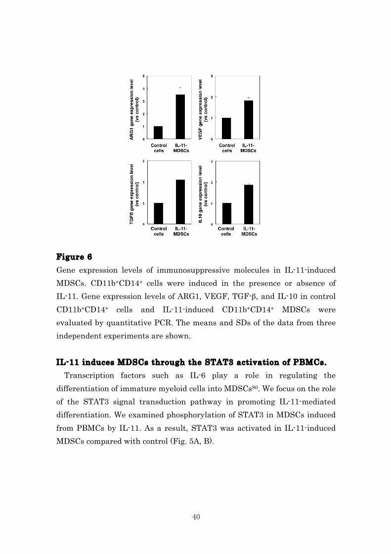
In addition, we confirmed that IFN-γ production by T cells significantly reduced by the IL-11-indced MDSCs (Fig. 5). These data suggest that IL-11-indeced MDSCs is related with the induction of immunosuppression that impairs the killing of cancer cells at tumor sites. Figure 5 IL-11-induced MDSCs suppress IFN-γ production by activated T cells. MDSCs were generated by culture of PBMCs with IL-11/GM-CSF for 7 days. CD11b+CD14+ MDSCs cells were then isolated by FACSAria and co-cultured with donor-matched autologous T-cells ± CD3/CD28 activation beads. IFN-γ production by T cells at 72 h after CD3/CD28 stimulation was measured by ELISA. The means and SDs of the data from three independent experiments are shown. *p < 0.05, compared with control, two-sided Student’s t test. IL-11-induced MDSCs enhanced arginase-1 gene expression. In parallel with the inhibitory activity, we demonstrated that the expression level of Arg-1 was up-regulated in culture of PBMCs with IL-11, whereas VEGF, TGF-β, and IL-10 were slightly enhanced (Fig. 6). As shown in Fig. 4, 5, and IL-11 caused the induction of cells with an immunosuppressive activity. Thus, these data clearly indicated that IL-11 influenced on the differentiation and activation of MDSCs in tumor microenvironment.
40
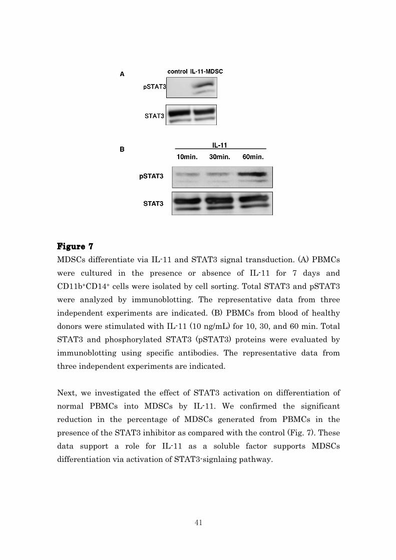
Figure 6 Gene expression levels of immunosuppressive molecules in IL-11-induced MDSCs. CD11b+CD14+ cells were induced in the presence or absence of IL-11. Gene expression levels of ARG1, VEGF, TGF-β, and IL-10 in control CD11b+CD14+ cells and IL-11-induced CD11b+CD14+ MDSCs were evaluated by quantitative PCR. The means and SDs of the data from three independent experiments are shown. IL-11 induces MDSCs through the STAT3 activation of PBMCs. Transcription factors such as IL-6 play a role in regulating the differentiation of immature myeloid cells into MDSCs80. We focus on the role of the STAT3 signal transduction pathway in promoting IL-11-mediated differentiation. We examined phosphorylation of STAT3 in MDSCs induced from PBMCs by IL-11. As a result, STAT3 was activated in IL-11-induced MDSCs compared with control (Fig. 5A, B).
41
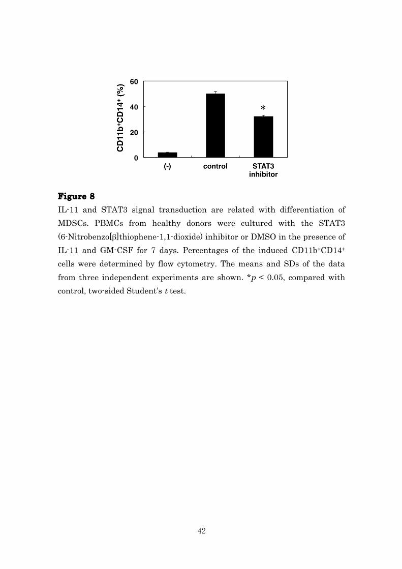
Figure 7 MDSCs differentiate via IL-11 and STAT3 signal transduction. (A) PBMCs were cultured in the presence or absence of IL-11 for 7 days and CD11b+CD14+ cells were isolated by cell sorting. Total STAT3 and pSTAT3 were analyzed by immunoblotting. The representative data from three independent experiments are indicated. (B) PBMCs from blood of healthy donors were stimulated with IL-11 (10 ng/mL) for 10, 30, and 60 min. Total STAT3 and phosphorylated STAT3 (pSTAT3) proteins were evaluated by immunoblotting using specific antibodies. The representative data from three independent experiments are indicated. Next, we investigated the effect of STAT3 activation on differentiation of normal PBMCs into MDSCs by IL-11. We confirmed the significant reduction in the percentage of MDSCs generated from PBMCs in the presence of the STAT3 inhibitor as compared with the control (Fig. 7). These data support a role for IL-11 as a soluble factor supports MDSCs differentiation via activation of STAT3-signlaing pathway.
42
Figure 8 IL-11 and STAT3 signal transduction are related with differentiation of MDSCs. PBMCs from healthy donors were cultured with the STAT3 (6-Nitrobenzo[β]thiophene-1,1-dioxide) inhibitor or DMSO in the presence of IL-11 and GM-CSF for 7 days. Percentages of the induced CD11b+CD14+ cells were determined by flow cytometry. The means and SDs of the data from three independent experiments are shown. *p < 0.05, compared with control, two-sided Student’s t test.
43
DISCUSSION It has been considered that overcoming of immunosuppressive tumor escape mechanisms is essential to induce tumor-specific CTLs for establishing more efficient tumor immunotherapy. A recent report indicated that dietary iron, enhanced colonic IL-6/IL-11-STAT3 signaling pathway, promoted the inflammation and subsequent tumor development in a mouse model of inflammation-associated colorectal tumorigenesis using DSS86. In addition, IL-6 directly promotes survival, proliferation, and differentiation of tumor cells and MDSCs in an IL-6-STAT3-dependent manner. It has also been demonstrated that IL-6 is a key cytokine that induces MDSCs under a tumor-bearing condition83. However, the precise mechanism of the effect of the MDSCs by IL-11 was still unknown. In this study, we first demonstrated that IL-11, produced as a soluble factor in tumor microenvironment of patients with colon cancer, promoted the differentiation of immune cells into functional MDSCs. We considered that MDSCs was induced by IL-11 from the local tumor microenvironment, which was required for generation of differentiation and immunosuppression that impairs cancer cell killing by CTLs at tumor sites (Fig. 1-5). These data are consistent with recent reports demonstrating that exposure of MDSCs to either tumor microenvironment or inflammatory site enhances the suppressive function87, 88. STAT3 regulates various target genes involved in growth, proliferation, survival and differentiation of MDSCs as well as numerous types of cells 51, 89,
90. In this study, we found that STAT3 was activated in IL-11-induced MDSCs (Fig. 6, 7). Moreover, we confirmed that the IL-11-mediated differentiation of MDSCs was blocked by the inhibition of STAT3 activation (Fig. 8). Intratumoral IL-11 expression in tumor sites of colorectal cancer patients strongly suggests that the induced MDSCs may be mediated by IL-11-STAT3 signaling in tumor microenvironment. Our data may supports a previous report indicating that IL-11 directly activates the neoplastic epithelium rather than promoting tumorigenesis indirectly by engaging hematopoietic cells such as MDSCs73. Recent reported has provided evidence that beyond their direct cytotoxic or
44
cytostatic effects on cancer cells, several conventional chemotherapeutic drugs and agents used in targeted therapies can promote the elimination or inactivation of suppressive Tregs or MDSCs, resulting in enhanced antitumor immunity91. Ernst. M et al. reviewed that some recent insights derived from genetic and therapeutic inhibition of the IL-6/IL-11 - gp130 - STAT3 signaling cascade in the context of preclinical mouse models of cancer92. Our present data suggests that IL-11-induced MDSCs might play a role in immunosuppression of tumor microenvironments. Therefore, regulation of the functional differentiation via IL-11-induced MDSCs may be a possible target for cancer immunotherapy. Given the critical roles served by MDSCs in immunosuppression, our elucidation for the effector functions in IL-11-induced MDSCs has implications in the rational design of strategies for modulation of immune responses and development of new efficient treatments for cancer patients.
45
CONCLUDING REMARKS New discoveries from this research l Three distinct splenic CD11b+ Gr-1+ cells (Myeloid-derived suppressor
cells; MDSCs) subsets and two distinct tumor-infiltrating CD11b+ Gr-1+ cells exist in tumor-bearing mouse.
l Splenic CD11b+Gr-1+ cells differentiate into immunosuppressor by
tumor-derived factors. l Administration of anti IL-6R mAb eliminates CD11b+ Gr-1+ cell subsets
and inhibits tumor growth. l Administration of anti IL-6R mAb enhances anti-tumor immunity such
as CD8+ T cell response. l IL-11 induces differentiation of CD11b+ CD14+ monocytic MDSCs
l IL-11 may be related with suppression of anti-tumor immunity by
induction of MDSCs at tumor sites of colorectal cancer patients.
l Activation of STAT3-signlaing pathway is required for differentiation of MDSCs by IL-11
l Control of IL-11-STAT3 signaling pathway in MDSCs may be useful target for improving cancer immunotherapy.
Immunotherapeutic significance of the discovery and future perspectives In Capter1, the therapeutic efficacy of cancer immunotherapy has been limited. Overcoming strong immunosuppression and tumor escape mechanisms in tumor microenvironment is necessary for development of an
46
effective immunotherapy. So, it is important to evaluate the immunosuppressive mechanisms of tumor-bearing hosts for successful treatment of tumors. Myeloid-derived suppressor cells (MDSCs) are one of the major immunosuppressive cell types in tumor microenvironments. In this study, we first found that the splenic CD11b+Gr-1+ cells (MDSCs) were classified into three different subsets according to their phenotypic and morphological characteristics. In contrast to spleen, CD11b+Gr-1+ cells infiltrated into tumor tissues inhibited T cell activation, indicating that CD11b+Gr-1+ cells acquired an immunosuppressive activity by the influence of tumor microenvironments. Moreover, we found that administration of Gemcitabine (GEM) and/or anti-IL-6 mAb, both of which revealed regulatory effect on MDSCs differentiation, into tumor-bearing mice caused the blocking of suppressive MDSCs accumulation at tumor site and inhibited the growth of tumor cells. It is considered that in the future, our results intended to suggest that the potential for combination of chemotherapy with immunotherapy effectively lead to a more developed application of effective treatments. Reacenty, reported that the combined treatment of immunotherapy with radiation therapy and tumor-specific Th1 cells are very efficiently to be capable of inducing anti-tumor immunity. Therefore, by the combined administration of anti-IL-6R mAb, chemotherapy, and cancer vaccine therapy, it would be possible to enhance the anti-tumor effects. We believe that anti-IL-6R mAb leads to decrease in number of MDSCs in cancer patients, and IL-6 signaling pathway is a good target for regulation of MDSCs function during cancer immunotherapy. Significance of the discovery and future perspectives in the regulation of MDSCs In Capter2, productions of inflammatory cytokines (IL-1β, IL-6, and IL-11) and inductions of MDSCs in the tumor microenvironment are crucial factors limiting the efficacy of immune-based therapy. In this study, we first found that CD11b+ CD14+ monocytic MDSCs were
47
generated from PBMCs of healthy donors in the presence of IL-11. The IL-11-induced MDSCs upregulated immunosuppressive molecules such as arginase-1. T-cell proliferation was significantly reduced when CD11b+CD14+ cells generated in the presence of IL-11 were co-cultured with CD3/CD28-stimulated CD4+ T and CD8+ T cells of autologous healthy donors. IL-11 stimulation induced STAT3 phosphorylation in normal PBMCs. Neutralization of IL-11 inhibited the STAT3 phosphorylation and differentiation into MDSCs. Blockade of STAT3 activation suppressed induction of MDSCs by IL-11. Immunohistochemical analysis indicated that both IL-11 and pSTAT3 was observed in stromal portion of tumor tissues from colon cancer patients. These finding suggest that IL-11-STAT3 signaling pathway is a potential target for induction of MDSCs in the tumor microenvironment. Thus, IL-11-STAT3 mediated regulation in functional differentiation of MDSCs will be a promising strategy for improving cancer immunotherapy.
48
ACKOWLEDGMENT First of all, I would like to acknowledge Prof. Toru Kondo (Division of Cancer-Related Genes, Institute for Genetic Medicine, Hokkaido University) for their thoughtful direction of all the experiment carried out in this dissertation. I sincerely thank them for tireless support and guidance throughout the experiment. I would like to thank Associate Prof. Hidemitsu Kitamura and all the members in my laboratory (Division of Functional Immunology, Institute for Genetic Medicine, Hokkaido University) for technical assistance during the experiment. This work was supported in part by a Grant-in-Aid from the Research Fellowship for Young Scientists (251464 to Kentaro Sumida)
49
REFERENCES 1. Nishimura, T. et al. Distinct role of antigen-specific T helper type 1
(Th1) and Th2 cells in tumor eradication in vivo. J Exp Med. 190, 617-627 (1999).
2. Trinchieri, G., Pflanz, S. & Kastelein, R. A. The IL-12 family of heterodimeric cytokines: new players in the regulation of T cell responses. Immunity .19, 641-644 (2003).
3. Kennedy, R. & Celis, E. Multiple roles for CD4+ T cells in anti-tumor immune responses. Immunol Rev. 222, 129-144 (2008).
4. Takeshima, T. et al. Local radiation therapy inhibits tumor growth through the generation of tumor-specific CTL: its potentiation by combination with Th1 cell therapy. Cancer Res. 70, 2697-2706 (2010).
5. Zou, W. Immunosuppressive networks in the tumour environment and their therapeutic relevance. Nat Rev Cancer. 5, 263-274 (2005).
6. Yu, H., Kortylewski, M. & Pardoll, D. Crosstalk between cancer and immune cells: role of STAT3 in the tumour microenvironment. Nat Rev Immunol. 7, 41-51 (2007).
7. Yu, H., Pardoll, D. & Jove, R. STATs in cancer inflammation and immunity: a leading role for STAT3. Nat Rev Cancer. 9, 798-809 (2009).
8. Narita, Y. et al. The key role of IL-6-arginase cascade for inducing dendritic cell-dependent CD4(+) T cell dysfunction in tumor-bearing mice. J Immunol. 190, 812-820 (2013).
9. Hodi, F. S. et al. Biologic activity of cytotoxic T lymphocyte-associated antigen 4 antibody blockade in previously vaccinated metastatic melanoma and ovarian carcinoma patients. Proc Natl Acad Sci U S A. 100, 4712-4717 (2003).
10. Zhang, Y. et al. Th1 cell adjuvant therapy combined with tumor vaccination: a novel strategy for promoting CTL responses while avoiding the accumulation of Tregs. Int Immunol. 19, 151-161 (2007).
11. Chamoto, K. et al. 3-Methylcholanthrene-induced transforming growth factor-beta-producing carcinomas, but not sarcomas, are refractory to regulatory T cell-depletion therapy. Cancer Sci 101, 855-861 (2010).
50
12. Youn, J. I., Nagaraj, S., Collazo, M. & Gabrilovich, D. I. Subsets of myeloid-derived suppressor cells in tumor-bearing mice. J Immunol. 181, 5791-5802 (2008).
13. Gabrilovich, D. I. & Nagaraj, S. Myeloid-derived suppressor cells as regulators of the immune system. Nat Rev Immunol. 9, 162-174 (2009).
14. Ostrand-Rosenberg, S. & Sinha, P. Myeloid-derived suppressor cells: linking inflammation and cancer. J Immunol. 182, 4499-4506 (2009).
15. Peranzoni, E. et al. Myeloid-derived suppressor cell heterogeneity and subset definition. Curr Opin Immunol. 22, 238-244 (2010).
16. Dolcetti, L. et al. Hierarchy of immunosuppressive strength among myeloid-derived suppressor cell subsets is determined by GM-CSF. Eur J Immunol. 40, 22-35 (2010).
17. Almand, B. et al. Increased production of immature myeloid cells in cancer patients: a mechanism of immunosuppression in cancer. J Immunol. 166, 678-689 (2001).
18. Rodriguez, P. C. et al. Arginase I in myeloid suppressor cells is induced by COX-2 in lung carcinoma. J Exp Med. 202, 931-939 (2005).
19. Huang, B. et al. Gr-1+CD115+ immature myeloid suppressor cells mediate the development of tumor-induced T regulatory cells and T-cell anergy in tumor-bearing host. Cancer Res. 66, 1123-1131 (2006).
20. Ochoa, A. C., Zea, A. H., Hernandez, C. & Rodriguez, P. C. Arginase, prostaglandins, and myeloid-derived suppressor cells in renal cell carcinoma. Clin Cancer Res. 13, 721s-726s (2007).
21. Bunt, S. K. et al. Reduced inflammation in the tumor microenvironment delays the accumulation of myeloid-derived suppressor cells and limits tumor progression. Cancer Res. 67, 10019-10026 (2007).
22. Bunt, S. K., Clements, V. K., Hanson, E. M., Sinha, P. & Ostrand-Rosenberg, S. Inflammation enhances myeloid-derived suppressor cell cross-talk by signaling through Toll-like receptor 4. J Leukoc Biol. 85, 996-1004 (2009).
23. Cheng, P. et al. Inhibition of dendritic cell differentiation and accumulation of myeloid-derived suppressor cells in cancer is regulated by S100A9 protein. J Exp Med. 205, 2235-2249 (2008).
51
24. Kusmartsev, S. et al. Reversal of myeloid cell-mediated immunosuppression in patients with metastatic renal cell carcinoma. Clin Cancer Res. 14, 8270-8278 (2008).
25. Murdoch, C., Muthana, M., Coffelt, S. B. & Lewis, C. E. The role of myeloid cells in the promotion of tumour angiogenesis. Nat Rev Cancer. 8, 618-631 (2008).
26. Kusmartsev, S. et al. Reversal of myeloid cell-mediated immunosuppression in patients with metastatic renal cell carcinoma. Clin Cancer Res. 14, 8270-8278 (2008).
27. Diaz-Montero, C. M. et al. Increased circulating myeloid-derived suppressor cells correlate with clinical cancer stage, metastatic tumor burden, and doxorubicin-cyclophosphamide chemotherapy. Cancer Immunol Immunother. 58, 49-59 (2009).
28. Solito, S. et al. A human promyelocytic-like population is responsible for the immune suppression mediated by myeloid-derived suppressor cells. Blood 118, 2254-2265 (2011).
29. Gros, A. et al. Myeloid cells obtained from the blood but not from the tumor can suppress T-cell proliferation in patients with melanoma. Clin Cancer Res. 18, 5212-5223 (2012).
30. Bronte, V. & Zanovello, P. Regulation of immune responses by L-arginine metabolism. Nat Rev Immunol. 5, 641-654 (2005).
31. Nagaraj, S. et al. Altered recognition of antigen is a mechanism of CD8+ T cell tolerance in cancer. Nat Med. 13, 828-835 (2007).
32. Sinha, P. et al. Proinflammatory S100 proteins regulate the accumulation of myeloid-derived suppressor cells. J Immunol. 181, 4666-4675 (2008).
33. Lu, T. et al. Tumor-infiltrating myeloid cells induce tumor cell resistance to cytotoxic T cells in mice. J Clin Invest. 121, 4015-4029 (2011).
34. Gabrilovich, D. I., Ostrand-Rosenberg, S. & Bronte, V. Coordinated regulation of myeloid cells by tumours. Nat Rev Immunol. 12, 253-268 (2012).
35. Lu, T. & Gabrilovich, D. I. Molecular pathways: tumor-infiltrating
52
myeloid cells and reactive oxygen species in regulation of tumor microenvironment. Clin Cancer Res. 18, 4877-4882 (2012).
36. Li, H., Han, Y., Guo, Q., Zhang, M. & Cao, X. Cancer-expanded myeloid-derived suppressor cells induce anergy of NK cells through membrane-bound TGF-beta 1. J Immunol. 182, 240-249 (2009).
37. Chung, Y. C. & Chang, Y. F. Serum interleukin-6 levels reflect the disease status of colorectal cancer. J Surg Oncol. 83, 222-226 (2003).
38. De Vita, F. et al. Serum interleukin-10 levels in patients with advanced gastrointestinal malignancies. Cancer 86, 1936-1943 (1999).
39. Karin, M. & Greten, F. R. NF-kappaB: linking inflammation and immunity to cancer development and progression. Nat Rev Immunol. 5, 749-759 (2005).
40. Nishimoto, N. et al. Improvement in Castleman's disease by humanized anti-interleukin-6 receptor antibody therapy. Blood 95, 56-61 (2000).
41. Nishimoto, N. et al. Treatment of rheumatoid arthritis with humanized anti-interleukin-6 receptor antibody: a multicenter, double-blind, placebo-controlled trial. Arthritis Rheum. 50, 1761-1769 (2004).
42. Yokota, S. et al. Efficacy and safety of tocilizumab in patients with systemic-onset juvenile idiopathic arthritis: a randomised, double-blind, placebo-controlled, withdrawal phase III trial. Lancet 371, 998-1006 (2008).
43. Wakita, D. et al. IFN-gamma-dependent type 1 immunity is crucial for immunosurveillance against squamous cell carcinoma in a novel mouse carcinogenesis model. Carcinogenesis 30, 1408-1415 (2009).
44. Nishimura, T., Burakoff, S. J. & Herrmann, S. H. Protein kinase C required for cytotoxic T lymphocyte triggering. J Immunol 139, 2888-2891 (1987).
45. Deaglio, S. et al. Adenosine generation catalyzed by CD39 and CD73 expressed on regulatory T cells mediates immune suppression. J Exp Med. 204, 1257-1265 (2007).
46. Jin, D. et al. CD73 on tumor cells impairs antitumor T-cell responses: a novel mechanism of tumor-induced immune suppression. Cancer Res. 70, 2245-2255 (2010).
53
47. Beavis, P. A., Stagg, J., Darcy, P. K. & Smyth, M. J. CD73: a potent suppressor of antitumor immune responses. Trends Immunol. 33, 231-237 (2012).
48. Suzuki, E., Kapoor, V., Jassar, A. S., Kaiser, L. R. & Albelda, S. M. Gemcitabine selectively eliminates splenic Gr-1+/CD11b+ myeloid suppressor cells in tumor-bearing animals and enhances antitumor immune activity. Clin Cancer Res. 11, 6713-6721 (2005).
49. Le, H. K. et al. Gemcitabine directly inhibits myeloid derived suppressor cells in BALB/c mice bearing 4T1 mammary carcinoma and augments expansion of T cells from tumor-bearing mice. Int Immunopharmacol. 9, 900-909 (2009).
50. Narita, Y., Wakita, D., Ohkuri, T., Chamoto, K. & Nishimura, T. Potential differentiation of tumor bearing mouse CD11b+Gr-1+ immature myeloid cells into both suppressor macrophages and immunostimulatory dendritic cells. Biomed Res. 30, 7-15 (2009).
51. Gabrilovich, D. I., Ostrand-Rosenberg, S. & Bronte, V. Coordinated regulation of myeloid cells by tumours. Nat Rev Immunol. 12, 253-268 (2012).
52. Kusmartsev, S. & Gabrilovich, D. I. Immature myeloid cells and cancer-associated immune suppression. Cancer Immunol Immunother. 51, 293-298 (2002).
53. Movahedi, K. et al. Identification of discrete tumor-induced myeloid-derived suppressor cell subpopulations with distinct T cell-suppressive activity. Blood 111, 4233-4244 (2008).
54. Youn, J. I., Nagaraj, S., Collazo, M. & Gabrilovich, D. I. Subsets of myeloid-derived suppressor cells in tumor-bearing mice. J Immunol. 181, 5791-5802 (2008).
55. Dolcetti, L. et al. Hierarchy of immunosuppressive strength among myeloid-derived suppressor cell subsets is determined by GM-CSF. Eur J Immunol. 40, 22-35 (2010).
56. Youn, J. I. & Gabrilovich, D. I. The biology of myeloid-derived suppressor cells: the blessing and the curse of morphological and functional heterogeneity. Eur J Immunol. 40, 2969-2975 (2010).
54
57. Condamine, T. & Gabrilovich, D. I. Molecular mechanisms regulating myeloid-derived suppressor cell differentiation and function. Trends Immunol. 32, 19-25 (2011).
58. Sumida, K. et al. Anti-IL-6 receptor mAb eliminates myeloid-derived suppressor cells and inhibits tumor growth by enhancing T-cell responses. Eur J Immunol. 42, 2060-2072 (2012).
59. Peranzoni, E. et al. Myeloid-derived suppressor cell heterogeneity and subset definition. Curr Opin Immunol. 22, 238-244 (2010).
60. Solito, S. et al. A human promyelocytic-like population is responsible for the immune suppression mediated by myeloid-derived suppressor cells. Blood 118, 2254-2265 (2011).
61. Montero, A. J., Diaz-Montero, C. M., Kyriakopoulos, C. E., Bronte, V. & Mandruzzato, S. Myeloid-derived suppressor cells in cancer patients: a clinical perspective. J Immunother. 35, 107-115 (2012).
62. Liu, C. et al. Expansion of spleen myeloid suppressor cells represses NK cell cytotoxicity in tumor-bearing host. Blood 109, 4336-4342 (2007).
63. Nagaraj, S. et al. Altered recognition of antigen is a mechanism of CD8+ T cell tolerance in cancer. Nat Med. 13, 828-835 (2007).
64. Cheng, P. et al. Inhibition of dendritic cell differentiation and accumulation of myeloid-derived suppressor cells in cancer is regulated by S100A9 protein. J Exp Med. 205, 2235-2249 (2008).
65. De Wilde, V. et al. Endotoxin-induced myeloid-derived suppressor cells inhibit alloimmune responses via heme oxygenase-1. Am J Transplant. 9, 2034-2047 (2009).
66. Li, H., Han, Y., Guo, Q., Zhang, M. & Cao, X. Cancer-expanded myeloid-derived suppressor cells induce anergy of NK cells through membrane-bound TGF-beta 1. J Immunol. 182, 240-249 (2009).
67. Oberlies, J. et al. Regulation of NK cell function by human granulocyte arginase. J Immunol. 182, 5259-5267 (2009).
68. Poschke, I., Mougiakakos, D., Hansson, J., Masucci, G. V. & Kiessling, R. Immature immunosuppressive CD14+HLA-DR-/low cells in melanoma patients are Stat3hi and overexpress CD80, CD83, and DC-sign. Cancer Res. 70, 4335-4345 (2010).
55
69. Lu, T. & Gabrilovich, D. I. Molecular pathways: tumor-infiltrating myeloid cells and reactive oxygen species in regulation of tumor microenvironment. Clin Cancer Res. 18, 4877-4882 (2012).
70. Nagaraj, S. et al. Antigen-specific CD4(+) T cells regulate function of myeloid-derived suppressor cells in cancer via retrograde MHC class II signaling. Cancer Res. 72, 928-938 (2012).
71. Teramura, M., Kobayashi, S., Hoshino, S., Oshimi, K. & Mizoguchi, H. Interleukin-11 enhances human megakaryocytopoiesis in vitro. Blood 79, 327-331 (1992).
72. Hirano, T. Interleukin 6 in autoimmune and inflammatory diseases: a personal memoir. Proc Jpn Acad Ser B Phys Biol Sci 86, 717-730 (2010).
73. Putoczki, T. & Ernst, M. More than a sidekick: the IL-6 family cytokine IL-11 links inflammation to cancer. J Leukoc Biol. 88, 1109-1117 (2010).
74. Gao, Y. B. et al. Enhanced production of CTGF and IL-11 from highly metastatic hepatoma cells under hypoxic conditions: an implication of hepatocellular carcinoma metastasis to bone. J Cancer Res Clin Oncol. 139, 669-679 (2013).
75. Ren, L., Wang, X., Dong, Z., Liu, J. & Zhang, S. Bone metastasis from breast cancer involves elevated IL-11 expression and the gp130/STAT3 pathway. Med Oncol. 30, 634 (2013).
76. Campbell, C. L. et al. Interleukin-11 receptor expression in primary ovarian carcinomas. Gynecol Oncol. 80, 121-127 (2001).
77. Bellone, G. et al. Cytokine expression profile in human pancreatic carcinoma cells and in surgical specimens: implications for survival. Cancer Immunol Immunother. 55, 684-698 (2006).
78. Nakayama, T. et al. Expression of interleukin-11 (IL-11) and IL-11 receptor alpha in human gastric carcinoma and IL-11 upregulates the invasive activity of human gastric carcinoma cells. Int J Oncol. 30, 825-833 (2007).
79. Necula, L. G. et al. IL-6 and IL-11 as markers for tumor aggressiveness and prognosis in gastric adenocarcinoma patients without mutations in Gp130 subunits. J Gastrointestin Liver Dis. 21, 23-29 (2012).
80. Putoczki, T. L. et al. Interleukin-11 is the dominant IL-6 family cytokine
56
during gastrointestinal tumorigenesis and can be targeted therapeutically. Cancer Cell 24, 257-271 (2013).
81. Xiang, Z. L., Zeng, Z. C., Fan, J., Tang, Z. Y. & Zeng, H. Y. Expression of connective tissue growth factor and interleukin-11 in intratumoral tissue is associated with poor survival after curative resection of hepatocellular carcinoma. Mol Biol Rep. 39, 6001-6006 (2012).
82. McCoy, E. M., Hong, H., Pruitt, H. C. & Feng, X. IL-11 produced by breast cancer cells augments osteoclastogenesis by sustaining the pool of osteoclast progenitor cells. BMC Cancer. 13, 16 (2013).
83. Mace, T. A. et al. Pancreatic cancer-associated stellate cells promote differentiation of myeloid-derived suppressor cells in a STAT3-dependent manner. Cancer Res. 73, 3007-3018 (2013).
84. Mace, T. A. et al. Bioactive compounds or metabolites from black raspberries modulate T lymphocyte proliferation, myeloid cell differentiation and Jak/STAT signaling. Cancer Immunol Immunother. 63, 889-900 (2014).
85. Yuan, H. et al. Axitinib augments antitumor activity in renal cell carcinoma via STAT3-dependent reversal of myeloid-derived suppressor cell accumulation. Biomed Pharmacother. 68, 751-756 (2014).
86. Chua, A. C. et al. Dietary iron enhances colonic inflammation and IL-6/IL-11-Stat3 signaling promoting colonic tumor development in mice. PLoS One 8, e78850 (2013).
87. Corzo, C. A. et al. HIF-1α regulates function and differentiation of myeloid-derived suppressor cells in the tumor microenvironment. J Exp Med. 207, 2439-2453 (2010).
88. Haverkamp, J. M., Crist, S. A., Elzey, B. D., Cimen, C. & Ratliff, T. L. In vivo suppressive function of myeloid-derived suppressor cells is limited to the inflammatory site. Eur J Immunol. 41, 749-759 (2011).
89. Nefedova, Y. et al. Regulation of dendritic cell differentiation and antitumor immune response in cancer by pharmacologic-selective inhibition of the janus-activated kinase 2/signal transducers and activators of transcription 3 pathway. Cancer Res. 65, 9525-9535 (2005).
90. Gabrilovich, D. I. & Nagaraj, S. Myeloid-derived suppressor cells as

57
regulators of the immune system. Nat Rev Immunol. 9, 162-174 (2009). 91. Alizadeh, D. & Larmonier, N. Chemotherapeutic targeting of
cancer-induced immunosuppressive cells. Cancer Res. 74, 2663-2668 (2014).
92. Ernst, M. & Putoczki, T. L. Molecular Pathways: IL11 as a Tumor-Promoting Cytokine-Translational Implications for Cancers. Clin Cancer Res. 20, 5579-5588 (2014).


![Author(s) Doc URL - HUSCAP...) for the English language review. Conflict of interest The authors declare that they have no conflict of interest. References [1] Brackmann DE, Toh EH](https://img.pdfslide.us/doc/110x75/5f88bb5cebb8b151a8670f3f/authors-doc-url-huscap-for-the-english-language-review-conflict-of-interest.jpg)