Embed Size (px)
Citation preview

DNA REPAIR PATHWAYS INVOLVED IN THE FORMATION OF ANAPHASE BRIDGES
by
Ceyda Açılan
BS, Bogaziçi University, 2001
Submitted to the Graduate Faculty of
Arts and Sciences in partial fulfillment
of the requirements for the degree of
Doctor of Philosophy
University of Pittsburgh
2006

ii
UNIVERSITY OF PITTSBURGH
FACULTY OF ART AND SCIENCES
This dissertation was presented
by
Ceyda Açılan
It was defended on
December 1st, 2006
and approved by
Graham F. Hatfull, PhD, Professor
Deborah Chapman, PhD, Associate Professor
Jeffrey D. Hildebrand, PhD, Associate Professor
Richard D. Wood, PhD, Professor
Dissertation Advisor: William S. Saunders, PhD, Associate Professor

iii
Copyright © by Ceyda Açılan
2006

iv
Chromosomal alterations can arise from numerous events, including errors during cell
division or repair of damaged DNA. Of these errors, segregational defects such as anaphase
bridges and multipolar spindles play a major role in chromosomal instability, leading to
tumorigenesis.
Bridges can theoretically be produced by several mechanisms including telomere-
telomere fusion, persistence of chromatid cohesion into anaphase or repair of broken DNA ends.
DNA damage can induce anaphase bridges following exposure to agents such as hydrogen
peroxide or ionizing radiation (IR). Our hypothesis is that while the majority of double strand
breaks (DSBs) are repaired, to restore the original chromosome structure, incorrect fusion events
also occur leading to bridging and that bridge formation allows cells to bypass the apoptotic
pathways that are activated in response to DNA damage. To test this, we set out to determine
what pathways the cells use to heal the damage and form bridges. Our data suggest that neither
of the two major pathways used by the cell for repair of double strand breaks, homologous
recombination (HR) and non-homologous end joining (NHEJ), is required for bridge formation.
In fact, the NHEJ pathway seems to play a role in the prevention of bridges. When NHEJ is
compromised, the cell appears to use HR to repair the break, resulting in increased anaphase
bridge formation. Moreover, intrinsic NHEJ activity of different cell lines appears to be
DNA REPAIR PATHWAYS INVOLVED IN THE FORMATION OF ANAPHASE
BRIDGES
Ceyda Açılan, PhD
University of Pittsburgh, 2006

v
correlated with induction of bridges from DNA damage. Our preliminary data also suggest that
cell lines with high levels of bridging are capable of apoptosis, yet further experiments are
required to test if blocking bridging can enhance cell death.
Multipolar spindles are aberrant mitotic figures that occur when a cell divides with two or
more poles, which can lead to uneven segregation of the chromosomes. In our studies, we found
that IR treatment can lead to an increase in multipolarity shortly after treatment and changes the
distribution of spindle pole components. Initial observations on the splitting of centrosomal
proteins following IR treatment are presented.

vi
TABLE OF CONTENTS
TITLE PAGE .................................................................................................................................. i
ABSTRACT .................................................................................................................................. iv
TABLE OF CONTENTS .............................................................................................................. vi
LIST OF TABLES ........................................................................................................................ xi
LIST OF FIGURES ..................................................................................................................... xii
PREFACE....................................................................................................................................XV
1.0 CHAPTER I: INTRODUCTION................................................................................... 1
1.1 CONCEPTS IN GENOMIC INSTABILITY ........................................................ 1
1.2 SOURCES OF CHROMOSOMAL INSTABILITY............................................. 3
1.2.1 Chromosome Segregation Defects.................................................................. 4
1.2.1.1 Lagging Chromosomes........................................................................... 4
1.2.1.2 Micronuclei............................................................................................. 5
1.3 ANAPHASE BRIDGES........................................................................................ 8
1.3.1 Double Stranded DNA Repair Mechanisms ................................................. 11
1.3.1.1 Non Homologous End Joining (NHEJ) ................................................ 11
1.3.1.2 Phenotypes Associated with deficiencies in NHEJ components.......... 15
1.3.1.3 Homologous Recombination (HR) ....................................................... 16
1.3.1.4 Phenotypes Associated with deficiencies in HR components .............. 20

vii
1.3.1.5 The choice between NHEJ and HR ...................................................... 20
1.3.2 Breakage-Fusion-Bridge (BFB) Cycles........................................................ 22
1.3.3 Fate of Anaphase Bridges ............................................................................. 26
1.4 MULTIPOLAR SPINDLES (MPS) .................................................................... 27
1.4.1 Centrosome Structure and Duplication......................................................... 27
1.4.2 Centrosome amplification and MPS ............................................................. 30
1.4.3 Other mechanisms leading to MPS............................................................... 32
1.4.4 MPS and tumorogenesis ............................................................................... 33
1.5 CORRELATION BETWEEN MULTIPOLAR SPINDLES AND ANAPHASE
BRIDGES............................................................................................................................. 34
2.0 CHAPTER II: ROLE OF DNA REPAIR PATHWAYS IN THE FORMATION OF
ANAPHASE BRIDGES ............................................................................................................... 36
2.1 INTRODUCTION ............................................................................................... 36
2.1.1 Oxidative stress and DNA damage ............................................................... 36
2.1.2 Secondary effects induced by H2O2.............................................................. 37
2.1.3 Anaphase Bridges ......................................................................................... 38
2.2 RESULTS............................................................................................................ 40
2.2.1 Screening genetic mutants of DNA repair proteins using H2O2 to induce
DSBs for deficiencies in formation of anaphase bridges ............................................. 40
2.2.1.1 Mutant cell lines exhibit similar levels of bridging in HR-deficient
backgrounds in response to H2O2 treatment ........................................................ 40
2.2.1.2 Mutant cell lines exhibit enhanced levels of bridging in NHEJ-deficient
backgrounds in response to H2O2 treatment ........................................................ 41

viii
2.2.1.3 There is no additive effect for induction of anaphase bridges with
deficiencies in different proteins of NHEJ pathway............................................ 41
2.2.1.4 p53 is not required for bridge formation............................................... 42
2.2.2 Role of DNA repair pathways involved in anaphase bridging ..................... 49
2.2.2.1 HR is not required for bridge formation ............................................... 49
2.2.2.2 NHEJ is essential for prevention of bridges induced by IR.................. 50
2.2.2.3 HR appears to form bridges, when NHEJ is compromised .................. 59
2.2.2.4 NHEJ/HR double deficient cells can still repair DSBs ........................ 59
2.2.3 Decrease in anaphase bridges does not appear to be due to a decrease in
mitotic index ................................................................................................................ 62
2.2.4 In vitro NHEJ ligation assay ......................................................................... 68
2.2.4.1 NHEJ activity is correlated with the induction of bridges in cancer cells
.............................................................................................................. 71
2.3 DISCUSSION...................................................................................................... 80
3.0 CHAPTER III: ANALYSIS OF NHEJ IN INDUCTION OF ANAPHASE BRIDGES
AND END JOINING ACTIVITY IN CANCER CELLS ............................................................ 86
3.1 INTRODUCTION ............................................................................................... 86
3.2 RESULTS............................................................................................................ 89
3.2.1 XRCC4 expression is correlated with both the induction of bridges and
NHEJ activity ............................................................................................................... 89
3.2.2 Restoring XRCC4 in cells with low endogenous levels does not rescue the
increased bridging phenotype ...................................................................................... 89
3.3 DISCUSSION.................................................................................................... 100

ix
4.0 CHAPTER IV: THE APOPTOTIC PATHWAYS AND THE ABILITY TO SENSE
DNA DAMAGE ARE INTACT IN ORAL CANCER CELLS WITH HIGH ANAPHASE
BRIDGES. .................................................................................................................................. 103
4.1 INTRODUCTION ............................................................................................. 103
4.2 RESULTS.......................................................................................................... 106
4.2.1 Oral cancer cells can sense DNA damage .................................................. 106
4.2.2 Cancer cells show induction of p53 and change in subcellular localization
following DNA damage ............................................................................................. 106
4.2.3 Oral cancer cells can undergo apoptosis upon DNA damage..................... 109
4.3 DISCUSSION.................................................................................................... 114
5.0 CHAPTER V: EVIDENCE FOR CENTROSOMAL SPLITTING AND
MULTIPOLAR SPINDLE FORMATION AS A RESULT OF IONIZING RADIATION ...... 117
5.1 INTRODUCTION ............................................................................................. 117
5.2 RESULTS.......................................................................................................... 120
5.2.1 DNA damage by ionizing radiation, but not H2O2 increases MPS formation ..
..................................................................................................................... 120
5.2.2 MPS formation might be a result of centrosomal splitting after ionizing
radiation ..................................................................................................................... 123
5.3 DISCUSSION.................................................................................................... 130
6.0 CHAPTER VI: SUMMARY AND SPECULATIONS ............................................. 133
7.0 CHAPTER VII: MATERIALS AND METHODS.................................................... 139
7.1.1 Cell Culturing.............................................................................................. 139
7.1.2 Transfections and ionizing radiation........................................................... 140

x
7.1.3 H2O2 and wortmannin treatment................................................................. 141
7.1.4 Isolation of genomic DNA for ladder formation ........................................ 141
7.1.5 Immunofluorescence staining ..................................................................... 142
7.1.6 Preparation of cell free extracts and Non homologous end joining activity
assay ..................................................................................................................... 143
7.1.7 Antibodies ................................................................................................... 144
7.1.8 Plasmids ...................................................................................................... 144
7.1.9 Statistical methods ...................................................................................... 145
BIBLIOGRAPHY....................................................................................................................... 147

xi
LIST OF TABLES
Table 1 Total number of coverslips and cells scored for each DNA double-strand break
repair deficient cell line…. ........................................................................................................... 70
Table 2 Total number of coverslips and cells scored for each cancer cell line ............... 78

xii
LIST OF FIGURES
Figure 1 Examples of cells with lagging chromosomes....................................................... 6
Figure 2 Example of a cell with micronucleus..................................................................... 7
Figure 3 Examples of cells with anaphase bridges. ............................................................. 9
Figure 4 Non-homologous end joining pathway in mammalian systems. ......................... 13
Figure 5 Homologous recombination pathway in mammalian systems. ........................... 18
Figure 6 Breakage-fusion-bridge cycles . .......................................................................... 25
Figure 7 Examples of multipolar mitoses in metaphase. ................................................... 28
Figure 8 Centrosome structure ........................................................................................... 29
Figure 9 Induction of anaphase bridges in HR defective cells upon H2O2 treatment. ....... 43
Figure 10 Induction of anaphase bridges in cells deficient for NHEJ proteins after H2O2
treatment ............................................................................................................................. 45
Figure 11 Inhibition of DNA-PK by wortmannin enhanced bridge induction in wild type,
but in NHEJ deficient cells ................................................................................................ .47
Figure 12 Induction of anaphase bridges in p53 deficient cells upon H2O2 treatment......... 48
Figure 13 HR-deleted cell lines RAD54B-/-/- and XRCC3-/- did not express the
corresponding proteins.................................................................................................................. 51
Figure 14 Anaphase bridges were induced to similar levels by IR in HR mutants ............. 53

xiii
Figure 15 Ku80 and XRCC4 levels were reduced after siRNA treatment based on
immunoblotting............................................................................................................................. 54
Figure 16 Immunofluorescence staining of Ku80 and XRCC4 was reduced after siRNA
treatment ............................................................................................................................. 56
Figure 17 Enhanced induction of anaphase bridges in siRNA-treated cells or genetic
mutants upon IR treatment............................................................................................................ 58
Figure 18 Induction of bridges in NHEJ/HR deficient HCT116 cells by IR ....................... 60
Figure 19 Induction of bridges in DSB-repair deficient chicken cells by IR....................... 61
Figure 20 Transient γ-H2AX staining in HR/NHEJ defective cells after IR ....................... 63
Figure 21 Cell scores for γ-H2AX foci formation. .............................................................. 64
Figure 22 Transient 53BP1 staining in HR/NHEJ defective cells ....................................... 65
Figure 23 Transient P-ATM staining in HR/NHEJ defective cells...................................... 67
Figure 24 Mitotic indices for HCT116 WT and repair deficient cells................................. 69
Figure 25 In vitro ligation activity assay as a measure of NHEJ activity. ........................... 75
Figure 26 Cancer cells exhibited different levels of bridges and intrisic NHEJ activity. .... 77
Figure 27 Correlation between in vitro NHEJ activity and induction of bridges by IR. ..... 79
Figure 28 Summary for DNA repair pathways involved in anaphase bridges .................... 82
Figure 29 Expression levels for NHEJ proteins in cancer cells ........................................... 91
Figure 30 Correlations between XRCC4 levels and bridge induction and NHEJ activity. . 93
Figure 31 Expression levels of DNA repair proteins were increased upon transfection of an
exogenous plasmid ....................................................................................................................... 96
Figure 32 Overexpression of XRCC4 did not rescue the elevated bridging phenotype in
cancer cells with low endogenous XRCC4 level .......................................................................... 98

xiv
Figure 33 Overexpression of XRCC4 corrected the increased bridging phenotype in
XRCC4-/- CHO cells .................................................................................................................... .99
Figure 34 Cancer cells appeared to be able to sense the DNA damage as judged by γ-H2AX
foci formation ........................................................................................................................... 107
Figure 35 p53 and p21 expression in UPCI:SCC103 cells after H2O2 treatment............... 110
Figure 36 Immunostaining of p53 in UPCI:SCC103 cells after H2O2 treatment............... 112
Figure 37 Cancer cells were capable of apoptosis as determined by formation of DNA
ladders after H2O2 treatment ...................................................................................................... 113
Figure 38 Frequency of multipolarity in HEK-293 cells following H2O2 treatment. ........ 121
Figure 39 Frequency of multipolarity in HEK-293 cells following IR.............................. 122
Figure 40 γ-tubulin staining in HEK-293 cells before and after IR ................................... 125
Figure 41 Centrin-2 staining at the spindle poles before and after IR in HEK-293 cells .. 126
Figure 42 Number of centrin-2 spots/pole in HEK-293 cells following ionizing radiation
treatment ........................................................................................................................... 127
Figure 43 Model for MPS formation following IR treatment............................................ 129

xv
PREFACE
FOR MY DEAR FAMILY
Melek Açılan
Muharrem Açılan
Burak Açılan

xvi
First and foremost, I would like to thank my thesis advisor, Dr. William Saunders, for
accepting me in his lab and giving me such a wonderful project to work on. He has guided me
through tough times with many helpful suggestions and endless discussions. I appreciate all the
insights he has given me in science and his encouragements in my personal life.
I would like to thank my thesis committee members, Dr. Graham Hatfull, Dr. Jeffrey
Hildebrand, Dr. Deborah Chapman and Dr. Richard Wood for their time and comments about
my project.
I am thankful to all Saunders’ Lab members for creating an enjoyable working
environment. I thank Dr. Nicholas Quintyne for discussing my problems, helpful feedbacks
about my project and editing my papers. I thank Dr. Li Luo and Qian Wu not only for their
insightful discussions and help with experiments, but also for their wonderful friendship. I will
not forget our conversations with Li in our little scope room during hours of counting. I also have
to thank Qian for the positive attitude she expresses at all times and her funny, short stories I
love listening to. I thank Diane Hoffelder and Janet Reign for their assistance when I joined the
lab and Kristen Bartoli for her considerate closeness. I am indebted to our collaborator Dr.
Susanne Gollin for sharing cell lines and her inputs for my dissertation.
I have gratitude toward the entire Biological Sciences department for the facilities and the
supportive and joyful environment. Everybody in this department has been very generous in
sparing their time and also sharing reagents. I particularly would like to thank the Brodsky Lab,

xvii
the Arndt lab and the Hildebrand lab for letting me use their equipment. I owe a lot to Ms. Carole
Lafave for her guidance in my teaching experience and for being kind and thoughtful. If it wasn’t
for Cathy Barr, I would not have managed to register to any class at Pitt or meet any deadline. I
should especially thank Natalie Marinzel for cheering me up with her laughter, her genuine
compliments to my outfits and her life-saving reminders. I also thank my fellow classmates, for
keeping me in good spirits and accompanying me on the streets of “Graduate School”.
I cannot thank enough my dear friends at the Turkish American Student Association
Executive Committee. They have made life more tolerable in Pittsburgh, with joyful and merry
committee meetings and entertaining activities. I would principally like to thank Gorkem Saka
and Basak Isin for the companionship they have shown in the past years. I should particularly
note Serpil Ugrasiz for always being there for me and especially for our past midnight
conversations. I am also grateful to Nihan Sivri for being a loyal friend since we were kids. She
has been amazingly caring in this long haul. I cannot thank her enough for her e-mails, phone
calls and visits when I needed her the most.
I dedicate this work to my beloved mom, dad and my dear brother. I can’t express my
appreciations not only for their faith in me, but also for accompanying me with their summer
visits during my PhD in Pittsburgh. My father’s optimistic nature, my mother’s caring advices,
and my brother’s energetic dialogues incredibly encouraged me to get this degree. I cannot think
of a better family to be raised in. Thank you for not giving up on me, I love you very much!!!

xviii
Lastly, I would like to thank Fatih Ayhan for thousands of minutes of phone calls from
thousands of miles away. He has been my light at the end of a dark tunnel and has played an
instrumental role in completing this experience. I really appreciate his companionship.
1
1.0 CHAPTER I: INTRODUCTION
1.1 CONCEPTS IN GENOMIC INSTABILITY
Cancer is a result of accumulation of mutations that control cell division or cell death. It
is widely accepted that a cell has to undergo multiple mutations before it becomes tumorogenic
(Lengauer et al., 1997). Hence, events speeding up the mutation rate or which interfere with the
stability of the genome are likely to give the some cells a growth advantage, which could
potentially proceed to cancer development. Some of these events that may contribute to
destabilization of the genome are described below:
Changes in nucleotide sequence: These are alterations in the DNA sequence by
substitution, addition or deletion of nucleotides. Examples include mutations in c-K-Ras, which
are found in most pancreatic cancers (Almoguera et al., 1988), BRCA1/2 genes, which are
associated with 30-50% of the heritable breast cancer cases (Rahman and Stratton 1998;
Nathanson et al., 2001), or the well-studied p53 gene, which is defective in 50% of all human
cancers (Hollstein et al., 1991; Hainaut and Hollstein 2000; Soussi and Beroud 2001).

2
Changes in chromosome number: This can be described as the gain or loss of an entire
chromosome, which leads to the state of aneuploidy. Aneuploidy is known to be associated with
almost all cancer types (Mitelman 1983) and its severity is correlated with the stage of
tumorogenesis (Cavalli et al., 1989).
Translocations: A translocation is defined as the exchange of DNA material between
non homologous chromosomes. Translocations may create fusion of genes with a change in
activity of hybrid sequences. One famous example includes the Philadelphia chromosome in
chronic myeloid leukemia, where the nuclear protein Bcr is translocationally juxtaposed to the
Abl kinase, producing a hybrid Bcr-Abl protein with increased kinase activity/localization (Ben-
Neriah et al., 1986; Jeffs et al., 1998). Likewise, in Burkitt's lymphoma, the myc oncogene is
translocated to the immunoglobin promoter leading to enhanced expression of Myc in
lymphocytes (Dean et al., 1983).
Amplifications: Gene amplifications involve an increase in the copy number of a proto-
oncogene, which may lead to over production of the associated gene product and drive the cell to
an abnormal growth stage. For instance, amplification of the N-Myc oncogene has been reported
in 30% of neuroblastomas (Seeger et al., 1985).
In normal cells, the biological processes preventing these changes are intact, thus they
occur rarely, if ever. On the other hand, all of these events are readily observed in different
cancer types, and are tightly linked to genomic instability. For example, failure in mismatch
repair system (MMR) has been shown to elevate the rate of DNA mutation by ~1000-fold

3
(Christmann et al., 2003) and results in the destabilization of simple repeat sequences. This
phenomenon, which is known as microsattelite instability (MIN), is found in most hereditary
non-polyposis colon cancers (HNPCC) (Aaltonen et al., 1993), and sporadic colon cancers
(Narayan and Roy 2003). Phenotypically, MIN cannot be detected by karyotypic analysis and
does not involve the large scale genomic changes discussed above.
The other major form of instability involves continuous gross alterations in chromosomal
structure and number, which is defined as chromosomal instability (CIN). While experimental
data support a direct link for MIN and carcinogenesis, the link between CIN and cancer is
ambiguous. Whilst it is clear that many tumor types exhibit the CIN phenotype (Lengauer et al.,
1997; Lengauer et al., 1998), there is a chicken-and-egg dilemma as to whether CIN is an early
event leading to cancer or an outcome of malignant transformation. Despite the modeling
studies, which indicate that CIN alone is sufficient to drive the cell into tumorogenesis (Nowak
et al., 2002), other studies challenge this hypothesis with the evidence of adenomas without any
obvious changes in CIN (Haigis et al., 2002). Regardless, CIN is a hallmark of cancer and
studying the molecular events leading to chromosomal instability will enlighten our
understanding of it.
1.2 SOURCES OF CHROMOSOMAL INSTABILITY
Although there are numerous events that may lead to CIN, the main reasons can be listed
as defects in chromosomal segregation, repair of DNA damage and cell-cycle checkpoint
regulation. Seemingly diverse at first, these events are actually very closely intertwined. The

4
different defects that lead to CIN usually coexist in tumor cell lines perhaps cooperatively
contributing to the multistep tumorogenesis process (Saunders et al., 2000; Jallepalli and
Lengauer 2001; Gisselsson 2003; Deng 2006). For instance, incompetency to repair a
checkpoint gene might allow mitosis despite incorrect chromosome congression. Experimental
evidence from BRCA2 deficient mice show that these mice which harbor mutations in spindle
checkpoint proteins (Bub1, BubR1) exhibit higher incidences of lymphomas (Lee et al., 1999).
BRCA2 is also reported to interact with the mitotic kinase PLK1 (Lee et al., 2004) and the
checkpoint kinase BubR1 (Futamura et al., 2000) indicating a tie between DNA repair and
checkpoint proteins.
While the links between DNA repair/checkpoint regulation and chromosomal
segregation/checkpoint regulation are well studied, it is less clear how errors in the repair of
DNA damage can lead to segregation defects. In this dissertation, evidence for the molecular
mechanisms regarding this connection will be presented. Below, I will start by discussing the
commonly observed segregation defects in cancer cells.
1.2.1 Chromosome Segregation Defects
The major segregation defects visible in cancer cells can be summarized as lagging
chromosomes, micronuclei, anaphase bridges and multipolar mitoses.
1.2.1.1 Lagging Chromosomes
Lagging chromosomes can be defined as chromosomes or fragments of chromosomes,
which fail to align properly at the metaphase plate or those that lag behind the separating

5
chromosomal masses during anaphase (Figure 1). Lagging chromosomes have been shown to
emerge as a consequence of failure in attachment of the spindle microtubules to the kinetochore
proteins (Dulout and Olivero 1984) or by attachment to a single spindle pole (merotelic
attachment) (Cimini et al., 2001; Cimini et al., 2002). It has also been uncovered in the Saunders
lab that resolution of anaphase bridges may also result in lagging chromosomes as pieces of
chromosomal material (Hoffelder et al., 2004). In any case, the daughter cells are under the risk
of losing a chromosome or having both copies of the sister chromatids, which contribute to CIN.
1.2.1.2 Micronuclei
Micronuclei are derived from whole chromosomes or chromosomal fragments, which
have a nuclear envelope and are anywhere from 1/16 to 1/3 of the size of the main nucleus
(Fenech 1993). Micronuclei are distinct from the nucleus, yet remain in the same cell (Figure 2).
While the major reason for micronuclei formation appears to be due to lagging chromosomes,
breakage of an anaphase bridge has also been shown to result in micronuclei in 50% of divisions
with a bridge (Hoffelder et al., 2004). Micronuclei occur frequently upon treatment with DNA
damaging reagents and have been proposed as a diagnostic tool to test for chemotherapy damage
(Driessens et al., 2003).
Once a micronucleus forms, many scenarios are possible for its fate. It may be excluded
from the cell, reincorporated into the main nucleus or function separately in the cell’s cytoplasm
(Leach and Jackson-Cook 2004). In the first situation, the cell would face the threat of losing
chromosomal material, resulting in aneuploidy. In the second scenario, the micronuclear mass
might potentially continue normal biological activity. In the third case, micronuclei do not

6
CA B
D E F
Figure 1 UPCI:SCC103 cells dividing normally in metaphase (A) or anaphase (D).
Examples of cells in corresponding stages with lagging chromosomes are indicated by arrows (B,
C, E, and F). DAPI is used for DNA staining and images are taken with an Olympus camera with
100X magnification lens.

7
Figure 2 Example of a cell with a micronucleus (shown by arrow) in UPCI:SCC103 cell
line. DAPI is used for DNA staining and images are taken with an Olympus camera with 100X
magnification lens. (Image: Courtesy of Qian Wu.)

8
appear to be fully transcriptionally active as they are incapable of nucleotide incorporation and
contain reduced numbers of nuclear pore complexes, which could potentially limit regular
transport (Hoffelder et al., 2004).
As described above, both lagging chromosomes and micronuclei could lead to genomic
instability if not corrected. Fortunately, these defects can trigger checkpoint proteins to pause
mitosis until all chromosomes are properly attached to both poles. There is evidence that
merotelic attachments may be corrected even prior to anaphase (Salmon et al., 2005; Cimini et
al., 2006). On the other hand, there is no indication of activation of checkpoints in cases of
anaphase bridging or formation of multipolar spindles. Hence these defects are potentially more
dangerous as will be discussed further.
1.3 ANAPHASE BRIDGES
Anaphase bridging is a commonly observed phenomenon in cancer cells, which can be
described as a chromatin fiber connecting the two chromosome masses together (Gisselsson et
al., 2000) (Figure 3B). This abnormality was first described by Barbara McClintock in 1941
(McClintock 1942) and since then, it has been shown that bridges lead to structural and
numerical chromosome changes which are strongly linked to tumorogenesis (Artandi et al.,
2000; Stewenius et al., 2005). Furthermore, anaphase bridges have major contributions to CIN
and have been reported in both cell culture (Gisselsson et al., 2000) and tissues (Montgomery et
al., 2003).

9
Figure 3 Examples of cells with bridges in UPCI:SCC103 cell line: normal anaphase (A),
anaphase with a bridge (B). Bridges are usually resolved by telophase (C), however can
sometimes persist to telophase (D) or interphase (E) and (F). Arrows indicate bridges. DAPI is
used for DNA staining and images are taken with an Olympus camera with 100X magnification
lens.
A B
C D
E F

10
A bridge is thought to form from the fusion of two broken DNA ends, or as a result of
shortened or absent telomere sequences (Fouladi et al., 2000; Gisselsson et al., 2001; Zhu et al.,
2002). Telomeres are composed of tandem (TTAGGG) repeats at the end of the chromosomes,
which span a region of 5-15 kb (Moyzis et al., 1988; de Lange et al., 1990). They form a
specialized structure called a t-loop, and are stabilized by many telomeric proteins (Callen and
Surralles 2004), which may either directly bind to telomeres or have a regulatory role in telomere
protection. Some of these proteins have a dual function in both telomere maintenance and DNA
damage repair, indicating a cross talk between telomeres and the DNA damage pathways
(Nugent et al., 1998; Surralles et al., 2004). An average of 65 bp of telomeric sequence tends to
be lost with each division in cells lacking telomerase or alternative ways to stabilize telomeres
(Counter et al., 1992). Once the shortening process starts, the t-loop can no longer form and the
protective proteins detach from the telomeres. In this case, the chromosome ends can act as free
DNA breaks and may be fused to other ends with short telomeres. Supporting this, anaphase
bridges have been shown to arise from telomere shortening, loss of telomeres or defects in
telomerase (Artandi et al., 2000; Fouladi et al., 2000; Gisselsson et al., 2001; Rudolph et al.,
2001; Lo et al., 2002). Moreover, there is a perfect correlation between telomere length in
cancer cells and the frequency of endogenous bridges (Stewenius et al., 2005).
Telomeres might alternatively be lost with the occurrence of a DNA double stranded
break (DSB) in the chromosome. This can also lead to a bridge, yet not necessarily through
telomere-telomere fusions. How bridges form through the repair of double stranded breaks is the

11
main focus of this report. This gains particular importance, since many cancer cells exhibit
stabilized telomeres.
1.3.1 Double Stranded DNA Repair Mechanisms
Cells are subjected to a number of DNA damaging events, threatening the genomic
integrity. This damage can be induced by various agents, including endogenous factors such as
free radicals formed as byproducts of metabolic functions, or stalled replication forks or
exogenous factors such as ionizing radiation or mutagenic chemicals. Among the most severe
lesions caused by these factors, DNA double stranded breaks (DSBs) are probably the most
difficult to repair. Upon formation of a DSB, generally, the original ends are fused back together,
restoring the chromosome’s original structure (Rief and Lobrich 2002). However, misligation of
two non-matching ends may also occur, resulting in an anaphase bridge and chromosomal
instability (Mills et al., 2003; Pfeiffer et al., 2004).
In order to deal with double stranded DNA breaks, mammalian cells have two major
repair pathways: Non-homologous end joining (NHEJ) and homologous recombination (HR).
While NHEJ requires little or no homology to fuse the broken ends, HR-dependent repair utilizes
the homologous strand as a template. These mechanisms will be discussed further below:
1.3.1.1 Non Homologous End Joining (NHEJ)
NHEJ is initiated by the binding of Ku proteins to the broken ends of the DNA (Smith
and Jackson 1999) (Figure 4B). The Ku heterodimer, Ku80/70, which is composed of two
subunits of molecular weight 86 and 73kDa, forms a hollow ring like structure large enough to

12
Double strand break
End Binding by Ku70/80
Recruitment of DNA-PKcs and Artemis
Artemis Artemis
DNA-PKcs DNA-PKcs
Artemis Artemis
DNA-PKcs DNA-PKcs
End-Bridging by DNA-PK and Phosphorylation of Artemis
Recruitment of LigaseIV/XRCC4/XLF
Ligation
Artemis Artemis
DNA-PKcsDNA-PKcsXRCC4
XRCC4
XRCC4
XRCC4
LigaseIV
LigaseIV
A)
B)
C)
D)
E)
F)
XLF
XLF

13
Figure 4 NHEJ in mammalian systems. Upon formation of a DSB (A), Ku70/80 proteins
are recruited to the damage site (B). The Ku heterodimer recruits DNA-PKcs, which is thought
to mediate end-bridging. DNA-PKcs both phosphorylates and forms a complex with Artemis and
this complex is involved in processing the 5’ and 3’ DNA overhangs (C, D). Finally, DNA
Ligase IV/XRCC4/XLF complex is recruited to the lesion site (E) and the ends are ligated
together (F). Figure adapted from (Hefferin and Tomkinson 2005).

14
accommodate the duplex DNA and protects DNA ends from being degraded (Walker et al.,
2001). Upon binding, the complex translocates along the DNA possibly to allow further end
processing (Ochem et al., 1997; Yavuzer et al., 1998). While the role of Ku in protecting DNA
ends from degradation is well described, there is some ambiguity for its function as an alignment
factor (Bliss and Lane 1997; Cary et al., 1997; Ramsden and Gellert 1998). Alignment and end
bridging seem hard to reconcile, since Ku proteins are known to recruit another protein, DNA
Dependent Protein Kinase catalytic subunit (DNA-PKcs) (Dvir et al., 1992; Gottlieb and Jackson
1993), which might also be involved in the alignment process (Figure 4C, D). It appears that the
association of Ku proteins and DNA-PKcs is dependent on DNA. While DNA-PKcs can bind to
DNA without the Ku proteins, the binding is enhanced by 100 fold in the presence of Ku (Dvir et
al., 1992; Gottlieb and Jackson 1993; Suwa et al., 1994; Yaneva et al., 1997). The Ku
heterodimer, together with DNA-PKcs, forms the active kinase unit, which is called the DNA-
PK complex.
Deficiencies in NHEJ proteins, such as mutations in DNA-PKcs, result in a syndrome
with severe combined immunodeficiency (SCID), since the NHEJ pathway is also required for
RAG1/RAG2 endonuclease initiated V(D)J recombination (Jhappan et al., 1997; Taccioli et al.,
1998). Recently a novel gene, which also causes the SCID phenotype, has been identified
(Moshous et al., 2000; Moshous et al., 2001). The Artemis nuclease is found to associate with
DNA-PKcs and is capable of hairpin opening, which is an intermediate structure formed during
V(D)J recombination. The Artemis/DNA-PKcs complex exhibits nuclease activity, where 5’
ends are blunted, and 3’ overhangs are trimmed (Ma et al., 2002) (Figure 4D). Moreover,

15
phosphorylation of Artemis by DNA-PK appears to be crucial for this nuclease activity (Ma et
al., 2002).
DNA-PK also appears to stimulate the last players of NHEJ: the XRCC4/DNA Ligase IV
complex, which initiates the final ligation step between the two juxtaposed DNA ends (Chen et
al., 2000) (Figure 4E, F). The activity of Ligase IV is strongly stimulated in vitro by its cofactor
XRCC4, which is also shown to increase its stability (Critchlow et al., 1997; Grawunder et al.,
1997; Bryans et al., 1999). XRCC4 is known to be phosphorylated by DNA-PKcs, however
mutations in these sites do not interfere with its complementation of radiation sensitivity
(Critchlow et al., 1997; Yu et al., 2003; Lee et al., 2004). Recently, a protein which has strong
predicted structural similarity to XRCC4 has been identified. The XRCC4 like factor, XLF (or
Cernunnos), can bind to XRCC4-Ligase IV complex and is possibly involved in this last step of
break repair (Ahnesorg et al., 2006; Buck et al., 2006; Callebaut et al., 2006).
1.3.1.2 Phenotypes Associated with deficiencies in NHEJ components
Deficiency in Ku80 results in defects in V(D)J recombination, and increased sensitivity
to irradiation (Nussenzweig et al., 1996; Zhu et al., 1996; Nussenzweig et al., 1997; Kabotyanski
et al., 1998). Mice deficient in Ku80 exhibit growth retardation and shorter life span (Vogel et
al., 1999). Moreover, spontaneous chromosome breaks appears to arise in fibroblasts derived
from these animals, indicating insufficient response to DNA damage (Karanjawala et al., 1999).
While heterozygous inactivation of Ku80 results in chromosomal instabilities such as
translocations and fusions, homozygous mutation leads to cell death after a certain number of
divisions (Li et al., 2002; Myung et al., 2004). Ku70 deficient mice manifest similar defects
(Ouyang et al., 1997; Li et al., 1998).

16
Deficiencies in DNA-PKcs or Artemis show milder phenotypes without any growth
retardation and similar incidences of T-cell lymphomas as wild type mice. However, as
mentioned previously, both defects result in SCID phenotype (Jhappan et al., 1997; Taccioli et
al., 1998; Moshous et al., 2000; Moshous et al., 2001) and lead to some sensitivity to DNA
damaging agents (Jeggo 1998; Convery et al., 2005; Musio et al., 2005).
Knockout mice for Ligase IV and XRCC4 exhibit the most severe defects. Deletion of
both genes results in embryonic lethality, possibly due to severe neurodegeneration (Barnes et
al., 1998; Frank et al., 1998; Gao et al., 1998). Defects in neurogenesis are also seen in Ku80/70
deficient mice, but not in DNA-PKcs null animals (Gu et al., 2000). Lethality can be rescued by
additional p53 deficiency, and these mice still suffer from high incidences of lymphomas
indicating the importance of these proteins in protection from cancer (Frank et al., 2000; Gao et
al., 2000).
1.3.1.3 Homologous Recombination (HR)
One of the earliest events in HR-dependent repair is converting the DSB ends to a
recombination competent structure. This step involves degradation of single strands by the
eukaryotic Mre11/RAD50/NBS1 (MRN) complex leaving a 3’ protruding end, which is
hundreds of bases long (Figure 5B) (D'Amours and Jackson 2002). This 3’ single-stranded DNA
(ssDNA), is pre-coated with Replication Protein A (RPA) (Figure 5C), which both resolves
secondary structures and facilitates RAD51 loading on the DNA with the assistance of mediator
protein RAD52 (Figure 5D) (Sung 1997; Shinohara and Ogawa 1998). Subsequently, this
nucleoprotein filament invades the intact DNA duplex, and forms a structure called the

17
DSBDouble strand break formation
Mre11-Rad50-NBS1
RPA ssDNA complex RPA
Rad51 ssDNA filament Rad51, Rad51 paralogues, Rad52, Rad54
Homology search, strand invasion Rad51, Rad51 paralogues,
Rad54
Repair synthesis, branch migration
Rad54
ResolutionXRCC3/Rad51C
5’-3’ degradation
A)
B)
C)
D)
E)
F)
G)

18
Figure 5 HR in mammalian systems. The DSB (A) ends are processed by the MRN
complex (B). The 3’ protruding end, which is precoated with RPA (C), is later loaded with
RAD51 (D), which is facilitated and stabilized by RAD51 paralogues, RAD54 and the mediator
RAD52 protein. Following strand invasion and branch migration (E-F), the broken strand is
repaired by copying the intact template and finally the complex is resolved (G).

19
“D-loop”. Strand displacement and branch migration are complicated tasks and involve many
energy-dependent processes such as breaking internal hydrogen bonds, unwinding the DNA, and
competing with other DNA binding proteins (Figure 5E). Hence, many proteins play roles during
these processes including RAD51, and all five RAD51-like proteins, namely XRCC2, XRCC3,
RAD51B (or RAD51L1), RAD51C (RAD51L2) and RAD51D (RAD51L3) (Albala et al., 1997;
Rice et al., 1997; Cartwright et al., 1998; Dosanjh et al., 1998; Liu et al., 1998; Pittman et al.,
1998; French et al., 2002). These RAD51 paralogues form four different complexes, XRCC2-
RAD51D, RAD51B-RAD51C, XRCC3-RAD51C and RAD51B-RAD51C-RAD51D-XRCC2,
which act to stimulate the activity of RAD51 in various aspects (Braybrooke et al., 2000;
Kurumizaka et al., 2001; Sigurdsson et al., 2001; Henry-Mowatt et al., 2003; Yokoyama et al.,
2003).
Other relevant proteins include RAD54, RAD54 paralogues, and the breast cancer
associated proteins BRCA1/2 (Jasin 2002). RAD54 likely has a dual function in both enabling
strand exchange and facilitating branch migration. It can stabilize RAD51 nucleofilament and
enhance D-loop formation by introducing negative supercoils (Figure 5D-F) (Sigurdsson et al.,
2002; Mazin et al., 2003). RAD54B, a RAD54 paralogue, also associates with RAD51 and
colocalizes with RAD51 foci (DSB sites where repair proteins accumulate) (Tanaka et al., 2000).
RAD54B has additive phenotypes with RAD54, indicating they have non-overlapping functions,
however the exact role is yet to be discovered (Wesoly et al., 2006). Cells deficient for either
BRCA1 or BRCA2 have impaired HR-dependent repair, and these proteins mediate their
function probably in conjunction with RAD51 (Moynahan et al., 1999; Moynahan et al., 2001).

20
Once new DNA is synthesized by copying the homologous template, the intertwined
molecules are resolved, possibly by the action of XRCC3/RAD51C complex (Figure 5G)
(Brenneman et al., 2002; French et al., 2002; Liu et al., 2004).
1.3.1.4 Phenotypes Associated with deficiencies in HR components
Defects in HR proteins have a range of phenotypes. RAD51 appears as a key protein,
since RAD51 disruption causes early embryonic lethality in mice, and mutant cell cultures do not
proliferate even in a p53-/- background (Lim and Hasty 1996; Tsuzuki et al., 1996). On the other
hand, knockout mice for RAD51 like proteins (XRCC2, RAD51B, and RAD51D) can progress
until later stages in development (Shu et al., 1999; Deans et al., 2000; Pittman and Schimenti
2000). Both RAD54- and RAD54B-deficient mice are alive, but defective cells exhibit mild
sensitivity to DNA damaging agents (Bezzubova et al., 1997; Essers et al., 1997; Wesoly et al.,
2006). While RAD52 defects reduce HR, no effect on mutagen sensitivity is observed (Rijkers et
al., 1998). BRCA1 or BRCA2 homozygous deletion results in embryonic death, like the loss of
RAD51, and deficient cell lines are sensitized to DNA damage (Gowen et al., 1996; Connor et
al., 1997; Ludwig et al., 1997; Shen et al., 1998; Scully et al., 1999).
1.3.1.5 The choice between NHEJ and HR
Both NHEJ and HR are important mechanisms in the repair of DSBs in all eukaryotes.
However there seem to be differences in the choice of pathway between different organisms.
While NHEJ is the prominent pathway in vertebrates, HR appears as the major mechanism in
yeast (Critchlow and Jackson 1998; Dudasova et al., 2004). One reason for this choice could be
due to the highly repetitive structure of the mammalian genome, which might make it harder to

21
find the correct template for HR to repair the damage. On the other hand, the yeast genome is
smaller, nearly all genes are intronless and usually consist of a single copy.
NHEJ has been long considered as the error-prone or even “illegitimate” repair pathway,
since the deletions can be harmful, if they occur in an essential gene or its regulatory sequences.
However it should be noted that HR also runs the risk of creating rearrangements by
recombining with related sequences in non-homologous chromosomes (albeit with reduced
frequencies) (Richardson et al., 1998), or might lead to loss of heterozygosity by copying
information from the homologous chromosome (Stark and Jasin 2003).
While both pathways seem to collaborate to maintain genomic stability, it also appears
that there is a competition as to which repair pathway will be used to repair the damage. It has
been shown that both NHEJ and HR can have access to the same DSB (Richardson and Jasin
2000). The HR proteins RAD51 and RAD52 can bind to DSB ends, possibly competing with the
Ku complex of NHEJ (Baumann and West 1998; Haber 2000). In support of this model,
reduction in NHEJ protein levels results in stimulation of HR-mediated repair (Fukushima et al.,
2001; Pierce et al., 2001; Allen et al., 2002; Delacote et al., 2002). This stimulation only occurs
if early NHEJ proteins, namely DNA-PKcs and Ku, are inhibited and not when late proteins are
blocked such as XRCC4 inhibition (Pierce et al., 2001). Furthermore, consistently inactivated
DNA-PKcs, which is a component of NHEJ, leads to decreased HR rates (presumably since it is
still present at the break site, yet non-functional), while its absence leads to an increase in HR
rates, indicating an “interactive competition” between the pathways (Allen et al., 2003). This
selection between pathways is currently an area of intense investigation. It is at least partly

22
influenced by cell-cycle. NHEJ predominates throughout G1 to early S, and HR becomes more
active during late S and mitosis, when the sister chromatids are available as template (Takata et
al., 1998; Saintigny et al., 2001; Rothkamm et al., 2003).
1.3.2 Breakage-Fusion-Bridge (BFB) Cycles
After the formation of a DSB, the ends are repaired by either of the pathways mentioned
above. However, if the initial fusion occurs between either the sister chromatids (after
replication) or with another chromosome, a dicentric chromosome will form with two
centromeres (Figure 6A, B). Dicentrics have the potential to attach to both spindle poles resulting
in a tug-of-war between the two spindle halves at anaphase (Gisselsson 2005). The bridge will
eventually resolve by breaking, most likely not at the site it fused (Gisselsson et al., 2001;
Hoffelder et al., 2004). In the next cell cycle, the telomere-less DNA ends are again primed to
fuse after replication, which is known as the Breakage-Fusion-Bridge (BFB) cycle, and can
result in loss or amplification of genetic information or translocation of chromosome arms.
Fusion events following DNA replication, as described, is known as “chromatid-type” BFB
cycles (Figure 6B).
In cases where fusion events precede DNA replication, the chromosome will replicate
producing two dicentrics. These dicentrics may segregate either in a parallel fashion or by
producing two anaphase bridges (Figure 6C). If they segregate in parallel, each daughter cell will
inherit a dicentric chromosome, which will face the same scenario in the next cell cycle. In cases
of bi-directional segregation, the bridges will form, and eventually break (Hoffelder et al., 2004).

23
replication
T T T T T
TT T T
T TT
DNADamage
T
T Break
T
T
A
B
T
replicated chromatids
fusion
breakage
bridge formationreplication
fusionreplication
T T T T T
TT T T
T TT
DNADamage
T
T Break
T
T
A
B
T
replicated chromatids
fusion
breakage
bridge formationreplication
fusion

24
C
T T
replication
Dicentric chromosome
T TT T
TT
TTT T
T T
ParalelsegregationDouble bridge
TTTT
T T
break
Fusion in next cell cycle
No breakage
C
T T
replication
Dicentric chromosome
T TT T
TT
TTT TT T
T TT T
ParalelsegregationDouble bridge
TTTT TT
T T
break
Fusion in next cell cycle
No breakage

25
Figure 6 Breakage-Fusion-Bridge cycles. Double stranded break formation followed by
replication may result in bridging between sister chromatids (A). Chromatid type BFB cycles
(B). Chromosome type BFB cycles and possible outcomes of chromosome segregation. See text
for details (C). Green lines indicate the sites of break, “T” stands for telomeres. Centromeres are
represented by black circles, and different chromosomes are color coded by red and blue.

26
The cells will have sticky telomere-free DNA ends, which may fuse back and enter a
“chromosome type” BFB cycle.
1.3.3 Fate of Anaphase Bridges
It is now known that the breakage along the anaphase bridge typically occurs at multiple
sites, leading to formation of micronuclei and extensive loss of DNA material (Gisselsson et al.,
2001; Hoffelder et al., 2004). In an extreme case, the entire bridge may be fragmented, resulting
in loss of whole chromosome arms, which may explain the high frequencies of isochromosomes
or whole-arm translocations in cancer cells (Gisselsson et al., 2005; Stewenius et al., 2005).
Furthermore, these rearrangements also result in amplifications of low copy genes. For example,
there is evidence indicating that both cyclinD1 and NuMA genes have been amplified via
breakage-fusion-bridge cycles (Jiang et al., 1992; Huang et al., 2002). Alternatively, a bridge
might result in detachment of sister chromatids from the spindle, which would give rise to loss of
the whole chromosome (Gisselsson et al., 2005; Stewenius et al., 2005). Lastly, anaphase
bridging might interfere with cytokinesis, resulting in a binucleate cell (Stewenius et al., 2005).
After another round of DNA replication and centrosome duplication, a binucleate has the
potential to divide in a multipolar fashion, which will eventually cause further chromosomal
instability (Luo L and Saunders WS, unpublished data).
Although bridges usually break in anaphase from the tension of the spindle pulling forces
or during cytokinesis, in rare cases they will persist until telophase or even after cytokinesis
resulting in formation of an interphase bridge seen as persistent chromatin strands connecting

27
daughter cells (Figure 3C-F). It is not yet known whether these cells continue BFB cycles,
function as a binucleate cell or cease proliferation.
1.4 MULTIPOLAR SPINDLES (MPS)
During mitosis, each daughter cell is ensured to obtain the correct amount of DNA
through carefully controlled attachments of the chromosomes to the highly organized spindle
fibers and symmetric separation in a bipolar fashion. However, divisions with more than two
poles are also observed which leaves the cells with abnormal numbers of chromosomes (Figure
7) (Wunderlich 2002). Multipolar spindle (MPS) formation is a defect observed in many cancer
cell lines, and is associated with an increase in centrosome number (Saunders 2005; Stewenius et
al., 2005).
1.4.1 Centrosome Structure and Duplication
Centrosomes are the microtubule organizing centers in mammalian cells. The major
structural elements of centrosomes are a pair of barrel shaped centrioles surrounded by an
amorphous framework of proteins called the pericentriolar material (PCM). Centrioles are small
cylindrical organelles (~200-400nm) composed of 9 triplet longitudinal fibrils and radial spokes
connecting fibrils to the center of the centriole (Figure 8) (Dutcher 2001; Dutcher 2001). Among
the major components of the PCM are the γ-tubulin ring complexes, the site of microtubule
nucleation (Zheng et al., 1995), and the Sfi1p and centrin fibers, which make connections
between different components of the centrosome, mediating overall structure and centriole

28
Figure 7 Multipolar mitoses in metaphase (A, B) and anaphase (C, D) observed in
UPCI:SCC103 cell line. Notice that bridging usually accompanies multipolarity. DAPI is used
for DNA staining and images are taken with an Olympus camera with 100X magnification lens.
A B
C D

29
Figure 8 Centrosome structure. Centriole configuration with an array of nine microtubule
triplets is shown in cartoon and in enlarged view by electron microscopy. Magnification ~
305,000X. Figure from:
http://users.rcn.com/jkimball.ma.ultranet/BiologyPages/C/Cytoskeleton.html and
http://io.uwinnipeg.ca/~jfranck/Bio3221_Pwrpt_lectures/Nov10_lecture_files/slide0016_image0
16.jpg

30
duplication (Salisbury 2004). Other structural components include several coiled coil proteins
(Salisbury 2003), such as pericentrin (Doxsey et al., 1994; Dictenberg et al., 1998), Cep135
(Ohta et al., 2002), AKAP-450 (Keryer et al., 1993; Witczak et al., 1999), and ninein (Bouckson-
Castaing et al., 1996), which regulate centrosomal activity.
Since centrosomes play a critical role in equal segregation of the chromosomes, they have
to be duplicated once and only once in the cell cycle to ensure bipolarity. In G1, cells typically
have two centrioles, which are orthogonally oriented relative to each other. Once cells pass the
G1 checkpoint, the centrioles separate slightly, and nascent procentrioles start emerging
perpendicular to the mother centrioles at the proximal end (Adams and Kilmartin 2000;
Khodjakov et al., 2002). Elongation of procentrioles continues throughout G2 and is completed
by the end of mitosis. Centrosomal maturation continues by recruitment of the PCM proteins
during the G2/M phase and completion takes ~1.5 cell cycles (Dictenberg et al., 1998). A
number of phosphorylation and ubiquitin dependent degradation events also take place to
regulate the centrosome duplication cycle (Freed et al., 1999; Fry et al., 2000; Wojcik et al.,
2000; Hinchcliffe and Sluder 2001; Meraldi and Nigg 2002).
The centriole pairs and the associated PCM, which forms the new centrosomes, migrate
away from each other shortly before mitosis, ensuring bipolar spindle formation.
1.4.2 Centrosome amplification and MPS
Extra numbers of centrosomes have been reported in many different tumor types
including breast, lung, brain, gall bladder, bone, pancreas, colorectal, head and neck cancers

31
(Lingle et al., 1998; Pihan et al., 1998; Weber et al., 1998; Carroll et al., 1999; Lingle and
Salisbury 1999; Sato et al., 1999; Gustafson et al., 2000; Kuo et al., 2000; Pihan et al., 2001;
Sato et al., 2001). Moreover, changes in centrosome number is strongly correlated with
aneuploidy and CIN in many cases (Lingle et al., 1998; Sato et al., 1999; Ghadimi et al., 2000;
Pihan et al., 2003).
Centrosomes can potentially be amplified by three different mechanisms (Nigg 2002).
The first model involves several rounds of duplication within a single cell cycle. Since
centrosome duplication requires a long time, there has to be a delay in cell cycle progression for
this model to be correct. Indeed, when cells are treated with drugs, such as hydroxyurea or
aphidicolin to stall DNA replication (Balczon et al., 1995; Meraldi et al., 1999), or when RAD51
deficient cells are arrested in G2 phase after irradiation (Dodson et al., 2004), centrosomes
continue to duplicate in the absence of DNA replication and completion of the cell cycle. In the
second model, over-replication occurs due to a failure of cytokinesis. Abortion of cell division
arises from a variety of reasons some of which are: persistence of DNA damage, an inactive
spindle-assembly checkpoint or abnormalities in mitotic progression (Meraldi et al., 2002;
Millband et al., 2002). For example, it has been shown that a reduction in the activity of the
myosin light chain kinase, which functions in contractile ring formation and completion of
cytokinesis, is a major source of cytokinesis failure in a variety of cancer cell lines (Matsumura
2005) (Wu Q. and Saunders WS., unpublished results). Thirdly, cell fusion might also result in
supernumerary centrosomes. This phenomenon has been observed following treatment of cells
with X-Ray or UV or overexpression of Rad6 in epithelial cells (Kura et al., 1978; Brathen et al.,
2000; Shekhar et al., 2002). Regardless of which initial event leads to an amplification in

32
centrosome number, the main outcome is the increased likelihood of formation of multipolar
spindles (Lingle et al., 1998; D'Assoro et al., 2002; Lingle et al., 2002).
It has been observed that both MPS and centrosome amplification appear together in
cancer cells, suggestive of a link between these abnormalities (Gisselsson et al., 2002). Despite
the intimate correlation, cancer cells do not always undergo multipolar mitosis when they acquire
extra centrosomes. A typical example is the N1E-115 cell line, where supernumerary
centrosomes coalesce to form a bipolar metaphase plate (Ring et al., 1982). Similar results are
also observed in UPCI:SCC114 cells indicating that there are mechanisms clustering the
centrosomes allowing bipolar division (Quintyne et al., 2005). There is evidence suggesting that
coalescence is achieved through a dynein-dependent mechanism, since this microtubule binding
motor is reduced in the spindle of most cancer cells. Restoring dynein to the spindle results in
centrosomal clustering and bipolar division (Quintyne et al., 2005).
1.4.3 Other mechanisms leading to MPS
Although centrosomal amplification is a major mechanism in MPS formation,
multipolarity can be induced in the absence of changes in centrosome number. Overexpression of
Nek2 kinase, which plays a role in centriole separation through phosphorylation of linker
proteins between the centrioles, has been shown to provoke centrosome splitting and
multipolarity (Fry et al., 1998; Fry et al., 1998). Centrosomal splitting can also occur via
treatment of microtubule destabilizing drugs such as nocodazole, colcemid, or disorazole (Jean et
al., 1999; Meraldi and Nigg 2001) (Acilan C, Saunders WS, unpublished observations).
Moreover, the splitting effect can be amplified with the combined action of nocodazole and

33
overexpression of Nek2, indicating that the effect can be additive (Meraldi and Nigg 2001).
Furthermore, treatment of cells with reagents other than microtubule destabilizers or cold-
shocking the cells have also been reported to induce splitting of centrosomes and MPS (Schliwa
et al., 1983; Callaini and Marchini 1989; Kojima and Czihak 1990).
In addition, there is evidence indicating that incomplete replication or DNA damage can
result in centrosomal splitting in both Drosophila and mammalian cells (Hut et al., 2003).
Treatment of cells with γ-irradiation has also shown to induce multipolarity and once again
centrosome overduplication has been proposed as the mechanism. In chapter 5, we shall provide
evidence suggesting other mechanisms as to how γ-irradiation might lead to MPS.
1.4.4 MPS and tumorogenesis
During a multipolar division, a balanced distribution of chromosomes to the daughter
cells is very unlikely, even if the sister chromatids segregate accurately. In point of fact, it has
been shown that the division of sister chromatids after a tripolar mitosis is almost random
(Stewenius et al., 2005). Under this assumption, some of the progeny cells will inherit no copies
of certain chromosomes (nullisomy) and it is likely that clonal expansion will not be favored in
cells containing nullisomies. So why do cancer cells exhibit multipolar mitoses but are not
eliminated by selection? It can be speculated that the majority of the daughter cells will not
undergo further cell divisions, which will be an evolutionary dead end, but a rare event can grant
a selective advantage. This would hold true especially after a multipolar division in binucleate
cells, which might provide extra number of chromosomes. Moreover, a genetic variation leading
to in vivo resistance to chemotherapeutic drugs or an ability to stimulate angiogenesis in a tumor

34
could also favor multipolar divisions. Alternatively, formation of MPS can be a secondary event
without any particular advantage. Cancer cells frequently lack cell cycle checkpoints and
abnormal mitotic figures could be expected to occur in these backgrounds. Supernumerary
centrosomes are only associated with a minority of cells contributing 1–15% of the population.
It is possible that those that divide in a multipolar fashion are eliminated from the culture and a
steady frequency of supernumerary centrosomes is achieved by de novo amplification of
centrosomes.
1.5 CORRELATION BETWEEN MULTIPOLAR SPINDLES AND ANAPHASE
BRIDGES
Both anaphase bridges and multipolar spindle formation can lead to chromosomal
instability and massive changes in chromosomal structure and number. They are usually found
concomitantly driving the cancer cell to aneuploidy (Gisselsson et al., 2004). Moreover, there is
a strong positive correlation between the frequency of MPS and the level of bridging in many
different tumors tested (r=0.96) (Gisselsson et al., 2002). The basis for such a correlation could
be a single event, environmental or genetic, causing both of these defects. For instance,
treatments such as ionizing radiation and X-ray exposure have been shown to induce both (Scott
and Zampetti-Bosseler 1980; Gisselsson et al., 2001; Sato et al., 2001). Furthermore, loss of p53
results in an increase in centrosome number probably through deficiencies in the regulation of
the centrosome duplication cycle and failure of cytokinesis. p53 might also control centrosome
cycle through transactivation independent ways such as physical binding to the centrosomes
(Carroll et al., 1999; Tarapore and Fukasawa 2002). Moreover, p53 loss also affects the

35
frequency of anaphase bridges in backgrounds with deficient DNA repair pathways or
telomerase enzyme (Zhu et al., 2002). Consistently, expression of human papillomavirus proteins
E6 or E7, which bind to and inactivate p53 and pRb tumor suppressor proteins, can both trigger
formation of anaphase bridges and lead to supernumerary centrosomes and multipolar mitoses
(Duensing and Munger 2002; Schaeffer et al., 2004). Alternatively, a correlation might exist due
to a dependence of anaphase bridges or multipolar spindles on one another. We have observed
that when cells undergo multipolar anaphase, they usually contain bridges connecting some or all
poles to each other (Figure 7C, D). Thus, within a mixed population of normal and aberrant
division, these events tend to occur together. It is known that anaphase bridges do not necessarily
resolve and persistence through interphase is observed (Figure 3D-F). In such instances, the
bridge might interfere with cytokinesis, which would lead to an increase in centrosome number,
hence multipolar spindles. While any of the above, alone, could explain the observed correlation,
they might also co-exist strengthening the link between these defects.

36
2.0 CHAPTER II: ROLE OF DNA REPAIR PATHWAYS IN THE FORMATION OF
ANAPHASE BRIDGES
2.1 INTRODUCTION
2.1.1 Oxidative stress and DNA damage
There are a number of DNA damaging events and oxidative damage constitutes a
significant source of DNA damage in all aerobic organisms. Oxidative damage can occur
through exogenous sources such as ionizing radiation (IR) (Skov 1984; Teoule 1987; Nikjoo et
al., 1994) or from endogenous sources such as production of H2O2 as a result of metabolic
processes associated with redox reduction reactions in the mitochondria (Chance et al., 1979).
While H2O2 does not react with DNA directly, it results in production of •OH radicals,
which are highly reactive with DNA, through the Fenton reaction (Imlay et al., 1988).
Fe2++H2O2→•OH+Fe3+.
Metals, such as Cu+ and Fe2+, catalyze this transition, and both of these metals are
abundant in cellular fluids, and are also associated with DNA in trace amounts. Interestingly,

37
DNA damage through H2O2 requires the presence of these metals, consistent with Fenton
reaction as the primary source of reactive oxygen species (Blakely et al., 1990). •OH can result in
nearly 100 different types of DNA damage including formation of single stranded DNA breaks
(SSB) (Michalik et al., 1995). It can also indirectly lead to double stranded breaks (DSB)
possibly due to close proximity of SSBs, when H2O2 is present at higher concentrations (Dahm-
Daphi et al., 2000; Jackson and Loeb 2001).
IR can also result in different DNA lesions, including DSBs (Hutchinson 1985). On the
other hand, unlike H2O2, ionizing radiation can exert its effects directly on DNA, by radiation
energy (Teoule 1987; Ward 1988). Ionization can release 6-7 times more energy than needed to
break a covalent C-C bond (Hall 1994), and DSBs can be induced by IR, even at low doses
(Dahm-Daphi et al., 2000). The effects of IR may also be indirect through production of reactive
oxygen species by ionizing water molecules (Skov 1984; Teoule 1987; Ward 1988; Nikjoo et al.,
1994).
2.1.2 Secondary effects induced by H2O2
While H2O2 is a useful experimental reagent to induce DNA damage, this treatment has
other side effects, such as damaging cells through oxidation of membrane lipids or proteins
(Howe et al., 2004; Huang et al., 2004). Moreover, it is known to stimulate apoptosis by altering
Ca2+ homeostasis (Lin et al., 2004; Shin et al., 2004; van Rossum et al., 2004). In contrast, direct
H2O2 exposure inhibits CaM-kinase activity, hence might have an inhibitory effect on apoptosis
as well (Franklin et al., 2006). Furthermore, certain oncogenes, such as K-ras, are shown to be
activated by oxidative stress, which suggests a tumor promoting role for H2O2 (Jackson 1994).

38
H2O2 treatment is able to influence cell volume as well by changing membrane potential
(Bychkov et al., 1999) or by activation of membrane serine-threonine phosphatases (Bize and
Dunham 1995; Bize et al., 1998). In addition, the cytoskeletal network is affected by hydrogen
peroxide, which can either selectively alter the structure of the cytoskeletal proteins (Aksenov et
al., 2001) or rearrange cellular actin networks (Huot et al., 1998). H2O2 also induces formation of
focal adhesion complexes possibly through activation of the SAPK2/p38 pathway (Dalle-Donne
et al., 2001).
2.1.3 Anaphase Bridges
Anaphase bridging, a commonly observed segregation defect in cancer cells, is one of the
major sources of genomic instability (Gisselsson et al., 2000) (Montgomery et al., 2003). It has
been proposed that bridging may have hazardous consequences like gene deletions,
amplifications or translocations, and is known to be strongly linked to carcinogenesis (Artandi et
al., 2000; Stewenius et al., 2005). Anaphase bridges can theoretically be produced by several
mechanisms such as telomeric fusions, or repair of DNA double-stranded breaks (Bryant 1984;
Artandi et al., 2000; Fouladi et al., 2000; Gisselsson et al., 2001; Rudolph et al., 2001; Lo et al.,
2002; O'Hagan et al., 2002). While a significant amount of evidence has been obtained on how
bridges form as a result of telomeric fusion, relatively little is known about bridge induction after
DSB formation.
In an attempt to determine how and why cells form bridges following DSB induction, we
first set out to determine what DNA repair pathways the cells use to heal the DNA damage that
cause bridges. Preliminary experiments were performed using hydrogen peroxide to induce

39
DSBs. To exclude the possible side effects of H2O2 mentioned above, the results were confirmed
and expanded using IR.
Our studies have indicated that neither of the two major DNA repair pathways in
mammalian cells, HR or NHEJ, alone, is required for bridge formation. In fact, the NHEJ
pathway seems to play a role in the prevention of bridges. Moreover, it appears that cancer cells
that have intrinsically high NHEJ activity are less likely to form bridges.

40
2.2 RESULTS
2.2.1 Screening genetic mutants of DNA repair proteins using H2O2 to induce DSBs for
deficiencies in formation of anaphase bridges
Whilst cells usually fuse the correct broken ends efficiently, the presence of dicentric
chromosomes and anaphase bridges indicate that incorrect fusions also occur. In order to explore
the mechanisms behind anaphase bridging in response to DSBs, we hypothesized that formation
of bridges is an enzymatic process, which involves the fusion of broken DNA ends via repair
proteins. Initially, we tested this hypothesis by using genetic mutants for numerous proteins
involved in either NHEJ or HR, the major DSB repair pathways in vertebrates. The DSBs were
induced by H2O2 treatment in unsynchronized cultures. Cells were fixed 24 hours following
treatment, which is approximately one cell-cycle duration after the breaks were introduced.
Anaphase bridges were scored based on DAPI staining and exclusively DNA links that are
entirely continuous between the condensed chromosome masses were counted as a bridge.
Consistent with previous reports, in all cell lines tested, treatment of H2O2 resulted in increase
bridges, albeit with varying frequencies (Figures 9-11) (Thomas et al., 2003).
2.2.1.1 Mutant cell lines exhibit similar levels of bridging in HR-deficient backgrounds in
response to H2O2 treatment
We began by examining whether HR plays a role in bridging. Figure 9 illustrates the
induction of anaphase bridges in HCT116 cells and the RAD54B mutant derived from this cell

41
line by targeting all three copies of the gene (HCT116 cells harbor three alleles of RAD54B).
The loss of RAD54B expression was verified by immunoblotting (Figure 13). RAD54B deletion
results in decreased HR rates and varying levels of sensitivity in response to different DNA
damaging reagents (Miyagawa et al., 2002; Wesoly et al., 2006). In this HR mutant, we observed
that anaphase bridges were induced to similar extents compared to the parental line, indicating
that HR pathway was not required for bridge formation.
2.2.1.2 Mutant cell lines exhibit enhanced levels of bridging in NHEJ-deficient
backgrounds in response to H2O2 treatment
In order to test whether NHEJ plays a role in bridging, we used Ku80 deficient Chinese
Hamster Ovary (CHO) and the DNA-PKcs deficient human glioblastoma (MO59) cell lines. For
both cell lines, the parental wild type cells showed an induction of bridges upon H2O2 treatment.
Strikingly however, the level of anaphase bridge formation in either NHEJ deficient cell line was
higher than seen in wild type cells (Figure 10). This result suggested that NHEJ was not required
in the formation of bridges, but rather it played an efficient role in their prevention.
2.2.1.3 There is no additive effect for induction of anaphase bridges with deficiencies in
different proteins of NHEJ pathway
DNA-PKcs is a member of phosphotidylinositol-3 (PI-3) kinase family (although there is
no evidence for its function as a lipid kinase like PI-3 kinase) (Smith et al., 1999). PI-3 kinases,
including DNA-PKcs, are known to be inhibited by wortmannin (Hosoi et al., 1998). Given this
tool, we wanted to test whether inhibition of DNA-PKcs by wortmannin will have similar effects
on bridging in other cell lines as well as in cells that are already defective for NHEJ. As
predicted, anaphase bridges were enhanced upon induction of DSBs in wild type cells after drug

42
treatment. Nevertheless, inhibition of DNA-PKcs in cells with an existing mutation in a NHEJ
protein, Ku80, did not further augment bridge frequency, indicating that there is no additive
effect between the NHEJ proteins for the induction of anaphase bridging (Figure 11). However,
it should be noted that wortmannin is not specific for DNA-PKcs and among the other PI-3
kinases, activity of DNA repair proteins, such as ATM or ATR, is also expected to be reduced.
2.2.1.4 p53 is not required for bridge formation
The tumor suppressor protein, p53, has a critical role in many signal transduction
pathways including DNA repair, distinguishing between growth arrest or apoptosis (Niida and
Nakanishi 2006; Sun 2006) p53 has been shown to inhibit deletion of bases during repair related
joining events by NHEJ and has been proposed to influence NHEJ during site-directed, but not
IR induced DSB repair (Tachibana 2004; Dahm-Daphi et al., 2005).
To test whether p53 is required for bridge formation, we assayed for bridging in p53
knockout mouse epithelial cells (Figure 12). Upon H2O2 treatment, anaphase bridges were
greatly induced from ~5% to an average of 80%. Likewise, another segregation defect, formation
of micronuclei that occurs as a consequence of bridging, was increased in accordance. Consistent
with this data, it has been reported by our lab that treatment of cells with cigarette smoke
condensate also induces anaphase bridge formation at a higher frequency in p53 deleted
backgrounds (Luo et al., 2004).
The induction of bridges in p53 deleted cells suggests that this protein is probably not
required for bridge formation. However, this experiment lacks control cells with wild type p53.
Hence these results are inconclusive for a definitive role for p53 in bridge formation.

43
Figure 9 Induction of anaphase bridges in human colon cancer cell line HCT116 following
H2O2 treatment. A RAD54B-deficient cell line was created by sequential gene targeting of all
three copies of the RAD54B gene in HCT116 (wild type, WT) cells. Cells were treated in culture
medium containing H2O2 at the indicated concentrations, switched to regular medium following
10 minutes of incubation and fixed in cold methanol 24 h after H2O2 treatment. Cells were
scored for the percentage of bridges in the total anaphase population.
0
5
10
15
20
25
30
35
0 0.2 0.4
WTRAD54B-/-/-
[H2O2, mM]
% B
ridg
es
0
5
10
15
20
25
30
35
0 0.2 0.4
WTRAD54B-/-/-
[H2O2, mM]
% B
ridg
es

44
05
10152025303540
0 0.2 0.4
Ku80 WT (CHOK1 cell line)
Ku80-/- (XRS6 cell line)
[H2O2, mM]
% B
ridg
es
05
10152025303540
0 0.2 0.4
Ku80 WT (CHOK1 cell line)
Ku80-/- (XRS6 cell line)
[H2O2, mM]
% B
ridg
es
0102030405060708090
100
0 0.2 0.4
DNA-PK WT (MO59K cell line)
DNA-PK-/- (MO59J cell line)
[H2O2, mM]
% B
ridg
es
0102030405060708090
100
0 0.2 0.4
DNA-PK WT (MO59K cell line)
DNA-PK-/- (MO59J cell line)
[H2O2, mM]
% B
ridg
esA)
B)

45
Figure 10 Induction of anaphase bridges in cells deficient for NHEJ proteins after H2O2
treatment. Ku80 (A) or DNA-PKcs deficient (B) cells are treated with H2O2 as described in
Figure 9, and scored for anaphase bridging. The XRS-6 cell line is derived from the parent
Chinese Hamster Ovary (CHOK1) cells, and is deficient for Ku80 expression (A). MO59K is the
sister cell line for MO59J, which fails to express the catalytic subunit of DNA-PK. The two cell
lines were established from the same tumor, and otherwise exhibit similar genomic instability
(Allalunis-Turner et al., 1993) (B).

46
05
10152025303540
0 0.2 0.4
Ku80 WT, -WM
Ku80 WT, +WM
[H2O2, mM]
% B
ridg
es
05
10152025303540
0 0.2 0.4
Ku80 WT, -WM
Ku80 WT, +WM
[H2O2, mM]
% B
ridg
es
05
10152025303540
0 0.2 0.4
Ku80-/-, -WMKu80-/-, +WM
[H2O2, mM]
% B
ridg
es
05
10152025303540
0 0.2 0.4
Ku80-/-, -WMKu80-/-, +WM
[H2O2, mM]
% B
ridg
es
A)
B)

47
Figure 11 Inhibition of DNA-PK by wortmannin (WM) enhances bridge induction. Wild
type (WT) CHOK1 (A), or Ku80 deficient (Ku80-/-) XRS-6 (B) cells are treated with H2O2 as
described previously, but grown in wortmannin (WM) containing medium, where indicated.
While inhibition of DNA-PKcs by wortmannin induces bridge formation in a background wild
type for NHEJ (A), an additive induction is not observed in Ku80 deficient backgrounds (B).

48
Figure 12 KE epithelial cells that are derived from p53 knockout mouse were treated with
H2O2 as described previously. Cells are scored for bridge formation and presence of micronuclei.
Both of these defects were induced following DSB induction.
Segregation defects in p53 deficient cells
0102030405060708090
100
0 0.4
%
BridgesMicronuclei
[H2O2, mM]
Segregation defects in p53 deficient cells
0102030405060708090
100
0 0.4
%
BridgesMicronuclei
[H2O2, mM]

49
2.2.2 Role of DNA repair pathways involved in anaphase bridging
Although formation of anaphase bridges is most likely a result of the DNA damaging
effects of H2O2, in order to rule out other secondary causes, IR was also used as an additional
source of DNA damaging agent.
Since cell lines used in these initial experiments were coming from different genetic
backgrounds and species, cross comparison of the data was hard to judge. Hence we used
deletion mutants of HR pathway or siRNA to reduce the levels of NHEJ proteins in the same
HCT116 background in the following sets of experiments.
2.2.2.1 HR is not required for bridge formation
A reduction in homologous recombination is generally tested by two different assays:
targeted integration frequency and sister chromatid exchange (SCE) rate. The two mutants used
in our assays were once again the RAD54B mutant, which is known to display reduced targeted
integration frequencies (>10 fold) with no change in sister chromatid exchange rates (Miyagawa
et al., 2002), and the XRCC3 mutant, which exhibits reduced SCE rates without altered targeted
integration frequencies (Yoshihara et al., 2004). Neither the XRCC3 nor the RAD54B mutant
cell lines express the corresponding proteins as confirmed by immunoblotting (Figure 13).
Consistent with our results using H2O2, ionizing radiation treatment led to similar levels
of anaphase bridging in either mutant background relative to the parental HCT116 cell line
(p<0.0001, both mutants) (Figure 14A). To rule out a possible influence of known deficiency of

50
MLH1 (Aaltonen et al., 1993; Branch et al., 1995), a DNA mismatch repair protein, or unknown
mutations in the HCT116 cell line, we also examined parental and HR-defective RAD51C
mutant Chinese Hamster Ovary cells and again found no difference in bridge induction with
mutation of HR (p<0.08) (Figure 14B).
2.2.2.2 NHEJ is essential for prevention of bridges induced by IR
To explore the role of NHEJ in anaphase bridging, we targeted both Ku80 and XRCC4
proteins using siRNA. These targets represent early and late acting proteins in the pathway
(Figure 4). siRNA transfection of HCT116 cells efficiently reduced the Ku80 and XRCC4
expression as judged by immunoblotting (Figure 15). The knockdown was also confirmed by
immunostaining of the cells for these proteins (Figure 16). These cells clearly showed decreased
staining upon siRNA treatment and only cells with no visible staining were scored. The average
transfection efficiency was between 90-95%, hence most of the population was included.
In both of the targeted knockdowns, we observed a significant increase in anaphase
bridge formation (p<0.0001, both knockdowns) (Figure 17A). To exclude the possibility that
these findings were specific for HCT116, we established similar conclusions using Ku80 and
XRCC4-deficient CHO cells, where enhanced levels of bridging are seen (p<0.0001, both
mutants) (Figure 17B, C). These observations, in combination with our findings using H2O2
treatment, reveal that NHEJ activity is critical to prevent bridge formation in response to DNA
damage.

51
Figure 13 HR-deleted cell lines RAD54B-/-/- (HCT116 cells that are disrupted in all three
copies of the RAD54B gene) and XRCC3-/- (HCT116 cells that are disrupted in both copies of
the XRCC3 gene) do not express RAD54B, or XRCC3 proteins, respectively. Whole cell
extracts were immunoblotted and both non-specific bands and γ-tubulin were used as loading
controls.
HC
T116
WT
RA
D54
B-/-
/-
XR
CC
3-/-
11892
5235
52
35
RAD54B
XRCC3
γ-Tubulin 5235

52
0
10
20
30
40
50
60
0 1.8 3
HCT116, WT
HCT116, Rad54B-/-/-
HCT116, XRCC3-/-
Dose IR (Gy)
% B
ridg
es
0
10
20
30
40
50
60
0 1.8 3
HCT116, WT
HCT116, Rad54B-/-/-
HCT116, XRCC3-/-
Dose IR (Gy)
% B
ridg
es
V79 (RAD51C WT)
irs3 (RAD51C Mutant)
0
5
10
15
20
25
0 1.8 3Dose IR (Gy)
% B
ridg
es
V79 (RAD51C WT)
irs3 (RAD51C Mutant)
0
5
10
15
20
25
0 1.8 3Dose IR (Gy)
% B
ridg
esA)
B)

53
Figure 14 Anaphase bridges were induced to similar levels in HR mutants by IR. (A)
HCT116 cells, or RAD54B or XRCC3 mutants in that background, were treated with IR, fixed
after one day, and DAPI stained. Percentage of bridges was calculated from the total anaphases.
(B) Mutation of the HR component RAD51C did not change bridge-induction in CHO cells.
Wild type and the RAD51C mutant cells were IR-treated and scored for anaphase bridges.

54
Figure 15 Both Ku80 and XRCC4 levels are reduced after siRNA treatment. Immunoblots
of HCT116 cells were transiently transfected with siRNA for Ku80 (siKu80) or XRCC4
(siXRCC4) and harvested three days after transfection using RIPA buffer.
XRCC4+r
ando
m si
RN
A
+siK
u80
+siX
RC
C4
Ku80
γ-Tubulin

55
+siKu80
Random siRNA
DNA Ku80
DNA XRCC4
+siXRCC4
Random siRNA
A)
B)

56
Figure 16 HCT116 cells were fixed in methanol and stained for Ku80 (A) or XRCC4 (B)
three days after transfection with either siKu80 or siXRCC4, respectively. siRNA treatment
results in reduction in the expression of both of these proteins as determined by fluorescence
intensity. Only anaphases with reduced intensity were scored for bridging.

57
Dose IR (Gy)
0
10
20
30
40
50
60
CHO-K1 (Ku80 WT)
XRS-6 (Ku80 Mutant)
0 1.8 3
% B
ridg
es
XR-1 TR (XRCC4+)
XR-1 (XRCC4-/-)
0
10
20
30
4050
60
70
% B
ridg
es
0 1.2 3Dose IR (Gy)
0
10
20
30
40
50
60
0 1.8 3
+siKu80
+siXRCC4
+random siRNA
Dose IR (Gy)
% B
ridg
es
A)
B)
C)

58
Figure 17 Enhanced induction of anaphase bridges in siRNA-treated cells or genetic mutants
upon IR treatment. (A) HCT116 cells were transiently-transfected with either siKu80 or
siXRCC4. Cells were treated with indicated doses of IR three days after transfection. Cells were
fixed one day after IR treatment and stained for DNA to count bridges and antibodies to XRCC4
or Ku80 to measure knockdown. Only anaphases with no visible staining were scored for
bridges. The transfection efficiency was ~ 90%. (B) Bridge induction in the CHO-K1 parental
line or the same background with a Ku80 mutation (XRS-6). (C) Bridge induction in CHO cell
lines that were either XRCC4-/- or XRCC4-/- stably-transfected with an XRCC4 expression
plasmid (XRCC4+).

59
2.2.2.3 HR appears to form bridges, when NHEJ is compromised
It has been previously reported that HR levels are increased in NHEJ deficient mutants
(Fukushima et al., 2001; Pierce et al., 2001; Allen et al., 2002; Delacote et al., 2002). In order to
explore whether HR is the source of bridging in the absence of NHEJ, we used siRNA
knockdown of NHEJ proteins (Ku80 and XRCC4) in HR deleted backgrounds (XRCC3 and
RAD54B) in all pair wise combinations and examined their capacity to form bridges.
Consistently, the increased bridge phenotype by NHEJ induction was reduced back to wild type
levels (p<0.001, all combinations) (Figure 18). We conclude that the increased bridge induction
from loss of NHEJ requires HR.
To confirm these observations, we performed similar experiments in chicken DT40 cells.
Again, Ku70 deficiency resulted in elevated levels of bridging (p<0.02), while RAD54 mutation
alone showed no further increase than wild type. In both cell lines, the HR/NHEJ double
deficiency led to a decrease in the high anaphase bridge phenotype seen in NHEJ mutants alone
(p<0.01) (Figure 19).
2.2.2.4 NHEJ/HR double deficient cells can still repair DSBs
To distinguish whether the reduction of bridging in these double mutants is due to
persistent breaks or by an unknown repair mechanism which does not lead to bridges we tested
the presence of DNA breaks by phosphorylated H2AX (γ−H2AX) staining, an established
marker for DSBs (Rogakou et al., 1998; Sedelnikova et al., 2002). While almost all cells were
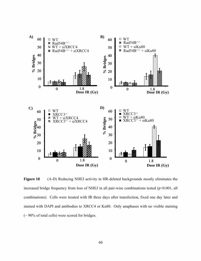
60
Figure 18 (A-D) Reducing NHEJ activity in HR-deleted backgrounds mostly eliminates the
increased bridge frequency from loss of NHEJ in all pair-wise combinations tested (p<0.001, all
combinations). Cells were treated with IR three days after transfection, fixed one day later and
stained with DAPI and antibodies to XRCC4 or Ku80. Only anaphases with no visible staining
(~ 90% of total cells) were scored for bridges.
A) B)
C)
0
10
20
30
40
50
60
0 1.80
10
20
30
40
50
60
0 1.8
WTRad54B-/-/-
WT + siKu80Rad54B-/-/- + siKu80
Dose IR (Gy)
% B
ridg
esWTXRCC3-/-
WT + siKu80XRCC3-/- + siKu80
Dose IR (Gy)
% B
ridg
es
0
10
20
30
40
50
60
0 1.80
10
20
30
40
50
60
0 1.8
0
10
20
30
40
50
60
0 1.80
10
20
30
40
50
60
0 1.8
WTRad54B-/-/-
WT + siXRCC4Rad54B-/-/- + siXRCC4
Dose IR (Gy)
% B
ridg
es
0
10
20
30
40
50
60
0 1.80
10
20
30
40
50
60
0 1.8
WTXRCC3-/-
WT + siXRCC4XRCC3-/- + siXRCC4
Dose IR (Gy)
% B
ridg
es
D)

61
Figure 19 Chicken DT40 cells also show HR-dependent induction of bridges in NHEJ
mutants. Wild type, RAD54-deficient, Ku70-deficient, or RAD54/Ku70 double-deficient DT40
cells were treated with the indicated doses of IR and scored for anaphase bridge formation. As
observed with human cells, HR-deficiency did not change bridge induction, and mutational loss
of NHEJ enhanced the bridge levels, while the HR/NHEJ double mutants showed levels of
induction similar to the parental line. Both DIC optics and DAPI staining were needed to
distinguish anaphase bridges in these cells.
DT40 (WT) Rad54-/-
Ku70-/-
Rad54-/-&Ku70-/-
0
10
20
30
40
50
60
0 0.3 0.6
Dose IR (Gy)
% B
ridg
es
DT40 (WT) Rad54-/-
Ku70-/-
Rad54-/-&Ku70-/-
0
10
20
30
40
50
60
0 0.3 0.6
Dose IR (Gy)
% B
ridg
es

62
positive for foci formation 20 minutes after IR, the staining disappeared by 24 hours, at the time
anaphase bridging is scored (Figure 20). Even though the differences between these samples
were dramatic, quantification for the presence of γ−H2AX foci was not very obvious due to the
differences in the size of the foci, which were scattered on many focal planes. Yet, approximate
scores for cells containing foci are shown in Figure 21.
Although these results suggest that DSB repair still occurred in the HR/NHEJ defective
background, breaks might simply lose γ-H2AX phosphorylation without repair. In order to
exclude this possibility, we tested other DNA double strand break markers, 53BP1 and
phosphorylated-ATM (P-ATM), which also form foci at the site of DSBs (Figures 22, 23)
(Anderson et al., 2001; Bakkenist and Kastan 2003). Once again, the majority of the foci
disappeared by 24h in every combination tested (siRNA knockdown of XRCC4 and Ku80 in
RAD54B and XRCC3 deficient backgrounds).
2.2.3 Decrease in anaphase bridges does not appear to be due to a decrease in mitotic
index
It has been previously shown that cells that are defective for Ku80, XRCC4 or XRCC3
are sensitive to ionizing radiation (Giaccia et al., 1990; Nussenzweig et al., 1997; Brenneman et
al., 2000). Moreover, cells deficient for both repair pathways exhibit reduced proliferation
(Gorski et al., 2003; Couedel et al., 2004; Mills et al., 2004). Correspondingly in our assays, if
cells with damage do not undergo mitosis, they would not form a bridge in anaphase, and this
could potentially account for the decrease in anaphase bridging in cells compromised for both
pathways.

63
Figure 20 γ-H2AX staining was transient in HR/NHEJ defective cells. RAD54B-/-/- cells
were transfected with siKu80 and monitored for the presence of γ-H2AX foci 20min or 24h after
IR treatment. Similar results are obtained for all pair-wise HR/NHEJ double deficiencies (data
not shown). The formation of foci in repair defective backgrounds was similar to the wild type
HCT116 cells. See Figure 21 for quantification.
γH2AX
untreated 20 min after 3 Gy IR 24 h after 3 Gy IR
DNA
Ku80

64
Figure 21 Cells were scored for formation of γ-H2AX foci following IR treatment. Majority
of the breaks disappear by 24h in HR/NHEJ defective cells. Similar levels of foci appearance
were observed in repair defective backgrounds compared to the wild type HCT116 cells 20min
after IR. Although both the number of foci/cell and the number of cells containing foci decreased
after 24h, the percentage of cells with γ-H2AX foci was slightly more in NHEJ/HR deficient
cells. The percentage of cells that contain foci is shown in the graph. Approximately 500 cells
were scored for each experiment.
0
10
20
30
40
50
60
70
80
90
100
untreated 20min after IR 24h after IR
WT + rdm siRNARad54B-/-/- + rdm siRNAXRCC3-/- + rdm siRNARad54B-/-/- + siKu80XRCC3-/- + siKu80Rad54B-/-/- + siXRCC4XRCC3-/- + siXRCC4
% C
ells
Pos
itive
for γ-
H2A
X fo
ci

65
Figure 22 53BP1 staining was transient in HR/NHEJ defective cells. RAD54B-/-/- cells
were transfected with siKu80 and monitored for the presence of 53BP1 foci 20min or 24h after
IR treatment. Similar results are obtained for all pair-wise HR/NHEJ double deficiencies (data
not shown).
DNA
53BP1
Ku80
untreated 20 min after 3 Gy IR 24 h after 3 Gy IR

66
P-ATM
DNA
siKu80
Merge
untreated 20 min after 3 Gy IR 24 h after 3 Gy IR

67
Figure 23 P-ATM staining was transient in HR/NHEJ defective cells. RAD54B-/-/- cells
were transfected with siKu80 and monitored for the presence of P-ATM foci 20min or 24h after
IR treatment. Both P-ATM and Ku80 antibodies were from the same species, hence a
fluorophore-labeled random siRNA was used as a cotransfection marker (third panel labeled
siKu80). Labeled siRNA is 100% associated with knockdown of Ku80 as determined on a
separate slide (data not shown), but probably underestimates the number of knocked down cells
since only accumulation of siRNA label can be detected by immunofluorescence. Arrows
indicate transfected cells. Similar results are obtained for all pair-wise HR/NHEJ double
deficiencies (data not shown).

68
In order to explore whether there is a reduction in proliferating cells, we scored mitotic
indices before and after ionizing radiation treatment. While there was a mild decrease in XRCC4
deficient backgrounds, cell division was mostly not affected despite the repair deficiencies
(Figure 24). Moreover we did not detect a reduction in cell proliferation in any background
during the course of the experiment (data not shown). These results suggest that the decrease in
anaphase bridge frequency in repair deficient cells for both pathways cannot simply be explained
by a decrease in mitotic population. Consistent with our results, XRCC3 deletion in the same
HCT116 cell line has been previously published not to cause sensitivity to ionizing radiation
(Yoshihara et al., 2004). Hence it appears that HCT116 cells lack the checkpoints required to
arrest cell division.
Since the anaphase bridges are expressed as a percentage in the total anaphase population
(number of cells scored in each category is displayed in Table 1), and the fact that mitotic index
is not affected in double deficient cells, the decrease in anaphase bridging in these cells does not
appear to be due to a decrease in the anaphase population.
2.2.4 In vitro NHEJ ligation assay
Since NHEJ clearly plays a role in the prevention of bridges, can we explain the anaphase
bridge variation in cancer cell lines by differences in NHEJ activity? We used an in vitro
ligation reaction to quantify NHEJ activity in different tumor cell lines (Diggle et al., 2003).
Linearized plasmid DNA was incubated with whole cell extracts (WCE) and religation measured
by the appearance of dimers and trimers. (The presence of WCE blocked monomeric ligation by
an unknown mechanism, as shown by comparison of T4 DNA ligase +/- WCE, (Figure 25D)).

69
Dose IR (Gy)
% C
ells
in M
itosi
s
0
1
2
3
4
5
6
7
0 3
WT + rdm siRNA
WT + siKu80
WT + siXRCC4
Rad54B-/-/- + rdm siRNA
Rad54B-/-/- + siKu80
Rad54B-/-/- + siXRCC4
XRCC3-/- + rdm siRNA
XRCC3-/- + siKu80
XRCC3-/- + siXRCC4
% C
ells
in M
itosi
s
0
1
2
3
4
5
6
7
0 3
WT + rdm siRNA
WT + siKu80
WT + siXRCC4
Rad54B-/-/- + rdm siRNA
Rad54B-/-/- + siKu80
Rad54B-/-/- + siXRCC4
XRCC3-/- + rdm siRNA
XRCC3-/- + siKu80
XRCC3-/- + siXRCC4
Figure 24 Mitotic indices for HCT116 parental cells and for repair deficient backgrounds.
HCT116 cells do not show growth abnormalities in repair defective backgrounds upon ionizing
radiation treatment. Each experiment was repeated three times and 500-600 cells were scored per
slide from five different positions on the coverslip. (rdm siRNA: random siRNA, siKu80: siRNA
to Ku80, siXRCC4: siRNA to XRCC4)

70
Table 1 Total number of coverslips and cells scored for each experiment. (WT: wild type,
def: deficiency)

71
Although this assay has been previously shown to measure NHEJ activity (Baumann and West
1998; Diggle et al., 2003), to confirm the specificity of the assay we tested various NHEJ
inhibitors.
Addition of PI3-kinase inhibitors (to block DNA-PKcs activity), or antibodies specific to
NHEJ proteins reduced ligation (Figure 25A, B). Interestingly, while siRNA to XRCC4
diminished ligation, siRNA to Ku80 did not reproducibly decrease ligation, which could be due
to residual Ku80 after knockdown (Figure 25C). Consistent with other reports, addition of Ku80
antibodies effectively reduced NHEJ activity with very little ligation (Figure 25B, lane 4)
(Baumann and West 1998). The difference could be due to residual Ku80 from the untransfected
population or incomplete siRNA knockdown. Alternatively, addition of antibodies might
interfere with the activity of other proteins that are in complex with Ku80 and result in a
reduction in the NHEJ activity. While this assay could be less sensitive to Ku80 levels, because it
was dependent on NHEJ components and we believe reflects cellular NHEJ activity. As
expected, the NHEJ activity of HR mutants was undiminished (Figure 25E).
2.2.4.1 NHEJ activity is correlated with the induction of bridges in cancer cells
We next tested for a correlation between NHEJ activity and bridge induction for eight
tumor-derived and two control cell lines (Figure 26). The RPE-hTERT normal control cell line
had very few anaphase bridges prior to treatment and arrested in interphase following IR
treatment (Figure 26A and data not shown. Total cell counts are shown in Table 2.). We
therefore utilized HEK-293, an immortalized nonmalignant cell line, for a reference. Anaphase
bridges in this line increased from ~20% to ~40% after IR exposure. Among the tumor-derived
cell lines, the frequency of both pre-existing and induced bridges varied dramatically (Figure

72
26A, 0Gy), suggesting that the pathway leading to bridge formation has varied levels of activity
in different cancer cells. The induced bridge levels are calculated as the difference of anaphase
bridge frequency between 1.8Gys and untreated levels, since some cell lines do not increase
bridge frequency at doses higher than 1.8Gys (data not shown). To compare levels, we defined
NHEJ activity as the ratio of multimers to all plasmid bands.
It appears that the NHEJ activity is generally inversely proportional to the induction of
bridges (Figure 27, p< 0.07). The relationship is not strictly linear; perhaps reflecting the genetic
variance of these independent cell lines. We find no correlation between pre-existing bridges
and NHEJ activity (data not shown). These results suggest that the level of induced bridges
depends on the NHEJ activity of the cancer cells, although the correlation is rather weak.

73
untre
ated
+WM
+ In
hII
+ In
hIII
A)
C) +ran
dom
siR
NA
+siK
u80
+siX
RC
C4
Ku80
XRCC4
γ-Tub
No
antib
ody
+α-K
u80
+α-X
RC
C4
+α-K
u70
+α-D
NA
Liga
seIV
+α-c
yclin
E
+α-H
A
B)
unligated, linear
dimer
trimer
unligated, linear
dimer
trimer
unligated, linear
dimertrimer

74
WT
RA
D54
B-/-
/-
XR
CC
3-/-
D)
Cut
Unc
ut
+T4
alon
e
+Ext
ract
alo
ne
+Ext
ract
, +T4
E)
unligatednicked circular
dimertrimer
closed circular

75
Figure 25 (A) Addition of DNA-PK inhibitors abolished ligation capability of WCEs. WM:
Wortmannin, InhII/ InhIII: DNA-PK Inhibitor II or III. Forty μg of total protein each reaction.
(B) Addition of antibodies specific for NHEJ proteins reduced NHEJ activity. (C) WCEs
prepared from cells that were transfected with siXRCC4, but not siKu80, show reduced ligation
activity. DNA gels of NHEJ ligation on top, immunoblots of WCEs below. (D) Intramolecular
ligation is inhibited by WCE. Equal amounts of WCE and T4 ligase were used in all indicated
lanes. Monomeric circles, like the supercoiled band near the bottom of the gel, can reform with
the addition of T4 ligase, but not when WCE is present. WCE also seems to suppress the total
ligation activity of T4 ligase. DNA was visualized with SYBR-Green (Molecular Probes). (E)
HR mutants have no change in NHEJ ligation efficiency.

76
RPE-hTERTHEK-293A549HCT116 HeLaUPCI:SCC103UPCI:SCC070MES-SAU2OSSK-HEP1
<1% N/A
% B
ridg
es
0
10
20
30
40
50
60
70
Dose IR (Gy)0 1.8
RPE-hTERTHEK-293A549HCT116 HeLaUPCI:SCC103UPCI:SCC070MES-SAU2OSSK-HEP1
<1% N/A
% B
ridg
es
0
10
20
30
40
50
60
70
Dose IR (Gy)0 1.8
RPE
-hTE
RT
HEK
-293
A54
9
HC
T116
HeL
a
UPC
I:SC
C10
3
UPC
I:SC
C07
0
MES
-SA
U2O
S
SK-H
EP1
Trimer
Dimer
Unligated
A)
B)

77
Figure 26 (A) Bridge induction varies between different cell lines. The indicated cell lines
were treated with IR, fixed 1 day after treatment, and stained with DAPI to visualize bridges.
The RPE-hTERT are primary retinal pigmented epithelial cells expressing human telomerase.
(B) NHEJ activity varies between different cell lines. WCEs from the indicated cell lines were
used for end joining reactions, as described in Materials and Methods.

78
Table 2 Total number of coverslips and cells analyzed for each experiment.

79
Figure 27 Correlation between NHEJ activity in vitro and induction of bridges by IR. The
ligation activity of WCEs from the cell lines in Figure 26 (except RPE-hTERT cells) was
quantified as described in the text. The median value of NHEJ for each cell line was plotted
versus the median percent increase in bridges at 1.8 Gy of IR. The values in this graph were the
median for 4-7 experiments for NHEJ activity and 4-12 experiments for bridge frequency. Each
experiment gave similar results, but the absolute values of the different data points varied
substantially from day-to-day (p<0.07).
NHEJ activity
% In
crea
se in
bri
dges
0
5
10
15
20
25
30
0 0.05 0.1 0.15 0.2 0.25
HEK-293A549HCT116HeLaUPCI:SCC103UPCI:SCC70MES-SAU2OSSK-HEP1

80
2.3 DISCUSSION
In this study, we investigated which DNA repair pathway contributes to formation of
anaphase bridges using cells compromised in NHEJ, HR or both pathways. Our data support a
role for NHEJ as an essential pathway in preventing the formation of chromatin bridges, which
lead to gross chromosomal changes. This is consistent with previous observations of
chromosomal instability in NHEJ mutants, such as non-reciprocal translocations, or deletions
(Difilippantonio et al., 2000; Gao et al., 2000; Bassing et al., 2002; Rooney et al., 2003), some of
which could be a consequence of anaphase bridges. Based on our data, HR is not required for
the formation of bridges. However surprisingly, in the absence of NHEJ, the HR pathway
apparently repairs DSBs in such a way that a chromatin bridge forms in anaphase (Figure 28).
It has been previously demonstrated that DSBs may be repaired by an NHEJ-independent
pathway resulting in formation of dicentric chromosomes and subsequent translocations and
gene amplifications in cells deficient for XRCC4 or Ligase IV (Zhu et al., 2002) or Ku80/70
(Difilippantonio et al., 2002). The authors have proposed mechanisms which involve incursion
of the DSB end into an intact template followed by break induced replication. Replication of the
broken chromatid leads to fusion of the sister chromatid ends, formation of dicentrics and cycles
of breakage-fusion-bridge. Furthermore, the authors also reported that these chromosomal
changes can occur in the absence of both Ku80 (NHEJ) and RAD54 (HR), which implies that
intact homologous recombination is not required for bridge formation in the absence of NHEJ.
However, these studies did not examine the change in the frequency of dicentrics (or

81
DNA Damage
Break
activeNHEJ
activeHR
I
restoration of original structure
inactiveHR
activeNHEJ
II
or
activeHR
inactiveNHEJ
III
duplicatechromatid
or
inactiveNHEJ
inactiveHR
IV
or
+
Another Pathway
No repair
ligation
DNA Damage
Break
activeNHEJ
activeHR
I
restoration of original structure
inactiveHR
activeNHEJ
II
or
activeHR
inactiveNHEJ
III
duplicatechromatid
or
inactiveNHEJ
inactiveHR
IV
or
+
Another Pathway
No repair
ligation

82
Figure 28 Cells with active NHEJ and HR remove DSBs without forming anaphase bridges
(I). NHEJ can still repair the breaks efficiently without bridging in the absence of HR (II).
However when NHEJ is compromised, HR repairs the damage, but forms anaphase bridges
during this repair (III). In the absence of both pathways, wild type levels of anaphase bridges are
observed, which could indicate either the presence of a third repair pathway or persistence of
breaks (IV). Our data suggest that majority of the DSBs are repaired, hence a slow acting third
pathway could act in the absence of both pathways.

83
translocations) following DNA damage. It should be noted that despite the reduction in bridging
in NHEJ/HR compromised cells, there is still a basal level of induction in bridging which could
account for the genomic aberrations observed in these studies.
The mechanism for HR-driven bridge formation in the NHEJ mutants is currently
unknown. One possibility is suggested by the observations that a reduction in NHEJ protein
levels results in a stimulation of HR-mediated repair (Fukushima et al., 2001; Pierce et al., 2001;
Allen et al., 2002; Delacote et al., 2002). The stimulation is greatest if early NHEJ proteins,
namely DNA-PKcs and Ku70/80, are diminished, and less if the late-acting XRCC4 is inhibited
(Pierce et al., 2001). Not surprisingly then, we found Ku80-deficiency leads to a higher level of
bridging compared to lack of the late-acting XRCC4 (Figure 17A-C). Moreover, studies on the
motility of free chromosome ends have indicated that immobilization of the ends requires Ku80
(personal communication with Dr. T. Misteli). This result is consistent with the increase in
bridging in the absence of Ku80, since formation of an anaphase bridge would require loss of
positional constraints for the fusion of incorrect ends. Perhaps enhanced levels of HR crossover
intermediates form in the NHEJ-deficient cells which cannot be completely resolved, thus
leading to bridges in anaphase. Further analysis will be required to test this model.
The tendency of HR to form anaphase bridges in NHEJ-deficient backgrounds may
explain why patients with increased frequencies of HR also carry elevated cancer risk and
chromosomal instability (Bishop and Schiestl 2000; Bishop and Schiestl 2001; Thompson and
Schild 2002; Bishop and Schiestl 2003; Griffin and Thacker 2004). Loss of heterozygosity,
amplifications, or translocations are genetic events common during carcinogenesis, and are

84
associated with HR. For example, in acute myleloid leukemia, duplication within the ALL-1 gene
involves recombination between Alu sequences (Schichman et al., 1994). In chronic myeloid
leukemia, the nuclear protein Bcr is translocationally juxtaposed to the Abl kinase, and has been
shown to have shared homology at the break site, possibly indicating HR as the mechanism
(Jeffs et al., 1998). A future line of investigation would be to test whether diminished NHEJ
activity is necessary for these HR-driven illegitimate recombinational changes.
Recently, attention has been focused on mice that are deficient for both HR and NHEJ
repair pathways. Derived cell lines have higher frequencies of DSBs as shown by elevated γ-
H2AX and chromosomal instability (Gorski et al., 2003; Couedel et al., 2004; Mills et al., 2004).
The reduction in bridge formation in our cell lines does not appear to be a consequence of
persistent breaks since γ-H2AX-foci disappear after IR. Moreover, other proteins involved in
DSB repair, 53BP1 and phospho-ATM, are also no longer present at the break sites. However,
we cannot discount the possibility that breaks may lose DSB repair markers without completing
repair.
While the disappearance of repair foci and the existence of bridges in the double mutants
suggest the possibility of a third pathway to repair the damage, it is also possible both NHEJ and
HR may function despite absence of these repair proteins albeit maybe with reduced accuracy or
efficiency. A third NHEJ-independent repair pathway has previously been proposed (DiBiase et
al., 2000; Wang et al., 2001; Wang et al., 2001). Whilst there is evidence that DNA ligase III and
PARP-1 might be involved in this alternative pathway (Wang et al., 2005; Wang et al., 2006), it
is not yet well characterized. The back up NHEJ path efficiently is shown remove the majority

85
of the DSBs in the absence of canonical NHEJ factors and is shown to operate with 20-30 fold
slower kinetics (Wang et al., 2001; Wang et al., 2003). This path could potentially account for
the repair we see in HR/NHEJ deficient backgrounds, since bridges are scored after 1 cell cycle
(~24h). More detailed kinetic studies of anaphase bridge induction in NHEJ/HR deficient
backgrounds, where foci disappearance rate is observed between 20min and 24h are required to
initially explore this possibility.
In our studies, we also found that intrinsic differences in NHEJ activity might explain
why cells form varying levels of bridges in response to IR. However, this correlation is not very
strong (p<0.07), possibly due to genetic differences in these cancer cells, such as aneuploidy.
Since the number of chromosomes in each cell may influence the number of bridges observed,
we obtained the karyotypes of these commonly used cell lines (from HyperCLDB and ATCC
Cell line databases and personal communication with Dr Susanne Gollin). These results indicate
that HCT116 and MES-SA cells display near diploidy, cell lines such as HEK-293, A549, HeLa,
UPCI:SCC070, U2OS and SK-HEP1 are close to triploid, and UPCI:SCC103 cells are near
pentaploid. Among these, while both HEK-293s and SK-HEP1 cells are hypotriploid, HEK-293
cells show the highest induction of bridges after IR (~26%), and SK-HEP1 cells exhibit only a
mild increase (~8%). On the other hand, the pentaploid cell line, UPCI:SCC103, shows an
intermittent value with a ~19% raise. Hence total chromosome number is not likely to be the
source for the differences in anaphase bridging, however the correlation between induction of
bridges and NHEJ activity might be weakened by this added variable.

86
3.0 CHAPTER III: ANALYSIS OF NHEJ IN INDUCTION OF ANAPHASE
BRIDGES AND END JOINING ACTIVITY IN CANCER CELLS
3.1 INTRODUCTION
As discussed in Chapter II, cancer cells which lack proper checkpoint signaling continue
cell cycle progression despite defects such as anaphase bridges. Briefly, we hypothesized that
anaphase bridging is a consequence of incorrect repair of double stranded breaks (DSBs),
defined as fusion of two non-contiguous DNA ends, and we wanted to explore which repair
pathway is involved in misfusion. Between the two major repair mechanisms in mammalian
cells, non homologous end joining (NHEJ) and homologous recombination (HR), our findings
indicated that NHEJ plays an important role in the prevention of bridges and that the intrinsic
NHEJ activity in cancer cells (as determined by an in vitro ligation assay) negatively correlates
with the induction of anaphase bridges.
In this chapter, I shall discuss our studies in determining whether overall NHEJ activity
or the expression of any one of the NHEJ proteins has a correlation with the bridging potential.
To answer this question, we examined the levels of various NHEJ proteins and compared their

87
expression to bridge induction as well as NHEJ activity. This analysis does not take into account
other changes in activity that are not caused by elevated expression.
During NHEJ mediated repair, the ligation is initiated with the recruitment of Ku80 and
Ku70 proteins to the damage site, which protect the DNA ends from degradation and allow
further processing (Smith et al., 1999). Subsequently, Ku80/70 recruits DNA Protein Kinase
catalytic subunit (DNA-PKcs), and together they form the DNA-PK complex (Dvir et al., 1992;
Gottlieb and Jackson 1993). DNA-PKcs both phosphorylates and associates with Artemis and
this complex is involved in processing the 5’ and 3’ DNA overhangs (Ma et al., 2002). Other
targets of DNA-PKcs include XRCC4 and Ligase IV, which function in the final ligation step of
NHEJ. XRCC4/Ligase IV complex seals the break along with the associated XLF protein (See
Figure 4 for illustration of the pathway) (Critchlow et al., 1997; Chen et al., 2000; Ahnesorg et
al., 2006; Buck et al., 2006).
XRCC4 is a small (38kDa), nuclear protein, which tightly binds to Ligase IV and
increases its stability (Critchlow et al., 1997; Bryans et al., 1999). This binding also enhances the
activity of Ligase IV in vitro, possibly at the adenylation step (Grawunder et al., 1997). The
deficit of XRCC4 causes sensitivity to ionizing radiation (IR), defects in proliferation, and
increased genomic instability including chromosomal translocations (Gao et al., 1998; Yan et al.,
2006). Moreover, cells which lack XRCC4 show enhanced levels of anaphase bridges after
ionizing radiation (Chapter II).

88
We report here our analysis of the NHEJ components involved in bridge formation.
Among these proteins, endogenous XRCC4 expression was correlated with resistance to
anaphase bridging. Furthermore, the levels of XRCC4 demonstrated a correlation with the NHEJ
activity of tumor cells, suggesting a limiting role of XRCC4 for NHEJ. However, elevation of
XRCC4 by itself was insufficient to prevent bridges in cancer cell lines that exhibited lower
XRCC4 levels. These results suggest that although XRCC4 appears to have an important role for
bridge formation, it is not the only factor that determines induction of bridging. Moreover,
coexpression of XRCC4 with its interacting partner, DNA Ligase IV, did not affect the increased
bridging phenotype, suggesting that factors other than XRCC4/Ligase IV are limiting in these
cells.

89
3.2 RESULTS
3.2.1 XRCC4 expression is correlated with both the induction of bridges and NHEJ
activity
To determine which NHEJ component, if any, correlates with induction of bridges,
expression of various NHEJ proteins was assessed in commonly used cancer cell lines.
Immunoblotting for DNA-PKcs did not give consistent results, possibly due to transfer
inefficiencies of this high molecular weight protein (~420 kDa) (data not shown). DNA Ligase
IV, Ku80 and Ku70 expression varied little between cell lines (Figure 29A), as reported formerly
(Kasten et al., 1999; Carlomagno et al., 2000; Merel et al., 2002). The levels of XRCC4
expression on the other hand, varied substantially (Figure 29B). Moreover, this variation was
inversely correlated with the frequency of induced bridges after IR treatment (p<0.04, Figure
30A). Next we determined the NHEJ activity of these cell lines as described previously (Chapter
II) and compared with XRCC4 expression. These results indicated that the intrinsic changes
observed in XRCC4 expression was correlated with NHEJ activity in vitro (p<0.05; Figure 30B).
3.2.2 Restoring XRCC4 in cells with low endogenous levels does not rescue the increased
bridging phenotype
To test if XRCC4 levels were limiting for the induction of bridges, we exogenously-
expressed XRCC4 in two low-expressing cell lines (HeLa and UPCI:SCC103). Overexpression

90
HEK
-293
HC
T116
HeL
a
UPC
I:SC
C10
3
MES
-SA
U2O
S
SK-H
EP1
UPC
I:SC
C07
0
A54
9
Ku70
γTubulin
Ku80
DNA Ligase IV
γTubulin
RPE
-hTE
RT
XRCC4
γTubulin
HEK
-293
HC
T116
HeL
a
UPC
I:SC
C10
3
MES
-SA
U2O
S
SK-H
EP1
UPC
I:SC
C07
0
A54
9
RPE
-hTE
RT
A)
B)

91
Figure 29 (A) Ku80, Ku70, and DNA Ligase IV-expression levels were relatively constant,
(B) while XRCC4 levels show variation among different cancer cell lines. Twenty μg of total
whole cell extracts (WCEs) (also used in NHEJ activity assays in Chapter II) were
immunoblotted and γ-tubulin was used as a loading control.

92
A)
B)
Normalized XRCC4 levels
% In
crea
se in
bri
dges
Normalized XRCC4 levels
NH
EJ
activ
ity
0
5
10
15
20
25
30
0 0.5 1 1.5 2 2.5 3
HEK-293A549HCT116HeLaUPCI:SCC103UPCI:SCC70MES-SAU2OSSK-HEP1
0
0.05
0.1
0.15
0.2
0.25
0 0.5 1 1.5 2 2.5 3
HEK-293A549HCT116HeLaUPCI:SCC103UPCI:SCC70MES-SAU2OSSK-HEP1

93
Figure 30 (A) Inverse correlation between XRCC4 levels and bridge induction. XRCC4-
expression levels were quantified using Image Guage software, normalized for γ-tubulin levels,
and the median plotted against the increase in bridges after ionizing radiation. (B) NHEJ-
dependent ligation was positively correlated with XRCC4 levels. The values in these graphs
were the median for 4-7 experiments for NHEJ activity and 4-12 experiments for bridge
frequency and 3-7 experiments for XRCC4 expression. While individual experiments gave
similar trends, there was a variation in the absolute values of experiments performed on different
days.

94
was confirmed by both immunoblotting and immunofluorescence (Figure 31A, B and data not
shown). Based on counting cells for the presence of Flag-tag staining, elevated expression of
XRCC4 did not reduce bridge formation in response to IR in either tested cell line (Figure 32).
Exogenously-expressed XRCC4 was able to suppress the elevated bridge phenotype of an
XRCC4-/- CHO mutant (Figure 33). These observations suggest that the exogenously-expressed
XRCC4 was active, but could not complement the bridging defect of the low-expressing cell
lines.
This result indicates the presence of other variables playing a role in determining bridge
frequency. To test whether this variable is DNA Ligase IV, the binding partner of XRCC4, we
coexpressed XRCC4 and Ligase IV (Figure 31A and data not shown). Transfection was
controlled by the presence of GFP cotransfection marker for Ligase IV overexpression.
However, we did not control for the activity of Ligase IV expressed from our construct.
Simultaneous expression of these proteins did not reduce anaphase bridging (Figure 32)
indicating that Ligase IV levels are not limiting in these cell lines.
While the levels of XRCC4 were correlated with bridge induction, other unknown
variables apparently influence bridge frequency, since restoring XRCC4 levels cannot rescue
increased anaphase bridging. Thus, we believe XRCC4 is a potentially useful marker for
anaphase bridges from DNA damage, but is not limiting bridge formation in the tested cancer
cell lines.

95
α-DNA Ligase IV
α -Flag
α−XRCC4
α−γTubulin
A)
Vector
alon
eXRC
C4-F
lag +
Liga
seIV
XRCC4
-Flag
alon
eLi
gase
IV al
one
B)
DNA XRCC4-Flag
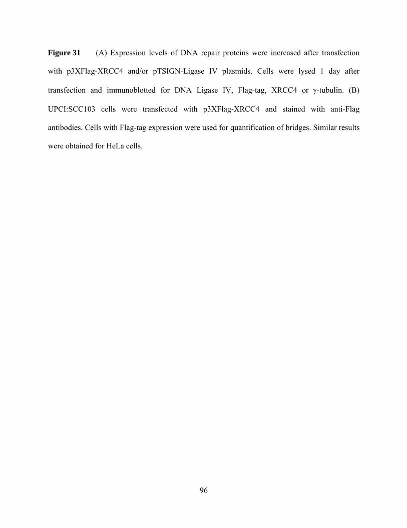
96
Figure 31 (A) Expression levels of DNA repair proteins were increased after transfection
with p3XFlag-XRCC4 and/or pTSIGN-Ligase IV plasmids. Cells were lysed 1 day after
transfection and immunoblotted for DNA Ligase IV, Flag-tag, XRCC4 or γ-tubulin. (B)
UPCI:SCC103 cells were transfected with p3XFlag-XRCC4 and stained with anti-Flag
antibodies. Cells with Flag-tag expression were used for quantification of bridges. Similar results
were obtained for HeLa cells.

97
UPCI:SCC 103
0
10
20
30
40
50
60
0 1.8Dose IR (Gy)
% B
ridg
es
Vector aloneXRCC4-Flag aloneLigase IV alone
XRCC4-Flag + Ligase IV
HeLa
0
10
20
30
40
50
60
0 1.8Dose IR (Gy)
% B
ridg
es
Vector aloneXRCC4-Flag aloneLigase IV aloneXRCC4-Flag + Ligase IV
A)
B)

98
Figure 32 Overexpression of XRCC4 does not rescue the bridge induction phenotype. Both
UPCI:SCC103 (A) and HeLa (B) cells were transfected with p3XFlag-XRCC4 alone, pTSIGN-
Ligase IV (with H2B-GFP marker) or both plasmids. Cells were scored for their bridge forming
potential based on either the presence of Flag-tag or the cotransfection marker.

99
Figure 33 Overexpression of XRCC4 corrected the increased bridging phenotype in
XRCC4-/- CHO cells. XR-1 (XRCC4-/- CHO cell line) cells were transfected with p3XFlag-
XRCC4 and were scored for their bridge forming potential based on the presence of Flag-tag.
XR-1 TR: XR-1 cells stably transfected with wild type XRCC4 gene.
01020304050607080
0 1.8Dose IR (Gy)
% B
ridg
es
XRCC4-/- (XR-1)
XRCC4-/- + XRCC4-Flag
XRCC4 + (XR-1 TR)
01020304050607080
0 1.8Dose IR (Gy)
% B
ridg
es
XRCC4-/- (XR-1)
XRCC4-/- + XRCC4-Flag
XRCC4 + (XR-1 TR)

100
3.3 DISCUSSION
In this study, we tested whether intrinsic genetic differences can explain why cells form
various levels of bridges in response to ionizing radiation. Our first candidates were NHEJ
proteins, since our previous studies suggested a preventative role for NHEJ in anaphase bridging
(Chapter II). Because there is no direct quantitative analysis for activity for each of the
components, we assayed for the expression of NHEJ proteins. Among the NHEJ players tested,
Ku80, Ku70, and DNA Ligase IV demonstrated similar expression between different cell lines,
while XRCC4 levels displayed variation. A variation in NHEJ proteins in cancer cells had
previously been of interest to other laboratories for various reasons. In agreement with our
findings, both Ku70 and Ku80 levels appeared to be similar, and differences in XRCC4/Ligase
IV levels were detected, although different cell lines were used in these studies (Kasten et al.,
1999; Carlomagno et al., 2000; Merel et al., 2002). However, these groups failed to find
correlations between expression of proteins and the phenotypic differences such as
radiosensitivity of the patients or in vitro ligation activity. Here we report that the reduced
expression of XRCC4 is correlated with the induction of anaphase bridging. Moreover, the
ligation capacity of the cancer cells also appears to positively correlate with XRCC4 levels.
If XRCC4 was the only factor playing a role in determining the tendency to form bridges,
restoring XRCC4 levels in cell lines with low endogenous expression would be expected to
rescue the increased frequencies of bridging. However, increasing XRCC4 expression alone does
not significantly change the induction of anaphase bridges, indicating that XRCC4 probably acts

101
with other factors in this process. Coexpression of XRCC4 with a potential candidate factor,
DNA-Ligase IV, did not change resistance to bridging suggesting DNA Ligase IV is not limiting
for these cells. Interestingly, the interaction between XRCC4 and Ligase IV has been reported to
affect the stability of Ligase IV and low XRCC4 expression corresponded to low Ligase IV
levels (Bryans et al., 1999). The authors demonstrated that increasing XRCC4 levels in these cell
lines stabilizes Ligase IV and enhances its expression (Bryans et al., 1999). In contrast to
previous reports, the change in XRCC4 expression did not correlate with the levels of DNA
Ligase IV, but different cell lines were used in these studies. These results suggest that cancer
cells might have alternative mechanisms to stabilize Ligase IV.
Based on assays using nuclear extracts, it has been previously suggested that
XRCC4/Ligase IV complex is rate limiting for NHEJ (Nick McElhinny et al., 2000; Huang and
Dynan 2002). This study further suggests that a natural correlation exists for the intrinsic
expression of XRCC4 and the intrinsic ligation capacity of the extracts supporting a limiting role
for this protein. This is in contrast to Merel et al. (2002) who found that XRCC4 levels did not
correlate with the in vitro NHEJ activity from different breast cancer lines. The differences
between the studies may be due to different methodology or different source material for the
malignant lines.
NHEJ factors are potential tools both as diagnostic markers and targets for chemotherapy
and radiotherapy, since deficiencies in these proteins factors confer sensitivity to DNA damaging
agents. Among the NHEJ proteins, XRCC4 has been previously demonstrated to acquire
mutations in cancer cells and small nucleotide polymorphisms (SNPs) in the XRCC4 gene have

102
been associated with elevated cancer risk (Fu et al., 2003). Moreover, XRCC4 has recently been
proposed as a diagnostic tool in non-BRCA1/2 heritable breast cancer cases (Allen-Brady et al.,
2006). Based on our studies, among the tested NHEJ proteins, XRCC4 seems to be correlated
with anaphase bridging, which is additional justification as a testing its importance for diagnostic
tool.

103
4.0 CHAPTER IV: THE APOPTOTIC PATHWAYS AND THE ABILITY TO SENSE
DNA DAMAGE ARE INTACT IN ORAL CANCER CELLS WITH HIGH ANAPHASE
BRIDGES.
4.1 INTRODUCTION
Cancer development is a consequence of DNA damage. Normal cells with intact
checkpoints and repair machinery either arrest until the damage is corrected or undergo
programmed cell death if the damage exceeds the cells’ repair capacity. On the other hand,
cancer cells continue proliferation despite the existing defects and frequently are resistant to
apoptotic stimuli.
Most of the current cancer therapies, including chemotherapy and radiation treatment, are
based on triggering apoptotic pathways and selective death of cancer cells. Once the decision to
die is made, several morphological and biochemical changes occur. Commonly, these can be
characterized as fragmentation of DNA, a change in cell size, membrane blebbing, or
chromosomal condensation (Hengartner 2000). Other intracellular changes include, but are not
limited to, induction of tumor suppressor proteins such as p53, activation of caspases, inhibition

104
of both Bcl-2 family proteins and inhibitor of apoptosis proteins (IAPs) (Fulda and Debatin
2006).
While most therapies are based on killing cells by apoptosis, cancer cells may have
disturbed apoptotic pathways, which contribute to failure of the treatment. These disturbances
could be at various levels, including overexpression of anti-death proteins, inactivation of the
proteins involved in apoptotic signaling pathways or inability to sense damage initially. In
support of this, cancer cells which cannot undergo apoptosis have been reported in the literature
(Zhao et al., 1997; Alam and Ratner 2001). However, apoptosis is not the only form of cell death
and these cells can still be effectively eradicated through other means, involving necrosis, mitotic
catastrophe, or autophagy (Abend 2003; Brown and Wilson 2003; Okada and Mak 2004).
A characteristic element of anti-tumor strategies that trigger apoptosis is combining
different drugs and killing cancer cells selectively with minimal damage to rest of the organism.
Therefore, various approaches have been taken in order to increase the cytotoxicity of the
damaging reagents and tendency of cancer cells to respond with apoptosis. For example, mutual
action of S-phase specific poisons and irradiation has been shown to sensitize cells to apoptosis
(Gorczyca et al., 1993). Moreover, triggering cell differentiation following DNA damage
increases programmed cell death and has been proposed as another anti-tumor strategy (Del Bino
et al., 1994; Darzynkiewicz 1995). Anti-apoptotic proteins, such as Bcl family members, have
been attractive targets for cancer therapy. Consequently, small, membrane permeable chemicals
inhibiting the activity of anti-apoptotic proteins have been identified and combining this
treatment with chemotherapy has been shown to increase cell death (Wang et al., 2000; Real et

105
al., 2004). All of these strategies have taken numerous facts about cancer cells into consideration.
Cancer cells undergo several rounds of cell division and have inactive checkpoints, which make
them more vulnerable to selective killing upon damage in S-phase, especially given that normal
cells are arrested in G1. In many cases, tumor cells may be de-differentiated and make extensive
amounts of anti-death proteins and become highly sensitive to apoptosis when the conditions are
reversed.
As described in the previous chapters, anaphase bridging is a common phenomenon in
cancer cells. We hypothesized that bridging occurs as a result of erroneous repair of the broken
ends by fusion of two wrong ends. Consequently, cells escape from the inhibitory effects of
DSBs since there are no further telomere-free DNA ends. Hence anaphase bridging can be
considered a method of uninterrupted cell cycle progression without undergoing arrest or
apoptosis. If this is true, a combined therapy of induction of DSBs by radiation treatment, and
blocking repair proteins involved in bridging could serve as another potential strategy to increase
apoptosis in tumors with high frequencies of anaphase bridges.
With this idea in mind, initial studies were performed in order to test whether the
apoptotic pathways are intact in cancer cells which exhibit high levels of bridges following DSB
induction. UPCI:SCC103 and UPCI:SCC040 cell lines were subjected to H2O2 treatment and
assayed for their ability to sense DNA damage, induce proteins involved in cell death signaling
and execute apoptosis.

106
4.2 RESULTS
4.2.1 Oral cancer cells can sense DNA damage
UPCI:SCC103 and UPCI:SCC040 cells were treated with H2O2 and the occurrence of
DSBs was confirmed by the detection of TUNEL positive cells (data not shown). DSB marker γ-
H2AX was used to observe cellular response to DNA damage via formation of foci. Cells were
fixed 30min and 1h after H2O2 treatment (Figure 34). Both cell lines showed intense γ-H2AX
foci after induction of DSBs at these time points, suggesting that pathways involved in sensing
DNA damage are intact.
4.2.2 Cancer cells show induction of p53 and change in subcellular localization following
DNA damage
p53 has been often considered as the “guardian of the genome” as it plays an important
role in making the decision between cell cycle arrest and cell death in response to DNA damage
and various cellular stresses. It acts as a signal transducer and results in changes in the
expression level of hundreds of genes via its ability to function as a transcription factor. These
genes include apoptotic genes such as BAX, Bcl-L, FAS1, and genes involved in cell cycle
regulation such as p21 WAF1/CIP1, GADD45, CyclinD1 or MDM2, which negatively regulates
p53 activity (reviewed in (Tokino and Nakamura 2000)).

107
Figure 34 Cancer cells appear to be able to sense the DNA damage as judged by γ-H2AX
foci formation. UPCI:SCC103 cells were treated with 4mM H2O2 for 10min, and fixed either
30min or 1h after treatment and stained for γ-H2AX and DNA. Foci formation was observed in
almost all cells.
DAPI
γH2AX
0 min 30 min 1h

108
p53 activity can be modulated by different mechanisms: an increase in levels, either by
enhanced half life or by new transcription, post-translational modifications such as
phosphorylation, acetylation, ubiquitination, cis/trans isomerization, neddylation, sumolytion,
methylation, ribosylation and O-glycosylation events or a change in subcellular localization from
the cytoplasm to the nucleus (Liang and Clarke 2001; Lacroix et al., 2006).
During the cell cycle, in normal cells, p53 is located mostly in the cytoplasm through G1,
translocates into the nucleus in G2/ early S, and shuttles back to the cytoplasm in late S
(Shaulsky et al., 1990). MDM2 protein, which binds to and promotes p53 degradation by
ubiquitination (Haupt et al., 1997), is involved in transportation of p53 from the nucleus to the
cytoplasm (Roth et al., 1998; Tao and Levine 1999). Upon damage, MDM2 is sequestered in the
nucleolus, which frees p53 to activate its target genes and p53 accumulates in the nucleus (Tao
and Levine 1999).
Mutations affecting p53 function are very frequent in cancer cells. Indeed, loss of p53 is
correlated with drug resistance and predisposes mice to tumor formation (Attardi and Jacks
1999; Wallace-Brodeur and Lowe 1999). Hence, we assayed for p53 activity and localization in
UPCI:SCC103 cells. While p53 levels increased only slightly upon treatment with H2O2, there
was significant augmentation of p21 levels, which is a target of p53 (Figure 35). Interestingly,
p53 appeared to be sequestered in a subnuclear area in all untreated cells. After DNA damage,
p53 was distributed diffusely in the nucleus and cytoplasm (Figure 36). While the intensity of
immunofluorescence staining for p53 increased after treatment, the enhancement was mostly

109
detected in the cytoplasm. These results suggest that p53 may be active; however we cannot
totally argue that it is fully functional, since the increase observed in its expression is not very
compelling and p21 is only one of the many targets of p53. More experiments testing p53
regulations have to be performed to test its activity and the genomic DNA be sequenced.
4.2.3 Oral cancer cells can undergo apoptosis upon DNA damage
Next we tested the ability of these cells to mediate apoptosis. As mentioned previously,
one characteristic feature of programmed cell death through apoptosis is DNA fragmentation,
which can be detected as formation of DNA ladders following gel electrophoresis. Cells were
treated with H2O2 and collected after 3, 6 and 9 days. It is important to note that the
concentration of H2O2 used in this assay was 10 times higher than what was used to induce
anaphase bridges.
We observed that H2O2 treatment under these conditions can lead to apoptosis starting
from 3 days after exposure (Figure 37). Cell death mostly occurred in cells collected from the
floating population and to lesser extent, at 9 days after treatment, in the attached cell population
(Figure 37, lane 8). This result indicates that at least some of the oral cancer cell lines tested in
this study can undergo apoptosis. However, there is also an apoptosis resistant population, which
is indicated by no ladder formation in the attached cells. It will be interesting to see whether
these cells can be sensitized further when repair proteins involved in bridge formation are
blocked.

110
Figure 35 While p53 levels are only slightly increased upon H2O2 treatment, p21 induction
is more pronounced. UPCI:SCC103 cells are subjected to 4mM H2O2 and lysed in RIPA buffer
24h after treatment. 100μg of total cell lysate is used for immunoblotting for p53 and p21. Actin
is used for a loading control.
0h
- +-
24hTime
H2O2
p53
actin
p21

111
0
p53
DAPI
12h 24h
+H2O2
-H2O2
p53
DAPI
Hours After H2O2Treatment

112
Figure 36 UPCI:SCC103 cells were treated with 4mM H2O2, fixed at indicated times and
stained with anti-p53 antibodies. Upon H2O2 treatment, subcellular localization of p53 was
predominantly nuclear.

113
Figure 37 UPCI:SCC103 and UPCI:SCC040 cells are treated with 4mM H2O2, lysed at
indicated times after treatment. The genomic DNA is isolated and electrophoresed on agarose
gels. Apoptotic DNA ladder formation is observed mostly in floating cells.
M 0 3 7 9 3 7 9 M 0 3 7 9 3 7 9
A F A FUPCI:SCC40 UPCI:SCC103
Days After H2O2Treatment
DNA ladder formation
A: Cells that are Attached on the plate
F: Cells that Float

114
4.3 DISCUSSION
This chapter summarizes my preliminary studies on the apoptotic capacity of cancer cells
with high bridge frequencies. We hypothesized that anaphase bridging may allow cells to bypass
apoptotic pathways that are activated in response to DNA damage and continue cell cycle
progression. Our initial data indicate that these cell lines can sense DNA damage and undergo
apoptosis, yet further experiments will be needed to test this hypothesis.
Our results demonstrated that while the cell lines under analysis were capable of DNA
ladder formation, a subpopulation of cells was still attached with very little cell death. This could
either indicate that cells are still resistant to apoptosis or that the dose of H2O2 used was not
sufficient to trigger apoptosis. Similar experiments using control cells with normal checkpoints
and apoptotic pathways should be able to resolve this question. Moreover, as mentioned
previously, H2O2 treatment both leads to and inhibits apoptosis through mechanisms other than
damage to DNA (Lin et al., 2004; Shin et al., 2004; van Rossum et al., 2004; Franklin et al.,
2006). Since our focus is apoptosis as a result of DNA damage, confirming these results by using
other means, such as ionizing radiation would strengthen these conclusions. Lastly, whilst there
is an increase in p53 and p21, there could still be mutations elsewhere in the apoptotic pathways.
Assays for caspase activation or cytochrome c release from mitochondria would provide further
evidence for the integrity of cell death signaling in these cancer cells.

115
Assuming intact apoptosis, which repair proteins should be blocked in order to test our
hypothesis? While we collected significant information on how bridges form, unfortunately the
induction of bridges was not completely eliminated in any of the DNA repair mutants. Even in
the absence of both pathways, although the level of bridging was reduced compared to NHEJ
mutants alone, the induction was still at wild type levels, which suggests the existence of a third
unknown pathway contributing to bridge formation. Hence the aim of testing our hypothesis by
inhibiting anaphase bridge formation is not as straightforward as we initially assumed. Based on
what we know about bridges, we can speculate that the double deficient cells or wild type cells
will undergo apoptosis more frequently than NHEJ deficient cells, since they form fewer bridges
in response to damage. However, such an experiment relies on the assumption that repair
deficiencies will only change anaphase bridge frequency and will not interfere with any of the
apoptotic pathways. Yet, there appears to be a cross-talk between both repair mechanisms and
apoptotic signaling. For example, physical interactions have been reported between p53 and the
HR proteins, RAD51 and RAD54 (Linke et al., 2003). HR rates increase in the absence of p53
and this p53 mediated limitation of HR is thought to be through p53-RAD51 interactions (Linke
et al., 2003). More recent studies demonstrated that the proapoptotic proteins, Bax and Bid, can
inhibit HR (Dumay et al., 2006). Furthermore, overexpression of RAD51 provides increased
resistance to ionizing radiation (Vispe et al., 1998) and elevated levels of RAD51 have been
observed in cancer cells, which might contribute to their insensitivity to apoptotic stimuli
(Raderschall et al., 2002). As for NHEJ, similar associations have been observed. Both Ku and
DNA-PKcs have been demonstrated to regulate apoptosis, but not cell-cycle arrest (Wang et al.,
2000; Sawada et al., 2003). Deficiencies in NHEJ or HR result in apoptosis, and defects in both
pathways are associated with more cell death than either one alone (Barnes et al., 1998; Gao et

116
al., 1998; Raderschall et al., 2002; Couedel et al., 2004). These observations are consistent with
our hypothesis suggesting a role for anaphase bridging in prevention of apoptosis. The increased
apoptosis phenotype observed in double deficient cells could partially be due to the decrease in
the frequency of bridges. Yet, these experiments should be performed where the expected
increase in apoptosis and decrease in bridges are confirmed in the same set of experiments.
Lastly, a comparison of cell death rates between cell lines with low and high levels of
bridge induction could be informative; however other differences in genetic backgrounds would
have to be considered during evaluations.

117
5.0 CHAPTER V: EVIDENCE FOR CENTROSOMAL SPLITTING AND
MULTIPOLAR SPINDLE FORMATION AS A RESULT OF IONIZING RADIATION
5.1 INTRODUCTION
It has been commonly accepted that genomic instability and cancer are closely related
phenomena. Almost every type of cancer has previously existing and ongoing rearrangements in
the genome. These chromosomal alterations can arise from numerous events including errors
during cell division and cell cycle progression (Charames and Bapat 2003). Of these errors,
segregation defects such as multipolar spindles (MPS) play a major role in chromosomal
instability, leading to tumor development.
MPS can be described as abnormal mitotic figures observed when cell division takes
place with more than two poles (Figure 7). Multipolarity is a commonly observed defect in
cancer cells and is mainly associated with supernumerary centrosomes (Saunders 2005;
Stewenius et al., 2005). Increase in centrosome number is reported in many carcinomas including
breast, lung, brain, gall bladder, bone, pancreas, colorectal, head and neck cancers (Lingle et al.,
1998; Pihan et al., 1998; Weber et al., 1998; Carroll et al., 1999; Lingle and Salisbury 1999; Sato
et al., 1999; Gustafson et al., 2000; Kuo et al., 2000; Pihan et al., 2001; Sato et al., 2001). Hence
centrosomal amplification and formation of aberrant mitotic spindles have been proposed to be

118
involved in tumorogenesis, as these defects can lead to missegregation of chromosomes and
genomic instability (Doxsey 1998).
Centrosomes are the microtubule organizing center (MTOC) of the cells, which play an
important role in cell-cycle progression. Centrioles, which are small barrel shaped structures and
the pericentriolar material (PCM), amorphous protein mass surrounding the centrioles, constitute
the major elements of centrosomes. Each centriole is composed of nine longitudal microtubule
triplets and proteins such as centrin, cenexin and tektin (Rieder et al., 2001). On the other hand,
the PCM contains proteins involved in microtubule nucleation and anchoring such as γ-tubulin,
pericentrin and ninein (Rieder et al., 2001).
Normally, centrosome duplication starts after the G1 checkpoint and maturation of the
centrosomes continues until the next cell cycle (Dictenberg et al., 1998; Adams and Kilmartin
2000; Khodjakov et al., 2002). During prophase, centrosomes begin migrating to opposite ends
of the cells and mitotic spindles form. After cytokinesis, each cell receives only one centrosome.
This is a carefully controlled process, since precise duplication ensures bipolar spindle formation
and proper segregation of chromosomes.
An increase in centrosome number can be a consequence of over-replication of
centrosomes, failure of cell division or cell-cell fusion (Nigg 2002). While it is not currently
known which mechanism contributes most in tumorogenesis, the final outcome of all these
events is the tendency to form multipolar spindles (Lingle et al., 1998; D'Assoro et al., 2002;
Lingle et al., 2002).

119
In Chapter I, the correlation between multipolar spindles and anaphase bridges was
discussed. In an attempt to find whether there is a direct causal relationship between bridging and
multipolarity, we performed experiments where either defect was induced and asked whether the
frequency of the other segregation defect changed in parallel. While we failed to find a direct
cause and effect relationship between these abnormalities, our studies indicated that ionizing
radiation (IR), but not H2O2, leads to formation of MPS in human embryonic kidney cells. These
preliminary studies demonstrated that IR results in an increase in multipolarity as fast as 30
minutes after treatment. Moreover, this effect appears to be through centrosomal
splitting/fragmentation as judged by centrosomal markers such as γ-tubulin and centrin-2.

120
5.2 RESULTS
5.2.1 DNA damage by ionizing radiation, but not H2O2 increases MPS formation
Anaphase bridges can be induced via either H2O2 or ionizing radiation, possibly due to
the double strand breaks induced by these agents. To test whether another segregation defect,
multipolar mitosis, which has a strong positive correlation with anaphase bridges in cancer cells
(Gisselsson et al., 2002 117) can be induced by DNA damage as well, we treated human
embryonic kidney (HEK-293) cells with both of these agents.
Upon H2O2 treatment, while anaphase bridges were induced as expected (data not
shown), we failed to see an elevation in the level of MPS (Figure 38). On the other hand, IR
treatment did show a slight, but notable and reproducible induction in multipolarity (Figure 39).
Consistent with our results, previous reports have suggested a role for gamma-irradiation on
centrosome overduplication (Scott and Zampetti-Bosseler 1980; Sato et al., 2000; Sato et al.,
2000). These studies have demonstrated that occurrence of supernumerary centrosomes depends
on p21Waf1/Cip1 and p27 (Sato et al., 2000; Sugihara et al., 2006) and that in some cell lines,
centrosome amplification appears to increase only in p53 deficient backgrounds (Armit et al.,
2002; Kawamura et al., 2006). There is also evidence showing that Kruppel-like transcription
factor, KLF4, prevents centrosome overduplication following IR by transcriptionally suppressing
expression of cyclinE (Yoon et al., 2005). Yet, how DNA damage causes an increase in
centrosome number/multipolarity is not very clear.

121
Figure 38 Scores for multipolar spindles in HEK-293 cells following H2O2 treatment. Cells
were treated with 1.6mM of H2O2 for 10 minutes, changed to regular medium, fixed at indicated
time points post-treatment, and scored for MPS by γ-tubulin staining and DNA shape.
Multipolar spindles after H2O2
0102030405060708090
100
0 30min 1h 24h
Time after H2O2 treatment
%
Bipolar spindlesMultipolar Spindles
Multipolar spindles after H2O2
0102030405060708090
100
0 30min 1h 24h
Time after H2O2 treatment
%
Bipolar spindlesMultipolar Spindles

122
Figure 39 Multipolarity in HEK-293 cells following ionizing radiation. Cells were treated
with 6Gys of IR, and fixed at indicated time points after treatment. MPS were scored by γ-
tubulin staining and DNA shape.
Induction of Multipolar Spindles after IR
0102030405060708090
100
0 30mins 1hr 24hrsTime after IR
%
Bipolar spindles
Multipolar Spindles

123
5.2.2 MPS formation might be a result of centrosomal splitting after ionizing radiation
Interestingly, in our experiments the induction in multipolarity was detectable as early as
1h, but not 30min (Figure 39). The time frame for this increase suggests a cell cycle independent
pathway, since MPS can be observed before the completion of one cell cycle (~24h). In order to
further explore the mechanism of MPS formation in response to IR, cells were stained with
common spindle pole markers, such as γ-tubulin and centrin-2, which are both centrosomal
proteins found in PCM and on centrioles respectively (Rieder et al., 2001).
Two distinct γ-tubulin spots were observed at the spindle poles in untreated, control cells
which represents the normal state (Figure 40). However, the staining appeared disperse and/or
broken up upon IR treatment (Figure 40). Likewise, the majority of the cells (>60%) have two
centrin-2 foci associated with each centriole at the poles in untreated cells (Figure 41, 42, blue
bars (2+2)). Interestingly, cells with normal numbers of centrin foci were reduced (~40%) after
ionizing radiation (Figure 41). The shift towards greater numbers of centrin foci occurred 30min
following IR (Figure 42, red bars), but was reduced 1h after treatment, at the time when an
increase in multipolar spindles was observed (Figure 42, yellow bars). Moreover, the size of
these spots was irregular, which is consistent with centrosomal fragmentation (Figure 41,
enlarged views).
Although this data is very preliminary, it suggests that ionizing radiation may cause
centrosomes to fragment or split, as early as 30 minutes after treatment. Fragmented/split

124
centrosomes may behave like extra copies of centrosomes and perhaps nucleate new
microtubules and eventually lead to abnormal spindles. Correspondingly, multipolar mitosis was
seen at 1h, when there was a reduction in the centrin foci/pole. It can be speculated that this
reduction was due the appearance of new multipolar spindles (Figure 43).
Other than centriolar fragmentation, this shift could also arise from displacement of
centrin-2 from the centrioles, or de novo formation of centrin-2 at the poles. These possibilities
will be covered further in the discussion.

125
DAPI
γ-Tubulin
Before IR After IR
Figure 40 γ-tubulin staining in HEK-293 cells before and after IR. The arrows indicate the γ-
tubulin at the poles. γ-tubulin staining becomes disperse or split at the spindle poles after
ionizing radiation.

126
Figure 41 Centrin-2 staining at the spindle poles before and after IR in HEK-293 cells.
Normally, cells have 2 centrin spots per pole in unradiated cells. Upon radiation treatment
multiple dots can be observed. Scale bar represents 10 microns. Insets represent enlarged centrin-
2 dots at the poles. Images are composed of the stack of focal planes, hence all foci are in focus.
Before IR After IR

127
Figure 42 Number of centrin-2 spots/pole in HEK-293 cells following ionizing radiation
treatment. Cells were treated with 6Gys of IR, fixed at the indicated time points, and stained for
centrin-2 and DNA. Number of centrin-2 dots at each pole was counted for bipolar metaphases.
(2+2) indicates 2 centrin dots at each pole, which is the normal state. ( 2+>3) indicates two dots
at one pole and greater than 3 dots at the other pole. Likewise, (3+>2) indicates 3 dots at one
pole and greater than 2 dots on the other. Ionizing radiation treatment can result in abnormal
numbers of centrin-2 foci 30min following IR, which is reduced by 1h.
Centrin-2 spots after IR treatment
0
10
20
30
40
50
60
70
80
1+=<
2
2+2
1+>2
2+3
2+>3
3+>2
4+>4
5+>5
7+>7
%
0 Gy IR 6 Gys IR, 30min fix6 Gys IR, 1hr fix
# of centrin-2 spots

128
Ionizing Radiation
30min
1h
+

129
Figure 43 Normally, cells exhibit two centrin-2 foci at each pole at the metaphase plate.
Upon irradiation, supernumerary centrin-2 foci is detected 30min following IR. On the other
hand, an increase in multipolar spindles is observed 1h after treatment, which coincides with a
drop in the number of centrin-2 spots.

130
5.3 DISCUSSION
This chapter summarizes the preliminary studies on how multipolar spindles form in
response to ionizing radiation. Our initial data indicated that IR can cause splitting of
centrosomal markers, namely γ-tubulin and centrin-2, as early as 30min after treatment and result
in multipolarity at 1h. Yet, other DNA damaging reagents, such as H2O2, do not lead to MPS. It
is important to note that while a link between IR and MPS was previously reported, these
changes were known to occur 24h after IR and cell-cycle dependent mechanisms were under
investigation (Sato et al., 2000). On the other hand, we detected an increase immediately after IR
and our data are reminiscent for a role of IR in centrosome fragmentation or splitting rather than
cell cycle changes or failure of cytokinesis. However, these results are not yet conclusive, and
additional experiments need to be performed to study the mechanisms leading to multipolarity as
a result of ionizing radiation.
Firstly, it has been demonstrated that IR can induce a transient change in cellular free
[Ca2+] concentration (Todd and Mikkelsen 1994) and that these rapid calcium fluxes can be
detected at 30sec (Lyng et al., 2001). Moreover, self assembly of centrin-2 has been shown to be
dependent on calcium (Tourbez et al., 2004; Yang et al., 2006) and at high [Ca2+] levels, large
centrin spots form at or near the center of the cell (Baron et al., 1994). Hence extra numbers of
centrin foci could be explained by formation of new centrin satellites due to increased [Ca2+]
concentrations as a consequence of IR. Alternatively, centrin-2 could be displaced from the

131
centrioles or de novo centrioles might truly start emerging, which all would lead to an increase in
centrin spots at metaphase.
In order to distinguish between these possibilities, other centrosomal markers could be
tested for the togetherness of staining at the poles following IR. Among the possible candidates,
dynein, dynactin, ninein, c-Nap1, Nek-2, EB1 or APC are all known to focus at the metaphase
poles (Smith et al., 1994; Gaglio et al., 1996; Berrueta et al., 1998; Quintyne et al., 1999; Helps
et al., 2000; Mayor et al., 2000; Mogensen et al., 2000) and alterations in the staining of these
proteins could potentially argue against both calcium dependent formation of centrin foci or
displacement of centrin from the centrioles, yet still would not be conclusive. On the other hand,
de novo centriole formation could be tracked by live cell imaging using cells expressing centrin-
GFP. While newly forming centrioles should emerge as a new spot, fragmentation of centrioles
would appear as splitting of an already existing signal. However, there are some caveats
associated with this experiment as well. First, it is not possible to irradiate cells under the
microscope and centrosome splitting is observed soon after IR treatment. Hence events under
observation may be missed by the time the experiment is set up. Second, centrin-GFP expression
is usually low and the signal is weak. Moreover, centrin spots are rarely on the same focal plane
and merging multiple planes increases the background making the results harder to interpret. To
resolve these problems, electron microscopy could be used to analyze centrioles following
radiation. While electron micrographs would be sufficient to distinguish among fragmentation,
splitting or de novo formation of centrosomes, the sample size is usually a limiting factor.
Therefore, a combination of these experiments will hopefully be able to enlighten our view about
how supernumerary centrosome-like structures form upon IR.

132
Lastly, this experiment was performed in HEK-293 cells and normal number of centrin
spots (2+2) was observed in only ~65% of the population (Figure 42, blue bars). This percentage
is lower than the published literature (85-90%) (Salisbury et al., 2002), suggesting that there
might be some intrinsic abnormalities in this cell line. It has been shown that in some cells IR
treatment does not result in an increase in MPS, unless p53 levels are reduced using siRNA
(Kawamura et al., 2006). Yet, we observe an elevation immediately after irradiation without any
other treatments. Hence, it is important to repeat these experiments using normal cells, which do
not exhibit any existing centrosomal defects.

133
6.0 CHAPTER VI: SUMMARY AND SPECULATIONS
Carcinogenesis requires multiple changes in the genome. It is estimated that at least 5-6
mutations should take place for a normal cell to become cancerous (Lengauer et al., 1997). Yet,
the process of mutagenesis is slow and how it leads to cancer is determined by many factors.
Less than 1.5% of the human genome encodes for proteins (Lander et al., 2001), and even when
a DNA damaging event takes place, the majority of these mutations will occur in non-coding
regions. Within the 1.5% coding region, only a minority of genes are involved in cell cycle
regulation, and only mutations in these genes will actually be relevant to carcinogenesis.
Furthermore, not all types of changes will lead to tumor formation, even if they occur in genes
controlling the cell cycle. For instance, alterations cannot be silent mutations and should trigger
the activity of oncogenes and often must deactivate both copies of the tumor suppressor genes.
On the other hand, the cell also has defense mechanisms to correct these changes. Repair
machinery actively removes lesions from the DNA. Cells either arrest or undergo apoptosis upon
damaging events. Moreover, the immune system efficiently kills cancerous cells (Burnet 1965),
as shown by the observation that organ transplant recipients with suppressed immune system
carry a 65-fold increased risk of squamous and basal cell carcinoma (Jensen et al., 1999). Yet,
despite the strong odds against it, cancer accounts for almost a quarter of all deaths in USA,
exceeded by only heart diseases. As of 2005, the risk of an American man developing cancer
over his lifetime is one in two, and one in three for women (results obtained from American

134
Cancer Society). So how is it possible that a single cell undergoes sufficient number of changes
that lead to tumor formation? It is almost certain that mutations that allow further changes in the
genome will speed up this process and lead to a hypermutable state (Nowell 1976). Among these
alterations, defects in the DNA repair machinery and events that result in gross chromosomal
changes could be expected to play a major role in tumorogenesis.
There are approximately 10,000 lesions that occur spontaneously in a mammalian cell per
day (Lindahl 1993). While these lesions are efficiently removed, failure to do so can result in a
catastrophic outcome. For instance, individuals with deficient nucleotide-excision repair (NER),
which is only one of the several DNA repair pathways (xeroderma pigmentosum patients), are
5,000-fold more likely to have squamous and basal cell carcinoma and carry 2000-fold increased
likelihood of melanomas (Kraemer et al., 1987). Higher incidences of malignancies are also
observed in Fanconi anemia, Nijmegen breakage syndrome, or ataxia teleangiectasia, which are
all associated with defects in repair pathways (Morrell et al., 1986; van der Burgt et al., 1996;
Alter 2003). The enhanced risk of cancer in these patients provides strong evidence on how
effectively DNA repair proteins protect the genome.
Chromosome segregation defects, such as anaphase bridges and multipolar spindles, can
be considered as the other major mechanisms that may lead to cancer, hence present an important
field of study. These defects are strongly associated with chromosomal instability (CIN)
(Gisselsson et al., 2000; Lingle et al., 2002; Montgomery et al., 2003). CIN, the increased
tendency for having abnormal chromosome number and structure, is a common feature observed
in many cancer cells. As proposed in the classic paper titled “The Clonal Evolution of Tumor

135
Cell Populations” by Peter C. Nowell, CIN is one of the early events during tumorogenesis
promoting other amplifications, losses or rearrangements (Nowell 1976). In his paper, Nowell
also proposed the more aneuploid a cell becomes, the more aggressive the resulting tumor.
Rationally, both bridges and MPS can result in aneuploidy, and once a cell becomes aneuploid,
these changes are most likely irreversible. Besides, formation of a single bridge can drive BFB
cycles triggering further instability and perhaps leads to a more aggressive tumor grade.
Moreover, there is no evidence yet that these defects can be detected by cell-cycle checkpoints,
which make them potentially more dangerous.
Interestingly, in our studies we found that these seemingly different mechanisms driving
carcinogenesis are in fact related and that there is a statistically significant correlation between
the efficiency of DNA repair and segregation defects, namely between double stranded break
repair pathway Non-Homologous End Joining (NHEJ) and induction of anaphase bridges. Since
cells that exhibit less NHEJ activity are more likely to form bridges, the effect could potentially
be amplified by having both defects. Indeed, deficiencies in NHEJ proteins are associated with
increased instability (Difilippantonio et al., 2000; Gao et al., 2000; Bassing et al., 2002; Rooney
et al., 2003) and some of these abnormalities could actually be a result of anaphase bridge
formation, not simply a failure of DNA repair.
The homologous recombination (HR) pathway has been generally considered as an error-
free pathway, since it uses the undamaged homologous template to repair the break. Yet, it
appears that this pathway can also lead to genomic imbalances, such as anaphase bridging, in the
absence of NHEJ. The physiological nature of chromatin joint sites in anaphase bridges is not

136
currently known. While these may represent continuous covalently-linked DNA molecules, they
may also be proteinous intermediate structures of the HR pathway. On the other hand, recent
reports indicate the presence of γ-H2AX foci on anaphase bridges without fragmentation (as
confirmed by metaphase spreads), and the authors have proposed that these “residual” foci
represent abnormal chromosome structures as a result of illegitimate repair (Suzuki et al., 2006).
Hence, it is entirely possible that HR repairs the break leading to continuous DNA strands in the
bridges, but this repair is not equivalent to normal rejoining of the two broken ends, since γ-
H2AX foci persist along these sites. In order to further characterize these joint sites, it will be
important to know where exactly the breaks are formed. In an experimental system, where
double stranded breaks are precisely induced by either the RAG recombinase or by the I-SceI
endonuclease could provide insights in revealing the nature of these break sites in different repair
deficient backgrounds. Moreover, fluorescence and PCR-based assays are described, which
could be used to identify the individual contribution of NHEJ and HR (Weinstock et al., 2006).
Both results from our lab and others show that reduction of p53 or blocking the activity
of either p53 or pRb by HPV encoded proteins E6 and E7 lead to enhanced levels of anaphase
bridging (Luo et al., 2004; Wentzensen et al., 2004). Both of these proteins are involved in repair
signaling pathways, but no direct involvement in the physical repair of DSBs has been described.
Since we show that a reduction in HR alone is not sufficient to increase the frequency of
radiation-induced bridges, enhanced levels of bridging does not appear to be a general
phenomenon as a result of knockdown of any repair protein or pathway. p53 has been
demonstrated to inhibit site-specific, but not radiation-induced NHEJ, suggesting a link between
p53 and NHEJ, which may partly explain these findings (Tachibana 2004; Dahm-Daphi et al.,

137
2005). On the other hand, it can also be speculated that perhaps anaphase bridges are sensed by
the cell by some mechanism such as tension on the DNA. p53 and pRb might be playing a role in
elimination of these cells from the dividing population, which would reduce the observed
frequency of bridges. Alternatively, the increase in the level of bridging could be a result of
abnormalities in the pRb or p53 regulated signaling pathways that are involved in bridge
formation. For instance, HR rates have been shown to increase in the absence of p53 (Linke et
al., 2003), which could explain increased bridging in these cases similar to our findings where
we demonstrated that HR is responsible for induction of bridges in the absence of NHEJ.
In this study, we also demonstrated preliminary evidence indicating that radiation
treatment leads to not only an increase in anaphase bridges, but also in multipolar spindles.
Surprisingly, these changes occur shortly after IR, suggesting cell-cycle independent
mechanisms as a cause. IR can physically change the structure of DNA by introducing numerous
types of lesions that include DSBs (Hutchinson 1985), but it will be really interesting to see if it
can cause physical disassociation of proteins, such as centrin-2 from the centrioles or if it is
strong enough to break bonds holding a centrosome together. Future lines of investigation will be
required to answer these questions.
During the tumorogenesis process, we already know that multiple events should take
place. However, while the potential contribution of segregation defects is not doubted, it is still
not known whether these events, on their own, are powerful enough to lead to genomic
instability or to cancer formation. Following clones from primary cells subsequent to an initial
segregation defect could improve our current understanding of these abnormalities and disclose

138
their relevance in carcinogenesis. Our work has just put some pieces in the big puzzle of this
process, revealing how these defects might be forming.

139
7.0 CHAPTER VII: MATERIALS AND METHODS
7.1.1 Cell Culturing
American Type Culture Collection (ATCC, Manassas, VA) cell lines, HCT116, SKHEP-
1, U2OS, HEK-293, HeLa, MES-SA, A549 and CHO-K1, are cultured in the culture medium
recommended by the supplier. Oral cancer cell lines, UPCI-SCC103 and UPCI-SCC70s, were
gifts from Dr. S. Gollin (University of Pittsburgh, USA). They were maintained in MEM,
supplemented with 10% fetal bovine serum (FBS), non-essential amino acids, L-Glutamine, and
gentamicin, and trypsinized in 0.05% trypsin, 0.053 mM EDTA. HCT116 RAD54B-/-/- and
XRCC3-/- deletion cell lines (described in Miyagawa et. al. 2002, Yoshihara et. al., 2004) were
generously donated by Dr. K. Miyagawa (Hiroshima University, Japan). They were maintained
in 10% FBS, McCoy’s 5A medium and trypsinized in 0.05% trypsin, 0.053 mM EDTA. All
DT40 chicken cell lines (kindly provided by Dr. Y. Wang, Thomas Jefferson University, USA)
were cultured in DMEM supplemented with 10% FBS, 5% chicken serum, and 10-5 M
betamercaptoethanol. Ku80 deficient CHO-K1 cell line, XRS-6 (gift from Dr. P. Jeggo,
University of Sussex, UK), and XRCC4 deficient XR-1 (gift from Dr. T. Stamato, Lankenau
Institute for Medical Research, USA) were grown in Kaign’s modification of F12 medium
supplemented with 10% FBS. Irs3 (RAD51C-mutant), and V79 (wild-type parent) cells (kindly
donated by Dr. J. Nickoloff, University of New Mexico School of Medicine, USA) were

140
maintained in MEMα with 10% FBS and with 1% penicillin/streptomycin (Gibco). KE p53-/- ,
mouse primary epithelial cells were a gift from Dr. Fukasawa (University of Cincinnati College
of Medicine, Cincinnati) and were derived from embryonic tissues from p53-null mice. These
cells were cultured as described (Chiba, Okuda et al. 2000). All cells were incubated at 37oC, in
5% CO2.
7.1.2 Transfections and ionizing radiation
HCT116 cells were grown on 22mm x 22mm sterile glass coverslips in six-well plates or
35mm dishes. Transfection with siKu80 (Dharmacon), siXRCC4 (Dharmacon), or the negative
control siRNA, Alexa Fluor 488 (Qiagen) was performed using Lipofectamine 2000 reagent
(Invitrogen) according to the manufacturer’s recommendations. 0.5μg of siRNA per dish was
used for transfection. Cells were transfected 16-18 hours after being seeded on the coverslip,
when they were around 50-60% confluent. Two days after transfection, cells were split 1:2.
Three days after transfection, they were exposed to indicated doses of ionizing radiation (IR)
using a gamma irradiator (137Cs source, Manufactured by Atomic Energy of Canada Limited,
Model CP-36-B-533, Serial number 1030), when the siRNA effect is determined to be maximum
by immunoblotting. 24 hours after IR treatment, cells were fixed in -20oC methanol for 5
minutes and air dried. Coverslips were either immunostained immediately or stored at -20oC.
HeLa and UPCI-SCC103 cells were transfected with 1μg p3XFlag-XRCC4 plasmid DNA using
Fugene6 reagent (Roche) with a 1:3 ratio (w/v) according to manufacturers instructions (cells
were kept in OPTI-MEM (Gibco) overnight during transfection, rather than regular growth
medium). Transfections with two plasmids were performed similarly as single transfections
except that 0.5μg of each plasmid DNA was mixed prior to incubation with transfection reagent.

141
These cells were treated with ionizing radiation 24 hours after plasmid transfection. Each
experiment was performed at least 3 times.
7.1.3 H2O2 and wortmannin treatment
H2O2 was purchased from CVS or Giant Eagle and the bottles were not reused after
opening. Cells were treated as a modification of (Kang et al., 2002). Briefly, H2O2 was diluted in
regular cell culture medium and filter sterilized before putting on cells for 10min at 37oC for both
inductions of anaphase bridges and apoptosis. The medium was replaced with regular medium
and cells were fixed at 1 day (for bridge formation) or collected at 3, 6 or 9 days (for apoptosis
assays) after treatment. Cells were preincubated with 20μM wortmannin diluted in medium for
1.5h prior to H2O2 treatment, changed to medium containing H2O2 for induction of DNA
damage, and switched back to medium with 20μM wortmannin until next day.
7.1.4 Isolation of genomic DNA for ladder formation
Cells grown on 100mm dishes were washed with 3mls of PBS and lysed with 2mls of 7M
Guanidine hydrochloride. The lysate was mixed with 1ml of Wizard DNA purification resin
(Promega) in 15ml Falcon tubes and incubated for 5min at room temperature. The tubes were
centrifuged at 2000g for 3min, the pellet was collected and rinsed in 3mls of washing solution
(90mM NaCl, 9mM Tris.Cl pH: 7.4, 2.25mM EDTA, 55% Ethanol). The pellet was then
transferred to a 3cc syringe attached to a Wizard miniprep column and washed twice with 3mls
of washing solution, and dried by centrifugation over a microfuge at 5000g for 5min. For eluting
DNA, 50μls of TE buffer pH: 8 was incubated with the pellet for 5min. The mixture was

142
transferred to a new eppendorf tube, and centrifuged at 5000g for 5min. Then, the supernatant
was incubated with RNAse A for 30min at 37oC and run on 1% agarose gel, 400ng DNA/well, at
5V/cm.
7.1.5 Immunofluorescence staining
Cover slips were either processed immediately after fixation or stored at -20°C for
methanol fixed cells or stored at 4oC in blocking solution up to two weeks for paraformaldehyde
fixed cells. Coverslips were rehydrated in PBS or 1.5% BSA/PBST. Staining for anaphase
bridges was carried out as described in (Luo et al., 2004). Cells were immunostained with anti-
XRCC4 (BD Biosciences, 1:300 dilution), anti-Ku80 (BD Biosciences, 1:300 dilution), anti-Flag
(Sigma, 1:1000) antibodies for 1h following methanol fixation. Cells that were efficiently
knocked down (no visible immunolabelling) for either XRCC4 or Ku80, or cells that had Flag
signal were scored for bridge formation. For γH2AX, 53BP1 and phospho-ATM (P-ATM)
staining, cells were fixed in 2% (γH2AX) or 4% paraformaldehyde (53BP1, P-ATM) in PBS at
room temperature for 30min (γH2AX) or 10min (53BP1, P-ATM), rinsed in PBS and
permeabilized in 1% Triton/PBS at 4°C (γH2AX) or room temperature (53BP1, P-ATM) for 5
min (γH2AX) or 30min (53BP1, P-ATM), and blocked in 4% goat serum/PBS in 37°C (γH2AX)
or 1.5% BSA/PBST (53BP1, P-ATM) for 1 hr. Primary antibodies were diluted in the blocking
solution and incubated on cells for 1h at room temperature. Cells were incubated with Alexa 488
goat anti-rabbit IgG and Alexa 568 goat anti-mouse IgG for 30min at room temperature and
stained with DAPI. The coverslips were then mounted with anti-fade solution (1 mg/ml p-
phenylenediamine, 0.1x PBS, pH: 9.0 with KOH, 90% glycerol, stored in the dark at -20°C).

143
Olympus BX60 epifluorescence microscope with 100x or 50x oil immersion objectives was used
to view slides. Hamamatsu Argus-20 CCD camera was used to capture images.
7.1.6 Preparation of cell free extracts and Non-homologous end joining activity assay
Cell free extracts were prepared following the described protocol in Diggle et. al. (2003)
with the following changes. 1-2 plates of (150 mm dish, 99% confluent) cells were resuspended
in 1.5 volume of hypotonic buffer and homogenized with a tissue tearor (Biospec products,
Model 985-370) Speed 2, 1min, 4oC, rather than a dounce homogenizer. The same extracts were
used for both immunoblotting and NHEJ activity reactions. DNA end joining assay was
performed using pSilencer U6-Linker plasmid as template DNA, which was end labeled with 32P
after KpnI digestion. End joining reactions were carried out in 20μl with 40μg protein extract
(determined by Bradford Assay) and 10ng radiolabeled DNA as described. The DNA from each
reaction was purified using Eppendorf Perfectprep Cleanup kit prior to electrophoresis (0.7%
agarose gel, 10V/cm, 1h). For DNA-PK inhibition reactions, cell lysates were preincubated with
a final concentration of 30μM wortmannin, 50μM DNA-PK InhibitorII, or 50μM DNA-PK
InhibitorIII (all Calbiochem) at 37oC for 15-20min or XRCC4/Ku80 antibodies (1:3 dilution, BD
Biosciences, in a total of 30μl) at room temperature for 45min prior to addition of DNA.
Phosphorimage data was collected using FujiFilm BAS-2500 (serial number 2702264), and the
data was quantified using ImageGuage software program (Fuji Film Science Lab).

144
7.1.7 Antibodies
rAb-XRCC3 (1:1000) and rAb-RAD54B (1:750) (gift from Dr. K. Miyagawa), mAb-
Ku80, mAb-Ku70 and mAb-XRCC4 (BD BioSciences, 1:1000 dilution for immunoblotting and
1:300 for immunofluorescence), mAb-γTubulin (1:1000, Sigma), rAb-DNA Ligase IV (1:1000,
GeneTex, Inc.), rAb- phosphorylated histone γH2AX (1:800, Upstate Cell Signaling Solutions,
Lake Placid, NY), rAb-53BP1(1:500, Novus Biologicals), mAb-phospho ATM (1:200, gift from
Dr. C. Bakkenist), mAb-Flag tag (1:1000, Sigma), mAb-p53 (DO-1, 1:50, Santa Cruz, for
immunoblotting), mAb-p53 (1:100, Oncogene, for immunofluorescence), mAb-p21 (1:100,
Santa Cruz), mAb-HA (Roche), rAb-actin (1:500, Sigma), mAb-centrin-2 (1:5000, gift from Dr.
J. Salisbury), rAb-cyclinE (SantaCruz Biotechnology) were used as primary antibodies. Alexa
Fluor488 and Alexa Fluor568 (1:250, Molecular Probes, Eugene, OR, for immunofluorescence),
ECL anti-mouse IgG-HRP linked F(ab’)α fragment and ECL anti-rabbit IgG-HRP linked
F(ab’)α fragment (1:5000, Amersham, GE Healthcare, UK) were used as secondary antibodies.
4’, 6-diamidino-2-phenylindole (DAPI) at 1 μg/ml (Sigma, St. Louis, MO) was used to stain
DNA. Antibodies were diluted in 1.5% BSA/PBST, DAPI in ddH2O or PBS for
immunofluorescence and the supernatant for 5% dry milk/TBST after centrifugation for 10
minutes, full speed on a tabletop centrifuge for immunoblotting.
7.1.8 Plasmids
pTSIGN-DNALigIV was generated by subcloning DNA ligase IV cDNA (ATCC, Cat #
5259632) into pTSIGN vector (gift from Dr. R.D. Wood) using the EcoRI and BamHI sites.

145
For pSilencerU6-Linker plasmid, two oligonucleotides (Invitrogen)
5’GATCCCGGGCCCGATATCGGTACCACGCGTTTCAAGAGAACGCGTGGTACCGATA
TCGGGCCCTTTTTTGGAAA3’and
5’AGCTTTTCCAAAAAAGGGCCCGATATCGGTACCACGCGTTCTCTTGAA
ACGCGTGGTACCGATATCGGGCCC GG 3’ are annealed according to manufacturers
instructions and cloned into pSilencerU6 plasmid (Ambion). p3XFlag-XRCC4 was a gift from
Dr. MJ. Sadofsky.
7.1.9 Statistical methods
Correlation between XRCC4 level and NHEJ activity was assessed by a permutation test
for which the test statistic was the sum of Spearman correlation coefficients for all possible
combinations of replicated experiments; each coefficient was weighted by the number of
XRCC4/NHEJ pairs that contributed. Tests of all other correlations were based on within-cell
line medians of replicated experiments, and utilized Spearman’s test. A t-test based on a pooled
variance was used on the data for determining significance of the difference in the induction of
bridges in NHEJ/HR mutant backgrounds.
For the assessment of correlation between NHEJ activity and induction of bridges, the
measure of induction of bridges for a cell line was defined as difference in the median number of
bridges before and after irradiation. Correlation of NHEJ activity and induction of bridges was
assessed with a permutation test based on the weighted sum (over replicated NHEJ experiments)
of Spearman correlation coefficients. Weights were proportional n-1, where n is the number of
NHEJ-bridge induction pairs contributing to each coefficient, and the test was evaluated with

146
2000 replicates. A t-test, utilizing pooled variance, was used to assess significance of the cell-
line differences in the induction of bridges.

147
BIBLIOGRAPHY
Aaltonen LA, Peltomaki P, Leach FS, Sistonen P, Pylkkanen L, Mecklin JP, Jarvinen H, Powell
SM, Jen J, Hamilton SR, et al. 1993. Clues to the pathogenesis of familial colorectal
cancer. Science 260:812-6.
Abend M. 2003. Reasons to reconsider the significance of apoptosis for cancer therapy. Int J
Radiat Biol 79:927-41.
Adams IR, Kilmartin JV. 2000. Spindle pole body duplication: a model for centrosome
duplication? Trends Cell Biol 10:329-35.
Ahnesorg P, Smith P, Jackson SP. 2006. XLF interacts with the XRCC4-DNA ligase IV complex
to promote DNA nonhomologous end-joining. Cell 124:301-13.
Aksenov MY, Aksenova MV, Butterfield DA, Geddes JW, Markesbery WR. 2001. Protein
oxidation in the brain in Alzheimer's disease. Neuroscience 103:373-83.
Alam M, Ratner D. 2001. Cutaneous squamous-cell carcinoma. N Engl J Med 344:975-83.
Albala JS, Thelen MP, Prange C, Fan W, Christensen M, Thompson LH, Lennon GG. 1997.
Identification of a novel human RAD51 homolog, RAD51B. Genomics 46:476-9.
Allalunis-Turner MJ, Barron GM, Day RS, 3rd, Dobler KD, Mirzayans R. 1993. Isolation of two
cell lines from a human malignant glioma specimen differing in sensitivity to radiation
and chemotherapeutic drugs. Radiat Res 134:349-54.

148
Allen-Brady K, Cannon-Albright LA, Neuhausen SL, Camp NJ. 2006. A role for XRCC4 in age
at diagnosis and breast cancer risk. Cancer Epidemiol Biomarkers Prev 15:1306-10.
Allen C, Halbrook J, Nickoloff JA. 2003. Interactive competition between homologous
recombination and non-homologous end joining. Mol Cancer Res 1:913-20.
Allen C, Kurimasa A, Brenneman MA, Chen DJ, Nickoloff JA. 2002. DNA-dependent protein
kinase suppresses double-strand break-induced and spontaneous homologous
recombination. Proc Natl Acad Sci U S A 99:3758-63.
Almoguera C, Shibata D, Forrester K, Martin J, Arnheim N, Perucho M. 1988. Most human
carcinomas of the exocrine pancreas contain mutant c-K-ras genes. Cell 53:549-54.
Alter BP. 2003. Cancer in Fanconi anemia, 1927-2001. Cancer 97:425-40.
Anderson L, Henderson C, Adachi Y. 2001. Phosphorylation and rapid relocalization of 53BP1
to nuclear foci upon DNA damage. Mol Cell Biol 21:1719-29.
Armit CJ, O'Dea S, Clarke AR, Harrison DJ. 2002. Absence of p53 in Clara cells favours
multinucleation and loss of cell cycle arrest. BMC Cell Biol 3:27.
Artandi SE, Chang S, Lee SL, Alson S, Gottlieb GJ, Chin L, DePinho RA. 2000. Telomere
dysfunction promotes non-reciprocal translocations and epithelial cancers in mice. Nature
406:641-5.
Attardi LD, Jacks T. 1999. The role of p53 in tumour suppression: lessons from mouse models.
Cell Mol Life Sci 55:48-63.
Bakkenist CJ, Kastan MB. 2003. DNA damage activates ATM through intermolecular
autophosphorylation and dimer dissociation. Nature 421:499-506.

149
Balczon R, Bao L, Zimmer WE, Brown K, Zinkowski RP, Brinkley BR. 1995. Dissociation of
centrosome replication events from cycles of DNA synthesis and mitotic division in
hydroxyurea-arrested Chinese hamster ovary cells. J Cell Biol 130:105-15.
Barnes DE, Stamp G, Rosewell I, Denzel A, Lindahl T. 1998. Targeted disruption of the gene
encoding DNA ligase IV leads to lethality in embryonic mice. Curr Biol 8:1395-8.
Baron AT, Suman VJ, Nemeth E, Salisbury JL. 1994. The pericentriolar lattice of PtK2 cells
exhibits temperature and calcium-modulated behavior. J Cell Sci 107 (Pt 11):2993-3003.
Bassing CH, Swat W, Alt FW. 2002. The mechanism and regulation of chromosomal V(D)J
recombination. Cell 109 Suppl:S45-55.
Baumann P, West SC. 1998. DNA end-joining catalyzed by human cell-free extracts. Proc Natl
Acad Sci U S A 95:14066-70.
Baumann P, West SC. 1998. Role of the human RAD51 protein in homologous recombination
and double-stranded-break repair. Trends Biochem Sci 23:247-51.
Ben-Neriah Y, Daley GQ, Mes-Masson AM, Witte ON, Baltimore D. 1986. The chronic
myelogenous leukemia-specific P210 protein is the product of the bcr/abl hybrid gene.
Science 233:212-4.
Berrueta L, Kraeft SK, Tirnauer JS, Schuyler SC, Chen LB, Hill DE, Pellman D, Bierer BE.
1998. The adenomatous polyposis coli-binding protein EB1 is associated with
cytoplasmic and spindle microtubules. Proc Natl Acad Sci U S A 95:10596-601.
Bezzubova O, Silbergleit A, Yamaguchi-Iwai Y, Takeda S, Buerstedde JM. 1997. Reduced X-
ray resistance and homologous recombination frequencies in a RAD54-/- mutant of the
chicken DT40 cell line. Cell 89:185-93.

150
Bishop AJ, Schiestl RH. 2000. Homologous recombination as a mechanism for genome
rearrangements: environmental and genetic effects. Hum Mol Genet 9:2427-334.
Bishop AJ, Schiestl RH. 2001. Homologous recombination as a mechanism of carcinogenesis.
Biochim Biophys Acta 1471:M109-21.
Bishop AJ, Schiestl RH. 2003. Role of homologous recombination in carcinogenesis. Exp Mol
Pathol 74:94-105.
Bize I, Dunham PB. 1995. H2O2 activates red blood cell K-Cl cotransport via stimulation of a
phosphatase. Am J Physiol 269:C849-55.
Bize I, Munoz P, Canessa M, Dunham PB. 1998. Stimulation of membrane serine-threonine
phosphatase in erythrocytes by hydrogen peroxide and staurosporine. Am J Physiol
274:C440-6.
Blakely WF, Fuciarelli AF, Wegher BJ, Dizdaroglu M. 1990. Hydrogen peroxide-induced base
damage in deoxyribonucleic acid. Radiat Res 121:338-43.
Bliss TM, Lane DP. 1997. Ku selectively transfers between DNA molecules with homologous
ends. J Biol Chem 272:5765-73.
Bouckson-Castaing V, Moudjou M, Ferguson DJ, Mucklow S, Belkaid Y, Milon G, Crocker PR.
1996. Molecular characterisation of ninein, a new coiled-coil protein of the centrosome. J
Cell Sci 109 (Pt 1):179-90.
Branch P, Hampson R, Karran P. 1995. DNA mismatch binding defects, DNA damage tolerance,
and mutator phenotypes in human colorectal carcinoma cell lines. Cancer Res 55:2304-9.
Brathen M, Banrud H, Berg K, Moan J. 2000. Induction of multinucleated cells caused by UVA
exposure in different stages of the cell cycle. Photochem Photobiol 71:620-6.

151
Braybrooke JP, Spink KG, Thacker J, Hickson ID. 2000. The RAD51 family member,
RAD51L3, is a DNA-stimulated ATPase that forms a complex with XRCC2. J Biol
Chem 275:29100-6.
Brenneman MA, Wagener BM, Miller CA, Allen C, Nickoloff JA. 2002. XRCC3 controls the
fidelity of homologous recombination: roles for XRCC3 in late stages of recombination.
Mol Cell 10:387-95.
Brenneman MA, Weiss AE, Nickoloff JA, Chen DJ. 2000. XRCC3 is required for efficient
repair of chromosome breaks by homologous recombination. Mutat Res 459:89-97.
Brown JM, Wilson G. 2003. Apoptosis genes and resistance to cancer therapy: what does the
experimental and clinical data tell us? Cancer Biol Ther 2:477-90.
Bryans M, Valenzano MC, Stamato TD. 1999. Absence of DNA ligase IV protein in XR-1 cells:
evidence for stabilization by XRCC4. Mutat Res 433:53-8.
Bryant PE. 1984. Effects of ara A and fresh medium on chromosome damage and DNA double-
strand break repair in X-irradiated stationary cells. Br J Cancer Suppl 6:61-5.
Buck D, Malivert L, de Chasseval R, Barraud A, Fondaneche MC, Sanal O, Plebani A, Stephan
JL, Hufnagel M, le Deist F, Fischer A, Durandy A, de Villartay JP, Revy P. 2006.
Cernunnos, a novel nonhomologous end-joining factor, is mutated in human
immunodeficiency with microcephaly. Cell 124:287-99.
Burnet M. 1965. Somatic Mutation And Chronic Disease. Br Med J 1:338-42.
Bychkov R, Pieper K, Ried C, Milosheva M, Bychkov E, Luft FC, Haller H. 1999. Hydrogen
peroxide, potassium currents, and membrane potential in human endothelial cells.
Circulation 99:1719-25.

152
Callaini G, Marchini D. 1989. Abnormal centrosomes in cold-treated Drosophila embryos. Exp
Cell Res 184:367-74.
Callebaut I, Malivert L, Fischer A, Mornon JP, Revy P, de Villartay JP. 2006. Cernunnos
interacts with the XRCC4 x DNA-ligase IV complex and is homologous to the yeast
nonhomologous end-joining factor Nej1. J Biol Chem 281:13857-60.
Callen E, Surralles J. 2004. Telomere dysfunction in genome instability syndromes. Mutat Res
567:85-104.
Carlomagno F, Burnet NG, Turesson I, Nyman J, Peacock JH, Dunning AM, Ponder BA,
Jackson SP. 2000. Comparison of DNA repair protein expression and activities between
human fibroblast cell lines with different radiosensitivities. Int J Cancer 85:845-9.
Carroll PE, Okuda M, Horn HF, Biddinger P, Stambrook PJ, Gleich LL, Li YQ, Tarapore P,
Fukasawa K. 1999. Centrosome hyperamplification in human cancer: chromosome
instability induced by p53 mutation and/or Mdm2 overexpression. Oncogene 18:1935-44.
Cartwright R, Dunn AM, Simpson PJ, Tambini CE, Thacker J. 1998. Isolation of novel human
and mouse genes of the recA/RAD51 recombination-repair gene family. Nucleic Acids
Res 26:1653-9.
Cary RB, Peterson SR, Wang J, Bear DG, Bradbury EM, Chen DJ. 1997. DNA looping by Ku
and the DNA-dependent protein kinase. Proc Natl Acad Sci U S A 94:4267-72.
Cavalli C, Danova M, Gobbi PG, Riccardi A, Magrini U, Mazzini G, Bertoloni D, Rutigliano L,
Rossi A, Ascari E. 1989. Ploidy and proliferative activity measurement by flow
cytometry in non-Hodgkin's lymphomas. Do speculative aspects prevail over clinical
ones? Eur J Cancer Clin Oncol 25:1755-63.

153
Chance B, Sies H, Boveris A. 1979. Hydroperoxide metabolism in mammalian organs. Physiol
Rev 59:527-605.
Charames GS, Bapat B. 2003. Genomic instability and cancer. Curr Mol Med 3:589-96.
Chen L, Trujillo K, Sung P, Tomkinson AE. 2000. Interactions of the DNA ligase IV-XRCC4
complex with DNA ends and the DNA-dependent protein kinase. J Biol Chem
275:26196-205.
Christmann M, Tomicic MT, Roos WP, Kaina B. 2003. Mechanisms of human DNA repair: an
update. Toxicology 193:3-34.
Cimini D, Fioravanti D, Salmon ED, Degrassi F. 2002. Merotelic kinetochore orientation versus
chromosome mono-orientation in the origin of lagging chromosomes in human primary
cells. J Cell Sci 115:507-15.
Cimini D, Howell B, Maddox P, Khodjakov A, Degrassi F, Salmon ED. 2001. Merotelic
kinetochore orientation is a major mechanism of aneuploidy in mitotic mammalian tissue
cells. J Cell Biol 153:517-27.
Cimini D, Wan X, Hirel CB, Salmon ED. 2006. Aurora kinase promotes turnover of kinetochore
microtubules to reduce chromosome segregation errors. Curr Biol 16:1711-8.
Connor F, Bertwistle D, Mee PJ, Ross GM, Swift S, Grigorieva E, Tybulewicz VL, Ashworth A.
1997. Tumorigenesis and a DNA repair defect in mice with a truncating Brca2 mutation.
Nat Genet 17:423-30.
Convery E, Shin EK, Ding Q, Wang W, Douglas P, Davis LS, Nickoloff JA, Lees-Miller SP,
Meek K. 2005. Inhibition of homologous recombination by variants of the catalytic
subunit of the DNA-dependent protein kinase (DNA-PKcs). Proc Natl Acad Sci U S A
102:1345-50.

154
Couedel C, Mills KD, Barchi M, Shen L, Olshen A, Johnson RD, Nussenzweig A, Essers J,
Kanaar R, Li GC, Alt FW, Jasin M. 2004. Collaboration of homologous recombination
and nonhomologous end-joining factors for the survival and integrity of mice and cells.
Genes Dev 18:1293-304.
Counter CM, Avilion AA, LeFeuvre CE, Stewart NG, Greider CW, Harley CB, Bacchetti S.
1992. Telomere shortening associated with chromosome instability is arrested in
immortal cells which express telomerase activity. Embo J 11:1921-9.
Critchlow SE, Bowater RP, Jackson SP. 1997. Mammalian DNA double-strand break repair
protein XRCC4 interacts with DNA ligase IV. Curr Biol 7:588-98.
Critchlow SE, Jackson SP. 1998. DNA end-joining: from yeast to man. Trends Biochem Sci
23:394-8.
D'Amours D, Jackson SP. 2002. The Mre11 complex: at the crossroads of dna repair and
checkpoint signalling. Nat Rev Mol Cell Biol 3:317-27.
D'Assoro AB, Barrett SL, Folk C, Negron VC, Boeneman K, Busby R, Whitehead C, Stivala F,
Lingle WL, Salisbury JL. 2002. Amplified centrosomes in breast cancer: a potential
indicator of tumor aggressiveness. Breast Cancer Res Treat 75:25-34.
Dahm-Daphi J, Hubbe P, Horvath F, El-Awady RA, Bouffard KE, Powell SN, Willers H. 2005.
Nonhomologous end-joining of site-specific but not of radiation-induced DNA double-
strand breaks is reduced in the presence of wild-type p53. Oncogene 24:1663-72.
Dahm-Daphi J, Sass C, Alberti W. 2000. Comparison of biological effects of DNA damage
induced by ionizing radiation and hydrogen peroxide in CHO cells. Int J Radiat Biol
76:67-75.

155
Dalle-Donne I, Rossi R, Milzani A, Di Simplicio P, Colombo R. 2001. The actin cytoskeleton
response to oxidants: from small heat shock protein phosphorylation to changes in the
redox state of actin itself. Free Radic Biol Med 31:1624-32.
Darzynkiewicz Z. 1995. Apoptosis in antitumor strategies: modulation of cell cycle or
differentiation. J Cell Biochem 58:151-9.
de Lange T, Shiue L, Myers RM, Cox DR, Naylor SL, Killery AM, Varmus HE. 1990. Structure
and variability of human chromosome ends. Mol Cell Biol 10:518-27.
Dean M, Kent RB, Sonenshein GE. 1983. Transcriptional activation of immunoglobulin alpha
heavy-chain genes by translocation of the c-myc oncogene. Nature 305:443-6.
Deans B, Griffin CS, Maconochie M, Thacker J. 2000. Xrcc2 is required for genetic stability,
embryonic neurogenesis and viability in mice. Embo J 19:6675-85.
Del Bino G, Li X, Traganos F, Darzynkiewicz Z. 1994. Altered susceptibility of differentiating
HL-60 cells to apoptosis induced by antitumor drugs. Leukemia 8:281-8.
Delacote F, Han M, Stamato TD, Jasin M, Lopez BS. 2002. An xrcc4 defect or Wortmannin
stimulates homologous recombination specifically induced by double-strand breaks in
mammalian cells. Nucleic Acids Res 30:3454-63.
Deng CX. 2006. BRCA1: cell cycle checkpoint, genetic instability, DNA damage response and
cancer evolution. Nucleic Acids Res 34:1416-26.
DiBiase SJ, Zeng ZC, Chen R, Hyslop T, Curran WJ, Jr., Iliakis G. 2000. DNA-dependent
protein kinase stimulates an independently active, nonhomologous, end-joining
apparatus. Cancer Res 60:1245-53.

156
Dictenberg JB, Zimmerman W, Sparks CA, Young A, Vidair C, Zheng Y, Carrington W, Fay
FS, Doxsey SJ. 1998. Pericentrin and gamma-tubulin form a protein complex and are
organized into a novel lattice at the centrosome. J Cell Biol 141:163-74.
Difilippantonio MJ, Petersen S, Chen HT, Johnson R, Jasin M, Kanaar R, Ried T, Nussenzweig
A. 2002. Evidence for replicative repair of DNA double-strand breaks leading to
oncogenic translocation and gene amplification. J Exp Med 196:469-80.
Difilippantonio MJ, Zhu J, Chen HT, Meffre E, Nussenzweig MC, Max EE, Ried T,
Nussenzweig A. 2000. DNA repair protein Ku80 suppresses chromosomal aberrations
and malignant transformation. Nature 404:510-4.
Diggle CP, Bentley J, Kiltie AE. 2003. Development of a rapid, small-scale DNA repair assay
for use on clinical samples. Nucleic Acids Res 31:e83.
Dodson H, Bourke E, Jeffers LJ, Vagnarelli P, Sonoda E, Takeda S, Earnshaw WC, Merdes A,
Morrison C. 2004. Centrosome amplification induced by DNA damage occurs during a
prolonged G2 phase and involves ATM. Embo J 23:3864-73.
Dosanjh MK, Collins DW, Fan W, Lennon GG, Albala JS, Shen Z, Schild D. 1998. Isolation and
characterization of RAD51C, a new human member of the RAD51 family of related
genes. Nucleic Acids Res 26:1179-84.
Doxsey S. 1998. The centrosome--a tiny organelle with big potential. Nat Genet 20:104-6.
Doxsey SJ, Stein P, Evans L, Calarco PD, Kirschner M. 1994. Pericentrin, a highly conserved
centrosome protein involved in microtubule organization. Cell 76:639-50.
Driessens G, Harsan L, Robaye B, Waroquier D, Browaeys P, Giannakopoulos X, Velu T,
Bruyns C. 2003. Micronuclei to detect in vivo chemotherapy damage in a p53 mutated
solid tumour. Br J Cancer 89:727-9.

157
Dudasova Z, Dudas A, Chovanec M. 2004. Non-homologous end-joining factors of
Saccharomyces cerevisiae. FEMS Microbiol Rev 28:581-601.
Duensing S, Munger K. 2002. The human papillomavirus type 16 E6 and E7 oncoproteins
independently induce numerical and structural chromosome instability. Cancer Res
62:7075-82.
Dulout FN, Olivero OA. 1984. Anaphase-telophase analysis of chromosomal damage induced by
chemicals. Environ Mutagen 6:299-310.
Dumay A, Laulier C, Bertrand P, Saintigny Y, Lebrun F, Vayssiere JL, Lopez BS. 2006. Bax and
Bid, two proapoptotic Bcl-2 family members, inhibit homologous recombination,
independently of apoptosis regulation. Oncogene 25:3196-205.
Dutcher SK. 2001. Motile organelles: the importance of specific tubulin isoforms. Curr Biol
11:R419-22.
Dutcher SK. 2001. The tubulin fraternity: alpha to eta. Curr Opin Cell Biol 13:49-54.
Dvir A, Peterson SR, Knuth MW, Lu H, Dynan WS. 1992. Ku autoantigen is the regulatory
component of a template-associated protein kinase that phosphorylates RNA polymerase
II. Proc Natl Acad Sci U S A 89:11920-4.
Essers J, Hendriks RW, Swagemakers SM, Troelstra C, de Wit J, Bootsma D, Hoeijmakers JH,
Kanaar R. 1997. Disruption of mouse RAD54 reduces ionizing radiation resistance and
homologous recombination. Cell 89:195-204.
Fenech M. 1993. The cytokinesis-block micronucleus technique: a detailed description of the
method and its application to genotoxicity studies in human populations. Mutat Res
285:35-44.

158
Fouladi B, Sabatier L, Miller D, Pottier G, Murnane JP. 2000. The relationship between
spontaneous telomere loss and chromosome instability in a human tumor cell line.
Neoplasia 2:540-54.
Frank KM, Sekiguchi JM, Seidl KJ, Swat W, Rathbun GA, Cheng HL, Davidson L, Kangaloo L,
Alt FW. 1998. Late embryonic lethality and impaired V(D)J recombination in mice
lacking DNA ligase IV. Nature 396:173-7.
Frank KM, Sharpless NE, Gao Y, Sekiguchi JM, Ferguson DO, Zhu C, Manis JP, Horner J,
DePinho RA, Alt FW. 2000. DNA ligase IV deficiency in mice leads to defective
neurogenesis and embryonic lethality via the p53 pathway. Mol Cell 5:993-1002.
Franklin RA, Rodriguez-Mora OG, Lahair MM, McCubrey JA. 2006. Activation of the
calcium/calmodulin-dependent protein kinases as a consequence of oxidative stress.
Antioxid Redox Signal 8:1807-17.
Freed E, Lacey KR, Huie P, Lyapina SA, Deshaies RJ, Stearns T, Jackson PK. 1999.
Components of an SCF ubiquitin ligase localize to the centrosome and regulate the
centrosome duplication cycle. Genes Dev 13:2242-57.
French CA, Masson JY, Griffin CS, O'Regan P, West SC, Thacker J. 2002. Role of mammalian
RAD51L2 (RAD51C) in recombination and genetic stability. J Biol Chem 277:19322-30.
Fry AM, Mayor T, Meraldi P, Stierhof YD, Tanaka K, Nigg EA. 1998. C-Nap1, a novel
centrosomal coiled-coil protein and candidate substrate of the cell cycle-regulated protein
kinase Nek2. J Cell Biol 141:1563-74.
Fry AM, Mayor T, Nigg EA. 2000. Regulating centrosomes by protein phosphorylation. Curr
Top Dev Biol 49:291-312.

159
Fry AM, Meraldi P, Nigg EA. 1998. A centrosomal function for the human Nek2 protein kinase,
a member of the NIMA family of cell cycle regulators. Embo J 17:470-81.
Fu YP, Yu JC, Cheng TC, Lou MA, Hsu GC, Wu CY, Chen ST, Wu HS, Wu PE, Shen CY.
2003. Breast cancer risk associated with genotypic polymorphism of the nonhomologous
end-joining genes: a multigenic study on cancer susceptibility. Cancer Res 63:2440-6.
Fukushima T, Takata M, Morrison C, Araki R, Fujimori A, Abe M, Tatsumi K, Jasin M, Dhar
PK, Sonoda E, Chiba T, Takeda S. 2001. Genetic analysis of the DNA-dependent protein
kinase reveals an inhibitory role of Ku in late S-G2 phase DNA double-strand break
repair. J Biol Chem 276:44413-8.
Fulda S, Debatin KM. 2006. Extrinsic versus intrinsic apoptosis pathways in anticancer
chemotherapy. Oncogene 25:4798-811.
Futamura M, Arakawa H, Matsuda K, Katagiri T, Saji S, Miki Y, Nakamura Y. 2000. Potential
role of BRCA2 in a mitotic checkpoint after phosphorylation by hBUBR1. Cancer Res
60:1531-5.
Gaglio T, Saredi A, Bingham JB, Hasbani MJ, Gill SR, Schroer TA, Compton DA. 1996.
Opposing motor activities are required for the organization of the mammalian mitotic
spindle pole. J Cell Biol 135:399-414.
Gao Y, Ferguson DO, Xie W, Manis JP, Sekiguchi J, Frank KM, Chaudhuri J, Horner J,
DePinho RA, Alt FW. 2000. Interplay of p53 and DNA-repair protein XRCC4 in
tumorigenesis, genomic stability and development. Nature 404:897-900.
Gao Y, Sun Y, Frank KM, Dikkes P, Fujiwara Y, Seidl KJ, Sekiguchi JM, Rathbun GA, Swat W,
Wang J, Bronson RT, Malynn BA, Bryans M, Zhu C, Chaudhuri J, Davidson L, Ferrini

160
R, Stamato T, Orkin SH, Greenberg ME, Alt FW. 1998. A critical role for DNA end-
joining proteins in both lymphogenesis and neurogenesis. Cell 95:891-902.
Ghadimi BM, Sackett DL, Difilippantonio MJ, Schrock E, Neumann T, Jauho A, Auer G, Ried
T. 2000. Centrosome amplification and instability occurs exclusively in aneuploid, but
not in diploid colorectal cancer cell lines, and correlates with numerical chromosomal
aberrations. Genes Chromosomes Cancer 27:183-90.
Giaccia AJ, Denko N, MacLaren R, Mirman D, Waldren C, Hart I, Stamato TD. 1990. Human
chromosome 5 complements the DNA double-strand break-repair deficiency and gamma-
ray sensitivity of the XR-1 hamster variant. Am J Hum Genet 47:459-69.
Gisselsson D. 2003. Chromosome instability in cancer: how, when, and why? Adv Cancer Res
87:1-29.
Gisselsson D. 2005. Mitotic instability in cancer: is there method in the madness? Cell Cycle
4:1007-10.
Gisselsson D, Bjork J, Hoglund M, Mertens F, Dal Cin P, Akerman M, Mandahl N. 2001.
Abnormal nuclear shape in solid tumors reflects mitotic instability. Am J Pathol 158:199-
206.
Gisselsson D, Jonson T, Petersen A, Strombeck B, Dal Cin P, Hoglund M, Mitelman F, Mertens
F, Mandahl N. 2001. Telomere dysfunction triggers extensive DNA fragmentation and
evolution of complex chromosome abnormalities in human malignant tumors. Proc Natl
Acad Sci U S A 98:12683-8.
Gisselsson D, Jonson T, Yu C, Martins C, Mandahl N, Wiegant J, Jin Y, Mertens F, Jin C. 2002.
Centrosomal abnormalities, multipolar mitoses, and chromosomal instability in head and
neck tumours with dysfunctional telomeres. Br J Cancer 87:202-7.

161
Gisselsson D, Lv M, Tsao SW, Man C, Jin C, Hoglund M, Kwong YL, Jin Y. 2005. Telomere-
mediated mitotic disturbances in immortalized ovarian epithelial cells reproduce
chromosomal losses and breakpoints from ovarian carcinoma. Genes Chromosomes
Cancer 42:22-33.
Gisselsson D, Palsson E, Yu C, Mertens F, Mandahl N. 2004. Mitotic instability associated with
late genomic changes in bone and soft tissue tumours. Cancer Lett 206:69-76.
Gisselsson D, Pettersson L, Hoglund M, Heidenblad M, Gorunova L, Wiegant J, Mertens F, Dal
Cin P, Mitelman F, Mandahl N. 2000. Chromosomal breakage-fusion-bridge events cause
genetic intratumor heterogeneity. Proc Natl Acad Sci U S A 97:5357-62.
Gorczyca W, Gong J, Ardelt B, Traganos F, Darzynkiewicz Z. 1993. The cell cycle related
differences in susceptibility of HL-60 cells to apoptosis induced by various antitumor
agents. Cancer Res 53:3186-92.
Gorski MM, Eeken JC, de Jong AW, Klink I, Loos M, Romeijn RJ, van Veen BL, Mullenders
LH, Ferro W, Pastink A. 2003. The Drosophila melanogaster DNA Ligase IV gene plays
a crucial role in the repair of radiation-induced DNA double-strand breaks and acts
synergistically with Rad54. Genetics 165:1929-41.
Gottlieb TM, Jackson SP. 1993. The DNA-dependent protein kinase: requirement for DNA ends
and association with Ku antigen. Cell 72:131-42.
Gowen LC, Johnson BL, Latour AM, Sulik KK, Koller BH. 1996. Brca1 deficiency results in
early embryonic lethality characterized by neuroepithelial abnormalities. Nat Genet
12:191-4.

162
Grawunder U, Wilm M, Wu X, Kulesza P, Wilson TE, Mann M, Lieber MR. 1997. Activity of
DNA ligase IV stimulated by complex formation with XRCC4 protein in mammalian
cells. Nature 388:492-5.
Griffin CS, Thacker J. 2004. The role of homologous recombination repair in the formation of
chromosome aberrations. Cytogenet Genome Res 104:21-7.
Gu Y, Sekiguchi J, Gao Y, Dikkes P, Frank K, Ferguson D, Hasty P, Chun J, Alt FW. 2000.
Defective embryonic neurogenesis in Ku-deficient but not DNA-dependent protein
kinase catalytic subunit-deficient mice. Proc Natl Acad Sci U S A 97:2668-73.
Gustafson LM, Gleich LL, Fukasawa K, Chadwell J, Miller MA, Stambrook PJ, Gluckman JL.
2000. Centrosome hyperamplification in head and neck squamous cell carcinoma: a
potential phenotypic marker of tumor aggressiveness. Laryngoscope 110:1798-801.
Haber JE. 2000. Partners and pathwaysrepairing a double-strand break. Trends Genet 16:259-64.
Haigis KM, Caya JG, Reichelderfer M, Dove WF. 2002. Intestinal adenomas can develop with a
stable karyotype and stable microsatellites. Proc Natl Acad Sci U S A 99:8927-31.
Hainaut P, Hollstein M. 2000. p53 and human cancer: the first ten thousand mutations. Adv
Cancer Res 77:81-137.
Hall E. 1994. Radiobiology for the Radiologist. 4 ed. Philadelphia: J. B. Lippincott.
Haupt Y, Maya R, Kazaz A, Oren M. 1997. Mdm2 promotes the rapid degradation of p53.
Nature 387:296-9.
Hefferin ML, Tomkinson AE. 2005. Mechanism of DNA double-strand break repair by non-
homologous end joining. DNA Repair (Amst) 4:639-48.

163
Helps NR, Luo X, Barker HM, Cohen PT. 2000. NIMA-related kinase 2 (Nek2), a cell-cycle-
regulated protein kinase localized to centrosomes, is complexed to protein phosphatase 1.
Biochem J 349:509-18.
Hengartner MO. 2000. The biochemistry of apoptosis. Nature 407:770-6.
Henry-Mowatt J, Jackson D, Masson JY, Johnson PA, Clements PM, Benson FE, Thompson LH,
Takeda S, West SC, Caldecott KW. 2003. XRCC3 and Rad51 modulate replication fork
progression on damaged vertebrate chromosomes. Mol Cell 11:1109-17.
Hinchcliffe EH, Sluder G. 2001. "It takes two to tango": understanding how centrosome
duplication is regulated throughout the cell cycle. Genes Dev 15:1167-81.
Hoffelder DR, Luo L, Burke NA, Watkins SC, Gollin SM, Saunders WS. 2004. Resolution of
anaphase bridges in cancer cells. Chromosoma 112:389-97.
Hollstein M, Sidransky D, Vogelstein B, Harris CC. 1991. p53 mutations in human cancers.
Science 253:49-53.
Hosoi Y, Miyachi H, Matsumoto Y, Ikehata H, Komura J, Ishii K, Zhao HJ, Yoshida M, Takai
Y, Yamada S, Suzuki N, Ono T. 1998. A phosphatidylinositol 3-kinase inhibitor
wortmannin induces radioresistant DNA synthesis and sensitizes cells to bleomycin and
ionizing radiation. Int J Cancer 78:642-7.
Howe CJ, Lahair MM, McCubrey JA, Franklin RA. 2004. Redox regulation of the
calcium/calmodulin-dependent protein kinases. J Biol Chem 279:44573-81.
Huang J, Dynan WS. 2002. Reconstitution of the mammalian DNA double-strand break end-
joining reaction reveals a requirement for an Mre11/Rad50/NBS1-containing fraction.
Nucleic Acids Res 30:667-74.

164
Huang X, Gollin SM, Raja S, Godfrey TE. 2002. High-resolution mapping of the 11q13
amplicon and identification of a gene, TAOS1, that is amplified and overexpressed in
oral cancer cells. Proc Natl Acad Sci U S A 99:11369-74.
Huang X, Moir RD, Tanzi RE, Bush AI, Rogers JT. 2004. Redox-active metals, oxidative stress,
and Alzheimer's disease pathology. Ann N Y Acad Sci 1012:153-63.
Huot J, Houle F, Rousseau S, Deschesnes RG, Shah GM, Landry J. 1998. SAPK2/p38-dependent
F-actin reorganization regulates early membrane blebbing during stress-induced
apoptosis. J Cell Biol 143:1361-73.
Hut HM, Lemstra W, Blaauw EH, Van Cappellen GW, Kampinga HH, Sibon OC. 2003.
Centrosomes split in the presence of impaired DNA integrity during mitosis. Mol Biol
Cell 14:1993-2004.
Hutchinson F. 1985. Chemical changes induced in DNA by ionizing radiation. Prog Nucleic
Acid Res Mol Biol 32:115-54.
Imlay JA, Chin SM, Linn S. 1988. Toxic DNA damage by hydrogen peroxide through the
Fenton reaction in vivo and in vitro. Science 240:640-2.
Jackson AL, Loeb LA. 2001. The contribution of endogenous sources of DNA damage to the
multiple mutations in cancer. Mutat Res 477:7-21.
Jackson JH. 1994. Potential molecular mechanisms of oxidant-induced carcinogenesis. Environ
Health Perspect 102 Suppl 10:155-7.
Jallepalli PV, Lengauer C. 2001. Chromosome segregation and cancer: cutting through the
mystery. Nat Rev Cancer 1:109-17.
Jasin M. 2002. Homologous repair of DNA damage and tumorigenesis: the BRCA connection.
Oncogene 21:8981-93.
165
Jean C, Tollon Y, Raynaud-Messina B, Wright M. 1999. The mammalian interphase centrosome:
two independent units maintained together by the dynamics of the microtubule
cytoskeleton. Eur J Cell Biol 78:549-60.
Jeffs AR, Benjes SM, Smith TL, Sowerby SJ, Morris CM. 1998. The BCR gene recombines
preferentially with Alu elements in complex BCR-ABL translocations of chronic myeloid
leukaemia. Hum Mol Genet 7:767-76.
Jeggo PA. 1998. Identification of genes involved in repair of DNA double-strand breaks in
mammalian cells. Radiat Res 150:S80-91.
Jensen P, Hansen S, Moller B, Leivestad T, Pfeffer P, Geiran O, Fauchald P, Simonsen S. 1999.
Skin cancer in kidney and heart transplant recipients and different long-term
immunosuppressive therapy regimens. J Am Acad Dermatol 40:177-86.
Jhappan C, Morse HC, 3rd, Fleischmann RD, Gottesman MM, Merlino G. 1997. DNA-PKcs: a
T-cell tumour suppressor encoded at the mouse scid locus. Nat Genet 17:483-6.
Jiang W, Kahn SM, Tomita N, Zhang YJ, Lu SH, Weinstein IB. 1992. Amplification and
expression of the human cyclin D gene in esophageal cancer. Cancer Res 52:2980-3.
Kabotyanski EB, Gomelsky L, Han JO, Stamato TD, Roth DB. 1998. Double-strand break repair
in Ku86- and XRCC4-deficient cells. Nucleic Acids Res 26:5333-42.
Kang KS, Yun JW, Lee YS. 2002. Protective effect of L-carnosine against 12-O-
tetradecanoylphorbol-13-acetate- or hydrogen peroxide-induced apoptosis on v-myc
transformed rat liver epithelial cells. Cancer Lett 178:53-62.
Karanjawala ZE, Grawunder U, Hsieh CL, Lieber MR. 1999. The nonhomologous DNA end
joining pathway is important for chromosome stability in primary fibroblasts. Curr Biol
9:1501-4.

166
Kasten U, Plottner N, Johansen J, Overgaard J, Dikomey E. 1999. Ku70/80 gene expression and
DNA-dependent protein kinase (DNA-PK) activity do not correlate with double-strand
break (dsb) repair capacity and cellular radiosensitivity in normal human fibroblasts. Br J
Cancer 79:1037-41.
Kawamura K, Morita N, Domiki C, Fujikawa-Yamamoto K, Hashimoto M, Iwabuchi K, Suzuki
K. 2006. Induction of centrosome amplification in p53 siRNA-treated human fibroblast
cells by radiation exposure. Cancer Sci 97:252-8.
Keryer G, Rios RM, Landmark BF, Skalhegg B, Lohmann SM, Bornens M. 1993. A high-
affinity binding protein for the regulatory subunit of cAMP-dependent protein kinase II in
the centrosome of human cells. Exp Cell Res 204:230-40.
Khodjakov A, Rieder CL, Sluder G, Cassels G, Sibon O, Wang CL. 2002. De novo formation of
centrosomes in vertebrate cells arrested during S phase. J Cell Biol 158:1171-81.
Kojima MK, Czihak GK. 1990. Mitotic patterns and DNA synthesis in dinitrophenol-treated sea
urchin eggs. Eur J Cell Biol 52:129-34.
Kraemer KH, Lee MM, Scotto J. 1987. Xeroderma pigmentosum. Cutaneous, ocular, and
neurologic abnormalities in 830 published cases. Arch Dermatol 123:241-50.
Kuo KK, Sato N, Mizumoto K, Maehara N, Yonemasu H, Ker CG, Sheen PC, Tanaka M. 2000.
Centrosome abnormalities in human carcinomas of the gallbladder and intrahepatic and
extrahepatic bile ducts. Hepatology 31:59-64.
Kura S, Sasaki H, Aramaki R, Yoshinaga H. 1978. Binucleate cell formation induced by X
irradiation. Radiat Res 76:83-94.

167
Kurumizaka H, Ikawa S, Nakada M, Eda K, Kagawa W, Takata M, Takeda S, Yokoyama S,
Shibata T. 2001. Homologous-pairing activity of the human DNA-repair proteins
Xrcc3.Rad51C. Proc Natl Acad Sci U S A 98:5538-43.
Lacroix M, Toillon RA, Leclercq G. 2006. p53 and breast cancer, an update. Endocr Relat
Cancer 13:293-325.
Lander ES, Linton LM, Birren B, Nusbaum C, Zody MC, Baldwin J, Devon K, Dewar K, Doyle
M, FitzHugh W, Funke R, Gage D, Harris K, Heaford A, Howland J, Kann L, Lehoczky
J, LeVine R, McEwan P, McKernan K, Meldrim J, Mesirov JP, Miranda C, Morris W,
Naylor J, Raymond C, Rosetti M, Santos R, Sheridan A, Sougnez C, Stange-Thomann N,
Stojanovic N, Subramanian A, Wyman D, Rogers J, Sulston J, Ainscough R, Beck S,
Bentley D, Burton J, Clee C, Carter N, Coulson A, Deadman R, Deloukas P, Dunham A,
Dunham I, Durbin R, French L, Grafham D, Gregory S, Hubbard T, Humphray S, Hunt
A, Jones M, Lloyd C, McMurray A, Matthews L, Mercer S, Milne S, Mullikin JC,
Mungall A, Plumb R, Ross M, Shownkeen R, Sims S, Waterston RH, Wilson RK, Hillier
LW, McPherson JD, Marra MA, Mardis ER, Fulton LA, Chinwalla AT, Pepin KH, Gish
WR, Chissoe SL, Wendl MC, Delehaunty KD, Miner TL, Delehaunty A, Kramer JB,
Cook LL, Fulton RS, Johnson DL, Minx PJ, Clifton SW, Hawkins T, Branscomb E,
Predki P, Richardson P, Wenning S, Slezak T, Doggett N, Cheng JF, Olsen A, Lucas S,
Elkin C, Uberbacher E, Frazier M, Gibbs RA, Muzny DM, Scherer SE, Bouck JB,
Sodergren EJ, Worley KC, Rives CM, Gorrell JH, Metzker ML, Naylor SL, Kucherlapati
RS, Nelson DL, Weinstock GM, Sakaki Y, Fujiyama A, Hattori M, Yada T, Toyoda A,
Itoh T, Kawagoe C, Watanabe H, Totoki Y, Taylor T, Weissenbach J, Heilig R, Saurin
W, Artiguenave F, Brottier P, Bruls T, Pelletier E, Robert C, Wincker P, Smith DR,

168
Doucette-Stamm L, Rubenfield M, Weinstock K, Lee HM, Dubois J, Rosenthal A,
Platzer M, Nyakatura G, Taudien S, Rump A, Yang H, Yu J, Wang J, Huang G, Gu J,
Hood L, Rowen L, Madan A, Qin S, Davis RW, Federspiel NA, Abola AP, Proctor MJ,
Myers RM, Schmutz J, Dickson M, Grimwood J, Cox DR, Olson MV, Kaul R, Raymond
C, Shimizu N, Kawasaki K, Minoshima S, Evans GA, Athanasiou M, Schultz R, Roe BA,
Chen F, Pan H, Ramser J, Lehrach H, Reinhardt R, McCombie WR, de la Bastide M,
Dedhia N, Blocker H, Hornischer K, Nordsiek G, Agarwala R, Aravind L, Bailey JA,
Bateman A, Batzoglou S, Birney E, Bork P, Brown DG, Burge CB, Cerutti L, Chen HC,
Church D, Clamp M, Copley RR, Doerks T, Eddy SR, Eichler EE, Furey TS, Galagan J,
Gilbert JG, Harmon C, Hayashizaki Y, Haussler D, Hermjakob H, Hokamp K, Jang W,
Johnson LS, Jones TA, Kasif S, Kaspryzk A, Kennedy S, Kent WJ, Kitts P, Koonin EV,
Korf I, Kulp D, Lancet D, Lowe TM, McLysaght A, Mikkelsen T, Moran JV, Mulder N,
Pollara VJ, Ponting CP, Schuler G, Schultz J, Slater G, Smit AF, Stupka E, Szustakowski
J, Thierry-Mieg D, Thierry-Mieg J, Wagner L, Wallis J, Wheeler R, Williams A, Wolf
YI, Wolfe KH, Yang SP, Yeh RF, Collins F, Guyer MS, Peterson J, Felsenfeld A,
Wetterstrand KA, Patrinos A, Morgan MJ, de Jong P, Catanese JJ, Osoegawa K, Shizuya
H, Choi S, Chen YJ. 2001. Initial sequencing and analysis of the human genome. Nature
409:860-921.
Leach NT, Jackson-Cook C. 2004. Micronuclei with multiple copies of the X chromosome: do
chromosomes replicate in micronuclei? Mutat Res 554:89-94.
Lee H, Trainer AH, Friedman LS, Thistlethwaite FC, Evans MJ, Ponder BA, Venkitaraman AR.
1999. Mitotic checkpoint inactivation fosters transformation in cells lacking the breast
cancer susceptibility gene, Brca2. Mol Cell 4:1-10.

169
Lee KJ, Jovanovic M, Udayakumar D, Bladen CL, Dynan WS. 2004. Identification of DNA-
PKcs phosphorylation sites in XRCC4 and effects of mutations at these sites on DNA end
joining in a cell-free system. DNA Repair (Amst) 3:267-76.
Lee M, Daniels MJ, Venkitaraman AR. 2004. Phosphorylation of BRCA2 by the Polo-like
kinase Plk1 is regulated by DNA damage and mitotic progression. Oncogene 23:865-72.
Lengauer C, Kinzler KW, Vogelstein B. 1997. Genetic instability in colorectal cancers. Nature
386:623-7.
Lengauer C, Kinzler KW, Vogelstein B. 1998. Genetic instabilities in human cancers. Nature
396:643-9.
Li G, Nelsen C, Hendrickson EA. 2002. Ku86 is essential in human somatic cells. Proc Natl
Acad Sci U S A 99:832-7.
Li GC, Ouyang H, Li X, Nagasawa H, Little JB, Chen DJ, Ling CC, Fuks Z, Cordon-Cardo C.
1998. Ku70: a candidate tumor suppressor gene for murine T cell lymphoma. Mol Cell
2:1-8.
Liang SH, Clarke MF. 2001. Regulation of p53 localization. Eur J Biochem 268:2779-83.
Lim DS, Hasty P. 1996. A mutation in mouse rad51 results in an early embryonic lethal that is
suppressed by a mutation in p53. Mol Cell Biol 16:7133-43.
Lin HJ, Wang X, Shaffer KM, Sasaki CY, Ma W. 2004. Characterization of H2O2-induced acute
apoptosis in cultured neural stem/progenitor cells. FEBS Lett 570:102-6.
Lindahl T. 1993. Instability and decay of the primary structure of DNA. Nature 362:709-15.
Lingle WL, Barrett SL, Negron VC, D'Assoro AB, Boeneman K, Liu W, Whitehead CM,
Reynolds C, Salisbury JL. 2002. Centrosome amplification drives chromosomal
instability in breast tumor development. Proc Natl Acad Sci U S A 99:1978-83.

170
Lingle WL, Lutz WH, Ingle JN, Maihle NJ, Salisbury JL. 1998. Centrosome hypertrophy in
human breast tumors: implications for genomic stability and cell polarity. Proc Natl Acad
Sci U S A 95:2950-5.
Lingle WL, Salisbury JL. 1999. Altered centrosome structure is associated with abnormal
mitoses in human breast tumors. Am J Pathol 155:1941-51.
Linke SP, Sengupta S, Khabie N, Jeffries BA, Buchhop S, Miska S, Henning W, Pedeux R,
Wang XW, Hofseth LJ, Yang Q, Garfield SH, Sturzbecher HW, Harris CC. 2003. p53
interacts with hRAD51 and hRAD54, and directly modulates homologous recombination.
Cancer Res 63:2596-605.
Liu N, Lamerdin JE, Tebbs RS, Schild D, Tucker JD, Shen MR, Brookman KW, Siciliano MJ,
Walter CA, Fan W, Narayana LS, Zhou ZQ, Adamson AW, Sorensen KJ, Chen DJ, Jones
NJ, Thompson LH. 1998. XRCC2 and XRCC3, new human Rad51-family members,
promote chromosome stability and protect against DNA cross-links and other damages.
Mol Cell 1:783-93.
Liu Y, Masson JY, Shah R, O'Regan P, West SC. 2004. RAD51C is required for Holliday
junction processing in mammalian cells. Science 303:243-6.
Lo AW, Sabatier L, Fouladi B, Pottier G, Ricoul M, Murnane JP. 2002. DNA amplification by
breakage/fusion/bridge cycles initiated by spontaneous telomere loss in a human cancer
cell line. Neoplasia 4:531-8.
Ludwig T, Chapman DL, Papaioannou VE, Efstratiadis A. 1997. Targeted mutations of breast
cancer susceptibility gene homologs in mice: lethal phenotypes of Brca1, Brca2,
Brca1/Brca2, Brca1/p53, and Brca2/p53 nullizygous embryos. Genes Dev 11:1226-41.

171
Luo LZ, Werner KM, Gollin SM, Saunders WS. 2004. Cigarette smoke induces anaphase
bridges and genomic imbalances in normal cells. Mutat Res 554:375-85.
Lyng FM, Seymour CB, Mothersill C. 2001. Oxidative stress in cells exposed to low levels of
ionizing radiation. Biochem Soc Trans 29:350-3.
Ma Y, Pannicke U, Schwarz K, Lieber MR. 2002. Hairpin opening and overhang processing by
an Artemis/DNA-dependent protein kinase complex in nonhomologous end joining and
V(D)J recombination. Cell 108:781-94.
Matsumura F. 2005. Regulation of myosin II during cytokinesis in higher eukaryotes. Trends
Cell Biol 15:371-7.
Mayor T, Stierhof YD, Tanaka K, Fry AM, Nigg EA. 2000. The centrosomal protein C-Nap1 is
required for cell cycle-regulated centrosome cohesion. J Cell Biol 151:837-46.
Mazin AV, Alexeev AA, Kowalczykowski SC. 2003. A novel function of Rad54 protein.
Stabilization of the Rad51 nucleoprotein filament. J Biol Chem 278:14029-36.
McClintock B. 1942. The Fusion of Broken Ends of Chromosomes Following Nuclear Fusion.
Proc Natl Acad Sci U S A 28:458-63.
Meraldi P, Honda R, Nigg EA. 2002. Aurora-A overexpression reveals tetraploidization as a
major route to centrosome amplification in p53-/- cells. Embo J 21:483-92.
Meraldi P, Lukas J, Fry AM, Bartek J, Nigg EA. 1999. Centrosome duplication in mammalian
somatic cells requires E2F and Cdk2-cyclin A. Nat Cell Biol 1:88-93.
Meraldi P, Nigg EA. 2001. Centrosome cohesion is regulated by a balance of kinase and
phosphatase activities. J Cell Sci 114:3749-57.
Meraldi P, Nigg EA. 2002. The centrosome cycle. FEBS Lett 521:9-13.

172
Merel P, Prieur A, Pfeiffer P, Delattre O. 2002. Absence of major defects in non-homologous
DNA end joining in human breast cancer cell lines. Oncogene 21:5654-9.
Michalik V, Spotheim Maurizot M, Charlier M. 1995. Calculation of hydroxyl radical attack on
different forms of DNA. J Biomol Struct Dyn 13:565-75.
Millband DN, Campbell L, Hardwick KG. 2002. The awesome power of multiple model
systems: interpreting the complex nature of spindle checkpoint signaling. Trends Cell
Biol 12:205-9.
Mills KD, Ferguson DO, Alt FW. 2003. The role of DNA breaks in genomic instability and
tumorigenesis. Immunol Rev 194:77-95.
Mills KD, Ferguson DO, Essers J, Eckersdorff M, Kanaar R, Alt FW. 2004. Rad54 and DNA
Ligase IV cooperate to maintain mammalian chromatid stability. Genes Dev 18:1283-92.
Mitelman F. 1983. Catalogue of chromosome aberrations in cancer. Cytogenet Cell Genet 36:1-
515.
Miyagawa K, Tsuruga T, Kinomura A, Usui K, Katsura M, Tashiro S, Mishima H, Tanaka K.
2002. A role for RAD54B in homologous recombination in human cells. Embo J 21:175-
80.
Mogensen MM, Malik A, Piel M, Bouckson-Castaing V, Bornens M. 2000. Microtubule minus-
end anchorage at centrosomal and non-centrosomal sites: the role of ninein. J Cell Sci
113 (Pt 17):3013-23.
Montgomery E, Wilentz RE, Argani P, Fisher C, Hruban RH, Kern SE, Lengauer C. 2003.
Analysis of anaphase figures in routine histologic sections distinguishes chromosomally
unstable from chromosomally stable malignancies. Cancer Biol Ther 2:248-52.

173
Morrell D, Cromartie E, Swift M. 1986. Mortality and cancer incidence in 263 patients with
ataxia-telangiectasia. J Natl Cancer Inst 77:89-92.
Moshous D, Callebaut I, de Chasseval R, Corneo B, Cavazzana-Calvo M, Le Deist F, Tezcan I,
Sanal O, Bertrand Y, Philippe N, Fischer A, de Villartay JP. 2001. Artemis, a novel DNA
double-strand break repair/V(D)J recombination protein, is mutated in human severe
combined immune deficiency. Cell 105:177-86.
Moshous D, Li L, Chasseval R, Philippe N, Jabado N, Cowan MJ, Fischer A, de Villartay JP.
2000. A new gene involved in DNA double-strand break repair and V(D)J recombination
is located on human chromosome 10p. Hum Mol Genet 9:583-8.
Moynahan ME, Chiu JW, Koller BH, Jasin M. 1999. Brca1 controls homology-directed DNA
repair. Mol Cell 4:511-8.
Moynahan ME, Pierce AJ, Jasin M. 2001. BRCA2 is required for homology-directed repair of
chromosomal breaks. Mol Cell 7:263-72.
Moyzis RK, Buckingham JM, Cram LS, Dani M, Deaven LL, Jones MD, Meyne J, Ratliff RL,
Wu JR. 1988. A highly conserved repetitive DNA sequence, (TTAGGG)n, present at the
telomeres of human chromosomes. Proc Natl Acad Sci U S A 85:6622-6.
Musio A, Marrella V, Sobacchi C, Rucci F, Fariselli L, Giliani S, Lanzi G, Notarangelo LD,
Delia D, Colombo R, Vezzoni P, Villa A. 2005. Damaging-agent sensitivity of Artemis-
deficient cell lines. Eur J Immunol 35:1250-6.
Myung K, Ghosh G, Fattah FJ, Li G, Kim H, Dutia A, Pak E, Smith S, Hendrickson EA. 2004.
Regulation of telomere length and suppression of genomic instability in human somatic
cells by Ku86. Mol Cell Biol 24:5050-9.

174
Narayan S, Roy D. 2003. Role of APC and DNA mismatch repair genes in the development of
colorectal cancers. Mol Cancer 2:41.
Nathanson KL, Wooster R, Weber BL. 2001. Breast cancer genetics: what we know and what we
need. Nat Med 7:552-6.
Nick McElhinny SA, Snowden CM, McCarville J, Ramsden DA. 2000. Ku recruits the XRCC4-
ligase IV complex to DNA ends. Mol Cell Biol 20:2996-3003.
Nigg EA. 2002. Centrosome aberrations: cause or consequence of cancer progression? Nat Rev
Cancer 2:815-25.
Niida H, Nakanishi M. 2006. DNA damage checkpoints in mammals. Mutagenesis 21:3-9.
Nikjoo H, O'Neill P, Terrissol M, Goodhead DT. 1994. Modelling of radiation-induced DNA
damage: the early physical and chemical event. Int J Radiat Biol 66:453-7.
Nowak MA, Komarova NL, Sengupta A, Jallepalli PV, Shih Ie M, Vogelstein B, Lengauer C.
2002. The role of chromosomal instability in tumor initiation. Proc Natl Acad Sci U S A
99:16226-31.
Nowell PC. 1976. The clonal evolution of tumor cell populations. Science 194:23-8.
Nugent CI, Bosco G, Ross LO, Evans SK, Salinger AP, Moore JK, Haber JE, Lundblad V. 1998.
Telomere maintenance is dependent on activities required for end repair of double-strand
breaks. Curr Biol 8:657-60.
Nussenzweig A, Chen C, da Costa Soares V, Sanchez M, Sokol K, Nussenzweig MC, Li GC.
1996. Requirement for Ku80 in growth and immunoglobulin V(D)J recombination.
Nature 382:551-5.

175
Nussenzweig A, Sokol K, Burgman P, Li L, Li GC. 1997. Hypersensitivity of Ku80-deficient
cell lines and mice to DNA damage: the effects of ionizing radiation on growth, survival,
and development. Proc Natl Acad Sci U S A 94:13588-93.
O'Hagan RC, Chang S, Maser RS, Mohan R, Artandi SE, Chin L, DePinho RA. 2002. Telomere
dysfunction provokes regional amplification and deletion in cancer genomes. Cancer Cell
2:149-55.
Ochem AE, Skopac D, Costa M, Rabilloud T, Vuillard L, Simoncsits A, Giacca M, Falaschi A.
1997. Functional properties of the separate subunits of human DNA helicase II/Ku
autoantigen. J Biol Chem 272:29919-26.
Ohta T, Essner R, Ryu JH, Palazzo RE, Uetake Y, Kuriyama R. 2002. Characterization of
Cep135, a novel coiled-coil centrosomal protein involved in microtubule organization in
mammalian cells. J Cell Biol 156:87-99.
Okada H, Mak TW. 2004. Pathways of apoptotic and non-apoptotic death in tumour cells. Nat
Rev Cancer 4:592-603.
Ouyang H, Nussenzweig A, Kurimasa A, Soares VC, Li X, Cordon-Cardo C, Li W, Cheong N,
Nussenzweig M, Iliakis G, Chen DJ, Li GC. 1997. Ku70 is required for DNA repair but
not for T cell antigen receptor gene recombination In vivo. J Exp Med 186:921-9.
Pfeiffer P, Goedecke W, Kuhfittig-Kulle S, Obe G. 2004. Pathways of DNA double-strand break
repair and their impact on the prevention and formation of chromosomal aberrations.
Cytogenet Genome Res 104:7-13.
Pierce AJ, Hu P, Han M, Ellis N, Jasin M. 2001. Ku DNA end-binding protein modulates
homologous repair of double-strand breaks in mammalian cells. Genes Dev 15:3237-42.
176
Pihan GA, Purohit A, Wallace J, Knecht H, Woda B, Quesenberry P, Doxsey SJ. 1998.
Centrosome defects and genetic instability in malignant tumors. Cancer Res 58:3974-85.
Pihan GA, Purohit A, Wallace J, Malhotra R, Liotta L, Doxsey SJ. 2001. Centrosome defects can
account for cellular and genetic changes that characterize prostate cancer progression.
Cancer Res 61:2212-9.
Pihan GA, Wallace J, Zhou Y, Doxsey SJ. 2003. Centrosome abnormalities and chromosome
instability occur together in pre-invasive carcinomas. Cancer Res 63:1398-404.
Pittman DL, Schimenti JC. 2000. Midgestation lethality in mice deficient for the RecA-related
gene, Rad51d/Rad51l3. Genesis 26:167-73.
Pittman DL, Weinberg LR, Schimenti JC. 1998. Identification, characterization, and genetic
mapping of Rad51d, a new mouse and human RAD51/RecA-related gene. Genomics
49:103-11.
Quintyne NJ, Gill SR, Eckley DM, Crego CL, Compton DA, Schroer TA. 1999. Dynactin is
required for microtubule anchoring at centrosomes. J Cell Biol 147:321-34.
Quintyne NJ, Reing JE, Hoffelder DR, Gollin SM, Saunders WS. 2005. Spindle multipolarity is
prevented by centrosomal clustering. Science 307:127-9.
Raderschall E, Stout K, Freier S, Suckow V, Schweiger S, Haaf T. 2002. Elevated levels of
Rad51 recombination protein in tumor cells. Cancer Res 62:219-25.
Rahman N, Stratton MR. 1998. The genetics of breast cancer susceptibility. Annu Rev Genet
32:95-121.
Ramsden DA, Gellert M. 1998. Ku protein stimulates DNA end joining by mammalian DNA
ligases: a direct role for Ku in repair of DNA double-strand breaks. Embo J 17:609-14.

177
Real PJ, Cao Y, Wang R, Nikolovska-Coleska Z, Sanz-Ortiz J, Wang S, Fernandez-Luna JL.
2004. Breast cancer cells can evade apoptosis-mediated selective killing by a novel small
molecule inhibitor of Bcl-2. Cancer Res 64:7947-53.
Rice MC, Smith ST, Bullrich F, Havre P, Kmiec EB. 1997. Isolation of human and mouse genes
based on homology to REC2, a recombinational repair gene from the fungus Ustilago
maydis. Proc Natl Acad Sci U S A 94:7417-22.
Richardson C, Jasin M. 2000. Coupled homologous and nonhomologous repair of a double-
strand break preserves genomic integrity in mammalian cells. Mol Cell Biol 20:9068-75.
Richardson C, Moynahan ME, Jasin M. 1998. Double-strand break repair by interchromosomal
recombination: suppression of chromosomal translocations. Genes Dev 12:3831-42.
Rieder CL, Faruki S, Khodjakov A. 2001. The centrosome in vertebrates: more than a
microtubule-organizing center. Trends Cell Biol 11:413-9.
Rief N, Lobrich M. 2002. Efficient rejoining of radiation-induced DNA double-strand breaks in
centromeric DNA of human cells. J Biol Chem 277:20572-82.
Rijkers T, Van Den Ouweland J, Morolli B, Rolink AG, Baarends WM, Van Sloun PP, Lohman
PH, Pastink A. 1998. Targeted inactivation of mouse RAD52 reduces homologous
recombination but not resistance to ionizing radiation. Mol Cell Biol 18:6423-9.
Ring D, Hubble R, Kirschner M. 1982. Mitosis in a cell with multiple centrioles. J Cell Biol
94:549-56.
Rogakou EP, Pilch DR, Orr AH, Ivanova VS, Bonner WM. 1998. DNA double-stranded breaks
induce histone H2AX phosphorylation on serine 139. J Biol Chem 273:5858-68.

178
Rooney S, Alt FW, Lombard D, Whitlow S, Eckersdorff M, Fleming J, Fugmann S, Ferguson
DO, Schatz DG, Sekiguchi J. 2003. Defective DNA repair and increased genomic
instability in Artemis-deficient murine cells. J Exp Med 197:553-65.
Roth J, Dobbelstein M, Freedman DA, Shenk T, Levine AJ. 1998. Nucleo-cytoplasmic shuttling
of the hdm2 oncoprotein regulates the levels of the p53 protein via a pathway used by the
human immunodeficiency virus rev protein. Embo J 17:554-64.
Rothkamm K, Kruger I, Thompson LH, Lobrich M. 2003. Pathways of DNA double-strand
break repair during the mammalian cell cycle. Mol Cell Biol 23:5706-15.
Rudolph KL, Millard M, Bosenberg MW, DePinho RA. 2001. Telomere dysfunction and
evolution of intestinal carcinoma in mice and humans. Nat Genet 28:155-9.
Saintigny Y, Delacote F, Vares G, Petitot F, Lambert S, Averbeck D, Lopez BS. 2001.
Characterization of homologous recombination induced by replication inhibition in
mammalian cells. Embo J 20:3861-70.
Salisbury JL. 2003. Centrosomes: coiled-coils organize the cell center. Curr Biol 13:R88-90.
Salisbury JL. 2004. Centrosomes: Sfi1p and centrin unravel a structural riddle. Curr Biol
14:R27-9.
Salisbury JL, Suino KM, Busby R, Springett M. 2002. Centrin-2 is required for centriole
duplication in mammalian cells. Curr Biol 12:1287-92.
Salmon ED, Cimini D, Cameron LA, DeLuca JG. 2005. Merotelic kinetochores in mammalian
tissue cells. Philos Trans R Soc Lond B Biol Sci 360:553-68.
Sato N, Mizumoto K, Nakamura M, Maehara N, Minamishima YA, Nishio S, Nagai E, Tanaka
M. 2001. Correlation between centrosome abnormalities and chromosomal instability in
human pancreatic cancer cells. Cancer Genet Cytogenet 126:13-9.

179
Sato N, Mizumoto K, Nakamura M, Nakamura K, Kusumoto M, Niiyama H, Ogawa T, Tanaka
M. 1999. Centrosome abnormalities in pancreatic ductal carcinoma. Clin Cancer Res
5:963-70.
Sato N, Mizumoto K, Nakamura M, Tanaka M. 2000. Radiation-induced centrosome
overduplication and multiple mitotic spindles in human tumor cells. Exp Cell Res
255:321-6.
Sato N, Mizumoto K, Nakamura M, Ueno H, Minamishima YA, Farber JL, Tanaka M. 2000. A
possible role for centrosome overduplication in radiation-induced cell death. Oncogene
19:5281-90.
Saunders W. 2005. Centrosomal amplification and spindle multipolarity in cancer cells. Semin
Cancer Biol 15:25-32.
Saunders WS, Shuster M, Huang X, Gharaibeh B, Enyenihi AH, Petersen I, Gollin SM. 2000.
Chromosomal instability and cytoskeletal defects in oral cancer cells. Proc Natl Acad Sci
U S A 97:303-8.
Sawada M, Sun W, Hayes P, Leskov K, Boothman DA, Matsuyama S. 2003. Ku70 suppresses
the apoptotic translocation of Bax to mitochondria. Nat Cell Biol 5:320-9.
Schaeffer AJ, Nguyen M, Liem A, Lee D, Montagna C, Lambert PF, Ried T, Difilippantonio MJ.
2004. E6 and E7 oncoproteins induce distinct patterns of chromosomal aneuploidy in
skin tumors from transgenic mice. Cancer Res 64:538-46.
Schichman SA, Caligiuri MA, Strout MP, Carter SL, Gu Y, Canaani E, Bloomfield CD, Croce
CM. 1994. ALL-1 tandem duplication in acute myeloid leukemia with a normal
karyotype involves homologous recombination between Alu elements. Cancer Res
54:4277-80.

180
Schliwa M, Pryzwansky KB, Borisy GG. 1983. Tumor promoter-induced centrosome splitting in
human polymorphonuclear leukocytes. Eur J Cell Biol 32:75-85.
Scott D, Zampetti-Bosseler F. 1980. The relationship between cell killing, chromosome
aberrations, spindle defects and mitotic delay in mouse lymphoma cells of differential
sensitivity to X-rays. Int J Radiat Biol Relat Stud Phys Chem Med 37:33-47.
Scully R, Ganesan S, Vlasakova K, Chen J, Socolovsky M, Livingston DM. 1999. Genetic
analysis of BRCA1 function in a defined tumor cell line. Mol Cell 4:1093-9.
Sedelnikova OA, Rogakou EP, Panyutin IG, Bonner WM. 2002. Quantitative detection of
(125)IdU-induced DNA double-strand breaks with gamma-H2AX antibody. Radiat Res
158:486-92.
Seeger RC, Brodeur GM, Sather H, Dalton A, Siegel SE, Wong KY, Hammond D. 1985.
Association of multiple copies of the N-myc oncogene with rapid progression of
neuroblastomas. N Engl J Med 313:1111-6.
Shaulsky G, Ben-Ze'ev A, Rotter V. 1990. Subcellular distribution of the p53 protein during the
cell cycle of Balb/c 3T3 cells. Oncogene 5:1707-11.
Shekhar MP, Lyakhovich A, Visscher DW, Heng H, Kondrat N. 2002. Rad6 overexpression
induces multinucleation, centrosome amplification, abnormal mitosis, aneuploidy, and
transformation. Cancer Res 62:2115-24.
Shen SX, Weaver Z, Xu X, Li C, Weinstein M, Chen L, Guan XY, Ried T, Deng CX. 1998. A
targeted disruption of the murine Brca1 gene causes gamma-irradiation hypersensitivity
and genetic instability. Oncogene 17:3115-24.

181
Shin SY, Kim CG, Jho EH, Rho MS, Kim YS, Kim YH, Lee YH. 2004. Hydrogen peroxide
negatively modulates Wnt signaling through downregulation of beta-catenin. Cancer Lett
212:225-31.
Shinohara A, Ogawa T. 1998. Stimulation by Rad52 of yeast Rad51-mediated recombination.
Nature 391:404-7.
Shu Z, Smith S, Wang L, Rice MC, Kmiec EB. 1999. Disruption of muREC2/RAD51L1 in mice
results in early embryonic lethality which can Be partially rescued in a p53(-/-)
background. Mol Cell Biol 19:8686-93.
Sigurdsson S, Van Komen S, Bussen W, Schild D, Albala JS, Sung P. 2001. Mediator function
of the human Rad51B-Rad51C complex in Rad51/RPA-catalyzed DNA strand exchange.
Genes Dev 15:3308-18.
Sigurdsson S, Van Komen S, Petukhova G, Sung P. 2002. Homologous DNA pairing by human
recombination factors Rad51 and Rad54. J Biol Chem 277:42790-4.
Skov KA. 1984. The contribution of hydroxyl radical to radiosensitization: a study of DNA
damage. Radiat Res 99:502-10.
Smith GC, Divecha N, Lakin ND, Jackson SP. 1999. DNA-dependent protein kinase and related
proteins. Biochem Soc Symp 64:91-104.
Smith GC, Jackson SP. 1999. The DNA-dependent protein kinase. Genes Dev 13:916-34.
Smith KJ, Levy DB, Maupin P, Pollard TD, Vogelstein B, Kinzler KW. 1994. Wild-type but not
mutant APC associates with the microtubule cytoskeleton. Cancer Res 54:3672-5.
Soussi T, Beroud C. 2001. Assessing TP53 status in human tumours to evaluate clinical
outcome. Nat Rev Cancer 1:233-40.

182
Stark JM, Jasin M. 2003. Extensive loss of heterozygosity is suppressed during homologous
repair of chromosomal breaks. Mol Cell Biol 23:733-43.
Stewenius Y, Gorunova L, Jonson T, Larsson N, Hoglund M, Mandahl N, Mertens F, Mitelman
F, Gisselsson D. 2005. Structural and numerical chromosome changes in colon cancer
develop through telomere-mediated anaphase bridges, not through mitotic multipolarity.
Proc Natl Acad Sci U S A 102:5541-6.
Sugihara E, Kanai M, Saito S, Nitta T, Toyoshima H, Nakayama K, Nakayama KI, Fukasawa K,
Schwab M, Saya H, Miwa M. 2006. Suppression of centrosome amplification after DNA
damage depends on p27 accumulation. Cancer Res 66:4020-9.
Sun Y. 2006. p53 and its downstream proteins as molecular targets of cancer. Mol Carcinog
45:409-15.
Sung P. 1997. Function of yeast Rad52 protein as a mediator between replication protein A and
the Rad51 recombinase. J Biol Chem 272:28194-7.
Surralles J, Jackson SP, Jasin M, Kastan MB, West SC, Joenje H. 2004. Molecular cross-talk
among chromosome fragility syndromes. Genes Dev 18:1359-70.
Suwa A, Hirakata M, Takeda Y, Jesch SA, Mimori T, Hardin JA. 1994. DNA-dependent protein
kinase (Ku protein-p350 complex) assembles on double-stranded DNA. Proc Natl Acad
Sci U S A 91:6904-8.
Suzuki M, Suzuki K, Kodama S, Watanabe M. 2006. Phosphorylated histone H2AX foci persist
on rejoined mitotic chromosomes in normal human diploid cells exposed to ionizing
radiation. Radiat Res 165:269-76.
Taccioli GE, Amatucci AG, Beamish HJ, Gell D, Xiang XH, Torres Arzayus MI, Priestley A,
Jackson SP, Marshak Rothstein A, Jeggo PA, Herrera VL. 1998. Targeted disruption of

183
the catalytic subunit of the DNA-PK gene in mice confers severe combined
immunodeficiency and radiosensitivity. Immunity 9:355-66.
Tachibana A. 2004. Genetic and physiological regulation of non-homologous end-joining in
mammalian cells. Adv Biophys 38:21-44.
Takata M, Sasaki MS, Sonoda E, Morrison C, Hashimoto M, Utsumi H, Yamaguchi-Iwai Y,
Shinohara A, Takeda S. 1998. Homologous recombination and non-homologous end-
joining pathways of DNA double-strand break repair have overlapping roles in the
maintenance of chromosomal integrity in vertebrate cells. Embo J 17:5497-508.
Tanaka K, Hiramoto T, Fukuda T, Miyagawa K. 2000. A novel human rad54 homologue,
Rad54B, associates with Rad51. J Biol Chem 275:26316-21.
Tao W, Levine AJ. 1999. Nucleocytoplasmic shuttling of oncoprotein Hdm2 is required for
Hdm2-mediated degradation of p53. Proc Natl Acad Sci U S A 96:3077-80.
Tao W, Levine AJ. 1999. P19(ARF) stabilizes p53 by blocking nucleo-cytoplasmic shuttling of
Mdm2. Proc Natl Acad Sci U S A 96:6937-41.
Tarapore P, Fukasawa K. 2002. Loss of p53 and centrosome hyperamplification. Oncogene
21:6234-40.
Teoule R. 1987. Radiation-induced DNA damage and its repair. Int J Radiat Biol Relat Stud
Phys Chem Med 51:573-89.
Thomas P, Umegaki K, Fenech M. 2003. Nucleoplasmic bridges are a sensitive measure of
chromosome rearrangement in the cytokinesis-block micronucleus assay. Mutagenesis
18:187-94.
Thompson LH, Schild D. 2002. Recombinational DNA repair and human disease. Mutat Res
509:49-78.

184
Todd DG, Mikkelsen RB. 1994. Ionizing radiation induces a transient increase in cytosolic free
[Ca2+] in human epithelial tumor cells. Cancer Res 54:5224-30.
Tokino T, Nakamura Y. 2000. The role of p53-target genes in human cancer. Crit Rev Oncol
Hematol 33:1-6.
Tourbez M, Firanescu C, Yang A, Unipan L, Duchambon P, Blouquit Y, Craescu CT. 2004.
Calcium-dependent self-assembly of human centrin 2. J Biol Chem 279:47672-80.
Tsuzuki T, Fujii Y, Sakumi K, Tominaga Y, Nakao K, Sekiguchi M, Matsushiro A, Yoshimura
Y, MoritaT. 1996. Targeted disruption of the Rad51 gene leads to lethality in embryonic
mice. Proc Natl Acad Sci U S A 93:6236-40.
van der Burgt I, Chrzanowska KH, Smeets D, Weemaes C. 1996. Nijmegen breakage syndrome.
J Med Genet 33:153-6.
van Rossum GS, Drummen GP, Verkleij AJ, Post JA, Boonstra J. 2004. Activation of cytosolic
phospholipase A2 in Her14 fibroblasts by hydrogen peroxide: a p42/44(MAPK)-
dependent and phosphorylation-independent mechanism. Biochim Biophys Acta
1636:183-95.
Vispe S, Cazaux C, Lesca C, Defais M. 1998. Overexpression of Rad51 protein stimulates
homologous recombination and increases resistance of mammalian cells to ionizing
radiation. Nucleic Acids Res 26:2859-64.
Vogel H, Lim DS, Karsenty G, Finegold M, Hasty P. 1999. Deletion of Ku86 causes early onset
of senescence in mice. Proc Natl Acad Sci U S A 96:10770-5.
Walker JR, Corpina RA, Goldberg J. 2001. Structure of the Ku heterodimer bound to DNA and
its implications for double-strand break repair. Nature 412:607-14.

185
Wallace-Brodeur RR, Lowe SW. 1999. Clinical implications of p53 mutations. Cell Mol Life Sci
55:64-75.
Wang H, Perrault AR, Takeda Y, Qin W, Wang H, Iliakis G. 2003. Biochemical evidence for
Ku-independent backup pathways of NHEJ. Nucleic Acids Res 31:5377-88.
Wang H, Rosidi B, Perrault R, Wang M, Zhang L, Windhofer F, Iliakis G. 2005. DNA ligase III
as a candidate component of backup pathways of nonhomologous end joining. Cancer
Res 65:4020-30.
Wang H, Zeng ZC, Bui TA, Sonoda E, Takata M, Takeda S, Iliakis G. 2001. Efficient rejoining
of radiation-induced DNA double-strand breaks in vertebrate cells deficient in genes of
the RAD52 epistasis group. Oncogene 20:2212-24.
Wang H, Zeng ZC, Perrault AR, Cheng X, Qin W, Iliakis G. 2001. Genetic evidence for the
involvement of DNA ligase IV in the DNA-PK-dependent pathway of non-homologous
end joining in mammalian cells. Nucleic Acids Res 29:1653-60.
Wang JL, Liu D, Zhang ZJ, Shan S, Han X, Srinivasula SM, Croce CM, Alnemri ES, Huang Z.
2000. Structure-based discovery of an organic compound that binds Bcl-2 protein and
induces apoptosis of tumor cells. Proc Natl Acad Sci U S A 97:7124-9.
Wang M, Wu W, Wu W, Rosidi B, Zhang L, Wang H, Iliakis G. 2006. PARP-1 and Ku compete
for repair of DNA double strand breaks by distinct NHEJ pathways. Nucleic Acids Res.
Wang S, Guo M, Ouyang H, Li X, Cordon-Cardo C, Kurimasa A, Chen DJ, Fuks Z, Ling CC, Li
GC. 2000. The catalytic subunit of DNA-dependent protein kinase selectively regulates
p53-dependent apoptosis but not cell-cycle arrest. Proc Natl Acad Sci U S A 97:1584-8.
Ward JF. 1988. DNA damage produced by ionizing radiation in mammalian cells: identities,
mechanisms of formation, and reparability. Prog Nucleic Acid Res Mol Biol 35:95-125.

186
Weber RG, Bridger JM, Benner A, Weisenberger D, Ehemann V, Reifenberger G, Lichter P.
1998. Centrosome amplification as a possible mechanism for numerical chromosome
aberrations in cerebral primitive neuroectodermal tumors with TP53 mutations.
Cytogenet Cell Genet 83:266-9.
Weinstock DM, Nakanishi K, Helgadottir HR, Jasin M. 2006. Assaying double-strand break
repair pathway choice in mammalian cells using a targeted endonuclease or the RAG
recombinase. Methods Enzymol 409:524-40.
Wentzensen N, Vinokurova S, von Knebel Doeberitz M. 2004. Systematic review of genomic
integration sites of human papillomavirus genomes in epithelial dysplasia and invasive
cancer of the female lower genital tract. Cancer Res 64:3878-84.
Wesoly J, Agarwal S, Sigurdsson S, Bussen W, Van Komen S, Qin J, van Steeg H, van Benthem
J, Wassenaar E, Baarends WM, Ghazvini M, Tafel AA, Heath H, Galjart N, Essers J,
Grootegoed JA, Arnheim N, Bezzubova O, Buerstedde JM, Sung P, Kanaar R. 2006.
Differential contributions of mammalian Rad54 paralogs to recombination, DNA damage
repair, and meiosis. Mol Cell Biol 26:976-89.
Witczak O, Skalhegg BS, Keryer G, Bornens M, Tasken K, Jahnsen T, Orstavik S. 1999.
Cloning and characterization of a cDNA encoding an A-kinase anchoring protein located
in the centrosome, AKAP450. Embo J 18:1858-68.
Wojcik EJ, Glover DM, Hays TS. 2000. The SCF ubiquitin ligase protein slimb regulates
centrosome duplication in Drosophila. Curr Biol 10:1131-4.
Wunderlich V. 2002. JMM---past and present. Chromosomes and cancer: Theodor Boveri's
predictions 100 years later. J Mol Med 80:545-8.

187
Yan CT, Kaushal D, Murphy M, Zhang Y, Datta A, Chen C, Monroe B, Mostoslavsky G,
Coakley K, Gao Y, Mills KD, Fazeli AP, Tepsuporn S, Hall G, Mulligan R, Fox E,
Bronson R, De Girolami U, Lee C, Alt FW. 2006. XRCC4 suppresses medulloblastomas
with recurrent translocations in p53-deficient mice. Proc Natl Acad Sci U S A 103:7378-
83.
Yaneva M, Kowalewski T, Lieber MR. 1997. Interaction of DNA-dependent protein kinase with
DNA and with Ku: biochemical and atomic-force microscopy studies. Embo J 16:5098-
112.
Yang A, Miron S, Duchambon P, Assairi L, Blouquit Y, Craescu CT. 2006. The N-terminal
domain of human centrin 2 has a closed structure, binds calcium with a very low affinity,
and plays a role in the protein self-assembly. Biochemistry 45:880-9.
Yavuzer U, Smith GC, Bliss T, Werner D, Jackson SP. 1998. DNA end-independent activation
of DNA-PK mediated via association with the DNA-binding protein C1D. Genes Dev
12:2188-99.
Yokoyama H, Kurumizaka H, Ikawa S, Yokoyama S, Shibata T. 2003. Holliday junction binding
activity of the human Rad51B protein. J Biol Chem 278:2767-72.
Yoon HS, Ghaleb AM, Nandan MO, Hisamuddin IM, Dalton WB, Yang VW. 2005. Kruppel-
like factor 4 prevents centrosome amplification following gamma-irradiation-induced
DNA damage. Oncogene 24:4017-25.
Yoshihara T, Ishida M, Kinomura A, Katsura M, Tsuruga T, Tashiro S, Asahara T, Miyagawa K.
2004. XRCC3 deficiency results in a defect in recombination and increased
endoreduplication in human cells. Embo J 23:670-80.

188
Yu Y, Wang W, Ding Q, Ye R, Chen D, Merkle D, Schriemer D, Meek K, Lees-Miller SP. 2003.
DNA-PK phosphorylation sites in XRCC4 are not required for survival after radiation or
for V(D)J recombination. DNA Repair (Amst) 2:1239-52.
Zhao X, Gschwend JE, Powell CT, Foster RG, Day KC, Day ML. 1997. Retinoblastoma protein-
dependent growth signal conflict and caspase activity are required for protein kinase C-
signaled apoptosis of prostate epithelial cells. J Biol Chem 272:22751-7.
Zheng Y, Wong ML, Alberts B, Mitchison T. 1995. Nucleation of microtubule assembly by a
gamma-tubulin-containing ring complex. Nature 378:578-83.
Zhu C, Bogue MA, Lim DS, Hasty P, Roth DB. 1996. Ku86-deficient mice exhibit severe
combined immunodeficiency and defective processing of V(D)J recombination
intermediates. Cell 86:379-89.
Zhu C, Mills KD, Ferguson DO, Lee C, Manis J, Fleming J, Gao Y, Morton CC, Alt FW. 2002.
Unrepaired DNA breaks in p53-deficient cells lead to oncogenic gene amplification
subsequent to translocations. Cell 109:811-21.