Embed Size (px)
Citation preview

Synthesis of Fused Tricyclic Compounds from Glycals &
Click Chemistry Inspired Imaging of Microbes
Mr. Ashish Tripathi
Dr. Srinivas Hotha
(Research Guide)
DIVISION OF ORGANIC CHEMISTRY
NATIONAL CHEMICAL LABORATORY
PUNE – 411008 (INDIA)
[May 2008]
SYNTHESIS OF FUSED TRICYCLIC COMPOUNDS FROM GLYCALS
& CLICK CHEMISTRY INSPIRED IMAGING OF MICROBES
A THESIS SUBMITTED TO
UNIVERSITY OF PUNE FOR THE DEGREE OF
DOCTOR OF PHILOSOPHY (IN CHEMISTRY)
BY
Mr. ASHISH TRIPATHI
DIVISION OF ORGANIC CHEMISTRY NATIONAL CHEMICAL LABORATORY
PUNE – 411008 (INDIA)
May 2008

Dedicated To
My Parents, Brother & Sister-in-law

CERTIFICATE
This is to certify that the research work presented in thesis entitled “Synthesis of Fused
Tricyclic Compounds from Glycals & Click Chemistry inspired Imaging of Microbes” has
been carried out under my supervision at National Chemical Laboratory, Pune and is a
bonafide work of Mr. Ashish Tripathi. This work is original and has not been submitted for
any other degree or diploma of this or any other University.
Pune-411008 (Dr. Srinivas Hotha)
May 2008 Research Guide
DECLARATION
The research work embodied in this thesis has been carried out at National Chemical
Laboratory, Pune under the supervision of Dr. Srinivas Hotha, Organic Chemistry Division,
National Chemical Laboratory, Pune - 411 008. This work is original and has not been
submitted in part or full, for any degree or diploma of this or any other university.
Organic Chemistry Division (Ashish Tripathi)
National Chemical Laboratory
Pune-411008
May 2008
Acknowledgements
Science is fun doing and pleasure giving. Above all we get paid reasonably well for
doing what we like most. It is a pleasant feeling for me to have this opportunity to express my
gratitude for all of them who have been accompanied and supported throughout the time I
spent working for my doctoral degree.
First and foremost, I would take this chance to express my unreserved thanks to my
guide and mentor Dr. S. Hotha for his excellent guidance, continuous encouragement, and
generous support during the every stage of my Ph.D. The confidence he had in me, willingness
to share new ideas, enthusiasm to initiate novel projects and determination to drive them to
completion helped me in a real sense to shape my research career. I do sincerely acknowledge
the freedom rendered by him in the laboratory for the independent thinking, planning and
execution of research. Although this eulogy is insufficient, I preserve an everlasting gratitude
for him.
It gives me immense pleasure to thank Prof. Gopala Krishna Aradhyam, IIT Madras for
providing me an opportunity to work under his guidance and use the facilities in his
laboratory. My work on fluorescent imaging of microorganisms would not have been complete
without his help, advice and co-operation.
Thanks are due to Dr. Sayam Sen Gupta and Dr. Mahesh Kulkarni for permitting me to
work in their respective laboratories. Both of them have been extremely co-operative and have
always provided me with valuable suggestions which have helped me to improve my research.
It would be very inappropriate of me not to make mention Mr. I. Sivakumar, Dr. C. V.
Ramana and Dr. H.V. Thulasiram who were always ready for their timely help whenever
required.
During my tenure in NCL, I learnt that a journey is easier when we travel together. I
would like to thank my labmates Sushil, Girish, Sudhir, Suresh, Rao, Ashif, Suneel, Ram,
Sandesh, Mahesh, Abhijeet and Shivaji for their kind help, invaluable discussions which we
shared and maintaining a lively environment in the laboratory during the course of my work.
My friends at IIT Madras, Bindu, Jeba, Harsha, Sai, Lavanya and Vijai were forever willing
me to teach me the basics of biochemistry, without their assistance my work at IIT Madras
would not have been successful.
Special thanks are due to my beloved friends Sameer, Divya, Sreedhar, James, Atul,
Patwa, Susheem, Noor, Dillu, Prasanna, Ashwani, Kannan, Ramanujan, Srikant, Mohsin, JP,
Saurabh, Ganya, Rishi, Somesh for their unconditional support and continuous encouragement
during my stay in NCL.
I wish to thank my fellow colleagues in NCL, Raman, Roopa, Mahesh, Satyendra,
Pushpesh, Dr. Umashankar, Nagendra, Kamendra, Abhishek, Nishant, Arshad, Baagh,
Dharmendra, Ankush, Dr. Khirud, Lakshi, Bhalchandra, Swaroop, Rajendra, Nagraj, Sachin,
Pankaj, Sanjay, Chetan, Sarvesh, Abhilash, Arun, Dr. Manish, Dr. Arif, Pinak, Amol, Satish,
Sashi, Ambrish, Bhuvan, Manje, Rakesh, Sunil and Sudharshan for their cheerful company and
making my life in NCL very lively and enjoyable.
My thesis would not have been complete without the timely help from the spectroscopy
group especially Dr. Rajmohan, Mr. Sathe and Mrs. Phalgune from NMR facility.
I am grateful to Council of Scientific and Industrial Research, Government of India, for
awarding the junior and senior research fellowships and Dr. S. Sivaram, Director, National
Chemical Laboratory to carry out my research works, extending all infrastructure facilities.
Finally, it has been a difficult task to capture and express my feelings for my family
members. What I am and intend to be in the future is because of the good will and unstinted
support of my parents, elder brother, Abhishek and my bhabhi, Suchitra, without knowing
much what I am doing exactly, just wishing me all the time with no expectations. No words are
enough to acknowledge them for their patience and sacrifice which were always remain a
source of inspiration and will remain throughout my life. My success now and always will be
dedicated to them.
Ashish
Contents
Page Number
General Remarks i
Abbreviations ii
Abstract iv
Chapter 1: Diversity Oriented Synthesis of Fused Tricyclic Compounds from Glycals
Introduction 1
Present work 12
Experimental Section 27
Spectra Charts 38
References 72
Chapter 2: Click Chemistry Inspired Imaging of Microorganisms
Introduction 74
Present work 82
Experimental Section 97
Spectra Charts 108
References 127
Chapter 3: Photocleavable Linkers for Bioconjugation of Proteins
Introduction 129
Present work 137
Experimental Section 146
Spectra Charts 153
References 164
List of Publications 166

i
General Remarks
• 1H NMR spectra were recorded on AV-200 MHz, AV-400 MHz, and DRX-500 MHz
spectrometer using tetramethylsilane (TMS) as an internal standard. Chemical shifts
have been expressed in ppm units downfield from TMS.
• 13C NMR spectra were recorded on AV-50 MHz, AV-100 MHz, and DRX-125 MHz
spectrometer.
• EI Mass spectra were recorded on Finngan MAT-1020 spectrometer at 70 eV using a
direct inlet system.
• Infrared spectra were scanned on Shimadzu IR 470 and Perkin-Elmer 683 or 1310
spectrometers with sodium chloride optics and are measured in cm-1.
• UV/Visible spectra were recorded on Perkin Elmer Lambda 35 spectrophotometer.
• Fluorescent Graphs were scanned with JASCO FP-6500 fluorescence
spectrophotometer.
• Fluorescent Images were analyzed under Leica DM5000B fluorescent microscope.
• Optical rotations were measured with a JASCO DIP 370 digital polarimeter.
• All reactions are monitored by Thin Layer chromatography (TLC) carried out on 0.25
mm E-Merck silica gel plates (60F-254) with UV light, I2, and anisaldehyde in ethanol
as developing agents.
• All reactions were carried out under nitrogen or argon atmosphere with dry, freshly
distilled solvents under anhydrous conditions unless otherwise specified. Yields refer to
chromatographically and spectroscopically homogeneous materials unless otherwise
stated.
• All evaporations were carried out under reduced pressure on Büchi rotary evaporator
below 40 °C unless otherwise specified.
• Silica gel (60–120), (100-200), and (230-400) mesh were used for column
chromatography.
• Scheme, Figure and Compound numbers in abstract and individual chapters are
different.
ii
Abbreviations
Ac Acetyl/Acetate
CAN Acetonitrile
Bz Benzoyl
Bn Benzyl
BSA Bovine Serum Albumin
COSY Correlation Spectroscopy
DIC N,N-Diisopropylcarobodiimide
DIPEA N,N-Diisopropylethyl amine
DMAP Dimethylaminopyridine
DME Dimethoxyethane
DMF Dimethylformamide
DMSO Dimethylsulphoxide
g Gram
h hour
Hz Hertz
J Coupling constant
M Molar
mL Milliliter
mol Mole
mmol Millimole
MsCl Methanesulphonyl chloride
Ms Methanesulphonyl
Me Methyl
MOM Methoxy-O-methyl
NOESY Nuclear Overhauser Enhancement Spectroscopy
r.t. Room temperature
TBDMSCl tert-Butyldimethylsilyl chloride
TBDPSCl tert-Butyldiphenylsilyl chloride
THF Tetrahydrofuran
Tr Trityl

Abstract
iv
Abstract
Thesis Organization
The research work has been divided into three parts each comprising of one chapter.
The first chapter elaborates the utility of Diversity Oriented Synthesis in the organic chemistry
with the aim to develop novel natural product like small compounds while in the second and
third chapter describe the work influenced by chemical biology which is a fast emerging field
of interfacial science. Taking advantage of the click chemistry, novel probes were synthesized
which were employed for the imaging of micro-organisms and bioconjugation of the proteins.
A short description of each chapter is provided here under.
1. Diversity Oriented Synthesis of Fused Tricyclic Compounds from Glycals
Cell permeable small molecules can be used to perturb protein function reversibly with
temporal and quantitative control in biological systems. One of the goals of chemical
genomics is to discover small molecules that affect biological processes through perturbation
of protein function. High-throughput screening of chemically diverse libraries provides
unprecedented opportunity for rapid identification of small molecules with better physiological
effects. The chances of finding a hit molecule depends grossly on the collection of compounds
present in the chemical library and ideally the library should be of highest level of structural,
skeletal and stereochemical diversity. Diversity oriented synthesis is a newly proposed
algorithm that enables synthesis of variety of scaffolds simultaneously in a predictable manner
exploiting concepts from parallel synthesis. Our interest in the utilization of carbohydrate
scaffolds for the development of DOS pathways prompted us to investigate synthesis of fused
tricyclic cyclopentenones. We have utilized complexity generating reactions comprising the
Pauson-Khand reaction, Ferrier reaction and Michael addition on glycals to achieve tricyclic
enones in a highly diastereoselective manner.
We choose to use the readily accessible unsaturated sugars as our starting material as
the whole system could be subjected to Ferrier Reaction to yield 2,3-unsaturated glycosides.
The double bond of the then glycoside can thus be subjected to various reactions e.g.
asymmetric dihydroxylation, epoxidation etc to achieve structural complexity and diversity.
The SN2’ attack of alcohols or allylic rearrangement of per-O-acetylated glycals was
discovered by Ferrier using BF3.Et2O as the Lewis acid catalyst is known as the

v
Ferrier reaction. The reaction is also known to occur in the presence of other Lewis Acid e.g.
InCl3, SnCl4, BiCl3, FeCl3, Sc(OTf)3, ZnCl2 etc. However, all the methods have some
drawbacks namely form varying yields to low stereoselectivity and long reaction times. We
NbCl5, AcetonitrileMicrowave
+ ROH
NbCl5, AcetonitrileMicrowave
+
ROH
Aglycones (ROH):
OH OHOH
OH
OOH
SH OH OH
O
OMeBnOBnO
BnO
HO
O
OMeBnOBnOHO
BnO
+
1
2
3
4
a b c d e
f g h i j
OAcO
OAcAcO
OAcO
OAc
OOAcAcO
OBn
OBn
OOAc
AcOAcO
Scheme 1. NbCl5 mediated microwave assisted synthesis of various 2,3 unsaturated glycosides demonstrated on per-O-acetylated glucal and galactal
then found out that NbCl5 was effecting the formation of 2,3-unsarutated glycosides from per-
O-acetylated glycals under microwave conditions. As a test reaction the 3,4,6-tri-O-acetyl D-
glucal was treated with a catalytic amount of NbCl5 in the presence of benzyl alcohol to yield
benzyl 4 ,6-di-O-acetyl-2, 3-dideoxy-D-erythro-hex-2-eno-α-D-glucopyranoside. The
methodology was then extended to per-O-acetylated glucal and galactal using ten aglycones
resulting in the formation of various mono- and disaccharides (3a-3j and 4a-4j) as shown in
Scheme 1. All the reactions were complete in less than a minute and gave only α isomer and
good yields.
a b c
Reagent and Conditions: (a) (i) NaOMe,MeOH, r,95%; (ii) Ph3CCl,Pyridine,CH2Cl2, 90%; (b) Propargyl bromide, NaH, DMF, nBu4N+I-, 0 - rt; 3 hr, 91%; (c) Co2(CO)8, Ch2Cl2,rt, 1 hr then acetonitrile-dimethoxyethane (4:1), 70 C, 4 hr, 92%.
7
O OBnTrO
O OH H
.......
H
3i 5 6
OBn
OOAc
AcO
OBn
OOTr
HOOBn
OOTr
O
Scheme 2. Synthesis of fused tricyclic cyclopentenones from 2,3 unsaturated glycosides and NOE interactions of the fused tricyclic enone

vi
Going ahead and creating the next level of diversity on the unsaturated glycoside we
thought that the 2,3-double bond would serve as an excellent platform for the 2+2+1 Pauson
Khand reaction of the enyne. Subsequently the obtained Ferrier product, 3i was deacetylated
under the Z�mplen conditions (NaOMe, MeOH). The C-4 hydroxyl of 5 was converted as
propargyl ether (NaH in DMF, Propargyl bromide), before which the primary hydroxyl group
was blocked with trityl chloride (CH2Cl2, Et3N, Trityl Chloride).
O
O O
OBnTrO
O
O O
OBn
O
O O
OBn O
O O
TrO
O OTrO
O O
O
O O
TrO
OOMe
OBn
OBnOBn
O
O
OMeBnOBnOO
BnO
8 9
10 11
12 13
14 15
16 17
18 19
O
OTrO
OBn
OBn
OO
O
CH3 OBn
OOTr
O
OOBnO
OMeBnO
OBn
OOTr
O
OBnOBnO
OMeBnO
O
O
OTr
O
O
O
Figure 1. Synthesis of natural product like fused tricyclic enones
Having synthesized the required enyne, 6 we performed the Pauson-Khand reaction on
the 6 employing Co2(CO)8, DCM under N2 atmosphere to obtain the cobalt-enyne complex as
thick red oil after passing through a pad of silica gel. Subsequently, the cobalt-enyne complex
was cleaved by the NMO in DCM to yield fused tricyclic cyclopentenone (7) in 70% yield
over the two steps. However, the cleavage of the cobalt complex affected by NMO was very
slow and also sensitive to the amount of moisture present in the reaction mixture. The other
protocols tried were wherein we used cyclohexylamine and acetonitrile instead of NMO did
not give the desired results but instead led to the decomposition of the complex. Meanwhile,
we found out that heating the cobalt-enyne complex in acetonitrile-dimethoxyethane (4:1)
effected the cleavage of the complex and gave the desired tricyclic enone, 7 (Scheme 2). We
observed no acetylenic proton in the 1H NMR spectrum of the enone, however the olefinic
proton was present at the δ 6.00 ppm as a singlet showing the presence of the α, β–unsaturated
system in the molecule. The carbonyl resonances were present in the 13 C NMR at the δ 206.7
ppm further confirming the success of the reaction. The other resonance signals e.g.
vii
corresponding to the presence of the benzylic moiety remained unchanged confirming the
configuration of the compound. The reaction was found to occur in a highly diastereoselective
fashion leading to the formation of single diastereomer although three new chiral centers
were formed simultaneously (confirmed by their NOESY experiments, Scheme 2). The
chirality of the C-4 center has a very important role in configuration of the resulting tricyclic
enones as the Co-enyne complex will be below the plane of the paper and thus resulting in the
unidirectional fusion. Having developed the synthetic strategy for the formation of natural
product like fused tricyclic cyclopentenones, we then successfully demonstrated the reaction on
the other glycals (per-O-acetylated galactal, rhamnal and xylal) and using various mono- and
di-saccharides, the results are elaborated in Figure 1. It is pertinent to mention that the
compound 9 possesses the complementary stereochemistry at the new chiral centre formed
when compared to 7 as the there is an inversion at C-4 in the enyne 8. The generality of the
Pauson-Khand reaction can be gauzed by the fact that all the compounds were synthesized in
excellent yields and were chirally pure. The fused tricyclic library was characterized by 1H, 13C NMR spectra and the elemental analysis and the results found were in conformity with the
assigned structure.
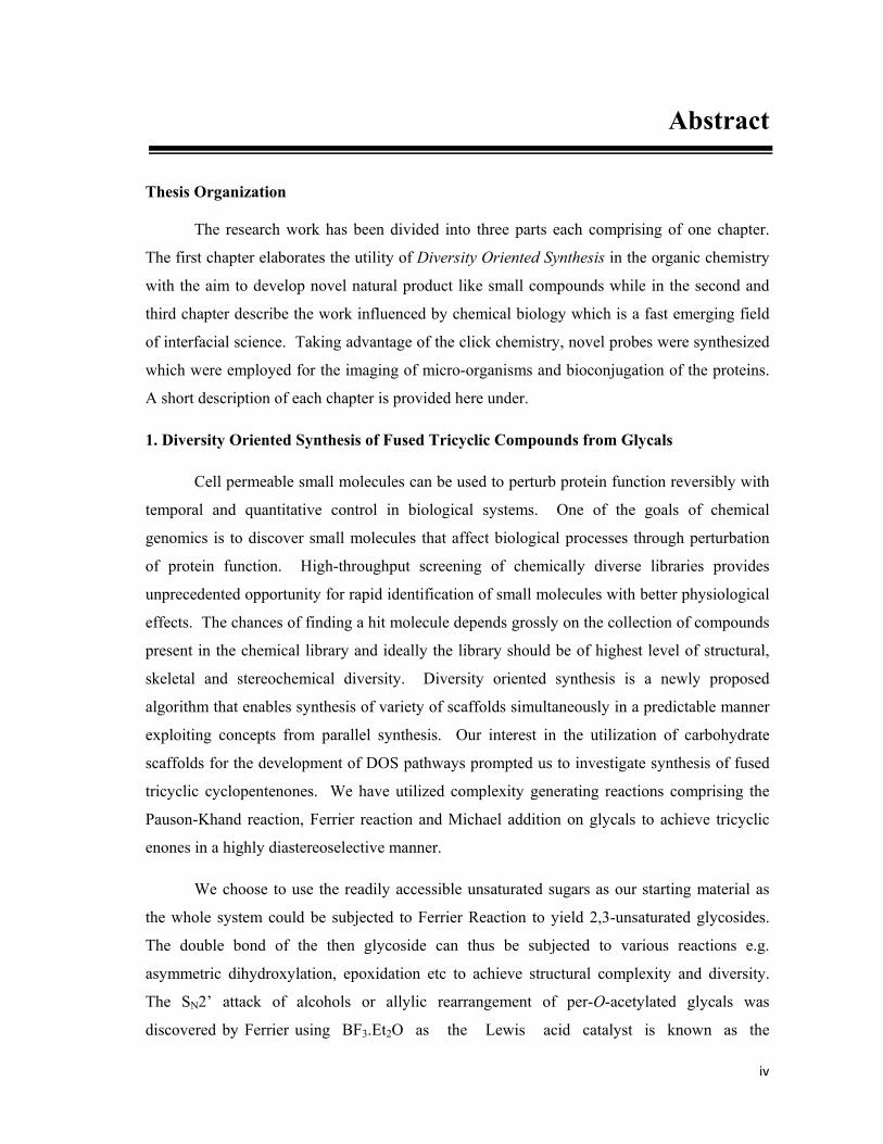
7, 9, 13
OMe
O
O
O O
OBnO
O O
TrOOBn
SSR1R1
R1SH, DMAP,Toluene, 4-12 h
R1 =
a b c d e f
22
O
O O
TrOOBn
SR1
20 21
Scheme 3. Michael Addition of Thiol onto the Tricyclic Enones
The presence of α,β–unsaturated system in the enones would enable to function as
excellent Michael acceptors. So we chose thiols due to the tremendous ability of sulphur
nucleophiles to undergo 1,4-addition. In a typical experiment the enone 7 was taken in toluene
and stirred along with the thiol in the presence of catalytic amount of DMAP. We observed the
formation of a single diastereomer in case of all the enones due to bowl-like configuration of
the fused tricyclic moiety. The incoming sulphur nucleophile attacked from the least sterically
viii
demanding side so as to furnish a single diastereomer in excellent yields. A total of six thiols
and three tricyclic enones were chosen for synthesizing the fused tricyclic library. The results
are presented in Scheme 3.
In conclusion, we have developed a practical protocol for the synthesis of natural
product-like tricyclic compounds and a total of 25 tricyclic compounds were synthesized as a
pilot library, using the complexity generating reactions like Ferrier, Pauson-Khand and the
Michael reactions. A little peep at the library reveals the unique mixture of diversity and
complexity among the members of the library. While the glucal and galactal derived scaffolds
demonstrate the complementary stereochemistry, on the other hand, the rhamnal and xylal
derived compounds provide the structural complexity to the carbohydrate library.
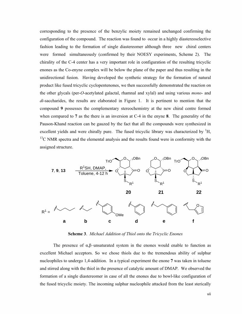
2. Click Chemistry Inspired Imaging of Microorganisms
The ability to visualize, track and quantify molecules and events in living cells with
high spatial and temporal resolution is essential for understanding biological systems. There
are very few techniques known in literature for probing the cellular pathways. Although
fluorescent tags have long been used in cell biology, the recent advances like confocal
microscopy fuelled it much further. In spite of all this, the major disadvantage is that
fluorescent tags available commercially are bulkier in nature and when attached changes the
structure and thus it tends to change the function of the biological molecules. Here in this effort
we showed the utility of a coumarinyl based pro-fluorophore, which can be easily be tethered
to any scaffold and can be triggered to become fluorescent through a Cu(I)-catalyzed 1,3
dipolar cycloaddition reaction with an incorporated alkyne functionality. Glucose is one of the
most abundant molecule and is the major source of the energy inside the cells. It is pertinent to
mention that bacteria use glucose as a sole C-source for their survival. So, we decided to track
the most active carbon of glucose. For this we decided to selectively block the hydroxyl
functionality of glucose sequentially with propargyl group and provide it to the microbial cell
as the carbon source. We can thus monitor the cellular uptake by addition of the coumarinyl
azide into the cell-lysate and take the fluorescence spectra of the cell-lysate.
All the five positional isomers of glucose (23a-23e) with propargyl appendages were
synthesized via the intermediates 24-33 following the protocol shown in Scheme 4, and made
to react with coumarinyl azide to yield the corresponding glucoconjugates (34a-34e, Scheme
5). It was heartwarming to notice that all the triazoles were fluorescent. Now having acquired

ix
the desired water-soluble propargyl modified glucose derivatives we decided check the
feasibility of the molecules to image the microorganisms. The E.coli cells (BL21) were
D-Glucose
a
b
c
d
e
f g h
i
j
k
l
m
n
o
23c
23a 23b 23d
23e
28
24 25 26
27
29 30
3132
33
OOMOMO
OMeMOMO
OPhOOHO
OMeHO
OPh
OHOMOMO
OMeMOMO
TPSO
OO
OH
OH
OMeHO
OOHO
OMeOBz
OPh
OOMOMO
OMeOH
OPhOHOHO
OMeO
HO
OHO
OH
OH
OHO
O
OO
O
OOH
O
OO
O
OO
OHO
OH
OH
OMe
O
O
OO
O
O
OMOM
O
OO
TBDMSO
HO
OMOM
O
OO
HO
MOMO
OMOMOHO
OH
O
OMe
HO
Reagents and conditions: (a) Propargyl Alcohol, Dioxane‐HCl, reflux, 6 h, 75% (b) Acetone, CuSO4, 24h, 80% (c) Propargyl bromide, NaH, DMF, nBu4N
+I‐, 0‐rt, 3h, 94% (d) MeOH‐HCl, reflux, 12h, 87% (e) MOMCl, DIPEA, CH2Cl2, 12h, 95% (f) (i) p‐toluene sulfonic acid, Methanol, 5h, 85% (ii) TBDMSCl, Triethylamine, CH2Cl2, 4h, 95% (g) (i) MOMCl, DIPEA, CH2Cl2, 12h, 90% (ii) p‐toluene sulfonic acid, Methanol, 1h, 94% (h) MeOH‐HCl, reflux, 12h, 85% (i) PhCHO, ZnCl2, 24h, 80% (j) MOMCl, DIPEA, CH2Cl2, 12h, 97% (k) (i) p‐toluene sulfonic acid, Methanol, 2h, 94% (ii) TBDPSCl, Triethylamine, CH2Cl2, 4h, 92% (l) (i)Propargyl bromide, NaH, DMF, nBu4N
+I‐, 0‐rt, 3h, 96% (ii) MeOH‐HCl, reflux, 4h, 88% (m) Dibutyltinoxide, Dioxane, Benzoyl Chloride, 6h, 85% (n) (i) MOMCl, DIPEA, CH2Cl2, 12h, 97% (ii) NaOMe, MeOH, rt, 30 min, 97% (o) (i) Propargyl bromide, NaH, DMF, nBu4N
+I‐, 0‐rt, 3h, 96% (ii) MeOH‐HCl, reflux, 2h, 92%
Scheme 4. Modified glucose derivative
allowed to grow in the presence of modified glucose for 12 h. The cells were then
subsequently washed and lysed with sonification. The fluorescent spectra of the cell lysate
were examined after their reaction with the coumarinyl azide. Analysis of the fluorescent
spectra illustrated that only compound 23b propargyl ether at C-2 position was observed to
have been incorporated into the cell wall more as compared to other propargyl ethers (Figure
2B). The feasibility of exploiting coumarin based probe for visualizing bacterial cells through
fluorescence microscope was then investigated using C-2 only. The cells were fixed on cover
slips and click reaction was carried out. E.coli. cells incubated in the presence of 23b showed
strong fluorescence. The images of the bacterial results are shown respectively in Figure 3A.

x
Furthermore, the experiments were also performed on Lactobaccilus lacti and yeast cells
(Sacchromyces wild type). These microorganisms also demonstrated the similar results in
accordance with that shown by E. coli. The C-2 propargyl compound, 23b, was found to be
more potent as compared to the other propargyl derivatives for incorporation into the cell wall
of the micro-organisms. It may be noted that the control for all the micro-organisms did not
exhibited any fluorescence. The control for all the experiments did not have any modified
glucose derivative added to itself but it was made to undergo the same procedures as the
experimental samples. This shows that modified glucose 23b is being incorporated into
microbes and after the conjugation with coumarin azide results in the fluorescence.
Scheme 5. Formation of Fluorescent Triazole
(A) Fluorescent images of BL21, Lactobaccilus lacti and Yeast cells; (B) Fluorescence spectra of the BL21, Lactobaccilus lacti and Yeast cells. Indicated cells were inoculated in normal complex media containing either glucose (control) or propargyl ether (with 23b) was allowed to grow overnight at 37°C. Cells were treated with coumarin azide in presence of CuSO4, sodium ascorbate after which the images and spectra were recorded.
Figure 2. Fluorescent Microscopic and Spectroscopic Studies

xi
In summary, the utility of profluorophoric coumarine-based azide for fluorescent
labelling and imaging of microorganism culture was demonstrated for the first time for
bacterial and yeast cultures. The current protocol of fluorescent labelling via the click
chemistry facilitates monitoring of glycan expression and the related processes which are
poorly accessible. Additionally, this protocol also scores over the reported fluorescence probes
with the size of the probe being smaller and the not interfering with the cellular processes.
3. Photocleavable Linkers for Bioconjugation of Proteins
Proteins are a large molecules comprising of various amino acids arranged from one
end to another. Various different class of proteins are present inside the cell namely –
hydrolases, proteases, kinases, nucleases, toxins, cell- matrix proteins, receptors, serum
proteins, galactosidases and antibodies and are involved in various important biological
functions inside the body. Manipulation of protein structure and thus its activity leads to
unravel critical details of protein function at molecular and cellular level. These can be
statistically modified utilizing the several active side chains of amino acid residues in them
(e.g. Lysine, Argnine, Cysteine etc.) free amino group and sulfuhydryl group can selectively be
targeted to introduce the desired moieties to tap the cellular mechanisms. Post translational
modifications affect the protein structure and thus its stability, activity and interactions with
other molecules. Combination of chemical and biological techniques to chemoselectively
modify proteins has proved to be an excellent resource for monitoring their function at
molecular level. Photosensitive protecting groups act as valuable tools for investigating the
biological phenomenon. Uses of photolabile groups have long been known for synthetic
purposes and they have come a long way since their first application in caging of ATP
molecule by Hoffman et. al. Photodeprotection is known to occur in high quantum yields at
shorter wavelengths (> 300 nm) with no harmful side products. These can be used for the
formation of stable covalent bonds on the reactive side chains of the proteins which can be
easily cleaved using the light. Photocleavable groups can be used as conditional trigger to
observe the various cellular mechanisms which in turn are controlled by the proteins. Various
other functions of protein such as folding and unfolding, interactions with other bio-molecules
etc. can also be monitored on a time dependent manner.
Herein we designed a photocleavable linker which can be easily introduced on the
available free amino groups of the proteins. Further the designed linker will have o-nitro
benzyl group on one end while on the other end it has alkyne or the azide moiety. The
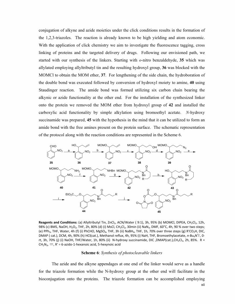
xii
conjugation of alkyne and azide moieties under the click conditions results in the formation of
the 1,2,3-triazoles. The reaction is already known to be high yielding and atom economic.
With the application of click chemistry we aim to investigate the fluorescence tagging, cross
linking of proteins and the targeted delivery of drugs. Following our envisioned path, we
started with our synthesis of the linkers. Starting with o-nitro benzaldehyde, 35 which was
allylated employing allyltributyl tin and the resulting hydroxyl group, 36 was blocked with the
MOMCl to obtain the MOM ether, 37. For lengthening of the side chain, the hydroboration of
the double bond was executed followed by conversion of hydroxyl moiety to amine, 40 using
Staudinger reaction. The amide bond was formed utilizing six carbon chain bearing the
alkynic or azide functionality at the other end. For the installation of the synthesized linker
onto the protein we removed the MOM ether from hydroxyl group of 42 and installed the
carboxylic acid functionality by simple alkylation using bromoethyl acetate. N-hydroxy
succinamide was prepared, 45 with the hypothesis in the mind that it can be utilized to form an
amide bond with the free amines present on the protein surface. The schematic representation
of the protocol along with the reaction conditions are represented in the Scheme 6.
d e
f g h
i j
CHONO2 NO2
HO
NO2
MOMO
NO2
MOMOOH
a b c NO2
MOMON3
NO2
MOMONH2
NO2
MOMONHBn
NO2
MOMONBn
O
RNO2
HONBn
O
R
NO2
ONBn
OR
O
O
NO ONO2
ONBn
OREtO2C
35 36 37 38 39
40 41 42 43
44 45
Reagents and Conditions: (a) Allyltributyl Tin, ZnCl2, ACN/Water ( 9:1), 3h, 95% (b) MOMCl, DIPEA, CH2Cl2, 12h, 98% (c) BMS, NaOH, H2O2, THF, 2h, 80% (d) (i) MsCl, CH2Cl2, 30min (ii) NaN3, DMF, 60°C, 4h, 90 % over two steps; (e) PPh3, THF, Water, 4h (f) (i) PhCHO, MgSO4, THF, 3h (ii) NaBH4, THF, 1h, 70% over three steps (g) R'CO2H, DIC, DMAP ( cat.), DCM, 4h, 90% (h) HCl(cat.), Methanol reflux, 4h, 95% (i) NaH, THF, Bromoethylacetate, n‐Bu4N
+I‐, 0‐rt, 3h, 70% (j) (i) NaOH, THF/Water, 1h, 80% (ii) N‐hydroxy succinamide, DIC ,DMAP(cat.),CH2Cl2, 2h, 85%. R = CH2N3, , R' = 6‐azido‐1‐hexanoic acid, 5‐hexynoic acid
Scheme 6: Synthesis of photocleavable linkers
The azide and the alkyne appendages at one end of the linker would serve as a handle
for the triazole formation while the N-hydroxy group at the other end will facilitate in the
bioconjugation onto the proteins. The triazole formation can be accomplished employing
xiii
another protein to form a homo- or hetro-dimeric conjugate of the protein or if the small
molecules are engaged then it would help unravel the involvement of protein inside the various
metabolic processes inside the cell. Having completed the synthesis of the linkers, we moved
on to investigate the utility of the linkers for the bioconjugation. We choose the readily
available Bovine Serum Albumin (BSA) for our experiments for its numerous biological
applications including ELISA (Enzyme-Linked Immunosorbent Assay), blots etc.
Additionally, it is also used as a nutrient in cell and microbial culture. One of the main
advantages for preferring BSA over the others was due to its stability as compared to the other
proteins.
BSA is known to have several free amines free in its tertiary structure. We choose to
covalently link only a few of them in our experiments, firstly it will be impossible to react all
the free amines and secondly, it will be tiresome job to cleave all the linked amines after the
click reaction. The stock solution of 4 mg/mL BSA was prepared in 0.1 M phosphate buffer
(pH 7.2) and mixed with NHS-linkers taken in DMSO. The resulting mixture was allowed to
rotate in the rotary for 2 h. After the completion of the bioconjugation, the protein was
dialysed overnight employing the cellulose membrane (12,000 kDa cut off). The samples were
analysed by MALDI-TOF. Delightfully, both the azide and alkyne linkers were found to link
onto the protein. Surprisingly, if the low concentration of the linkers were employed for the
reaction then MALDI-TOF analysis showed that only one molecule of the linker is covalently
attached to the protein.
To conclude we have successfully synthesized the photocleavable linkers having the
azide and alkyne appendages and also exploited them to covalently link onto BSA which can
be further employed for linking small molecules as drugs or fluorescence tags to the proteins.
On the other hand, the linkers can also be exploited for protein-protein bioconjugation to form
homo- and hetro-dimeric conjugates to assist in the studies regarding the behaviour of proteins
in promiscuity with the other proteins.
Note: Compound numbers in abstract are different from those in the thesis.

Chapter 1 Diversity Oriented Synthesis of Fused Tricyclic Compounds from Glycals

1
Chapter 1: Introduction
Nature provides a plethora of structurally diverse and biologically active compounds.
Natural products have a profound effect on both chemical biology and drug discovery. These
natural products have been attractive probes for cell-cell signalling pathways, protein-protein
interactions, and provide suitable cures for various disorders, disabilities and syndromes.1
Cell-permeable and selective small molecules that are used to perturb protein function rapidly,
reversibly and conditionally with temporal and quantitative control in any biological function
helps in providing a better insight of the biological pathways.2 The rich structural diversity and
complexity of the natural products have prompted synthetic chemists to produce them in
laboratory, often with medicinal applications in mind. Nature produces an astonishing array of
structurally complex and diverse molecules as the secondary metabolites. Many drugs in
clinical use are either natural products or natural product derivatives. For example over the last
20 years, 5% of the 1031 chemical entities approved as drugs were natural products, and
another 23% were natural product-derived.3
The advent of the twentieth century saw the isolation of a large number of natural
products from the plant extracts and with the progression of characterization techniques, UV;
IR; and more important NMR and MS, there have been great ease in the identification of their
structures.4 Even with the sophistication of purification and chromatographic methods, the
separation and characterization of natural product extracts still remains a challenging task.
Unfortunately, there are serious disadvantages of working with the extracts. Firstly, the nature
does not synthesize secondary metabolites in pure form waiting to be isolated; therefore, the
extracts comprise a collection of huge number of compounds with complementary structure
and properties. This leaves us with the problem of isolation, identification of the active
component(s). Secondly, there is always a limited supply of the active component(s) from the
extract, usually in few milligrams from the huge plant extracts. Thirdly, the active component
may be so complex structurally, such as Taxol that making analogues to characterize or
optimize its activity remains a difficult exercise. Fourthly, the chemical space encompassed by
the natural products is a limited space which may not be the only region useful for discovering
physical or biological properties of interest.
The increasing entrepreneurship of synthetic organic chemists to in the art of total
synthesis has helped to reduce the limited supply of natural products, even with the materials
possessing very complex structures. In principle, the total synthesis is the complete chemical

2
synthesis of complex natural product starting with the much simpler starting materials without
the aid of any biological processes. The target molecule can be a secondary metabolite from a
plant; bacteria, or a medicinally active ingredient or organic compounds of theoretical interest
in chemistry or biology.
The earliest demonstration of organic total synthesis was Friedrich W�hler’s synthesis
of urea in 1828, which showed that organic molecules can be produced from inorganic
precursors. The first commercialized total synthesis was Gustaf Komppa’s synthesis and
industrial production of camphor in 1903.5 Early efforts focussed on building chemicals which
were extracted from plants or isolated from microorganisms. Today, the total synthesis
remains a playground for synthetic chemists to develop new reactions, methodologies and
highlight the sophistication of modern synthetic organic chemistry.5b Sometimes, it inspires
the development of novel mechanisms, catalysts or techniques. From a chemist’s perspective,
the art of total synthesis demands an accurate sense of chemical intuition and encyclopaedic
knowledge of chemical reactions. Some of the classical examples of total synthesis include
that of Cholesterol, Cortisone, Chlorophyll, Colchicine, Taxol, Vancomycin etc.5b, 5c It is
impertinent to mention E. J. Corey for his contribution in advancing the art and science of total
synthesis by introducing retrosynthetic analysis.6
Nature produces molecules which can be simple, such as Serotonin and Histamine, or
structurally complex, like Vancomycin, Taxol etc. These accommodate a variety of functional
groups and are usually supplemented with the problem of instability, for most of these are
stable inside the biological systems which provide an amazing stability and also enhance their
activity. Target Oriented Synthesis (TOS) starts with the powerful planning algorithm,
retrosynthetic analysis and subjecting simple and easily available synthons. In majority of the
cases, the well trained eye of the professional is required in order to identify the basic
fragment(s) present in the complex target molecule which can be derived from the suitable
precursor. In the elaboration of the synthetic strategy, one should never forget that even the
well established procedures may fail when applied in a specific structural context and thus an
otherwise chemically sound synthetic plan may prove to be unworkable. Hence, the most risky
synthetic operations should be shifted to the earliest possible step of the entire synthetic
scheme so as to avoid the possible loss of time. A number of other criteria must also be
considered when making a final selection between the options that emerge for the total
synthesis of any given natural product. Among the most important are the length of the
scheme(the fewer steps the better); availability and price of the starting materials, catalysts, the
complexity of the equipments needed and last but not the least an in-depth knowledge of a rich

3
arsenal of available synthetic methods and a clear understanding of the ultimate goal of the
whole endeavour. In fact a synthetic plan designed for the laboratory may appear nearly ideal
but may be totally unacceptable for the industrial applications considering the cost measures,
the amount of toxic waste being produced or simply the number of steps being involved.
It is heart-warming to see how the synthetic chemists solve the problems of synthetic
chemistry using the rich arsenal of complementary methods. Woodward’s synthesis of steroids
beautifully illustrates the value of a carefully thought out general plan for a synthesis.3 His
syntheses were most often described as having an element of art in them, and since then, the
synthetic chemists have always looked for elegance as well as utility in their synthetic plan.
Thus in the process of target oriented synthesis we come across the invention of new general
reactions and reagents for organic synthesis, the design and execution of various efficient
multi-step synthesis of complex organic molecules. However, inspite of the tremendous
amount of effort, meticulous planning and money being poured into the total synthesis, it has
failed to provide any suitable answer to the needs of pharmaceutical companies for regular and
surplus supply of drug molecules. With the demand of pharmaceutical drugs shooting up to
astronomical heights with the population boom, it has prompted the scientists to venture into
more novel fields of science rather than banking on mother nature to provide us with the cure
to our problems.
Chemical Space:
Chemical space is the space spanned by all possible (i.e. energetically stable) stoichiometric
combinations of electrons and atomic nuclei and topologies (isomers) in molecules and also
compounds in general (Figure 1).7 Chemical reactions allow us to move in chemical space.
The mapping between chemical space and molecular properties is often not unique, meaning
that there is usually more than one molecule which exhibits the same property which is being
explored when carrying out material design and drug discovery.
Figure 1: Three dimensional view of Chemical Space.
4
The number of drug like molecules in the chemical space has been estimated to be 1062.
By comparison, there are approximately 1051 atoms on earth. Therefore, it is impossible to
synthesize every drug-like molecule and chemists must hence be selective. This becomes
evident when knowing that only 27,000,000 molecules have been registered (and thus been
synthesized) so far. Systematic exploration of chemical space is possible by creating in silico
databases of virtual molecules.8 By representing these compounds as a series of chemical
descriptors (molecular weight, lipophilicity, dipole moment, etc.) it is possible to plot them in
chemical space.
Not all biologically active compounds encountered in the chemical space have the
desired physicochemical properties to be a drug. A biologically active compound may be too
lipophilic to be orally absorbed, too polar to be able to cross the gastrointestinal wall or may
have too vulnerable chemical functionality that can be attacked by metabolizing systems in the
body, and therefore not remain intact long enough to have a fruitful in vivo effect. The
determination of characteristics of compounds that are more likely to be safe, orally bio-
available medicines has led to the concept of ‘drug-likeness’. Compounds that are drug-like
have the potential to be developed into orally administered drugs, which are generally favoured
owing to their use by the patients. In this regard, the Christopher Lipinski has postulated the
Rule of Five which states that, in general, an orally active drug has no more than one violation
of the following criteria: (i) Not more than 5 hydrogen bond donors (nitrogen or oxygen atoms
with one or more hydrogen atoms); (ii) Not more than 10 hydrogen bond acceptors (nitrogen or
oxygen atoms); (iii) A molecular weight under 500 gm/mol; (iv) A partition coefficient log P
less than 5. Note that all the numbers are multiples of five and thus the postulation became
popular as Lipinski’s Rule of Five. 9
With the introduction of various combinatorial techniques and high-throughput
screening assays, there is an increasing demand for the small molecules within the chemical
space which are more probable to be drug-like. Surprisingly, after the billions of years of
evolution, nature still thrives on small molecules for signalling, protection and other essential
functions. Simple life forms can function on a few hundred small molecules. In order to reap
the benefits of small molecule approach for chemical genetics, advances must be made in
finding the selective small molecules that bind to the protein target quickly, cheaply and with
adequate selectively. If chemical space is huge and we cannot synthesize everything, then we
should decide on what to make and how to make it.
Many research groups have calculated where currently available drugs are located in
chemical space and it had been noted that they cluster together. There are two schools of
5
thought on how to use this information. One approach is to look for new molecular entities
based on ‘privileged’ core structures (pioneered by Waldmann group),10 which are known to be
commonly bioactive. Another approach is to look for new molecular entities in unchartered
regions of chemical space, by synthesizing new core structures (introduced by Schreiber
group)11. Diversity Oriented Synthesis (DOS) finds application in both of them. Generating
libraries around new or unexplored templates with the aim of generating structurally, skeletally
and functionally diverse compounds has become more common.12
Diversity Oriented Synthesis:
The search for new molecular entities on the drug discovery and chemical genetics
programs requires the screening of high-quality collections of structurally diverse small
molecules. The design and synthesis of such collections remains a major challenge to synthetic
chemists. As mentioned earlier that the natural products do not occupy all regions of chemical
space that are relevant to discovering bioactive compounds. Therefore, a question posed is, are
there more productive, unchartered areas of chemical space that should be investigated to
discover new molecular entities? In the early 1990’s chemist turned to combinatorial
chemistry as a technique to efficiently synthesize large number of compounds by appending
building blocks onto a core structure in search of finding novel active compounds. Despite,
resulting in a large number of compounds being synthesized, this methodology was not
successful as initially expected. The failure of the approach to discover a broad range of
activities was due to the lack of structural diversity being attained. Any structural diversity of
the molecules was only supplied by the building blocks and starting scaffold, while the
resulting molecular framework was the same in every case. In order to achieve the highest
level of structural diversity the following factors must be varied: (i) the building blocks; (ii) the
stereochemistry; (iii) the functional groups; (iv) the molecular framework. Today chemists are
investigating ways to synthesize libraries of compounds with high degree of structural
diversity. Efficiently enriching chemical space in this way has been termed as Diversity-
Oriented Synthesis (DOS), which concentrates on the synthesis of combinatorial libraries of
structurally diverse (and complex) small molecules for biological screening.
The approach to DOS is in contrast to target-oriented synthesis (TOS), which aims to
synthesize a single target, or traditional combinatorial chemistry, which generates structurally
similar target structure (Figure 2). In target oriented synthesis we follow a convergent pathway
to achieve the synthesis of any single natural product while in combinatorial chemistry we
subject a set of similar compounds to a same set of reactions to compose a library which is
more or less structurally similar. Complementary to these practices, the synthetic pathways in

6
DOS are branched and divergent, and the planning strategy extends from simple and similar
compounds to more complex and diverse compounds both in terms of structure and activity.
Retrosynthetic analysis traditionally focuses on the existence of a defined target structure. In
DOS there is no single target structure and, therefore, retrosynthesis analysis cannot be used
directly; instead a forward synthetic analysis algorithm is required which involves the
transformation of a collection of substrates into a group of products by
SingleTarget
Target-Oriented Synthesis:Convergent
Diversity-Oriented Synthesis:Divergent
DiverseTarget
Structure
SimpleRetrosynthetic
Complex Simple &Similar
Complex &Diverse
ForwardSynthetic
Analysis Analysis Figure 2: Target-Oriented Synthesis and Diversity-Oriented Synthesis
performing a number of chemical reactions together in forward direction. The inherent
chemical reactivity common to all the substrates remains a key element for the implementation
of forward synthetic analysis. Although, retrosynthetic analysis cannot be applied for the DOS,
but its foundations have regularly been practised there to develop a complementary strategy to
facilitate planning and implementation of synthetic strategies for the development of diverse
libraries. Also complementary to TOS, the DOS does not involve a huge number of steps, the
structural diversity and complexity is planned to achieve over as few steps as possible. With
the help of various complexity generating reaction, reagent variations, substrate differentiation
and stereochemical modifications DOS libraries aims to introduce rich diversity to enable them
to target a wide range of proteins and thus inhibit the biological processes. However, the
degree of diversity obtained within libraries is subjective: some libraries are reliant on a large
number of appendages while others focus on synthesizing different skeletal structures. DOS
libraries may seem to be inherited from a natural product scaffold or can be found out in an
entire new region within the chemical space. Both of these categories are discussed
henceforth.
7
Libraries inspired from the natural products:
The functional group diversity and architectural platforms engineered into natural
products during their biosynthesis continue to attract several synthetic and medicinal chemists
in their strategies for the creation of biologically active mimics, and provide selective ligands
for cellular targets. Moreover, the natural products have also been effective in teaching us
about chemical functionality that is compatible with most of the biological systems so they
remain an invaluable tool for deciphering the logic for biosynthesis and as a platform for the
development of frontline drugs. Individual natural products are proving to be valuable,
biologically validated starting points for the library design. With the underlying structural
frameworks selected for binding to certain protein domains, structural diversity arising from
variations in appended functional groups can provide selectivity for related targets. These are
thus used as a major source of innovation for therapeutic drugs for infectious diseases.
However, the core scaffold of a natural product can also provide a biologically validated
framework which can be altered with diverse functional groups to optimize its activity.
Library design strategies have been divided into three broad categories: (i) libraries based in
core scaffold of an individual product; (ii) libraries based in specific structural motifs; (iii)
libraries that mimic the structural characteristic of natural products.
(i) Libraries based on core scaffold of an individual product: This approach has primarily
been used to enhance the inherent activity of the parent natural product. Libraries designed on
a privileged core of a natural product, known to inhibit a specific protein target, offer an
excellent platform to provide an array of compounds which will have more chances to provide
the lead molecule. Compounds in libraries that are based on core structures known to exhibit
biological activity will have a higher intrinsic ability to bind to targets than those compounds in
a library not based on natural products. A further extension suggests that if the parent core is a
fortunate motif, defined as being able to target a specific class of proteins, then the offshoot
library will be extremely be useful for targeting multiple classes of protein having different
folds. In a recent example, Schreiber and co-workers synthesized a library of 3520 spiro-
oxindoles based on spirotryprostatin B, a mammalian cell-cycle inhibitor produced by
Aspergillus fumigatus.13 Using a yeast cell-based screen, they identified 19 enhancers of
latrunculin B, an actin polymerization inhibitor that induces growth arrest, and concurrently
identified preliminary structure-activity relationship. In contrast, the consecutive library may
not bind to the same target but can also display a wide range of activity against multiple protein
targets having different folds. An important early demonstration in this approach was by Shair
and co-workers discovery of secramine, a novel secretory pathway inhibitor from a library of

8
2527 compounds based on enantiomeric scaffold of galanthamine, a known
acetylcholinesterase inhibitor.14 Notably, galanthamine had no effect upon the secretory
pathway at upto 100 μM (Figure 3).
HN
NN
O
OO
MeMe
Spirotryprostatin B
N
N O
O
O
O
MeO
HN
O
N
Ph
Ph
OHO
H
Enhancer of Latrunculin B
N
OMeO
Me
OH
Galanthamine
NH
OBr
N
O OH
S
OMeO
Secramine
Figure 3. Some of the natural product derived active Pharmacophore molecules obtained from the DOS libraries synthesized by Shair group Individual natural products are proving to be valuable starting materials for the library
design. These provide biologically active framework, which when exploited to various
chemical reactions and decorated with the various functional groups have been one of the most
successful ways of drug design. However, the foremost limitation, in this approach is that the
fixed scaffold is most likely suited to address a narrow range of biological targets with related
activity.
(ii) Libraries based on specific sub-structural motifs of natural products: This strategy exploits
the use of natural products sub-structures which are distributed across in most of the natural
products. Inherent activities of the drugs are due to the presence of these core scaffolds.
Ornamenting them with various functional groups provides an increased chance for the
multiple target inhibition.
O
O
O
OMe
OMe
MeO
O
CN
OH
O
NADH:ubiquinoneoxireductase inhibitor Anti-MRSA antibacterial
Figure 4. Nicolaou and group discovered two of the active drugs originated from 2,2-dimethylbenzopyran skeleton
One of the earliest discoveries in this regard was the approach of Nicolaou and co-
workers’ synthesis of a collection of 10,000-membered compounds derived from the skeleton
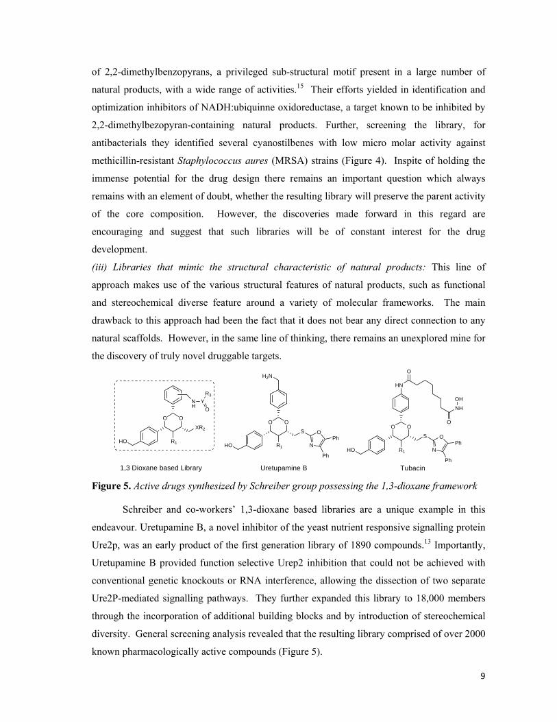
9
of 2,2-dimethylbenzopyrans, a privileged sub-structural motif present in a large number of
natural products, with a wide range of activities.15 Their efforts yielded in identification and
optimization inhibitors of NADH:ubiquinne oxidoreductase, a target known to be inhibited by
2,2-dimethylbezopyran-containing natural products. Further, screening the library, for
antibacterials they identified several cyanostilbenes with low micro molar activity against
methicillin-resistant Staphylococcus aures (MRSA) strains (Figure 4). Inspite of holding the
immense potential for the drug design there remains an important question which always
remains with an element of doubt, whether the resulting library will preserve the parent activity
of the core composition. However, the discoveries made forward in this regard are
encouraging and suggest that such libraries will be of constant interest for the drug
development.
(iii) Libraries that mimic the structural characteristic of natural products: This line of
approach makes use of the various structural features of natural products, such as functional
and stereochemical diverse feature around a variety of molecular frameworks. The main
drawback to this approach had been the fact that it does not bear any direct connection to any
natural scaffolds. However, in the same line of thinking, there remains an unexplored mine for
the discovery of truly novel druggable targets.
HO
OO
NH
YO
R3
R1
XR2
HO
OO
R1
S
H2N
N
OPh
PhHO
OO
R1
S
HN
N
OPh
Ph
O
NH
O
OH
Uretupamine B Tubacin1,3 Dioxane based Library Figure 5. Active drugs synthesized by Schreiber group possessing the 1,3-dioxane framework
Schreiber and co-workers’ 1,3-dioxane based libraries are a unique example in this
endeavour. Uretupamine B, a novel inhibitor of the yeast nutrient responsive signalling protein
Ure2p, was an early product of the first generation library of 1890 compounds.13 Importantly,
Uretupamine B provided function selective Urep2 inhibition that could not be achieved with
conventional genetic knockouts or RNA interference, allowing the dissection of two separate
Ure2P-mediated signalling pathways. They further expanded this library to 18,000 members
through the incorporation of additional building blocks and by introduction of stereochemical
diversity. General screening analysis revealed that the resulting library comprised of over 2000
known pharmacologically active compounds (Figure 5).

10
This final approach of the DOS of deriving the libraries from the natural products has
yielded significant number of the pharmacologically active molecules. The exploitation of
skeletal diversity coupled with the stereochemical features of a variety of the natural product
core scaffolds which are classified across a vast region of chemical space provides us with an
unexplored gold mine to reveal novel drugs. The region of the chemical space occupied by
these molecules neighbours that to the natural products, thus shows us an important pathway
for discovering the novel biologically molecules which can be used for the further challenging
targets.
Each of the above three strategies balance the degree of connection to natural-product
structure space against the accessibility of structural diversity that is likely required to address
multiple different biological targets. This approach has primarily been used to enhance the
original activity of the parent natural product. However, the complexity of the natural products
coupled with the limited supply can limit the structural modification and thus library design.
But still the advances made forward in understanding the natural product biosynthesis, have
helped us in the identification of the active motifs. The libraries designed around the natural
product scaffolds lead us into an unchartered area of the chemical space to yield numerous
small molecules which hold the potential of giving us a better insight of the understanding of
the biological processes.
Novel libraries which do not bear any resemblance to the natural products:
Synthetic chemists get inspired after considering the vast amount of chemical space
being under-utilised and also being aware of the fact that most of the natural products or their
derived libraries are clustered together or occupy adjacent space to venture into new territories
of chemical space in search for novel drug-like molecules. Generating libraries around new or
underexploited templates with the aim of synthesizing structurally, skeletally and functionally
diverse and novel scaffolds has become more common in the recent years. The idea is to
exploit number of various diverse and unique skeletons and synthesize libraries exploiting the
various complexity generating reactions, the resulting libraries although do not have any
likeliness to natural products but are still able to bear the fruitful results. It is significant to
mention the library synthesis by Mitchell and Shaw group which prepared a series of oxazoline
compounds which were either spirocyclic or fused lactams (Figure 6).17 The researchers
produced five core structures overall and found several compounds comprising this core which
promote the growth of yeast, while others were cytotoxic to HeLa cells in a dose dependent
manner.

11
However, together with the excitement of exploring new frontiers of chemical space is
also comes the fear of failure. The key to the success of these libraries lies behind the careful
planning and execution of the synthetic plan. If the core scaffold around which the whole
library has been generated is biologically inactive or toxic then the whole effort can prove to be
a futile exercise. Quite lately, a lot of effort has been forward in this direction and the chemists
have exploited both linear as well as the convergent pathways for the generation of libraries
with high degree of structural as well as stereochemical diversity.
HN
NO
Ar
O
N3
NO
Ar
N3
NH
NO
O
Ar
N3
NHO
NO
Ar
N3
NH
O
N
OAr
O
OCH3
N3
N3
Figure 6. Small molecule inhibitors discovered by Mitchell and Shaw group using DOS
The ability of high-throughput screening methods in the rapid screening of huge
number of molecules for various biological assays has made DOS a favourite sport amongst
most researchers involved in the drug discovery. DOS has scored over the traditional target-
oriented synthesis and lately introduced combinatorial chemistry with its uniqueness for the
rapid synthesis of drug-like molecules. The success for DOS lies in the fact that its techniques
had evolved from both of its precursors which were the traditional methods of drug design.
The clever exploitation of forward synthetic analysis (the basic concepts of which finds its
roots in retrosynthetic plans) and the combinatorial techniques to generate libraries with high
degree of complexity has been the success of this technique. However, the real challenge for
the future is to maintain a continuous supply of the libraries with high degree of structural
diversity. This would enable the exploration of the previously unchartered regions of chemical
space in an effort for the search of novel pharmacologically active compounds.
*****
12
Chapter 1: Present Work
Carbohydrates are the most abundant of the four major classes of biomolecules, which
also include proteins, lipids and nucleic acids, found in the living organisms which are the least
exploited for the Diversity Oriented Synthesis (DOS). They along with their derivatives play a
direct role in the smooth functioning of cellular mechanisms like immune system, fertilization,
pathogenesis, blood clotting etc. and are also involved as the receptors for cell-cell recognition
and pathogen defence. In most of the cases, these have a decisive role in the biological
processes rather than being present only as a source of energy. To add to all this, sugars are
also involved in providing a structural framework for the cells and in turn to the living
organisms.
Renewed interest in the biology and chemistry of carbohydrates over the past two
decades has dramatically accelerated the development of carbohydrate based therapeutics.17
Modern medicine already employs a range of bioactive carbohydrates, including heparin-based
anticoagulants, polysaccharide vaccines, aminoglycoside antibiotics, azasugars for diabetes and
with further developments, they also promise a cure for influenza and cancer. Besides,
carbohydrates are also being used to modify the pharmacological profile of other drugs,
including recombinant proteins. Some hybrid molecules often display superior efficacy and
reduced side effects when compared with the parent drug. The use of such biomolecules for
drug and gene delivery is actively being pursued. The development of many carbohydrate
based pharmaceuticals has been facilitated by the gains made in the field of glycobiology,
which focuses on defining the biological significance of carbohydrate interactions.18 The
physical chemistry of carbohydrate-protein interactions strongly influences the pharmacology
of many carbohydrate drugs. As relatively polar substances, they are unable to rapidly cross
the lipid membranes and as a result they often act on the cell surfaces or in extracellular
domain. And it will not be surprising to know that these are also distributed among a wide
variety of natural products originating from different classes and having diverse applications.
These are widely found in a number of the antibiotics produced by the microorganisms.
Notably carbohydrates also possess many of the favourable physical attributes,
including their ready availability, chiral purity and structural diversity, even simple
oligosaccharides have huge number of structural isomers owing to the poly-functionality of the
constituent monomer.8 Although much of the efforts are being poured into the glycobiology,
carbohydrates are not been utilized to their potential in DOS. The inherent chirality and the
13
poly-functionality of the carbohydrates can be easily manipulated to introduce the high degree
of complexity in the resulting libraries, which help us in expanding our boundaries in the
chemical space. The starting template in the DOS occupies a predominant role in dictating the
resulting skeletal and stereochemical diversities. As evident by the Lipinski’s Rule of Five and
from various records generated from the data-mining, large number of biologically active
natural products is oxygen-rich compared with corresponding congeners thus far synthesized
from the combinatorial libraries. In view of the above facts, we became interested in initiating
a diversity oriented synthesis using glycals as the starting template to enable oxygen-rich
stereochemically pure scaffolds. The unsaturated bond as well the poly-hydroxyl functionality
coupled with the inherent chirality of the glycals can be easily exploited to generate structurally
diverse libraries in short steps. We planned to introduce a three level of diversity into the
library using the range of complexity generating reactions like Ferrier, Pauson-Khand and
Michael Addition reactions, which are discussed in detail hereunder.
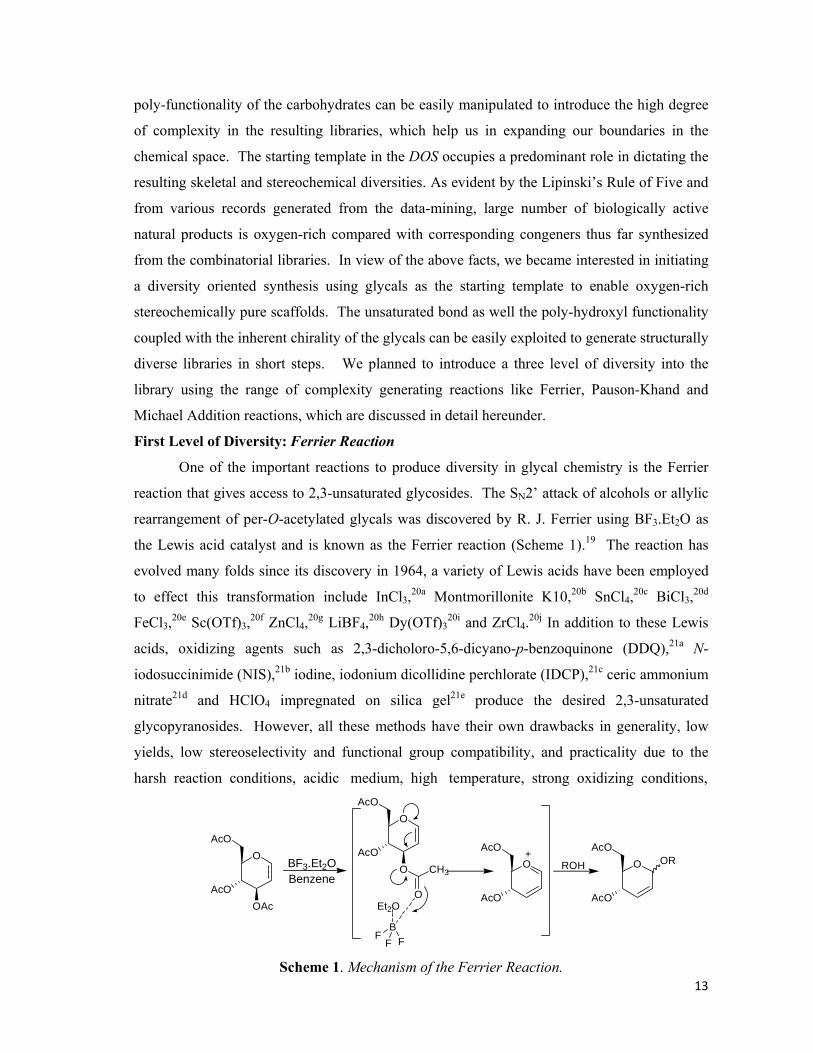
First Level of Diversity: Ferrier Reaction
One of the important reactions to produce diversity in glycal chemistry is the Ferrier
reaction that gives access to 2,3-unsaturated glycosides. The SN2’ attack of alcohols or allylic
rearrangement of per-O-acetylated glycals was discovered by R. J. Ferrier using BF3.Et2O as
the Lewis acid catalyst and is known as the Ferrier reaction (Scheme 1).19 The reaction has
evolved many folds since its discovery in 1964, a variety of Lewis acids have been employed
to effect this transformation include InCl3,20a Montmorillonite K10,20b SnCl4,20c BiCl3,20d
FeCl3,20e Sc(OTf)3,20f ZnCl4,20g LiBF4,20h Dy(OTf)320i and ZrCl4.20j In addition to these Lewis
acids, oxidizing agents such as 2,3-dicholoro-5,6-dicyano-p-benzoquinone (DDQ),21a N-
iodosuccinimide (NIS),21b iodine, iodonium dicollidine perchlorate (IDCP),21c ceric ammonium
nitrate21d and HClO4 impregnated on silica gel21e produce the desired 2,3-unsaturated
glycopyranosides. However, all these methods have their own drawbacks in generality, low
yields, low stereoselectivity and functional group compatibility, and practicality due to the
harsh reaction conditions, acidic medium, high temperature, strong oxidizing conditions,
O
AcOOAc
AcO
BF3.Et2OBenzene
O
AcOO
AcO
O
CH3
BF
F F
Et2O
O
AcO
AcOO
AcO
AcO + ORROH
Scheme 1. Mechanism of the Ferrier Reaction.
14
longer reaction time and stoichiometric use of the costly reagents being employed. In view of
the above and in our efforts to utilize glycal chemistry to achieve diverse scaffolds, we
focussed on developing a practical procedure for synthesizing 2,3-dideoxy glycopyranosides.
Recently niobium(V)chloride has emerged as a Lewis acid for a variety of the reactions.21g In
particular NbCl5 has many advantages compared with other Lewis acids such as ease of
handling, moisture stability, shelf life and economic viability. So we decided to take advantage
of the Lewis acidity of the NbCl5 for effecting the Ferrier reaction. To check our hypothesis,
per-O-acetylated glucal in the presence of benzyl alcohol was treated with one mole percent of
NbCl5 in acetonitrile under microwave irradiation in an open vessel for 2 minutes (Scheme
2).22
OBn
OAcO
OAcO
OAc
BnOH NbCl5(cat.)Acetonitrile+AcO AcO
Scheme 2. NbCl5 promoted Ferrier reaction of per-O-acetylated glucal
Not only is the desired product formed with 97% yield but also only α-anomer was
observed. The use of microwave reaction conditions with carbohydrate is not yet a mature
subject, albeit various successful efforts in conventional organic synthesis exist. The use of
microwaves in the Ferrier reaction was explored using Montmorillonite K1020a and InCl3.20b
The microwave induced Montmorillonite K10 mediated Ferrier rearrangement of per-O-
acetylated glucal resulted in the formation of 2,3-unsaturated glucosides; the corresponding
InCl3 reaction gave α-anomeric selectivity, however, 30 mol% of InCl3 was required.
Complementary to the other Lewis acids, only catalytic amounts of NbCl5 effected the reaction
with excellent yields and stereoselectivity. It is also pertinent to mention that the corresponding
reaction under room temperature or reflux conditions took almost one hour for completion as
evident by the consumption of the starting material, thereby making this reaction a microwave
oven-induced reaction enhancement (MORE) technique.
O
OAc
OAcO
OAc
O
O
H3CAcOAcO
NbCl4
Cl
O
OAc
AcOCl
O
H
R:
O
OAc
AcO
OR
NbCl5SN2'
- NbCl4OAc-HCl
Figure 7. Mechanism of the NbCl5 promoted Ferrier reaction

15
HO
HO
HO
HO
O
HO
HO
HO
HS
OBnO
OMe
OH
BnO
OHO
OMe
OBn
BnO
BnO
BnO
2.0
2.5
95
92
2.0 80
1.5 85
2.0 93
2.0 97
3.0 84
2.0 87
3.0 90
3.0 87
Entry Substrate Alcohol Product Time(min) Yield(%)
1
2
3
4
5
6
7
8
9
10
O
O
O
O
O
OO
OMeBnO
BnO
OO
OMe
OBn
BnOBnO
1a
1b
1c
1d
1e
1h
1i
1j
OO
O
OAcO
AcO
OAc
OAcO
OAc
OAcO
OAc
OAcO
OAc
OAcO
OAc
OAcO
OAc
OAcO
OAc
OAcO
OAc
OAcO
OAc
OAcO
OAc S
OAcO
OAc
1f
BnO
1g
Table 1. NbCl5 promoted 2,3-unsaturated Ferrier rearranged products from glucal
A recent report by Sinou et. al. provided the plausible mechanistic details of the NbCl5
catalyzed Ferrier reaction.23 Niobium complexes with the carbonyl oxygen atom of the O-
acetyl group at C-3. Then one of the chlorine atom of NbCl5 could attack at C-1 via a 8-
membered transition state to furnish 4,6-di-O-acetyl-2,3-dideoxy-β-D-erytho-hex-2-
enopyranosyl chloride by a SN2’ mechanism. This instantaneously reacts with the alcohol to
form the Ferrier product. The exclusive formation of the α-anomer was justified with the
explanation that the displacement of the chlorine atom with that of the alcohol is an
instantaneous reaction which takes place before the anomerization of the intermediate halide
(Figure 7). The generality of this reaction with NbCl5 was shown by exposing per-O-

16
acetylated glucal to various alcohols including allylic, benzylic, aliphatic, phenolic and
monosaccharide donors to the corresponding glycosides of mono- and disaccharides. As it is
evident from the results revealed in Table 1, all the donors reacted with great ease in shorter
time to give α-anomer as the sole product of the reaction in excellent yields. All the glycoside
products were confirmed by comparing with the reported values.20,21
Encouraged by our success, we decided to test our protocol on 3,4,6-tri-O-acetyl
galactal. Treatment of per-O-acetylated galactal in the presence of an alcohol with NbCl5(cat.)
in acetonitrile and irradiation with microwaves for less than 3 min resulted in the formation of
the Ferrier product 4,6-di-O-acetyl-2,3-dideoxy-D-threo-hex-2-eno-α-D-galactopyranoside
along with a minor amount of 2-deoxy-α-D-lyxo-hexopyranoside (Scheme 3). It is important
to mention that not so many synthetic procedures are available in the literature for the Ferrier
reaction involving 3,4,6-tri-O-acetyl galactal and many report the formation of 2-deoxy
OBn
OAcO
OAcAcOO
OAcAcO
BnOH NbCl5(cat.)Acetonitrile+ + O
OAc
OBn
AcOAcO
Scheme 3. NbCl5 catalysed Ferrier reaction of per-O-acetylated galactal
compound as the frustrating side product in considerable yield. In order to check the versatility
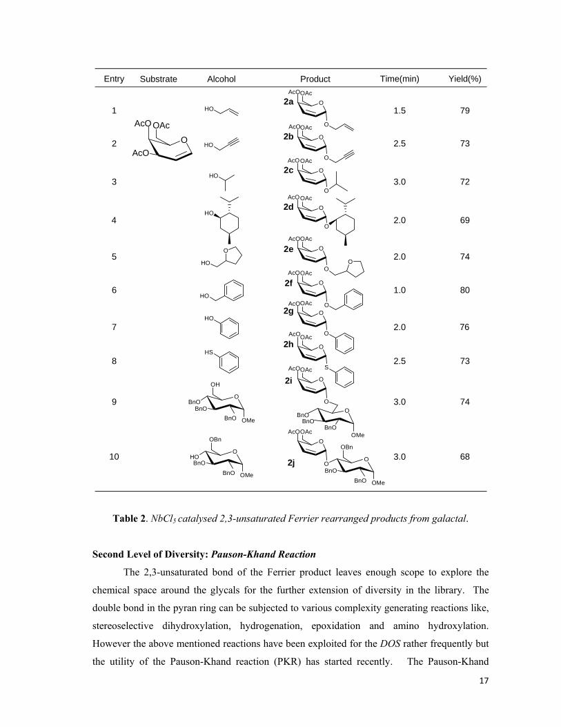
of this reaction we exposed a variety of aglycones to per-O-acetylated galactal (Table 2). As is
evident from the results shown, all of the reactions were completed within no time and
enhanced amount of Ferrier product was obtained in contrast to the earlier published results.
The ratio of the Ferrier product versus the 2-deoxy compound was found to be 4:1 after the
isolation of the respective compounds by conventional silica gel column chromatography. 1H
and 13C NMR spectra of all the various saccharides synthesized were in conformity with those
of reported values.20,21
Successful in our efforts of exploiting the chirality and structural complexity of the
carbohydrates, we were able to synthesize a library of 20 members. All the
constituents/members of the library are chirally pure and structurally diverse even though they
are derived from the same skeleton. The derivatives of 2,3-unsaturated glycosides constitute
the structural motifs of several antibiotics and these are also found to reduce the plasma
cholesterol and triglyceride levels significantly in mice.
17
HO
HO
HO
HO
O
HO
HO
HO
HS
OBnO
OMe
OH
BnO
OHO
OMe
OBn
BnO
BnO
BnO
1.5
2.5
79
73
3.0 72
2.0 69
2.0 74
1.0 80
2.0 76
2.5 73
3.0 74
3.0 68
Entry Substrate Alcohol Product Time(min) Yield(%)
1
2
3
4
5
6
7
8
9
10
O
O
O
O
O
OO
OMeBnO
BnO
OO
OMe
OBn
BnOBnO
2a
2b
2c
2d
2e
2h
2i
2j
OO
O
OAcO
OAc
OOAc
OOAc
OOAc
OOAc
OOAc
OOAc
OOAc
OOAc
OOAc S
OOAc
2f
BnO
2g
AcO
AcO
AcO
AcO
AcO
AcO
AcO
AcO
AcO
AcO
AcO
Table 2. NbCl5 catalysed 2,3-unsaturated Ferrier rearranged products from galactal.
Second Level of Diversity: Pauson-Khand Reaction
The 2,3-unsaturated bond of the Ferrier product leaves enough scope to explore the
chemical space around the glycals for the further extension of diversity in the library. The
double bond in the pyran ring can be subjected to various complexity generating reactions like,
stereoselective dihydroxylation, hydrogenation, epoxidation and amino hydroxylation.
However the above mentioned reactions have been exploited for the DOS rather frequently but
the utility of the Pauson-Khand reaction (PKR) has started recently. The Pauson-Khand
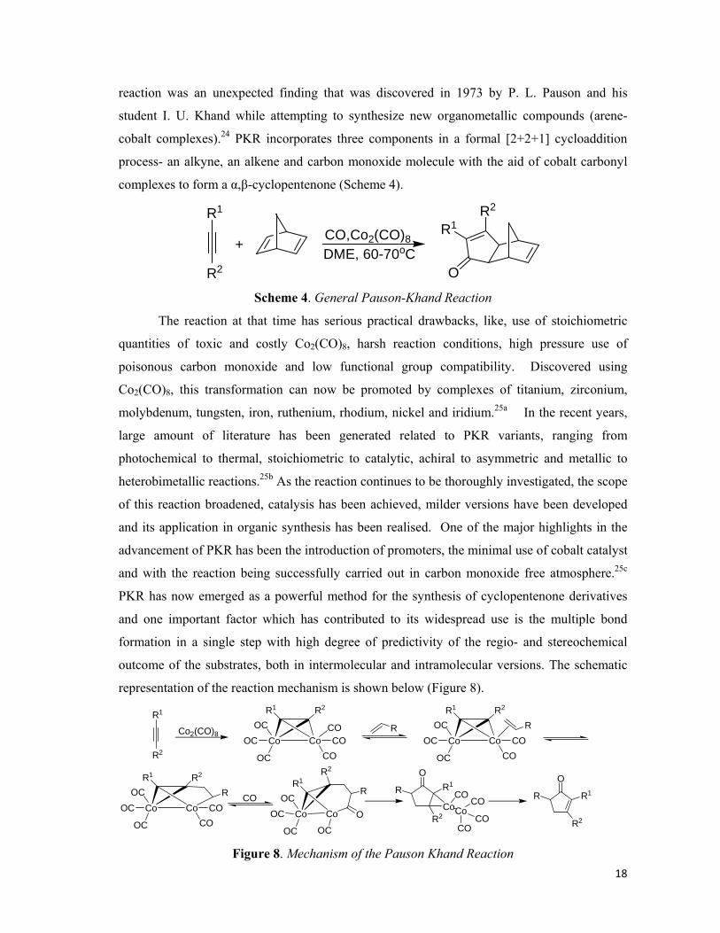
18
reaction was an unexpected finding that was discovered in 1973 by P. L. Pauson and his
student I. U. Khand while attempting to synthesize new organometallic compounds (arene-
cobalt complexes).24 PKR incorporates three components in a formal [2+2+1] cycloaddition
process- an alkyne, an alkene and carbon monoxide molecule with the aid of cobalt carbonyl
complexes to form a α,β-cyclopentenone (Scheme 4).
CO,Co2(CO)8
R1
R2 O
R1R2
DME, 60-70oC+
Scheme 4. General Pauson-Khand Reaction
The reaction at that time has serious practical drawbacks, like, use of stoichiometric
quantities of toxic and costly Co2(CO)8, harsh reaction conditions, high pressure use of
poisonous carbon monoxide and low functional group compatibility. Discovered using
Co2(CO)8, this transformation can now be promoted by complexes of titanium, zirconium,
molybdenum, tungsten, iron, ruthenium, rhodium, nickel and iridium.25a In the recent years,
large amount of literature has been generated related to PKR variants, ranging from
photochemical to thermal, stoichiometric to catalytic, achiral to asymmetric and metallic to
heterobimetallic reactions.25b As the reaction continues to be thoroughly investigated, the scope
of this reaction broadened, catalysis has been achieved, milder versions have been developed
and its application in organic synthesis has been realised. One of the major highlights in the
advancement of PKR has been the introduction of promoters, the minimal use of cobalt catalyst
and with the reaction being successfully carried out in carbon monoxide free atmosphere.25c
PKR has now emerged as a powerful method for the synthesis of cyclopentenone derivatives
and one important factor which has contributed to its widespread use is the multiple bond
formation in a single step with high degree of predictivity of the regio- and stereochemical
outcome of the substrates, both in intermolecular and intramolecular versions. The schematic
representation of the reaction mechanism is shown below (Figure 8).
R1
R2
Co2(CO)8Co Co
OC
OC
CO
COCOOC
R1 R2
Co CoOC
OC COCOOC
R1 R2
R
Co Co
OC
OC COCOOC
R1 R2
R
Co CoOC
OC OC
OC
R1R2
R
O
OR1
R2
R
CoCo
COCO
COCO R
O
R1
R2
R
CO
Figure 8. Mechanism of the Pauson Khand Reaction
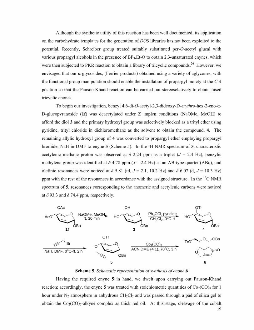
19
Although the synthetic utility of this reaction has been well documented, its application
on the carbohydrate templates for the generation of DOS libraries has not been exploited to the
potential. Recently, Schreiber group treated suitably substituted per-O-acetyl glucal with
various propargyl alcohols in the presence of BF3.Et2O to obtain 2,3-unsaturated enynes, which
were then subjected to PKR reaction to obtain a library of tricyclic compounds.26 However, we
envisaged that our α-glycosides, (Ferrier products) obtained using a variety of aglycones, with
the functional group manipulation should enable the installation of propargyl moiety at the C-4
position so that the Pauson-Khand reaction can be carried out stereoselctively to obtain fused
tricyclic enones.
To begin our investigation, benzyl 4,6-di-O-acetyl-2,3-dideoxy-D-erythro-hex-2-eno-α-
D-glucopyranoside (1f) was deacetylated under Z�mplen conditions (NaOMe, MeOH) to
afford the diol 3 and the primary hydroxyl group was selectively blocked as a trityl ether using
pyridine, trityl chloride in dichloromethane as the solvent to obtain the compound, 4. The
remaining allylic hydroxyl group of 4 was converted to propargyl ether employing propargyl
bromide, NaH in DMF to enyne 5 (Scheme 5). In the 1H NMR spectrum of 5, characteristic
acetylenic methane proton was observed at δ 2.24 ppm as a triplet (J = 2.4 Hz), benzylic
methylene group was identified at δ 4.78 ppm (J = 2.4 Hz) as an AB type quartet (ABq), and
olefinic resonances were noticed at δ 5.81 (td, J = 2.1, 10.2 Hz) and δ 6.07 (d, J = 10.3 Hz)
ppm with the rest of the resonances in accordance with the assigned structure. In the 13C NMR
spectrum of 5, resonances corresponding to the anomeric and acetylenic carbons were noticed
at δ 93.3 and δ 74.4 ppm, respectively.
O OBnTrO
O O
NaOMe, MeOHrt, 30 min
Ph3CCl, pyridineCH2Cl2, 0oC-rt
NaH, DMF, 0oC-rt, 2 h
Co2(CO)8ACN:DME (4:1), 70oC, 3 h
1f 3 4
5 6
Br
OBn
OOAc
AcO
OBn
OOH
HO
OBn
OOTr
HO
OBn
OOTr
O
Scheme 5. Schematic representation of synthesis of enone 6
Having the required enyne 5 in hand, we dwelt upon carrying out Pauson-Khand
reaction; accordingly, the enyne 5 was treated with stoichiometric quantities of Co2(CO)8 for 1
hour under N2 atmosphere in anhydrous CH2Cl2 and was passed through a pad of silica gel to
obtain the Co2(CO)6-alkyne complex as thick red oil. At this stage, cleavage of the cobalt

20
complex and carbonyl insertion became an arduous task. Initially we used N-methyl
morpholine N-oxide (NMO) in CH2Cl2 to effect cleavage of the cobalt complex, and we
obtained 6 in 70% yield. However, to our surprise, the reaction was sluggish (3 days) and
sensitive to the quality of NMO; hence, we sought alternate protocols. Heating the Co2(CO)6-
alkyne complex in the presence of other cleavage reagents such as cyclohexylamine or
acetonitrile did not result in the formation of the 6 but led to the decomposition of the cobalt
complex. Treatment of the Co2(CO)6-alkyne complex with H2O2 furnished the desired tricyclic
enone 6 in 30% yield. Use of dimethoxyethane and acetonitrile at 70 °C as the Co2(CO)6-
alkyne complex cleavage cocktail afforded the enone 6 in 92% yield (Scheme 5).
O OBnTrO
O O
6
H H H
.......
NOE interactions
Figure 9. NOE correlations of enone 6.
The 1H NMR spectrum of 6 showed no resonances corresponding to the enyne, and
new resonances characteristic of olefinic proton associated with the enone were observed at δ
6.00 ppm as a singlet. Resonances due to the benzylic -CH2 group were noticed at δ 4.67 ppm
as an ABq (J = 12.2 Hz), and that of anomeric proton were evident at δ 5.22 ppm as a doublet
(J = 7.8 Hz). It is pertinent to mention that the Pauson-Khand annulation occurred in a highly
diastereoselective fashion; the NOESY spectrum showed NOE cross-peaks between H-2, H-3,
H-4, and H-6 (Figure 9). Configuration at the C-4 position plays a predominant role in
dictating the chirality of the Pauson-Khand annulations as the Co2(CO)6-alkyne will be below
the plane. As a consequence, carbonyl insertion can take place from below only; hence, the
observation of a single diastereomer 6 with the simultaneous formation of three chiral centres.
NaOMe, MeOHrt, 30 min
Ph3CCl, pyridineCH2Cl2, 0oC-rt
1d 7 8
9 10
Co2(CO)8ACN:DME (4:1), 70oC, 3 h
OOAc
AcO
O
OOH
HO
O
OOTr
HO
O
OOTr
O
O
O OTrO
O ONaH, DMF, 0oC-rt, 2 h
Br
Scheme 6. Schematic representation for the synthesis of enone 10.

21
O
O O
TrO OOMe
OBn
OBnOBn
O
NaOMe, MeOHrt, 30 min
Ph3CCl, pyridineCH2Cl2, 0oC-rt
1i 11 12
13 14
Co2(CO)8ACN:DME (4:1), 70oC, 3 h
OOAc
AcO
OBnOBnO
OMeBnO
O
OOH
HO
OBnOBnO
OMeBnO
O
OOTr
HO
OBnOBnO
OMeBnO
O
OOTr
O
OBnOBnO
OMeBnO
ONaH, DMF, 0oC-rt, 2 h
Br
Scheme 7. Schematic representation for the synthesis of enone 14
Excited with our success and in progression with the introduction of more structural
complexity and diversity, we subjected the Ferrier products 1d, 1i and 1j to the same set of
reactions to get enones 10, 14 and 18 via intermediates 7-9, 11-13 and 15-17 as shown in
Scheme 6, 7 and 8 respectively. The practicality of the whole procedure was established, as
both of them resulted in the formation of the single diastereomers. The products were
confirmed by their 1H and 13C NMR spectra. Thrilled after having identified a practical route
for the synthesis of fused tricyclic enones with the successful formation of the three glucal
derived cyclopentenones, we decided to check the generality of the reaction on the other
glycals namely, galactal, xylal and rhamnal. This will add another dimension to our library
and would offer the much needed stereochemical diversity to our DOS library. Not only the
resulting library will be uniquely diverse but would also explore the new boundaries of the
chemical space. Persistent in our synthetic endeavours, we then subjected benzyl 4,6-di-O-
acetyl-2,3-dideoxy-D-erythro-hex-2-eno-α-D-galactopyranoside (2f), the Ferrier product, to
tritylation followed by alkylation of the resulting allylic alcohol with propargyl bromide, NaH
in DMF to yield compound 21. In the 1H NMR spectrum of the galactal derived enyne 21, the
distinguishable acetylenic proton was observed at δ 2.28 Hz as the triplet (J = 2.28 Hz), the
olefinic protons were located at δ 6.03 (dd, J = 2.79, 9.99 Hz) and δ 6.21 (td, J = 0.77, 5.19,
9.90 Hz). The 13C NMR spectrum of enyne 21, resonances corresponding to anomeric and
NaOMe, MeOHrt, 30 min
Ph3CCl, pyridineCH2Cl2, 0oC-rt
1j 15 16
Co2(CO)8ACN:DME (4:1), 70oC, 3 h
OOAc
AcO
OOBnO
OMeBnO
OBnO
OH
HO
OOBnO
OMeBnO
OBnO
OTr
HO
OOBnO
OMeBnO
OBn
OOTr
O
OOBnO
OMeBnO
OBn O
O O
TrOO
OMeBnO
BnOO
BnO
17 18
NaH, DMF, 0oC-rt, 2 h
Br
Scheme 8. Schematic representation for the synthesis of enone 18.

22
O OBnTrO
O O
NaOMe, MeOHrt, 30 min
Ph3CCl, pyridineCH2Cl2, 0oC-rt
NaH, DMF, 0oC-rt, 2 h
Co2(CO)8ACN:DME (4:1), 70oC, 3 h
2f 19 20
21 22
Br
OBn
OOAc
OBn
OOH
OBn
OOTr
OBn
OOTrO
AcO HO HO
Scheme 9. Schematic representation for the synthesis of enone 22
acetylenic carbon were noticed at δ 92.9 and δ 74.5 ppm respectively. Rest of the spectrum was
in accordance with the structure. Exposing enyne 21 to Pauson-Khand reaction conditions, we
were thrilled to have the enone 22 in 90% yield and as a single diastereomer (Scheme 9). The
product was confirmed by the presence of the distinguishable resonances of olefinic proton
associated with the enone at δ 5.93 as a singlet and with the disappearance of the respective
acetylenic peaks in the 1H NMR spectrum. The anomeric proton was observed at δ 97.5 ppm
in the 13C NMR spectrum. In accordance with the earlier observation, the formation of 22 was
envisioned to occur in a highly diastereoselective fashion. It is heart warming to mention that
contrary to glucal derived enyne 23, the insertion of the olefin into the cobalt-enyne complex
occurs from above the plane due to the configuration at C-4 atom and results in the formation
of a single diastereomer which is having complementary stereochemistry to the compound 24.
Continuing in our attempts to provide more structural complexity to our DOS and
improve the diversity of the carbohydrate library, we envisaged the formation of the rhamnal
and xylal based enones. Starting with the per-O-acetylated xylal, we performed the Ferrier
reaction employing benzyl alcohol as the aglycone in the presence of one mole percent of
NbCl5 in acetonitrile under the microwave conditions for one min. The corresponding
monosaccharide, 24, was subjected to same set of reactions as cited above for the sister
template to yield the enyne, 26. On the examination of 1H NMR spectrum of 26, the distinctive
triplet of the methine proton was observed at δ 2.45 (J = 2.28 Hz) along with the olefinic
protons at δ 6.01 (dd, J = 2.72, 10.23 Hz) and δ 6.14 (td, J = 1.12, 4.98 Hz).
O OBn
O O
NaOMe, MeOHrt, 30 min
NaH, DMF, 0oC-rt, 2 h
Co2(CO)8ACN:DME (4:1), 70oC, 3 h
23 24 25
26 27
Br
OBn
OAcO
OBn
OHO
OBn
OO
OAcO NbCl5(cat.), BnOHAcetonitrileAcO
Scheme 10. Schematic representation for the synthesis of enone 27

23
The 13C NMR spectrum showed anomeric and alkynic carbon at δ 92.2 and 74.7 ppm
respectively. The synthesis of the PKR product of 26 was carried out by heating the cobalt-
enyne complex in acetonitrile-dimethoxyethane at 70°C for 3 h to affect the cleavage of the
complex and result in the enone 27 (Scheme 10). The tricyclic enone, 27 was confirmed by the
disappearance of the acetylenic proton in the 1H NMR spectrum and the appearance of a singlet
at δ 6.01, the olefinic peak characteristic of the enone. The 13C NMR spectrum also confirmed
the product with the emergence of the carbonyl peak at δ 207.3 ppm. All the other peaks were
in agreement with the assigned structure.
O OBn
O O
NaOMe, MeOHrt, 30 min
NaH, DMF, 0oC-rt, 2 h
Co2(CO)8ACN:DME (4:1), 70oC, 3 h
23 24 25
26 27
Br
NbCl5(cat.), BnOHAcetonitrile
OAcO
CH3
OAcO
CH3
OAcO
CH3
OH
O
CH3
O
Scheme 11. Schematic representation for the synthesis of enone 27.
To add another feather into our carbohydrate template based library, we performed the
same string of reactions as mentioned for per-O-acetylated xylal on per-O-acetylated rhamnal
to acquire the final natural product like enone 32 (Scheme 11). All the substrates were
confirmed through NMR and elemental analysis. After the successful completion of the
synthesis of six natural product-like tricyclic enones, we performed an initial survey of the
substrate scope. We could successfully develop a general protocol for the synthesis of various
mono- and disaccharides derived from a variety of glycals such as per-O-acetylated glucal,
galactal, xylal, and rhamnal adopting our optimized aforementioned conditions. Not only are
the six compounds structurally complex but also offer elegant stereochemical diversity.
Third Level of Diversity: Michael Addition
The Michael addition of nucleophiles to electron deficient alkenes is one of the most
important reactions in organic chemistry for constructing carbon-sulfur bond. Among various
nucleophilic additions, the reaction of thiols to form a carbon-sulfur bond constitutes a key
reaction in the biosynthesis as well as in the synthesis of biologically active compounds such as
calcium antagonist diltiazem. The α,β-unsaturation of the products from Pauson-Khand
products offers enough possibility for the diversification for exposing them to the Michael
addition using thiols. In continuation of our efforts for offering more complexity to our
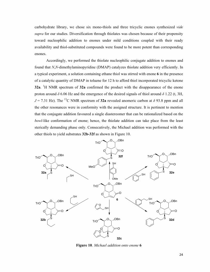
24
carbohydrate library, we chose six mono-thiols and three tricyclic enones synthesized vide
supra for our studies. Diversification through thiolates was chosen because of their propensity
toward nucleophilic addition to enones under mild conditions coupled with their ready
availability and thiol-substituted compounds were found to be more potent than corresponding
enones.
Accordingly, we performed the thiolate nucleophilic conjugate addition to enones and
found that N,N-dimethylaminopyridine (DMAP) catalyzes thiolate addition very efficiently. In
a typical experiment, a solution containing ethane thiol was stirred with enone 6 in the presence
of a catalytic quantity of DMAP in toluene for 12 h to afford thiol incorporated tricyclic ketone
32a. 1H NMR spectrum of 32a confirmed the product with the disappearance of the enone
proton around δ 6.06 Hz and the emergence of the desired signals of thiol around δ 1.22 (t, 3H,
J = 7.31 Hz). The 13C NMR spectrum of 32a revealed anomeric carbon at δ 93.8 ppm and all
the other resonances were in conformity with the assigned structure. It is pertinent to mention
that the conjugate addition favoured a single diastereomer that can be rationalized based on the
bowl-like conformation of enone; hence, the thiolate addition can take place from the least
sterically demanding phase only. Consecutively, the Michael addition was performed with the
other thiols to yield substrates 32b-32f as shown in Figure 10.
O
O O
TrOOBn
S
O
O O
TrOOBn
S
O
O O
TrOOBn
S
O
O O
TrOOBn
S
MeO
O
O O
TrOOBn
SO
O O
TrOOBn
SO
O
O O
TrOOBn
6
32a
32b
32c
32d
32e
32f
SH
SH
SHO
SH
SHOMe
SH
Figure 10. Michael addition onto enone 6

25
With the established protocol and successful formation of the six Michael adducts from
enone 6, we tried the same reaction on the xylal-derived enone 27. As a general synthetic
procedure, the enone, 27 was taken in toluene and allowed to stir in the presence of ethane thiol
to afford the product 33a as a single isomer only. The stereochemistry of the resulting Michael
adduct was defined taking a cue from the spatial arrangement of 33a, as the fundamental
skeleton is identical to both of them. Hereafter, the other thiols were promptly were reacted
with the enone 27 to afford the Michael products 33b-33f (Figure 11).
O
O O
OBn
S
O
O O
OBn
S
O
O O
OBn
S
O
O O
OBn
S
MeO
O
O O
OBn
SO
O O
OBn
SO
O
O O
OBn
27
33a
33b
33c
33d
33e
33f
SH
SH
SHO
SH
SHOMe
SH
Figure 11. Michael addition onto enone 27.
Delighted with the synthesis of the Michael adducts for glucal derived library, we
undertook synthesis of the same Michael products with the galactal derived enone to have more
stereochemical flexibility in our library. We envisaged that the conjugate addition onto enone
22 would afford the complementary stereochemistry to compound 6. This could be rationally
explained on the grounds that both them are having complementary structures to each other,
bowl-like structures, and the addition takes place from the least hindered side to afford single
isomer only. In the usual experiment, the enone 22 was dissolved in toluene and was stirred
with ethane thiol for 12 hr. After the completion of the reaction, inspection of the 1H NMR
spectrum of 34a revealed the characteristic resonances of thiol at δ 1.24 (t, 3H, J = 7.48 Hz)

26
and the 13C NMR spectrum also confirmed the product with all the carbons at the expected
positions along with the anomeric carbon at δ 95.5 ppm. Consecutively, the remaining thiol
adducts were synthesized following the same protocol to yield the compounds 34b-34f (Figure
12).
O
O O
TrOOBn
S
O
O O
TrOOBn
S
O
O O
TrOOBn
S
O
O O
TrOOBn
S
MeO
O
O O
TrOOBn
SO
O O
TrOOBn
SO
O
O O
TrOOBn
22
34a
34b
34c
34d
34e
34f
SH
SH
SHO
SH
SHOMe
SH
Figure 12. Michael addition onto enone 22
In conclusion, we have developed a practical protocol for the synthesis of natural
product-like tricyclic compounds and a total of 25 tricyclic compounds were synthesized as a
pilot library, using the Ferrier, Pauson-Khand reactions and the conjugate thiolate addition to
enones were performed under mild reaction conditions to afford a thiol-substituted library.27 It
is pertinent to mention that the complexity generating reactions (viz., the Ferrier, Pauson-
Khand, and Michael addition reactions) were highly diastereoselective thereby enabling
chirally pure, oxygen-rich, tricyclic derivatives from easily accessible glycals. A little
examination of the pilot library beautifully illustrates the fact that the collection of the
compounds possesses a high level of structural complexity and diversity. On one hand, the
complementary spatial arrangement of the glucal and galactal provided the stereochemical
flexibility on the other hand the structural dissimilarity of the xylal and rhamnal provides an
advantage of structural variation as well.
*****
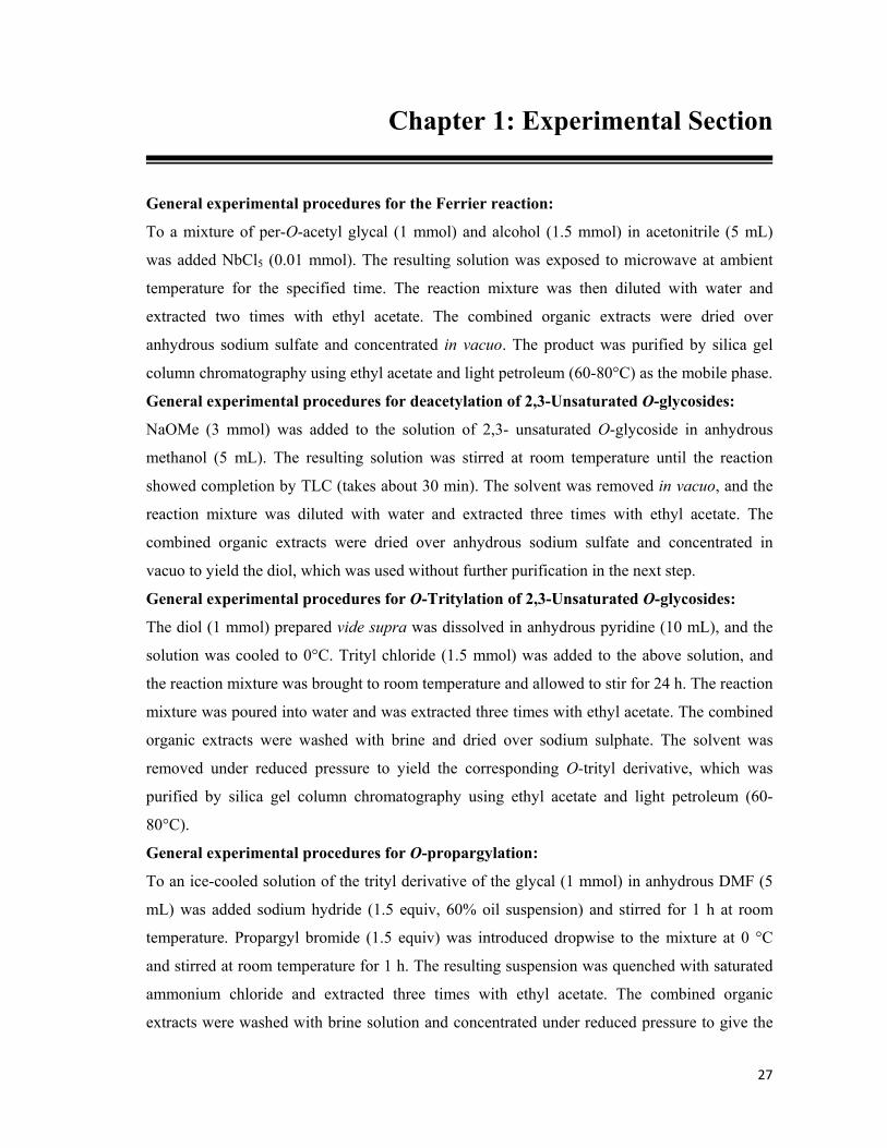
27
Chapter 1: Experimental Section
General experimental procedures for the Ferrier reaction:
To a mixture of per-O-acetyl glycal (1 mmol) and alcohol (1.5 mmol) in acetonitrile (5 mL)
was added NbCl5 (0.01 mmol). The resulting solution was exposed to microwave at ambient
temperature for the specified time. The reaction mixture was then diluted with water and
extracted two times with ethyl acetate. The combined organic extracts were dried over
anhydrous sodium sulfate and concentrated in vacuo. The product was purified by silica gel
column chromatography using ethyl acetate and light petroleum (60-80°C) as the mobile phase.
General experimental procedures for deacetylation of 2,3-Unsaturated O-glycosides:
NaOMe (3 mmol) was added to the solution of 2,3- unsaturated O-glycoside in anhydrous
methanol (5 mL). The resulting solution was stirred at room temperature until the reaction
showed completion by TLC (takes about 30 min). The solvent was removed in vacuo, and the
reaction mixture was diluted with water and extracted three times with ethyl acetate. The
combined organic extracts were dried over anhydrous sodium sulfate and concentrated in
vacuo to yield the diol, which was used without further purification in the next step.
General experimental procedures for O-Tritylation of 2,3-Unsaturated O-glycosides:
The diol (1 mmol) prepared vide supra was dissolved in anhydrous pyridine (10 mL), and the
solution was cooled to 0°C. Trityl chloride (1.5 mmol) was added to the above solution, and
the reaction mixture was brought to room temperature and allowed to stir for 24 h. The reaction
mixture was poured into water and was extracted three times with ethyl acetate. The combined
organic extracts were washed with brine and dried over sodium sulphate. The solvent was
removed under reduced pressure to yield the corresponding O-trityl derivative, which was
purified by silica gel column chromatography using ethyl acetate and light petroleum (60-
80°C).
General experimental procedures for O-propargylation:
To an ice-cooled solution of the trityl derivative of the glycal (1 mmol) in anhydrous DMF (5
mL) was added sodium hydride (1.5 equiv, 60% oil suspension) and stirred for 1 h at room
temperature. Propargyl bromide (1.5 equiv) was introduced dropwise to the mixture at 0 °C
and stirred at room temperature for 1 h. The resulting suspension was quenched with saturated
ammonium chloride and extracted three times with ethyl acetate. The combined organic
extracts were washed with brine solution and concentrated under reduced pressure to give the

28
OBn
OOTr
HO
crude O-propargyl derivative that was purified by silica gel column chromatography using
ethyl acetate and light petroleum (60-80°C).
General experimental procedures for Pauson-Khand Reaction:
To an ice-cooled solution of alkyne in anhydrous dichloromethane was added Co2(CO)8 (1.2
equiv) and stirred at room temperature for 1 h. The reaction mixture was adsorbed on silica gel
and was eluted with ethyl acetate and light petroleum to yield the cobalt-enyne complex as
thick red oil. Subsequently, the Co-alkyne complex was redissolved in anhydrous 1,2-
dimethoxy ethane (5 mL) and anhydrous acetonitrile (20 mL) and was refluxed until the colour
of the solution changed from thick red to greyish black. The solution was filtered through a pad
of silica gel and concentrated to give light yellow oil, which was purified by silica gel column
chromatography using ethyl acetate and light petroleum (60-80°C).
General experimental procedures for thiolate additions on enones:
The tricyclic enone (1 equiv) was dissolved in anhydrous toluene (10 mL/mmol), and the thiol
(2 equiv) was added to it. The resulting solution was stirred in the presence of catalytic amount
of DMAP until the reaction showed completion by TLC (typically takes about 12-24 h).
Thereafter the solvent was removed in vacuo, and the adduct was purified by silica gel column
chromatography using ethyl acetate and light petroleum (60-80°C).
Compound Characterization Data:
Compound 4: [α]D (CHCl3, c 0.9) = + 50.6. IR (cm-1) = 3455. 1H
NMR (200 MHz, CDCl3): δ 3.29-3.46 (m, 2H), 3.83-3.94 (m, 1H),
4.02-4.12 (m, 1H), 4.72 (ABq, 2H, J = 11.77 Hz), 5.05 (s, 1H), 5.76
(td, 1H, 2.38, 10.17 Hz), 5.92 (td, 1H, 1.56, 10.16 Hz), 7.23-7.35 ( m,
14H), 7.44-7.50 ( m, 6H). 13C NMR (50 MHz, CDCl3): δ 61.8, 63.4, 70.0, 71.5, 93.4, 125.5,
127.7-129.6, 137.5, 133.6, 142.5. Anal. Calcd. for C32H30O3: C, 83.09; H, 6.54. Found C,
82.95; H, 6.64.
Compound 5: [α]D (CHCl3, c 0.7) = +33.6°. IR (cm-1) = 3303. 1H
NMR (200 MHz, CDCl3): δ 2.24 (t, 1H, J = 2.39 Hz), 3.19 (dd, 1H, J =
5.44, 10.31 Hz), 3.37 (dd, 1H, J = 1.34, 10.23 Hz), 4.01 (m, 3H), 4.23
(dd, 1H, J = 1.18, 9.46 Hz), 4.78 (ABq, 2H, J = 11.80 Hz), 5.15 (d, 1H,
J = 1.62 Hz), 5.81 (td, 1H, J = 2.14, 10.22 Hz), 6.07 (d, 1H, J = 10.28 Hz), 7.15-7.42 (m, 14H),
7.47-7.54 (m, 6H). 13C NMR (50 MHz, CDCl3): δ 56.6, 63.1, 69.4, 69.7, 70.1, 74.4, 79.7, 86.4,
OBn
OOTr
O

29
93.3, 126.7-128.8, 131.0, 137.9, 144.1. Anal. Calcd. for C35H32O4: C, 81.37; H, 6.24. Found: C,
80.96; H, 6.21.
Compound 6: [α]D (CHCl3, c 0.9) = +177.9°. IR (cm-1) = 1716, 1647. 1H NMR (500 MHz, CDCl3): δ 3.21-3.33 (m, 4H), 3.59 (td, 1H, J =
1.98, 7.34 Hz), 3.95 (t, 1H, J = 9.13 Hz), 4.45 (m, 2H), 4.67 (ABq, 2H,
J = 12.18 Hz), 5.22 (d, 1H, J = 7.76 Hz), 6.00 (s, 1H), 7.12-7.31 (m, 14H), 7.33-7.45 (m, 6H). 13C NMR (50 MHz, CDCl3): δ 45.5, 1.5, 64.0, 65.1, 65.7, 68.9, 71.2, 86.8, 94.3, 125.2, 126.8-
128.7, 137.3, 143.9, 178.36, 206.7. Anal. Calcd. for C36H32O5: C, 79.39; H, 5.92. Found: C,
79.89; H, 6.20. MALDI-TOF: mol wt calcd 544.64; found 567.17 (M+ + 23 for Na).
Compound 8: [α]D (CHCl3, c 0.7) = -48.35°. IR (cm-1) = 3460. 1H NMR (500 MHz, CDCl3): δ 0.64-1.01 (m, 11H), 1.12-1.31 (m,
3H), 1.58-1.65 (m, 2H), 2.05-2.12 (m, 2H), 2.61 (d, 1H, J = 4.68
Hz), 3.25-3.53 (m, 3H), 3.85-3.95 (m, 1H), 4.08-4.15 (m,1H), 5.01
(s, 1H), 5.73 (td, 1H, J = 2.28, 10.20 Hz), 5.88 (m, 1H), 7.17-7.34
(m, 9H), 7.43-7.49 (m, 6H). 13C NMR (50 MHz, CDCl3): δ 16.2, 21.1, 22.3, 23.2, 25.5, 31.7,
34.3, 43.5, 48.8, 65.5, 66.6, 69.4, 80.3, 87.4, 95.8, 126.0-128.5, 132.6, 143.5. Anal. calcd. for
C35H42O3: C, 82.31; H, 8.29. Found: C, 82.39; H, 8.32.
Compound 9: [α]D (CHCl3, c 1.6) = +8.76°. IR (cm-1): 3216. 1H
NMR (200 MHz, CDCl3): δ 0.72, 0.75, 0.79, 0.83, 0.89, 0.93 (6s,
9H), 0.98-1.11 (m, 3H), 1.19-1.70 (m, 4H), 2.05-2.35 (m, 3H),
3.23 (dd, 1H, J = 4.04, 10.23 Hz), 3.38-3.58 (m, 2H), 3.85-4.08
(m, 3H), 4.24 (td, 1H, J = 0.9, 9.23 Hz), 5.14 (s, 1H), 5.73-6.10
(m, 2H), 7.16-7.35 (m, 9H), 7.40-7.56 (m, 6H). 13C NMR (50 MHz, CDCl3): δ 16.2, 21.1, 22.2,
23.2, 25.6, 31.8, 34.4, 43.3, 48.8, 56.5, 63.1, 69.0, 70.5, 74.3, 79.8, 80.5, 86.4, 96.1, 126.8-
128.8, 130.5, 143.9, 144.0. Anal. Calcd. for C38H44O4: C, 80.82; H, 7.85. Found: C, 80.49; H,
7.89.
Compound 10: [α]D (CHCl3, c 1.0) = +147.5°. IR (cm-1): 1749,
1454. 1H NMR (200 MHz, CDCl3): δ 0.60, 0.63 (2s, 3H), 0.76, 0.79,
0.86, 0.92 (4s, 6 H), 0.83-0.97 (m, 3H), 1.08-1.22 (m, 2H), 1.58 (m,
3H), 2.11 (m, 2 H), 3.24-4.45 (m, 5H), 4.27 (dd, 1 H, J = 8.40, 9.61
Hz), 4.56 (bs, 2 H), 5.26 (d, 1H, J = 7.59 Hz), 5.99 (d, 1 H, J = 1.51
Hz), 7.10-7.35 (m, 9H), 7.42-7.53 (m, 6H). 13C NMR (50 MHz, CDCl3): δ 15.8, 21.4, 22.2,
22.6, 24.7, 31.6, 34.2, 42.3, 45.7, 49.0, 52.2, 63.8, 65.2, 65.4, 71.3, 80.1, 86.8, 96.3, 125.0,
O OBnTrO
O O
O OTrO
O O
OOTr
HO
O
OOTr
O
O
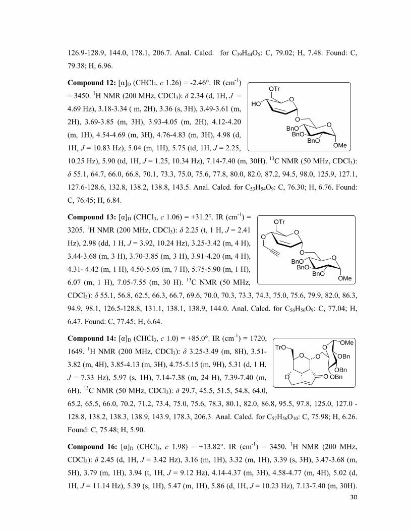
30
126.9-128.9, 144.0, 178.1, 206.7. Anal. Calcd. for C39H44O5: C, 79.02; H, 7.48. Found: C,
79.38; H, 6.96.
Compound 12: [α]D (CHCl3, c 1.26) = -2.46°. IR (cm-1)
= 3450. 1H NMR (200 MHz, CDCl3): δ 2.34 (d, 1H, J =
4.69 Hz), 3.18-3.34 ( m, 2H), 3.36 (s, 3H), 3.49-3.61 (m,
2H), 3.69-3.85 (m, 3H), 3.93-4.05 (m, 2H), 4.12-4.20
(m, 1H), 4.54-4.69 (m, 3H), 4.76-4.83 (m, 3H), 4.98 (d,
1H, J = 10.83 Hz), 5.04 (m, 1H), 5.75 (td, 1H, J = 2.25,
10.25 Hz), 5.90 (td, 1H, J = 1.25, 10.34 Hz), 7.14-7.40 (m, 30H). 13C NMR (50 MHz, CDCl3):
δ 55.1, 64.7, 66.0, 66.8, 70.1, 73.3, 75.0, 75.6, 77.8, 80.0, 82.0, 87.2, 94.5, 98.0, 125.9, 127.1,
127.6-128.6, 132.8, 138.2, 138.8, 143.5. Anal. Calcd. for C53H54O9: C, 76.30; H, 6.76. Found:
C, 76.45; H, 6.84.
Compound 13: [α]D (CHCl3, c 1.06) = +31.2°. IR (cm-1) =
3205. 1H NMR (200 MHz, CDCl3): δ 2.25 (t, 1 H, J = 2.41
Hz), 2.98 (dd, 1 H, J = 3.92, 10.24 Hz), 3.25-3.42 (m, 4 H),
3.44-3.68 (m, 3 H), 3.70-3.85 (m, 3 H), 3.91-4.20 (m, 4 H),
4.31- 4.42 (m, 1 H), 4.50-5.05 (m, 7 H), 5.75-5.90 (m, 1 H),
6.07 (m, 1 H), 7.05-7.55 (m, 30 H). 13C NMR (50 MHz,
CDCl3): δ 55.1, 56.8, 62.5, 66.3, 66.7, 69.6, 70.0, 70.3, 73.3, 74.3, 75.0, 75.6, 79.9, 82.0, 86.3,
94.9, 98.1, 126.5-128.8, 131.1, 138.1, 138.9, 144.0. Anal. Calcd. for C56H56O9: C, 77.04; H,
6.47. Found: C, 77.45; H, 6.64.
Compound 14: [α]D (CHCl3, c 1.0) = +85.0°. IR (cm-1) = 1720,
1649. 1H NMR (200 MHz, CDCl3): δ 3.25-3.49 (m, 8H), 3.51-
3.82 (m, 4H), 3.85-4.13 (m, 3H), 4.75-5.15 (m, 9H), 5.31 (d, 1 H,
J = 7.33 Hz), 5.97 (s, 1H), 7.14-7.38 (m, 24 H), 7.39-7.40 (m,
6H). 13C NMR (50 MHz, CDCl3): δ 29.7, 45.5, 51.5, 54.8, 64.0,
65.2, 65.5, 66.0, 70.2, 71.2, 73.4, 75.0, 75.6, 78.3, 80.1, 82.0, 86.8, 95.5, 97.8, 125.0, 127.0 -
128.8, 138.2, 138.3, 138.9, 143.9, 178.3, 206.3. Anal. Calcd. for C57H56O10: C, 75.98; H, 6.26.
Found: C, 75.48; H, 5.90.
Compound 16: [α]D (CHCl3, c 1.98) = +13.82°. IR (cm-1) = 3450. 1H NMR (200 MHz,
CDCl3): δ 2.45 (d, 1H, J = 3.42 Hz), 3.16 (m, 1H), 3.32 (m, 1H), 3.39 (s, 3H), 3.47-3.68 (m,
5H), 3.79 (m, 1H), 3.94 (t, 1H, J = 9.12 Hz), 4.14-4.37 (m, 3H), 4.58-4.77 (m, 4H), 5.02 (d,
1H, J = 11.14 Hz), 5.39 (s, 1H), 5.47 (m, 1H), 5.86 (d, 1H, J = 10.23 Hz), 7.13-7.40 (m, 30H).
O
O O
TrO OOMe
OBn
OBnOBn
O
OOTr
HO
OBnOBnO
OMeBnO
O
OOTr
O
OBnOBnO
OMeBnO
O
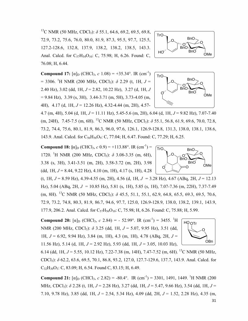
31
13C NMR (50 MHz, CDCl3): δ 55.1, 64.6, 69.2, 69.5, 69.8,
72.9, 73.2, 75.6, 76.0, 80.0, 81.9, 87.3, 95.5, 97.7, 125.5,
127.2-128.6, 132.8, 137.9, 138.2, 138.2, 138.5, 143.3.
Anal. Calcd. for C57H54O10: C, 75.98; H, 6.26. Found: C,
76.08; H, 6.44.
Compound 17: [α]D (CHCl3, c 1.08) = +35.34°. IR (cm-1)
= 3306. 1H NMR (200 MHz, CDCl3): δ 2.29 (t, 1H, J =
2.40 Hz), 3.02 (dd, 1H, J = 2.82, 10.22 Hz), 3.27 (d, 1H, J
= 9.84 Hz), 3.39 (s, 3H), 3.44-3.71 (m, 5H), 3.73-4.05 (m,
4H), 4.17 (d, 1H, J = 12.26 Hz), 4.32-4.44 (m, 2H), 4.57-
4.7 (m, 4H), 5.04 (d, 1H, J = 11.11 Hz), 5.45-5.6 (m, 2H), 6.04 (d, 1H, J = 9.82 Hz), 7.07-7.40
(m, 24H), 7.45-7.5 (m, 6H). 13C NMR (50 MHz, CDCl3): δ 55.1, 56.8, 61.9, 69.6, 70.0, 72.8,
73.2, 74.4, 75.6, 80.1, 81.9, 86.3, 96.0, 97.6, 126.1, 126.9-128.8, 131.3, 138.0, 138.1, 138.6,
143.9. Anal. Calcd. for C56H56O9: C, 77.04; H, 6.47. Found: C, 77.29; H, 6.25.
Compound 18: [α]D (CHCl3, c 0.9) = +113.88°. IR (cm-1) =
1720. 1H NMR (200 MHz, CDCl3): δ 3.08-3.35 (m, 6H),
3.38 (s, 3H), 3.41-3.51 (m, 2H), 3.58-3.72 (m, 2H), 3.98
(dd, 1H, J = 8.44, 9.22 Hz), 4.10 (m, 1H), 4.17 (s, 1H), 4.28
(t, 1H, J = 8.59 Hz), 4.39-4.55 (m, 2H), 4.56 (d, 1H, J = 3.28 Hz), 4.67 (ABq, 2H, J = 12.13
Hz), 5.04 (ABq, 2H, J = 10.85 Hz), 5.81 (s, 1H), 5.85 (s, 1H), 7.07-7.36 (m, 22H), 7.37-7.49
(m, 8H). 13C NMR (50 MHz, CDCl3): δ 45.5, 51.1, 55.1, 62.9, 64.8, 65.5, 69.3, 69.5, 70.6,
72.9, 73.2, 74.8, 80.3, 81.9, 86.7, 94.6, 97.7, 125.0, 126.9-128.9, 138.0, 138.2, 139.1, 143.9,
177.9, 206.2. Anal. Calcd. for C57H56O10: C, 75.98; H, 6.26. Found: C, 75.88; H, 5.99.
Compound 20: [α]D (CHCl3, c 2.84) = - 52.99°. IR (cm-1) = 3455. 1H
NMR (200 MHz, CDCl3): δ 3.25 (dd, 1H, J = 5.07, 9.95 Hz), 3.51 (dd,
1H, J = 6.92, 9.94 Hz), 3.84 (m, 1H), 4.3 (m, 1H), 4.78 (ABq, 2H, J =
11.56 Hz), 5.14 (d, 1H, J = 2.92 Hz), 5.93 (dd, 1H, J = 3.05, 10.03 Hz),
6.14 (dd, 1H, J = 5.55, 10.12 Hz), 7.22-7.38 (m, 14H), 7.47-7.52 (m, 6H). 13C NMR (50 MHz,
CDCl3): δ 62.2, 63.6, 69.5, 70.1, 86.8, 93.2, 127.0, 127.7-129.6, 137.7, 143.9. Anal. Calcd. for
C32H30O3: C, 83.09; H, 6.54. Found C, 83.15; H, 6.49.
Compound 21: [α]D (CHCl3, c 2.82) = -80.4°. IR (cm-1) = 3301, 1491, 1449. 1H NMR (200
MHz, CDCl3): δ 2.28 (t, 1H, J = 2.28 Hz), 3.27 (dd, 1H, J = 5.47, 9.66 Hz), 3.54 (dd, 1H, J =
7.10, 9.78 Hz), 3.85 (dd, 1H, J = 2.54, 5.34 Hz), 4.09 (dd, 2H, J = 1.52, 2.28 Hz), 4.35 (m,
O
O O
TrOO
OMeBnO
BnOO
BnO
O
HO
TrO
O
OMeBnO
BnOO
BnO
O
O
TrO
O
OMeBnO
BnOO
BnO
OBn
OOTrHO

32
1H), 4.73 (ABq, 2H, J = 11.63 Hz), 5.15 (d, 1H, J ) 2.98 Hz), 6.02
(dd, 1H, J = 2.79, 9.99 Hz), 6.21 (ddd, 1H, J = 0.77, 5.19, 9.90 Hz),
7.15-7.42 (m, 14H), 7.45-7.55 (m, 6H). 13C NMR (50 MHz,
CDCl3): δ 56.2, 63.1, 67.0, 69.4, 69.8, 74.5, 79.8, 86.6, 92.9, 126.7-
128.7, 129.9, 137.6, 143.9. Anal. Calcd. for C35H32O4: C, 81.37; H, 6.24. Found: C, 80.65; H,
6.58.
Compound 22: [α]D (CHCl3, c 0.8) = -130.9°. IR (cm-1) = 1738, 1713. 1H NMR (200 MHz, CDCl3): δ 2.79 (d, 1 H, J = 6.39 Hz), 3.25 (dd, 1
H, J = 2.29, 10.53 Hz), 3.33 (m, 1 H), 3.43-3.64 (m, 1H), 4.09 (dd, 1H,
J = 3.24, 9.62 Hz), 4.18 (dd, 1H, J = 4.10, 7.79 Hz), 4.31 (ABq, 2H, J = 14.75 Hz), 4.74 (ABq,
2H, J = 11.45 Hz), 4.03 (d, 1H, J = 11.85 Hz), 5.93 (s, 1H), 7.12-7.38 (m, 14H), 7.41-7.48 (m,
6H). 13C NMR (50 MHz, CDCl3): δ 44.1, 51.7, 63.0, 65.8, 68.0, 69.3, 71.0, 86.9, 97.5, 124.0,
126.9-128.8, 137.0, 144.0, 183.6, 209.4. Anal. Calcd. for C36H32O5: C, 79.39; H, 5.92. Found:
C, 79.61; H, 5.58.
Compound 26: [α]D (CHCl3, c 1.0) = +69.4°. IR (cm-1) = 3306. 1H
NMR (200 MHz, CDCl3): δ 2.45 (t, 1H, J = 2.28 Hz), 3.87 m, 1H), 3.96
(td, 1H, J = 1.10, 12.81 Hz), 4.12 (dd, 1H, J = 2.69, 4.12 Hz), 4.26 (d,
2H, J = 2.41 Hz), 4.68 (ABq, 2H, J = 11.77 Hz), 5.09 (d, 1H, J = 2.35 Hz), 6.01 (dd, 1H, J =
2.72, 10.23 Hz), 6.14 (td, 1H, J = 1.12, 4.98 Hz), 7.25-7.38 (m, 5H). 13C NMR (50 MHz,
CDCl3): δ 55.5, 61.1, 66.6, 69.7, 74.7, 79.7, 92.2, 126.2, 127.7, 128.1, 128.4, 130.1, 137.6.
Anal. Calcd. for C15H16O3: C, 73.75; H, 6.60. Found: C, 73.69; H, 6.61.
Compound 27: [α]D (CHCl3, c 1.5) = +52.2°. IR (cm-1) = 1738, 1713. 1H
NMR (200 MHz, CDCl3): δ 2.92 (d, 1H, J = 6.78 Hz), 3.51 (m, 2H), 4.03
(dd, 1H, J = 5.30, 13.09 Hz), 4.38 (m, 1H), 4.58 (ABq, 2H, J = 11.50 Hz),
4.67 (t, 2H, J = 14.29 Hz), 4.84 (s, 1H), 6.10 (s, 1H), 7.28-7.40 (m, 5H). 13C NMR (50 MHz,
CDCl3): δ 43.8, 51.5, 62.2, 65.2, 70.0, 70.1, 96.8, 124.6, 128.0, 128.5, 136.8, 183.5, 208.7.
Anal. Calcd. for C16H16O4: C, 70.57; H, 5.92. Found: C, 70.74; H, 6.29.
Compound 31: [α]D (CHCl3, c 1.7) = -96.4°. IR (cm-1) = 3288. 1H
NMR (200 MHz, CDCl3): δ 1.27 (d, 3H, J = 5.78 Hz), 2.43 (t, 1H, J =
2.39 Hz), 3.85 (m, 2H), 4.23 (d, 2H, J = 2.32 Hz), 4.68 (ABq, 2H, J =
12.00 Hz), 5.03 (d, 1H, J = 2.61 Hz), 5.79 (ddd, 1H, J = 1.76, 2.65,
O OBnTrO
O O
O OBn
O O
OBn
OOTrO
OBn
OO
O
CH3 OBn
O
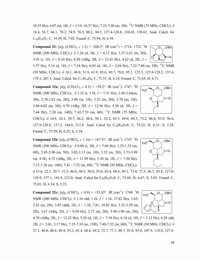
33
10.25 Hz), 6.07 (td, 1H, J = 1.19, 10.37 Hz), 7.23-7.40 (m, 5H). 13C NMR (75 MHz, CDCl3): δ
18.4, 56.7, 66.1, 70.3, 74.9, 76.5, 80.2, 94.1, 127.4-128.8, 130.65, 138.62. Anal. Calcd. for
C16H18O3: C, 74.39; H, 7.02. Found: C, 73.94; H, 6.58.
Compound 32: [α]D (CHCl3, c 1.2) = -268.2°. IR (cm-1) = 1714, 1722. 1H
NMR (200 MHz, CDCl3): δ 1.24 (d, 3H, J = 6.13 Hz), 3.27-3.61 (m, 3H),
3.93 (t, 1H, J = 9.10 Hz), 4.50 (ABq, 2H, J = 12.41 Hz), 4.62 (d, 2H, J =
1.57 Hz), 5.16 (d, 1H, J = 7.54 Hz), 6.05 (d, 1H, J = 2.04 Hz), 7.22-7.40 (m, 5H). 13C NMR
(50 MHz, CDCl3): δ 18.1, 46.0, 51.9, 61.9, 85.6, 69.7, 78.0, 95.1, 125.3, 125.8-128.2, 137.6,
179.1, 207.3. Anal. Calcd. for C17H18O4: C, 71.31; H, 6.34. Found: C, 71.65; H, 6.71.
Compound 32a: [α]D (CH2Cl2, c 0.3) = +59.2°. IR (cm-1): 1747. 1H
NMR (200 MHz, CDCl3): δ 1.22 (t, 3 H, J = 7.31 Hz), 2.48-2.64(m,
3H), 2.70-2.83 (m, 2H), 3.08 (m, 1H), 3.32 (m, 2H), 3.70 (m, 1H),
3.86-4.02 (m, 3H), 4.70 (ABq, 2H, J = 12.56 Hz), 5.30 (d, 1H, J =
7.44 Hz), 7.20 (m, 14H), 7.42-7.53 (m, 6H). 13C NMR (75 MHz,
CDCl3): δ 14.6, 24.1, 29.7, 46.2, 48.6, 50.1, 52.2, 65.5, 69.0, 69.3, 73.2, 86.8, 93.9, 96.6,
127.0-128.8, 137.2, 144.0, 211.8. Anal. Calcd for C38H38O5S: C, 75.22; H, 6.31; S, 5.28.
Found: C, 75.59; H, 6.25; S, 5.34.
Compound 32b: [α]D (CHCl3, c 1.16) = +67.51°. IR (cm-1): 1747. 1H
NMR (200 MHz, CDCl3): δ 0.80 (t, 3H, J = 7.04 Hz), 1.25-1.53 (m,
4H), 2.45-2.90 (m, 5H), 3.02-3.13 (m, 1H), 3.32 (m, 2H), 3.73-3.99
(m, 4 H), 4.72 (ABq, 2H, J = 11.99 Hz), 5.30 (d, 1H, J = 7.50 Hz),
7.15-7.36 (m, 14H), 7.41 - 7.52 (m, 6H). 13C NMR (50 MHz, CDCl3):
δ 13.6, 22.1, 29.7, 31.5, 46.0, 48.5, 50.0, 55.0, 65.4, 68.8, 69.1, 73.0, 77.3, 86.7, 93.8, 127.0-
128.9, 137.1, 143.9, 212.0. Anal. Calcd for C40H42O5S: C, 75.68; H, 6.67; S, 5.05. Found: C,
75.01; H, 6.34; S, 5.23.
Compound 32c: [α]D (CHCl3, c 0.9) = +53.85°. IR (cm-1): 1748. 1H
NMR (200 MHz, CDCl3): δ 1.16 (dd, 1 H, J = 1.16, 17.82 Hz), 2.65-
2.82 (m, 2H), 3.05 (ddd, 1H, J = 1.38, 7.81, 10.82 Hz), 3.25-3.39 (m,
2H), 3.67 (ABq, 2H, J = 9.58 Hz), 3.77 (m, 2H), 3.90-3.98 (m, 2H),
4.70 (ABq, 2H, J = 12.22 Hz), 5.28 (d, 1H, J = 7.54 Hz), 6.14 (d, 1H, J = 3.12 Hz), 6.28 (dd,
1H, J = 2.01, 3.17 Hz), 7.15-7.39 (m, 15H), 7.40-7.52 (m, 6H). 13C NMR (50 MHz, CDCl3): δ
27.1, 46.0, 48.4, 49.4, 55.3, 65.4, 68.8, 69.2, 72.7, 77.1, 86.7, 91.0, 93.8, 107.9, 110.8, 127.0-
O OBn
O O
O
O O
TrOOBn
S
O
O O
TrOOBn
S
O
O O
TrOOBn
SO
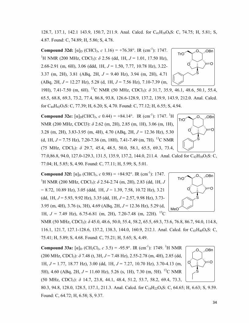
34
128.7, 137.1, 142.1 143.9, 150.7, 211.9. Anal. Calcd. for C41H38O6S: C, 74.75; H, 5.81; S,
4.87. Found: C, 74.89; H, 5.86; S, 4.78.
Compound 32d: [α]D (CHCl3, c 1.16) = +76.38°. IR (cm-1): 1747. 1H NMR (200 MHz, CDCl3): δ 2.56 (dd, 1H, J = 1.01, 17.50 Hz),
2.68-2.91 (m, 6H), 3.06 (ddd, 1H, J = 1.50, 7.77, 10.78 Hz), 3.22-
3.37 (m, 2H), 3.81 (ABq, 2H, J = 9.40 Hz), 3.94 (m, 2H), 4.71
(ABq, 2H, J = 12.27 Hz), 5.28 (d, 1H, J = 7.56 Hz), 7.10-7.39 (m,
19H), 7.41-7.50 (m, 6H). 13C NMR (50 MHz, CDCl3): δ 31.7, 35.9, 46.1, 48.6, 50.1, 55.4,
65.5, 68.8, 69.3, 73.2, 77.4, 86.8, 93.8, 126.6-128.9, 137.2, 139.9, 143.9, 212.0. Anal. Calcd.
for C44H42O5S: C, 77.39; H, 6.20; S, 4.70. Found: C, 77.12; H, 6.55; S, 4.94.
Compound 32e: [α]D(CHCl3, c 0.44) = +84.14°. IR (cm-1): 1747. 1H
NMR (200 MHz, CDCl3): δ 2.62 (m, 2H), 2.85 (m, 1H), 3.06 (m, 1H),
3.28 (m, 2H), 3.83-3.95 (m, 4H), 4.70 (ABq, 2H, J = 12.36 Hz), 5.30
(d, 1H, J = 7.75 Hz), 7.20-7.36 (m, 18H), 7.41-7.49 (m, 7H). 13C NMR
(75 MHz, CDCl3): δ 29.7, 45.4, 48.5, 50.0, 58.1, 65.5, 69.3, 73.4,
77.0,86.8, 94.0, 127.0-129.3, 131.5, 135.9, 137.2, 144.0, 211.4. Anal. Calcd for C42H38O5S: C,
77.04; H, 5.85; S, 4.90. Found: C, 77.11; H, 5.99; S, 5.01.
Compound 32f: [α]D (CHCl3, c 0.98) = +84.92°. IR (cm-1): 1747. 1H NMR (200 MHz, CDCl3): δ 2.54-2.74 (m, 2H), 2.83 (dd, 1H, J
= 8.72, 10.89 Hz), 3.05 (ddd, 1H, J = 1.39, 7.58, 10.72 Hz), 3.21
(dd, 1H, J = 5.93, 9.92 Hz), 3.35 (dd, 1H, J = 2.57, 9.98 Hz), 3.73-
3.95 (m, 4H), 3.76 (s, 3H), 4.69 (ABq, 2H, J = 12.36 Hz), 5.29 (d,
1H, J = 7.49 Hz), 6.75-6.81 (m, 2H), 7.20-7.48 (m, 22H). 13C
NMR (50 MHz, CDCl3): δ 45.0, 48.6, 50.0, 55.4, 58.2, 65.5, 69.3, 73.6, 76.8, 86.7, 94.0, 114.8,
116.1, 121.7, 127.1-128.6, 137.2, 138.3, 144.0, 160.9, 212.1. Anal. Calcd. for C43H40O6S: C,
75.41; H, 5.89; S, 4.68. Found: C, 75.21; H, 5.65; S, 4.49.
Compound 33a: [α]D (CH2Cl2, c 3.5) = -95.9°. IR (cm-1): 1749. 1H NMR
(200 MHz, CDCl3): δ 7.48 (t, 3H, J = 7.48 Hz), 2.55-2.78 (m, 4H), 2.85 (dd,
1H, J = 1.77, 18.77 Hz), 3.00 (dd, 1H, J = 7.27, 10.70 Hz), 3.70-4.13 (m,
5H), 4.60 (ABq, 2H, J = 11.60 Hz), 5.26 (s, 1H), 7.30 (m, 5H). 13C NMR
(50 MHz, CDCl3): δ 14.7, 23.8, 44.1, 48.4, 51.2, 53.7, 58.2, 69.4, 73.3,
80.3, 94.8, 128.0, 128.5, 137.1, 211.3. Anal. Calcd. for C18H22O4S: C, 64.65; H, 6.63; S, 9.59.
Found: C, 64.72; H, 6.58; S, 9.37.
O
O O
TrOOBn
S
O
O O
TrOOBn
S
O
O O
TrOOBn
S
MeO
O
O O
OBn
S
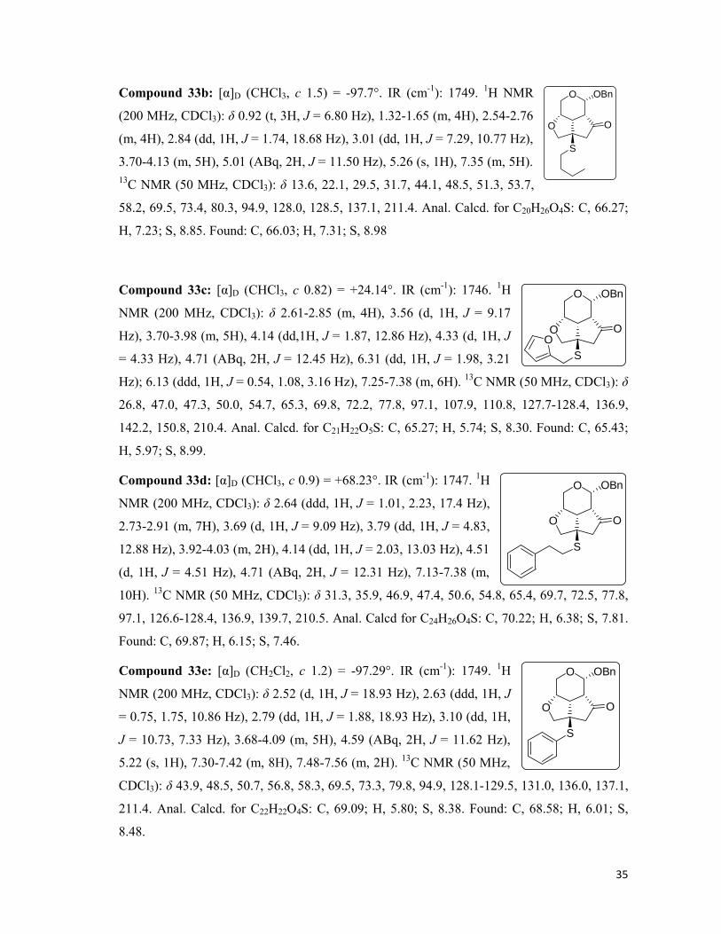
35
Compound 33b: [α]D (CHCl3, c 1.5) = -97.7°. IR (cm-1): 1749. 1H NMR
(200 MHz, CDCl3): δ 0.92 (t, 3H, J = 6.80 Hz), 1.32-1.65 (m, 4H), 2.54-2.76
(m, 4H), 2.84 (dd, 1H, J = 1.74, 18.68 Hz), 3.01 (dd, 1H, J = 7.29, 10.77 Hz),
3.70-4.13 (m, 5H), 5.01 (ABq, 2H, J = 11.50 Hz), 5.26 (s, 1H), 7.35 (m, 5H). 13C NMR (50 MHz, CDCl3): δ 13.6, 22.1, 29.5, 31.7, 44.1, 48.5, 51.3, 53.7,
58.2, 69.5, 73.4, 80.3, 94.9, 128.0, 128.5, 137.1, 211.4. Anal. Calcd. for C20H26O4S: C, 66.27;
H, 7.23; S, 8.85. Found: C, 66.03; H, 7.31; S, 8.98
Compound 33c: [α]D (CHCl3, c 0.82) = +24.14°. IR (cm-1): 1746. 1H
NMR (200 MHz, CDCl3): δ 2.61-2.85 (m, 4H), 3.56 (d, 1H, J = 9.17
Hz), 3.70-3.98 (m, 5H), 4.14 (dd,1H, J = 1.87, 12.86 Hz), 4.33 (d, 1H, J
= 4.33 Hz), 4.71 (ABq, 2H, J = 12.45 Hz), 6.31 (dd, 1H, J = 1.98, 3.21
Hz); 6.13 (ddd, 1H, J = 0.54, 1.08, 3.16 Hz), 7.25-7.38 (m, 6H). 13C NMR (50 MHz, CDCl3): δ
26.8, 47.0, 47.3, 50.0, 54.7, 65.3, 69.8, 72.2, 77.8, 97.1, 107.9, 110.8, 127.7-128.4, 136.9,
142.2, 150.8, 210.4. Anal. Calcd. for C21H22O5S: C, 65.27; H, 5.74; S, 8.30. Found: C, 65.43;
H, 5.97; S, 8.99.
Compound 33d: [α]D (CHCl3, c 0.9) = +68.23°. IR (cm-1): 1747. 1H
NMR (200 MHz, CDCl3): δ 2.64 (ddd, 1H, J = 1.01, 2.23, 17.4 Hz),
2.73-2.91 (m, 7H), 3.69 (d, 1H, J = 9.09 Hz), 3.79 (dd, 1H, J = 4.83,
12.88 Hz), 3.92-4.03 (m, 2H), 4.14 (dd, 1H, J = 2.03, 13.03 Hz), 4.51
(d, 1H, J = 4.51 Hz), 4.71 (ABq, 2H, J = 12.31 Hz), 7.13-7.38 (m,
10H). 13C NMR (50 MHz, CDCl3): δ 31.3, 35.9, 46.9, 47.4, 50.6, 54.8, 65.4, 69.7, 72.5, 77.8,
97.1, 126.6-128.4, 136.9, 139.7, 210.5. Anal. Calcd for C24H26O4S: C, 70.22; H, 6.38; S, 7.81.
Found: C, 69.87; H, 6.15; S, 7.46.
Compound 33e: [α]D (CH2Cl2, c 1.2) = -97.29°. IR (cm-1): 1749. 1H
NMR (200 MHz, CDCl3): δ 2.52 (d, 1H, J = 18.93 Hz), 2.63 (ddd, 1H, J
= 0.75, 1.75, 10.86 Hz), 2.79 (dd, 1H, J = 1.88, 18.93 Hz), 3.10 (dd, 1H,
J = 10.73, 7.33 Hz), 3.68-4.09 (m, 5H), 4.59 (ABq, 2H, J = 11.62 Hz),
5.22 (s, 1H), 7.30-7.42 (m, 8H), 7.48-7.56 (m, 2H). 13C NMR (50 MHz,
CDCl3): δ 43.9, 48.5, 50.7, 56.8, 58.3, 69.5, 73.3, 79.8, 94.9, 128.1-129.5, 131.0, 136.0, 137.1,
211.4. Anal. Calcd. for C22H22O4S: C, 69.09; H, 5.80; S, 8.38. Found: C, 68.58; H, 6.01; S,
8.48.
O
O O
OBn
S
O
O O
OBn
SO
O
O O
OBn
S
O
O O
OBn
S
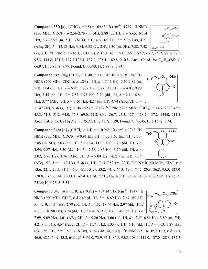
36
Compound 33f: [α]D (CHCl3, c 0.8) = +64.4°. IR (cm-1): 1749. 1H NMR
(200 MHz, CDCl3): δ 2.56-2.75 (m, 3H), 2.88 (dd,1H, J = 8.65, 10.14
Hz), 3.72-3.95 (m, 5H), 3.81 (s, 3H), 4.66 (d, 1H, J = 5.06 Hz), 4.71
(ABq, 2H, J = 12.18 Hz), 6.84, 6.88 (2s, 2H), 7.30 (m, 5H), 7.38, 7.42
(2s, 2H). 13C NMR (50 MHz, CDCl3): δ 46.1, 47.5, 50.3, 55.3, 57.7, 65.7, 69.7, 72.7, 77.3,
97.3, 114.8, 121.2, 127.7-128.4, 137.0, 138.1, 160.8, 210.5. Anal. Calcd. for C23H24O5S: C,
66.97; H, 5.86; S, 7.77. Found: C, 66.73; H, 5.99; S, 7.94.
Compound 34a: [α]D (CHCl3, c 0.48) = +10.04°. IR (cm-1): 1747. 1H
NMR (200 MHz, CDCl3): δ 1.24 (t, 3H, J = 7.42 Hz), 2.50-2.89 (m,
5H), 3.04 (dd, 1H, J = 6.95, 10.97 Hz), 3.27 (dd, 1H, J = 4.03, 9.99
Hz), 3.45 (dd, 1H, J = 7.57, 9.97 Hz), 3.70 (dd, 1H, J = 2.14, 6.84
Hz), 3.77 (ABq, 2H, J = 9.10 Hz), 4.24 (m, 1H), 4.74 (ABq, 2H, J =
11.47 Hz), 5.36 (s, 1H), 7.10-7.52 (m, 20H). 13C NMR (75 MHz, CDCl3): δ 14.7, 23.9, 45.9,
48.3, 51.3, 53.2, 64.0, 66.3, 69.0, 74.2, 80.9, 86.7, 95.5, 127.0-128.7, 137.2, 144.0, 211.1.
Anal. Calcd. for C38H38O5S: C, 75.22; H, 6.31; S, 5.28. Found: C, 75.45; H, 6.13; S, 5.34.
Compound 34b: [α]D (CHCl3, c 1.4) = +10.96°. IR (cm-1): 1743. 1H
NMR (200 MHz, CDCl3): δ 0.91 (m, 3H), 1.32-1.63 (m, 4H), 2.50-
2.85 (m, 5H), 3.03 (dd, 1H, J = 6.94, 11.02 Hz), 3.26 (dd, 1H, J =
3.94, 9.87 Hz), 3.50 (dd, 1H, J = 7.58, 9.85 Hz), 3.70 (dd, 1H, J =
2.02, 6.96 Hz), 3.76 (ABq, 2H, J = 9.04 Hz), 4.25 (m, 1H), 4.74
(ABq, 2H, J = 11.49 Hz), 5.36 (s, 1H), 7.11-7.52 (m, 20H). 13C NMR (50 MHz, CDCl3): δ
13.6, 22.1, 29.5, 31.7, 45.9, 48.3, 51.4, 53.2, 64.1, 66.3, 69.0, 74.2, 80.8, 86.8, 95.5, 127.0-
128.8, 137.3, 144.0, 211.1. Anal. Calcd. for C40H42O5S: C, 75.68; H, 6.67; S, 5.05. Found: C,
75.34; H, 6.76; S, 5.33.
Compound 34c: [α]D (CHCl3, c 0.82) = +24.14°. IR (cm-1): 1747. 1H
NMR (200 MHz, CDCl3): δ 2.49 (d, 1H, J = 18.69 Hz), 2.67 (dd, 1H,
J = 1.10, 11.14 Hz), 2.79 (dd, 1H, J = 1.52, 18.66 Hz), 2.97 (dd, 1H, J
= 6.85, 10.98 Hz), 3.24 (dd, 1H, J = 4.16, 9.98 Hz), 3.48 (dd, 1H, J =
7.69, 9.09 Hz), 3.61 (ABq, 2H, J = 9.26 Hz), 3.66 (dd, 1H, J = 2.l5, 6.86 Hz), 3.80 (m, 2H),
4.21 (m, 1H), 4.67 (ABq, 2H, J = 11.71 Hz), 5.35 (s, 1H), 6.16 (dd, 1H, J = 0.63, 3.27 Hz),
6.31 (dd, 1H, J = 1.89, 3.16 Hz), 7.15-7.48 (m, 21H). 13C NMR (50 MHz, CDCl3): δ 27.1,
46.0, 48.1, 50.9, 53.2, 64.1, 66.3, 69.0, 73.9, 81.1, 86.8, 95.5, 108.0, 111.0, 127.0-128.8, 137.3,
O
O O
OBn
S
MeO
O
O O
TrOOBn
S
O
O O
TrOOBn
S
O
O O
TrOOBn
SO
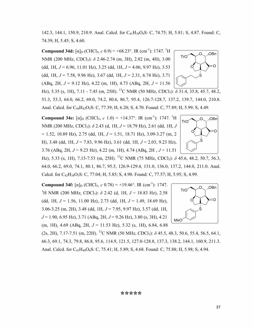
37
142.3, 144.1, 150.9, 210.9. Anal. Calcd. for C41H38O6S: C, 74.75; H, 5.81; S, 4.87. Found: C,
74.39; H, 5.45; S, 4.60.
Compound 34d: [α]D (CHCl3, c 0.9) = +68.23°. IR (cm-1): 1747. 1H
NMR (200 MHz, CDCl3): δ 2.46-2.74 (m, 3H), 2.82 (m, 4H), 3.00
(dd, 1H, J = 6.96, 11.01 Hz), 3.25 (dd, 1H, J = 4.06, 9.97 Hz), 3.53
(dd, 1H, J = 7.58, 9.96 Hz), 3.67 (dd, 1H, J = 2.31, 6.74 Hz), 3.71
(ABq, 2H, J = 9.12 Hz), 4.22 (m, 1H), 4.73 (ABq, 2H, J = 11.56
Hz), 5.35 (s, 1H), 7.11 - 7.45 (m, 25H). 13C NMR (50 MHz, CDCl3): δ 31.4, 35.8, 45.7, 48.2,
51.3, 53.3, 64.0, 66.2, 69.0, 74.2, 80.6, 86.7, 95.4, 126.7-128.7, 137.2, 139.7, 144.0, 210.8.
Anal. Calcd. for C44H42O5S: C, 77.39; H, 6.20; S, 4.70. Found: C, 77.89; H, 5.99; S, 4.49.
Compound 34e: [α]D (CHCl3, c 1.0) = +14.37°. IR (cm-1): 1747. 1H
NMR (200 MHz, CDCl3): δ 2.43 (d, 1H, J = 18.79 Hz), 2.61 (dd, 1H, J
= 1.52, 10.89 Hz), 2.75 (dd, 1H, J = 1.51, 18.71 Hz), 3.09-3.27 (m, 2
H), 3.48 (dd, 1H, J = 7.83, 9.96 Hz), 3.61 (dd, 1H, J = 2.03, 9.23 Hz),
3.76 (ABq, 2H, J = 9.23 Hz), 4.22 (m, 1H), 4.74 (ABq, 2H , J = 11.51
Hz), 5.33 (s, 1H), 7.15-7.53 (m, 25H). 13C NMR (75 MHz, CDCl3): δ 45.6, 48.2, 50.7, 56.3,
64.0, 66.2, 69.0, 74.1, 80.1, 86.7, 95.5, 126.9-129.4, 131.0, 136.0, 137.2, 144.0, 211.0. Anal.
Calcd. for C42H38O5S: C, 77.04; H, 5.85; S, 4.90. Found: C, 77.57; H, 5.95; S, 4.99.
Compound 34f: [α]D (CHCl3, c 0.78) = +19.46°. IR (cm-1): 1747. 1H NMR (200 MHz, CDCl3): δ 2.42 (d, 1H, J = 18.83 Hz), 2.58
(dd, 1H, J = 1.56, 11.00 Hz), 2.73 (dd, 1H, J = 1.49, 18.69 Hz),
3.06-3.25 (m, 2H), 3.48 (dd, 1H, J = 7.95, 9.97 Hz), 3.57 (dd, 1H,
J = 1.90, 6.95 Hz), 3.71 (ABq, 2H, J = 9.26 Hz), 3.80 (s, 3H), 4.21
(m, 1H), 4.69 (ABq, 2H, J = 11.53 Hz), 5.32 (s, 1H), 6.84, 6.88
(2s, 2H), 7.17-7.51 (m, 22H). 13C NMR (50 MHz, CDCl3): δ 45.5, 48.3, 50.6, 55.4, 56.5, 64.1,
66.3, 69.1, 74.3, 79.8, 86.8, 95.6, 114.9, 121.5, 127.0-128.8, 137.3, 138.2, 144.1, 160.9, 211.3.
Anal. Calcd. for C43H40O6S: C, 75.41; H, 5.89; S, 4.68. Found: C, 75.88; H, 5.98; S, 4.94.
*****
O
O O
TrOOBn
S
O
O O
TrOOBn
S
O
O O
TrOOBn
S
MeO

38
Chapter 1: Spectral Charts
1H NMR (200 MHz, CDCl3) of Compound 5
8.0 7.5 7.0 6.5 6.0 5.5 5.0 4.5 4.0 3.5 3.0 2.5 2.0 1.5 1.0 0.5 0.0
0.0
0.1
0.2
0.3
0.4
0.5
0.6
0.7
0.8
0.9
1.0
20.00 2.760.97 0.950.950.940.93 0.78
0.002.
232.24
2.25
3.153.183.
203.233.
35
3.40
3.95
4.00
4.01
4.21
4.25
4.62
4.68
4.88
4.945.
16
5.775.
78
5.82
5.83
5.84
6.04
6.10
7.17
7.18
7.18
7.21
7.25
7.28
7.30
7.32
7.34
7.50
7.53
7.54
13C NMR (50 MHz, CDCl3) of Compound 5
200 150 100 50 0
0.0
0.1
0.2
0.3
0.4
0.5
0.6
0.7
0.8
0.9
1.0
Chloroform-d
56.56
63.08
69.43
69.73
70.58
74.40
76.36
77.00
77.63
79.67
86.3593
.33
126.77
126.90
127.71
128.78
130.96
137.89
144.07
DEPT NMR (50 MHz, CDCl3) of compound 5
200 150 100 50 0
-0.2
-0.1
0.0
0.1
0.2
0.3
0.4
0.5
0.6
0.7
0.8
0.9
1.0
56.56
63.08
69.43
69.73
70.57
93.33
126.77
126.90
127.70
128.78
130.96
OBn
OOTr
HO

39
1H NMR (200 MHz, CDCl3) of Compound 6
7.5 7.0 6.5 6.0 5.5 5.0 4.5 4.0 3.5 3.0 2.5 2.0 1.5 1.0 0.5 0.0
0.0
0.1
0.2
0.3
0.4
0.5
0.6
0.7
0.8
0.9
1.0
24.04 4.473.41 1.231.15 1.051.00 0.99
0.003.29
3.323.33
3.34
3.35
3.37
3.38
3.97
4.01
4.02
4.50
4.62
4.84
4.905.26
5.306.06
7.177.18
7.21
7.24
7.27
7.28
7.45
7.48
7.49
13C NMR (50 MHz, CDCl3) of Compound 6
200 150 100 50 0
0.0
0.1
0.2
0.3
0.4
0.5
0.6
0.7
0.8
0.9
1.0
Chloroform-d45
.45
51.48
64.03
65.14
65.70
68.88
71.1676
.36
77.00
77.63
86.75
94.32
125.20
126.99
127.71
128.69
137.26
143.87
178.36
206.70
DEPT NMR (50 MHz, CDCl3) of Compound 6
200 150 100 50 0
-0.2
-0.1
0.0
0.1
0.2
0.3
0.4
0.5
0.6
0.7
0.8
0.9
1.0
45.45
51.48
64.04
65.15
65.70
68.88
71.16
94.32
125.20
126.99
127.71
128.69
O OBnTrO
O O

40
COSY (500 MHz, CDCl3) Spectrum of Compound 6

41
NOESY (500 MHz, CDCl3) Spectrum of Compound 6

42
1H NMR (200 MHz, CDCl3) of compound 9
7.5 7.0 6.5 6.0 5.5 5.0 4.5 4.0 3.5 3.0 2.5 2.0 1.5 1.0 0.5 0.0
0.0
0.1
0.2
0.3
0.4
0.5
0.6
0.7
0.8
0.9
1.0
15.00 11.802.662.48 2.222.101.730.84 0.820.81
0.00
0.00
0.68
0.720.75
0.79
0.830.89
0.93
1.04
1.10
1.26
1.56
1.60
2.13
2.172.
202.23
2.24
2.26
3.193.213.
243.26
3.41
3.46
3.943.95
3.96
4.004.00
4.22
4.26
5.14
5.765.816.
03
6.087.
177.21
7.24
7.25
7.29
7.32
7.46
7.50
7.54
13C NMR (50 MHz, CDCl3) of Compound 9
200 150 100 50 00.0
0.1
0.2
0.3
0.4
0.5
0.6
0.7
0.8
0.9
1.0
Chloroform-d
16.23
21.12
22.22
23.23
25.58
31.80
34.35
43.2748.76
56.49
63.07
69.04
70.51
74.31
76.37
77.00
77.64
80.48
86.4196.14
126.84
127.64
128.81
130.45
143.97
144.04
DEPT NMR (50 MHz, CDCl3) of Compound 9
200 150 100 50 0
-0.2
-0.1
0.0
0.1
0.2
0.3
0.4
0.5
0.6
0.7
0.8
0.9
1.0
16.23
21.13
22.22
23.23
25.58
31.80
34.35
43.27
48.77
56.49
63.08
69.04
70.51
80.48
96.14
126.84
127.64
128.81
130.45
OOTr
O
O
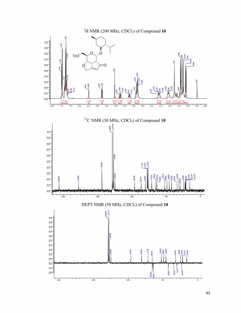
43
1H NMR (200 MHz, CDCl3) of Compound 10
8.0 7.5 7.0 6.5 6.0 5.5 5.0 4.5 4.0 3.5 3.0 2.5 2.0 1.5 1.0 0.5 0.0 -0.5
0.0
0.1
0.2
0.3
0.4
0.5
0.6
0.7
0.8
0.9
1.0
17.60 11.305.38 5.303.022.222.10 1.091.091.021.00
0.00
0.60
0.63
0.760.79
0.88
0.92
0.94
1.26
1.55
1.561.60
2.08
2.092.122.13
2.15
3.263.
273.29
3.34
3.39
3.71
3.723.764.224.26
4.31
4.56
5.24
5.28
5.99
6.00
7.17
7.18
7.19
7.21
7.24
7.25
7.32
7.46
7.50
7.51
13C NMR (50 MHz, CDCl3) of Compound 10
200 150 100 50 0
0.0
0.1
0.2
0.3
0.4
0.5
0.6
0.7
0.8
0.9
1.0
15.75
21.37
22.15
22.61
24.65
29.7331
.59
34.16
42.26
45.67
48.95
52.23
63.80
65.24
65.39
71.25
76.43
77.07
77.71
80.56
86.75
96.28
124.99
126.99
127.76
128.87
143.99
178.06
206.65
DEPT NMR (50 MHz, CDCl3) of Compound 10
200 150 100 50 0
-0.2
-0.1
0.0
0.1
0.2
0.3
0.4
0.5
0.6
0.7
0.8
0.9
1.0
15.76
21.37
22.15
22.62
24.65
29.73
31.60
34.16
42.26
45.67
48.96
52.23
63.8165.24
65.3971.25
80.56
96.29
124.99
126.99
127.77
128.88
O OTrO
O O
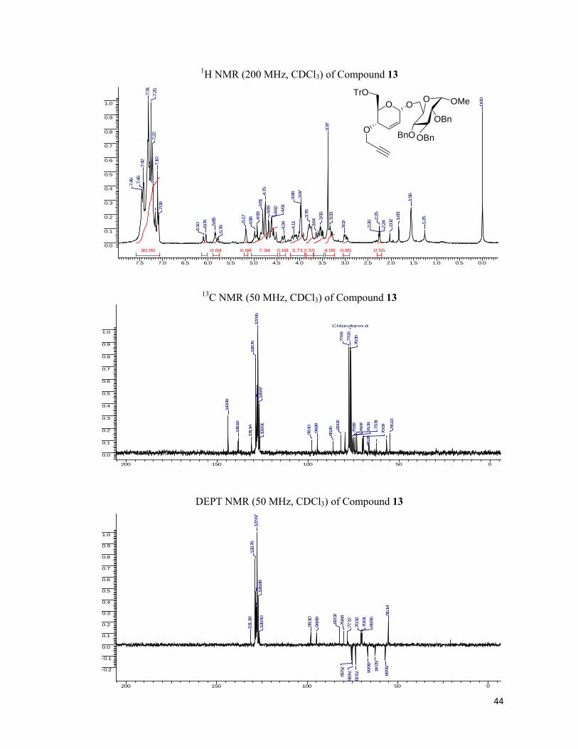
44
1H NMR (200 MHz, CDCl3) of Compound 13
7.5 7.0 6.5 6.0 5.5 5.0 4.5 4.0 3.5 3.0 2.5 2.0 1.5 1.0 0.5 0.0
0.0
0.1
0.2
0.3
0.4
0.5
0.6
0.7
0.8
0.9
1.0
30.00 7.34 4.063.712.55 0.850.84 0.680.68 0.55
0.00
1.25
1.56
1.83
2.02
2.24
2.25
2.26
3.02
3.33
3.37
3.55
3.64
3.78
3.97
3.98
4.11
4.34
4.61
4.62
4.68
4.75
4.81
4.93
4.985.17
5.78
5.85
6.05
6.10
7.08
7.10
7.22
7.257.31
7.42
7.45
7.46
13C NMR (50 MHz, CDCl3) of Compound 13
200 150 100 50 0
0.0
0.1
0.2
0.3
0.4
0.5
0.6
0.7
0.8
0.9
1.0Chloroform-d
55.13
66.28
70.03
73.31
74.33
74.97
75.58
76.36
77.00
77.63
82.02
86.26
94.88
98.10
126.51
126.87
127.65
128.76
131.14
138.18
143.99
DEPT NMR (50 MHz, CDCl3) of Compound 13
200 150 100 50 0
-0.2
-0.1
0.0
0.1
0.2
0.3
0.4
0.5
0.6
0.7
0.8
0.9
1.0
55.14
56.84
62.44
66.64
69.56
70.01
70.32
73.33
74.98
75.59
77.72
79.84
82.02
94.89
98.10
126.50
126.88
127.67
128.76
131.18
O
O
TrOO OMe
OBn
BnO
O
OBn

45
1H NMR (200 MHz, CDCl3) of Compound 14
8.0 7.5 7.0 6.5 6.0 5.5 5.0 4.5 4.0 3.5 3.0 2.5 2.0 1.5 1.0 0.5 0.0
0.0
0.1
0.2
0.3
0.4
0.5
0.6
0.7
0.8
0.9
1.0
30.00 8.095.15 4.514.27 3.100.88 0.84
0.00
0.88
1.201.21
3.21
3.233.
263.29
3.33
3.35
3.38
3.52
3.703.76
3.85
3.87
4.464.47
4.60
4.624.68
4.76
4.794.
82
4.844.
94
4.995.29
5.325.97
7.21
7.22
7.23
7.32
7.42
7.45
7.46
13C NMR (50 MHz, CDCl3) of Compound 14
200 150 100 50 0
0.0
0.1
0.2
0.3
0.4
0.5
0.6
0.7
0.8
0.9
1.0
Chloroform-d
29.65
45.49
51.48
54.79
63.97
65.52
70.22
71.1873.39
75.55
76.37
77.00
77.64
78.27
86.75
95.54
97.80
125.02
126.97
127.55
127.71
128.75
138.22
143.89
178.29
206.30
DEPT NMR (50 MHz, CDCl3) of Compound 14
200 150 100 50 0
-0.2
-0.1
0.0
0.1
0.2
0.3
0.4
0.5
0.6
0.7
0.8
0.9
1.0
45.57
51.56
54.87
64.0565.29
65.60
66.09
70.29
71.26
73.4775.63
78.35
80.21
82.12
95.62
97.89
125.10
127.05
127.63
127.79
128.83
O
O O
TrO OOMe
OBn
OBnOBn
O
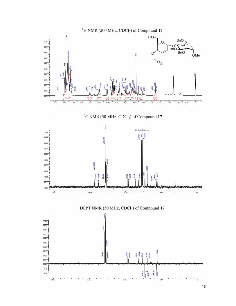
46
1H NMR (200 MHz, CDCl3) of Compound 17
8.0 7.5 7.0 6.5 6.0 5.5 5.0 4.5 4.0 3.5 3.0 2.5 2.0 1.5 1.0 0.5 0.0
0.0
0.1
0.2
0.3
0.4
0.5
0.6
0.7
0.8
0.9
1.0
30.00 8.133.97 3.692.07 1.71 1.190.95 0.760.71
0.00
2.28
2.29
2.30
3.04
3.053.25
3.39
3.46
3.58
3.60
3.63
3.97
3.97
4.00
4.204.35
4.41
4.624.68
4.74
4.80
5.01
5.075.
48
5.56
5.576.02
6.07
7.08
7.10
7.12
7.14
7.24
7.34
7.44
7.48
7.48
7.85
13C NMR (50 MHz, CDCl3) of Compound 17
200 150 100 50 0
0.0
0.1
0.2
0.3
0.4
0.5
0.6
0.7
0.8
0.9
1.0Chloroform-d
55.06
56.80
61.9
0
69.55
75.64
76.3
677
.00
77.63
80.07
86.31
96.01
97.5
7
126.05
126.92
127.71
128.76
131.30
137.96
138.55
143.86
DEPT NMR (50 MHz, CDCl3) of Compound 17
200 150 100 50 0
-0.2
-0.1
0.0
0.1
0.2
0.3
0.4
0.5
0.6
0.7
0.8
0.9
1.0
55.07
56.80
61.89
69.51
69.96
72.78
73.20
75.64
76.41
80.07
81.91
96.01
97.57
126.05
126.92
127.71
128.76
131.30
O
O
TrO
O
OMeBnO
BnOO
BnO
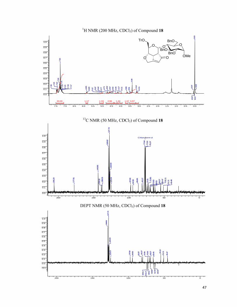
47
1H NMR (200 MHz, CDCl3) of Compound 18
7.5 7.0 6.5 6.0 5.5 5.0 4.5 4.0 3.5 3.0 2.5 2.0 1.5 1.0 0.5 0.0
0.0
0.1
0.2
0.3
0.4
0.5
0.6
0.7
0.8
0.9
1.0
30.00 6.574.56 1.671.341.331.17
-0.02
0.00
0.01
0.02
0.07
3.113.14
3.38
3.513.66
4.114.17
4.44
4.504.574.62
4.71
4.775.02
5.07
5.125.805.857.177.19
7.207.21
7.22
7.26
7.29
7.42
7.43
7.46
13C NMR (50 MHz, CDCl3) of Compound 18
200 150 100 50 0
0.0
0.1
0.2
0.3
0.4
0.5
0.6
0.7
0.8
0.9
1.0Chloroform-d
45.48
51.1155.11
65.47
69.27
69.50
70.6372.85
73.21
76.37
77.00
77.64
80.27
86.66
94.59
97.69
125.04
126.93
127.74
128.82
138.03
138.18
143.86
177.91
206.20
DEPT NMR (50 MHz, CDCl3) of Compound 18
200 150 100 50 0
-0.1
0.0
0.1
0.2
0.3
0.4
0.5
0.6
0.7
0.8
0.9
1.0
45.47
51.11
55.10
62.92
64.79
65.46
69.27
69.50
70.63
72.84
72.95
73.21
74.83
80.27
81.87
94.59
97.69
125.03
126.93
127.74
128.81
O
O O
TrOO
OMeBnO
BnOO
BnO
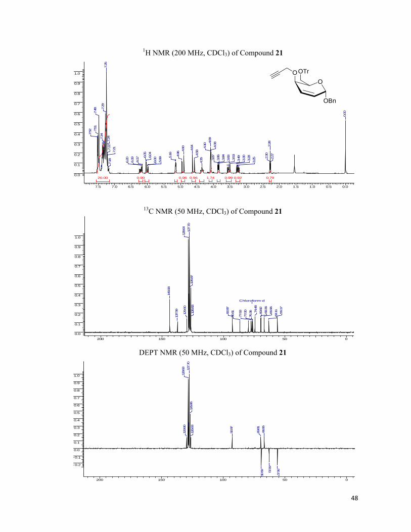
48
1H NMR (200 MHz, CDCl3) of Compound 21
7.5 7.0 6.5 6.0 5.5 5.0 4.5 4.0 3.5 3.0 2.5 2.0 1.5 1.0 0.5 0.0
0.0
0.1
0.2
0.3
0.4
0.5
0.6
0.7
0.8
0.9
1.0
20.00 1.74 0.990.96 0.96 0.920.90 0.79
0.00
2.27
2.28
2.30
3.253.28
3.30
3.493.53
3.833.84
3.86
3.87
4.084.09
4.10
4.35
4.58
4.64
4.90
4.96
5.16
5.99
6.00
6.04
6.05
6.17
6.19
6.20
7.18
7.217.
227.24
7.25
7.29
7.34
7.48
7.51
7.52
13C NMR (50 MHz, CDCl3) of Compound 21
200 150 100 50 00.0
0.1
0.2
0.3
0.4
0.5
0.6
0.7
0.8
0.9
1.0
Chloroform-d
56.17
63.11
66.96
69.36
69.82
74.48
76.36
77.00
77.63
86.6192.87
126.63
126.87
127.70
128.64
129.90
137.59
143.93
DEPT NMR (50 MHz, CDCl3) of Compound 21
200 150 100 50 0
-0.2
-0.1
0.0
0.1
0.2
0.3
0.4
0.5
0.6
0.7
0.8
0.9
1.0
56.17
63.11
66.95
69.35
69.81
92.87
126.63
126.86
127.70
128.63
129.90
OBn
OOTrO

49
1H NMR (200 MHz, CDCl3) of Compound 22
7.5 7.0 6.5 6.0 5.5 5.0 4.5 4.0 3.5 3.0 2.5 2.0 1.5 1.0
0.0
0.1
0.2
0.3
0.4
0.5
0.6
0.7
0.8
0.9
1.0
20.00 1.591.551.51 0.95 0.620.51 0.25
1.27
2.792.80
3.243.26
3.333.363.38
3.40
4.084.18
4.184.204.
234.38
4.40
4.564.58
4.91
4.92
4.94
5.31
5.93
7.15
7.17
7.20
7.22
7.23
7.35
7.437.44
7.45
7.47
13C NMR (50 MHz, CDCl3) of Compound 22
200 150 100 50 00.0
0.1
0.2
0.3
0.4
0.5
0.6
0.7
0.8
0.9
1.0
43.74
51.35
62.86
65.49
67.75
68.92
70.74
76.63
76.89
77.13
86.60
95.97
97.26
123.81
126.78
127.56
128.57
136.95
143.83
143.92
182.72
208.33
DEPT NMR (50 MHz, CDCl3) of Compound 22
200 150 100 50 0
-0.2
-0.1
0.0
0.1
0.2
0.3
0.4
0.5
0.6
0.7
0.8
0.9
1.0
44.07
51.74
63.00
65.83
68.00
69.29
71.03
97.47
124.04
126.95
127.74
128.74
O OBnTrO
O O
50
1H NMR (200 MHz, CDCl3) of Compound 26
7.5 7.0 6.5 6.0 5.5 5.0 4.5 4.0 3.5 3.0 2.5 2.0 1.5 1.0 0.5 0.0
0.0
0.1
0.2
0.3
0.4
0.5
0.6
0.7
0.8
0.9
1.0
5.00 2.061.911.120.94 0.93 0.91
0.00
2.44
2.45
2.46
3.873.87
3.893.93
3.99
4.08
4.25
4.26
4.54
4.60
4.77
4.83
5.09
5.98
5.99
6.036.04
6.10
6.13
7.26
7.35
7.36
7.37
13C NMR (50 MHz, CDCl3) of compound 26
200 150 100 50 0
0.0
0.1
0.2
0.3
0.4
0.5
0.6
0.7
0.8
0.9
1.0
Chloroform-d
55.4961.10
66.64
69.66
74.67
76.37
77.00
77.63
79.66
92.22
126.24
127.73
128.08128.38
130.13
137.63
DEPT NMR (50 MHz, CDCl3) of Compound 26
170 160 150 140 130 120 110 100 90 80 70 60 50 40 30 20 10 0
-1.0
-0.5
0.0
0.5
1.0
55.62
61.21
66.76
69.79
92.34
126.37
127.85
128.21
128.51
130.26
OBn
OO
51
1H NMR (200 MHz, CDCl3) of Compound 27
7.5 7.0 6.5 6.0 5.5 5.0 4.5 4.0 3.5 3.0 2.5 2.0 1.5 1.0 0.5 0.0 -0.5
0.0
0.1
0.2
0.3
0.4
0.5
0.6
0.7
0.8
0.9
1.0
5.98 3.05 2.531.38 1.151.00
0.00
2.91
2.912.94
2.953.513.52
3.57
3.59
3.994.01
4.054.084.
484.54
4.63
4.75
4.84
4.85
6.11
6.127.
267.34
7.34
7.35
7.37
7.38
13C NMR (50 MHz, CDCl3) of compound 27
200 150 100 50 0
0.0
0.1
0.2
0.3
0.4
0.5
0.6
0.7
0.8
0.9
1.0
Chloroform-d
43.83
51.53
62.19
65.2269
.97
70.06
76.37
77.00
77.64
96.76
124.65
127.96
128.49
136.78183.48
208.69
DEPT NMR (50 MHz, CDCl3) of Compound 27
200 150 100 50 0
-0.5
0.0
0.5
1.0
43.83
51.52
62.19
65.22
69.96
70.06
96.76
124.65
127.96
128.49
O OBn
O O

52
1H NMR (200 MHz, CDCl3) of Compound 31
7.5 7.0 6.5 6.0 5.5 5.0 4.5 4.0 3.5 3.0 2.5 2.0 1.5 1.0 0.5 0.0
0.0
0.1
0.2
0.3
0.4
0.5
0.6
0.7
0.8
0.9
1.0
0.19 0.140.08 0.080.040.04 0.04 0.03 0.03
0.00
1.26
1.29
1.391.61
2.42
2.43
2.44
3.79
3.80
3.83
3.86
3.89
3.93
4.23
4.24
4.55
4.61
4.75
4.81
5.02
5.03
5.805.81
5.81
5.826.
036.04
6.097.25
7.297.31
7.34
7.35
7.36
7.38
13C NMR (50 MHz, CDCl3) of Compound 31
200 150 100 50 0
0.0
0.1
0.2
0.3
0.4
0.5
0.6
0.7
0.8
0.9
1.0
Chloroform-d
18.37
56.67
66.07
70.29
74.89
76.45
77.00
77.43
77.85
80.24
94.12
127.42
127.94
128.24
128.73
130.65
138.62
DEPT NMR (50 MHz, CDCl3) of Compound 31
200 150 100 50 0
-0.5
0.0
0.5
1.0
18.4
3
56.73
66.1
0
69.7070.35
76.3
9
94.1
2
127.
4212
8.03
128.
3312
8.79
130.
68
O
CH3 OBn
O

53
1H NMR (200 MHz, CDCl3) of Compound 32
7.5 7.0 6.5 6.0 5.5 5.0 4.5 4.0 3.5 3.0 2.5 2.0 1.5 1.0
0.0
0.1
0.2
0.3
0.4
0.5
0.6
0.7
0.8
0.9
1.0
6.99 5.80 4.35 4.341.371.331.00
1.22
1.25
1.29
3.28
3.32
3.36
3.493.54
3.573.893.93
3.984.
414.474.53
4.62
4.62
5.14
5.18
6.05
6.067.
237.257.25
7.26
7.29
7.32
7.33
7.34
7.36
13C NMR (50 MHz, CDCl3) of Compound 32
200 150 100 50 0
0.0
0.1
0.2
0.3
0.4
0.5
0.6
0.7
0.8
0.9
1.0
17.92
45.76
51.67
61.69
65.32
69.46
76.47
76.78
77.11
77.74
94.87
125.04
127.1912
8.38
137.36178.88
207.08
DEPT NMR (50 MHz, CDCl3) of Compound 32
200 150 100 50 0
-0.5
0.0
0.5
1.0
17.9245
.76
51.67
61.69
65.32
69.46
76.78
94.87
125.04
127.19
128.38
O OBn
O O
H3C

54
1H NMR (200 MHz, CDCl3) of Compound 32a
7.5 7.0 6.5 6.0 5.5 5.0 4.5 4.0 3.5 3.0 2.5 2.0 1.5 1.0 0.5 0.0
0.0
0.1
0.2
0.3
0.4
0.5
0.6
0.7
0.8
0.9
1.0
20.00 5.683.092.63 2.261.020.90 0.86 0.50
TMS
-0.01
0.00
0.071.
111.19
1.221.
251.271.
56
2.51
2.53
2.55
2.62
2.73
2.79
2.923.303.
32
3.33
3.35
3.74
3.76
3.79
3.88
3.913.95
3.984.564.60
4.81
4.83
4.87
5.25
5.295.31
7.23
7.26
7.29
7.46
7.48
13C NMR (50 MHz, CDCl3) of Compound 32a
200 150 100 50 0
0.0
0.1
0.2
0.3
0.4
0.5
0.6
0.7
0.8
0.9
1.0
31.72
35.90
46.09
48.59
50.12
55.35
65.45
68.83
69.26
73.17
76.46
77.10
77.73
86.77
93.84126.69
127.12
127.89128.78
137.15
139.88
143.94
211.97
DEPT NMR (50 MHz, CDCl3) of Compound 32a
200 150 100 50 0
-0.2
-0.1
0.0
0.1
0.2
0.3
0.4
0.5
0.6
0.7
0.8
0.9
1.0
14.68
23.9324.20
29.79
46.21
48.65
50.11
65.53
68.9
5
69.3170.41
73.16
77.4979.20
93.91
127.15
127.
9112
8.82
O
O O
TrOOBn
S
55
1H NMR (200 MHz, CDCl3) of Compound 32b
7.5 7.0 6.5 6.0 5.5 5.0 4.5 4.0 3.5 3.0 2.5 2.0 1.5 1.0 0.5 0.0
0.0
0.1
0.2
0.3
0.4
0.5
0.6
0.7
0.8
0.9
1.0
20.00 8.654.95 4.133.51 1.730.86 0.820.77
TMS
-0.02
0.00
0.85
0.89
0.93
1.26
1.31
1.57
2.472.
512.69
2.72
2.81
3.32
3.74
3.79
3.87
3.91
3.984.554.
61
4.82
4.895.
28
5.327.19
7.227.23
7.26
7.31
7.45
7.49
7.50
13C NMR (50 MHz, CDCl3) of Compound 32b
200 150 100 50 0
0.0
0.1
0.2
0.3
0.4
0.5
0.6
0.7
0.8
0.9
1.0Chloroform-d
13.58
22.06
29.71
31.54
46.02
48.50
50.03
55.00
65.39
69.14
73.04
76.37
77.00
77.64
86.66
93.77
127.00
127.78
128.68
137.09
143.85
212.01
DEPT NMR (50 MHz, CDCl3) of Compound 32b
200 150 100 50 0
-0.1
0.0
0.1
0.2
0.3
0.4
0.5
0.6
0.7
0.8
0.9
1.0
13.5
8
22.04
29.7031.38
46.0
048
.48
49.8965.37
68.7
5
69.13
73.0
3
77.29
93.7
6
127.
0012
7.78
128.
67
O
O O
TrOOBn
S

56
1H NMR (200 MHz, CDCl3) of Compound 32c
7.5 7.0 6.5 6.0 5.5 5.0 4.5 4.0 3.5 3.0 2.5 2.0 1.5 1.0 0.5 0.0
0.0
0.1
0.2
0.3
0.4
0.5
0.6
0.7
0.8
0.9
1.0
23.39 4.39 2.212.202.14 1.061.00
0.00
2.59
2.60
2.67
2.76
3.043.
283.30
3.31
3.59
3.64
3.69
3.77
3.92
3.96
4.54
4.60
4.80
4.86
5.26
5.30
6.146.
15
6.276.28
6.28
6.29
7.19
7.22
7.25
7.30
7.44
7.48
7.49
13C NMR (50 MHz, CDCl3) of Compound 32c
200 150 100 50 0
0.0
0.1
0.2
0.3
0.4
0.5
0.6
0.7
0.8
0.9
1.0
Chloroform-d
27.12
46.02
48.40
49.35
55.28
65.3668.75
69.18
72.73
76.37
77.00
77.63
86.68
91.03
93.76
107.89
110.81
127.03
127.81
128.69
137.05
142.16
143.85
150.73
211.93
DEPT NMR (50 MHz, CDCl3) of Compound 32c
200 150 100 50 0
-0.2
-0.1
0.0
0.1
0.2
0.3
0.4
0.5
0.6
0.7
0.8
0.9
1.0
27.12
46.03
48.40
49.34
65.36
68.75
69.17
72.73
77.11
93.76
107.89
110.80
127.03
127.81
128.69
142.16
O
O O
TrOOBn
SO

57
1H NMR (200 MHz, CDCl3) of Compound 32d
7.5 7.0 6.5 6.0 5.5 5.0 4.5 4.0 3.5 3.0 2.5 2.0 1.5 1.0 0.5 0.0
0.0
0.1
0.2
0.3
0.4
0.5
0.6
0.7
0.8
0.9
1.0
26.61 6.433.97 2.091.211.001.00 0.94
0.00
1.261.562.60
2.71
2.77
2.79
2.80
3.043.053.28
3.31
3.72
3.77
3.84
3.93
3.96
4.54
4.60
4.81
4.875.
26
5.30
7.13
7.167.
177.22
7.24
7.24
7.30
7.45
7.48
7.49
13C NMR (50 MHz, CDCl3) of Compound 32d
200 150 100 50 0
0.0
0.1
0.2
0.3
0.4
0.5
0.6
0.7
0.8
0.9
1.0
31.72
35.90
46.09
48.59
50.12
55.35
65.45
68.83
69.26
73.17
76.46
77.10
77.73
86.77
93.84126.69
127.12
127.89128.78
137.15
139.88
143.94
211.97
DEPT NMR (50 MHz, CD Cl3) of Compound 32d
200 150 100 50 0
-0.2
-0.1
0.0
0.1
0.2
0.3
0.4
0.5
0.6
0.7
0.8
0.9
1.0
31.7235.91
46.09
48.59
50.12
65.46
68.83
69.26
73.17
77.35
93.84
126.69
127.12
127.89
128.78
O
O O
TrOOBn
S

58
1H NMR (200 MHz, CDCl3) of Compound 32e
7.5 7.0 6.5 6.0 5.5 5.0 4.5 4.0 3.5 3.0 2.5 2.0 1.5 1.0 0.5 0.0
0.0
0.1
0.2
0.3
0.4
0.5
0.6
0.7
0.8
0.9
1.0
25.00 3.49 1.87 1.440.930.860.84
0.00
2.64
3.33
3.85
4.59
4.80
5.28
5.32
7.25
7.28
7.43
7.47
13C NMR (50 MHz, CDCl3) of Compound 32e
200 150 100 50 0
0.0
0.1
0.2
0.3
0.4
0.5
0.6
0.7
0.8
0.9
1.0
Chloroform-d
29.6645
.41
48.53
49.99
58.11
65.49
69.28
73.40
76.57
77.00
77.43
86.8093.97
127.02
127.78
128.76
129.25
131.45
135.93
137.22
143.96
211.38
DEPT NMR (50 MHz, CDCl3) of Compound 32e
200 150 100 50 0
-0.2
-0.1
0.0
0.1
0.2
0.3
0.4
0.5
0.6
0.7
0.8
0.9
1.0
29.66
45.26
48.46
49.89
65.40
69.09
69.18
73.24
76.97
93.84
126.99
127.78
128.67
129.21
135.96
O
O O
TrOOBn
S
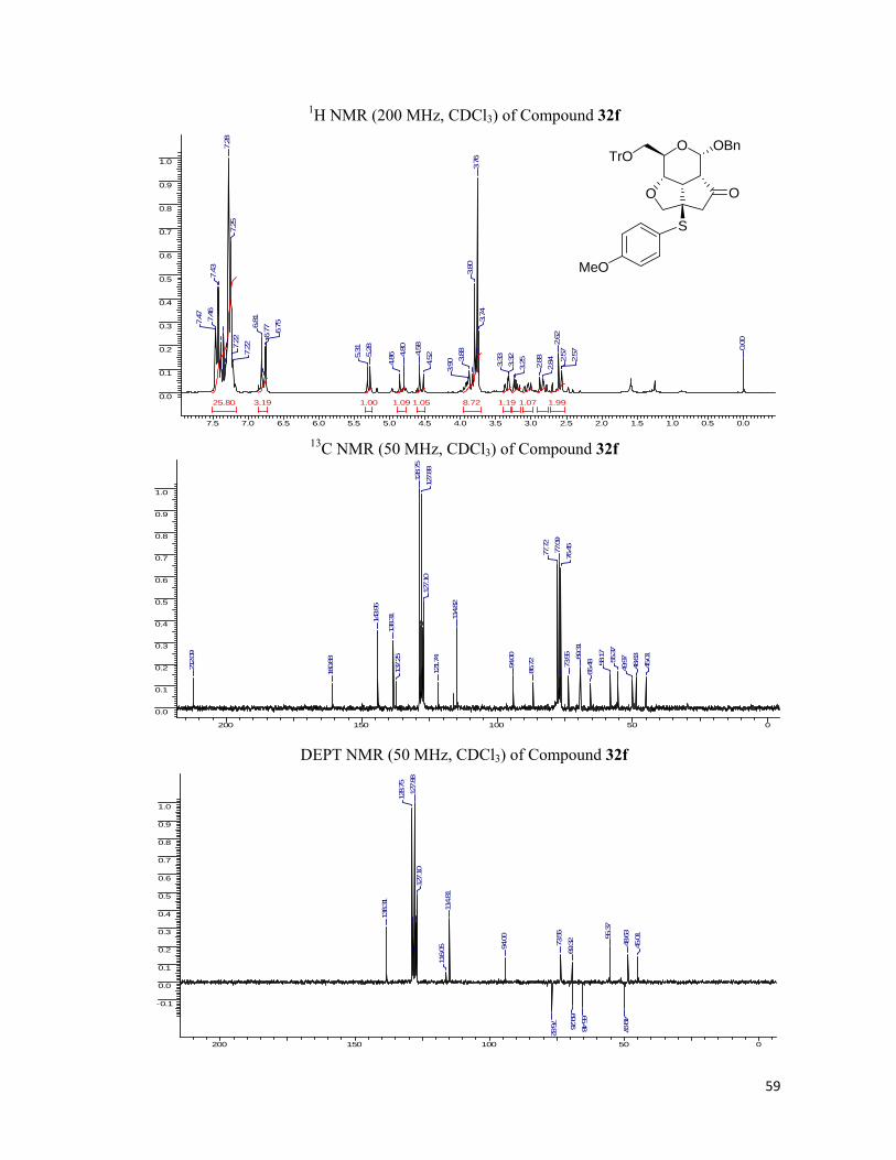
59
1H NMR (200 MHz, CDCl3) of Compound 32f
7.5 7.0 6.5 6.0 5.5 5.0 4.5 4.0 3.5 3.0 2.5 2.0 1.5 1.0 0.5 0.0
0.0
0.1
0.2
0.3
0.4
0.5
0.6
0.7
0.8
0.9
1.0
25.80 8.723.19 1.991.191.09 1.071.051.00
0.00
2.57
2.57
2.62
2.842.88
3.253.32
3.33
3.74
3.76
3.80
3.88
3.904.
524.58
4.80
4.865.
285.31
6.75
6.776.
817.
227.22
7.25
7.28
7.43
7.46
7.47
13C NMR (50 MHz, CDCl3) of Compound 32f
200 150 100 50 0
0.0
0.1
0.2
0.3
0.4
0.5
0.6
0.7
0.8
0.9
1.0
45.01
48.63
49.9755.37
58.17
65.4869
.31
73.55
76.45
77.09
77.72
86.72
94.00
114.82
121.74
127.10
127.88
128.75
137.25
138.31143.95
160.88
212.09
DEPT NMR (50 MHz, CDCl3) of Compound 32f
200 150 100 50 0
-0.1
0.0
0.1
0.2
0.3
0.4
0.5
0.6
0.7
0.8
0.9
1.0
45.01
48.63
49.97
55.37
65.48
69.26
69.3273.55
76.82
94.00
114.81
116.05
127.10
127.88
128.75
138.31
O
O O
TrOOBn
S
MeO

60
1H NMR (200 MHz, CDCl3) of Compound 33a
7.5 7.0 6.5 6.0 5.5 5.0 4.5 4.0 3.5 3.0 2.5 2.0 1.5 1.0 0.5 0.0
0.0
0.1
0.2
0.3
0.4
0.5
0.6
0.7
0.8
0.9
1.0
6.375.255.00 3.671.070.88
TMS
0.00
1.23
1.26
1.30
2.562.58
2.59
2.63
2.68
2.80
2.81
2.963.00
3.01
3.72
3.76
3.923.99
4.03
4.04
4.094.
474.53
4.67
4.73
5.26
7.33
7.34
7.40
13C NMR (50 MHz, CDCl3) of Compound 33a
200 150 100 50 0
0.0
0.1
0.2
0.3
0.4
0.5
0.6
0.7
0.8
0.9
1.0
Chloroform-d
14.72
23.8044.10
48.41
51.21
53.68
58.18
69.44
73.34
76.36
77.00
77.63
80.3094.78
127.97
128.47
137.0721
1.29
DEPT NMR (50 MHz, CDCl3) of Compound 33a
200 150 100 50 0
0.0
0.5
1.0
31.27
35.93
46.9
247
.38
50.62
65.4169.70
72.5
2
77.75
97.1
1
126.
5812
7.73
128.34
O
O O
OBn
S
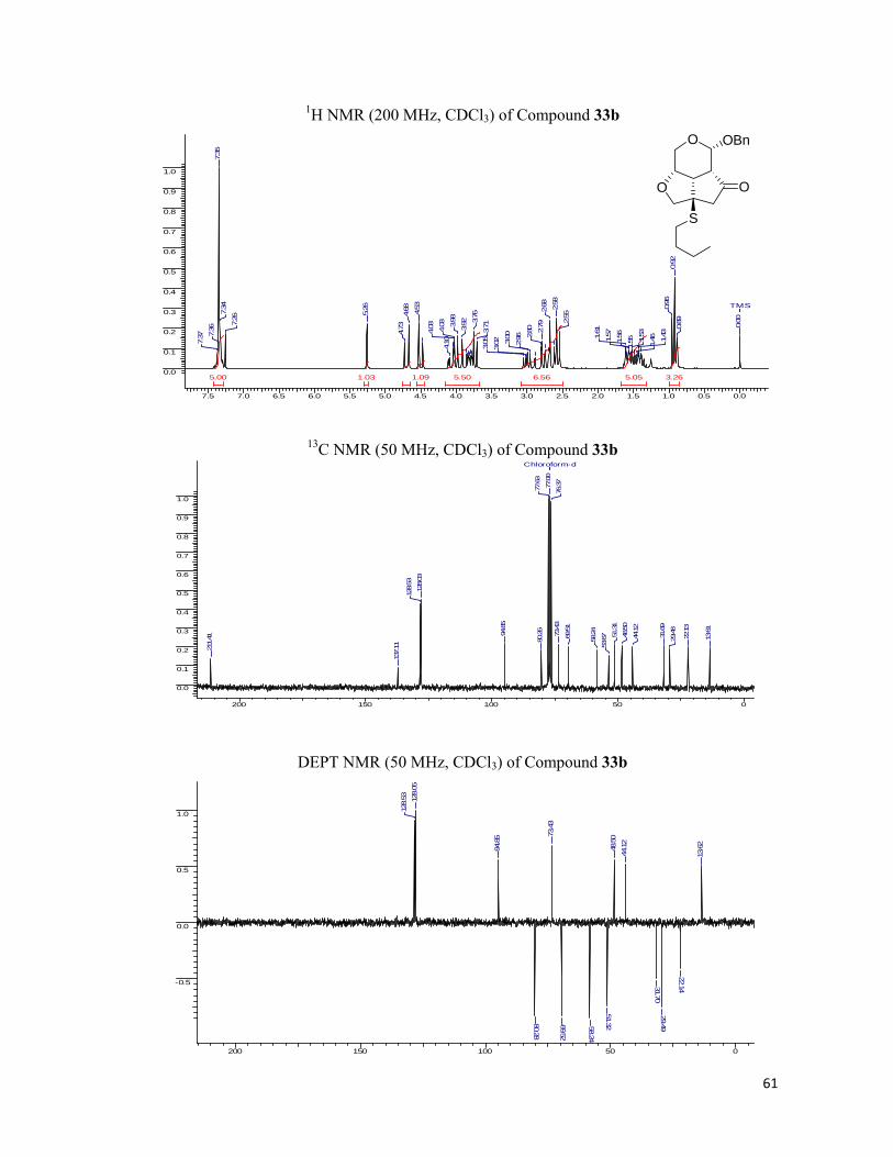
61
1H NMR (200 MHz, CDCl3) of Compound 33b
7.5 7.0 6.5 6.0 5.5 5.0 4.5 4.0 3.5 3.0 2.5 2.0 1.5 1.0 0.5 0.0
0.0
0.1
0.2
0.3
0.4
0.5
0.6
0.7
0.8
0.9
1.0
6.565.50 5.055.00 3.261.091.03
TMS
0.00
0.89
0.92
0.96
1.43
1.461.53
1.551.
561.571.61
2.55
2.58
2.68
2.79
2.80
2.963.00
3.023.05
3.71
3.76
3.923.98
4.03
4.03
4.10
4.53
4.68
4.73
5.26
7.26
7.34
7.35
7.36
7.37
13C NMR (50 MHz, CDCl3) of Compound 33b
200 150 100 50 0
0.0
0.1
0.2
0.3
0.4
0.5
0.6
0.7
0.8
0.9
1.0
Chloroform-d
13.61
22.13
29.48
31.69
44.12
48.50
51.31
53.67
58.24
69.51
73.43
76.37
77.00
77.63
80.26
94.85
128.03
128.53
137.11211.41
DEPT NMR (50 MHz, CDCl3) of Compound 33b
200 150 100 50 0
-0.5
0.0
0.5
1.0
13.62
22.14
29.4931.70
44.12
48.50
51.3258.24
69.52
73.43
80.28
94.85
128.05
128.53
O
O O
OBn
S

62
1H NMR (200 MHz, CDCl3) of Compound 33c
7.5 7.0 6.5 6.0 5.5 5.0 4.5 4.0 3.5 3.0 2.5 2.0 1.5 1.0 0.5 0.0
0.0
0.1
0.2
0.3
0.4
0.5
0.6
0.7
0.8
0.9
1.0
5.88 5.07 3.541.92 1.011.011.00 0.89
0.00
2.17
2.70
2.73
2.76
2.79
3.53
3.58
3.76
3.783.79
3.83
4.10
4.11
4.16
4.54
4.60
4.67
4.82
4.88
6.176.
19
6.306.31
6.32
6.337.26
7.30
7.32
7.33
7.35
13C NMR (50 MHz, CDCl3) of Compound 33c
200 150 100 50 0
0.0
0.1
0.2
0.3
0.4
0.5
0.6
0.7
0.8
0.9
1.0
Chloroform-d
26.81
47.01
47.33
49.99
54.68
65.34
69.75
72.19
76.3777.00
77.64
77.81
97.07
107.92110.84
127.74
128.37
136.93
142.15
150.78
210.41
DEPT NMR (50 MHz, CDCl3) of Compound 33c
200 150 100 50 0
-0.5
0.0
0.5
1.0
26.80
47.01
47.32
49.99
65.3269.74
72.18
77.80
97.07
107.92
110.84
127.76
128.37
142.15
O
O O
OBn
SO

63
1H NMR (200 MHz, CDCl3) of Compound 33d
7.5 7.0 6.5 6.0 5.5 5.0 4.5 4.0 3.5 3.0 2.5 2.0 1.5 1.0 0.5 0.0
0.0
0.1
0.2
0.3
0.4
0.5
0.6
0.7
0.8
0.9
1.0
11.01 9.022.291.70 1.03
0.00
2.60
2.68
2.76
2.79
2.81
2.82
2.83
2.853.
66
3.71
3.83
3.95
3.99
4.10
4.11
4.54
4.60
4.68
4.82
4.88
7.157.
16
7.207.277.30
7.31
7.33
7.34
13C NMR (50 MHz, CDCl3) of Compound 33d
200 150 100 50 0
0.0
0.1
0.2
0.3
0.4
0.5
0.6
0.7
0.8
0.9
1.0
Chloroform-d
31.28
35.94
46.93
47.39
50.63
54.77
65.42
69.71
72.54
76.37
77.00
77.63
77.76
97.11
126.59
127.74
128.35
128.52
136.89
139.69
210.47
DEPT NMR (50 MHz, CDCl3) of Compound 33d
200 150 100 50 0
0.0
0.5
1.0
31.2735.93
46.9
247
.38
50.62
65.4169.70
72.5
2
77.75
97.1
1
126.
5812
7.73
128.
34
O
O O
OBn
S

64
1H NMR (200 MHz, CDCl3) of Compound 33e
8.0 7.5 7.0 6.5 6.0 5.5 5.0 4.5 4.0 3.5 3.0 2.5 2.0 1.5 1.0 0.5 0.0 -0.5
0.0
0.1
0.2
0.3
0.4
0.5
0.6
0.7
0.8
0.9
1.0
10.00 5.14 2.731.01 1.000.96
0.00
2.47
2.57
2.74
2.75
3.09
3.71
3.75
3.79
3.81
3.83
3.94
4.00
4.01
4.07
4.46
4.52
4.66
4.71
5.22
7.26
7.35
7.38
7.49
7.53
7.54
13C NMR (50 MHz, CDCl3) of Compound 33e
200 150 100 50 0
0.0
0.1
0.2
0.3
0.4
0.5
0.6
0.7
0.8
0.9
1.0
Chloroform-d
43.9
448
.49
50.7
056
.7758.2
5
69.5
373
.30
76.3
777
.00
77.6
479
.75
94.9
0
128.10
128.
5312
9.38
129.
4913
0.96
135.
9813
7.06
211.42
DEPT NMR (50 MHz, CDCl3) of Compound 33e
140 130 120 110 100 90 80 70 60 50 40 30 20 10 0
-0.5
0.0
0.5
1.0
43.9
3
48.4
9
50.70
58.2469.53
73.3
079.75
94.9
0
128.
0212
8.10
129.
39
135.
98
O
O O
OBn
S

65
1H NMR (200 MHz, CDCl3) of Compound 33f
7.5 7.0 6.5 6.0 5.5 5.0 4.5 4.0 3.5 3.0 2.5 2.0 1.5 1.0 0.5 0.0
0.0
0.1
0.2
0.3
0.4
0.5
0.6
0.7
0.8
0.9
1.0
7.377.26 2.841.98 1.09 1.051.00
0.00
2.61
2.65
2.74
2.873.753.763.
793.81
3.88
3.92
4.10
4.104.
534.60
4.64
4.67
4.81
4.87
6.846.
88
7.267.
317.32
7.38
7.42
13C NMR (50 MHz, CDCl3) of Compound 33f
200 150 100 50 0
0.0
0.1
0.2
0.3
0.4
0.5
0.6
0.7
0.8
0.9
1.0
Chloroform-d
46.13
47.49
50.33
55.32
57.7465
.72
69.70
72.71
76.36
77.00
77.63
97.31
114.79
121.24
127.70
127.76
128.35
136.95
138.13
160.82210.54
DEPT NMR (50 MHz, CDCl3) of Compound 33f
200 150 100 50 0
-0.5
0.0
0.5
1.0
46.12
47.48
50.32
55.31
65.7269.68
72.70
77.33
97.31
114.78
127.76
128.3513
8.13
O
O O
OBn
S
MeO
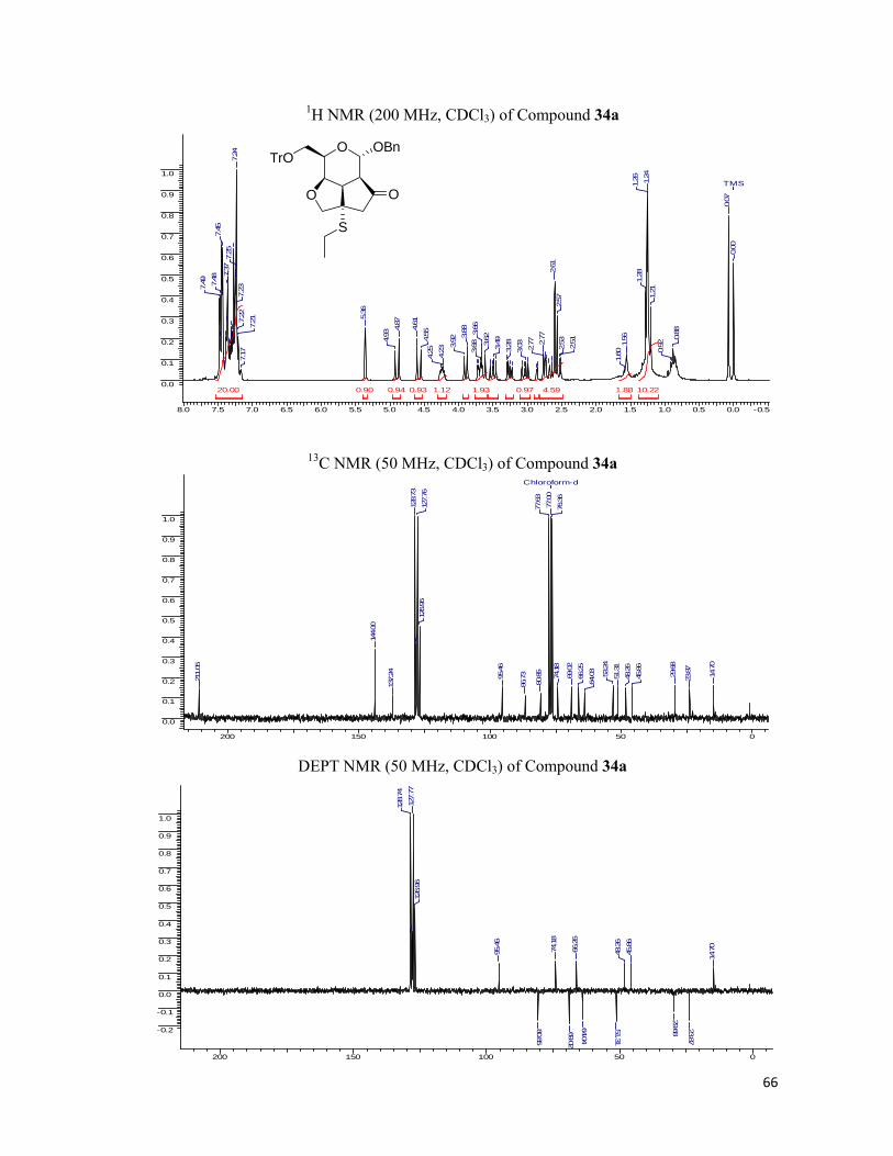
66
1H NMR (200 MHz, CDCl3) of Compound 34a
8.0 7.5 7.0 6.5 6.0 5.5 5.0 4.5 4.0 3.5 3.0 2.5 2.0 1.5 1.0 0.5 0.0 -0.5
0.0
0.1
0.2
0.3
0.4
0.5
0.6
0.7
0.8
0.9
1.0
20.00 10.224.591.93 1.881.12 0.970.94 0.930.90
TMS
0.00
0.07
0.88
0.92
1.21
1.24
1.26
1.28
1.56
1.60
2.51
2.53
2.57
2.61
2.77
2.77
3.03
3.283.493.62
3.66
3.68
3.88
3.92
4.23
4.25
4.55
4.61
4.87
4.93
5.36
7.17
7.217.
227.23
7.24
7.25
7.37
7.45
7.48
7.49
13C NMR (50 MHz, CDCl3) of Compound 34a
200 150 100 50 0
0.0
0.1
0.2
0.3
0.4
0.5
0.6
0.7
0.8
0.9
1.0
Chloroform-d
14.70
23.87
29.68
45.86
48.26
51.31
53.24
64.03
66.25
69.02
74.18
76.36
77.00
77.63
80.85
86.7395.46
126.96
127.76
128.73
137.24
144.00
211.05
DEPT NMR (50 MHz, CDCl3) of Compound 34a
200 150 100 50 0
-0.2
-0.1
0.0
0.1
0.2
0.3
0.4
0.5
0.6
0.7
0.8
0.9
1.0
14.70
23.87
29.68
45.86
48.26
51.31
64.04
66.26
69.02
74.18
80.85
95.46
126.96
127.77
128.74
O
O O
TrOOBn
S

67
1H NMR (200 MHz, CDCl3) of Compound 34b
7.5 7.0 6.5 6.0 5.5 5.0 4.5 4.0 3.5 3.0 2.5 2.0 1.5 1.0 0.5 0.0 -0.5
0.0
0.1
0.2
0.3
0.4
0.5
0.6
0.7
0.8
0.9
1.0
20.00 6.46 3.032.441.680.94 0.860.810.800.78
0.00
0.87
0.91
0.94
1.26
1.37
1.41
1.55
1.59
2.522.
552.60
2.74 2.76
3.02
3.27
3.293.493.613.
653.71
3.86
3.91
4.25
4.55
4.61
4.87
4.93
5.36
7.17
7.18
7.21
7.24
7.29
7.45
7.48
7.49
13C NMR (50 MHz, CDCl3) of Compound 34b
200 150 100 50 0
0.0
0.1
0.2
0.3
0.4
0.5
0.6
0.7
0.8
0.9
1.0
Chloroform-d
13.60
22.12
29.50
31.66
45.85
48.31
51.37
53.20
64.06
66.27
69.04
74.24
76.37
77.00
77.63
80.75
86.75
95.49
126.96
127.76
128.75
137.25
144.02
211.07
DEPT NMR (50 MHz, CDCl3) of Compound 34b
200 150 100 50 0
-0.2
-0.1
0.0
0.1
0.2
0.3
0.4
0.5
0.6
0.7
0.8
0.9
1.0
13.62
22.13
29.5131.65
45.84
48.31
51.37
64.06
66.27
69.04
74.24
80.76
95.49
126.96
127.78
128.74
O
O O
TrOOBn
S
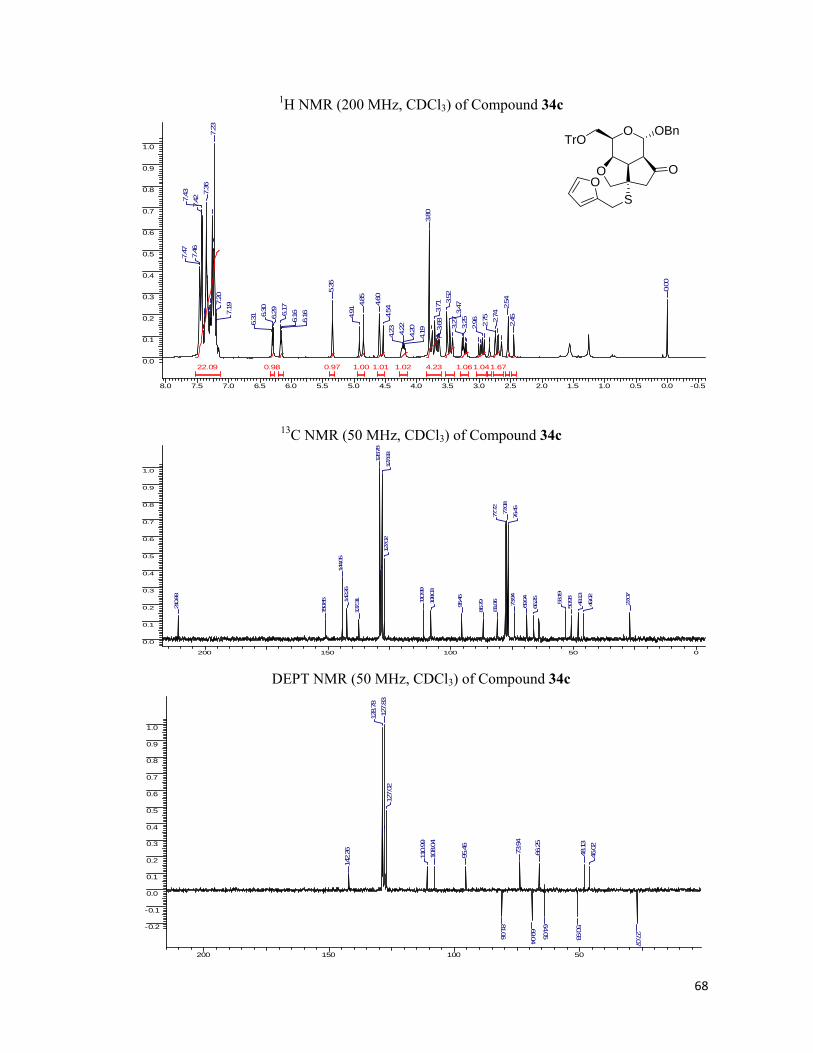
68
1H NMR (200 MHz, CDCl3) of Compound 34c
8.0 7.5 7.0 6.5 6.0 5.5 5.0 4.5 4.0 3.5 3.0 2.5 2.0 1.5 1.0 0.5 0.0 -0.5
0.0
0.1
0.2
0.3
0.4
0.5
0.6
0.7
0.8
0.9
1.0
22.09 4.23 1.671.061.041.021.011.000.98 0.97
0.00
2.45
2.54
2.74
2.75
2.96
3.25
3.27
3.47
3.52
3.68
3.71
3.80
4.194.204.22
4.23
4.54
4.60
4.85
4.91
5.35
6.16
6.166.
176.
296.30
6.31
7.19
7.20
7.23
7.36
7.427.
437.
467.47
13C NMR (50 MHz, CDCl3) of Compound 34c
200 150 100 50 0
0.0
0.1
0.2
0.3
0.4
0.5
0.6
0.7
0.8
0.9
1.0
27.07
46.02
48.13
50.93
53.19
66.25
69.04
73.94
76.45
77.08
77.72
81.06
86.79
95.46
108.03
110.99
127.02
127.83128.78
137.3114
2.26
144.05
150.85
210.88
DEPT NMR (50 MHz, CDCl3) of Compound 34c
200 150 100 50
-0.2
-0.1
0.0
0.1
0.2
0.3
0.4
0.5
0.6
0.7
0.8
0.9
1.0
27.07
46.02
48.13
50.93
64.05
66.25
69.04
73.94
81.06
95.46
108.04
110.99
127.02
127.83
128.78
142.26
O
O O
TrOOBn
SO
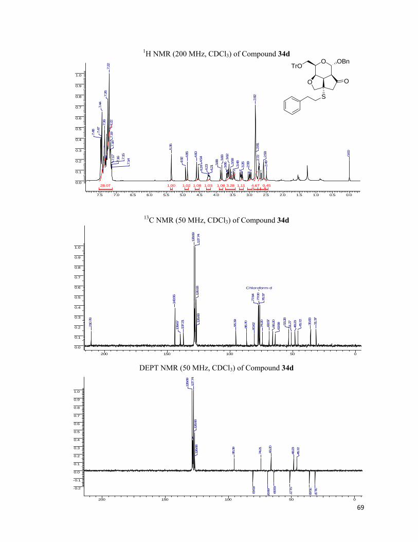
69
1H NMR (200 MHz, CDCl3) of Compound 34d
7.5 7.0 6.5 6.0 5.5 5.0 4.5 4.0 3.5 3.0 2.5 2.0 1.5 1.0 0.5 0.0
0.0
0.1
0.2
0.3
0.4
0.5
0.6
0.7
0.8
0.9
1.0
28.07 4.673.28 1.111.081.08 1.031.021.00 0.45
0.00
2.49
2.58
2.72
2.81
2.82
2.99
3.263.483.583.
623.66
3.83
3.88
4.214.23
4.24
4.544.60
4.86
4.92
5.35
7.14
7.15
7.167.17
7.18
7.19
7.22
7.22
7.26
7.36
7.44
7.47
7.48
13C NMR (50 MHz, CDCl3) of Compound 34d
200 150 100 50 0
0.0
0.1
0.2
0.3
0.4
0.5
0.6
0.7
0.8
0.9
1.0
Chloroform-d
31.37
35.83
45.72
48.23
51.27
53.28
63.99
66.20
68.97
74.20
76.37
77.00
77.64
80.62
86.70
95.39126.68
126.93
127.74
128.69
137.21
139.67
143.96
210.78
DEPT NMR (50 MHz, CDCl3) of Compound 34d
200 150 100 50 0
-0.2
-0.1
0.0
0.1
0.2
0.3
0.4
0.5
0.6
0.7
0.8
0.9
1.0
31.37
35.83
45.72
48.23
51.27
63.99
66.20
68.97
74.21
80.63
95.39
126.68
126.93
127.74
128.69
O
O O
TrOOBn
S
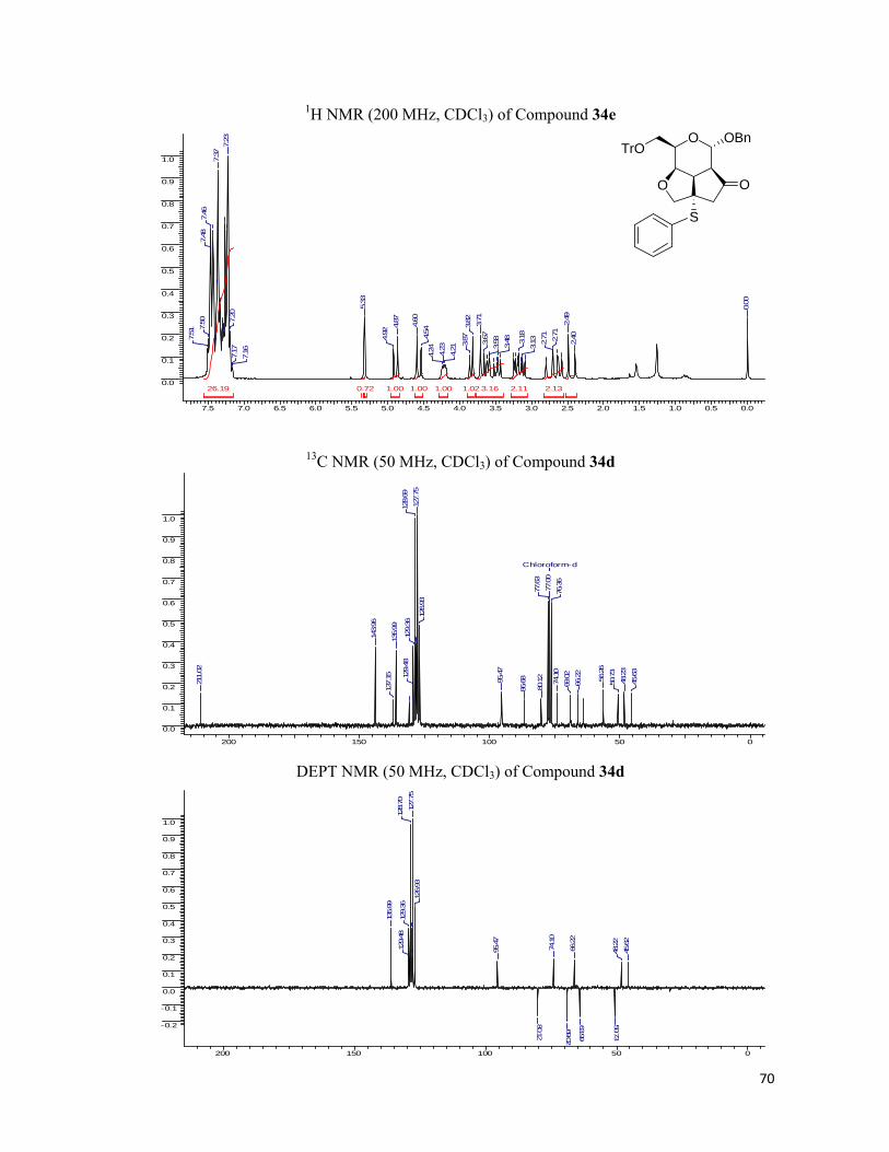
70
1H NMR (200 MHz, CDCl3) of Compound 34e
7.5 7.0 6.5 6.0 5.5 5.0 4.5 4.0 3.5 3.0 2.5 2.0 1.5 1.0 0.5 0.0
0.0
0.1
0.2
0.3
0.4
0.5
0.6
0.7
0.8
0.9
1.0
26.19 3.16 2.132.111.021.001.00 1.000.72
0.00
2.40
2.49
2.71
2.71
3.133.18
3.48
3.583.67
3.71
3.82
3.87
4.21
4.23
4.24
4.54
4.60
4.87
4.92
5.33
7.167.17
7.20
7.23
7.37
7.46
7.48
7.50
7.51
13C NMR (50 MHz, CDCl3) of Compound 34d
200 150 100 50 0
0.0
0.1
0.2
0.3
0.4
0.5
0.6
0.7
0.8
0.9
1.0
Chloroform-d
45.63
48.23
50.73
56.26
66.22
69.02
74.10
76.36
77.00
77.63
80.12
86.6895.47
126.93
127.75
128.69
129.36
129.48
135.99
137.15
143.96
211.02
DEPT NMR (50 MHz, CDCl3) of Compound 34d
200 150 100 50 0
-0.2
-0.1
0.0
0.1
0.2
0.3
0.4
0.5
0.6
0.7
0.8
0.9
1.0
45.62
48.22
50.73
63.99
66.22
69.02
74.10
80.12
95.47
126.93
127.75
128.70
129.36
129.48
135.99
O
O O
TrOOBn
S

71
1H NMR (200 MHz, CDCl3) of Compound 34e
7.5 7.0 6.5 6.0 5.5 5.0 4.5 4.0 3.5 3.0 2.5 2.0 1.5 1.0 0.5 0.0
0.0
0.1
0.2
0.3
0.4
0.5
0.6
0.7
0.8
0.9
1.0
24.37 4.752.60 2.242.231.08 1.051.031.00
0.00
2.37
2.46
2.612.67
3.17
3.223.63
3.67
3.753.
773.80
3.82
4.544.
60
4.87
4.93
5.32
6.75
6.76
6.846.
88
7.20
7.23
7.37
7.43
7.44
7.46
7.47
13C NMR (50 MHz, CDCl3) of Compound 34e
200 150 100 50 0
0.0
0.1
0.2
0.3
0.4
0.5
0.6
0.7
0.8
0.9
1.0
45.49
48.34
50.61
55.40
56.51
64.1266.34
69.10
74.26
76.44
77.07
77.71
79.84
86.7595.57
114.94
121.50
127.01
127.82
128.79
137.27
138.18144.05
160.92
211.27
DEPT NMR (50 MHz, CDCl3) of Compound 34e
200 150 100 50 0
-0.2
-0.1
0.0
0.1
0.2
0.3
0.4
0.5
0.6
0.7
0.8
0.9
1.0
45.4
948
.34
50.61
55.4
0
64.12
66.3
3
69.10
74.2
6
79.84
95.5
7
114.
94
127.
0112
7.83
128.
79
138.
18
O
O O
TrOOBn
S
MeO
72
Chapter 1: References
1. (a) Schreiber, S. L. Chem. Eng. News 2003, 81, 51-61; (b) Spring, D. R. Chem. Soc.
Rev. 2005, 34, 472-482.
2. Lee, Y. K.; Puong, K. Y. Br. J. Nutr. Med. 2002, 88, S101-S108.
3. Bohacek, R. S.; McMartin, C.; Guida, W. C. Med. Res. Rev. 1996, 16, 3-50.
4. Clardy, J.; Walsh, C. Nature 2004, 432, 829-837.
5. (a) Nichlaou, K. C.; Vourloumis, D.; Winssinger, N.; Baran, P. S. Angew. Chem.
Int. Ed. 2000, 39(1), 44-122; (b) Nicolaou, K. C.; Sorenson, E. J. Classics in Total
Synthesis 1996; (c) Nicolaou, K. C.; Snyder, S. A. Classics in Total Synthesis II
2003.
6. Corey, E. J. Chem. Soc. Rev. 1988, 17, 111-133.
7. (a) Thomas, G. L.; Wyatt, E. E.; Spring, D. R. Curr. Opin. Drug Disc. & Develop.
2006, 9(6), 700-712; (b) Kirkpatrick, P.; Ellis, C. Nature 2004, 432, 823-865; (c)
Shang, S.; Tan, D. S. Curr. Opin. Chem. Biol. 2005, 9, 1-11.
8. Fink, T.; Reymond, J. L. J. Chem. Inf. Model 2007, 47(2), 342.
9. Lipinski, C. A. et. al. Adv. Drug Del. Rev. 2001, 46, 3-26.
10. Breinbauer, R.; Vetter, I. R.; Waldmann, H. Angew. Chem., Int. Ed. 2002, 41, 2878-
2890.
11. Schreiber, S. L. Science 2000, 287, 1964-1969.
12. (a) Schreiber, S. L. Science 2000, 287, 1964-1969; (b) Kwon, O.; Park, S. B.;
Schreiber, S. L. J. Am. Chem. Soc. 2002, 124, 13402-13404; (c) Wu, C.-Y.; Chang,
C.-F.; Chen, J. S-y.; Wong, C.-H.; Lin, C.-H. Angew. Chem. Int. Ed. 2003, 42,
4661-4664. (d) Micalizio, G. C.; Schreiber, S. L. Angew. Chem., Int. Ed. 2002, 41,
3272-3276; (e) Tan, D. S.; M. A.; Shair, M. D.; Schreiber, S. L. J. Am. Chem. Soc.
1998, 120, 8565-8566; (f) Burke, M. D.; Schreiber, S. L. Angew. Chem., Int. Ed.
2004, 43, 46-58.
13. Neumann, C. S.; Nagayama, S.; Peristein, E. O.; Schreiber, S. L. J. Am. Chem. Soc.
2004, 126, 16077-16086.
14. Pelish, H. E.; Westwood, N. J.; Shair, M. D. J. Am. Chem. Soc. 2001, 123, 6740-
6741.
15. Nicolaou, K. C.; Pfefferkorn, J. A.; Mithcell, H. J. J. Am. Chem. Soc. 2000, 122,
9968-9976.

73
16. Thomas, G. L.; Wyatt, E. E.; Spring, D. R. Curr. Opin. Drug Disc. & Develop.
2006, 9(6), 700-712.
17. Williams, N. R.; Wander, J. D. The Carbohydrates in Chemistry and Biochemistry;
Academic Press: New York 1980, 761.
18. Varki, A.; Cummings, R.; Marth, J. Essentials of Glycobiology 1999.
19. (a) Ferrier, R. J.; Prasad, N. J. Chem. Soc. (C) 1969, 570–574.
20. (a) Babu, B. S.; Balasubramanian, K. K. Tetrahedron Lett. 2000, 41, 1271–1274;
(b) Shanmugasundaram, B.; Bose, A. K.; Balasubramanian, K. K. Tetrahedron Lett.
2002, 43, 6795–6798; (c) Grynkiewiez, G.; Priebe, W.; Zamojski, A. Carbohydr.
Res. 1979, 68, 33–41; (d) Swami, N. R.; Venkateswarlu, A. Synthesis 2002, 598–
600; (e) Masson, C.; Soto, J.; Bessodes, M. Synlett 2000, 1281–1282; (f) Yadav, J.
S.; Reddy, B. V. S.; Murthy, C. V. S. R.; Kumar, G. M. Synlett 2000, 1450–1451;
(g) Bettadaih, B. K.; Srinivas, P. Tetrahedron Lett. 2003, 44, 7257–7259; (h) Babu,
B. S.; Balasubramanian, K. K. Synth. Commun. 1998, 29, 4299–4305; (i) Yadav, J.
S.; Reddy, B. V. S.; Reddy, J. S. S. J. Chem. Soc., Perkin Trans. 1 2002, 2390–
2394; (j) Smitha, G.; Reddy, C. S. Synthesis 2004, 834–836.
21. (a) Toshima, K.; Ishizuka, T.; Matsuo, G.; Nakata, M.; Konoshita, M. J. Chem.
Soc., Chem. Commun. 1993, 704–706; (b) Fraser-Reid, B.; Madsen, R. J. Org.
Chem. 1995, 60, 3851–3858; (c) Koreeda, M.; Houston, T. A.; Shull, B. K.;
Klemke, E.; Tuinman, R. J. Synlett 1995, 90–92; (d) Lo´pez, J. C.; Fraser-Reid, B.
J. Chem. Soc., Chem. Commun. 1992, 94–95; (e) Yadav, J. S.; Reddy, B. V. S.;
Pandey, S. K. New J. Chem. 2001, 25, 538–540; (f) Agarwal, A.; Rani, S.; Vankar,
Y. D. J. Org. Chem. 2004, 69, 6137–6140; (g) Suzuki, K.; Hashimoto, T.; Maeta,
H.; Matsumoto, T. Synlett 1992, 125-128.
22. Hotha, S.; Tripathi, A. Tetrahedron Lett. 2005, 46, 4555-4558.
23. Sinou, D. Hetrocycles 2006, 68, 2607-2613.
24. Khand, I. U.; Knox, G. R.; Pauson, P. L. J. Chem. Soc. Perkin Trans 1973, 1, 977.
25. (a) Gibson, S. E.; Stevenazzi, A. Angew. Chem. Int. Ed. 2003, 42, 1800-1810. (b)
Krafft, M. E.; Bonaga, L. V. R. Tetrahedron 2004, 60, 9795-9833; (c) Jeong, N.;
Chung, Y. K.; Yoo, S. E. Synlett 1991, 204.
26. Kubota, H.; Lim, J.; Depew, K. M.; Schreiber, S. L. Chem. & Biol. 2002, 9, 265-
276.
27. Hotha, S.; Tripathi, A. J. Comb. Chem. 2005, 7, 968-976.
******

Chapter 2 ‘Click’ Chemistry Inspired Imaging of Microorganisms
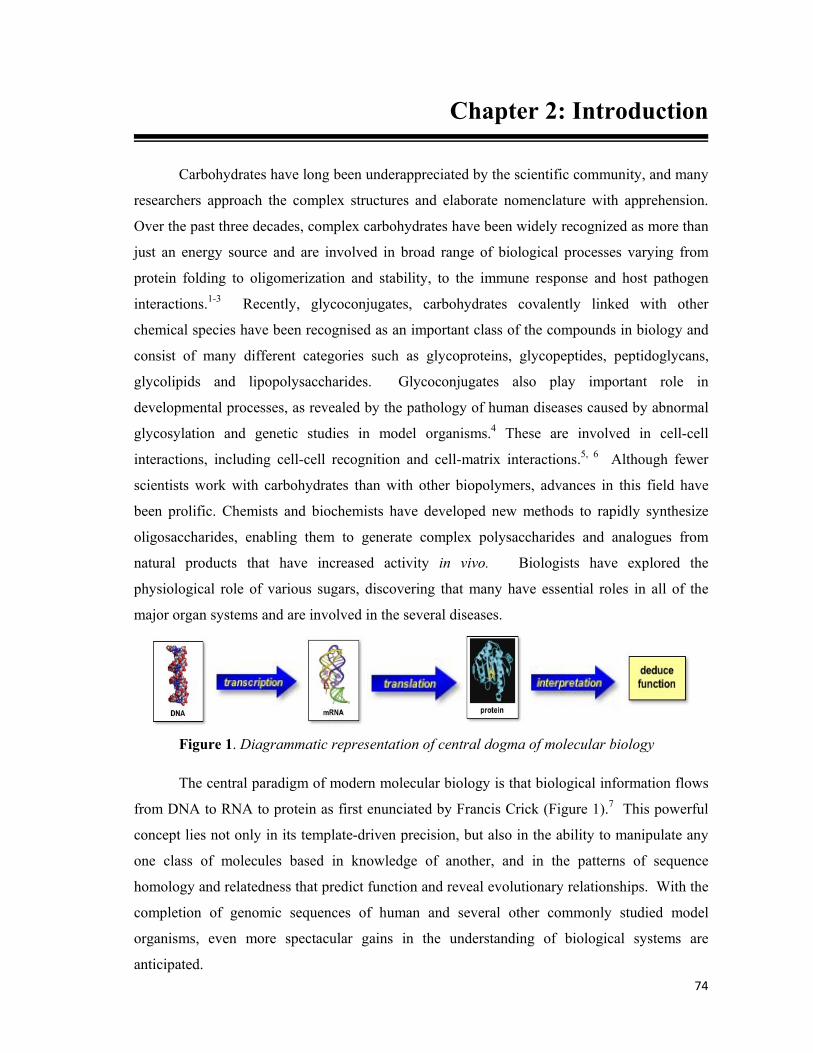
74
Chapter 2: Introduction
Carbohydrates have long been underappreciated by the scientific community, and many
researchers approach the complex structures and elaborate nomenclature with apprehension.
Over the past three decades, complex carbohydrates have been widely recognized as more than
just an energy source and are involved in broad range of biological processes varying from
protein folding to oligomerization and stability, to the immune response and host pathogen
interactions.1-3 Recently, glycoconjugates, carbohydrates covalently linked with other
chemical species have been recognised as an important class of the compounds in biology and
consist of many different categories such as glycoproteins, glycopeptides, peptidoglycans,
glycolipids and lipopolysaccharides. Glycoconjugates also play important role in
developmental processes, as revealed by the pathology of human diseases caused by abnormal
glycosylation and genetic studies in model organisms.4 These are involved in cell-cell
interactions, including cell-cell recognition and cell-matrix interactions.5, 6 Although fewer
scientists work with carbohydrates than with other biopolymers, advances in this field have
been prolific. Chemists and biochemists have developed new methods to rapidly synthesize
oligosaccharides, enabling them to generate complex polysaccharides and analogues from
natural products that have increased activity in vivo. Biologists have explored the
physiological role of various sugars, discovering that many have essential roles in all of the
major organ systems and are involved in the several diseases.
Figure 1. Diagrammatic representation of central dogma of molecular biology
The central paradigm of modern molecular biology is that biological information flows
from DNA to RNA to protein as first enunciated by Francis Crick (Figure 1).7 This powerful
concept lies not only in its template-driven precision, but also in the ability to manipulate any
one class of molecules based in knowledge of another, and in the patterns of sequence
homology and relatedness that predict function and reveal evolutionary relationships. With the
completion of genomic sequences of human and several other commonly studied model
organisms, even more spectacular gains in the understanding of biological systems are
anticipated.

75
Infact, creating a cell wall requires two major classes of molecule: lipids and
carbohydrates.8b, 8c Carbohydrates are indispensable to life on Earth. These biomolecules in
their simplest life forms serve as a primary energy source for sustaining life. For the most part,
however, carbohydrates exist not as simple sugars but as complex molecular conjugates, or
glycans. Glycans come in many shapes and sizes, from linear chains (polysaccharides) to
highly branched molecules bristling with antennae-like arms. And although proteins and
nucleic acids such as DNA have traditionally attracted far more scientific attention, glycans are
also key to life. They are ubiquitous in nature, forming the intricate sugar coat that surrounds
the cells of virtually every organism and occupying the spaces between these cells.
DNA RNA PROTEINS ENZYMES
CARBOHYDRATES
GLYCOCONJUGATESCELL
ORGANISM LIPIDS
Figure 2. Extended paradigm of the molecular biology
The structural role of carbohydrates becomes particularly important in constructing
complex multi-cellular organs and organisms, which requires interactions of cell with one
another and with the surrounding matrix.8 Indeed, all cells and many macromolecules in nature
carry a dense and complex array of covalently attached sugar chains (called oligosaccharides or
glycans). Since most glycans are in the outer surface of cellular and secreted macromolecules,
they are in a position to modulate or mediate a wide variety of events in cell-cell and cell-
matrix interactions crucial to the development and function of a complex multicellular
organism. They are also in a position to mediate interactions between organism (e.g. between
host and parasite). In addition, simple, highly dynamic protein-bound glycans are abundant in
the nucleus and cytoplasm, where they appear to serve as regulatory switches (Figure 2).
During the last century, the chemistry, biochemistry and biology of carbohydrates were
prominent matters of interest. However, during the initial phase of the modern revolution in
molecular biology, studies of glycans lagged far behind those major class of molecules. This
was in large part due to their inherent structural complexity, the difficulty in easily determining

76
their sequence, and the fact that their biosynthesis can not be easily predicted from the DNA
template. The development of a variety of new technologies for exploring the structures of
these glycan chains has opened a new frontier of molecular biology which has been called
glycobiology. This word was first coined by Rademacher, Parekh and Dwek to recognize the
coming together of the traditional disciplines of carbohydrate chemistry and biochemistry with
modern understanding of the cellular and molecular biology of glycans.9 Emerging from its
roots in classical carbohydrate chemistry and biochemistry, glycobiology has become a vibrant,
expanding and important extension of molecular biology. Glycobiology combines the
expertise of synthetic, analytical and carbohydrate biochemistry, as well as molecular and
cellular biology, to unravel the structural complexity, chemistry, biosynthesis, and biological
functions of the sugar-bearing biomolecules.
As the surface of almost every living cell is decorated with the a layer of complex
carbohydrates, many of the advances in complex carbohydrate synthesis revolve around
methods to form the glycosidic bond, because this is the primary means by which
monosaccharide building blocks are assembled into more complex oligosaccharide structures.
The complexity of the glycosylation pathways that manufacture and constantly remodel surface
sugars is truly astounding. Glycosylation can have a profound influence on the function of a
variety of eukaryotic cells. It can be described as a complex process that requires a major
commitment of cellular resources; it employs an estimated 2–3% of the genes in humans and
many high-energy intermediates.10 The first step in glycosylation is the import of dietary
sugars, such as glucose, into a cell. This is followed by a series of phosphorylation,
epimerization, and acetylation reactions that diversify these sugars and convert them into high-
energy nucleotide sugar donors. These compounds serve as the “building blocks” for the
assembly of complex carbohydrates in the endoplasmic reticulum and Golgi apparatus. The
newly synthesized carbohydrates, which are generally attached to proteins or lipids, are then
transported to the cell surface where they contribute to the interaction between a cell and its
environment by playing important structural and signalling roles. In particular, it can affect
signal transduction and cell–cell communication properties and thus shape critical cell
decisions, including the regulation of differentiation and apoptosis. Regulation of
glycosylation has multiple layers of complexity, both structural and functional, which make its
experimental and theoretical analysis difficult to perform and interpret.11
Carbohydrate chemists and biochemists tend to be controlling personalities and aren't
satisfied with the way sugar groups are attached naturally to glycosylated proteins and natural
products. Around the world researchers are occupied most days devising new ways to tame

77
fractious glycosylated biomolecules into shape. Glycosylation of protein or a natural product
can be achieved by the bioengineering of bacteria, yeast, plant cells, insect cells, and
mammalian cells. Apart from this, it can also be achieved by manipulating protein or natural
product glycosylation pathways in genetically engineered organisms. Compounds also can be
glycosylated by exploiting the liberality with which sugar-attaching enzymes (glycosyl
transferases) tolerate a wide range of different substrates. And one can glycosylate proteins
and natural products through a variety of chemical approaches: with enzymes, protein ligation,
metabolic bioengineering, or total synthesis.
More than 50% of human proteins are glycoproteins, which tend to be heterogeneously
glycosylated.12 That means the structures of attached sugars and the sites to which they're
attached are variable. A wide range of bioactive natural products are glycosylated as well,
including anthracycline antitumor antibiotics, avermectin antiparasitics, enediyne antibiotics,
macrolides such as erythromycin, and glycopeptide natural products like vancomycin. Some
natural products are also heterogeneously glycosylated, although generally to a lesser degree
than glycoproteins. Whether glycoproteins and glycosylated natural products are
heterogeneously glycosylated or not, researchers have felt the need to better control which
sugars go where on these molecules. The idea is to improve on Mother Nature because sugars
are often essential to the functions of biomolecules, and the ability to modify and control which
ones are attached and exactly how they're attached can make it possible to tailor biomolecular
structure, folding, stability, immunogenicity, uptake, distribution, target recognition, and other
properties and functions of these compounds. For example, glycosylated biomolecules can be
turned into better drugs by manipulating their sugars. Careful glycosylation of erythropoietin
(EPO), an approved glycoprotein drug for anemia and cancer, is critical for its activity and
longevity of action, and its properties have been improved by modifying its glycosylation
through genetic engineering. Novel research methodologies provided by glycobiology can
help to address many outstanding issues and integrate glycosylation with other metabolic and
cell regulation processes. Furthermore, glycosylation often lies at the interface between
metabolism and cell signalling, thereby making analysis even more challenging and interesting.
Recently, researchers around the world are actively pursuing the cell surface remodelling
exploiting glycosylation pathways through chemical means. As the cell surface govern many
of the vital biological events, perturbation of any of these metabolic processes will be guided
by the glycosylation pathways.
One of the recent advances in probing the glycosylation pathways was pioneered by C.
Bertozzi and her group when they synthesized the unnatural sialic acids as tool for the

78
penetrating metabolic process of the cells.13a Sialic acid is a generic term for the N- or O-
substituted derivatives of neuraminic acid, a nine-carbon monosaccharide. It is also the name
for the most common member of this group, N-acetylneuraminic acid. The amino group bears
either an acetyl or a glycolyl group. The hydroxyl substituents may vary considerably: acetyl,
lactyl, methyl, sulphate, and phosphate groups have been found. Sialic acids are the most
abundantly distributed terminal components of oligosaccharides on mammalian glycoproteins
and glycolipids. Sialic acids are biosynthesized from the six carbon precursor N-
acetylmannosamine.13b-13e Bertozzi group exploited the tolerance of enzymes, which take part
in sialic acid biosynthesis, to unnatural mannosamine derivatives; ManNAc was substituted
with N-levulinoyl having the ketone functionality at the position normally occupied by the N-
acetyl group in the natural substrate. The ketone group on the surface was useful when
covalently ligated with complementary functional group hydrazide (Figure 3).
OHO
AcHN
CO2H
OH
OH
HOOH
OHO
N
CO2H
OH
OH
HOOH
O
O
HO
HON
CO2H
O
OH
HOOH
O
O
H
OHO
N
CO2H
O
OH
HOOH
O
N
H
HN
R
O
NHNH2
OR
Cell
Cell
Cell
R =Inactive participantFluorescent TagDrug
Sialic acid
Figure 3. Biosynthetic incorporation of ketone groups of unnatural mannosamine derivatives onto cell surface
They selected three human cell lines for their studies- Jurkat (T cell derived), HL-60
(neutrophil derived), and HeLa (cervical epithelial carcinoma). After performing a series of
experiments, they were able to successfully demonstrate the incorporation of ManLev on the
cell surface exploiting the glycosylation pathway of the human cell lines. The presence of the
ketone group on the cell surface was determined by the chemoselective ligation of a hydrazide
based probe, biotinamidocaproyl hydrazide. The cells were then analysed by flow cytometry
employing fluorescein isothiocynate which shows a strong binding affinity for biotin.
Researchers were also successful in their attempt to demonstrate the inhibition of the ketone
group display on the cell surface via the use of Tunicamycin, known to inhibit the N-linked
protein glycosylation.
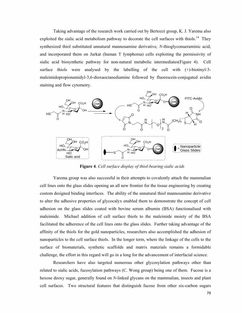
79
Taking advantage of the research work carried out by Bertozzi group, K. J. Yarema also
exploited the sialic acid metabolism pathway to decorate the cell surfaces with thiols.14 They
synthesized thiol substituted unnatural mannosamine derivative, N-thioglyconueraminic acid,
and incorporated them on Jurkat (human T lymphoma) cells exploiting the permissivity of
sialic acid biosynthetic pathway for non-natural metabolic intermediates(Figure 4). Cell
surface thiols were analysed by the labelling of the cell with (+)-biotinyl-3-
maleimidopropionamidyl-3,6-dioxaoctanediamine followed by fluorescein-conjugated avidin
staining and flow cytometry.
OHO
AcHN
CO2H
OH
OH
HOOH
O
HON
CO2H
OH
OH
HOOH
HSO
H
Cell
Sialic acid
NH
S
H
HO
O
(CH2)4
O
HN
OHN
O
N
O
O 2
OHO
N
CO2H
O
OH
HOOH
HSO
HCell
FITC-Avidin
OHO
N
CO2H
O
OH
HOOH
SO
HCell
NanoparticleGlass Slides=
Figure 4. Cell surface display of thiol-bearing sialic acids
Yarema group was also successful in their attempts to covalently attach the mammalian
cell lines onto the glass slides opening an all new frontier for the tissue engineering by creating
custom designed binding interfaces. The ability of the unnatural thiol mannosamine derivative
to alter the adhesive properties of glycocalyx enabled them to demonstrate the concept of cell
adhesion on the glass slides coated with bovine serum albumin (BSA) functionalised with
maleimide. Michael addition of cell surface thiols to the maleimide moiety of the BSA
facilitated the adherence of the cell lines onto the glass slides. Further taking advantage of the
affinity of the thiols for the gold nanoparticles, researchers also accomplished the adhesion of
nanoparticles to the cell surface thiols. In the longer term, where the linkage of the cells to the
surface of biomaterials, synthetic scaffolds and matrix materials remains a formidable
challenge, the effort in this regard will go in a long for the advancement of interfacial science.
Researchers have also targeted numerous other glycosylation pathways other than
related to sialic acids, fucosylation pathways (C. Wong group) being one of them. Fucose is a
hexose deoxy sugar, generally found on N-linked glycans on the mammalian, insects and plant
cell surfaces. Two structural features that distinguish fucose from other six-carbon sugars

80
found abundantly in mammals is the lack of hydroxyl functionality on C-6 and the L-
configuration. It is most often the terminal sugar in glycans that participate in important
biological events that are involved in various physiological and pathological processes of the
body. Wong et.al. demonstrated the visualization of the incorporation of an unnatural fucose
analog into glycoproteins via the fucose salvage pathway (Figure 5).15
O
OH
OH
ON3
OH
O
OH
OH
O
OH
NO O
N3
NO O
O
OH
OH
O
OH
N
O
O
N NN
O
OH
OH
O
OHN
O
O
N
NN
+
+
Cu(I)
Cu(I)
NapthalamideProbes
Unnatural Fucose Fluorescent Conjugates
Cell
Cell
Cell
Cell
Figure 5. Acetylene and azido probes for fucosylation pathways
The unnatural fucose analogs were designed so as to exploit the tolerance of the
fucosylating enzymes to a range of substituents at position 6 on fucose. Likewise, they
synthesized the azido/alkynyl functionalised fucose and allowed them to enter the cells. The
modified fucose derivatives were supplied to the cells in acetylated form facilitating
incorporation into glycoproteins through the salvage biosynthetic pathway and monitored them
by employing the fluorescent probes with the application of the click chemistry. The use of a
click activated probe is highly practical in biological systems as a fluorescent signal is obtained
only after a highly selective, bioorthogonal ligation takes place. The researchers were
successful in their attempt to probe the fucosylating pathway using fluorogenic probes
activated by click reaction allowing them to permit imaging of fucosylated glycoconjugates at
cell surface and inside the cells.
Apart from the mammalian cells, scientists are also engaged in discovering novel
probes for other microorganisms, like bacteria etc., in an attempt to display the cell wall with
unnatural substrates with a promise to provide novel vaccines against infectious diseases
caused by bacteria. In this regard, Nishimura et.al. reported a versatile chemical approach for
displaying the target compounds on the bacterial cell wall.16-18 Bacterial cell wall differs from
that of all the organisms by the presence of peptidoglycans (poly-N-acetylglucosamine and N-

81
acetylmuramic acid), which is located immediately outside the cytoplasmic membrane. The
bacterial cell wall consists of repeating units of N-acetylglucosamine and N-acetylmuramic
acid linked with a pentapeptide.19
OHO
O
O
OH
OHN
ONH
HO2CO
HN
O NH
O
HN CO2H
HN
POH
O
P
O
OH
NH
O
ONO
OH
O
OH
OO OH
RO
NHAc
O
O
R
Figure 6. Unnatural UDP-MurNAc pentapeptide synthesized by Nishimura for bacterial cell wall engineering
Nishimura group cleverly designed the unnatural probe for the Lactobaccilus bacterial
cell wall comprising of a pentapeptide attached to the N-acetylglucosamine along with Uracil
diphosphate moiety. The pentapeptide has the handle along which a fluorescent or a ketone
probe can be tethered which can later be tracked down by a hydrazide based fluorescent probe.
After examining a number of combinations of peptide, they arrived to a conclusion that
presence of the pentapeptide was vital for the activity of the unnatural disaccharide. After a
series of experiments, they were successful in demonstrating modification of the bacterial cell
wall through the artificial precursors which were displayed across the membrane.
As the global scientific efforts in the field of glycobiology have grown, the success of
these endeavours has prompted parallel efforts towards the next layer of biological complexity,
namely post-translational modifications, with an emphasis on glycosylation. The complex
glycosylation pathways of a cell do not exist in isolation; instead they are intimately
intertwined with other critical metabolic and regulatory networks of the cells, illustrate the
level of complexity involved to unravel the mechanistic details of glycosylation pathways.20
However, the tremendous benefits which are seemingly guaranteed have stimulated an
explosion of interest towards glycobiology and have been successful in luring lot of researchers
to put up a spirited effort to come with the challenge.
*****
82
Chapter 2: Present Work
Strep throat, cholera, pneumonia, whooping cough etc. these diseases, and more, are
often the only things bacteria get credit for causing in human and animals. Bacteria are the
oldest, the simplest and the most numerous forms of life on earth. Although we are never able
to notice these tiny forms of life with the naked eye, these are present almost everywhere.
These were first observed by Antonie van Leeuwenhoek in 1676, using a single-lens
microscope of his own design.21-23 Much later, the name bacterium was coined by Christian
Gottfried Ehrenberg in 1828, which is derived from the Greek word meaning small mass.
Afterwards, Robert Koch put forward his “Germ Theory of Disease”, for which he was
awarded the Nobel Prize in 1908, laying down some benchmarks to test if any organism is the
cause of a disease.
Bacteria are unicellular microorganisms, possessing a wide range of shapes from
spheres to rods to spirals. These are prokaryotes and unlike eukaryotes do not contain a nucleus
and rarely harbour membrane bound organelles. Bacterial cell wall is made of peptidoglycan,
unlike the cell wall of plants and fungi which are made up of cellulose and chitin respectively
(Figure 7).19 Broadly speaking, there are two different types of cell wall in bacteria, called
Gram-positive and Gram-negative. The name originates from the reaction of cells to the Gram
stain, a test long employed for the classification of bacterial species.24 Gram-positive
possesses a thick cell wall containing many layers of peptidoglycans and lipoteichoic acids
external to cytoplasmic membrane. In contrast, Gram-negative have relatively thin cell wall
consisting of a few layer of peptidoglycans surrounded by a second lipid membrane containing
lipopolysaccharides and lipoproteins. Most bacteria have Gram-negative cell wall. This
dissimilarity in structure is apparent in behavioral differences towards antibiotic susceptibility;
for instance, vancomycin can kill only Gram-positive bacteria and is ineffective against Gram-
negative pathogens. Peptidoglycan is a polymer of alternating N-acetylmuramic acid and N-
acetylglucosamine.25 Long strands of this alternating polymer may be linked by amino acids.
Gram-positive cells have a much more highly cross-linked peptidoglycan structure than Gram-
negative cells. Teichoic acid is a polymer of glycerol or ribitol via phosphodiester bonds. They
can be either covalently bonded to N-acetylmuramic acid of the peptidoglycan layer, linked to
the plasma membrane lipids found in cytoplasmic membrane or linked to the terminal D-
alanine in the tetrapeptide cross-links between molecules of N-acetylmuramic acid. The
combined unit s comprised of teichoic acid and lipids are referred to as lipoteichoic acid.

83
Teichoic acids are negatively charged and therefore contribute to the negative charge of the
Gram-positive cell wall.
Figure 7. The overall cell wall structure of (a) Gram-positive bacteria; (b) Gram-negative bacteria
Understanding glycomics in general and surface exposed carbohydrates of many
organisms in particular will play a vital role in enhancing our knowledge of human health
ranging from congenital disorders to infectious diseases.26 Advances in glycobiology have
established the significance of oligosaccharides and glycoconjugates, often present on the cell
surfaces.27 Carbohydrate engineering by exogenously added monosaccharide supplements is a
tool to probe various disease processes which would eventually facilitate discovery of highly
specific small molecule inhibitors.28 One of the major hindrances to the development of the
field is the lack of proper glycan oriented imaging techniques.29 Labeling via sialic acid and
fucose metabolic pathway in mammalian cells by exogenously supplied monosaccharides,
using a Staudinger ligation,28a a ketone-aminoxy/hydrazide ligation13, 28b and a Michael
addition14 have been extensively studied. However, this strategy will not be appropriate for
those cells (e.g. many bacteria, yeast – mostly causing infectious diseases) which do not
display appropriate sugars on their cell surfaces. Nishimura et.al. did study the cell surface
display of fluorescein attached UDP-MurNAc which allows imaging of the bacterial cells.16
Nishimura’s fluorescent conjugates are accessed through a multistep process and the total
glycosyl character of the chemical probe was reduced to the minimum due to the presence of
the appendages that would facilitate selective incorporation. As is evident from the Figure 8,
that researchers have exploited various appendages on carbohydrate motifs for probing the
glycosylation pathways utilizing fluorescence tags. But all the available methods have their
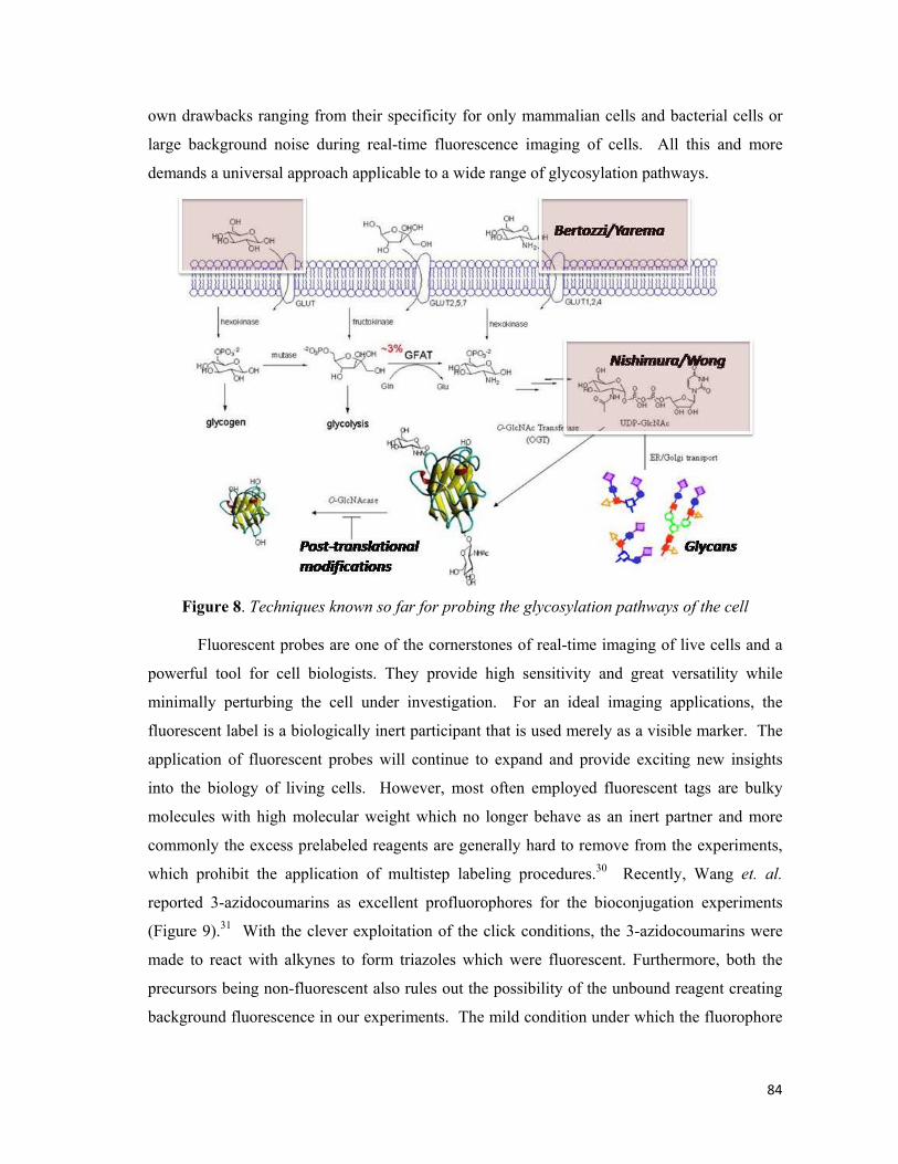
84
own drawbacks ranging from their specificity for only mammalian cells and bacterial cells or
large background noise during real-time fluorescence imaging of cells. All this and more
demands a universal approach applicable to a wide range of glycosylation pathways.
Figure 8. Techniques known so far for probing the glycosylation pathways of the cell
Fluorescent probes are one of the cornerstones of real-time imaging of live cells and a
powerful tool for cell biologists. They provide high sensitivity and great versatility while
minimally perturbing the cell under investigation. For an ideal imaging applications, the
fluorescent label is a biologically inert participant that is used merely as a visible marker. The
application of fluorescent probes will continue to expand and provide exciting new insights
into the biology of living cells. However, most often employed fluorescent tags are bulky
molecules with high molecular weight which no longer behave as an inert partner and more
commonly the excess prelabeled reagents are generally hard to remove from the experiments,
which prohibit the application of multistep labeling procedures.30 Recently, Wang et. al.
reported 3-azidocoumarins as excellent profluorophores for the bioconjugation experiments
(Figure 9).31 With the clever exploitation of the click conditions, the 3-azidocoumarins were
made to react with alkynes to form triazoles which were fluorescent. Furthermore, both the
precursors being non-fluorescent also rules out the possibility of the unbound reagent creating
background fluorescence in our experiments. The mild condition under which the fluorophore

85
was generated non-fluorescent precursors provides an excellent platform to capitalize it for the
real-time imaging of microorganisms.
O O
N3
HOR
O O
N
HO
NN
R
+ 'click' conditions
Non-Fluorescent Fluorescent Figure 9. Schematic illustration of formation of fluorescent triazole
One of the significant procedures in probing glycosylation pathways involves
modification of cell wall components chemically or enzymatically and thereby attaching a
probe on the cell wall surface which can serve as a handle for fluorescence imaging or selective
delivery of the drugs. Continuing in our efforts for the study and development of
glycoconjugates, we hypothesized that an azidocoumarin based profluorophore would be
immensely significant if it can be triggered to become fluorescent via Cu(I)-catalyzed 1,3
dipolar cycloaddition or ‘click’ reaction with an incorporated alkyne functionality by the
carbohydrate engineering of bacterial cells. Thus we dwelt upon exploiting a simple
appendage (a propargyl moiety) onto a monosaccharide which can be activated to become
fluorescent via the conjugation with the coumarin azide.
Click Chemistry:
“Strategy for the rapid and efficient assembly of molecules with diverse functionality... enabled
by a few nearly perfect reactions, it guarantees reliable synthesis of the desired product in high
yield and purity...” – K. B. Sharpless.
Click chemistry is a chemical philosophy introduced by Sharpless in 2001 and
describes chemistry tailored to generate substances quickly and reliably by joining small units
together.32 This is inspired by the fact that nature also generates substances by joining small
modular units. Click chemistry can be summarized neatly in one sentence: all searches must
be restricted to molecules that are easy to make. A set of stringent criteria that a process must
meet to be useful in the context of click chemistry has been defined by Sharpless et al., as
reactions that are wide in scope, stereospecific, easy to perform, and uses only readily available
reagents to produce the best yields and highest rates.32b More ideally, the reaction work-up and
purification should involve the use of benign solvents and should avoid chromatography.
Although meeting the requirements of a click reaction is a tall order, several processes have
been identified which step up to the mark: nucleophilic ring opening reactions; non-aldol

86
carbonyl chemistry; additions to carbon–carbon multiple bonds; and cycloaddition reactions
shown respectively in Figure 10.
R'R''O OR''
NN
NR'
R''
N
NH
N
R' O
R' N3
O
R'
X
R'
Nuc
HX
R'
N
R'
R''X
R'
[O]
Diels-AlderCycloaddition
1,3 Dipolarcycloaddition
Aromatic Hetrocycloaddition
C=C Additions
Nucleophilic Ring Opening
Non-AldolCarbonyl Chemistry
Natural SourcesR''XNH2
Nuc
[X]
Catalyst
X = O, NR, RS+, R2+
X = O, NR, RS+, R2+
X = N, O
Figure 10. Schematic illustration of different types of Click reactions
Among the carefully selected reactions, as depicted in Figure 10, Cu(I)-catalyzed
variant of the Huisgen 1,3-dipolar cycloaddition of azides and alkynes to afford 1,2,3-triazoles
become the gold standard of click chemistry due to its reliability, specificity and
biocompatibility. Rolf Huisgen was the first one to understand the scope of this reaction but it
was K. Barry Sharpless who referred to this cycloaddition as the “cream of the crop” of click
reactions.33, 32b Further interest in this reaction stems from the interesting biological activity of
1,2,3-triazoles. These heterocycles function as rigid linking units that can mimic the atom
placement and electronic properties of a peptide bond without the same susceptibility to
hydrolytic cleavage. Since the foundations of click reactions were laid, there has been an
explosive growth in publications describing a wealth of applications of this practical and
sensible chemical approach.
A notable variant of the Huisgen 1,3-dipolar cycloaddition is the copper(I) mediated
conjugation of organic azide and terminal alkynes to afford 1,4-regioisomers of 1,2,3-triazoles
as the sole products. Nowadays, this reaction is better termed the Copper(I)-catalyzed Azide-
Alkyne Cycloaddition (CuAAC) (Figure 11). While the reaction can be performed using
commercial sources of copper(I) such as cuprous bromide or iodide, the reaction gave the best
yields when a mixture of copper(II) salt (e.g. copper(II) sulfate) and a reducing agent (e.g.

87
sodium ascorbate) is employed to generate Cu(I) in situ. For improving the outcome of click
reaction in biological systems, most often the ligands are required to stabilize Cu(I) which is
NN
N R''
R'
R'
NN
R''+
-N N NNR' R''+
Cu(I)
Syn
Anti
Anti
NN
NR''
R'
NN
NR''
R' CuLn
NN
NR''
R'
R' H
NN
NCuLn
R''
R'
N N NR''
R' CuLn
R' CuLn
NN
R''+
-N
[CuLn+]DirectStepwise
(a)
(b)
+-
Figure 11. Schematic illustration of (a) Huisgen 1,3 dipolar cycloaddition; (b) Mechanistic pathway for the Cu(I) catalyzed azide-alkyne cycloaddition
otherwise unstable in aqueous solvents. True to the merits of click reaction, the reaction can be
performed in a wide range of solvents and mixtures of water and a variety of (partially)
miscible organic solvents including alcohols, DMSO, DMF, tBuOH and acetone work well.
OHO
OH
OH
O
1a
OHO
O
OH
OMe
1b
OHO
OH
OH
1c
OO
OH
OH
OMe
OHO
OH
O
HOHO O
HO HO
1d 1e
OMe
OMe
Figure 12. Positional isomers of propargylated glucose

88
Cell CellCell
Non-f luorescent Fluorescent
= Modif iedGlucose
= Dye
Figure 13. Cartoon representation of the overall strategy
Moving ahead with our intensions of providing a more generalist and universal
approach to probe the glycosylation pathways, we decided to choose the pyranose form of
glucose for our experiments. Glucose, a monosaccharide, is an important carbohydrate in
biological studies for its involvement in most of the metabolic processes inside the cells. It is
pertinent to mention that bacteria uses glucose as a sole C-source for their survival. Following
this, we hypothesized that it would be possible to probe the bacterial cell wall, if modified
monosaccharide is provided to the bacteria as a carbon source. This would enable us to monitor
the cellular uptake by the addition of azidocoumarin into the cell lysate which is further
analysed for fluorescence (Figure 13). We envisioned synthesizing all the positional isomers
of glucose (pyranose form) by selectively blocking the hydroxyl functionality sequentially with
propargyl group (Figure 12). To begin our investigation, we started with the synthesis of the
compounds which are discussed hereunder in detail.
D-Glucose Dioxane-HCl,reflux
HO
1a
OHO
OH
OH
OHO
Scheme 1. Synthesis of 1a
Starting with the synthesis of 1a, D-Glucose was refluxed with propargyl alcohol under
acidic conditions provided by dioxane-HCl to afford the propargylated glucose derivative, 1a
as an anomeric mixture (Scheme 1). The compound was confirmed by its 1H and 13C NMR
spectra in which all the resonances were in conformity with the reported values.34
Commercially available methyl 4,6-O-benzylidine-α-D-glucopyranoside, 2 was
converted to methyl 4,6-O-benzylidine-2-O-benzoyl-α-D-glucopyranoside, 3 reproducing the
reported literature protocols. Further, the hydroxyl functionality of compound 3 was blocked
employing the MOMCl in DIPEA using CH2Cl2 to yield the MOM ether, 4. Successive
deprotection of the benzoyl moiety of 4 under Z�mplen conditions methanol resulted
in

89
a
c2 3 4
561b
OOHO
OMeOH
OPh OOHO
OMeOBz
OPh OOMOMO
OMeOBz
OPh
OOMOMO
OMeOH
OPhOOMOMO
OMeO
OPhOHOHO
OMeO
HO
b
e d
Reagents and conditions: (a) reference 35; (b) MOMCl, DIPEA, CH2Cl2, 12 hr, 97%; (c) NaOMe, MeOH, rt, 30 min, 96%; (d) Propargyl bromide, NaH, DMF, nBu4N
+I‐, 0°C‐rt, 3 hr, 94%; (e) MeOH‐HCl, reflux, 2 hr, 87 %
Scheme 2. Schematic illustration of the synthesis of 1b
compound 5 which gave us an access to the free hydroxyl functionality at C-2 that enabled us
to appropriately position the propargyl group at C-2. This was achieved by alkylation of the
free alcohol 5 employing propargyl bromide in NaH using DMF as a solvent to result in
propargyl ether 6. Global deprotection of the alkylated MOM ether, 6 via refluxing in
methanolic-HCl afforded the required C-2 isomer, 1b as a free flowing brown solid (Scheme
2). The 1H NMR spectrum of the propargylated glucose revealed resonances at δ 2.81 (t, 1H, J
= 2.4 Hz) indicating the presence of alkyne functionality and a singlet at δ 3.31 (3H) of the
methyl group positioned at anomeric position. Compound 1b was further confirmed by the 13C
NMR spectrum which indicated the presence of a single isomer with the resonances due to the
anomeric carbon at δ 96.5 ppm.
O
OO
O
OOa b
7 8 1c
OHO
OH
OH
OMeOO
OO
O
OOH
Reagents and conditions: (a) Propargyl bromide, NaH, DMF, nBu4N
+I‐, 0 °C‐rt, 3 h, 90% (b) MeOH‐HCl, reflux, 2 h, 80%
Scheme 3. Schematic illustration of the synthesis of 1c
Successful in our efforts towards the synthesis of the two modified glucose derivatives,
we continued with the synthesis of 1c. Free hydroxyl functionality of the commercially
available, 1,2:5,6-Di-O-isopropylidene glucofuranose, 7 was alkylated with the propargyl
bromide employing NaH in DMF to yield, propargyl ether 8. The furanose form of compound,
8 was respectively transformed to the compound 1c as an anomeric mixture by refluxing in
methanolic-HCl (Scheme 3). The 1H NMR spectrum of compound 1c established the existence
of propargyl ether at δ 2.76 (t, 1H, J = 2.76 Hz) and due to the presence of the anomeric

90
mixture the 13C NMR spectrum indicated two anomeric carbon at δ 98.8 and 102.4 ppm
respectively.
Commercially available methyl 4,6-O-benzylidine-α-D-glucopyranoside, 2 was
transformed to di-MOM ether, 9 using the conditions as indicated in Scheme 4. The
benzylidene protecting group of 9 was removed employing acidic conditions provided by
PTSA in methanol to result in the diol, 10. The primary hydroxyl functionality of 10 was
selectively blocked as silyl ether exploiting the steric hindrance at C-4 carbon. The resulting
alcohol, 11 was then successfully converted to propargyl ether, 12 employing the basic
environment. Respective removal of all the protecting groups from 12 gave the desired
modified monosaccharide, 1d as a free flowing white solid (Scheme 4). In the 1H NMR
spectrum of 1d, resonances at δ 2.87 (t, 1H, J = 2.37 Hz), 3.35 (s, 3H) ppm indicated the
presence of the alkyne functionality and methyl glucoside respectively, while 13C spectrum
pointed towards the occurrence of single isomer with the anomeric carbon resonances at δ 98.6
ppm.
OO
OH
OH
OMeHO
c9 10
11121d
2
OOHO
OMeOH
OPh OOMOMO
OMeMOMO
OPh OHOMOMO
OMeMOMO
HO
OHOMOMO
OMeMOMO
TPSOOO
MOMO
OMeMOMO
TPSO
a b
e d
Reagents and conditions : (a) MOMCl, DIPEA, CH2Cl2, 12h, 90%; (b) PTSA (cat.), CH3OH, 2 h, 95%; (c) TBDPSCl, Triethylamine, CH2Cl2, 4 hr,97%; (d) Propargyl bromide, NaH, DMF, nBu4N
+I‐, 0 °C‐rt, 3 h, 92%; (e) CH3OH‐HCl, 2 h, 85%.
Scheme 4. Schematic elaboration of the synthesis of 1d
Once again 1,2:5,6-Di-O-isopropylidene glucofuranose, 7 was employed as the starting
material for the synthesis of 1e. The 3-OH group of 7 was converted to MOM ether after
which the primary 5,6-isopropylidene protecting group was carefully deprotected under acidic
environment provided by PTSA in methanol to afford the diol 14. The primary alcoholic
group was then protected as the silyl ether employing TBDMSCl, triethylamine in CH2Cl2 to
afford the secondary alcohol 15 which was subsequently converted to MOM ether 16. For the
installation of propargyl group at C-6 position, the silyl moiety was removed under acidic
conditions to yield the primary alcohol, 17, which was then propargylated using propargyl
bromide and NaH in DMF to afford the modified monosaccharide, 18. Refluxing the
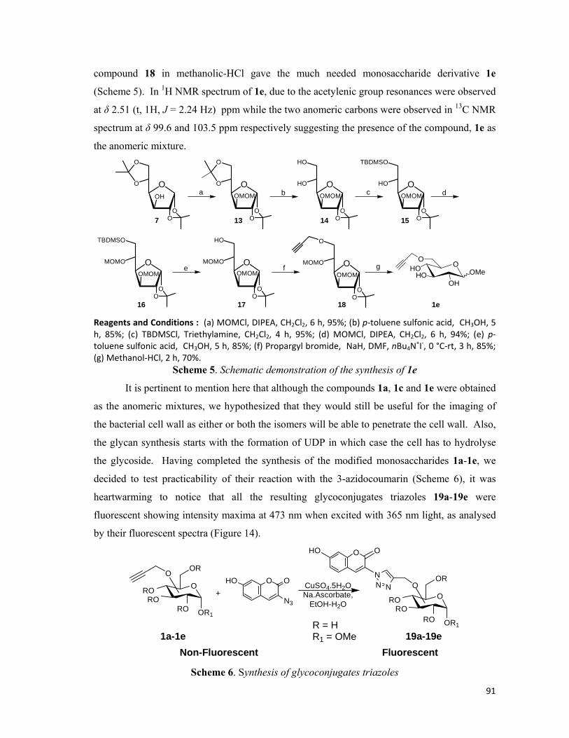
91
compound 18 in methanolic-HCl gave the much needed monosaccharide derivative 1e
(Scheme 5). In 1H NMR spectrum of 1e, due to the acetylenic group resonances were observed
at δ 2.51 (t, 1H, J = 2.24 Hz) ppm while the two anomeric carbons were observed in 13C NMR
spectrum at δ 99.6 and 103.5 ppm respectively suggesting the presence of the compound, 1e as
the anomeric mixture.
OHO
OH
O
OMeHO
O
OO
O
O
OH
O
OO
O
O
OMOM
O
OO
HO
HO
OMOM
O
OO
TBDMSO
HO
OMOM
O
OO
TBDMSO
MOMO
OMOM
O
OO
HO
MOMO
OMOMO
OO
O
MOMO
OMOM
a
1e
b c d
e f g
7 13 14 15
16 17 18 Reagents and Conditions : (a) MOMCl, DIPEA, CH2Cl2, 6 h, 95%; (b) p‐toluene sulfonic acid, CH3OH, 5 h, 85%; (c) TBDMSCl, Triethylamine, CH2Cl2, 4 h, 95%; (d) MOMCl, DIPEA, CH2Cl2, 6 h, 94%; (e) p‐toluene sulfonic acid, CH3OH, 5 h, 85%; (f) Propargyl bromide, NaH, DMF, nBu4N
+I‐, 0 °C‐rt, 3 h, 85%; (g) Methanol‐HCl, 2 h, 70%.
Scheme 5. Schematic demonstration of the synthesis of 1e
It is pertinent to mention here that although the compounds 1a, 1c and 1e were obtained
as the anomeric mixtures, we hypothesized that they would still be useful for the imaging of
the bacterial cell wall as either or both the isomers will be able to penetrate the cell wall. Also,
the glycan synthesis starts with the formation of UDP in which case the cell has to hydrolyse
the glycoside. Having completed the synthesis of the modified monosaccharides 1a-1e, we
decided to test practicability of their reaction with the 3-azidocoumarin (Scheme 6), it was
heartwarming to notice that all the resulting glycoconjugates triazoles 19a-19e were
fluorescent showing intensity maxima at 473 nm when excited with 365 nm light, as analysed
by their fluorescent spectra (Figure 14).
O
OR
OR1RO
+NN
N
O OHO
1a-1e
O
N3
O OHO CuSO4.5H2ONa.Ascorbate,
EtOH-H2O
Non-Fluorescent Fluorescent
RORO O
OR
OR1RO
O
RORO
19a-19eR = HR1 = OMe
Scheme 6. Synthesis of glycoconjugates triazoles
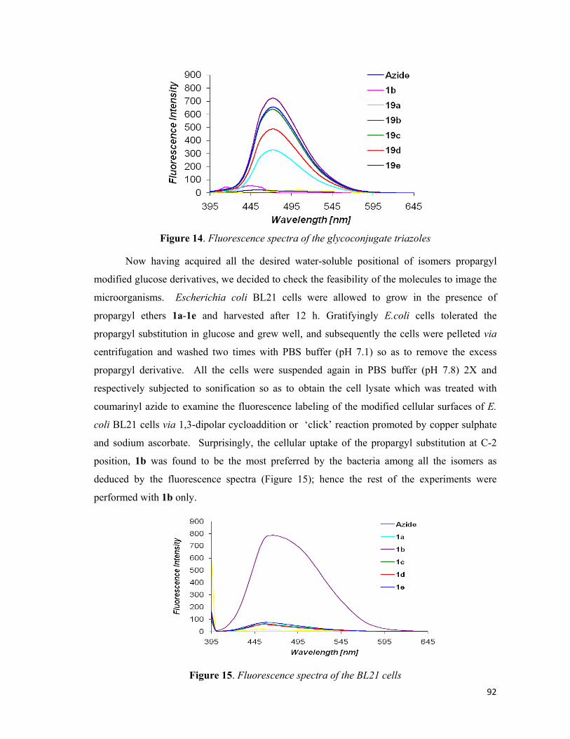
92
Figure 14. Fluorescence spectra of the glycoconjugate triazoles
Now having acquired all the desired water-soluble positional of isomers propargyl
modified glucose derivatives, we decided to check the feasibility of the molecules to image the
microorganisms. Escherichia coli BL21 cells were allowed to grow in the presence of
propargyl ethers 1a-1e and harvested after 12 h. Gratifyingly E.coli cells tolerated the
propargyl substitution in glucose and grew well, and subsequently the cells were pelleted via
centrifugation and washed two times with PBS buffer (pH 7.1) so as to remove the excess
propargyl derivative. All the cells were suspended again in PBS buffer (pH 7.8) 2X and
respectively subjected to sonification so as to obtain the cell lysate which was treated with
coumarinyl azide to examine the fluorescence labeling of the modified cellular surfaces of E.
coli BL21 cells via 1,3-dipolar cycloaddition or ‘click’ reaction promoted by copper sulphate
and sodium ascorbate. Surprisingly, the cellular uptake of the propargyl substitution at C-2
position, 1b was found to be the most preferred by the bacteria among all the isomers as
deduced by the fluorescence spectra (Figure 15); hence the rest of the experiments were
performed with 1b only.
Figure 15. Fluorescence spectra of the BL21 cells
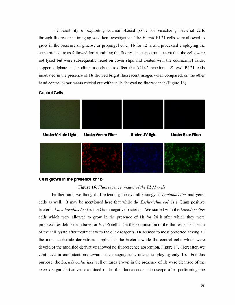
93
The feasibility of exploiting coumarin-based probe for visualizing bacterial cells
through fluorescence imaging was then investigated. The E. coli BL21 cells were allowed to
grow in the presence of glucose or propargyl ether 1b for 12 h, and processed employing the
same procedure as followed for examining the fluorescence spectrum except that the cells were
not lysed but were subsequently fixed on cover slips and treated with the coumarinyl azide,
copper sulphate and sodium ascorbate to effect the ‘click’ reaction. E. coli BL21 cells
incubated in the presence of 1b showed bright fluorescent images when compared; on the other
hand control experiments carried out without 1b showed no fluorescence (Figure 16).
Figure 16. Fluorescence images of the BL21 cells
Furthermore, we thought of extending the overall strategy to Lactobaccilus and yeast
cells as well. It may be mentioned here that while the Escherichia coli is a Gram positive
bacteria, Lactobaccilus lacti is the Gram negative bacteria. We started with the Lactobaccilus
cells which were allowed to grow in the presence of 1b for 24 h after which they were
processed as delineated above for E. coli cells. On the examination of the fluorescence spectra
of the cell lysate after treatment with the click reagents, 1b seemed to most preferred among all
the monosaccharide derivatives supplied to the bacteria while the control cells which were
devoid of the modified derivative showed no fluorescence absorption, Figure 17. Hereafter, we
continued in our intentions towards the imaging experiments employing only 1b. For this
purpose, the Lactobaccilus lacti cell cultures grown in the presence of 1b were cleansed of the
excess sugar derivatives examined under the fluorescence microscope after performing the

94
click reaction. Delightfully the experimental cultures exhibited brilliant fluorescence as
compared to the control cells (Figure 18).
Figure 17. Fluorescence spectra of the Lactobaccilus lacti cells
Figure 18. Fluorescence images of the Lactobaccilus lacti cells
Still not satisfied after having been awarded with successful results in lieu of our
efforts, we attempted to widen the scope of our experiments beyond the bacterial cultures. In
this regard, Sacchromyces (wild type) cells were selected for our endeavors. The yeast cells
were grown overnight with the propargyl derivatives 1a-1e, and isolated by centrifugation.
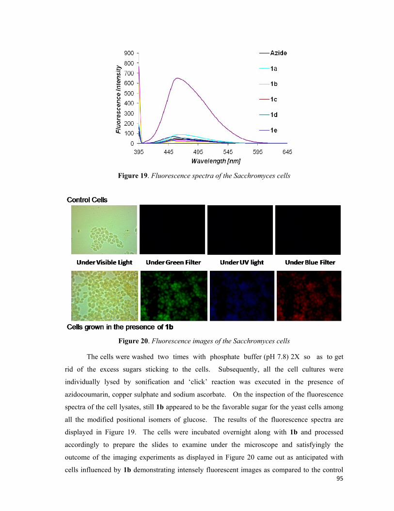
95
Figure 19. Fluorescence spectra of the Sacchromyces cells
Figure 20. Fluorescence images of the Sacchromyces cells
The cells were washed two times with phosphate buffer (pH 7.8) 2X so as to get
rid of the excess sugars sticking to the cells. Subsequently, all the cell cultures were
individually lysed by sonification and ‘click’ reaction was executed in the presence of
azidocoumarin, copper sulphate and sodium ascorbate. On the inspection of the fluorescence
spectra of the cell lysates, still 1b appeared to be the favorable sugar for the yeast cells among
all the modified positional isomers of glucose. The results of the fluorescence spectra are
displayed in Figure 19. The cells were incubated overnight along with 1b and processed
accordingly to prepare the slides to examine under the microscope and satisfyingly the
outcome of the imaging experiments as displayed in Figure 20 came out as anticipated with
cells influenced by 1b demonstrating intensely fluorescent images as compared to the control

96
experiments which were fed with the normal glucose instead of the modified propargylated
sugar 1b.
In conclusion, carbohydrate engineering by exogenously added monosaccharide
supplements is a technique of immense importance for studying various glycan specific
diseases. The utility of profluorophoric coumarine-based azide for fluorescent labelling and
imaging of bacterial, (both Gram positive as well as Gram negative cells), and yeast cultures
was demonstrated for the first time. In this endeavour, we showed that a propargyl group of
monosaccharide can be incorporated into the bacterial and yeast cultures which was probed
using azido coumarinyl profluorophore under 1,3-dipolar cycloaddition or ‘click’ chemistry
conditions. Often, it is difficult to identify the role of bacterial glycans as they exist in micro
and heterogeneous forms. The current protocol of fluorescent labelling via the click chemistry
facilitates monitoring of glycan expression and the related processes which are otherwise
poorly accessible.
*****

97
OHO
OH
OH
OHO
Chapter 2: Experimental Section
Synthesis of propargyl α/β-D-glucopyranoside: D-Glucose
(180mg, 1mmol) was suspended in 3 mL of dioxane-HCl and
heated with propargyl alcohol (0.445mL, 5 mmol) at 100°C for
5 h. After the completion of the reaction, as evident by the
TLC (CH2Cl2-MeOH; 5:1), the reaction was neutralised with triethylamine and the solvent was
then concentrated in vacuo to give a black syrup. The syrup was subjected to column
chromatography, excess of propargyl alcohol was eluted using ethyl acetate followed by the
elution of the product 1a (174 mg, 80%) as an anomeric mixture. The NMR resonances peaks
were found to be in conformity with the literature values reported for the same compound.34
Synthesis of methyl 4,6-O-benzylidine-2-O-benzoyl-3-O-
(methoxy-O-methyl)-α-D-glucopyranoside: To an ice cooled
stirred solution of methyl 4,6-O-benzylidine-2-O-benzoyl-α-D-
glucopyranoside (386 mg, 1 mmol) 3 and diisopropyl amine (
0.347 mL, 2 mmol) in dichloromethane (7 mL) was added MOMCl (0.150 mL, 2 mol) over a
period of 5 min. The reaction was allowed to stir at room temperature for 12 h. TLC
examination of the reaction indicated the formation of the compound as a single compound
(Ethyl Acetate-Petroleum Benzine 1:3). The reaction was diluted with water and extracted
with dichloromethane (2X); the combined extracts were dried over anhydrous sodium sulphate.
The solvent was evaporated under reduced pressure to give the crude MOM ether. It was
purified over silica gel column chromatography (Ethyl Acetate-Petroleum Benzine; 1:3) to give
Compound 4 (418 mg, 97% yield). [α]D (CHCl3, c 1.65) = +120.1°. 1H NMR (200 MHz,
CDCl3): δ 3.32 (s, 3H), 3.39 (s, 3H), 3.66-3.95 (m, 3H), 4.29-4.43 (m, 2 H), 4.79 (ABq , 2 H, J
= 6.86 Hz), 5.04-5.15(m , 2 H), 5.59 (s, 1H), 7.33-7.64 (m, 8H), 8.10-8.15 (m, 2H); 13C NMR
(50 MHz, CDCl3): δ 55.4, 55.8, 62.3, 69.0, 72.8, 73.3, 81.5, 97.1, 97.8, 101.6, 126.1, 126.1,
128.2, 128.2, 128.4, 128.4, 129.0, 129.5, 129.9, 129.9, 133.3, 137.2, 165.9; Anal Calcd. for
C23H26O8: C, 64.18; H, 6.09. Found: C, 63.75; H, 6.48.
Synthesis of methyl 4,6-O-benzylidine-3-O-(methoxy-O-methyl)-α-D-glucopyranoside:
NaOMe (108 mg, 2 mmol) was added to the solution of methyl 4,6-O-benzylidine-2-O-benzoyl
3-O-(methoxy-O-methyl)-α-D-glucopyranoside, 4 (430 mg, 1 mmol) in anhydrous methanol (5
OOMOMO
OMeOBz
OPh
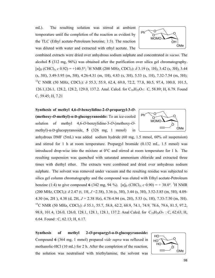
98
mL). The resulting solution was stirred at ambient
temperature until the completion of the reaction as evident by
the TLC (Ethyl acetate-Petroleum benzine; 1:3). The reaction
was diluted with water and extracted with ethyl acetate. The
combined extracts were dried over anhydrous sodium sulphate and concentrated in vacuo. The
alcohol 5 (312 mg, 96%) was obtained after the purification over silica gel chromatography.
[α]D (CHCl3, c 0.92) = +140.5°; 1H NMR (200 MHz, CDCl3): δ 3.19 (s, 1H), 3.42 (s, 3H), 3.44
(s, 3H), 3.49-3.95 (m, 5H), 4.26-4.31 (m, 1H), 4.83 (s, 3H), 5.53 (s, 1H), 7.32-7.54 (m, 5H); 13C NMR (50 MHz, CDCl3): δ 55.3, 55.9, 62.4, 69.0, 72.2, 77.8, 80.5, 97.4, 100.0, 101.5,
126.1,126.1, 128.2, 128.2, 129.0, 137.2. Anal. Calcd. for C16H22O7: C, 58.89; H, 6.79. Found
C, 59.45; H, 7.21
Synthesis of methyl 4,6-O-benzylidine-2-O-propargyl-3-O-
(methoxy-O-methyl)-α-D-glucopyranoside: To an ice-cooled
solution of methyl 4,6-O-benzylidine-3-O-(methoxy-O-
methyl)-α-D-glucopyranoside, 5 (326 mg, 1 mmol) in
anhydrous DMF (5mL) was added sodium hydride (60 mg, 1.5 mmol, 60% oil suspension)
and stirred for 1 h at room temperature. Propargyl bromide (0.132 mL, 1.5 mmol) was
introduced drop-wise into the mixture at 0°C and stirred at room temperature for 1 h. The
resulting suspension was quenched with saturated ammonium chloride and extracted three
times with diethyl ether. The extracts were combined and dried over anhydrous sodium
sulphate. The solvent was removed under vacuum and the resulting residue was subjected to
silica gel column chromatography and the compound was eluted with Ethyl acetate-Petroleum
benzine (1:4) to give compound 6 (342 mg, 94 %). [α]D (CHCl3, c 0.90) = + 38.0°. 1H NMR
(200 MHz, CDCl3): δ 2.47 (t, 1H, J =2.38), 3.36 (s, 3H), 3.44 (s, 3H), 3.52-3.85 (m, 5H), 4.09-
4.30 (m, 2H ), 4.38 (d, 2H, J = 2.38 Hz), 4.78-4.94 (m, 2H), 5.53 (s, 1H), 7.33-7.50 (m, 5H). 13C NMR (50 MHz, CDCl3): δ 55.1, 55.7, 58.8, 62.2, 68.9, 74.1, 74.9, 78.6, 79.6, 81.5, 97.2,
98.8, 101.4, 126.0, 126.0, 128.1, 128.1, 128.1, 137.2. Anal Calcd. for C19H24O7 : C, 62.63; H,
6.64. Found : C, 62.13; H, 6.17.
Synthesis of methyl 2-O-propargyl-α-D-glucopyranoside:
Compound 6 (364 mg, 1 mmol) prepared vide supra was refluxed in
methanolic-HCl (10 mL) for 2 h. After the completion of the reaction,
the solution was neutralised with triethylamine, the solvent was
OOMOMO
OMeOH
OPh
OOMOMO
OMeO
OPh
OHOHO
OMeO
HO

99
removed under pressure and was transferred onto a silica gel column and the compound 1b
(201 mg, 87%) was eluted with dichloromethane-methane (9:1) and obtained as a white solid.
[α]D (H2O, c 2.0) = + 124.0O. 1H NMR (200 MHz, CD3OD): δ 2.81 (t, 1H, J = 2.4 Hz), 3.31 (s,
3H), 3.36-3.80 (m, 9H), 4.25 (d, 2H, J = 2.27 Hz), 4.98 (d, 1H, J = 3.53 Hz). 13C NMR (50
MHz, Acetone-d6): δ 54.2, 57.5, 60.0, 69.1, 70.9, 71.7, 75.7, 78.2, 79.2, 96.5. Anal Cacld. for
C10H16O6 : C, 51.72; H, 6.94. Found : C, 51.99; H, 7.32.
Synthesis of 1,2:5,6-di-O-isopropylidene-3-O-propargyl-α-D-
glucofuranose:1,2:5,6-Di-O-isopropylidene-2-O-propargyl glucofura-
nose, 7 (261 mg, 1 mmol) was dissolved in anhydrous DMF (5mL),
cooled to 0°C and was added sodium hydride (60 mg, 1.5 mmol, 60% oil
suspension). The resulting reaction mixture was stirred at room
temperature for 1h. Propargyl bromide (0.132 mL, 1.5 mmol) was introduced drop-wise at 0°C
and stirred at room temperature for 1 h. The reaction was quenched by the addition of methanol
and extracted with diethyl ether (3X). The solvent was concentrated in vacuo to yield the
propargyl furanoside derivative 8 (268 mg, 90%). [α]D (CHCl3, c 0.60) = -12.1o; 1H NMR (200
MHz, CDCl3): δ 1.32 (s, 3H), 1.35( s, 3H), 1.43 (s, 3H), 1.50 (s, 3H), 2.49 (t, 1H, J = 2.40 Hz),
3.96-4.15 (m, 4H), 4.23-4.28 (m, 3H), 4.63 (s, 1H), 5.88 (s, 1H); 13C NMR (50 MHz, CDCl3):
δ 25.3, 26.2, 26.7, 26.7, 58.0, 67.1, 72.4, 74.9, 79.2, 80.9, 81.5, 82.8, 105.1, 108.9, 111.8; Anal.
Calcd. for C15H22O6: C, 60.39; H, 7.43. Found: C, 60.61; H, 7.65.
Synthesis of methyl 3-O-propargyl-α/β-D-glucopyranoside:
1,2:5,6-Di-O-isopropylidene-3-O-propargyl glucofuranose, 8
(298 mg, 1 mmol) was dissolved in methanolic-HCl (7 ml) and
refluxed for 2 h. Crude 1c was purified by silica gel column
chromatography (CH3OH:CH2Cl2-1:10) to yield 1c (185 mg, 80%) as the anomeric mixture
which was analyzed as such for the characterization purpose. 1H NMR (200 MHz, CD3OD): δ
2.76 (t, 1H, J = 2.76 Hz), 3.28 (s, 3H), 3.33-3.75 (m, 9H), 4.36 (d, 2H, J = 2.37 Hz), 4.65 (m,
1H) ; 13C NMR (50 MHz, CDCl3): δ 54.6, 55.4, 57.2, 57.4, 59.4, 60.0, 62.7, 68.7, 68.9, 70.4,
71.1, 74.2, 75.4, 75.7, 76.8, 78.9, 79.4, 80.5, 81.6, 82.6, 98.8, 102.4, 108.8. Anal. Calcd. for
C10H16O6: C, 51.72; H, 6.94. Found: C, 51.85; H, 6.83.
Synthesis of methyl 4,6-O-benzylidene-2,3-di-O-(methoxy-O-methyl)-α-D-
glucopyranoside: To a solution of methyl 4,6-O-benzylidene-α-D-glucopyranose, 2 (282 mg, 1
O
OO
O
OO
OHO
OH
OH
OMeO

100
mmol) in CH2Cl2 (7 mL), DIPEA (0.695 mL, 4 mmol) was
added and the resulting mixture was cooled in an ice bath.
MOMCl (0.301 mL, 4 mmol) was added drop-wise over a
period of 10 min, the reaction mixture was stirred at room
temperature for 12 h, was diluted with water and extracted with CH2Cl2 (3X). The combined
organic extracts were dried over anhydrous sodium sulphate and the solvent was removed
under reduced pressure and crude syrup was purified using silica gel column chromatography
with ethyl acetate-petroleum benzine as the mobile phase to obtain the respective di-MOM
ether 9 (333 mg, 90%). [α]D (CHCl3, c1.05) = +9.05o; 1H NMR (200 MHz, CDCl3): δ 3.35 (s,
3H), 3.43 (s, 3H), 3.53-3.85 ( m, 7H), 4.09-4.27 (m, 2H), 4.73-4.83 (s, 5H), 5.53 (s, 1H), 7.34-
7.47 (m, 5H); 13C NMR (50 MHz, CDCl3): δ 55.2, 55.5, 55.7, 62.2, 69.0, 73.9, 78.4, 81.5, 97.3,
97.8, 99.8, 101.4, 126.0, 126.0, 128.1, 128.1, 128.9, 137.2; Anal. Calcd. for C18H26O8: C,
58.37; H, 7.08. Found : C, 57.82; H, 7.52.
Synthesis of methyl 2,3-di-O-(methoxy-O-methyl)-α-D-
glucopyranoside: Compound 9 (370 mg, 1 mmol) was dissolved in
methanol (5 mL) and deprotection of the benzylidene was effected
by the addition of catalytic amount of p-toluene sulfonic acid. After
usual work-up, the diol 10 was obtained which was directly used in the next step without
further characterization.
Synthesis of methyl 6-O-t-butyl diphenyl silyl-2,3-di-O-(methoxy-
O-methyl)-α-D-glucopyranoside: The diol 10 (282 mg, 1 mmol)
was dissolved in CH2Cl2 (5 mL) and was added triethylamine
(0.208 mL, 1.5 mmol) and cooled to 0oC. TBDPSCl (0.256 mL, 1mmol) was added portion-
wise and the reaction mixture was stirred at room temperature until the completion of the
reaction as indicated on the TLC (Ethyl acetate-Petroleum Benzine, 1:3). The reaction mixture
was diluted with water and extracted with CH2Cl2 (3X). The combined extracts were dried
over anhydrous sodium sulphate, the solvent was removed in vacuo and the title compound
was purified by column chromatography to give compound 11 (505 mg, 97%). [α]D (CHCl3, c
1.22) = - 13.8o; 1H NMR (200 MHz, CDCl3): δ 1.06 (s, 9H), 3.41 (s, 3H), 3.43 (s, 3H), 3.44 (s,
3H), 3.49-3.74 (m, 4H), 3.83-4.02 (m, 3H), 4.69–4.86 (m, 5H), 7.35 – 7.42 (m, 6H), 7.69- 7.75
(m, 4H): 13C NMR(50 MHz, CDCl3): δ 19.3, 26.8, 26.8, 26.8, 54.8, 55.4, 55.7, 63.7, 69.8,
77.1, 71.6, 83.9, 97.5, 98.4, 98.7, 127.6, 127.6, 127.6, 127.6, 129.5, 129.5, 133.5, 133.5, 135.6,
OOMOMO
OMeMOMO
OPh
OHOMOMO
OMeMOMO
HO
OHOMOMO
OMeMOMO
TPSO

101
135.6, 135.6, 135.6; Anal Calcd. for C27H40O8Si : C, 62.28; H, 7.74; Si, 5.39. Found: C, 62.55;
H, 7.95; Si, 5.82.
Synthesis of methyl 6-O-t-butyl diphenyl silyl-2,3-di-O-
(methoxy-O-methyl)-4-O-propargyl-α-D-glucopyranoside:
Preparative procedure is same as delineated above for
compound 6. [α]D (CHCl3, c 0.82) = +27.7o; 1H NMR (200
MHz, CDCl3): δ 1.06 (s, 9H), 2.32 ( t, 1H, J = 2.25 Hz), 3.37 (s, 3H), 3.43 (s, 3H), 3.46 (s, 3H),
3.49 – 3.68 (m, 3H), 3.90- 4.0 (m, 3 H), 4.36–4.40 (m, 2H), 4.70-4.88 (m, 5H), 7.37–7.42 (m,
6H), 7.70-7.75 (m, 4H); 13C NMR (50 MHz, CDCl3): δ 19.3, 26.8, 26.8, 26.8, 54.8, 55.5, 56.2,
56.2, 59.8, 62.9, 70.9, 74.3, 77.4, 78.1, 79.2, 79.8, 97.7, 98.3, 98.8, 127.5, 127.5, 127.6, 127.6,
129.6, 129.6, 133.3, 133.7, 135.6, 135.6, 135.8, 135.8; Anal Calcd. for C30H42O8Si : C, 64.49;
H, 7.58. Found : C, 65.12; H, 7.21.
Synthesis of methyl 4-O-propargyl-α-D-glucopyranoside:
Preparative protocol is same as described for compound 1b. [α]D
(H2O, c 2.0) = +170.6o; 1H NMR (200 MHz, CD3OD): δ 2.87 (t,
1H, J = 2.37 Hz), 3.35 (s, 3H), 3.39 (d, 2H, J = 9.69 Hz), 3.48-
3.89 (m, 6H), 4.38 (d, 2H, J = 1.74 Hz), 4.74 (d, 2H, J = 3.72 Hz); 13C NMR (50 MHz,
Acetone-d6): δ 54.5, 59.2, 60.0, 70.0, 70.7, 72.6, 75.6, 76.9, 79.1, 98.6; Anal. Calcd. for
C10H16O6: C, 51.72; H, 6.94. Found: C, 51.44; H, 6.58.
Synthesis of 1,2:5,6-Di-O-isopropylidene-3-O-(methoxy-O-
methyl)-α-D-glucofuranose: Preparative procedure is as delineated
above for compound 4. [α]D (CHCl3, c 0.84) = -12.0o; 1H NMR (200
MHz, CDCl3): δ 1.32 (s, 3H), 1.34 (s, 3H), 1.42 (s, 3H), 1.50 (s, 3H),
3.41 (s,3H), 3.96 -4.33 (m, 4H), 4.56 (d, 1H, J = 3.52 Hz), 4.73 (d,
2H, J = 1.87 Hz), 5.89 (d, 1H, J = 3.53 Hz); 13C NMR (50 MHz,
CDCl3): δ 25.3, 26.2, 26.8, 30.9, 55.8, 67.5, 72.3, 79.1, 81.0, 83.3, 96.0, 105.2, 109.1, 111.9.
Anal Calcd. for C14H24O7 : C, 55.25; H, 7.95. Found: C, 55.75; H, 8.02.
Synthesis of 1,2-O-isopropylidene-3-O-(methoxy-O-methyl)-α-D-glucofuranose: To a
solution of 1,2:5,6-Di-O-isopropylidene-3-O- (methoxy-O-methyl)-α-D-glucofuranose, 13
(500 mg, 1mmol) in methanol (5 mL) was added a catalytic amount of PTSA, the resulting
solution was stirred at room temperature until the completion of the reaction as indicated by the
OOMOMO
OMeMOMO
TPSO
OO
OH
OH
OMeHO
O
OO
O
O
OMOM

102
TLC (Ethyl Acetate; Petroleum Benzine 1:1). The solvent was then removed
under reduced pressure and the diol thus obtained was carried further as such
for the next step.
Synthesis of 1,2-O-isopropylidene-3-O-(methoxy-O-methyl)-6-
O-t-butyl dimethylsilyl-α-D-glucofuranose: To a solution of the
diol 14 (305 mg, 1 mmol) in dichloromethane (5 mL) was added
triethylamine (0.208 mL, 1.5 mmol) and TBDMSCl (150 mg, 1
mmol) portion-wise at 0oC. The resulting reaction mixture was
stirred at room temperature for 4 h. The reaction mixture was
diluted with water and extracted with CH2Cl2 (3x50 mL). The combined extracts were dried
on anhydrous sodium sulphate and purified on silica gel to obtain compound 15 (398 mg, 95
%). [α]D (CHCl3, c 0.90) = -14.17o; 1H NMR (200 MHz, CDCl3): δ 0.09 (s, 6H), 0.91 (s, 9H),
1.32 (s, 3H), 1.48 (s, 3H), 2.74 (b, 1H), 3.43 (s, 3H), 3.71-3.89 (m, 3H), 4.09–4.14 (m, 1H),
4.26 (d, 1H, J = 2.87 Hz), 4.56 (d, 1H, J = 3.70 Hz), 4.76 (dd, 2 H, J = 6.57, 12.37 Hz), 5.89
(d, 1H, J = 3.65 Hz); 13C NMR (50 MHz, CDCl3): δ -5.5, -5.5, 18.3, 25.8, 25.8, 25.8, 26.3,
26.7, 55.8, 64.3, 68.2, 79.2, 79.9, 83.2, 96.5, 105.1, 111.7; Anal Calcd. for C17H34O7Si: C,
53.94; H, 9.05; Si, 7.42. Found : C, 54.32; H, 8.89; Si, 7.82.
Synthesis of 1,2-O-isopropylidene-3,5-di-O-(methoxy-O-
methyl)-6-O-t-butyl dimethylsilyl-α-D-glucofuranose:
Preparative procedure is same as delineated above for compound 4.
[α]D (CHCl3, c 0.72) = +7.3o; 1H NMR (200 MHz, CDCl3): δ 0.07
(s, 6H), 0.09 (s, 9H), 1.31 (s, 3H ), 1.48 (s, 3H), 3.40 (s, 3H), 3.41
(s, 3H), 3.73–3.89 (m, 2H), 3.97- 4.04 (m, 1H), 4.13 (d, 1H, J =
2.74 Hz), 4.25-4.31 (m, 1H), 4.65 ( d, 1H, J = 3.51 Hz), 4.70- 4.82 (m, 4 H), 5.88 ( d, 1 H, J =
3.66 Hz); 13C NMR (50 MHz, CDCl3): δ - 5.5, - 5.4, 18.3, 25.9, 25.9, 25.9, 26.3, 26.7, 55.7,
56.0, 63.7, 75.9, 78.1, 81.3, 83.0, 97.0, 97.3, 104.8, 111.7; Anal Calcd. for C19H38O8Si : C,
54.00; H, 9.06; Si, 6.65. Found : C, 54.49; H, 8.58; Si, 6.35.
Synthesis of 1,2-O-isopropylidene-3,5-di-O-(methoxy-O-methyl)-α-D-glucofuranose: To a
solution of the silyl ether 16 (464 mg, 1 mmol) was added catalytic amount of PTSA in
methanol (5 mL) and stirred at room temperature for 5 h. The solvent was then evaporated in
vacuo to yield a syrup which was purified by the silica gel column chromatography to achieve
O
OO
HO
HO
OMOM
O
OO
TBDMSO
HO
OMOM
O
OO
TBDMSO
MOMO
OMOM
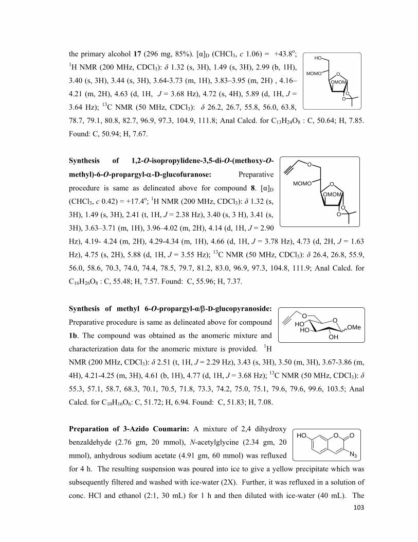
103
the primary alcohol 17 (296 mg, 85%). [α]D (CHCl3, c 1.06) = +43.8o; 1H NMR (200 MHz, CDCl3): δ 1.32 (s, 3H), 1.49 (s, 3H), 2.99 (b, 1H),
3.40 (s, 3H), 3.44 (s, 3H), 3.64-3.73 (m, 1H), 3.83–3.95 (m, 2H) , 4.16–
4.21 (m, 2H), 4.63 (d, 1H, J = 3.68 Hz), 4.72 (s, 4H), 5.89 (d, 1H, J =
3.64 Hz); 13C NMR (50 MHz, CDCl3): δ 26.2, 26.7, 55.8, 56.0, 63.8,
78.7, 79.1, 80.8, 82.7, 96.9, 97.3, 104.9, 111.8; Anal Calcd. for C13H24O8 : C, 50.64; H, 7.85.
Found: C, 50.94; H, 7.67.
Synthesis of 1,2-O-isopropylidene-3,5-di-O-(methoxy-O-
methyl)-6-O-propargyl-α-D-glucofuranose: Preparative
procedure is same as delineated above for compound 8. [α]D
(CHCl3, c 0.42) = +17.4o; 1H NMR (200 MHz, CDCl3): δ 1.32 (s,
3H), 1.49 (s, 3H), 2.41 (t, 1H, J = 2.38 Hz), 3.40 (s, 3 H), 3.41 (s,
3H), 3.63–3.71 (m, 1H), 3.96–4.02 (m, 2H), 4.14 (d, 1H, J = 2.90
Hz), 4.19- 4.24 (m, 2H), 4.29-4.34 (m, 1H), 4.66 (d, 1H, J = 3.78 Hz), 4.73 (d, 2H, J = 1.63
Hz), 4.75 (s, 2H), 5.88 (d, 1H, J = 3.55 Hz); 13C NMR (50 MHz, CDCl3): δ 26.4, 26.8, 55.9,
56.0, 58.6, 70.3, 74.0, 74.4, 78.5, 79.7, 81.2, 83.0, 96.9, 97.3, 104.8, 111.9; Anal Calcd. for
C16H26O8 : C, 55.48; H, 7.57. Found: C, 55.96; H, 7.37.
Synthesis of methyl 6-O-propargyl-α/β-D-glucopyranoside:
Preparative procedure is same as delineated above for compound
1b. The compound was obtained as the anomeric mixture and
characterization data for the anomeric mixture is provided. 1H
NMR (200 MHz, CDCl3): δ 2.51 (t, 1H, J = 2.29 Hz), 3.43 (s, 3H), 3.50 (m, 3H), 3.67-3.86 (m,
4H), 4.21-4.25 (m, 3H), 4.61 (b, 1H), 4.77 (d, 1H, J = 3.68 Hz); 13C NMR (50 MHz, CDCl3): δ
55.3, 57.1, 58.7, 68.3, 70.1, 70.5, 71.8, 73.3, 74.2, 75.0, 75.1, 79.6, 79.6, 99.6, 103.5; Anal
Calcd. for C10H16O6: C, 51.72; H, 6.94. Found: C, 51.83; H, 7.08.
Preparation of 3-Azido Coumarin: A mixture of 2,4 dihydroxy
benzaldehyde (2.76 gm, 20 mmol), N-acetylglycine (2.34 gm, 20
mmol), anhydrous sodium acetate (4.91 gm, 60 mmol) was refluxed
for 4 h. The resulting suspension was poured into ice to give a yellow precipitate which was
subsequently filtered and washed with ice-water (2X). Further, it was refluxed in a solution of
conc. HCl and ethanol (2:1, 30 mL) for 1 h and then diluted with ice-water (40 mL). The
O
OO
HO
MOMO
OMOM
O
OO
O
MOMO
OMOM
OHO
OH
O
OMeHO
OHO O
N3

104
solution was cooled in an ice bath and sodium nitrite (2.75 gm, 40 mmol.) was added in
portions and after which the reaction was allowed to stir for 15 min. at 0°C and then sodium
azide (3.9 gm, 60 mmol) was added to the stirred solution maintaining the temperature of the
reaction. After stirring for another 15 min., the resulting precipitate was filtered off and
washed with water, dried and purified over silica gel chromatography to afford a brown solid
(2.2 gm, 54%). The NMR characterization data was found to be in conformity with the
reported values.31a
General procedure for the synthesis of Triazoles (19a-19e): To an ethanol-water (5 mL; 1:1)
solution of modified sugars 1a (1 mol) was added coumarinyl azide (1 mol) and stirred at room
temperature. Freshly prepared solution of CuSO4.5H2O (0.1 mol %) and sodium ascorbate (0.5
mol%) was added and the reaction mixture was stirred at room temperature for 30 min after
which the TLC examination of the reaction mixture revealed formation of the fluorescent
triazoles. We were able to characterize these compounds by 1H NMR spectra only and not by 13C NMR spectral studies as we observed that these compounds have greater propensity to
form gels at higher concentrations. These compounds were directly analysed for their UV
(Figure 21) and fluorescence spectra.
Compound 19a: 1H NMR (200 MHz, CD3OD): δ 3.18-
3.46 (m, 6H), 3.62-3.85 (m, 5H), 4.75 (m, 2H), 4.97 (m,
1H), 6.80 (d, 1H, J = 1.89 Hz), 6.89 (dd, 1H, J = 1.90,
8.57 Hz), 7.64 (d, 1H, J = 8.61 Hz), 8.49 (s, 1H), 8.63 (s,
1H). Anal. Calcd. for C18H19N3O9: C, 51.31; H, 4.55; N,
9.97. Found: C, 51.25; H, 4.63; N, 10.09.
Compound 19b: 1H NMR (200 MHz, CD3OD): δ 3.29-
3.41 (m, 7H), 3.44-3.55 (m, 2H), 3.63- 3.86 (m, 5H),
4.83 (m, 2H), 6.78 (d, 1H, J = 2.13 Hz), 6.87 (dd, 1H, J =
2.28, 8.46 Hz), 7.61 (d, 1H, J = 8.59 Hz), 8.48 (s, 1H),
8.59 (s, 1H). Anal. Calcd. for C19H21N3O9: C, 52.41; H, 4.86; N, 9.65. Found C, 52.33; H, 4.96;
N, 9.73.
Compound 19c: 1H NMR (200 MHz, CD3OD): δ 3.29-3.35 (m, 3H), 3.42 (s, 3H), 3.53-4.24
(m, 7H), 4.68-4.78 (m, 1H), 5.03 (m, 2H), 6.77 (d, 1H, J = 2.14 Hz), 6.86 (dd, 1H, J = 2.27,
OHO
OH
OH
OHO
O OHO
NNN
OHOHO
OMeO
HO
O
HO
O
NNN

105
8.56 Hz), 7.61 (d, 1H, J = 8.61 Hz), 8.46 (s, 1H), 8.58
(s, 1H). Anal. Calcd. for C19H21N3O9: C, 52.41; H,
4.86; N, 9.65. Found C, 52.33; H, 4.96; N, 9.73.
Compound 19d: 1H NMR (200 MHz, CD3OD): δ 3.29-
3.32 (m, 2H), 3.35 (s, 2H), 3.39-3.47 (m, 6H), 3.51-3.86
(m, 5H), 4.67 (m, 1H), 5.07 (m, 1H), 6.81 (d, 1H, J = 2.15
Hz), 6.86 (dd, 1H, J = 2.17, 8.60 Hz), 7.63 (d, 1H, J =
8.60 Hz), 8.49 (s, 1H), 8.57 (s, 1H). Anal. Calcd. for
C19H21N3O9: C, 52.41; H, 4.86; N, 9.65. Found C, 52.31; H, 4.94; N, 9.71.
Compound 19e: 1H NMR (200 MHz, CD3OD): δ
3.29-3.43 (m, 7H), 3.51 (s, 1H), 3.57-3.87 (m, 4H),
4.04-4.20 (m, 1H), 4.67 (d, 1H, J = 3.68 Hz), 4.75 (m,
2H), 6.78 (d, 1H, J = 2.15 Hz), 6.87 (dd, 1H, J = 2.15,
8.60 Hz), 7.62 (d, 1H, J = 8.60 Hz), 8.47 (s, 1H), 8.57
(s, 1H). Anal. Calcd. for C19H21N3O9: C, 52.41; H, 4.86; N, 9.65. Found C, 52.61; H, 4.93; N,
9.51.
Figure 21. UV absorption spectra of sugar1b, triazole 19b and azidocoumarin
Preparation of the stock solutions:
Sugar Stock Solution (1a-1e): Individual solutions of compound 1a-1e were prepared in
27mg/ml in dist. water and then sterilized via autoclave.
3-azidocoumarin stock: 5 mg of the coumarinyl azide was dissolved in 2 ml of ethanol and
used as such for the experiments.
OHO
OH
HO
OMeO
OHO O
N NN
OO
OH
OH
OMeHO
OHO O
N NN
OHO
OH
O
OMeHO
OHO
O
N NN

106
100 mmol and 100 mmol stock solutions of copper sulphate and sodium ascorbate were
prepared.
Composition of LB and Mineral Medium:
Luria Broth: Tryptone (1 gm), Yeast extract (0.5 gm), NaCl (1gm) were dissolved in 100 ml
of dist. water and sterilised via autoclave.
Mineral Medium: Glucose (5 gm), Na2HPO4 (6 gm), KH2PO4 (3 gm), NH4Cl (1 gm), NaCl
(0.5 gm), MgSO4 (0.12 gm), CaCl2 (0.01 gm) were dissolved in 1000ml dist. H2O and used
after sterilisation.
General procedure for cell culturing:
Two sets of cultures were separately incubated simultaneously each time for all the cell
variants employed. Firstly, the control which was devoid of the modified sugars and secondly,
the cell cultures which were grown in the presence of modified sugar derivatives 1a-1e, 40
μL/mL of the propargylated monosaccharides were added individually to all the tubes.
However, after the cells have attained their maximum growth, both the sets were treated
likewise separately. The general procedure for the cell culturing is given hereunder for all the
cells used.
Escherichia coli: BL21 cells were inoculated into the 5 ml of LB medium. The cell culture
was then incubated at 37°C and shaken at 180 rpm for 12 h. Cells were then isolated by
centrifugation at 8000 rpm for 5 min and subsequently were washed with PBS buffer pH 7.8
(2x) and resuspended in 1 mL of PBS buffer (pH 7.8). The cells were subsequently cooled in
ice-bath before being sonicated for 2 min (pulse 2 sec). The cell lysate was isolated by
centrifugation of the resulting mixture at 8000 rpm for 5 min. The cell lysate was used as such
for UV and Fluorescence spectroscopic analysis.
Lactobaccilus lacti: Lactobaccilus cells were inoculated into the 5 mL mineral medium. The
culture was then incubated at 30°C without shaking for 24 h. Cells were then isolated by
centrifugation at 8000 rpm for 5 min and subsequently were washed with PBS buffer pH 7.8
(2X) and resuspended in 1 mL of PBS buffer (pH 7.8). The cells were cooled in ice-bath and
sonicated for 2 min (pulse 2 sec). The cell lysate was isolated by centrifugation of the resulting
mixture at 8000 rpm for 5 min. The cell lysate was used as such for UV and Fluorescence
spectroscopic analysis.
Sacchromyces (Wild Type): Sacchromyces cells were inoculated into the 5 mL LB medium.
The medium was shaken at 37°C for 12 hr at 180 rpm and cells were harvested by
centrifugation at 8000 rpm for 5 min. The cells were resuspended in 1ml PBS buffer pH 7.8
107
before they were washed two times with 2 mL of PBS buffer pH 7.8 and subsequently
sonicated in an ice-bath to yield the cell lysate after centrifugation at 8000 rpm for 5 min which
was used as such for UV and Fluorescence spectroscopic analysis.
Recording of UV and Fluorescence Spectra:
Cell lysates were treated with 5 μl of the above prepared azide stock solution and 5 μl of
CuSO4 and 10 μl of sodium ascorbate. The resulting solution was kept at room temperature
for 30 min and then utilized for UV and Florescence studies.
Preparation of Slides:
In a typical experiment, growing cells (OD600<1.0) were washed with 1 mL of PBS buffer (pH
7.8) and treated with 2 μl of azide stock solution and 1 μl and 2 μl of CuSO4 and sodium
ascorbate stock solutions. Cells were then incubated for 30 min at rt and then a drop of the
uniformly suspended cells was loaded onto a glass slide and kept at room temperature for air
drying, after which the cells were analysed under the fluorescence microscope and the images
recorded.
*****
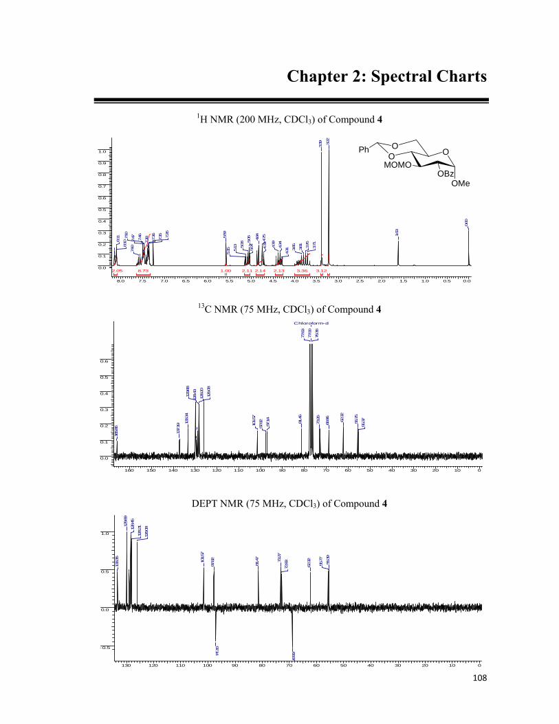
108
8.0 7.5 7.0 6.5 6.0 5.5 5.0 4.5 4.0 3.5 3.0 2.5 2.0 1.5 1.0 0.5 0.0
0.0
0.1
0.2
0.3
0.4
0.5
0.6
0.7
0.8
0.9
1.0
8.73 3.36 3.122.14 2.132.112.05 1.00
0.00
1.63
3.22
3.39
3.713.76
3.81
3.85
4.31
4.34
4.39
4.72
4.754.84
5.04
5.06
5.08
5.13
5.15
5.59
7.26
7.35
7.36
7.387.46
7.477.50
7.60
8.108.
11
Chapter 2: Spectral Charts
1H NMR (200 MHz, CDCl3) of Compound 4
13C NMR (75 MHz, CDCl3) of Compound 4
160 150 140 130 120 110 100 90 80 70 60 50 40 30 20 10 0
0.0
0.1
0.2
0.3
0.4
0.5
0.6
Chloroform-d
55.37
55.75
62.32
68.96
73.26
76.36
77.00
77.63
81.46
97.14
97.82
101.57
126.08
128.20
128.43
129.88
133.34
137.19
165.86
DEPT NMR (75 MHz, CDCl3) of Compound 4
130 120 110 100 90 80 70 60 50 40 30 20 10 0
-0.5
0.0
0.5
1.0
55.39
55.77
62.32
68.97
72.8373
.27
81.47
97.14
97.82
101.57
126.08
128.2112
8.45
129.89
133.35
OOMOMO
OMeOBz
OPh

109
1H NMR (200 MHz, CDCl3) of Compound 5
7.5 7.0 6.5 6.0 5.5 5.0 4.5 4.0 3.5 3.0 2.5 2.0 1.5 1.0 0.5 0.0
0.0
0.1
0.2
0.3
0.4
0.5
0.6
0.7
0.8
0.9
1.0
11.294.99 3.021.00 0.99 0.92
3.19
3.42
3.78
3.91
4.26
4.31
4.83
5.53
7.26
7.35
7.47
13C NMR (75 MHz, CDCl3) of Compound 5
130 120 110 100 90 80 70 60 50 40 30 20 10 0
0.0
0.1
0.2
0.3
0.4
0.5
0.6
0.7
0.8
Chloroform-d
55.34
55.67
62.43
68.96
72.19
76.37
77.00
77.64
77.75
80.50
97.42
99.97
101.53
126.09
128.16
128.97
137.20
DEPT NMR (75 MHz, CDCl3) of Compound 5
130 120 110 100 90 80 70 60 50 40 30 20 10 0
-0.5
0.0
0.5
1.0
55.36
55.70
62.44
68.9772
.20
77.79
80.50
97.44
99.97101.54
126.11
128.18
129.00
OOMOMO
OMeOH
OPh

110
1H NMR (200 MHz, CDCl3) of Compound 6
7.5 7.0 6.5 6.0 5.5 5.0 4.5 4.0 3.5 3.0 2.5 2.0 1.5 1.0 0.5 0.0
0.0
0.1
0.2
0.3
0.4
0.5
0.6
0.7
4.97 4.194.083.05 3.041.00 0.90
0.00
1.262.46
2.47
2.49
3.33
3.34
3.36
3.44
3.46
3.61
3.703.71
3.77
3.78
4.09
4.11
4.13
4.27
4.37
4.39
4.78
4.814.84
4.92
4.94
4.96
5.53
7.33
7.34
7.36
7.46
13C NMR (75 MHz, CDCl3) of Compound 6
130 120 110 100 90 80 70 60 50 40 30 20 10 0
0.0
0.1
0.2
0.3
0.4
0.5
0.6
0.7
0.8
0.9
1.0
Chloroform-d
55.14
55.68
58.8462
.20
68.91
74.1274.85
76.36
77.00
77.63
78.63
79.61
81.52
97.20
98.81
101.38
125.98
128.07
128.85
137.16
DEPT NMR (75 MHz, CDCl3) of Compound 6
130 120 110 100 90 80 70 60 50 40 30 20 10 0
-0.5
0.0
0.5
1.0
55.1
555
.69
58.85
62.2
0
68.9274
.1078
.62
81.5
1
97.20
98.8
110
1.38
125.
9912
8.08
OOMOMO
OMeO
OPh

111
1H NMR (200 MHz, CD3OD) of Compound 1b
5.0 4.5 4.0 3.5 3.0 2.5 2.0 1.5 1.0 0.5 0.0
0.0
0.1
0.2
0.3
0.4
0.5
0.6
0.7
0.8
5.79 3.802.001.08 0.68
2.81
3.31
3.47
3.493.56
3.60
3.65
3.73
3.74
3.79
4.244.25
4.25
4.27
4.72
4.97
4.99
13C NMR (75 MHz, CD3OD) of Compound 1b
90 80 70 60 50 40 30 20 10 0
0.0
0.1
0.2
0.3
0.4
0.5
0.6
0.7
0.8
0.9
1.0
Acetone-d6
29.80
54.23
57.50
60.04
69.1070.87
71.67
75.69
78.17
79.16
96.53
DEPT NMR (75 MHz, CD3OD) of Compound 1b
90 80 70 60 50 40 30 20 10 0
-0.5
0.0
0.5
1.0
Acetone-d6
29.80
54.22
57.50
60.04
69.0870.87
71.6778
.16
96.52
OHOHO
OMeO
HO

112
100 90 80 70 60 50 40 30 20 10 0
-0.5
0.0
0.5
1.0
25.29
26.16
26.74
58.0167.10
72.44
74.89
79.18
80.92
81.45
82.75
105.14
1H NMR (200 MHz, CDCl3) of Compound 8
5.5 5.0 4.5 4.0 3.5 3.0 2.5 2.0 1.5 1.0 0.5 0.0
0.0
0.1
0.2
0.3
0.4
0.5
0.6
0.7
0.8
0.9
1.0
12.724.062.981.000.94 0.83
1.26
1.321.35
1.43
1.50
1.79
2.49
3.96
4.004.03
4.06
4.10
4.15
4.23
4.28
4.63
5.88
1H NMR (200 MHz, CDCl3) of Compound 8
110 100 90 80 70 60 50 40 30 20 10 0
0.0
0.1
0.2
0.3
0.4
0.5
0.6
0.7
0.8
0.9
1.0
Chloroform-d
25.29
26.16
26.74
58.01
67.10
72.43
74.88
77.00
77.63
79.18
80.92
81.45
82.75
105.13
108.92111.78
DEPT NMR (75 MHz, CDCl3) of Compound 8
O
OO
O
OO

113
1H NMR (200 MHz, CD3OD) of Compound 1c
4.5 4.0 3.5 3.0 2.5 2.0 1.5 1.0 0.5 0.0
0.0
0.1
0.2
0.3
0.4
0.5
0.6
0.7
6.21 5.153.29 1.000.900.38
2.75
2.793.23
3.28
3.31
3.483.50
3.51
3.55
3.69
3.754.06
4.21
4.23
4.36
4.37
4.644.66
4.69
4.91
4.93
13C NMR (75 MHz, CD3OD) of Compound 1c
100 90 80 70 60 50 40 30 20 10 0
0.0
0.1
0.2
0.3
0.4
0.5
0.6
0.7
0.8
0.9
Acetone-d6
29.80
48.42
54.55
55.36
57.24
59.42
59.95
62.68
63.12
68.67
70.47
71.06
73.30
74.19
75.42
75.68
78.94
79.38
80.54
82.58
82.78
98.79
102.41
108.77
DEPT NMR (75 MHz, CD3OD) of Compound 1c
110 100 90 80 70 60 50 40 30 20 10 0
-1.0
-0.5
0.0
0.5
1.0
Acetone-d6
29.80
54.55
55.37
57.25
59.4459.93
62.6863.13
68.67
70.47
71.06
74.16
76.79
80.34
80.52
82.58
82.76
95.30
98.79
102.43
108.76
OHO
OH
OH
OMeO

114
130 120 110 100 90 80 70 60 50 40 30 20 10 0
-0.5
0.0
0.5
1.0
55.26
55.68
62.19
69.01
73.9178.42
81.53
97.2597.81
99.83101.43
126.04
128.13
1H NMR (200 MHz, CDCl3) of Compound 9
7.5 7.0 6.5 6.0 5.5 5.0 4.5 4.0 3.5 3.0 2.5 2.0 1.5 1.0 0.5 0.0
0.0
0.1
0.2
0.3
0.4
0.5
0.6
0.7
0.8
0.9
13.535.24 5.19 2.051.00
0.00
3.35
3.43
3.623.79
4.094.27
4.83
5.53
7.34
7.47
13C NMR (75 MHz, CDCl3) of Compound 9
130 120 110 100 90 80 70 60 50 40 30 20 10 0
0.0
0.1
0.2
0.3
0.4
0.5
0.6
0.7
0.8
0.9
1.0
Chloroform-d
55.24
55.45
55.66
62.19
69.00
73.93
76.36
77.00
77.63
81.54
97.25
97.80
99.83
101.43
126.03
128.12
128.90
137.23
DEPT NMR (75 MHz, CDCl3) of Compound 9
OOMOMO
OMeMOMO
OPh
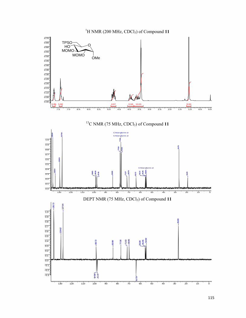
115
1H NMR (200 MHz, CDCl3) of Compound 11
7.5 7.0 6.5 6.0 5.5 5.0 4.5 4.0 3.5 3.0 2.5 2.0 1.5 1.0 0.5 0.0
0.00
0.05
0.10
0.15
0.20
0.25
0.30
0.35
0.40
0.45
0.50
0.55
0.60
0.65
0.70
13.01 9.005.82 4.973.85 3.04
13C NMR (75 MHz, CDCl3) of Compound 11
130 120 110 100 90 80 70 60 50 40 30 20 10 0
0.0
0.1
0.2
0.3
0.4
0.5
0.6
0.7
0.8
0.9
1.0
Chloroform-d
Chloroform-d
Chloroform-d
19.28
26.75
54.84
55.35
55.72
63.73
69.79
71.57
76.36
77.00
77.63
83.88
97.48
98.38
98.65
127.56
129.53
133.54
135.64
DEPT NMR (75 MHz, CDCl3) of Compound 11
130 120 110 100 90 80 70 60 50 40 30 20 10 0
-0.3
-0.2
-0.1
0.0
0.1
0.2
0.3
0.4
0.5
0.6
0.7
0.8
0.9
1.0
26.8
3
54.9
255
.45
55.8
0
63.79
69.8
571
.63
77.0
8
83.9
8
97.5798.45
98.7
2
127.
6412
9.62
135.
72
OHOMOMO
OMeMOMO
TPSO
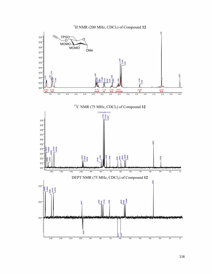
116
1H NMR (200 MHz, CDCl3) of Compound 12
7.5 7.0 6.5 6.0 5.5 5.0 4.5 4.0 3.5 3.0 2.5 2.0 1.5 1.0 0.5 0.0
0.0
0.1
0.2
0.3
0.4
0.5
0.6
0.7
0.8
0.9
1.0
9.50 9.005.89 5.223.84 2.911.70 0.89
0.00
1.06
2.312.
32
3.373.
433.46
3.54
3.90
3.91
4.36
4.394.754.80
4.814.83
4.87
7.267.37
7.41
7.42
7.70
13C NMR (75 MHz, CDCl3) of Compound 12
130 120 110 100 90 80 70 60 50 40 30 20 10 0
0.0
0.1
0.2
0.3
0.4
0.5
0.6
0.7
0.8
0.9
1.0
Chloroform-d
19.33
26.80
54.82
55.48
56.23
59.76
62.94
70.90
74.32
76.37
77.00
77.64
79.15
79.76
97.67
98.31
98.76
127.53
127.64
129.58
133.25
133.66
135.59
135.83
DEPT NMR (75 MHz, CDCl3) of Compound 12
130 120 110 100 90 80 70 60 50 40 30 20 10 0
0.0
0.5
1.0
26.80
54.84
55.51
56.25
59.77
62.93
70.90
77.35
78.12
79.16
97.69
98.77
127.54
127.67
129.6013
5.61
135.85
OOMOMO
OMeMOMO
TPSO

117
1H NMR (200 MHz, CD3OD) of Compound 1d
4.5 4.0 3.5 3.0 2.5 2.0 1.5 1.0 0.5 0.0
0.0
0.1
0.2
0.3
0.4
0.5
0.6
0.7
0.8
0.9
1.0
5.56 4.282.00 0.28
2.873.33
3.35
3.373.55
3.663.71
3.75
3.773.82
3.83
3.88
4.384.39
4.734.75
4.77
13C NMR (75 MHz, CD3OD) of Compound 1d
100 90 80 70 60 50 40 30 20 10 0
0.0
0.1
0.2
0.3
0.4
0.5
0.6
0.7
0.8
0.9
1.0
Acetone-d6
29.80
54.57
59.19
59.93
69.95
70.7072.57
76.9198.62
DEPT NMR (75 MHz, CD3OD) of Compound 1d
100 90 80 70 60 50 40 30 20 10 0
-1.0
-0.5
0.0
0.5
1.0
29.80
54.55
59.21
59.90
69.95
70.68
72.57
76.8898.61
OO
OH
OH
OMeHO

118
1H NMR (200 MHz, CDCl3) of Compound 13
6.0 5.5 5.0 4.5 4.0 3.5 3.0 2.5 2.0 1.5 1.0 0.5 0.0
0.0
0.1
0.2
0.3
0.4
0.5
0.6
0.7
0.8
0.9
1.0
12.805.05 3.082.000.89
0.00
1.321.34
1.42
1.50
2.17
3.41
4.004.034.084.11
4.12
4.134.
21
4.224.574.58
4.73
4.74
5.88
5.90
13C NMR (75 MHz, CDCl3) of Compound 13
110 100 90 80 70 60 50 40 30 20 10 0
0.0
0.1
0.2
0.3
0.4
0.5
0.6
0.7
0.8
0.9
1.0
Chloroform-d
25.33
26.21
26.78
30.85
55.75
56.06
64.36
67.48
68.70
72.30
76.36
77.00
77.63
79.0881.04
83.27
83.38
96.01
97.14
105.18
109.05111.87
DEPT NMR (75 MHz, CDCl3) of Compound 13
100 90 80 70 60 50 40 30 20 10 0
-0.5
0.0
0.5
1.0
25.3
426
.20
26.7
9
30.8
7
55.7
656
.07
67.48
68.6
8
72.2
8
79.0
581
.02
83.2
583
.37
96.00
105.
18
O
OO
O
O
OMOM

119
1H NMR (200 MHz, CDCl3) of Compound 15
5.5 5.0 4.5 4.0 3.5 3.0 2.5 2.0 1.5 1.0 0.5 0.0
0.00
0.05
0.10
0.15
0.20
0.25
9.15 5.943.05 3.032.951.98 1.010.95 0.82
TMS
0.00
0.09
0.89
0.91
1.321.48
2.74
3.43
3.84
4.09
4.274.57
4.75
4.78
5.90
13C NMR (75 MHz, CDCl3) of Compound 15
110 100 90 80 70 60 50 40 30 20 10 0
0.0
0.1
0.2
0.3
0.4
0.5
0.6
0.7
0.8
0.9
1.0
Chloroform-d
-5.45
18.26
25.84
26.68
55.84
64.3468.24
76.36
77.00
77.63
79.21
79.93
83.25
96.53105.08
111.71
DEPT NMR (75 MHz, CDCl3) of Compound 15
100 90 80 70 60 50 40 30 20 10 0
-0.4
-0.3
-0.2
-0.1
0.0
0.1
0.2
0.3
0.4
0.5
0.6
0.7
0.8
0.9
1.0
-5.43
25.86
26.69
55.86
64.34
68.25
79.21
79.92
83.24
96.54
105.08
O
OO
TBDMSO
HO
OMOM
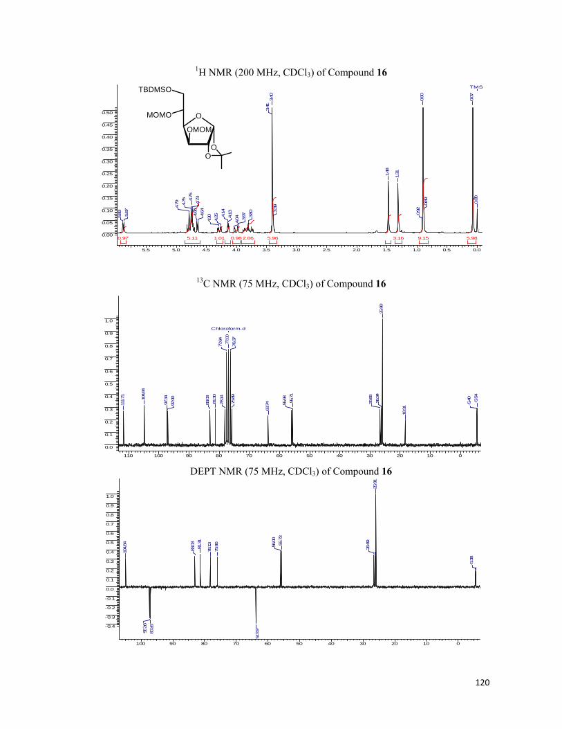
120
1H NMR (200 MHz, CDCl3) of Compound 16
5.5 5.0 4.5 4.0 3.5 3.0 2.5 2.0 1.5 1.0 0.5 0.0
0.00
0.05
0.10
0.15
0.20
0.25
0.30
0.35
0.40
0.45
0.50
9.15 5.985.965.11 3.162.061.01 0.980.97
TMS
0.00
0.07
0.89
0.90
0.92
1.311.48
3.39
3.40
3.41
3.80
3.97
4.044.
134.14
4.25
4.30
4.64
4.66
4.734.75
4.75
4.79
5.87
5.89
13C NMR (75 MHz, CDCl3) of Compound 16
110 100 90 80 70 60 50 40 30 20 10 0
0.0
0.1
0.2
0.3
0.4
0.5
0.6
0.7
0.8
0.9
1.0
Chloroform-d
-5.54
-5.40
18.31
25.90
26.34
26.68
55.71
55.98
63.7475
.89
76.37
77.00
77.64
78.14
81.30
83.03
97.00
97.34104.84
111.71
DEPT NMR (75 MHz, CDCl3) of Compound 16
100 90 80 70 60 50 40 30 20 10 0
-0.4
-0.3
-0.2
-0.1
0.0
0.1
0.2
0.3
0.4
0.5
0.6
0.7
0.8
0.9
1.0
-5.38
25.91
26.69
55.73
56.00
63.75
75.90
78.13
81.31
83.03
97.03
97.35
104.84
O
OO
TBDMSO
MOMO
OMOM

121
1H NMR (200 MHz, CDCl3) of Compound 17
5.5 5.0 4.5 4.0 3.5 3.0 2.5 2.0 1.5 1.0 0.5 0.0
0.00
0.05
0.10
0.15
0.20
0.25
0.30
0.35
0.40
0.45
0.50
5.983.93 3.002.011.98 0.970.95
0.00
1.32
1.49
3.40
3.44
3.683.71
3.873.
89
4.16
4.20
4.62
4.64
4.72
5.88
5.90
13C NMR (75 MHz, CDCl3) of Compound 17
110 100 90 80 70 60 50 40 30 20 10 0
0.0
0.1
0.2
0.3
0.4
0.5
0.6
0.7
0.8
0.9
1.0
Chloroform-d
26.1926.69
55.82
55.98
63.79
76.3677.00
77.63
78.65
82.74
96.90
97.30
104.88
111.81
DEPT NMR (75 MHz, CDCl3) of Compound 17
100 90 80 70 60 50 40 30 20 10 0
-0.4
-0.3
-0.2
-0.1
0.0
0.1
0.2
0.3
0.4
0.5
0.6
0.7
0.8
0.9
1.0
-5.38
25.91
26.69
55.73
56.00
63.75
75.90
78.13
81.31
83.03
97.03
97.35
104.84
O
OO
HO
MOMO
OMOM

122
1H NMR (200 MHz, CDCl3) of Compound 18
7.5 7.0 6.5 6.0 5.5 5.0 4.5 4.0 3.5 3.0 2.5 2.0 1.5 1.0 0.5 0.0
0.0
0.1
0.2
0.3
0.4
0.5
0.6
0.7
0.8
0.9
1.0
5.984.08 3.101.991.17 1.000.97 0.88
0.00
1.321.49
2.40
2.41
2.433.39
3.41
3.41
3.974.154.19
4.21
4.23
4.24
4.654.67
4.73
4.74
4.75
5.88
5.89
13C NMR (75 MHz, CDCl3) of Compound 18
110 100 90 80 70 60 50 40 30 20 10 0
0.0
0.1
0.2
0.3
0.4
0.5
0.6
0.7
0.8
0.9
1.0
Chloroform-d
26.38
26.77
55.85
56.03
58.60
70.31
74.00
74.35
76.37
77.00
77.64
78.51
79.73
82.96
96.85
97.2710
4.84
111.88
DEPT NMR (75 MHz, CDCl3) of Compound 18
100 90 80 70 60 50 40 30 20 10 0
-1.0
-0.5
0.0
0.5
26.39
26.78
55.87
56.05
58.61
70.29
74.00
74.38
78.49
79.73
81.23
82.96
96.8697.28
104.84
O
OO
O
MOMO
OMOM
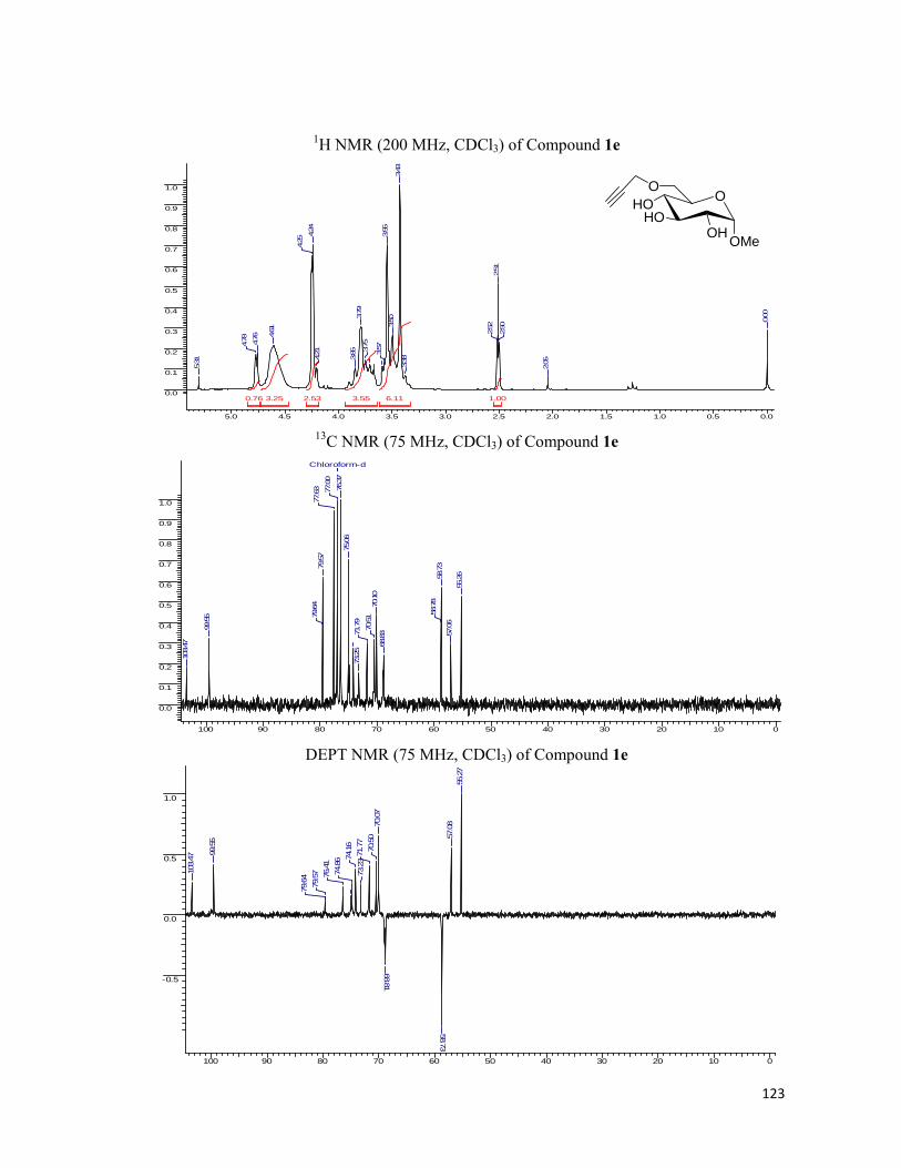
123
1H NMR (200 MHz, CDCl3) of Compound 1e
5.0 4.5 4.0 3.5 3.0 2.5 2.0 1.5 1.0 0.5 0.0
0.0
0.1
0.2
0.3
0.4
0.5
0.6
0.7
0.8
0.9
1.0
6.113.553.25 2.53 1.000.76
0.00
2.05
2.50
2.51
2.52
3.38
3.43
3.50
3.55
3.573.75
3.79
3.85
4.21
4.24
4.25
4.61
4.76
4.78
5.31
13C NMR (75 MHz, CDCl3) of Compound 1e
100 90 80 70 60 50 40 30 20 10 0
0.0
0.1
0.2
0.3
0.4
0.5
0.6
0.7
0.8
0.9
1.0
Chloroform-d55
.26
57.06
58.73
58.78
68.83
70.10
70.51
71.79
73.25
75.06
76.37
77.00
77.63
79.57
79.64
99.55
103.47
DEPT NMR (75 MHz, CDCl3) of Compound 1e
100 90 80 70 60 50 40 30 20 10 0
-0.5
0.0
0.5
1.0
55.2
757
.08
58.73
68.81
70.0
770
.50
71.7
773
.2374
.16
74.8
676
.41
79.5
779
.64
99.5
5
103.
47
OHO
OH
O
OMe
HO
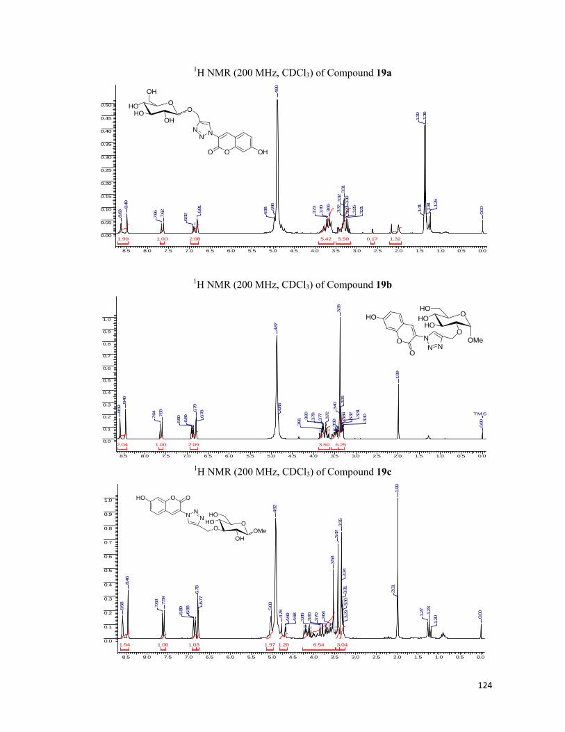
124
1H NMR (200 MHz, CDCl3) of Compound 19a
8.5 8.0 7.5 7.0 6.5 6.0 5.5 5.0 4.5 4.0 3.5 3.0 2.5 2.0 1.5 1.0 0.5 0.0
0.00
0.05
0.10
0.15
0.20
0.25
0.30
0.35
0.40
0.45
0.50
5.505.422.081.99 1.321.00 0.17
0.00
1.25
1.34
1.36
1.39
1.41
3.213.25
3.29
3.30
3.31
3.32
3.323.66
3.70
3.73
4.90
4.96
4.986.81
6.927.
62
7.66
8.49
8.63
1H NMR (200 MHz, CDCl3) of Compound 19b
8.5 8.0 7.5 7.0 6.5 6.0 5.5 5.0 4.5 4.0 3.5 3.0 2.5 2.0 1.5 1.0 0.5 0.0
0.0
0.1
0.2
0.3
0.4
0.5
0.6
0.7
0.8
0.9
1.0
6.253.502.092.04 1.00
TMS
0.00
1.99
3.303.
31
3.32
3.34
3.35
3.38
3.40
3.50
3.72
3.773.793.80
3.81
4.83
4.87
6.78
6.79
6.89
6.90
7.59
7.64
8.46
8.59
1H NMR (200 MHz, CDCl3) of Compound 19c
8.5 8.0 7.5 7.0 6.5 6.0 5.5 5.0 4.5 4.0 3.5 3.0 2.5 2.0 1.5 1.0 0.5 0.0
0.0
0.1
0.2
0.3
0.4
0.5
0.6
0.7
0.8
0.9
1.0
6.54 3.041.971.94 1.201.031.00
0.00
1.20
1.23
1.27
1.99
2.01
3.29
3.30
3.31
3.34
3.35
3.42
3.53
3.64
3.70
3.80
3.85
4.68
4.694.78
4.92
5.036.
776.78
6.88
6.89
7.59
7.63
8.46
8.58
OHO
OH
OH
OHO
O OHO
NNN
OHOHO
OMeO
HO
O
HO
O
NNN
OHO
OH
HO
OMeO
OHO O
N NN

125
1H NMR (200 MHz, CDCl3) of Compound 19d
8.5 8.0 7.5 7.0 6.5 6.0 5.5 5.0 4.5 4.0 3.5 3.0 2.5 2.0 1.5 1.0 0.5 0.0
0.0
0.1
0.2
0.3
0.4
0.5
0.6
0.7
9.144.772.062.01 1.241.161.00
0.00
2.00
2.01
3.29
3.30
3.31
3.35
3.39
3.42
3.423.73
3.744.
674.
684.
864.
905.
045.
106.806.
82
6.917.
617.
66
8.49
1H NMR (200 MHz, CDCl3) of Compound 19e
8.5 8.0 7.5 7.0 6.5 6.0 5.5 5.0 4.5 4.0 3.5 3.0 2.5 2.0 1.5 1.0 0.5 0.0
0.0
0.1
0.2
0.3
0.4
0.5
0.6
0.7
0.8
0.9
1.0
5.814.012.252.031.97 1.371.181.00
TMS
0.001.
201.
241.
27
1.99
2.01
2.02
3.303.
313.
323.
353.
403.
513.
623.77
3.814.
084.
11
4.66
4.68
4.75
4.90
6.786.
79
6.89
6.907.
607.
64
8.47
8.57
OO
OH
OH
OMeHO
OHO O
N NN
OHO
OH
O
OMeHO
OHO
O
N NN
126
Chapter 2: References
1. Friedman, R. J.; An, Y. H. Handbook of Bacterial Adhesion: Principles, Methods,
and Applications.
2. Sharon, N.; Ofek, I.; Glycoconjugate. J. 2001, 17, 659-664.
3. Lee, Y. K.; Puong, K. Y. Br. J. Nutr. Med. 2002, 88, S101-S108.
4. Isolauri, E.; Joensuu, J.; Vesikari, T. Vaccine 1995, 13, 310-312.
5. Hakamori, S. I. Proc. Natl. Acad. Sci. USA 2002, 99, 225-232.
6. Sorice, M.; Longo, A.; Pavan, A.; Glyoconjugate J. 2004, 20, 63-70.
7. Crick, F. H. C. Symp. Soc. Exp. Biol. XII 1958, 139-163.
8. (a) Rees, D. A. Biochem. J. 1972, 126, 257-273; (b) Holzl, G., Dormann, P. Prog.
Lipid Res. 2007, 46, 225-243; (c) Wang, X. Curr. Opin. Plant Biol. 2004, 7(3), 329-
336.
9. Rademacher, T. W.; Parekh, R. B.; Dwek, R. A. Annu. Rev. Biochem. 1988, 57,
785-838.
10. Bertozzi, C. R.; Kiessling, L. L. Science 2001, 291, 2357-2364.
11. Zachara, N. E.; Hart, G. W. Chem. Rev. 2002, 102, 431-438.
12. Aubert, A. K.; Desai, N. N.; Neuberger, A. C.; Michael, J. Arch. Biochem. Biophys.
1976, 175(2), 410-418.
13. (a) Mahal, L. K.; Yarema, K. J.; Bertozzi, C. R. Science 1997, 276, 1125-1128; (b)
Varki, A. FASEB, 1991, 5, 226. (c) Fltz, W.; Wong, C.-H. J. Org. Chem. 1994, 59,
8279; (d) Shames, S. L. et al. Glycobiology, 1991, 1, 187; (e) Lin, C.-H.; Sugai, T.;
Halcomb, R. L.; Ichikawa Y, Wong, C.-H. J. Am. Chem. Soc. 1992, 114, 10138.
14. Sampathkumar, S. G.; Li, A. V.; Jones, M. B.; Sun, Z.; Yarema, K. J. Nat. Chem.
Biol. 2006, 2, 149-152.
15. Sawa, M.; Hsu, T. L.; Itoh, T.; Sugiyama, M.; Hanson, S. R.; Vogt, P. K.; Wong,
C.-H. Proc. Nat. Acad. Sci. 2006, 103, 12371-1276.
16. Sadamoto, R.; Niikura, K.; Sears, P. S.; Liu, H.; Wong, C.-H.; Suksomcheep, A.;
Tomita, F.; Monde, K.; Nishimura, S. I. J. Am. Chem. Soc. 2002, 124, 9018-9019.
17. Sadamoto, R.; Niikura, K.; Monde, K.; Nishimura, S.-I. Methods Enzymol. 2003,
362, 273-286.
18. Sadamoto, R.; Niikura, K.; Ueda, T.; Mode, K.; Fukuhar, N.; Nishimura, S.-I. J.
Am. Chem. Soc. 2004, 126, 3755-3761.

127
19. Koch, A. Clin. Microbiol. Rev. 2003, 16(4), 673-687.
20. Hanover, J. A. FASEB 2001, 15, 1865-1876.
21. van Leeuwenhoek, A. Philosophical Transactions, 1684, 14, 568-574.
22. van Leeuwenhoek, A. Philosophical Transactions 1700, 22, 509-518.
23. van Leeuwenhoek, A. Philosophical Transactions 1702, 23, 1304-1311.
24. Gram, H. C. Fortschr. Med. 1884, 2, 185-189.
25. Madigan, M.; Martinki, J. Brock Biology of Microorganisms.
26. (a) Sharon, N. Biochem. Biophys. Acta 2006, 1760, 527-537. (b) Sharon, N. FEBS
Lett. 1987, 217, 145-157. (c) Yarema, K. J.; Bertozzi, C. R. Curr. Opin. Chem. Biol.
1998, 2, 49-61.
27. (a) Rudd, P. M.; Elliott, T.; Cresswell, P.; Wilson, I. A.; Dwek, R. A. Science
2001, 291, 2370-2376. (b) McAuliffe, J. C.; Hindsgaul, O. Front. Mol. Biol. 2000,
30, 249. (c) Varki, A. Glycobiology 1993, 3, 97-130. (c) Campbell C. T.; Yarema,
K. J. Genome Biol. 2005, 6, 236.1-236.7. (d) Murrell, M.P., Yarema, K.J.,
Levchenko, A. ChemBiochem. 2004, 5, 1334-1347.
28. (a) Saxon, E.; Bertozzi, C. R. Science 2000, 287, 2007-2010. (b) Tai, H. C.;
Kidekel, N.; Ficarro, S. B.; Peters, E. C.; Hsieh-Wilson, L. C. J. Am. Chem. Soc.
2004, 126, 10500-10501; (c) Mahal, L. K.; Bertozzi, C. R. Chem. Biol. 1997, 4,
415-422. (d) Prescher, J. A.; Bertozzi, C. R. Cell 2006, 126, 851-854. (e) Dube, D.
H.; Bertozzi, C. R. Curr. Opin. Chem. Biol. 2003, 7, 616-625. (f) Campbell, C.T.,
Sampathkumar, S.-G., Yarema, K.J. Mol. Biosys. 2007, 3, 187-194. (g) Sadamoto,
R. Trends Glycosci. Glycotechnol. 2005, 17, 97-105.
29. (a) Zhang, J.; Campbell, R. E.; Ting, A. Y.; Tsien, R. Y. Nat. Rev. Mol. Cell. Biol.
2002, 3, 906-918. (b) Laughlin, S. T.; Agard, N. J.; Baskin, J. M.; Carrico, I. S.;
Chang, P. V.; Ganguli, A. S.; Hangauer, M. J.; Lo, A.; Prescher, J. A.; Bertozzi, C.
R. Methods Enzymol. 2006, 415, 230-250. (c) Chang, P. V.; Prescher, J. A.;
Hangauer, M. J.; Bertozzi, C. R. J. Am. Chem. Soc. 2007, 129, 8400-8401. (d) Link,
A. J.; Tirrell, D. A. J. Am. Chem. Soc. 2003, 125, 11164-11165.
30. Zhang, J.; Campbell, R. E.; Ting, A. Y.; Tsein, R. Y. Nature 2002, 3, 906-918.
31. (a) Sivakumar, K.; Xie, F.; Cash, B. M.; Long, S.; Barnhill, H. N.; Wang, Q. Org.
Lett. 2004, 6, 4603-4606. (b) Zhou, Z.; Fahrni, C. J. J. Am. Chem. Soc. 2004, 126,
8862-8863.
32. (a) Huisgen, R. In 1,3-Dipolar Cycloaddition Chemistry, Padwa, A., Ed., Wiley:
New York, 1984, Chapter 1, pp 1-176. (b) Kolb, H. C.; Finn, M. G.; Sharpless, K.

128
B. Angew. Chem., Int. Ed. 2001, 40, 2004-2021. (c) Lewis, W. G.; Green, L. G.;
Grynszpan, F.; Radic, Z.; Carlier, P. R.; Taylor, P.; Finn, M. G.; Sharpless, K. B.
Angew. Chem., Int. Ed. 2002, 41, 1053-1057. (d) Rostovtsev, V. V.; Green, L. G.;
Fokin, V. V.; Sharpless, K. B. Angew. Chem., Int. Ed. 2002, 41, 2596-2599. (e) (a)
TornØe, C.; Christensen, C.; Meldal, M. J. Org. Chem. 2002, 67, 3057-3064. (f)
Buchardt, J.; SchiØdt, C. B.; Krog-Jensen, C.; Delaissé, J.-M.; Foged, N. T.;
Meldal, M. J. Comb. Chem. 2000, 2, 634-638. (g) Wang, Q.; Chan, T. r.; Hilgraf,
R.; Fokin, V. V.; Sharpless, K. B.; Finn, M. G. J. Am. Chem. Soc. 2003, 125, 3192-
3193. (h) Speers, A. E.; Cravatt, B. F. Chem. Biol. 2004, 11, 535-545.
33. Huisgen, R. “Centaury Lecture – 1,3-dipolar cycloadditions” – Proceedings of the
Chemical Society of London 1961, 357.
34. Roy, B.; Mukhopadhyay, B. Tetrahedron Lett. 2007, 48, 3783-3787.
35. Munavu, R. M.; Szmant, H. H. J. Org. Chem. 1976, 41, 1832-1836.
*****
Chapter 3 Photocleavable Linkers for Bioconjugation of Proteins

129
Chapter 3: Introduction
Living organisms are composed of lifeless molecules. Yet living organisms possess
extraordinary attributes not exhibited by random collection of molecules.1 First is their degree
of complexity and organisation in which the thousands of different molecules make up a cell’s
intricate internal structures. In contrast, inanimate matter-clay, sand, rocks etc. usually consists
of mixtures of relatively simple chemical compounds. Secondly, living organisms extract,
transform, and use energy from their environment usually in the form of chemical nutrients or
sunlight enabling them to build and maintain their complicated structure and support various
cellular metabolisms. Complementarily, inanimate matter exploits energy in an unsystematic
manner which tends to decay towards a more disordered state to come to the equilibrium with
its surroundings. The third attribute if the living organisms is the capability for precise self-
replication and self-assembly, a property that is quintessence of the living state. A single
bacterium placed in a sterile nutrient medium can give rise to billion identical “daughter” cells
in 24 hours.2 Each of the cells contain thousands of different molecules, some extremely
complicated; yet each bacterium is a faithful copy of the original, its construction directed
entirely from the information contained within the genetic material of the original cell.
Although the ability to self-replicate has no true analog in the nonliving world, there is an
instructive analogy in the growth of the crystals in saturated solutions. Crystallization produce
more material identical in lattice structure to the original “seed” crystal but these are less
complex and their structure is static as compared to the dynamic for of life in living organisms.
Each component of living organisms has a specific function. This is true not only of
macroscopic structures, such as leaves and stems or heart and lungs, but also true for the
variety of bio-molecules present inside the cell. The interplay among the chemical components
of a living organism is dynamic; changes in one component cause coordinating or
compensating changes in another, with the whole ensemble displaying a character beyond that
is its individual constituents. The collection of molecules carries out a program, the end result
of which is reproduction of the program and self-perpetuation of that collection of molecules;
in short, life. The fundamental building blocks of living cells are biomolecules which provide
the bedrock of life and now provide a new research frontier for the interfacial science. All life
forms can be considered to be self-organized systems assembled from these building blocks.
Even though one can subdivide life into substructures like cell organelles, the entire cell,
tissues, multicellular organisms, and even colonies, biomolecules make their presence felt
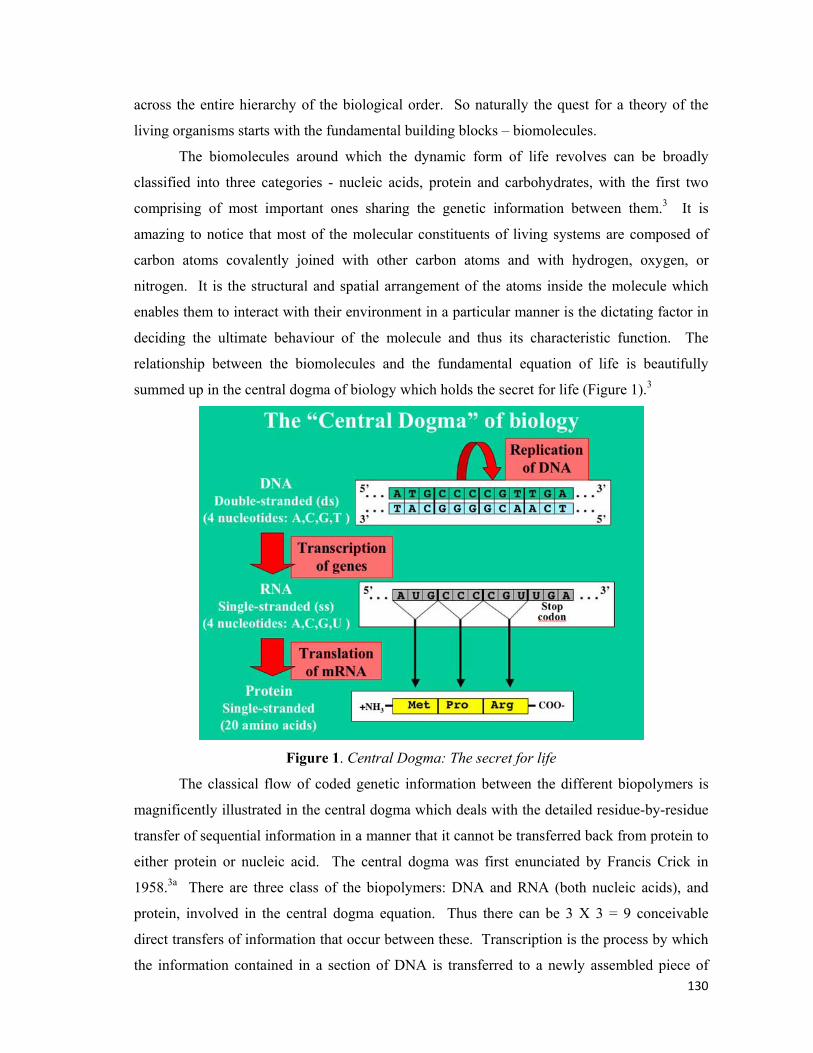
130
across the entire hierarchy of the biological order. So naturally the quest for a theory of the
living organisms starts with the fundamental building blocks – biomolecules.
The biomolecules around which the dynamic form of life revolves can be broadly
classified into three categories - nucleic acids, protein and carbohydrates, with the first two
comprising of most important ones sharing the genetic information between them.3 It is
amazing to notice that most of the molecular constituents of living systems are composed of
carbon atoms covalently joined with other carbon atoms and with hydrogen, oxygen, or
nitrogen. It is the structural and spatial arrangement of the atoms inside the molecule which
enables them to interact with their environment in a particular manner is the dictating factor in
deciding the ultimate behaviour of the molecule and thus its characteristic function. The
relationship between the biomolecules and the fundamental equation of life is beautifully
summed up in the central dogma of biology which holds the secret for life (Figure 1).3
Figure 1. Central Dogma: The secret for life
The classical flow of coded genetic information between the different biopolymers is
magnificently illustrated in the central dogma which deals with the detailed residue-by-residue
transfer of sequential information in a manner that it cannot be transferred back from protein to
either protein or nucleic acid. The central dogma was first enunciated by Francis Crick in
1958.3a There are three class of the biopolymers: DNA and RNA (both nucleic acids), and
protein, involved in the central dogma equation. Thus there can be 3 X 3 = 9 conceivable
direct transfers of information that occur between these. Transcription is the process by which
the information contained in a section of DNA is transferred to a newly assembled piece of
131
messenger RNA (mRNA). It is facilitated by RNA polymerase and transcription factors.3b, 3e
Eventually, this mature mRNA finds its way to a ribosome, where it delivers the genetic code
for the protein assembly, a process commonly known as Translation.3b In prokaryotic cells,
which have no nuclear compartment, the process of transcription and translation may be linked
together.4 In eukaryotic cells, the site of transcription (the cell nucleus) is usually separated
from the site of translation (the cytoplasm), so the mRNA must be transported out of the
nucleus into the cytoplasm, where it can be bound by ribosomes. The mRNA is read by the
ribosome as triplet codons, usually beginning with an AUG, or initiator methonine codon
downstream of the ribosome binding site.5 Complexes of initiation factors and elongation
factors bring aminoacylated transfer RNAs (tRNAs) into the ribosome-mRNA complex,
matching the codon in the mRNA to the anti-codon in the tRNA, thereby adding the correct
amino acid in the sequence encoding the gene. As the amino acids are linked into the growing
peptide chain, they begin folding into the correct conformation. This folding continues until
the nascent polypeptide chains are released from the ribosome as a mature protein. However,
ultimately the whole story boils down to the type of the protein translated from the mRNA to
carry out the different metabolic functions inside the cells.
Proteins are the most abundant biological macromolecules, occurring in all parts of the
cells. They also occur in great variety; thousands of different kinds, ranging in size from
relatively small peptides to huge polymers with molecular weights in millions, may be found in
a single cell. Moreover, proteins exhibit enormous diversity of biological function and are the
most important final products of the information pathways (Central Dogma). Proteins are the
molecular instruments through which the genetic information is expressed.3 Relatively simple
monomeric subunits provide the key to the structure of the thousands different kinds of the
proteins. All proteins, whether from the most ancient lines of bacteria or found among the
complex forms of life are constructed from the same ubiquitous set of amino acids, covalently
linked characteristic linear sequence to make up a polypeptide chain which is nothing but the
primary structure of the protein. Because each of the amino acid has a side chain with
distinctive chemical properties, this group of 20 precursor molecules may be regarded as the
alphabet in which the language of protein structure is written. What is the most remarkable
thing is that cells can produce proteins with strikingly different properties and activities by
joining the same 20 amino acids in many different combinations and sequences. From these
building blocks different organisms can make such widely diverse products as enzymes,
hormones, antibodies, transporters, muscles, milk proteins and a myriad of other substances
having distinct biological activities.6

132
For large macromolecules such as proteins, the tasks of describing and understanding
structure are approached at several levels of complexity, arranged in a kind of conceptual
hierarchy. Four levels of protein structure are commonly defined.7 A description of all
covalent bonds (mainly peptides and disulfide bonds) linking amino acids residues in a
polypeptide chain is its primary structure. The most important element of the primary structure
is the sequence of the amino acids. Secondary structure refers to the particularly stable
arrangements of amino acid residues giving rise to recurring structural patterns. In
biochemistry and structural biology, secondary structure is the general term for local segments
of biopolymers (proteins and nucleic acids) which is formally defined by the weak interactions
arising within the molecule. By building models of peptides using known information about
bond lengths and angles, the first elements of secondary structure, the alpha helix and beta-
sheet, were first suggested by Linus Pauling and co-workers in 1951.8 Both the alpha helix and
beta-sheet represent a way of saturating all the hydrogen bond donors and acceptors in the
peptide backbone. Although the other elements of the secondary structures have been
discovered, like various forms of loop and helices, but alpha helix and beta-sheet are the most
predominant secondary structures found in protein.
Figure 2. Diagrammatic illustration of the various structures of proteins
133
The elements of the secondary structure are usually folded into a compact shape using a
variety of loops and turns giving rise to the tertiary structure of the proteins.9 It formally
describes all aspects of the three-dimensional folding of the peptide backbone. The formation
of the tertiary structure is usually driven by the burial of hydrophobic residues, but other
interactions such as hydrogen bonding, ionic interactions and disulfide bonds can also stabilize
the tertiary structure to encompass all the non-covalent interactions which are not covered in
the secondary structure, and is what defines the overall fold of the protein. It is considered
indispensable for the function of the protein and is usually prone to any influence (e.g. certain
chemicals, heat etc.) that disrupts the weak interactions, a process which is commonly termed
as denaturation of the protein. Quaternary structure of the protein involves the spatial
relationship of multiple polypeptide chain (subunits) that are stably associated.9 Not all
proteins are found to have quaternary structure. The individual subunits may not be covalently
connected, but are stabilized by the same range of interactions as the tertiary structure. All the
structures of the protein are diagrammatically depicted in Figure 2.
As is evident from the central dogma, proteins are an important class of molecules
considering the variety of the cellular reactions they take part in. In the recent times, a wealth
of methods had been developed to manipulate the protein structure and thus its activity to
unravel the critical details of protein function at the molecular and cellular level. Notably, the
use of genetic tools such as site-directed mutagenesis, knock-out and knock-in techniques, and
the use of encoded reporters such as fluorescent proteins have facilitated the discovery of many
protein functions within their molecular networks.10 One of the next formidable challenges in
biology is directed towards understanding the molecular details of how post-translational
modifications (PTMs) affect protein function, stability, cellular localization, three-dimensional
conformation, and interaction with other molecules. Many of these biological investigations of
protein function require access to protein molecules that are hard to obtain with purely
biological methods such as site-directed mutagenesis and recombinant protein expression.11
The combination of chemical and biological techniques to chemoselectively modify proteins,
however has proved to be an excellent resource for studying protein function on a molecular
level. A few of the prominent techniques related to post-translational modification of proteins
can broadly be classified into the following categories: (a) PTMs involving addition of
functional groups; (b) PTMs involving addition of small peptides or other proteins; (c) PTMs
inducing the changes in the chemical nature of amino acids present on the peptide backbone;
(d) PTMs involving structural changes in the tertiary structure of protein.

134
Based on early work by Wieland et. al., a synthetic method known as native chemical
ligation (NCL) has evolved to be a powerful approach in peptide chemistry.12 NCL allows the
construction of large polypeptides through the condensation of two or more smaller peptides
with C-terminal thioesters and N-terminal cysteines, in the presence of an added thiol catalyst.
In a free reversible first step, a transthioesterification occurs to yield a thioesters-linked
intermediate; this intermediate rearranges irreversibly under the usual reaction conditions to
form a native amide bond at the ligation site. A short diagrammatic description is provided in
Figure 3a. NCL of unprotected peptide segments was developed by Dawson et. al. but
Wieland’s work led to the ‘active ester’ method for making protected peptide segments in
conventional solution synthesis in organic solvents.13 An elegant display of NCL was
demonstrated by C. H. Wong group when they successfully synthesized N-linked
glycopeptides using sugar assisted ligation (SAL) strategy from cysteines free peptides.14 This
approach is based on SAL involving a peptide thioesters and a glycopeptide in which N-
acetylglucosamine linked to serine side chain is modified at C-2 acetamide with a thiol handle
to mimic the cysteines function. They also reported the similar rates of ligation for both N-
and O-linked glycopeptides coupled with the wide tolerance for a variety of amino acids. The
detailed diagrammatic representation is provided in Figure 3b.
Figure 3. (a) Native Chemical Ligation: A well established method for preparing peptides, R is typically a phenyl ring; (b) Wong’s modification of the NCL to prepare the glycopeptides, in which sugars are attached to peptide chains, R1 represents the amino acid side chain

135
Figure 4. Schematic representation of Expressed Protein Ligation. CBD is the Chitin Binding Domain
The semi-synthetic version of NCL, known as Expressed Protein Ligation (EPL),
permits the in vitro ligation of a chemically synthesized C-terminal segment of a protein to a
recombinant N-terminal segment fused through its C terminus to an intein protein splicing
element.15 EPL, also named as intein-mediated protein ligation, facilitates the site-specific
protein labelling with a broad range of physical probes such as fluorophores, PTMs, stable
isotopes, and unnatural amino acids. The growing popularity of EPL can be estimated by the
increasing application of this technique to a wide range of protein-engineering problems, for
e.g. generation of cyclic peptides and proteins, segmental isotopic labelling, site-specific
protein modification, generation of cytotoxic proteins etc. Inteins are internal segments of
precursor proteins that catalyze their ipso excision, in an intramolecular process called protein
slicing, with the concurrent ligation of to flanking external regions (N- and C- inteins) through
a native peptide bond, to finally yield a protein.16 The schematic illustration is shown in Figure
4 for the EPL. Note that the addition of the external as an additive is the key to the process
since it generated reactive α-thioester derivative of the protein in situ. This underlines the
extension of ligation techniques to the emerging area of chemical genetics providing innovative
tools to probe protein function through perturbation of protein activity or protein-protein
interactions in a rapid tunable manner that is necessary to track protein function within the
dynamic and variable nature of biological processes.

136
The only disadvantage of NCL and EPL is the necessity of cysteines residue or a
homologue at the ligation site. The occurrence of this amino acid in globular proteins is very
low and the insertion of additional cycteine residues can alter the protein structure and function
by the formation of disulfide bridges. Several approaches have been developed to circumvent
this limitation. A few of the promising and most sought after techniques include NCL with
Cys-mimetics,17 Staudinger ligation on proteins,18 expressed enzymatic ligation,19 maleimide
and succinamide assisted protein conjugation19 etc. Complementary to the development of
techniques for protein-protein ligation, researchers are also involved in the probing the
behaviour of proteins in vicinity of abiotic surfaces like nanotubes/nanoparticles, glass slides,
transition metal catalysts etc.
However, the mild and selective chemical modification of complex biomolecules such
as proteins under physiological conditions remains a huge challenge. There are so many
factors governing the stability of the proteins, like temperature and chemical sensitivity,
denaturation and precipitation etc. which make the protein handling fairly difficult to handle.
However, the power of protein ligation to investigation protein function is evident and the
technology is constantly improving, aiming towards minimally invasive methods that allow
analysis of target proteins within the ensemble of all the other proteins in vivo.
*****

137
Chapter 3: Present Work
The continuing success of genome sequencing efforts has laid the foundation for
understanding the molecular basis of life in its many forms. However, this task involves not
only the characterization of genes but the products of gene expression as well. As primary
mediators of most physiological and pathological processes, proteins may be viewed as the
next major challenge, especially considering the difficulties involved in developing and
applying methods for probing protein interactions as compared to DNA or RNA molecules.20
For example, unlike oligonucleotides, proteins exhibit a diverse array of chemical and
biochemical properties, are not amenable to molecular amplification, and do not possess
predefined complementary binding partners.
Despite the many technical challenges that accompany the analysis of proteins, the need
for global strategies to characterize the expression and function of these biomolecules is clear,
especially given the multitude of post-transcriptional and post-translational processes that
regulate the activity of proteins in cells and tissues.21 In most cases, the initial polypeptide-
translation product undergoes some type of modification before it assumes its functional role in
a living system. These changes are broadly termed as “post-translational modifications” and
encompass a wide variety of reversible and irreversible chemical reactions. Specific delivery
of the protein to specific subcellular or extracellular components is often achieved with leader
or signal sequences, which may be proteolytically cleaved. At their cellular destinations,
proteins carry out their many functions, and their activities are controlled by post-translational
modifications. Most proteins function in collaboration with other proteins found within the
perimeter of the cells. Sensing extracellular signals is a matter of receptor to adaptor
interactions and the shape of the cell is maintained by an intricate network of structural protein
interactions. The interactions between the proteins are central importance for virtually every
process in a living cell, probing these interactions is a way to get a broader view of how they
work cooperatively in a cell.3 Information about these interactions improves our understanding
of diseases and can provide the basis for new therapeutic approaches.
The temporal and spatial regulation of protein function is central to biological
processes. Hence, the ability to artificially trigger molecular events in a biologically relevant
context is useful for the study of living organism.22 By chemically modifying a protein with
groups that can respond to external inputs (such as reagents, light etc.), will enable us to reveal
the complex mechanism through which proteins operate. The ability to alter the protein

138
structure and function by introducing unnatural amino acids has great potential to enhance our
understanding of the proteins and thus generate new tool for biochemical research. The
delicate structure of the proteins being vulnerable to the chemicals coupled with the thermal
stability makes task of biochemists all the even more complicated. In spite of these short
comings, innumerable methods have been reported in the literature for the construction of
protein-protein conjugates via the chemical modifications of this complex biomolecules.18, 19, 20
Most of the reactions employed for the protein derivatization involve the formation of a
covalent bond with the reactive functional groups (of amino acids) present on the polypeptide
backbone. Efficiency in this case depends entirely on the number of these reactive functional
residues present and the ease in the accessibility of these groups. Indeed, Schultz and co-
workers have demonstrated an elegant method for the selective modifications of the protein
surfaces via the genetic incorporation of azide- or acetylene-containing unnatural amino acids
in E. coli and yeast proteins.23 These unnatural amino acids were then modified by Huisgen [3
+ 2] cycloaddition reaction with the complementary derivatives respectively enabling them to
bioconjugate the two proteins residues. They had earlier showed the incorporation of
genetically encoded amino acids in bacteria and yeast cells possessing the ketone residues in
amino acids. This methodology allowed them to selectively label the proteins with a host of
reagents, including fluorophores and cytotoxic molecules.
HN
O O
HN N3
n
HN
O O
HN
n
NN
N
HNO
R1
nNN
NNH
O
R2
n
a
b
O
CO2H
HO O
O NH
O
CO2H
HO O
O NH
N3
R1 R2
Figure 5. (a) Modified viral capsules using click ligands; (b) Viral capsules after click reaction. Modified Fluorescein dyes employed for click reaction

139
The exploitation of click chemistry protocols on the proteins is a relatively growing
field of biochemistry and developments in this area are rapid. Earlier Finn group has decorated
the surface of cowpea mosaic virus (genetic material encapsulated by a protein coat) with azide
and alkyne linkers taking advantage of the reactivity of lysine and cysteine residues.24 As is
evident from the Figure 5, the researchers have efficiently modified the virus surface and
further prepared the conjugates by reacting with them with the fluorescein derivatives through
the Cu(I)-catalyzes azide-alkyne cycloaddition.
Figure 6. Hypothetical representation of the scheme for the bioconjugation of the proteins
Although powerful, each of the currently existing techniques to introduce unnatural
molecules into proteins has associated with it certain synthetic or practical limitations that have
limited their widespread application. There is a considerable demand for chemical reactions
that can selectively modify under physiological conditions. Herein we felt the need for the
development of the chemically synthesized linkers that can be covalently appended onto the
proteins and removed thereafter as per the requirement. Inspired by the recent developments in
the click chemistry, we hypothesized the structure of a bifunctional linker such that they will
possess the click appendages (alkyne or azide functionality) along with a photocleavable
moiety engrossed in it. Once installed on the protein, it will enable us to bioconjugate the two
proteins by clicking them together, and would allow us a unique advantage to again separate
the two proteins by cleaving the photochemical groups under light induced conditions. A short
hypothetical scheme is displayed in Figure 6. Although, there are a few cleavable linkers
reported earlier also for the protein-protein bioconjugation but those have taken advantage of
140
the disulfide bridges on the proteins but these are prone to disulfide exchange and show
premature cleavage in cellular systems and reducing buffers.25 The photocleavable group
being inert to the biological mechanisms does not interfere in the cellular mechanisms and also
can be triggered to expulsion from the biological systems on the exposure to light.
Photoactivable ligands are becoming increasingly common as remotely controllable
tools in drug-discovery process.26 These probes enable researchers to identify the target of
drugs, to determine the affinity and selectivity of the drug-target interaction, and to identify the
binding site on the target. In many cases, the cells/biomolecules do not react to light unless
these are highly specialized cells, such as the photoreceptors of the eye (Rhodopsin) etc.
Although, the photolabile groups were consistently engaged for the synthetic purposes but their
utility for the biological systems was first demonstrated by Hoffman et.al. with the caging of
adenosine triphosphate (ATP) derivative.27 Since then photolabile groups are consistently
being used for a wide range of biological experiments. Nowadays, a variety of photoactive
functionalities, such as o-nitrobenzyl derivatives, azobenzenes, are available to suit the
requirement of the designed experiments. A biosynthetic approach to site-specific
incorporation of unnatural amino acids, such as o-nitrobenzyl serine, was shown by Schultz et.
al. by incorporating the synthesized unnatural photolabile amino acid into the peptide
sequences of the thermostable DNA polymerase.28 One of the more practical application of the
photocleavable groups in drug development is in the field of photodynamic therapy, which is
currently under development for the cancer therapy. The DNA-alkylating site of a prodrug is
blocked by a photocleavable group to give non-cytotoxic prodrugs.29 Local irradiation releases
the cytotoxic agent and inhibits cell growth.
Inspite of all the positive developments which have taken place in this regards, we
noticed that the simultaneous use of “click chemistry” along with the photolabile groups have
not been exploited. As is evident from the proposed design of the linkers proposed in Figure 6,
it would allow us to synthesize the protein-protein hetro- or homo-dimeric conjugates and
would also permit us to again split the dimeric complex by photochemical detachment of the
photosensitive groups on the linkers. Unlike, the previous linkers which offered the reversible
conjugation of the proteins employing the disulfide bridges on the peptide backbone, the
clickable photolabile linkers will not be prone to the reducing agents or unnecessary cleavage
under the physiological conditions.
Having, the virtual design of our linkers, we decided to make use of o-nitrobenzyl
functionality as the photosensitive group. The wide popularity of this reagent as a protecting
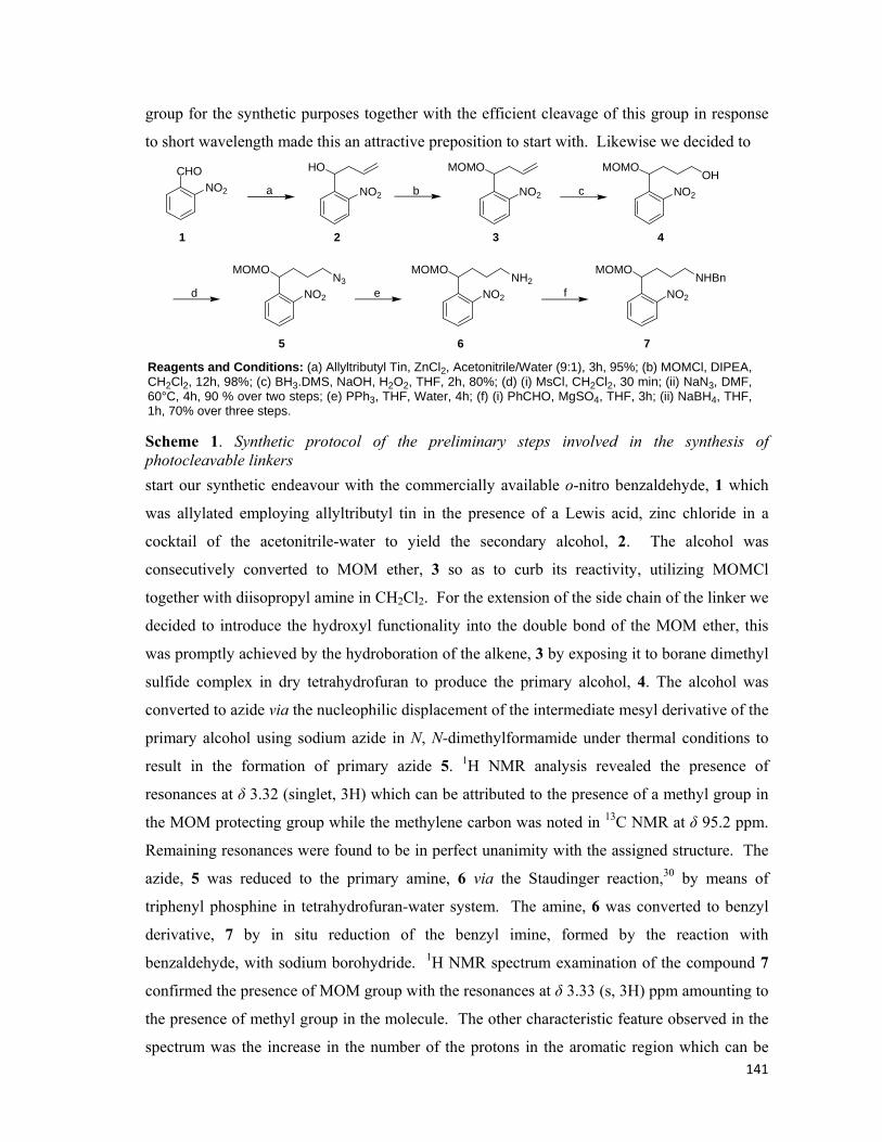
141
group for the synthetic purposes together with the efficient cleavage of this group in response
to short wavelength made this an attractive preposition to start with. Likewise we decided to
d e f
CHONO2 NO2
HO
NO2
MOMO
NO2
MOMOOH
a b c
NO2
MOMON3
NO2
MOMONH2
NO2
MOMONHBn
1 2 3 4
5 6 7
Reagents and Conditions: (a) Allyltributyl Tin, ZnCl2, Acetonitrile/Water (9:1), 3h, 95%; (b) MOMCl, DIPEA, CH2Cl2, 12h, 98%; (c) BH3.DMS, NaOH, H2O2, THF, 2h, 80%; (d) (i) MsCl, CH2Cl2, 30 min; (ii) NaN3, DMF,60°C, 4h, 90 % over two steps; (e) PPh3, THF, Water, 4h; (f) (i) PhCHO, MgSO4, THF, 3h; (ii) NaBH4, THF,1h, 70% over three steps. Scheme 1. Synthetic protocol of the preliminary steps involved in the synthesis of photocleavable linkers
start our synthetic endeavour with the commercially available o-nitro benzaldehyde, 1 which
was allylated employing allyltributyl tin in the presence of a Lewis acid, zinc chloride in a
cocktail of the acetonitrile-water to yield the secondary alcohol, 2. The alcohol was
consecutively converted to MOM ether, 3 so as to curb its reactivity, utilizing MOMCl
together with diisopropyl amine in CH2Cl2. For the extension of the side chain of the linker we
decided to introduce the hydroxyl functionality into the double bond of the MOM ether, this
was promptly achieved by the hydroboration of the alkene, 3 by exposing it to borane dimethyl
sulfide complex in dry tetrahydrofuran to produce the primary alcohol, 4. The alcohol was
converted to azide via the nucleophilic displacement of the intermediate mesyl derivative of the
primary alcohol using sodium azide in N, N-dimethylformamide under thermal conditions to
result in the formation of primary azide 5. 1H NMR analysis revealed the presence of
resonances at δ 3.32 (singlet, 3H) which can be attributed to the presence of a methyl group in
the MOM protecting group while the methylene carbon was noted in 13C NMR at δ 95.2 ppm.
Remaining resonances were found to be in perfect unanimity with the assigned structure. The
azide, 5 was reduced to the primary amine, 6 via the Staudinger reaction,30 by means of
triphenyl phosphine in tetrahydrofuran-water system. The amine, 6 was converted to benzyl
derivative, 7 by in situ reduction of the benzyl imine, formed by the reaction with
benzaldehyde, with sodium borohydride. 1H NMR spectrum examination of the compound 7
confirmed the presence of MOM group with the resonances at δ 3.33 (s, 3H) ppm amounting to
the presence of methyl group in the molecule. The other characteristic feature observed in the
spectrum was the increase in the number of the protons in the aromatic region which can be

142
attributed to the existence of the benzyl functionality present in the secondary amine, 7.
Moreover, the presence of methylene carbon at δ 94.3 ppm further confirmed the assigned
structure of the compound 7. The synthetic steps involved are displayed in Scheme 1 along
with the reagents and conditions employed.
Meanwhile, the functional side chain for the linkers was prepared. Commercially
available 6-bromo hexanol, 8 was heated with sodium azide in DMF to result in the azido
alcohol, 9. The alcohol was subsequently converted to the corresponding 6-azido-1-hexynoic
acid, 10 under the oxidizing conditions provided by the Jones reagent. The schematic display
of the reagents and conditions involved are shown in Scheme 2.
HOBr
HON3 HO2C N3
NaN3,DMF, 60oC
Jones Reagent,Acetone, 0o-r.t.
8 9 10 Scheme 2. Synthetic scheme for the azido side chain
Likewise, the alkyne functionalized side chain was prepared employing the
commercially available 6-hexynol, 11 which was thereafter converted to 5-hexynoic acid, 12
by oxidation of alcohol with Jones reagent. The reaction conditions are displayed in Scheme 3.
The compound was characterized by 1H and 13 C NMR spectra and the resonances were found
to be in accordance with the reported values.31
HO HO2CJones Reagent,Acetone, 0o-r.t.
11 12 Scheme 3. Synthetic scheme for the alkyne side chain
The acid functionality at one of the side chains, 10 and 12 would enable us to conjugate
them onto the amine, 7 thereby extending the alkyl chain. The extension of the side chain is
necessary because when appended onto the proteins, the azide/alkyne part of the alkyl chain
should protrude out from the bulky structure of the protein so as to be freely available for
modification. In continuation with our synthetic efforts, the alkyne derived hetro-bifunctional
linker, 17 was synthesized. The free secondary amine, 7 previously prepared was joined with
the alkyne side chain with the coupling conditions offered by N, N-diisopropyl carbodimide in
CH2Cl2 to grant us the amide, 13. Now having completed one part of the linker which seemed
suitable for the click modification on the proteins, we continued in our task to introduce the
proper functionalities on the linker which would enable us to chemically attach on the protein
surface. For the purpose, the MOM ether on 13 was cleaved under acidic environment (HCl,
Methanol) to afford the free secondary alcohol, 14 which was alkylated with bromoethylacetate
facilitated by sodium hydride in DMF to afford the ester, 15. Hydrolysis of the ester, 15 with
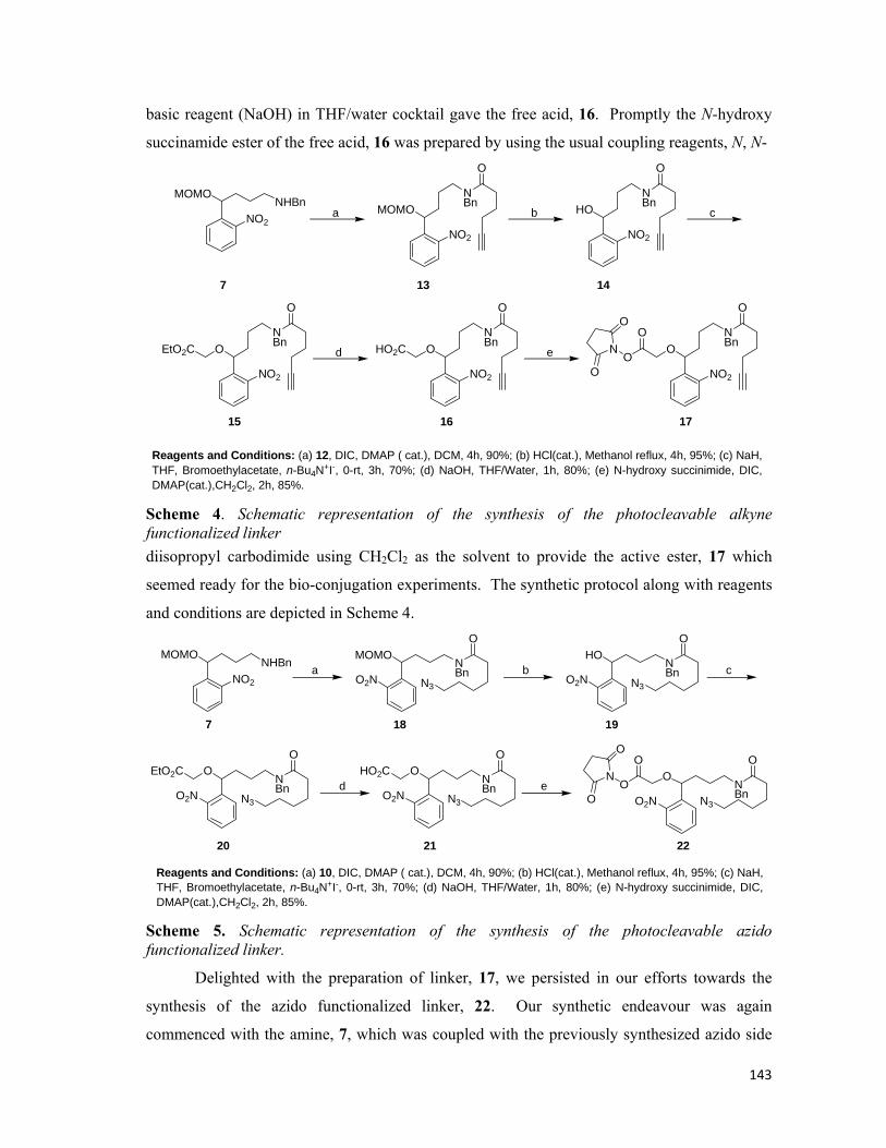
143
basic reagent (NaOH) in THF/water cocktail gave the free acid, 16. Promptly the N-hydroxy
succinamide ester of the free acid, 16 was prepared by using the usual coupling reagents, N, N-
NO2
ONBn
O
HO2C
NO2
ONBn
O
EtO2C
NO2
HONBn
O
NO2
MOMONBn
O
NO2
ONBn
O
O
ON
O
O
Reagents and Conditions: (a) 12, DIC, DMAP ( cat.), DCM, 4h, 90%; (b) HCl(cat.), Methanol reflux, 4h, 95%; (c) NaH, THF, Bromoethylacetate, n-Bu4N+I-, 0-rt, 3h, 70%; (d) NaOH, THF/Water, 1h, 80%; (e) N-hydroxy succinimide, DIC,DMAP(cat.),CH2Cl2, 2h, 85%.
b c
d
NO2
MOMONHBn
7
a
e
13 14
15 16 17
Scheme 4. Schematic representation of the synthesis of the photocleavable alkyne functionalized linker
diisopropyl carbodimide using CH2Cl2 as the solvent to provide the active ester, 17 which
seemed ready for the bio-conjugation experiments. The synthetic protocol along with reagents
and conditions are depicted in Scheme 4.
Reagents and Conditions: (a) 10, DIC, DMAP ( cat.), DCM, 4h, 90%; (b) HCl(cat.), Methanol reflux, 4h, 95%; (c) NaH, THF, Bromoethylacetate, n-Bu4N+I-, 0-rt, 3h, 70%; (d) NaOH, THF/Water, 1h, 80%; (e) N-hydroxy succinimide, DIC,DMAP(cat.),CH2Cl2, 2h, 85%.
NO2
MOMONHBn
7
O2N
MOMONBn
O
N3O2N
HONBn
O
N3
O2N
ONBn
O
N3
EtO2C
O2N
ONBn
O
N3
HO2C
O2N
ONBn
O
N3
O
ON
O
O
a b c
d e
18 19
20 21 22
Scheme 5. Schematic representation of the synthesis of the photocleavable azido functionalized linker.
Delighted with the preparation of linker, 17, we persisted in our efforts towards the
synthesis of the azido functionalized linker, 22. Our synthetic endeavour was again
commenced with the amine, 7, which was coupled with the previously synthesized azido side
144
chain, 10 with the coupling conditions afforded by N, N-diisopropyl amine in CH2Cl2 to
provide the amide, 18. Following the same synthetic protocol as standardized in the Scheme 4,
the secondary alcohol, 19 was obtained after the deprotection of the MOM ether in 18 in acidic
environment employing HCl in methanol. Likewise we then progressed with the alkylation of
the hydroxyl functionality with bromoethylacetate in sodium hydride using DMF as the solvent
to result in the product, 20. It was subsequently hydrolysed to afford the free acid, 21 and
converted to the active succinamide ester, 22 following the same protocol as standardized
previously (Scheme 5).
We moved on to investigate the utility of the linkers for the bioconjugation, with both
the functionalised clickable linkers in hand. For the purpose readily available Bovine Serum
Albumin (BSA) was chosen for our experiments due to its numerous biological applications
including ELISA (Enzyme-Linked Immunosorbent Assay), used as additive for hybridoma cell
cultures etc. Additionally, it is also used as a nutrient in cell and microbial culture. One of the
main advantages for preferring BSA over the others was due to its stability as compared to the
other proteins.
BSA is known to have several free amines in its tertiary structure. We choose to
covalently link only a few of them in our experiments, firstly it will be impossible to react all
the free amines and secondly, it will be tiresome job to cleave all the linked amines after the
click reaction. Proceeding with our conjugation experiments, BSA was taken in 0.1 M
phosphate buffer (pH 7.2) and allowed to stir with the NHS-functionalized linker, 17 for 2 h.
Assuming the completion of the experiment, the functionalized protein was dialysed overnight
employing the cellulose membrane (12, 000kD cut off) so as to remove the excess linker
present in the protein mixture. The covalently modified BSA sample was then analyzed with
MALDI-TOF. It was heartwarming to observe that there was an increase in mass of the
protein sample. The natural BSA showed a mass of 66,682 kDa while the alkyne modified
BSA revealed a mass of 68,084 kDa clearly indicating the presence of functionalized linker on
the peptide backbone.
Delighted in having succeeded with initial bioconjugation experiments, we then
proceeded to check the feasibility of the azido functionalized linker as well. Following the
same protocol, the azido linker, 22 was engaged instead of 17. Gratifyingly, upon the analysis
with MALDI-TOF, the BSA modified with azido linker displayed a mass of 67,970 kDa as
compared to the normal mass of BSA, 69,568 kDa, signalling towards the presence of azido
linker on the protein surfaces.

145
We further decided to check the generality of the synthesized linkers, 17 and 22, for the
other proteins. For the purpose, Insulin was chosen as another protein to be engaged in our
bioconjugation experiments. Insulin is an animal hormone whose presence in body makes
liver and muscle cells to regulate the blood glucose levels. As compared to BSA, it is a small
protein with molecular weight of around 5800 kDa. The utility of the linkers was probed by
employing both the photocleavable linkers, 17 and 22, for biological studies. Following the
standardized protocol, the Insulin was made to undergo through the same conditions as
established for BSA. It was pleasant to notice the modification of the Insulin samples as was
evident through the MALDI-TOF analysis of protein samples. The azido- and alkyne-modified
Insulin samples revealed the mass of 6238 kDa and 6187 kDa in their respective mass analysis
as compared to the normal mass of the Insulin (5800 kDa).
To conclude, we have successfully synthesized the two photocleavable linkers having
the azide and alkyne appendages and also exploited them to covalently link onto BSA and
Insulin. Furthermore, these linkers can be employed for linking small molecules, such as drugs
or fluorescence tags on to the proteins. On the other hand, the linkers can also be exploited for
protein-protein bioconjugation to form homo- and hetro-dimeric conjugates to assist in the
studies regarding the behaviour of proteins in promiscuity of the other proteins.
*****

146
Chapter 3: Experimental Section
Synthesis of 1-(2-nitrophenyl)but-3-en-1-ol: Commercially available o-
nitro benzaldehyde, 1 (151 mg, 1 mmol) was dissolved in a cocktail of 10
mL acetonitrile:water (9:1), to this was added allyltributyl tin (306 μL, 1
mmol) along with zinc chloride (136 mg, 1 mmol) was allowed to stir at
ambient temperature for 3 h. After the completion of the reaction, as indicated by the
consumption of the starting material on TLC examination, the reaction was subsequently
filtered through a pad of celite so as to remove insoluble impurities. The filtrate was diluted
with water and extracted with CH2Cl2 (3X), the combined extracts were dried over anhydrous
sodium sulphate and consequently the solvent was removed in vacuo to yield crude compound
2 which was purified via silica gel chromatography (Ethyl Acetate-Petroleum Benzine; 1:3) to
afford the pure allylated product 2 in 95% yield (182 mg). 1H NMR (200 MHz, CDCl3): δ
2.34-2.5 (m, 2H), 2.64-2.77 (m, 1H), 5.14-5.17 (m, 1H), 5.21-5.25 (m, 1H), 5.31 (dd, 1H, J =
3.79, 8.39 Hz), 5.79-6.00 (m, 1H), 7.38-7.47 (m, 1H), 7.60-7.69 (m, 1H), 7.82 (dd, 1H, J =
1.49, 7.84 Hz), 7.92 (dd, 1H, J = 1.29, 8.10 Hz). 13C NMR (50 MHz, CDCl3): δ 42.8, 68.4,
119.0, 124.4, 128.1, 128.1, 133.4, 134.0, 139.2, 147.8. Anal Calcd. for C10H11NO3: C, 62.17;
H, 5.74; N, 7.25. Found C, 62.22; H, 5.64; N, 7.33.
Synthesis of 1-(1-(methoxy-O-methyl)-but-3-enyl-2-nitrobenzene: To
an ice cooled solution of 2 (192 mg, 1 mmol) and diisopropylethyl amine
(347 μL, 2 mmol) in dichloromethane (5 mL) was added MOMCl (146
μL, 2 mmol) over a period of 5 min. The reaction was stirred at room
temperature for 12 h. TLC examination of the reaction indicated the formation of the
compound as a single compound, after which it was diluted with water and extracted with
dichloromethane (2X); the combined extracts were evaporated under reduced pressure before
which they were dried over anhydrous sodium sulphate, to provide the crude MOM ether, 3. It
was subsequently purified over silica gel column chromatography (Ethyl Acetate-Petroleum
Ether; 1:9) to afford the pure compound 3 in 98% yield (231 mg). 1H NMR (200 MHz,
CDCl3): δ 2.46-2.65 (m, 2H), 3.31 (s, 3H), 4.52 (ABq, 2H, J = 14.74 Hz), 5.07-5.17 (m, 2H),
5.27-5.33 (m, 1H), 5.82-6.03 (m, 1H), 7.38-7.47 (m, 1H), 7.60-7.68 (m, 1H), 7.74-7.79 (m,1H),
7.93 (dd, 1H, J = 1.27, 8.22 Hz). 13C NMR (50 MHz, CDCl3): δ 41.9, 55.8, 73.2, 95.2, 117.7,
NO2
HO
NO2
MOMO
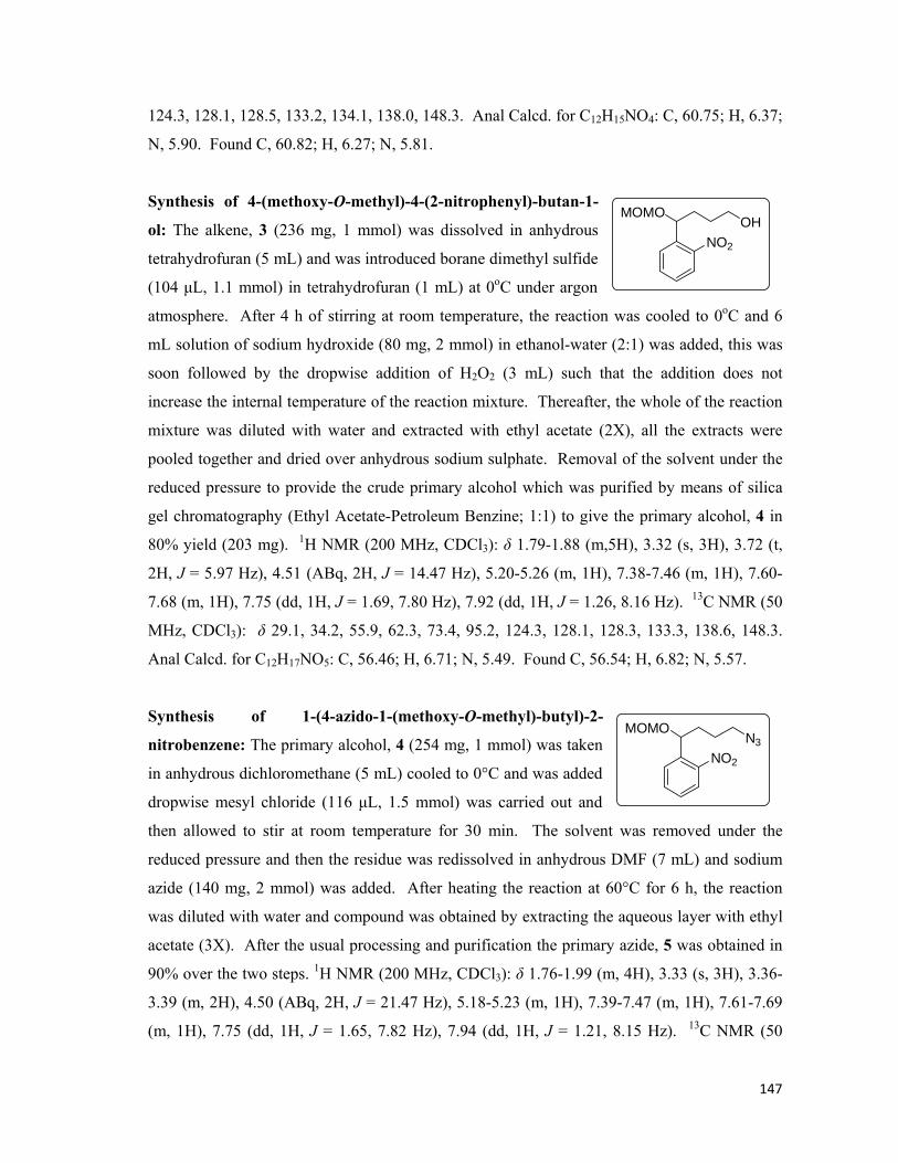
147
124.3, 128.1, 128.5, 133.2, 134.1, 138.0, 148.3. Anal Calcd. for C12H15NO4: C, 60.75; H, 6.37;
N, 5.90. Found C, 60.82; H, 6.27; N, 5.81.
Synthesis of 4-(methoxy-O-methyl)-4-(2-nitrophenyl)-butan-1-
ol: The alkene, 3 (236 mg, 1 mmol) was dissolved in anhydrous
tetrahydrofuran (5 mL) and was introduced borane dimethyl sulfide
(104 μL, 1.1 mmol) in tetrahydrofuran (1 mL) at 0oC under argon
atmosphere. After 4 h of stirring at room temperature, the reaction was cooled to 0oC and 6
mL solution of sodium hydroxide (80 mg, 2 mmol) in ethanol-water (2:1) was added, this was
soon followed by the dropwise addition of H2O2 (3 mL) such that the addition does not
increase the internal temperature of the reaction mixture. Thereafter, the whole of the reaction
mixture was diluted with water and extracted with ethyl acetate (2X), all the extracts were
pooled together and dried over anhydrous sodium sulphate. Removal of the solvent under the
reduced pressure to provide the crude primary alcohol which was purified by means of silica
gel chromatography (Ethyl Acetate-Petroleum Benzine; 1:1) to give the primary alcohol, 4 in
80% yield (203 mg). 1H NMR (200 MHz, CDCl3): δ 1.79-1.88 (m,5H), 3.32 (s, 3H), 3.72 (t,
2H, J = 5.97 Hz), 4.51 (ABq, 2H, J = 14.47 Hz), 5.20-5.26 (m, 1H), 7.38-7.46 (m, 1H), 7.60-
7.68 (m, 1H), 7.75 (dd, 1H, J = 1.69, 7.80 Hz), 7.92 (dd, 1H, J = 1.26, 8.16 Hz). 13C NMR (50
MHz, CDCl3): δ 29.1, 34.2, 55.9, 62.3, 73.4, 95.2, 124.3, 128.1, 128.3, 133.3, 138.6, 148.3.
Anal Calcd. for C12H17NO5: C, 56.46; H, 6.71; N, 5.49. Found C, 56.54; H, 6.82; N, 5.57.
Synthesis of 1-(4-azido-1-(methoxy-O-methyl)-butyl)-2-
nitrobenzene: The primary alcohol, 4 (254 mg, 1 mmol) was taken
in anhydrous dichloromethane (5 mL) cooled to 0°C and was added
dropwise mesyl chloride (116 μL, 1.5 mmol) was carried out and
then allowed to stir at room temperature for 30 min. The solvent was removed under the
reduced pressure and then the residue was redissolved in anhydrous DMF (7 mL) and sodium
azide (140 mg, 2 mmol) was added. After heating the reaction at 60°C for 6 h, the reaction
was diluted with water and compound was obtained by extracting the aqueous layer with ethyl
acetate (3X). After the usual processing and purification the primary azide, 5 was obtained in
90% over the two steps. 1H NMR (200 MHz, CDCl3): δ 1.76-1.99 (m, 4H), 3.33 (s, 3H), 3.36-
3.39 (m, 2H), 4.50 (ABq, 2H, J = 21.47 Hz), 5.18-5.23 (m, 1H), 7.39-7.47 (m, 1H), 7.61-7.69
(m, 1H), 7.75 (dd, 1H, J = 1.65, 7.82 Hz), 7.94 (dd, 1H, J = 1.21, 8.15 Hz). 13C NMR (50
NO2
MOMOOH
NO2
MOMON3
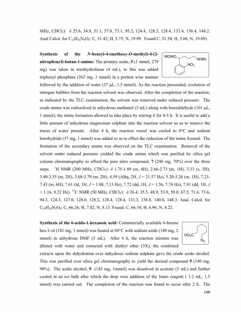
148
MHz, CDCl3): δ 25.6, 34.9, 51.1, 57.0, 73.1, 95.2, 124.4, 128.3, 128.4, 133.4, 138.4, 148.2.
Anal Calcd. for C12H16N4O4: C, 51.42; H, 5.75; N, 19.99. Found C, 51.54; H, 5.66, N, 19.89).
Synthesis of the N-benzyl-4-(methoxy-O-methyl)-4-(2-
nitrophenyl)-butan-1-amine: The primary azide, 5 (1 mmol, 279
mg) was taken in tetrahydrofuran (4 mL), to this was added
triphenyl phosphine (262 mg, 1 mmol) in a portion wise manner
followed by the addition of water (27 μL, 1.5 mmol). As the reaction proceeded, evolution of
nitrogen bubbles from the reaction solvent was observed. After the completion of the reaction,
as indicated by the TLC examination, the solvent was removed under reduced pressure. The
crude amine was redissolved in anhydrous methanol (5 mL) along with benzaldehyde (101 μL,
1 mmol), the imine formation allowed to take place by stirring it for 4-5 h. It is useful to add a
little amount of anhydrous magnesium sulphate into the reaction solvent so as to remove the
traces of water present. After 4 h, the reaction vessel was cooled to 0°C and sodium
borohydride (37 mg, 1 mmol) was added so as to effect the reduction of the imine formed. The
formation of the secondary amine was observed on the TLC examination. Removal of the
solvent under reduced pressure yielded the crude amine which was purified by silica gel
column chromatography to afford the pure nitro compound, 7 (240 mg, 70%) over the three
steps. 1H NMR (200 MHz, CDCl3): δ 1.75-1.89 (m, 4H), 2.66-2.73 (m, 1H), 3.33 (s, 3H),
3.40-3.55 (m, 2H), 3.68-3.79 (m, 2H), 4.59 (ABq, 2H, J = 21.57 Hz), 5.20-5.26 (m, 1H), 7.23-
7.43 (m, 6H), 7.61 (td, 1H, J = 1.08, 7.13 Hz), 7.72 (dd, 1H, J = 1.56, 7.76 Hz), 7.91 (dd, 1H, J
= 1.16, 8.22 Hz). 13C NMR (50 MHz, CDCl3): δ 26.4, 35.5, 48.9, 53.9, 59.0, 67.5, 71.6, 73.6,
94.3, 124.3, 127.0, 128.0, 128.2, 128.4, 128.4, 133.3, 138.8, 140.0, 148.3. Anal. Calcd. for
C19H24N2O4: C, 66.26; H, 7.02; N, 8.13. Found: C, 66.34; H, 6.96; N, 8.22.
Synthesis of the 6-azido-1-hexanoic acid: Commercially available 6-bromo
hex-1-ol (181 mg, 1 mmol) was heated at 60°C with sodium azide (140 mg, 2
mmol) in anhydrous DMF (5 mL). After 6 h, the reaction mixture was
diluted with water and extracted with diethyl ether (3X), the combined
extracts upon the dehydration over anhydrous sodium sulphate gave the crude azido alcohol.
This was purified over silica gel chromatography to yield the desired compound 9 (140 mg,
98%). The azido alcohol, 9 (143 mg, 1mmol) was dissolved in acetone (5 mL) and further
cooled in an ice bath after which the drop wise addition of the Jones reagent ( 1.2 mL, 1.5
mmol) was carried out. The completion of the reaction was found to occur after 2 h. The
NO2
MOMONHBn
HO2CN3

149
reaction mixture was diluted with water and the consequently extracted with diethyl ether (3X)
to yield the crude azido acid. Further the purification of the crude acid afforded the pure side
chain, 10 (125 mg, 80%). Whole spectrum features matched with those of the reported
values.31
Synthesis of N-benzyl-N-(4-(methoxy-O-methyl)-4-(2-
nitrophenyl)-butyl)-hex-5-ynamine: The free amine, 7 (343 mg,
1mmol), was dissolved in anhydrous CH2Cl2 (5 mL) along with 5-
hexynoic acid (112 mg, 1 mmol) prepared vide supra and cooled to
0°C in an ice-bath. N, N-diisopropyl carbodimide (311 μL, 1.2
mmol) was introduced into the reaction mixture so as to effect the
coupling of the two molecules, the reaction mixture was allowed to stir at room temperature
until the completion. After the completion of the reaction, the solvent was removed under the
reduced pressure and the crude residue was purified by silica gel column chromatography
(Ethyl Acetate-Petroleum Benzine; 2:3) to furnish desired amide 13 (393 mg, 90%). 1H NMR
(200 MHz, CDCl3): δ 1.71-1.99 (m, 8H), 2.19-2.35 (m, 2H), 2.43-2.59 (m, 2H), 3.27 (s, 3H),
3.43-3.44 (m, 1H), 4.47 (ABq, 2H, J = 20.97 Hz), 4.59 (d, 2H, J = 9.73 Hz), 5.13 (m, 1H),
7.15-7.48 (m, 6H), 7.55-7.73 (m, 2H), 7.88-7.97 (m, 1H). 13C NMR (50 MHz, CDCl3): δ 17.8,
23.9, 25.3, 31.6, 35.1, 45.7, 46.8, 48.2, 50.8, 55.9, 69.1, 73.5, 83.8, 95.2, 124.5, 126.2-128.5,
133.5, 136.9, 137.7, 138.3, 148.2, 172.7. Anal Calcd. for C25H30N2O5: C, 68.47; H, 6.90; N,
6.39. Found C, 68.58; H, 6.97; N, 6.43.
Synthesis of N-benzyl-N-(4-hydroxy-4-(2-nitrophenyl) butyl) hex-5-
ynamide: The amide, 13 (437 mg, 1 mmol) was refluxed in methanol
(5 mL) in the presence of HCl (cat.) until the completion of the
reaction as evident by TLC examination. Thereafter, the reaction was
diluted with water and extracted with ethyl acetate (3X) which were
pooled together and dried over anhydrous sodium sulphate. Solvent
removal under reduced pressure and subsequent purification over the silica gel column yielded
the free alcohol, 14 (372 mg, 95%). 1H NMR (200 MHz, CDCl3): δ 1.70-1.97 (m, 6H), 2.20-
2.32 (m, 2H), 2.45-2.57 (m, 2H), 2.85 (d, 1H, J = 3.88 Hz), 3.21-3.31 (m , 1H), 3.66-3.80 (m,
1H), 4.10-4.14 (m, 1H), 4.56-4.61 (m, 2H), 5.21-5.31 (m, 1H), 7.15-7.46 (m, 6H), 7.58-7.68
(m, 1H), 7.77-7.94 (m, 2H). 13C NMR (50 MHz, CDCl3): δ 17.8, 23.8, 24.6, 31.6, 35.0, 45.7,
NO2
MOMONBn
O
NO2
HONBn
O

150
48.2, 51.1, 69.1, 83.6, 124.2, 126.2-128.9, 133.5, 136.5, 140.3, 140.9, 147.4, 173.2. Anal
Calcd. for C23H26N2O4: C, 70.03; H, 6.64; N, 7.10. Found C, 70.12; H, 6.75; N, 7.19.
Synthesis of compound 16: To an ice-cooled solution of
alcohol, 14 (392 mg, 1mmol) in anhydrous DMF (5 mL) was
added sodium hydride (44 mg, 1.1 mmol, 60% oil suspension)
and stirred for 1 h at room temperature. Bromoethylacetate
(220 μL, 2 mmol) was introduced dropwise at 0°C and stirred
at room temperature for 1 h. The resulting suspension was
quenched with saturated ammonium chloride and extracted with diethyl ether (3X). The
combined extracts were dried over anhydrous sodium sulphate and the solvent was removed in
vacuo and the resulting residue was directly used for the next step for hydrolysis of the ethyl
ester. For the purpose, the alkylated product, 15 (478 mg, 1 mmol) was dissolved in a mixture
of THF/water (1:1, 5 mL) was added sodium hydroxide solution (1 M, 2 mL) and allowed to
stir until the completion of the reaction, 5 h. After the completion of the reaction, the reaction
mixture was diluted with water and extracted with ethyl acetate (3X) to offer the crude acid as
a syrup after the evaporation of the solvent under reduced pressure. Purification achieved via
the silica gel column chromatography to afford the pure acid, 16 (384 mg, 75% over the two
steps). 1H NMR (200 MHz, CDCl3): δ 0.83-0.88 (m, 1H), 1.14-1.30 (m, 3H), 1.79-1.95 (m,
7H), 2.22-2.25 (m, 2H), 2.48-2.61 (m, 2H), 3.27-3.48 (m, 1H), 3.37-4.14 (m, 2H), 4.58-4.63
(m, 2H), 5.03-5.06 (m, 1H), 6.03 (b, 1H), 7.15-7.50 (m, 6H), 7.60-7.87 (m, 2H), 7.91-7.99 (m,
1H). 13C NMR (50 MHz, CDCl3): δ 14.1, 17.8, 23.2, 23.8, 25.0, 31.3, 34.8, 42.5, 45.7, 46.9,
48.2, 50.9, 60.4, 66.1, 69.0, 83.8, 124.7, 126.2, 127.9-128.9, 133.8, 136.6, 137.6, 148.5, 172.6,
173.2. Anal. Calcd for C29H31N3O8: C, 63.38; H, 5.69; N, 7.65. Found C, 63.47; H, 5.76; N,
7.73.
Synthesis of the compound 17: The free acid, 16 (513
mg, 1 mmol) was stirred along with N-hydroxy
succinamide (230 mg, 1.2 mmol) in anhydrous CH2Cl2 (5
mL). N, N-diisopropyl carbodimide (186 μL, 1.2 mmol)
was introduced into the reaction mixture at 0°C and the
resulting mixture was made to stir at room temperature for
2 h. After the completion of the reaction, any attempts to further purify the active ester were
foiled by the degradation of the compound inside the silica gel column. Hereafter, the reaction
NO2
ONBn
O
HO2C
NO2
ONBn
O
O
ON
O
O

151
was then cooled at 0°C so as to precipitate the insoluble unwanted side product (urea) and the
resulting upper layer was carefully decanted to obtain the alkyne functionalized linker which
was found to be pure enough to be utilized for the biological experiments.
Synthesis of 6-azido-N-benzyl-N-(4-(methoxy-O-methyl)-4-(2-
nitrophenyl) butyl) hexanamide: The synthetic protocol was
same as followed for the compound 13). 1H NMR (200 MHz,
CDCl3): δ 1.52-1.78 (m, 10H), 2.28-2.45 (m, 2H), 3.19-3.32 (m,
6H), 3.44 (m, 1H), 4.40-4.62 (m, 4H), 5.13 (m, 1H), 7.14-7.48
(m, 6H), 7.57-7.74 (m, 2H), 7.88-7.97 (m, 1H). 13C NMR (50 MHz, CDCl3): δ 23.2- 26.5,
28.9, 32.8, 35.1, 42.0, 45.7, 46.7, 48.1, 51.1, 55.8, 73.5, 95.2, 124.2, 124.4, 126.0, 127.8-128.8,
133.3, 136.9, 137.7, 138.2, 148.1, 172.7. Anal Calcd. for C25H33N5O5: C, 62.10; H, 6.88; N,
14.48. Found C, 62.19; H, 6.75; N, 14.56.
Synthesis of 6-azido-N-benzyl-N-(4-hydroxy-4-(2-
nitrophenyl)-butyl) hexanamide: Synthetic procedure same as
followed for the synthesis of the compound 14. 1H NMR (200
MHz, CDCl3): δ 1.18-1.81 (m, 10H), 2.29-2.42 (m, 2H), 3.19-
3.31 (m, 3H), 3.66-3.76 (m, 1H), 4.10-4.13 (m, 1H), 4.54 (d, 1H,
J = 1.90 Hz), 4.58 (d, 1H, J = 3.93 Hz), 5.26 (t, 1H, J = 8.94 Hz), 7.13-7.46 (m, 6H), 7.58-7.68
(m, 1H), 7.77-7.94 (m, 2H). 13C NMR (50 MHz, CDCl3): δ 24.8, 26.4, 28.7, 32.9, 33.1, 35.0,
45.8, 46.8, 51.1, 51.3, 68.9, 69.3, 124.3, 126.1-129.0, 133.5, 136.7, 141.0, 147.5, 173.8. Anal
Calcd. for C23H29N5O4: C, 62.85; H, 6.65; N, 15.93. Found C, 62.93; H, 6.76; N, 15.86.
Synthesis of 21: Synthetic procedure same as followed for
the compound 16. 1H NMR (200 MHz, CDCl3): δ 1.22-1.77
(m, 10H), 2.29-2.47 (m, 2H), 3.16-3.30 (m, 3H), 3.58-4.18
(m, 3H), 4.54-4.62 (m, 2H), 5.04 (m, 1H), 6.68 (b, 1H),
7.12-7.45 (m, 6H), 7.59-7.72 (m, 2H), 7.90-7.98 (m, 1H). 13C NMR (50 MHz, CDCl3): δ 23.9, 24.7, 25.0, 26.3, 28.5, 32.7, 33.0, 35.0, 46.0, 46.9, 48.2,
51.1, 51.2, 124.5, 126.1, 127.9-128.9, 133.9, 136.5, 137.6, 148.4, 173.4. Anal. Calcd. for
C25H31N5O6: C, 60.35; H, 6.28; N, 14.08. Found: C, 60.46; H, 6.35; N, 14.17.
Synthesis of 22: Synthetic procedure same as followed for the compound 17.
O2N
MOMONBn
O
N3
O2N
HONBn
O
N3
O2N
ONBn
O
N3
HO2C
152
Stock solution of BSA/Insulin: A 4 mg/mL stock solution of the protein was prepared.
General procedure for the bioconjugation of the linkers on the protein: 1 mg (10 mmol)
of the functionalized linker was dissolved in DMSO (200 μL) this was added to the stock
solution of the protein (800 μL), which was allowed to rotate on a rotary shaker for 2 hr. The
total concentration of the DMSO in the reaction was maintained as such that it doesn’t exceed
more than 20 percent of the total volume at any time. The protein samples were then dialysed
overnight in 0.1 M tris-HCl buffer (pH 8.1), with the buffer being replaced three times at
regular intervals. Thereafter, the protein samples were poured into an eppendroff and directly
utilized for MALDI-TOF analysis.
*****
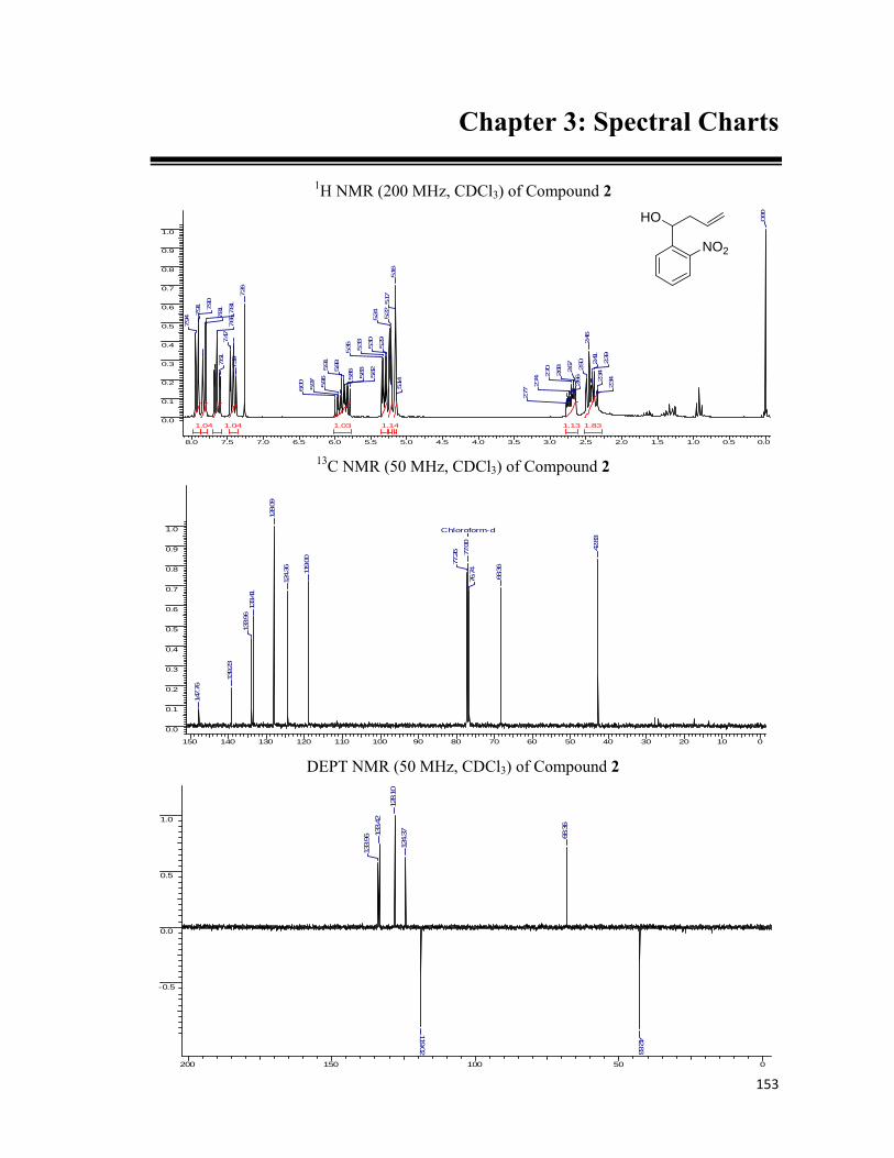
153
Chapter 3: Spectral Charts
1H NMR (200 MHz, CDCl3) of Compound 2
8.0 7.5 7.0 6.5 6.0 5.5 5.0 4.5 4.0 3.5 3.0 2.5 2.0 1.5 1.0 0.5 0.0
0.0
0.1
0.2
0.3
0.4
0.5
0.6
0.7
0.8
0.9
1.0
1.831.14 1.131.04 1.04 1.03
0.00
2.342.
342.39
2.41
2.46
2.50
2.66
2.67
2.68
2.70
2.74
2.77
5.14
5.16
5.17
5.22
5.24
5.29
5.30
5.33
5.35
5.82
5.83
5.86
5.885.91
5.96
5.97
6.00
7.26
7.39
7.47
7.61
7.65
7.81
7.81
7.90
7.91
7.94
13C NMR (50 MHz, CDCl3) of Compound 2
150 140 130 120 110 100 90 80 70 60 50 40 30 20 10 0
0.0
0.1
0.2
0.3
0.4
0.5
0.6
0.7
0.8
0.9
1.0 Chloroform-d
42.83
68.36
76.74
77.00
77.26
119.00
124.36
128.09
133.41
133.96
139.23
147.76
DEPT NMR (50 MHz, CDCl3) of Compound 2
200 150 100 50 0
-0.5
0.0
0.5
1.0
42.83
68.36
119.02
124.37
128.10
133.42
133.96
NO2
HO

154
1H NMR (200 MHz, CDCl3) of Compound 3
8.0 7.5 7.0 6.5 6.0 5.5 5.0 4.5 4.0 3.5 3.0 2.5 2.0 1.5 1.0 0.5 0.0
0.00
0.05
0.10
0.15
0.20
0.25
0.30
0.35
0.40
0.45
0.50
3.09 2.162.09 1.061.04 1.031.02
0.00
2.46
2.54
2.572.
58
2.58
2.592.60
2.61
2.65
3.31
4.46
4.49
4.56
4.60
5.07
5.16
5.17
5.29
5.31
5.33
5.82
5.91
6.037.
38
7.39
7.42
7.60
7.64
7.64
7.74
7.75
7.91
7.95
7.96
13C NMR (50 MHz, CDCl3) of compound 3
150 140 130 120 110 100 90 80 70 60 50 40 30 20 10 0
0.0
0.1
0.2
0.3
0.4
0.5
0.6
0.7
0.8
0.9
1.0
Chloroform-d
41.9
1
55.76
73.2
1
76.37
77.0
077
.63
95.15
117.
71
124.27
128.08
128.51
133.
1713
4.10
138.01
148.
30
DEPT NMR (50 MHz, CDCl3) of Compound 3
200 150 100 50 0
-1.0
-0.5
0.0
0.5
41.94
55.78
73.22
95.15
117.74
124.29
128.10
128.51133.20
134.12
NO2
MOMO

155
1H NMR (200 MHz, CDCl3) of Compound 4
8.0 7.5 7.0 6.5 6.0 5.5 5.0 4.5 4.0 3.5 3.0 2.5 2.0 1.5 1.0 0.5 0.0
0.00
0.05
0.10
0.15
0.20
0.25
0.30
0.35
0.40
0.45
0.50
5.093.162.101.071.05 1.04 1.02
0.00
1.79
1.80
1.82
1.84
1.86
1.88
3.32
3.69
3.72
3.75
4.45
4.48
4.55
4.58
5.20
5.26
7.28
7.647.
73
7.74
7.90
7.91
7.94
13C NMR (50 MHz, CDCl3) of Compound 4
150 140 130 120 110 100 90 80 70 60 50 40 30 20 10 0
0.0
0.1
0.2
0.3
0.4
0.5
0.6
0.7
0.8
0.9
1.0
Chloroform-d
29.1
2
34.1
6
55.9
3
62.3
2
73.3
676
.36
77.0
077
.63
95.2
4
124.
3112
8.07
128.
31
133.
30
138.
56
148.
26
DEPT NMR (50 MHz, CDCl3) of Compound 4
200 150 100 50 0
-1.0
-0.5
0.0
0.5
29.19
34.24
56.02
61.89
62.1062.39
62.74
63.17
63.40
73.42
95.3012
4.40
128.15128.38
133.39
NO2
MOMOOH
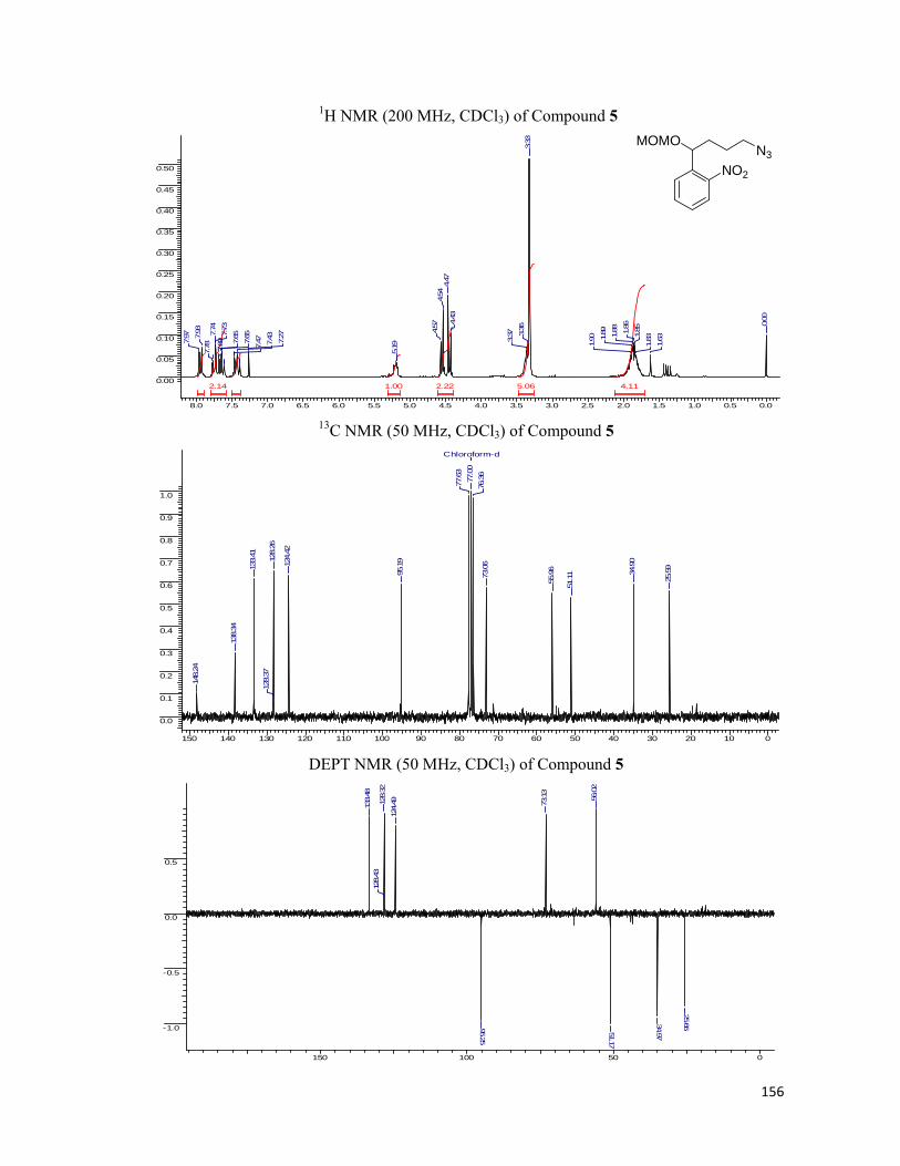
156
1H NMR (200 MHz, CDCl3) of Compound 5
8.0 7.5 7.0 6.5 6.0 5.5 5.0 4.5 4.0 3.5 3.0 2.5 2.0 1.5 1.0 0.5 0.0
0.00
0.05
0.10
0.15
0.20
0.25
0.30
0.35
0.40
0.45
0.50
5.06 4.112.222.14 1.00
0.00
1.63
1.83
1.851.86
1.88
1.89
1.90
3.33
3.36
3.37
4.43
4.47
4.54
4.57
5.19
7.27
7.43
7.477.65
7.65
7.69
7.73
7.74
7.78
7.93
7.97
13C NMR (50 MHz, CDCl3) of Compound 5
150 140 130 120 110 100 90 80 70 60 50 40 30 20 10 0
0.0
0.1
0.2
0.3
0.4
0.5
0.6
0.7
0.8
0.9
1.0
Chloroform-d
25.5
9
34.90
51.11
55.96
73.0
676
.36
77.00
77.63
95.1
9124.
42
128.
2612
8.37
133.41
138.34
148.24
DEPT NMR (50 MHz, CDCl3) of Compound 5
150 100 50 0
-1.0
-0.5
0.0
0.5
25.6634.9751.17
56.02
73.13
95.25
124.4912
8.32
128.43
133.48
NO2
MOMON3

157
1H NMR (200 MHz, CDCl3) of Compound 7
8.0 7.5 7.0 6.5 6.0 5.5 5.0 4.5 4.0 3.5 3.0 2.5 2.0 1.5 1.0 0.5 0.0
0.0
0.1
0.2
0.3
0.4
0.5
0.6
0.7
0.8
0.9
1.0
4.28 4.002.261.47 1.390.88 0.740.710.70
0.00
1.81
1.82
1.851.871.89
2.662.69
2.73
3.33
3.44
3.48
3.79
4.50
4.54
4.64
4.68
5.205.24
5.267.237.257.
277.29
7.31
7.33
7.70
7.71
7.89
7.89
13C NMR (50 MHz, CDCl3) of Compound 7
150 140 130 120 110 100 90 80 70 60 50 40 30 20 10 0
0.0
0.1
0.2
0.3
0.4
0.5
0.6
0.7
0.8
0.9
1.0
26.3635.52
48.85
53.89
59.02
67.48
71.59
76.48
77.12
77.76
94.2612
4.34
127.00
128.03
128.24
128.42
133.28
138.76
140.02
148.33
DEPT NMR (50 MHz, CDCl3) of Compound 7
200 150 100 50 0
-0.5
0.0
0.5
1.0
26.39
35.5448.86
53.92
59.06
67.49
71.61
73.61
94.26
124.39
127.04
128.07
128.28
128.45
132.02
133.33
NO2
MOMONHBn

158
1H NMR (200 MHz, CDCl3) of Compound 13
8.0 7.5 7.0 6.5 6.0 5.5 5.0 4.5 4.0 3.5 3.0 2.5 2.0 1.5 1.0 0.5 0.0
0.0
0.1
0.2
0.3
0.4
0.5
0.6
0.7
0.8
0.9
1.0
8.467.18 5.80 2.752.25 2.011.00 0.99
0.00
1.71
1.741.75
1.821.
851.89
1.95
1.96
2.21
2.27
2.34
2.47
2.56
2.59
3.27
3.30
3.43
4.40
4.444.51
4.62
5.137.15
7.19
7.24
7.27
7.32
7.40
7.43
7.697.69
7.89
7.93
7.97
13C NMR (50 MHz, CDCl3) of Compound 13
150 100 50 0
0.0
0.1
0.2
0.3
0.4
0.5
0.6
0.7
0.8
0.9
1.0
Chloroform-d
17.79
23.89
25.25
31.57
35.14
45.70
46.81
48.20
50.8355.8869.11
73.50
76.37
77.00
77.64
83.78
95.21
124.47
126.18127.85
128.51
133.45
136.87
137.71
138.29
148.17172.67
DEPT NMR (50 MHz, CDCl3) of Compound 13
200 150 100 50 0
-0.5
0.0
0.5
1.0
17.86
23.95
25.3131.3735.06
35.2045.7646.8748.2750.89
55.9
456
.04
73.3
773
.56
95.2712
4.36
124.
5412
6.24
127.
9212
8.57
128.
9013
3.37
133.
52
NO2
MOMONBn
O

159
1H NMR (200 MHz, CDCl3) of Compound 14
8.0 7.5 7.0 6.5 6.0 5.5 5.0 4.5 4.0 3.5 3.0 2.5 2.0 1.5 1.0 0.5 0.0
0.0
0.1
0.2
0.3
0.4
0.5
0.6
0.7
0.8
0.9
1.0
6.856.38 2.072.062.03 1.76 1.391.00 0.66 0.63 0.38
0.00
1.10
1.131.22
1.26
1.63
1.70
1.74
1.79
1.81
1.85
1.90
1.91
1.95
2.20
2.25
2.29
2.45
2.49
2.54
2.57
2.84
2.863.
263.29
3.31
3.70
3.73
3.764.10
4.124.14
4.50
4.56
4.58
4.61
4.68
5.26
7.15
7.18
7.19
7.27
7.33
7.37
7.37
7.42
7.45
7.46
7.777.78
7.81
7.81
7.87
7.90
7.94
13C NMR (50 MHz, CDCl3) of Compound 14
150 100 50 0
0.0
0.1
0.2
0.3
0.4
0.5
0.6
0.7
0.8
0.9
1.0
Chloroform-d
17.78
23.77
24.56
31.57
34.96
45.72
48.17
51.08
69.05
76.37
77.00
77.64
83.6212
4.18
126.25
128.91
133.47
136.53
140.30140.90
147.4217
3.22
DEPT NMR (50 MHz, CDCl3) of Compound 14
200 150 100 50 0
-0.5
0.0
0.5
1.0
17.85
23.8424.62
25.26
31.6335.02
35.35
45.78
48.2351.14
63.86
68.84
69.22
124.25
126.32
128.97
133.55
NO2
HONBn
O
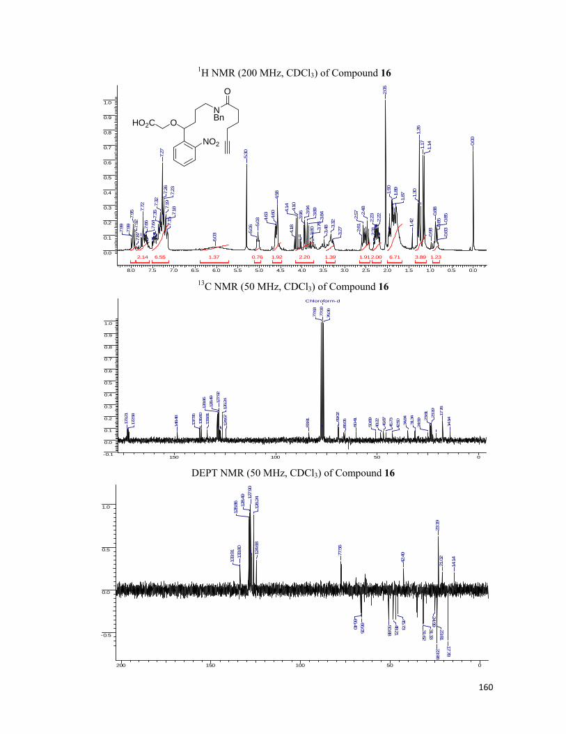
160
1H NMR (200 MHz, CDCl3) of Compound 16
8.0 7.5 7.0 6.5 6.0 5.5 5.0 4.5 4.0 3.5 3.0 2.5 2.0 1.5 1.0 0.5 0.0
0.0
0.1
0.2
0.3
0.4
0.5
0.6
0.7
0.8
0.9
1.0
6.716.55 3.892.202.14 2.001.92 1.911.391.37 1.230.76
0.00
0.83
0.85
0.85
0.88
0.98
1.141.17
1.26
1.30
1.42
1.871.
891.90
2.05
2.22
2.23
2.31
2.48
2.57
2.61
3.27
3.32
3.483.76
3.80
3.843.
893.94
3.96
4.10
4.14
4.18
4.58
4.60
4.63
5.03
5.06
5.30
6.03
7.15
7.187.
197.
237.26
7.27
7.32
7.35
7.64
7.66
7.72
7.87
7.92
7.95
7.99
7.99
13C NMR (50 MHz, CDCl3) of Compound 16
150 100 50 0-0.1
0.0
0.1
0.2
0.3
0.4
0.5
0.6
0.7
0.8
0.9
1.0
Chloroform-d
14.14
17.78
23.19
23.81
24.99
31.34
34.84
42.50
45.73
46.87
48.22
50.89
60.41
66.0569
.02
76.36
77.00
77.63
83.81
124.67
126.2412
7.92
128.49
128.86
133.81
136.60
137.55
148.48
172.58
173.21
DEPT NMR (50 MHz, CDCl3) of Compound 16
200 150 100 50 0
-0.5
0.0
0.5
1.0
14.14
17.78
21.02
23.19
23.8123.98
24.9931.3331.62
42.49
45.7348.2150.88
66.0566.40
77.56
124.68
126.2412
7.90
128.49
128.86
133.80
133.91
NO2
ONBn
O
HO2C

161
1H NMR (200 MHz, CDCl3) of Compound 18
8.0 7.5 7.0 6.5 6.0 5.5 5.0 4.5 4.0 3.5 3.0 2.5 2.0 1.5
0.0
0.1
0.2
0.3
0.4
0.5
0.6
0.7
0.8
0.9
1.0
10.005.625.56 3.74 1.931.800.81 0.79
1.12
1.15
1.37
1.42
1.47
1.611.64
1.69
1.701.74
1.76
1.77
1.78
2.282.32
2.362.
422.45
3.19
3.22
3.26
3.27
3.28
3.32
3.44
4.404.40
4.43
4.44
4.51
4.62
5.137.147.
177.18
7.24
7.277.28
7.33
7.367.
697.70
7.88
7.92
7.96
13C NMR (50 MHz, CDCl3) of Compound 18
170 160 150 140 130 120 110 100 90 80 70 60 50 40 30 20 10 0
0.0
0.1
0.2
0.3
0.4
0.5
0.6
0.7
0.8
0.9
1.0
Chloroform-d
23.3
224
.64
24.8
626
.31
26.4
528.5
832
.76
35.0
942
.0445
.67
46.7
448
.10
51.1
3
55.8
1
73.4
776
.3777.0
077
.63
95.1
5
124.
4312
5.99
127.
8112
8.44
128.
8113
3.25
136.
8713
7.67
138.
21
148.
10
172.
67
DEPT NMR (50 MHz, CDCl3) of Compound 18
150 100 50 0
-1.0
-0.5
0.0
0.5
23.45
24.76
24.98
26.43
28.70
32.88
33.02
35.20
42.16
45.78
48.21
50.9051.25
51.29
55.93
56.02
73.34
73.58
95.26
124.35
124.55
126.11
127.92
128.93
133.38
133.56
O2N
MOMONBn
O
N3
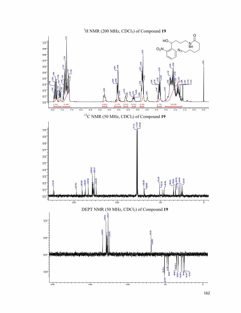
162
1H NMR (200 MHz, CDCl3) of Compound 19
8.0 7.5 7.0 6.5 6.0 5.5 5.0 4.5 4.0 3.5 3.0 2.5 2.0 1.5 1.0 0.5 0.0
0.0
0.1
0.2
0.3
0.4
0.5
0.6
0.7
0.8
0.9
1.0
10.005.48 2.90 1.761.751.61 0.81 0.53 0.52
0.00
1.101.13
1.261.34
1.37
1.52
1.56
1.59
1.63
1.79
1.81
2.04
2.29
2.33
2.35
2.37
2.39
2.42
3.00
3.19
3.22
3.26
3.27
3.31
3.66
3.69
3.73
3.76
4.10
4.13
4.50
4.54
4.54
4.58
4.59
5.26
7.13
7.167.17
7.27
7.33
7.37
7.42
7.42
7.58
7.62
7.63
7.67
7.817.
867.90
7.91
7.93
13C NMR (50 MHz, CDCl3) of Compound 19
150 100 50 0
0.0
0.1
0.2
0.3
0.4
0.5
0.6
0.7
0.8
0.9
1.0
24.7
8
26.4
428
.70
32.8
533.0
635
.00
45.8
1
51.1
451.2
5
68.8
569.3
0
76.4
377
.07
77.7
1
124.
2512
6.17
129.
00
133.
5213
6.65
140.
98
147.
50173.
78
DEPT NMR (50 MHz, CDCl3) of Compound 19
200 150 100 50 0
-0.5
0.0
0.5
1.0
24.70
24.78
26.4328.70
33.06
34.9935.33
45.8146.77
51.2551.30
68.85
69.30
124.26
126.17
129.01
133.53
O2N
HONBn
O
N3

163
1H NMR (200 MHz, CDCl3) of Compound 21
8.0 7.5 7.0 6.5 6.0 5.5 5.0 4.5 4.0 3.5 3.0 2.5 2.0 1.5 1.0 0.5 0.0
0.0
0.1
0.2
0.3
0.4
0.5
0.6
0.7
0.8
0.9
1.0
10.006.00 2.852.401.83 1.771.670.95 0.76
0.00
1.14
1.17
1.22
1.26
1.30
1.52
1.62
1.65
1.77
2.05
2.29
2.32
2.43
2.47
3.16
3.203.23
3.26
3.30
3.58
3.73
3.81
3.95
4.10
4.14
4.18
4.54
4.59
4.62
5.04
6.68
7.12
7.167.16
7.22
7.25
7.27
7.31
7.35
7.667.
71
7.72
7.94
7.98
13C NMR (50 MHz, CDCl3) of Compound 21
150 100 50 0
0.00
0.05
0.10
0.15
0.20
0.25
0.30
0.35
0.40
0.45
0.50
Chloroform-d
23.9
2
24.7
224.9
926
.33
28.5
332
.72
33.0
135
.04
46.0
246
.87
48.1
551
.15
51.2
3
76.3
777
.00
77.6
3
124.
53
126.
1112
7.93
128.
4812
8.91
136.
5313
7.5714
8.43
173.
41
DEPT NMR (50 MHz, CDCl3) of Compound 21
200 150 100 50 0
-0.5
0.0
0.5
1.0
-0.05
24.71
24.97
26.32
26.40
28.53
32.71
35.04
48.1451.14
66.58
77.17
124.52
126.11127.92
128.90
133.91
O2N
ONBn
O
N3
HO2C

164
Chapter 3: References
1. (a) Abdallah, D. J.; Weiss, R. G. Adv. Mater. 2000, 12, 1237-1247; (b) Terech, P.;
Weiss, R. G. Chem. Rev. 1997, 97, 3133-3159.
2. Eagon, R. J. Bacteriol. 1995, 83, 736-737.
3. (a) Crick, F. H. C. Symp. Soc. Exp. Biol XII 1958, 139-163; (b) Crick, F. H. C.
Nature 1970, 227, 561-563; (c) Uzawa, T.; Yamagishi, A.; Oshima, T. The Journal
of Biochem. 2002, 131, 849-853; (d) Werner, E. FEBS Lett. 2005, 579, 1779-1782;
(e) Ptashne, M.; Gann, A. Nature 1997, 386, 569-577.
4. Thanbichler, M.; Wang, S.; Shapiro, L. J. Cell Biochem. 2005, 96(3), 506-521.
5. Knight, R. D.; Freeland, S. J.; Landweber, L. F. Trends in Biochem. Sci. 1999,
24(6), 241-247.
6. (a) Dwek, D. Chem. Rev. 1996, 96, 683-720; (b) Hakamori, S.; Zhang, Y. Chem.
Biol. 1997, 4, 97-104.
7. Branden, C.; Tooze, J. Introduction to Protein Structure 2nd ed. (1999).
8. Pauling, L.; Corey, R. B.; Branson, H. R. Proc. Nat. Acad. Sci. 1951, 37, 205-211.
9. (a) Back, J. W.; de Jong, L.; Muijisers, A. O.; de Koster, C. G. J. Mol. Biol. 2003,
331, 303-313.; (b) Young, M. M.; Tang, N.; Hempel, J. C.; Oshiro, C. M.; Taylor,
E. W.; Kuntz, I. D.; Gibson B. W.; Dollinger, G. Proc. Nat. Acad. Sci. 2000, 97,
5802-5806; (c) Doyle, D. A.; Chait, B. T.; Mackinnon, R. Science 1998, 280, 69-77.
10. (a) Noren, C. J.; Anthony-Cahill, S. J.; Griffith, M. C.; Schultz, P. G. Science 1989,
244, 182-188; (b) Wallace, C. J. A. Curr. Opin. Biotechnol. 1995, 6, 403-410; (c)
Witte, K.; Sears, P.; Wong, C. H. J. Am. Chem. Soc. 1997, 119, 2114-2118.
11. Rauh, D.; Waldmann, H. Angew. Chem. Int. Ed. 2007, 46, 826-829.
12. Wieland, T.; Bockelmann, E.; Bauer, L.; Lau H. U. Justus Leibigs Ann. Chem.
1958, 583, 129.
13. (a) Dawson, P. E.; Muir, T.; Clark-Lewis, I.; Kent, S. B. Science 1994, 776-779; (b)
Dawson, P. E.; Kent, S. B. Annu. Rev. Biochem. 2000, 69, 923-960.
14. Ashraf, B.; Ficht, S.; Yang, Y. Y.; Bennett, C. S.; Wong, C. H. J. Am. Chem. Soc.
2006, 128, 15026-15033.
15. David, R.; Richter, M. P. O.; Beck-Sickinger, A. G. Eur. J. Biochem. 2004, 271,
663-677.
16. Perler, F. B. Nucleic Acids Res. 2000, 28, 344-345.
165
17. Canne, L. E.; Bark, S. J.; Kent, S. B. H. J. Am. Chem. Soc. 1996, 118, 5891-5896.
18. Nilsson, B. L.; Kiessling, L. L.; Raines, R. T. Org. Lett. 2000, 2, 1939-1941.
19. Bordusa, F. Chem. Rev. 2002, 102, 4817-4868.
20. Gavin, A. C. Nature 2006, 440, 631-636.
21. Kobe, B.; Kemp, B. E. Nature 1999, 402, 373-376.
22. Cambridge, S. B.; Davis, R. L.; Minden, J. S. Science 1997, 277, 825.
23. Schultz, P. G. et.al. J. Am. Chem. Soc. 2003, 125, 11782-11783.
24. Wang, Q.; Chan, T. R.; Hilgraf, R.; Fokin, V. V.; Sharpless, K. B.; Finn, M. G. J.
Am. Chem. Soc. 2003, 125, 3192-3193.
25. Verhelst, S. H. L.; Fonovic, M.; Bogoyo, M. Angew. Chem. Int. Ed. 2007, 46, 1284-
1286.
26. Brunner, J. Annu. Rev. Biochem. 1993, 62, 483-514.
27. Kaplan, J. H.; Hoffman, J. F. Biochemistry 1978, 17, 1929-1935.
28. Cornish, V.; Schultz, P. G. Curr. Opin. Stru. Biol. 1998, 4, 601-607.
29. Wei, Y. et. al. Bioorg. Med. Chem. Lett. 1998, 8, 2419-2422.
30. Staudinger, H.; Meyer, J. Helv. Chim. Acta. 1919, 2, 635.
31. Grandjean, C.; Boutonnier, A.; Guerreiro, C.; Fournier, J.-M.; Mulard, L. A. J. Org.
Chem. 2005, 70, 7123.
*****

166
List of Publications
1. “Niobium(V) chloride catalyzed microwave assisted synthesis of 2,3-unsaturated O-
glycosides by the Ferrier reaction”: Srinivas Hotha and Ashish Tripathi, Tetrahedron
Lett.2005, 46, 4555-4558.
2. “Diversity Oriented Synthesis of Tricyclic Compounds from Glycals Using the Ferrier
and the Pauson-Khand Reaction”: Srinivas Hotha and Ashish Tripathi, Journal of
Combinatorial Chemistry, 2005, 7, 968-976.
3. “Click Chemistry Guided Carbohydrate Mediated Imaging of Bacteria and Yeast”:
Ashish Tripathi, Gopala Krishna Aradhyam and Srinivas Hotha. (Manuscript under
preparation).
*****