Embed Size (px)
Citation preview
Submitted 10 July 2015, Accepted 28 November 2015, Published online 8 December 2015 Corresponding Author: Maba L. Dao – e-mail – [email protected] 737
Diversity of Lactifluus (Basidiomycota, Russulales) in West Africa: 5
new species described and some considerations regarding their
distribution and ecology
Maba DL 1,2*, Guelly AK 1, Yorou NS 3 and Agerer R 2
1Département de Botanique et Écologie Végétale, Faculté des Sciences, Université de LoméBP 1515, Lomé, Togo.
2Department Biology I, Organismic Biology: Mycology, Ludwig-Maximilians-Universität München, Menzinger Str. 67, 80638 München, Germany.
3Faculty of Agronomy, University of Parakou, BP 123, Parakou, Benin.
Maba DL, Guelly AK, Yorou NS, Agerer R 2015 – Diversity of Lactifluus (Basidiomycota,
Russulales) in West Africa: 5 new species described and some considerations regarding their
distribution and ecology. Mycosphere 6(6), 737–759, Doi 10.5943/mycosphere/6/6/9
Abstract
The genus Lactifluus is one of the common ectomycorrhizal fungal taxa in tropical African
forest ecosystems. Recent morphological and anatomical mycological studies based on specimens
we sampled from 2007 to 2013 in West African forest ecosystems, including dry, dense, riparian
forests and woodlands, enable to assess the diversity and the occurrence of Lactifluus species in the
Guineo-Sudanian domain. A total of 51 ITS rDNA sequences generated from our samples were
aligned against tropical African and worldwide Lactifluus sequences available in GenBank. A
Maximum Likelihood phylogenetic tree was inferred from 113 sequences. The phylogenetic
placement of the species, combined with our morpho-anatomical data, supported the description of
five new species distributed among Lactifluus species. Our data further confirm that the species
richness of the genus Lactifluus is high and partly unexplored in the Guineo-Sudanian domain, and
confirmed that, in both the Guineo-Sudanian and the Congo-Zambezian domain many common
species occur. Patterns of occurrence of the recorded Lactifluus species from Guineo-Sudanian
ecozones are also highlighted.
Key words – Guineo-Sudanian – milkcaps – morpho-anatomy – molecular phylogeny – taxonomy
Introduction
Although there has been remarkable progress in tropical mycological investigations for the
last twenty years, tropical African ecozones, and particularly West Africa, remain very poorly
explored (van Roiij et al. 2003, Rivière et al. 2007, Diédhiou et al. 2013). In Guineo-Sudanian
forest ecosystems, the genera Scleroderma, Tomentella, Russula, Lactarius (L.) and Lactifluus (Lf.)
are among the most studied ectomycorrhizal fungi (ECM) (van Rooij et al. 2003, Yorou et al. 2011,
2012, Verbeken & Walleyn 2010, Sanon et al. 2013, Maba et al. 2013, 2014, 2015, Sanon et al.
2014). Recent progress in molecular phylogenetic analyses and morpho-anatomical investigations
within lactarioid taxa (Buyck et al. 2008, Verbeken & Walleyn 2010, Van de Putte et al. 2010,
Stubbe et al. 2010, Verbeken et al. 2011, De Crop et al. 2014, Maba et al. 2013, 2014, 2015)
Mycosphere 6 (6): 737–759 (2015) ISSN 2077 7019
www.mycosphere.org Article Mycosphere
Copyright © 2015 Online Edition
Doi 10.5943/mycosphere/6/6/9
738
highlighted the high genetic diversity of the genus Lactifluus, with numerous new species and
cryptic species described from tropical Africa (Verbeken et al. 2008, Van de Putte et al. 2009, De
Crop et al. 2012, Maba et al. 2013, 2015). In order to better circumscribe species limits and the
ecological plasticity of Lactifluus species, mycological investigations have been undertaken from
specimens collected in West African forest ecosystems (Maba et al. 2013, 2014, 2015).
Maba et al. (2015) undertook molecular analysis of Lactifluus taxa, including unidentified
specimens from West Africa. In addition to considerations regarding ecology, the present study
provides morpho-anatomical support for new Lactifluus species identification. Thus, from
numerous specimens we sampled, five Lactifluus specimens of which the molecular phylogenetic
positions are supported by Maba et al. (2015), are described hereby as new species to science,
based on morpho-anatomical differences with most closely related species. The newly described
Lactifluus species are accommodated in subgenera Lactariopsis (two species), Russulopsis (one
species) and Edules (two species). Chorological patters of recorded Lactifluus species in West
Africa and their putative host trees are discussed.
Material and Methods
Specimens were sampled between 2007 and 2013 in various West African forest
ecosystems, including the northern Guinean seasonal, dry, dense, riparian and open forests,
woodlands and savannas following a megatransect through five countries (Benin, Togo, Burkina
Faso, Mali, and Guinea). The specimens described here were sampled from DAN riverside forest in
south-western part of Burkina Faso (MD355); in Malouwaita rainforest of Guinea (MD219B,
MD224 and MD234). Specimens (C2349, MD123 and MD131) were sampled in Fazao-Malfakassa
National Park, whereas specimens DPM05, C2157 and C2163 were sampled in Aledjo Reserve
forest, all in central Togo. Sampling techniques, records of preliminary morphological data as well
as specimens’ preparation for conservation are detailed previously (Maba et al. 2013). Colours
were recorded following Kornerup & Wanscher (1978). Holotypes of the new species are
conserved in TOGO herbarium and isotypes in GENT and M (Thiers 2012).
Light and Scanning Electron Microscopy
Microscopical studies were performed focusing on the lactarioids anatomical diagnostic
features, following Verbeken & Walleyn (2010) and Maba et al. (2013, 2014, 2015). Measurements
are given referring to Buyck (1991) and detailed by Maba et al. (2013). Comparative microscopic
studies also integrated specimens of Lactifluus zenkeri (A MA. 20) and L. sesemotani (AV94-471
and AV94-82) received from Ghent University as loans. SEM micrographs were obtained using the
procedures explained by Maba et al. (2013). Preliminary identification of specimens were made
using the Lactarius s. l. study based on material collected in similar ecosystems in the neighboring
country Benin (van Rooij et al. 2003) and the monograph of Verbeken & Walleyn (2010) about
tropical African Lactarius s. l.
DNA Extraction, sequencing and PCR amplification
Genomic DNA extraction, sequencing and PCR amplification were undertaken by Maba et
al. (2015). The internal transcribed spacer regions (ITS) of the nuclear ribosomal DNA including
ITS1, ITS2 and 5.8S regions were amplified using the fungal specific primer ITS1F in combination
with the basidiomycete specific primer ITS4B (Gardes & Bruns 1993). A total of 51 ITS sequences
were obtained and the sequences of the newly described species have been deposited at European
Nucleotide Archive/ENA (Table 1).
Sequence editing, analyses and molecular phylogenetic inference
We refer to Maba et al. (2015) for sequence editing, analyses and phylogenetic inference
(Supplement). Four new sequences (three newly generated and one obtained from GenBank) were
added to the sequence dataset compiled in Maba et al. (2015). The multiple sequence alignment and
procedure for phylogenetic tree inference refer to Maba et al. (2015) and the Maximum Likelihood
739
Table 1 – List of our generated and public Genbank sequences included in phylogenetic analyses
Species ENA, accession numbers Localities
Lactifluus (81 sequences)
Lactifluus allardii KF220017, KF220015 USA
Lactifluus annulatoangustifolius HG426475 Togo
Lactifluus annulatoangustifolius AY606981 Madagascar
Lactifluus annulatolongisporus sp. nov.* HG426470, LK392606 Togo
Lactifluus atrovelutinus GU258231 Malaysia
Lactifluus burkinabei sp. nov.* LK392609 Burkina Faso Lactifluus brunneocarpus sp. nov.* LK392608 Guinea
Lactifluus chamaeleontinus AY606980 Zambia
Lactifluus chiapanensis GU258297 Mexico
Lactifluus clarkeae HQ318283 Australia
Lactifluus clarkeae GU222280 New Zealand
Lactifluus crocatus HQ318265, Q318248, HQ318266 Thailand
Lactifluus denigricans AY606983 Benin
Lactifluus densifolius HG917385 Togo
Lactifluus edulis HG917384 Togo
Lactifluus emergens HG426467 Togo
Lactifluus emergens AY606979 Zimbabwe Lactifluus fazaoensis HG426477 Togo
Lactifluus flammans HG426471 Togo
Lactifluus flammans UDB016931 Benin
Lactifluus flavellus LK392594, LK392595 Togo
Lactifluus flocktonae JX2666621, JX266622 Australia
Lactifluus foetens HG917381 Togo
Lactifluus foetens LK392603 Burkina Faso
Lactifluus genevievae GU258294 Australia
Lactifluus glaucescens KF220117 Italy
Lactifluus glaucescens KF220094 Belgium
Lactifluus glaucescens KF220075 France
Lactifluus guellii sp. nov.* HG426466 Togo Lactifluus gymnocarpoides LK392601 Benin
Lactifluus gymnocarpoides LK392600 Benin
Lactifluus gymnocarpus HG426472 Togo
Lactifluus heimii LK392612 Togo
Lactifluus hygrophoroides JN129397 China
Lactifluus inversus AY606976 Guinea
Lactifluus longibasidius LK392596, HG426473 Togo
Lactifluus longipes HG917391, HG917383 Togo
Lactifluus longipilus HQ318235, HQ318258, KF432958 Thailand
Lactifluus longisporus DQ421971 Zambia
Lactifluus luteopus LK392602 Togo Lactifluus luteopus LK392611 Burundi
Lactifluus medusae HG426474 Togo
Lactifluus madagascariensis AY606977 Madagascar
Lactifluus melleus LK392598, LK392597 Togo
Lactifluus membranaceus sp. nov.* LK392610 Guinea
Lactifluus membranaceus sp. nov.* HG426478 Togo
Lactifluus nodosicystidiosus AY606975 Madagascar
Lactifluus nonpiscis HG426468 Togo
Lactifluus pectinatus LK392599 Togo
Lactifluus piperatus KF220122, KF220120 France
Lactifluus pelliculatus AY606978 Madagascar
Lactifluus phlebophyllus AY606074 Madagascar Lactifluus pseudoluteopus HQ318286 Thailand
Lactifluus rubroviolascens AY606984 Zambia
Lactifluus rubroviolascens AY606985 Madagascar
Lactifluus rubiginosus HG917386 Togo
Lactifluus sudanicus HG426469, HG426476 Togo
Lactifluus velutissimus AY606982 Zimbabwe
740
Species ENA, accession numbers Localities
Lactifluus volemus HQ318279, HQ318275 Thailand
Lactifluus volemoides UDB016930 Benin
Lactifluus sp. LK392607 Togo
Lactifluus sp. LK931501 Togo
Lactifluus sp. LK392604 Benin
Lactifluus sp. LK392605 Benin
Lactifluus sp. LM999911 Benin Lactifluus sp. LN651269 Burkina Faso
Lactifluus sp. LM999910 Togo
Lactifluus sp. UDB014027 Cameroon
Lactarius (17 sequences)
Lactarius baliophaeus GU258277 Zambia
Lactarius kabansus HG917376 Togo
Lactarius kabansus HG917390 Zimbabwe
Lactarius miniatescens HG917375 Burkina Faso
Lactarius miniatescens HG917374 Togo
Lactarius tenellus HG917373 Togo
Lactarius saponaceus HG917379 Guinea
Lactarius saponaceus HG917378 Togo Lactarius subbaliophaeus HG917372 Togo
Lactarius sp. UDB013804 Zambia
Lactarius sp. UDB015091 Gabon
Lactarius sp. UDB018664 Zambia
Lactarius sp. UDB018662 Zambia
Lactarius sp. UDB013845 Zambia
Lactarius sp. UDB013930 Cameroon
Lactarius sp. UDB016860 Zambia
Lactarius sp. UDB013836 Zambia
Multifurca (5 sequences)
Multifurca zonaria DQ422000, DQ421990 Thailand Multifurca furcata DQ421995, DQ421994 USA
Multifurca ochricompacta DQ421984 USA
Russula (8 sequences)
Russula cremeirosea EU819424 USA
Russula congoana HG917387 Togo
Russula congoana UDB016932 Benin
Russula compressa UDB016985 Benin
Russula discopus JQ902046 Burundi
Russula discopus JQ902050 Senegal
Russula lipida JF908663 Italy
Russula xerampilina KF386758 USA
Out group (2 sequences) Gloeocystidiellum sp.? KJ140715 USA
Hericium erinaceum EU784265 Kew
(*) Newly described species
(ML) tree obtained has included a total of 113 ITS sequences (Tab. 1, Supplement). The alignment
is submitted to TreeBASE (http://purl.org/phylo/treebase/phylows/study/TB2:S17549).
Results
ITS sequence analyses
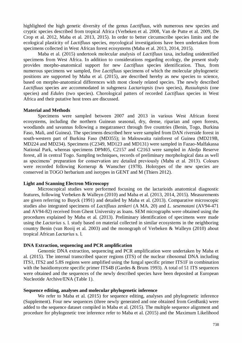
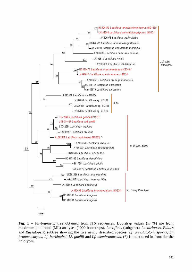
Sequences of the target specimens (MD123, MD131, MD224, MD234, MD355, C2157, and
C2349) are supported in the clade that encompasses representatives of Lactifluus subgenera
Lactariopsis, Edules and Russulopsis (Fig. 1). The sequences of two newly described species
(MD123 and MD131, C2349 and MD234) cluster within the Lf. subg. Lactariopsis subclade, with
98% and 57% of bootstrap support respectively, with already known species (subclade I). The
subclade II encompasses sequences of unidentified specimens from Togo (MD154) and Benin
741
H G 426470 Lac tifluus annulatolongisporus (M D 123) *
LK 392606 Lac tifluus annulatolongisporus (M D 131)
A Y 606978 Lac tifluus pelliculatus
HG 426475 Lac tifluus annulatoangus tifolius
A Y 606981 Lac tifluus annulatoangus tifolius
A Y 606980 Lac tifluus cham aeleontinus
LK 392612 Lac tifluus heim ii
A Y 606982 Lac tifluus velutiss im us
H G 426478 Lac tifluus m em branaceus (C 2349) *
LK 392610 Lac tifluus m em branaceus (M 234)
A Y 606977 Lac tifluus m adagascariens is
H G 426467 Lac tifluus em ergens
A Y 606979 Lac tifluus em ergens
I, Lf. subg.
Lactariopsis
LK 392607 Lac tifluus s p . M D 154
LK 392604 Lac tifluus s p . M D 304
LM 999911 Lac tifluus s p . M D326
LK 392605 Lac tifluus s p . M D 317
II, NA
HG 426466 Lac tifluus guellii (C 2157) *
UD B 014027 Lac tifluus conf. guellii
LK 392598 Lac tifluus m elleus
LK 392597 Lac tifluus m elleus
K L392609 Lac tifluus burk inabei (M D 355) *
A Y 606976 Lac tifluus inversus
A Y 606974 Lac tifluus phlebophy llus
H G 426477 Lac tifluus fazaoens is
H G 917385 Lac tifluus dens ifolius
H G 917384 Lac tifluus edulis
A Y 606975 Lac tifluus nodos icys tidiosus
III, Lf. subg. Edules
LK 392596 Lac tifluus longibas idius
H G 426473 Lac tifluus longibas idius
LK 392599 Lac tifluus pec tinatus
LK 392608 Lac tifluus brunneocarpus (M D 224) *
H G 917383 Lac tifluus longipes
H G 917391 Lac tifluus longipes
IV, Lf. subg. Russulopsis
100
98
99
69
90
100
92
100
98
100
57
96
100
71
89
99
96
71
99
100
86
94
99
73
86
47
98
100
100
99
60
0.005
63
Fig. 1 – Phylogenetic tree obtained from ITS sequences. Bootstrap values (in %) are from
maximum likelihood (ML) analyses (1000 bootstraps). Lactifluus (subgenera Lactariopsis, Edules
and Russulopsis) subtree showing the five newly described species: Lf. annulatolongisporus, Lf.
brunneocarpus, Lf. burkinabei, Lf. guellii and Lf. membranaceus. (*) is mentioned in front for the
holotypes.
742
(MD304, MD317, and MD326). At the same time the sequences of the specimens C2157 and
MD355 (two newly described species), fit in Lf. subg. Edules, with respectively 71 % and 99% of
bootstrap support with already known species (subclade III). In Lf. subg. Russulopsis subclade (IV),
nested the sequence of one new described species (MD224), supported by 99% of bootstrap value
as sister species of Lf. longipes.
The morpho-anatomical analyses reveal deviating features between specimen MD123,
MD131, MD219B, MD224, MD234, MD355, C2157, C2163, C2349 and DPM05, and their
morphological closely related species. These deviating features, coupled with the phylogenetic
placement of the specimens, accommodates them into five new species, notably: Lactifluus
annulatolongisporus (specimens MD123 and MD131) and Lactifluus membranaceus (C2349,
DPM05 and MD234) within Lf. subg. Lactariopsis; Lactifluus brunneocarpus (MD219B and
MD224) in Lf. subg. Russulopsis, and Lactifluus burkinabei (MD355) and Lactifluus guellii (C2157
and C2163), both within Lf. subg. Edules.
Taxonomy
Lactifluus annulatolongisporus Maba, sp. nov. Figs 2 – 4
Mycobank MB811601,
Facesoffungi Number: FoF 01642
Genbank ENA, accession number HG426470
Etymology – Referring to the presence of an annulus and the basidiospores that are strongly
elongate.
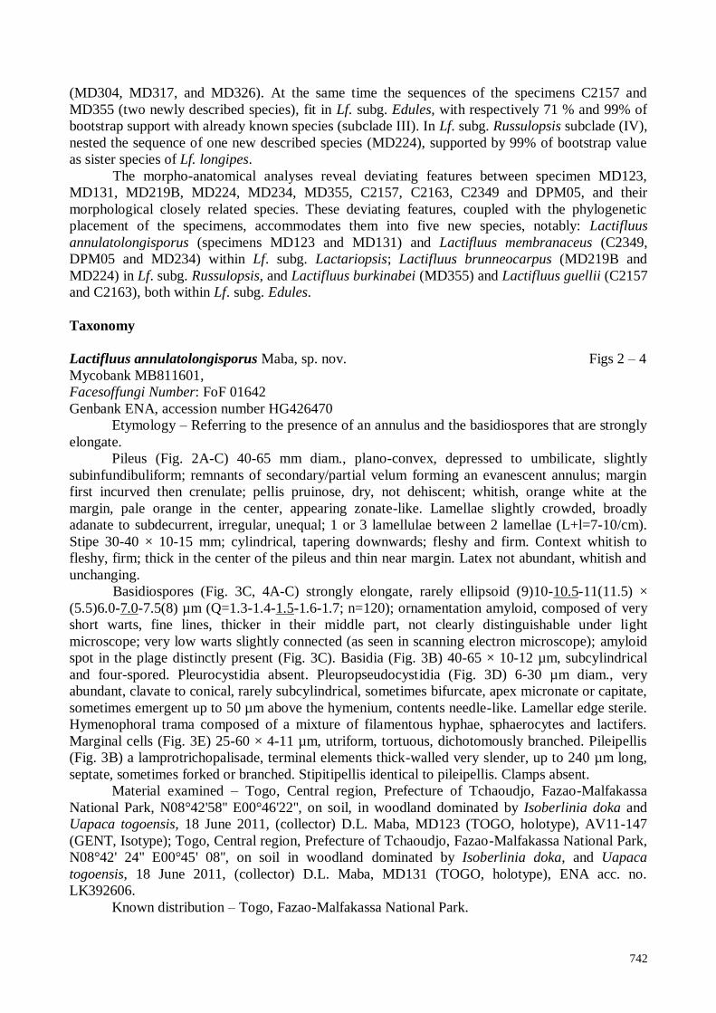
Pileus (Fig. 2A-C) 40-65 mm diam., plano-convex, depressed to umbilicate, slightly
subinfundibuliform; remnants of secondary/partial velum forming an evanescent annulus; margin
first incurved then crenulate; pellis pruinose, dry, not dehiscent; whitish, orange white at the
margin, pale orange in the center, appearing zonate-like. Lamellae slightly crowded, broadly
adanate to subdecurrent, irregular, unequal; 1 or 3 lamellulae between 2 lamellae (L+l=7-10/cm).
Stipe 30-40 × 10-15 mm; cylindrical, tapering downwards; fleshy and firm. Context whitish to
fleshy, firm; thick in the center of the pileus and thin near margin. Latex not abundant, whitish and
unchanging.
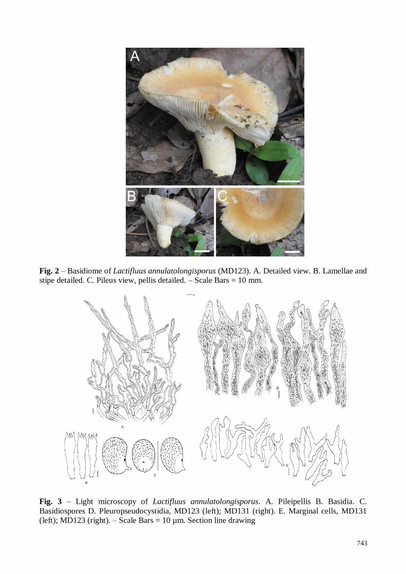
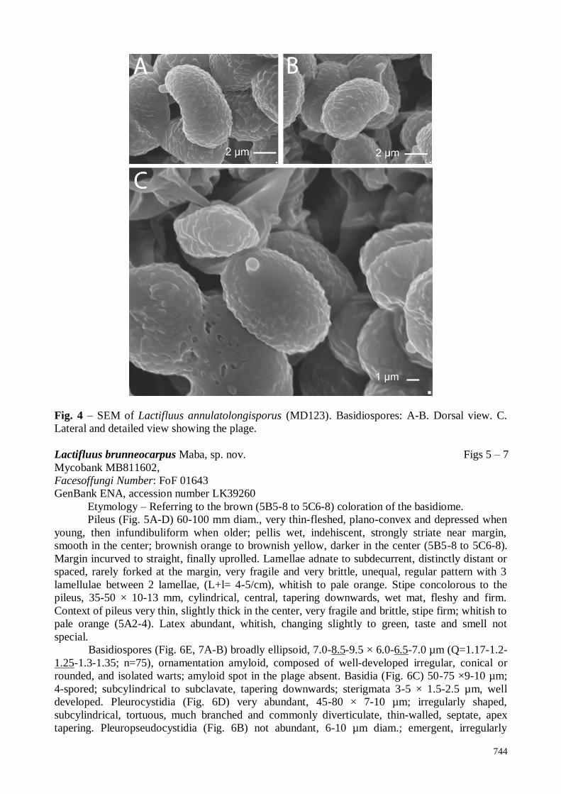
Basidiospores (Fig. 3C, 4A-C) strongly elongate, rarely ellipsoid (9)10-10.5-11(11.5) ×
(5.5)6.0-7.0-7.5(8) µm (Q=1.3-1.4-1.5-1.6-1.7; n=120); ornamentation amyloid, composed of very
short warts, fine lines, thicker in their middle part, not clearly distinguishable under light
microscope; very low warts slightly connected (as seen in scanning electron microscope); amyloid
spot in the plage distinctly present (Fig. 3C). Basidia (Fig. 3B) 40-65 × 10-12 µm, subcylindrical
and four-spored. Pleurocystidia absent. Pleuropseudocystidia (Fig. 3D) 6-30 µm diam., very
abundant, clavate to conical, rarely subcylindrical, sometimes bifurcate, apex micronate or capitate,
sometimes emergent up to 50 µm above the hymenium, contents needle-like. Lamellar edge sterile.
Hymenophoral trama composed of a mixture of filamentous hyphae, sphaerocytes and lactifers.
Marginal cells (Fig. 3E) 25-60 × 4-11 µm, utriform, tortuous, dichotomously branched. Pileipellis
(Fig. 3B) a lamprotrichopalisade, terminal elements thick-walled very slender, up to 240 µm long,
septate, sometimes forked or branched. Stipitipellis identical to pileipellis. Clamps absent.
Material examined – Togo, Central region, Prefecture of Tchaoudjo, Fazao-Malfakassa
National Park, N08°42'58'' E00°46'22'', on soil, in woodland dominated by Isoberlinia doka and
Uapaca togoensis, 18 June 2011, (collector) D.L. Maba, MD123 (TOGO, holotype), AV11-147
(GENT, Isotype); Togo, Central region, Prefecture of Tchaoudjo, Fazao-Malfakassa National Park,
N08°42' 24'' E00°45' 08'', on soil in woodland dominated by Isoberlinia doka, and Uapaca
togoensis, 18 June 2011, (collector) D.L. Maba, MD131 (TOGO, holotype), ENA acc. no.
LK392606.
Known distribution – Togo, Fazao-Malfakassa National Park.
743
Fig. 2 – Basidiome of Lactifluus annulatolongisporus (MD123). A. Detailed view. B. Lamellae and
stipe detailed. C. Pileus view, pellis detailed. – Scale Bars = 10 mm.
Fig. 3 – Light microscopy of Lactifluus annulatolongisporus. A. Pileipellis B. Basidia. C.
Basidiospores D. Pleuropseudocystidia, MD123 (left); MD131 (right). E. Marginal cells, MD131
(left); MD123 (right). – Scale Bars = 10 µm. Section line drawing
744
Fig. 4 – SEM of Lactifluus annulatolongisporus (MD123). Basidiospores: A-B. Dorsal view. C.
Lateral and detailed view showing the plage.
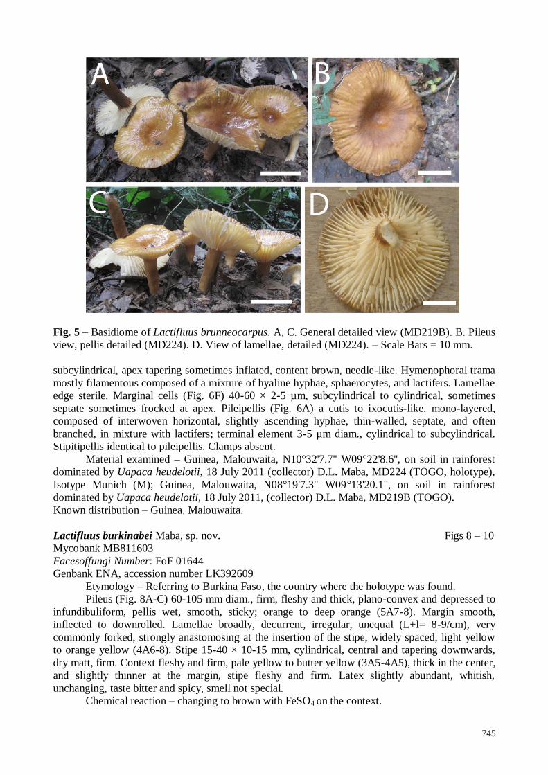
Lactifluus brunneocarpus Maba, sp. nov. Figs 5 – 7
Mycobank MB811602,
Facesoffungi Number: FoF 01643
GenBank ENA, accession number LK39260
Etymology – Referring to the brown (5B5-8 to 5C6-8) coloration of the basidiome.
Pileus (Fig. 5A-D) 60-100 mm diam., very thin-fleshed, plano-convex and depressed when
young, then infundibuliform when older; pellis wet, indehiscent, strongly striate near margin,
smooth in the center; brownish orange to brownish yellow, darker in the center (5B5-8 to 5C6-8).
Margin incurved to straight, finally uprolled. Lamellae adnate to subdecurrent, distinctly distant or
spaced, rarely forked at the margin, very fragile and very brittle, unequal, regular pattern with 3
lamellulae between 2 lamellae, (L+l= 4-5/cm), whitish to pale orange. Stipe concolorous to the
pileus, 35-50 × 10-13 mm, cylindrical, central, tapering downwards, wet mat, fleshy and firm.
Context of pileus very thin, slightly thick in the center, very fragile and brittle, stipe firm; whitish to
pale orange (5A2-4). Latex abundant, whitish, changing slightly to green, taste and smell not
special.
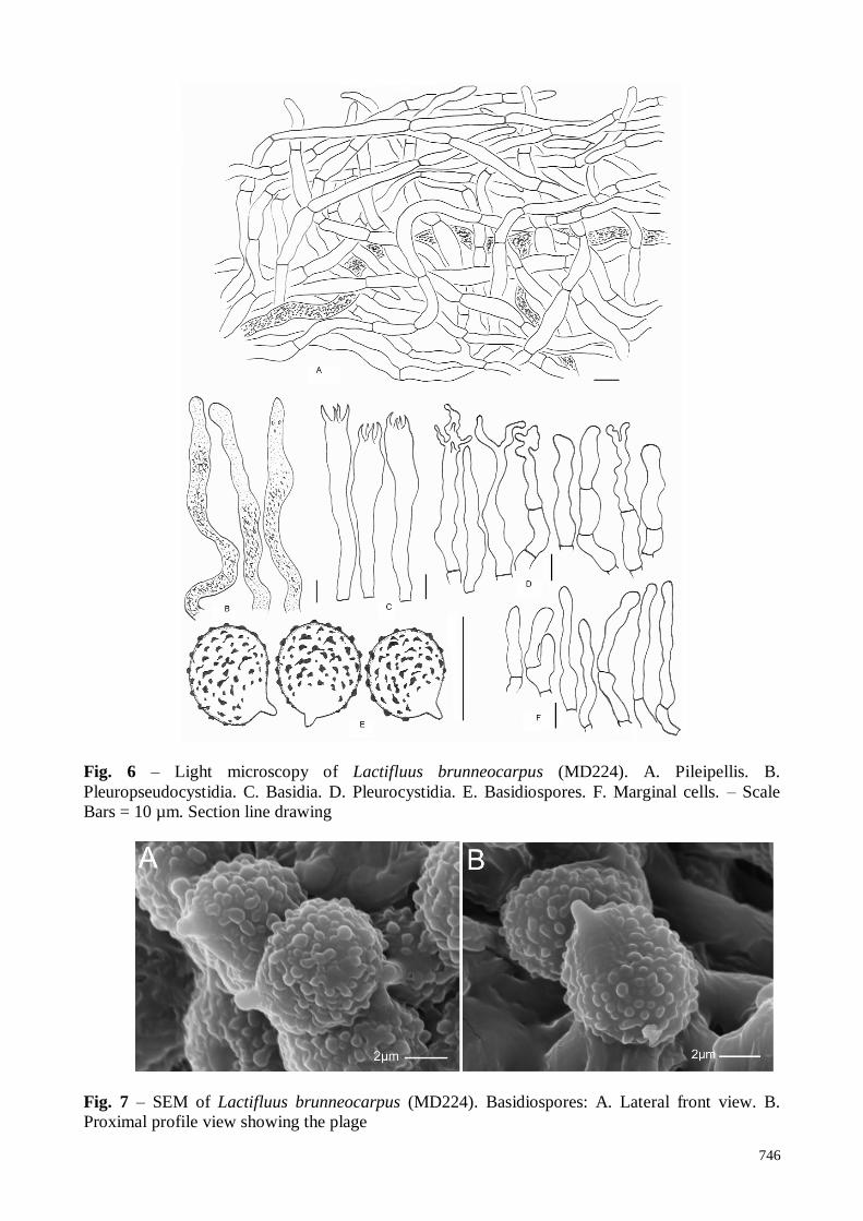
Basidiospores (Fig. 6E, 7A-B) broadly ellipsoid, 7.0-8.5-9.5 × 6.0-6.5-7.0 µm (Q=1.17-1.2-
1.25-1.3-1.35; n=75), ornamentation amyloid, composed of well-developed irregular, conical or
rounded, and isolated warts; amyloid spot in the plage absent. Basidia (Fig. 6C) 50-75 ×9-10 µm;
4-spored; subcylindrical to subclavate, tapering downwards; sterigmata 3-5 × 1.5-2.5 µm, well
developed. Pleurocystidia (Fig. 6D) very abundant, 45-80 × 7-10 µm; irregularly shaped,
subcylindrical, tortuous, much branched and commonly diverticulate, thin-walled, septate, apex
tapering. Pleuropseudocystidia (Fig. 6B) not abundant, 6-10 µm diam.; emergent, irregularly
745
Fig. 5 – Basidiome of Lactifluus brunneocarpus. A, C. General detailed view (MD219B). B. Pileus
view, pellis detailed (MD224). D. View of lamellae, detailed (MD224). – Scale Bars = 10 mm.
subcylindrical, apex tapering sometimes inflated, content brown, needle-like. Hymenophoral trama
mostly filamentous composed of a mixture of hyaline hyphae, sphaerocytes, and lactifers. Lamellae
edge sterile. Marginal cells (Fig. 6F) 40-60 × 2-5 µm, subcylindrical to cylindrical, sometimes
septate sometimes frocked at apex. Pileipellis (Fig. 6A) a cutis to ixocutis-like, mono-layered,
composed of interwoven horizontal, slightly ascending hyphae, thin-walled, septate, and often
branched, in mixture with lactifers; terminal element 3-5 µm diam., cylindrical to subcylindrical.
Stipitipellis identical to pileipellis. Clamps absent.
Material examined – Guinea, Malouwaita, N10°32'7.7'' W09°22'8.6'', on soil in rainforest
dominated by Uapaca heudelotii, 18 July 2011 (collector) D.L. Maba, MD224 (TOGO, holotype),
Isotype Munich (M); Guinea, Malouwaita, N08°19'7.3" W09°13'20.1", on soil in rainforest
dominated by Uapaca heudelotii, 18 July 2011, (collector) D.L. Maba, MD219B (TOGO).
Known distribution – Guinea, Malouwaita.
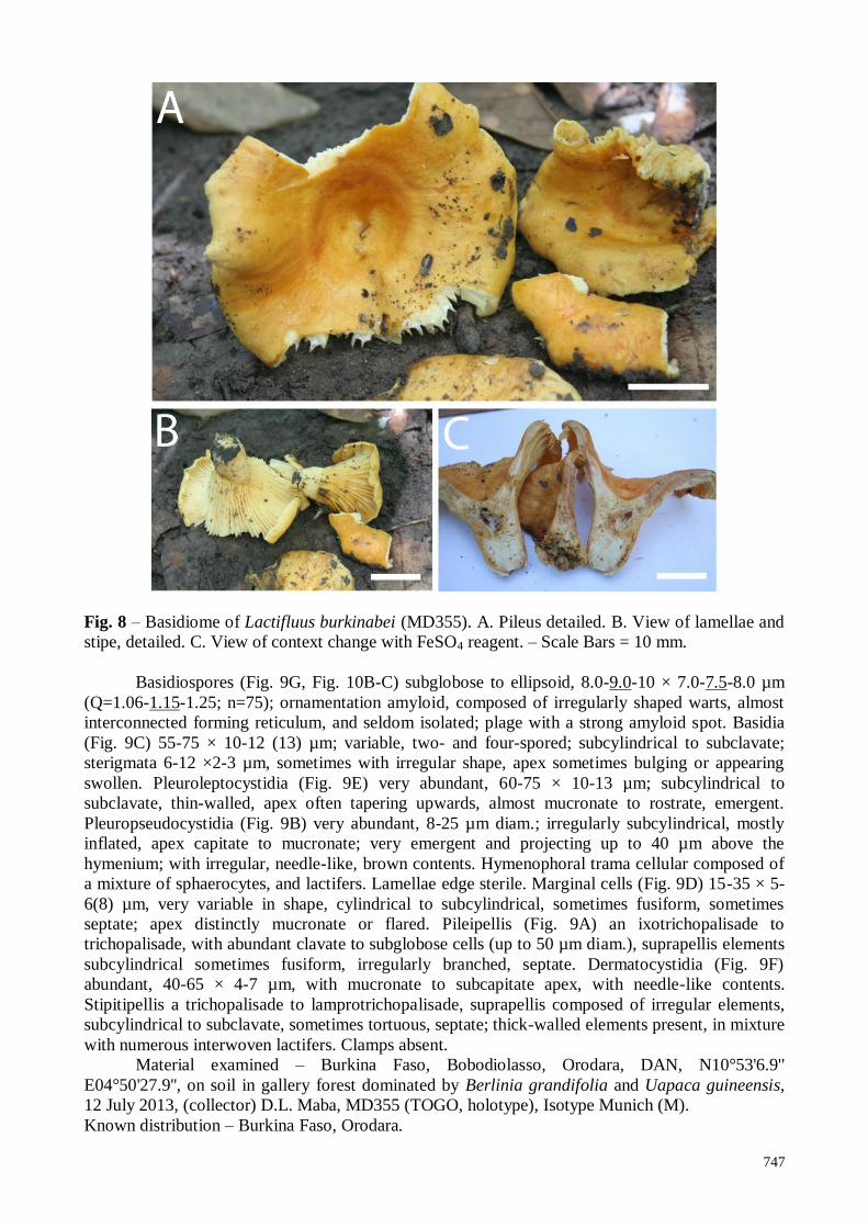
Lactifluus burkinabei Maba, sp. nov. Figs 8 – 10
Mycobank MB811603
Facesoffungi Number: FoF 01644
Genbank ENA, accession number LK392609
Etymology – Referring to Burkina Faso, the country where the holotype was found.
Pileus (Fig. 8A-C) 60-105 mm diam., firm, fleshy and thick, plano-convex and depressed to
infundibuliform, pellis wet, smooth, sticky; orange to deep orange (5A7-8). Margin smooth,
inflected to downrolled. Lamellae broadly, decurrent, irregular, unequal (L+l= 8-9/cm), very
commonly forked, strongly anastomosing at the insertion of the stipe, widely spaced, light yellow
to orange yellow (4A6-8). Stipe 15-40 × 10-15 mm, cylindrical, central and tapering downwards,
dry matt, firm. Context fleshy and firm, pale yellow to butter yellow (3A5-4A5), thick in the center,
and slightly thinner at the margin, stipe fleshy and firm. Latex slightly abundant, whitish,
unchanging, taste bitter and spicy, smell not special.
Chemical reaction – changing to brown with FeSO4 on the context.
746
Fig. 6 – Light microscopy of Lactifluus brunneocarpus (MD224). A. Pileipellis. B.
Pleuropseudocystidia. C. Basidia. D. Pleurocystidia. E. Basidiospores. F. Marginal cells. – Scale
Bars = 10 µm. Section line drawing
Fig. 7 – SEM of Lactifluus brunneocarpus (MD224). Basidiospores: A. Lateral front view. B.
Proximal profile view showing the plage
747
Fig. 8 – Basidiome of Lactifluus burkinabei (MD355). A. Pileus detailed. B. View of lamellae and
stipe, detailed. C. View of context change with FeSO4 reagent. – Scale Bars = 10 mm.
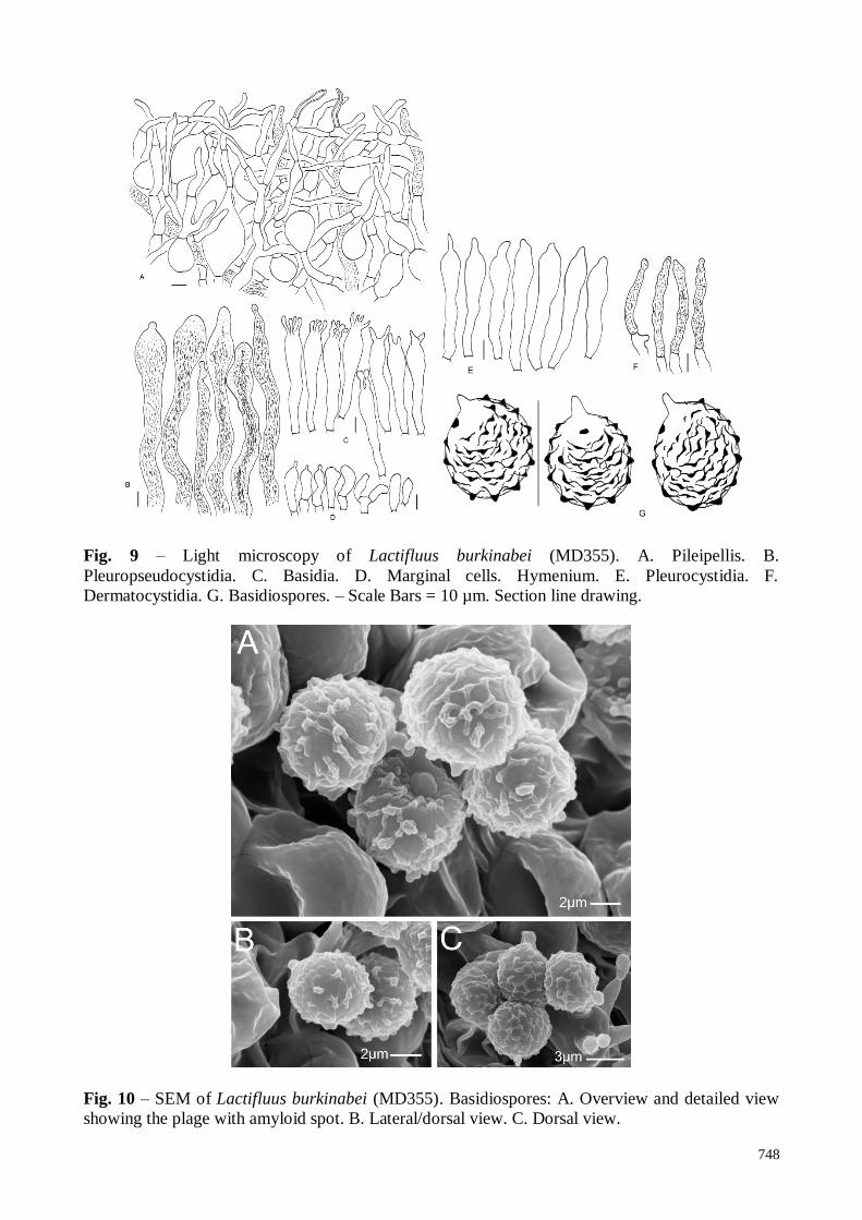
Basidiospores (Fig. 9G, Fig. 10B-C) subglobose to ellipsoid, 8.0-9.0-10 × 7.0-7.5-8.0 µm
(Q=1.06-1.15-1.25; n=75); ornamentation amyloid, composed of irregularly shaped warts, almost
interconnected forming reticulum, and seldom isolated; plage with a strong amyloid spot. Basidia
(Fig. 9C) 55-75 × 10-12 (13) µm; variable, two- and four-spored; subcylindrical to subclavate;
sterigmata 6-12 ×2-3 µm, sometimes with irregular shape, apex sometimes bulging or appearing
swollen. Pleuroleptocystidia (Fig. 9E) very abundant, 60-75 × 10-13 µm; subcylindrical to
subclavate, thin-walled, apex often tapering upwards, almost mucronate to rostrate, emergent.
Pleuropseudocystidia (Fig. 9B) very abundant, 8-25 µm diam.; irregularly subcylindrical, mostly
inflated, apex capitate to mucronate; very emergent and projecting up to 40 µm above the
hymenium; with irregular, needle-like, brown contents. Hymenophoral trama cellular composed of
a mixture of sphaerocytes, and lactifers. Lamellae edge sterile. Marginal cells (Fig. 9D) 15-35 × 5-
6(8) µm, very variable in shape, cylindrical to subcylindrical, sometimes fusiform, sometimes
septate; apex distinctly mucronate or flared. Pileipellis (Fig. 9A) an ixotrichopalisade to
trichopalisade, with abundant clavate to subglobose cells (up to 50 µm diam.), suprapellis elements
subcylindrical sometimes fusiform, irregularly branched, septate. Dermatocystidia (Fig. 9F)
abundant, 40-65 × 4-7 µm, with mucronate to subcapitate apex, with needle-like contents.
Stipitipellis a trichopalisade to lamprotrichopalisade, suprapellis composed of irregular elements,
subcylindrical to subclavate, sometimes tortuous, septate; thick-walled elements present, in mixture
with numerous interwoven lactifers. Clamps absent.
Material examined – Burkina Faso, Bobodiolasso, Orodara, DAN, N10°53'6.9''
E04°50'27.9'', on soil in gallery forest dominated by Berlinia grandifolia and Uapaca guineensis,
12 July 2013, (collector) D.L. Maba, MD355 (TOGO, holotype), Isotype Munich (M).
Known distribution – Burkina Faso, Orodara.
748
Fig. 9 – Light microscopy of Lactifluus burkinabei (MD355). A. Pileipellis. B.
Pleuropseudocystidia. C. Basidia. D. Marginal cells. Hymenium. E. Pleurocystidia. F.
Dermatocystidia. G. Basidiospores. – Scale Bars = 10 µm. Section line drawing.
Fig. 10 – SEM of Lactifluus burkinabei (MD355). Basidiospores: A. Overview and detailed view
showing the plage with amyloid spot. B. Lateral/dorsal view. C. Dorsal view.
749
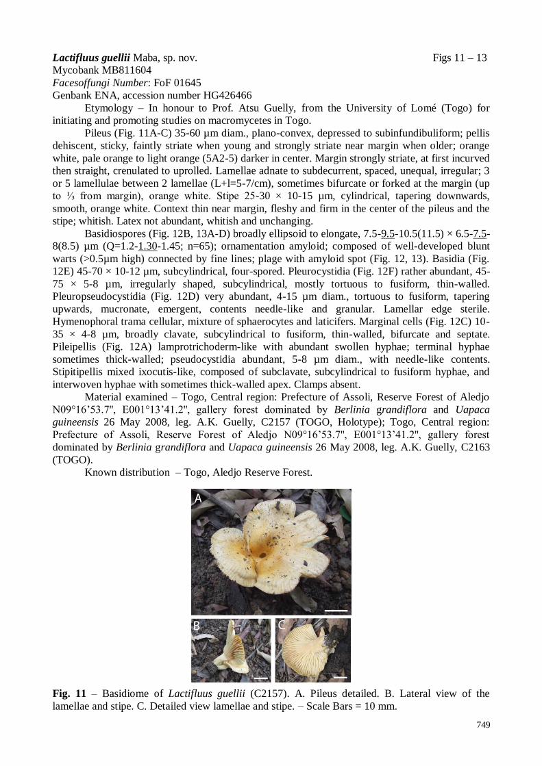
Lactifluus guellii Maba, sp. nov. Figs 11 – 13
Mycobank MB811604
Facesoffungi Number: FoF 01645
Genbank ENA, accession number HG426466
Etymology – In honour to Prof. Atsu Guelly, from the University of Lomé (Togo) for
initiating and promoting studies on macromycetes in Togo.
Pileus (Fig. 11A-C) 35-60 µm diam., plano-convex, depressed to subinfundibuliform; pellis
dehiscent, sticky, faintly striate when young and strongly striate near margin when older; orange
white, pale orange to light orange (5A2-5) darker in center. Margin strongly striate, at first incurved
then straight, crenulated to uprolled. Lamellae adnate to subdecurrent, spaced, unequal, irregular; 3
or 5 lamellulae between 2 lamellae (L+l=5-7/cm), sometimes bifurcate or forked at the margin (up
to ⅓ from margin), orange white. Stipe 25-30 × 10-15 µm, cylindrical, tapering downwards,
smooth, orange white. Context thin near margin, fleshy and firm in the center of the pileus and the
stipe; whitish. Latex not abundant, whitish and unchanging.
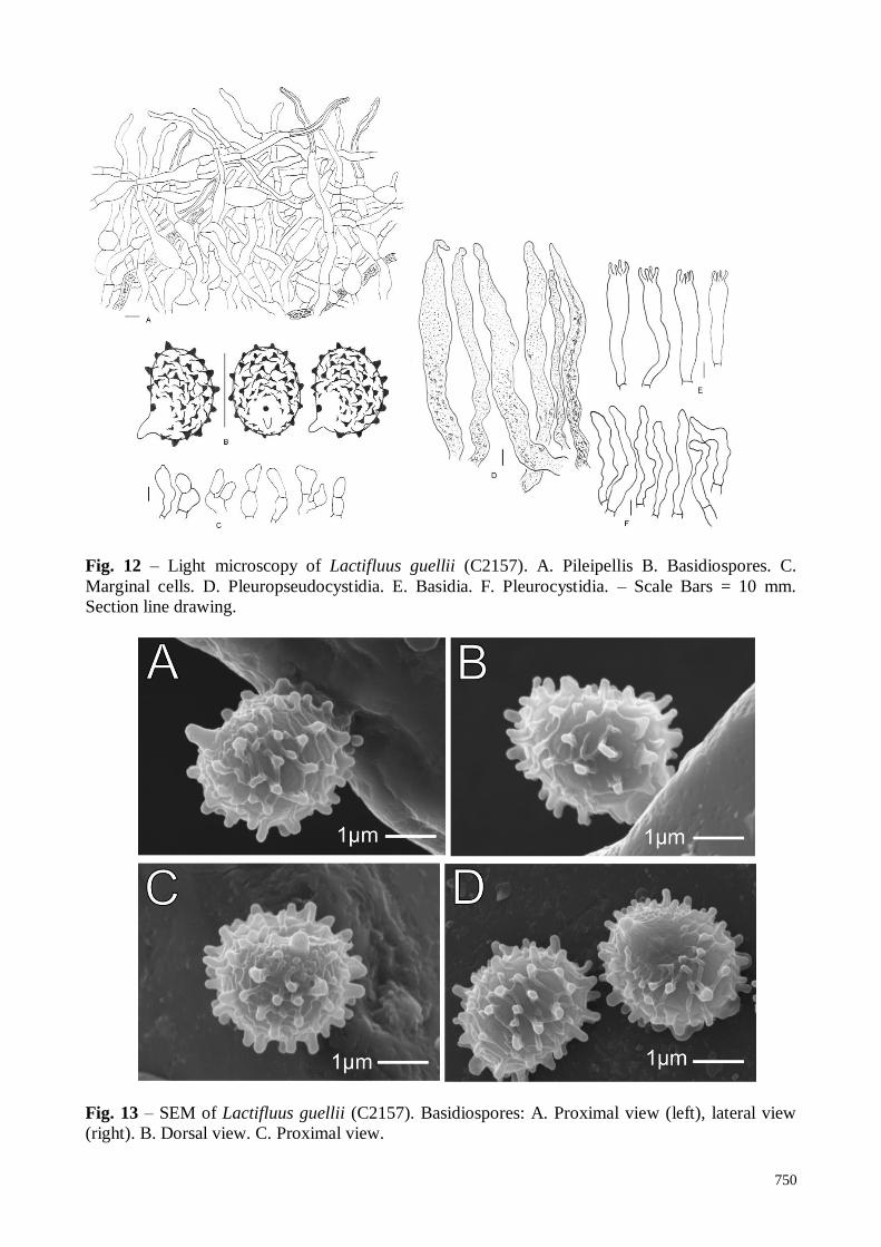
Basidiospores (Fig. 12B, 13A-D) broadly ellipsoid to elongate, 7.5-9.5-10.5(11.5) × 6.5-7.5-
8(8.5) µm (Q=1.2-1.30-1.45; n=65); ornamentation amyloid; composed of well-developed blunt
warts (>0.5µm high) connected by fine lines; plage with amyloid spot (Fig. 12, 13). Basidia (Fig.
12E) 45-70 × 10-12 µm, subcylindrical, four-spored. Pleurocystidia (Fig. 12F) rather abundant, 45-
75 × 5-8 µm, irregularly shaped, subcylindrical, mostly tortuous to fusiform, thin-walled.
Pleuropseudocystidia (Fig. 12D) very abundant, 4-15 µm diam., tortuous to fusiform, tapering
upwards, mucronate, emergent, contents needle-like and granular. Lamellar edge sterile.
Hymenophoral trama cellular, mixture of sphaerocytes and laticifers. Marginal cells (Fig. 12C) 10-
35 × 4-8 µm, broadly clavate, subcylindrical to fusiform, thin-walled, bifurcate and septate.
Pileipellis (Fig. 12A) lamprotrichoderm-like with abundant swollen hyphae; terminal hyphae
sometimes thick-walled; pseudocystidia abundant, 5-8 µm diam., with needle-like contents.
Stipitipellis mixed ixocutis-like, composed of subclavate, subcylindrical to fusiform hyphae, and
interwoven hyphae with sometimes thick-walled apex. Clamps absent.
Material examined – Togo, Central region: Prefecture of Assoli, Reserve Forest of Aledjo
N09°16’53.7'', E001°13’41.2'', gallery forest dominated by Berlinia grandiflora and Uapaca
guineensis 26 May 2008, leg. A.K. Guelly, C2157 (TOGO, Holotype); Togo, Central region:
Prefecture of Assoli, Reserve Forest of Aledjo N09°16’53.7'', E001°13’41.2'', gallery forest
dominated by Berlinia grandiflora and Uapaca guineensis 26 May 2008, leg. A.K. Guelly, C2163
(TOGO).
Known distribution – Togo, Aledjo Reserve Forest.
Fig. 11 – Basidiome of Lactifluus guellii (C2157). A. Pileus detailed. B. Lateral view of the
lamellae and stipe. C. Detailed view lamellae and stipe. – Scale Bars = 10 mm.
750
Fig. 12 – Light microscopy of Lactifluus guellii (C2157). A. Pileipellis B. Basidiospores. C.
Marginal cells. D. Pleuropseudocystidia. E. Basidia. F. Pleurocystidia. – Scale Bars = 10 mm.
Section line drawing.
Fig. 13 – SEM of Lactifluus guellii (C2157). Basidiospores: A. Proximal view (left), lateral view
(right). B. Dorsal view. C. Proximal view.
751
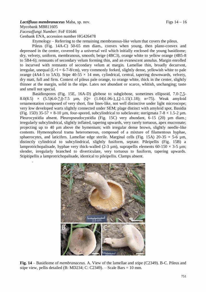
Lactifluus membranaceus Maba, sp. nov. Figs 14 – 16
Mycobank MB811605
Facesoffungi Number: FoF 01646
Genbank ENA, accession number HG426478
Etymology – Referring to the remaining membranous-like velum that covers the pileus.
Pileus (Fig. 14A-C) 50-65 mm diam., convex when young, then plano-convex and
depressed in the center, covered by a universal veil which initially enclosed the young basidiome;
dry, velvety, uniform, membranous, smooth; beige (4BC3), orange white to yellow orange (4B5-8
to 5B4-6); remnants of secondary velum forming thin, and an evanescent annulus. Margin enrolled
to incurved with remnants of secondary velum at margin. Lamellae thin, broadly decurrent,
irregular, unequal (L+l = 6-7-8/cm), very commonly forked, slightly dense, yellowish white to pale
orange (4A4-5 to 5A3). Stipe 40-55 × 14 mm, cylindrical, central, tapering downwards, velvety,
dry matt, full and firm. Context of pileus pale orange, to orange white, thick in the center, slightly
thinner at the margin, solid in the stipe. Latex not abundant or scarce, whitish, unchanging; taste
and smell not special.
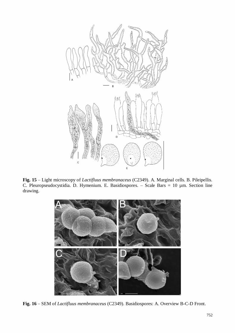
Basidiospores (Fig. 15E, 16A-D) globose to subglobose, sometimes ellipsoid, 7.0-7.5-
8.0(8.5) × (5.5)6.0-7.0-7.5 µm, (Q= (1.04)1.06-1.12-1.15(1.18); n=75). Weak amyloid
ornamentation composed of very short, fine lines-like, not well distinctive under light microscope;
very low developed warts slightly connected under SEM; plage distinct with amyloid spot. Basidia
(Fig. 15D) 35-57 × 8-10 µm, four-spored, subcylindrical to subclavate; sterigmata 7-8 × 1.5-2 µm.
Pleurocystidia absent. Pleuropseudocystidia (Fig. 15C) very abundant, 6-15 (20) µm diam.;
irregularly subcylindrical, slightly inflated, tapering upwards, very rarely tortuous, apex mucronate;
projecting up to 40 µm above the hymenium; with irregular dense brown, slightly needle-like
contents. Hymenophoral trama heteromerous, composed of a mixture of filamentous hyphae,
sphaerocytes, and laticifers. Lamellae edge sterile. Marginal cells (Fig. 15A) 20-35 × 5-6 µm,
distinctly cylindrical to subcylindrical, slightly fusiform, septate. Pileipellis (Fig. 15B) a
lamprotrichopalisade, hyphae very thick-walled (2-3 µm), suprapellis elements 60-150 × 3-5 µm;
slender, irregularly branched to diverticulate, very tortuous to fusiform, tapering upwards.
Stipitipellis a lamprotrichopalisade, identical to pileipellis. Clamps absent.
.
Fig. 14 – Basidiome of membranaceus. A. View of the lamellae and stipe (C2349). B-C. Pileus and
stipe view, pellis detailed (B: MD234; C: C2349). – Scale Bars = 10 mm.
752
Fig. 15 – Light microscopy of Lactifluus membranaceus (C2349). A. Marginal cells. B. Pileipellis.
C. Pleuropseudocystidia. D. Hymenium. E. Basidiospores. – Scale Bars = 10 µm. Section line
drawing.
Fig. 16 – SEM of Lactifluus membranaceus (C2349). Basidiospores: A. Overview B-C-D Front.
753
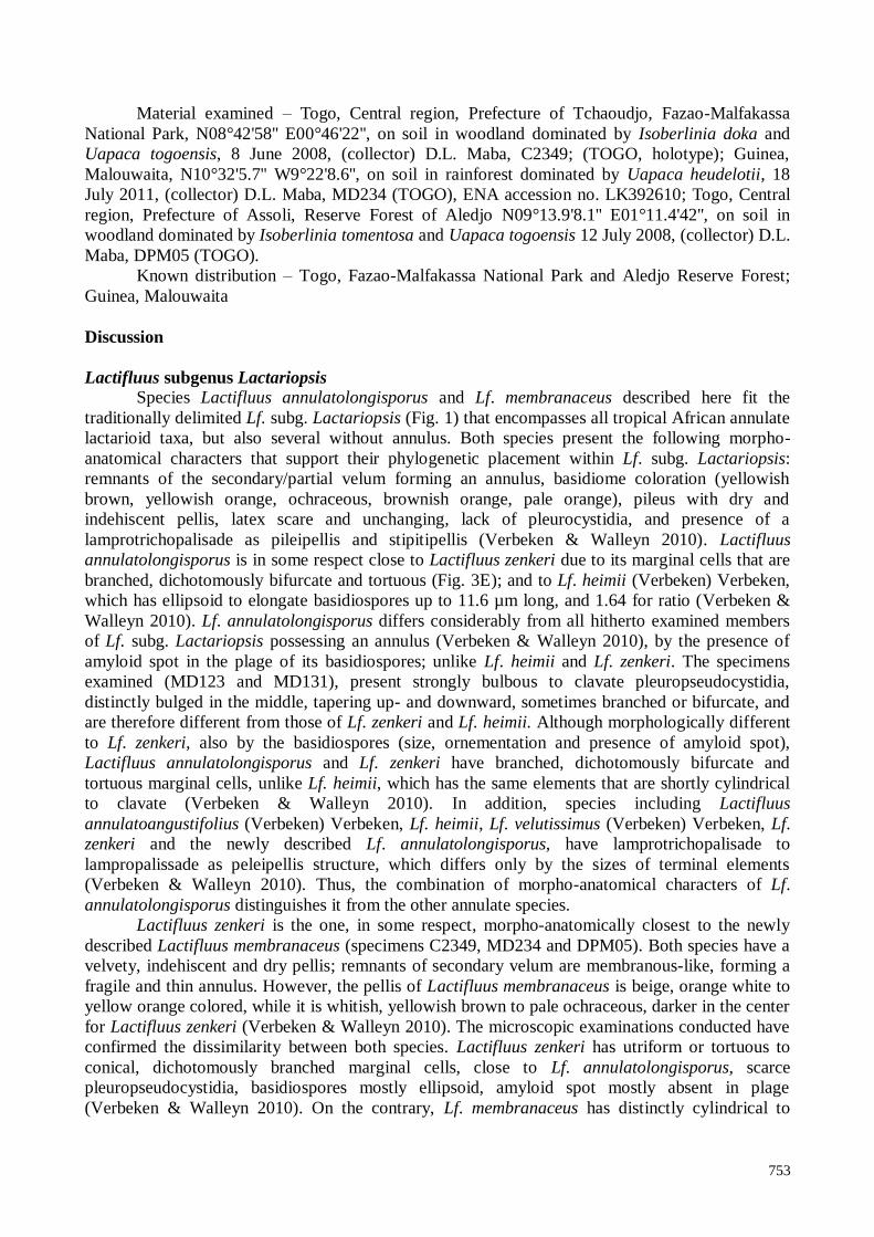
Material examined – Togo, Central region, Prefecture of Tchaoudjo, Fazao-Malfakassa
National Park, N08°42'58'' E00°46'22'', on soil in woodland dominated by Isoberlinia doka and
Uapaca togoensis, 8 June 2008, (collector) D.L. Maba, C2349; (TOGO, holotype); Guinea,
Malouwaita, N10°32'5.7'' W9°22'8.6'', on soil in rainforest dominated by Uapaca heudelotii, 18
July 2011, (collector) D.L. Maba, MD234 (TOGO), ENA accession no. LK392610; Togo, Central
region, Prefecture of Assoli, Reserve Forest of Aledjo N09°13.9'8.1'' E01°11.4'42'', on soil in
woodland dominated by Isoberlinia tomentosa and Uapaca togoensis 12 July 2008, (collector) D.L.
Maba, DPM05 (TOGO).
Known distribution – Togo, Fazao-Malfakassa National Park and Aledjo Reserve Forest;
Guinea, Malouwaita
Discussion
Lactifluus subgenus Lactariopsis
Species Lactifluus annulatolongisporus and Lf. membranaceus described here fit the
traditionally delimited Lf. subg. Lactariopsis (Fig. 1) that encompasses all tropical African annulate
lactarioid taxa, but also several without annulus. Both species present the following morpho-
anatomical characters that support their phylogenetic placement within Lf. subg. Lactariopsis:
remnants of the secondary/partial velum forming an annulus, basidiome coloration (yellowish
brown, yellowish orange, ochraceous, brownish orange, pale orange), pileus with dry and
indehiscent pellis, latex scare and unchanging, lack of pleurocystidia, and presence of a
lamprotrichopalisade as pileipellis and stipitipellis (Verbeken & Walleyn 2010). Lactifluus
annulatolongisporus is in some respect close to Lactifluus zenkeri due to its marginal cells that are
branched, dichotomously bifurcate and tortuous (Fig. 3E); and to Lf. heimii (Verbeken) Verbeken,
which has ellipsoid to elongate basidiospores up to 11.6 µm long, and 1.64 for ratio (Verbeken &
Walleyn 2010). Lf. annulatolongisporus differs considerably from all hitherto examined members
of Lf. subg. Lactariopsis possessing an annulus (Verbeken & Walleyn 2010), by the presence of
amyloid spot in the plage of its basidiospores; unlike Lf. heimii and Lf. zenkeri. The specimens
examined (MD123 and MD131), present strongly bulbous to clavate pleuropseudocystidia,
distinctly bulged in the middle, tapering up- and downward, sometimes branched or bifurcate, and
are therefore different from those of Lf. zenkeri and Lf. heimii. Although morphologically different
to Lf. zenkeri, also by the basidiospores (size, ornementation and presence of amyloid spot),
Lactifluus annulatolongisporus and Lf. zenkeri have branched, dichotomously bifurcate and
tortuous marginal cells, unlike Lf. heimii, which has the same elements that are shortly cylindrical
to clavate (Verbeken & Walleyn 2010). In addition, species including Lactifluus
annulatoangustifolius (Verbeken) Verbeken, Lf. heimii, Lf. velutissimus (Verbeken) Verbeken, Lf.
zenkeri and the newly described Lf. annulatolongisporus, have lamprotrichopalisade to
lampropalissade as peleipellis structure, which differs only by the sizes of terminal elements
(Verbeken & Walleyn 2010). Thus, the combination of morpho-anatomical characters of Lf.
annulatolongisporus distinguishes it from the other annulate species.
Lactifluus zenkeri is the one, in some respect, morpho-anatomically closest to the newly
described Lactifluus membranaceus (specimens C2349, MD234 and DPM05). Both species have a
velvety, indehiscent and dry pellis; remnants of secondary velum are membranous-like, forming a
fragile and thin annulus. However, the pellis of Lactifluus membranaceus is beige, orange white to
yellow orange colored, while it is whitish, yellowish brown to pale ochraceous, darker in the center
for Lactifluus zenkeri (Verbeken & Walleyn 2010). The microscopic examinations conducted have
confirmed the dissimilarity between both species. Lactifluus zenkeri has utriform or tortuous to
conical, dichotomously branched marginal cells, close to Lf. annulatolongisporus, scarce
pleuropseudocystidia, basidiospores mostly ellipsoid, amyloid spot mostly absent in plage
(Verbeken & Walleyn 2010). On the contrary, Lf. membranaceus has distinctly cylindrical to
754
subcylindrical, septate marginal cells (Fig. 15A), rarely fusiform, very abundant
pleuropseudocystidia (Fig. 15C), and basidiospores (Fig. 15E, 16A-D) mostly globose to
subglobose, with distinctly amyloid spot present in plage. Both Lf. annulatolongisporus and Lf.
membranaceus fit Lf. subg. Lactariopsis and their sequences are well supported in this subclade.
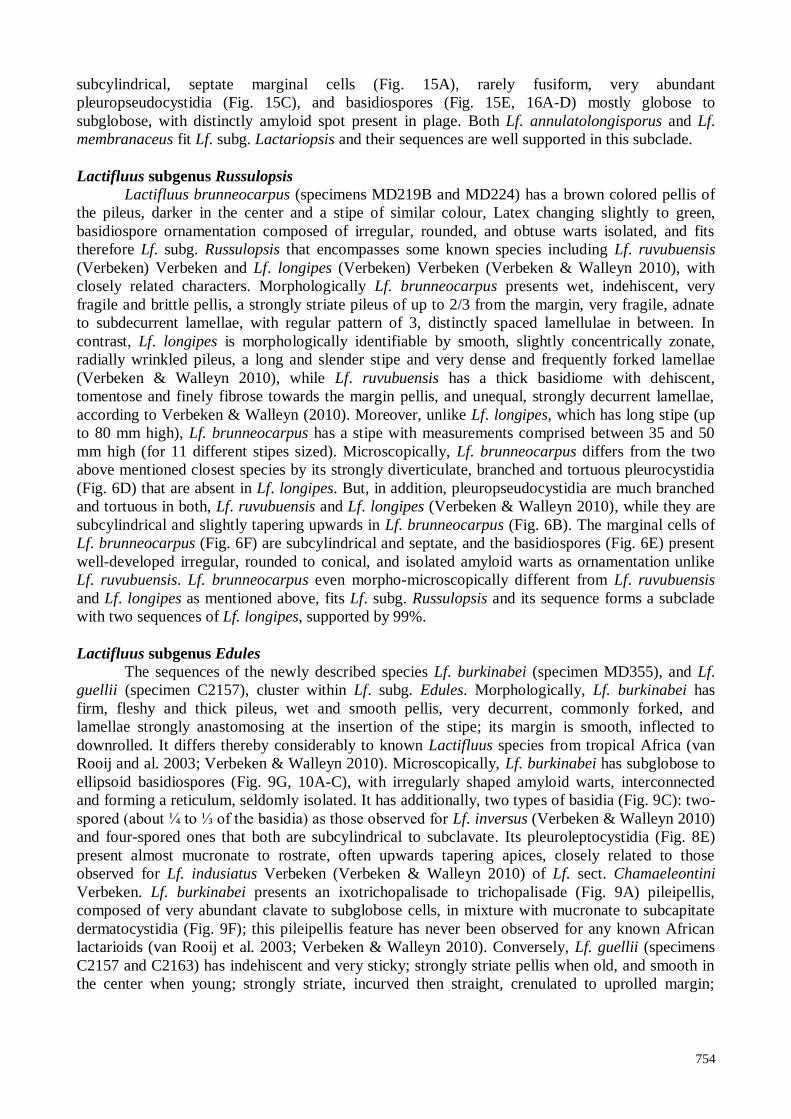
Lactifluus subgenus Russulopsis
Lactifluus brunneocarpus (specimens MD219B and MD224) has a brown colored pellis of
the pileus, darker in the center and a stipe of similar colour, Latex changing slightly to green,
basidiospore ornamentation composed of irregular, rounded, and obtuse warts isolated, and fits
therefore Lf. subg. Russulopsis that encompasses some known species including Lf. ruvubuensis
(Verbeken) Verbeken and Lf. longipes (Verbeken) Verbeken (Verbeken & Walleyn 2010), with
closely related characters. Morphologically Lf. brunneocarpus presents wet, indehiscent, very
fragile and brittle pellis, a strongly striate pileus of up to 2/3 from the margin, very fragile, adnate
to subdecurrent lamellae, with regular pattern of 3, distinctly spaced lamellulae in between. In
contrast, Lf. longipes is morphologically identifiable by smooth, slightly concentrically zonate,
radially wrinkled pileus, a long and slender stipe and very dense and frequently forked lamellae
(Verbeken & Walleyn 2010), while Lf. ruvubuensis has a thick basidiome with dehiscent,
tomentose and finely fibrose towards the margin pellis, and unequal, strongly decurrent lamellae,
according to Verbeken & Walleyn (2010). Moreover, unlike Lf. longipes, which has long stipe (up
to 80 mm high), Lf. brunneocarpus has a stipe with measurements comprised between 35 and 50
mm high (for 11 different stipes sized). Microscopically, Lf. brunneocarpus differs from the two
above mentioned closest species by its strongly diverticulate, branched and tortuous pleurocystidia
(Fig. 6D) that are absent in Lf. longipes. But, in addition, pleuropseudocystidia are much branched
and tortuous in both, Lf. ruvubuensis and Lf. longipes (Verbeken & Walleyn 2010), while they are
subcylindrical and slightly tapering upwards in Lf. brunneocarpus (Fig. 6B). The marginal cells of
Lf. brunneocarpus (Fig. 6F) are subcylindrical and septate, and the basidiospores (Fig. 6E) present
well-developed irregular, rounded to conical, and isolated amyloid warts as ornamentation unlike
Lf. ruvubuensis. Lf. brunneocarpus even morpho-microscopically different from Lf. ruvubuensis
and Lf. longipes as mentioned above, fits Lf. subg. Russulopsis and its sequence forms a subclade
with two sequences of Lf. longipes, supported by 99%.
Lactifluus subgenus Edules
The sequences of the newly described species Lf. burkinabei (specimen MD355), and Lf.
guellii (specimen C2157), cluster within Lf. subg. Edules. Morphologically, Lf. burkinabei has
firm, fleshy and thick pileus, wet and smooth pellis, very decurrent, commonly forked, and
lamellae strongly anastomosing at the insertion of the stipe; its margin is smooth, inflected to
downrolled. It differs thereby considerably to known Lactifluus species from tropical Africa (van
Rooij and al. 2003; Verbeken & Walleyn 2010). Microscopically, Lf. burkinabei has subglobose to
ellipsoid basidiospores (Fig. 9G, 10A-C), with irregularly shaped amyloid warts, interconnected
and forming a reticulum, seldomly isolated. It has additionally, two types of basidia (Fig. 9C): two-
spored (about ¼ to ⅓ of the basidia) as those observed for Lf. inversus (Verbeken & Walleyn 2010)
and four-spored ones that both are subcylindrical to subclavate. Its pleuroleptocystidia (Fig. 8E)
present almost mucronate to rostrate, often upwards tapering apices, closely related to those
observed for Lf. indusiatus Verbeken (Verbeken & Walleyn 2010) of Lf. sect. Chamaeleontini
Verbeken. Lf. burkinabei presents an ixotrichopalisade to trichopalisade (Fig. 9A) pileipellis,
composed of very abundant clavate to subglobose cells, in mixture with mucronate to subcapitate
dermatocystidia (Fig. 9F); this pileipellis feature has never been observed for any known African
lactarioids (van Rooij et al. 2003; Verbeken & Walleyn 2010). Conversely, Lf. guellii (specimens
C2157 and C2163) has indehiscent and very sticky; strongly striate pellis when old, and smooth in
the center when young; strongly striate, incurved then straight, crenulated to uprolled margin;
755
adnate to subdecurrent and spaced lamellae that are sometimes forked at the margin.
Microscopically it has ellipsoid to elongate basidiospores (Fig. 12B, 13A-D), with strong, well-
developed blunt amyloid warty ornamentation (>0.5µm high), finely interconnected at the base,
closely related to those observed in Lf. melleus Maba (Maba et al. 2015). The pleurocystidia of Lf.
guellii (Fig. 12F) are irregularly shaped, mostly tortuous to fusiform in contrast to the
pleuroleptocystidia of Lf. burkinabei (Fig. 9E) that are almost mucronate to rostate with often
upwards tapering apices. These features are unlike that of Lf. melleus. Pleuropseudocystidia of Lf.
guellii (Fig. 12D) are emergent, fusiform, tortuous and mucronate, closely related to those of Lf.
corbula R. Heim & Gooss.-Font. This latter mentioned species has a cutis-like pileipellis,
cylindrical to subclavate marginal cells, and ellipsoid basidiospores (up to 10.4 µm high, and up to
1.35 as ratio; n=60) with no amyloid spot in plage (Verbeken & Walleyn 2010). Lf. guellii on the
contrary has lamprotrichoderm-like pileipellis, with abundant swollen hyphae; its marginal cells are
broadly clavate, subcylindrical to fusiform, bifurcate and septate, and its basidiospores are broadly
ellipsoid to elongate (up to 11.5 µm high, and up to 1.45 as ratio; n=65), with strong amyloid spot
in plage. In the phylogeny analyses, Lf. guellii is supported (97%) as same species with one
unidentified from Cameroon (UDB014027), with that it forms a terminal clade; together with Lf.
melleus a clade with 71% of bootstrap support. At the same time the sequence of Lf. burkinabei is
well supported by 99% as a subclade within subgenus Edules, the subgenus itself is supported by
86% of bootstrap support value.
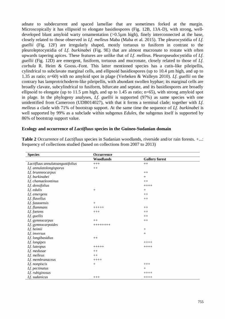
Ecology and occurrence of Lactifluus species in the Guineo-Sudanian domain
Table 2 Occurrence of Lactifluus species in Sudanian woodlands, riverside and/or rain forests. +...:
frequency of collections studied (based on collections from 2007 to 2013)
Species Occurrence
Woodlands Gallery forest
Lactifluus annulatoangustifolius +++ ++
Lf. annulatolongisporus ++
Lf. brunneocarpus ++
Lf. burkinabei +
Lf. chamaeleontinus ++
Lf. densifolius ++++
Lf. edulis + +
Lf. emergens ++
Lf. flavellus ++
Lf. fazaoensis +
Lf. flammans +++++ ++ Lf. foetens +++ ++
Lf. guellis ++
Lf. gymnocarpus ++ ++
Lf. gymnocarpoides ++++++++
Lf. heimii +
Lf. inversus +
Lf. longibasidius ++
Lf. longipes ++++
Lf. luteopus +++++ ++++
Lf. medusae ++
Lf. melleus ++
Lf. membranaceus ++++ Lf. nonpiscis + +++
Lf. pectinatus +
Lf. rubiginosus ++++
Lf. sudanicus +++ ++++
756
H G426470 Lactifluus annulatolongisporus *
LK392606 Lactifluus annulatolongisporus
AY606978 Lactifluus pe llicu la tus
H G426475 Lactifluus annu la toangus tifo lius
AY606981 Lactifluus annu la toangus tifo lius
AY606980 Lactifluus cham aeleontinus
LK392612 Lactifluus he im ii
AY606982 Lactifluus ve lu tis s im us
H G426478 Lactifluus membranaceus *
LK392610 Lactifluus membranaceus
AY606977 Lactifluus m adagas cariens is
H G426467 Lactifluus em ergens
AY606979 Lactifluus em ergens
LK392607 Lactifluus s p . MD 154
LK392604 Lactifluus s p . MD 304
LM999911 Lactifluus s p . MD 326
LK392605 Lactifluus s p . MD 317
HG426466 Lactifluus guellii *
U D B014027 Lactifluus aff. guellii
LK392598 Lactifluus m e lleus
LK392597 Lactifluus m elleus
LK392609 Lactifluus burkinabei *
AY606976 Lactifluus invers us
AY606974 Lactifluus ph lebophyllus
H G426477 Lactifluus fazaoens is
H G917385 Lactifluus dens ifo lius
H G917384 Lactifluus edu lis
AY606975 Lactifluus nodos icys tid ios us
LK392596 Lactifluus long ibas id ius
HG426473 Lactifluus s p long ibas id ius
LK392599 Lactifluus pectinatus
LK392608 Lactifluus brunneocarpus *
H G917383 Lactifluus long ipes
HG917391 Lactifluus long ipes
JX266621 Lactifluus flocktonae
JX266622 Lactifluus flocktonae
GU 258297 Lactifluus ch iapanens is
H Q318283 Lactifluus cla rckeae
GU 222280 Lactifluus cla rkeae
LM99910 Lactifluus s p .
LN651269 Lactfluus s p .
U D B016931 Lactifluus flam m ans
H G426471 Lactifluus flam m ans
H G426472 Lactifluus gym nocarpus
H G426468 Lactifluus nonp is cis
LK392603 Lactifluus foe tens
H G917381 Lactifluus foe tens
AY606984 Lactifluus rubrovio las cens
AY606985 Lactifluus rubrovio las cens
AY606983 Lactifluus den igricans
JN 129397 Lactarius hygrophoro ides
H Q318286 Lactifluus ps eudo lu teopus
LK392602 Lactifluus lu teopus
LK392611 Lactifluus lu teopus
H G426469 Lactifluus s udan icus
H G426476 Lactifluus s udan icus
H G917386 Lactifluus rub ig inos us
LK392594 Lactifluus flavellus
LK392595 Lactifluus flave llus
D Q421971 Lactifluus long is porus
H G426474 Lactifluus a ff. m edus ae
U D B016930 Lactifluus vo lem oides
LK392600 Lactifluus gym nocarpo ides
LK392601 Lactifluus gym nocarpo ides
H Q318258 Lactifluus long ip ilus
KF432958 Lactifluus long ip ilus
H Q318235 Lactifluus long ip ilus
H Q318265 Lactifluus crocatus
H Q318266 Lactifluus crocatus
H Q318248 Lactifluus crocatus
H Q318279 Lactifluus vo lem us
H Q318275 Lactifluus vo lem us
GU 258294 Lactifluuss genevievae
GU 258231 Lactifluus a trove lu tinus
KF220017 Lactifluus a lla rd ii
KF220015 Lactifluus a lla rd ii
KF220120 Lactifluus p ipera tus
KF220122 Lactarius p ipera tus
KF220117 Lactifluus g lauces cens
KF220075 Lactifluus g lauces cens
KF220094 Lactifluus g lauces cens
Lf, La
ctifluus
D Q421990 Murltifu rca zonaria
D Q422000 Multifu rca zonaria
D Q421984 Multifu rca ochricom pacta
D Q421995 Multifu rca fu rca ta
D Q421994 Multifu rca fu rca ta
U D B016860 Lactarius s p .
U D B013836 Lactarius s p .
H G917373 Lactarius tene llus
U D B013930 Lactarius s p .
H G917390 Lactarius kabans us
H G917376 Lactarius kabans us
U D B018662 Lactarius s p .
HG917378 Lactarius s aponaceus
HG917379 Lactarius s aponaceus
U D B015091 Lactarius s p .
U D B 015091 Lactarius s p .
HG917375 Lactarius mIn ia tes cens
H G917374 Lactarius m in ia tes cens
U D B013804 Lactarius s p .
H G917372 Lactarius s ubba liophaeus
U D B016864 Lactarius s p .
GU 258277 Lactarius ba liophaeus
VIII,
Lactarius
H G917387 R us s u la congoana
U D B016932 R us s u la congoana
U D B016985 R us s u la com pres s a
JQ902050 R us s ula d is copus
JQ902046 R us s u la d is copus
D Q422013 R us s u la lep ida
EU 819424 R us s u la crem eiros ea
FJ8 4 5 4 3 3 R us s u la xeram pelina
IX,
Russula
EU 784265 H ericium erinaceum
KJ140715 Gloeocys tid ie llum s p . Out group
100
100
100
100
97
100
100
100
95
100
98
94
51
100
100
97
69
100
100
95
100
100
77
50
83
79
83
96
82
82
94
99
100
100
98
98
96
99
100
95
99
95
99
59
69
100
57
99
100
100
100
100
100
69
100
100
71
100
99
90
100
100
100
98
100
92
92
94
68
99
98
96
100
55
98
92
57
78
53
66
61
68
100
99
99
83
65
55
74
96
54
61
96
71
86
96
99
71
100
73
89
86
60
97
63
47
0.05
I , Lf . s
ubg.
Lacta
ri op
sis
VI, Complex clade
II, N A
C.
Lf. subg.
Piperati
B, Lf. subg.
Gerardii
A,
Lf. subg.
Lactifluus
V, Lf. subg.
Lactifluus
III, Lf. subg.
Edules
IV, Lf. subg.
Russulopsis
VII,
Multifurca
NA: Not Applicable
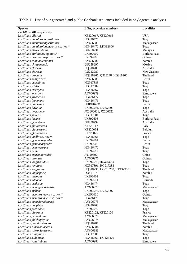
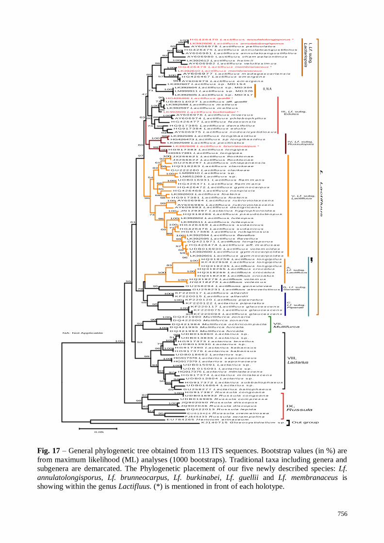
Fig. 17 – General phylogenetic tree obtained from 113 ITS sequences. Bootstrap values (in %) are
from maximum likelihood (ML) analyses (1000 bootstraps). Traditional taxa including genera and
subgenera are demarcated. The Phylogenetic placement of our five newly described species: Lf.
annulatolongisporus, Lf. brunneocarpus, Lf. burkinabei, Lf. guellii and Lf. membranaceus is
showing within the genus Lactifluus. (*) is mentioned in front of each holotype.
757
Species of the genus Lactifluus are widespread in Guineo-Sudanian ecosystems and occur
preferentially earliest between the end of May to July and latest between the end of August and
September (Verbeken and Buyck 2001; van Rooij et al. 2003; Verbeken & Walleyn 2010; Maba et
al. 2013, 2014, 2015). In collections that have been continuously sampled since 2007 in various
ectomycorrhizal dominated ecosystems, certain Lactifluus species including Lf.
annulatoangustifolius, Lf. edulis, Lf. foetens, Lf. gymnocarpus, Lf. luteopus, Lf. nonpiscis and Lf.
sudanicus have shown no preference regarding vegetation type, as they had been collected in both
woodlands and riverside/rain forests (Table 2). Lactifluus brunneocarpus, Lf. chamaeleontinus, Lf.
densifolius, Lf. guellii, Lf. rubiginosus, Lf. longipes, and Lf. flavellus were collected only in
riverside forests. Whereas Lf. annulatolongisporus, Lf. emergens, Lf. gymnocarpoides, Lf. medusae,
Lf. membranaceus, and Lf. melleus were collected only in woodlands (supplement), Lf. flammans is
collected mostly in woodlands (five times), but also in riverside forest (twice). Lactifluus
burkinabei, Lf. fazaoensis, Lf. heimii, Lf. inversus, Lf. pectinatus were collected once, either in
woodlands or in riverside forests. Thus, future additional mycological investigations including new
inventories, should therefore contribute for better understanding of their ecological status including
their distribution/occurrence, and phenology, and will also highlight whether any species are
endemic in the Sudanian domain.
In contrast, some species including Lf. medusae, Lf. densifolius, Lf. edules, Lf. heimii, Lf.
velutissimus are not restricted to Zambezian domain as suggested by Verbeken and Buyck (2001),
as the recent mycological investigations have provided collections from Sudanian domain (Maba et
al. 2013, 2015). Additionally, the occurrence in both Guineo-Sudanian and Congo-Zambezian
domain, in woodland or in riverside forests, of numerous Lactifluus species including Lf.
annulatoangustifolius, Lf. aurantiifolius, Lf. chamaeleontinus, Lf. carmineus, Lf. densifolius, Lf.
edulis, Lf. emergens, Lf. flammans, Lf. gymnocarpus, Lf. gymnocarpoides, Lf. heimii, Lf. inversus,
Lf. luteopus, Lf. medusae Lf. nonpiscis, Lf. longipes, Lf. longisporus, Lf. pelliculatus, Lf. pumilus,
Lf. rubiginosus, Lf. ruvubuensis, Lf. sesemotani, Lf. velutissimus, Lf. volemoides and Lf. zenkeri
(Table 2), confirm that in both the Guineo-Sudanian and the Congo-Zambezian domain, several
common species occur as mentioned by Verbeken and Buyck (2001). Clearly, as suggested by the
latter cited authors, many common Lactifluus and Lactarius species still need to be described from
both domains.
Species of the genus Lactifluus are common and widespread in Guineo-Sudanian forest
ecosystems, and display important anatomical features (Maba et al. 2013, 2015) of taxonomic
relevance. The present study and the previously undertaken (Maba et al. 2013, 2015) support the
high species richness of the genus Lactifluus in tropical Africa. This study provided additional new
Lactifluus species of tropical African domains, and in some respect supports those of Van de Putte
et al. (2009, 2010), and De Crop et al. (2014), which have suggested that the genus Lactifluus
contains cryptic and/or semi-cryptic species, based respectively on investigations undertaken within
Lf. subg. Lactifluus, section- Lactifluus, and Lf. subg. Piperati, section Piperati. Evidently, a
combination of anatomical and molecular analyses is the best way for interspecific discrimination,
as well as species richness assessment by providing relevant arguments for supporting or denying
traditional morphological diagnosis for species identification. In addition, West African forests
ecosystems remain very poorly investigated. Thus, continuous specimen sampling/collecting as
well as accelerated DNA sequencing and anatomical characterization of ectomycorrhizae, will
contribute to a better understanding of ecological process within this genus.
Acknowledgement
We are much indebted to the International Foundation of Sciences (IFS, grant D/5178-1) for
financial support and the German Academic Exchange Service (DAAD, grant A/11/72562). Special
thanks to Eva Facher from the Department Biologie I, Ludwig-Maximilians-Universität, München,
for the obtained SEM–pictures of spores, André De Kesel from National Botanic Garden, Belgium,
and Annemieke Verbeken from Ghent University, Belgium, for their valuable instructions and
advices during collecting trips and species identification.
758
References
Buyck B. 1991 – The study of microscopic features in Russula. 1. Spores and Basidia. Russulales
News 1, 8–26.
Buyck B, Hofstetter V, Eberhardt U, Verbeken A & Kauff F. 2008 – Walking the thin line between
Russula and Lactarius: the dilemma of Russula subsect. Ochricompactae. Fungal Diversity
28, 15–40.
De Crop E, Nuytinck J, Van de Putte K, Lecomte M, Eberhardt U, Verbeken A. 2014 – Lactifluus
piperatus (Russulales, Basidiomycota) and allied species in Western Europe and a
preliminary overview of the group worldwide. Mycological Progress 13, 493–511.
De Crop E, Tibuhwa D, Baribwegure D, Verbeken A 2012 – Lactifluus kigomaensis sp. nov. from
Kigoma Province, Tanzani. Cryptogamie Mycolologie 33(4): 421–426.
Diédhiou AG, Ebenye HCM, Selossé MA, Awana NO, Bâ AM 2013 – Diversity and community
structure of ectomycorrhizal fungi in mixed and monodominant African tropical rainforest. In
Tropical and Neotropical forest. (eds) Bâ AM, McGuire KL and Diédhiou AG. 2013 – CRC
Press.
Gardes M and Bruns TD. 1993 – ITS primers with enhanced specificity for basidiomycetes
application to the identification of mycorrhizes and rusts. Molecular Ecology 2, 113–118.
Kornerup A & Wanscher JH 1978 – Methuen Handbook of Colour. Methuen, London.
Maba DL, Guelly AK, Yorou NS, Verbeken A & Agerer R. 2013 – Two New Lactifluus species
(Basidiomycota, Russulales) from Fazao Malfakassa National Park (Togo, West Africa),
Mycological Progress 13, 513–524.
Maba DL, Guelly AK, Yorou NS, De Kesel A, Verbeken A & Agerer R. 2014 – The genus
Lactarius s. str. (Basidiomycota, Russulales) in Togo (West Africa): phylogeny and a new
species described. IMA Fungus 5, 39–49.
Maba DL, Guelly AK, Yorou NS, Verbeken A & Agerer R. 2015 – Phylogeny and microscopic
studies within the genus Lactifluus (Basidiomycota, Russulales) in West Africa including four
new species. IMA fungus 6, 13–24.
Rivière T, Diédhiou AG, Diabaté M, Senthilarasu G, Hatarajan K, Verbeken A, Buyck B, Dreyfus
B, Béna G, Bâ AM. 2007 – Genetic diversity of ectomycorrhizal Basidiomycetes from
African and Indian tropical rain forests. Mycorrhiza 17, 415–428.
Sanon E, Guissou KM-L, Yorou NS & Buyck B. 2014 – Le genre Russula au Burkina Faso
(Afrique de l’Ouest): quelques espèces nouvelles de couleur brunâtre. Cryptogamie,
Mycologie 35, 377–397.
Sanon KB, Bâ AM and Duponnois R. 2013 – Diversity and Function of Ectomycorrhiza between
Scleroderma and Afzelia species in Burkina Faso. In Ectomycorriizal symbioses. In Tropical
and Neotropical forest. (eds) Bâ AM, McGuire KL and Diédhiou AG. 2013 – CRC Press
Stubbe D, Nuytinck J, Verbeken A. 2010 – Critical assessment of the Lactarius gerardii species
complex (Russulales). Fungal Biology 114, 271–283.
Thiers B. 2012 – Index Herbariorum: a global directory of public herbaria and associated staff.
New York Garden’s Virtual Herbarium. <http://sweetgum.nybg.org/ih/> access 19 January
2012
Van De Putte K, De Kesel A, Nuytinck J & Verbeken A. 2009 – A new Lactarius species from
Togo with an isolated phylogenetic position. Cryptogamie Mycologia 30, 39–44.
Van de Putte K, Nuytinck J, Stubbe D, Le HT & Verbeken A. 2010 – Lactarius volemus sensu lato
(Russulales) from northern Thailand: morphological and phylogenetic species concepts
explored. Fungal Diversity 45, 99–130.
van Rooij P, De Kesel A & Verbeken A. 2003 – Studies in tropical African Lactarius species
(Russulales, Basidiomycota) 11. Records from Benin. Nova Hedwigia 77, 221–251.
Verbeken A, Buyck B. 2001 – Diversity and ecology of tropical ectomycorrhizal fungi in Africa.
In: Tropical Mycology (Watling R, Frankland JC, Ainsworth AM, Isaac S, Robinson G, eds):
11–24. Wallingford: CABI Publishing.
759
Verbeken A, Stubbe D, Nuytinck J. – 2008 Two new Lactarius species from Cameroon.
Cryptogamie Mycologie. 29, 137-143.
Verbeken A & Walleyn R. 2010 – Monograph of Lactarius in Tropical Africa. National Botanic
Garden of Belgium Fungus Flora of Tropical Africa, Vol. 2. 161 p.54 pl
Verbeken A, Nuytinck J & Buyck B. 2011 – New combinations in Lactifluus. 1. Lactifluus
subgenera Edules, Lactariopsis, and Russulopsis. Mycotaxon 118, 447–453.
Verbeken A, Van De Putte K & De Crop E. 2012 – New combinations in Lactifluus. 3. Lactifluus
subgenera Lactifluus and subgenera Piperati. Mycotaxon 120, 443–450.
Verbeken A & Nuytinck J. 2013 – Not every milkcap is a Lactarius. Scripta Botanica Belgica 51,
162–168.
Yorou NS, Diabaté M, Agerer R. 2011 – Phylogenetic placement and anatomical characterization
of two new West African Tomentella (Basidiomycota, Fungi) species. Mycological Progress
11, 171–180.
Yorou NS, Gardt S, Diabaté M, Guissou M-L, Agerer R. 2012 – Three new Tomentella species
from West Africa identified by anatomical and molecular data. Mycological Progress 11, 449–462.


![EAST-SAHEL 6 geozone 1999-2000...05-AAA-aa bura -maba ng maba, mabangi, maban, mabak, bura-mabang, bora-mabang, kana-mabang, bourgu, borgu, borgotke; maba-a community, wadai, in [51=]](https://img.pdfslide.us/doc/110x75/5d3f430a88c993f37c8cd6e1/east-sahel-6-1999-200005-aaa-aa-bura-maba-ng-maba-mabangi-maban-mabak-bura-mabang.jpg)


![[XLS]Data Registrasi Maba UNY - 12-08-2015 - 14-00-57kemahasiswaan.uny.ac.id/sites/kemahasiswaan.uny.ac.id... · Web viewALFIAH NURUL UTAMI SM PRESTASI 20150627 TURUNKE UKT III DIS](https://img.pdfslide.us/doc/110x75/5c8bd69b09d3f2016f8ce61e/xlsdata-registrasi-maba-uny-12-08-2015-14-00-web-viewalfiah-nurul-utami.jpg)