Embed Size (px)
Citation preview

Supporting InformationWu et al. 10.1073/pnas.1117113108
Fig. S1. Map of China with the location of Maba (Left) and detailed map of Maba with the location of the Lion Rock (Right).
Fig. S2. (Upper) Lion Rock Mountain and (Lower) Lion Head Mountain. The latter contains the karst cave in which the Maba 1 human cranium was found.
Wu et al. www.pnas.org/cgi/content/short/1117113108 1 of 8

Fig. S3. Schematic cross-section of the Lion Head Mountain Cave, with the positions of the Maba human cranium and the faunal remains indicated (Upper)and the crevice in the north portion of the second stratigraphic level of Lion Head Mountain Cave shown (Lower). The red arrow indicates the locus where theMaba cranium was found.
Wu et al. www.pnas.org/cgi/content/short/1117113108 2 of 8

Fig. S4. The reconstructed Maba 1 cranium. (A) Right lateral view; (B) anterior view; (C) left lateral view; (D) posterior view; (E) superior view; (F) basal view.The frontal lesion is indicated by the arrow.
Wu et al. www.pnas.org/cgi/content/short/1117113108 3 of 8

Fig. S6. Anterior left view of the Maba cranium, showing gnawing marks by Hystrix (porcupine) on the borders of (A) the right orbit, (B) the supraorbitaltorus, (C) the inferior edge of the nasal bone, (D) area to the right of glabella, and (E) area superior of the glabellar region.
Fig. S5. Areas of postmortem erosion on the left parietal bone of Maba 1. (Left) Surface erosion area. (Right) Cracks with associated bone loss.
Wu et al. www.pnas.org/cgi/content/short/1117113108 4 of 8
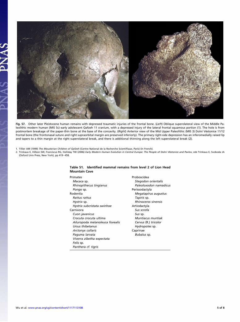
Table S1. Identified mammal remains from level 2 of Lion HeadMountain Cave
Primates ProboscideaMacaca sp. Stegodon orientalisRhinopithecus tingianus Paleoloxodon namadicusPongo sp. Perissodactyla
Rodentia Megatapirus augustusRattus rattus Tapiris sp.Hystrix sp. Rhinoceros sinensisHystrix subcristata swinhoe Artiodactyla
Carnivora Sus scrofaCuon javanicus Sus sp.Crocuta crocuta ultima Muntiacus muntiakAiluropoda melanoleuca fovealis Cervus (R.) tricolorUrsus thibetanus Hydropotes sp.Arctonyx collaris CaprinaePaguma larvata Bubalus sp.Viverra zibetha expectataFelis sp.Panthera cf. tigris
Fig. S7. Other later Pleistocene human remains with depressed traumatic injuries of the frontal bone. (Left) Oblique superolateral view of the Middle Pa-leolithic modern human (MIS 5c) early adolescent Qafzeh 11 cranium, with a depressed injury of the lateral frontal squamous portion (1). The hole is frompostmortem breakage of the paper-thin bone at the base of the concavity. (Right) Anterior view of the Mid Upper Paleolithic (MIS 3) Dolní V�estonice 11/12frontal bone (the frontonasal suture and right supraorbital margin are preserved inferiorly). The primary right-side depression has an inferomedially raised lipand tapers to a thin margin at the right superolateral break, and there is additional thinning along the left superolateral break (2).
1. Tillier AM (1999) The Mousterian Children of Qafzeh (Centre National de la Recherche Scientifique, Paris) (in French).2. Trinkaus E, Hillson SW, Franciscus RG, Holliday TW (2006) Early Modern Human Evolution in Central Europe: The People of Dolní Vĕstonice and Pavlov, eds Trinkaus E, Svoboda JA
(Oxford Univ Press, New York), pp 419e458.
Wu et al. www.pnas.org/cgi/content/short/1117113108 5 of 8

Table S2. Craniofacial and postcranial traumatic lesions, plus major developmental or degenerative abnormalities, of pre-last glacialmaximum (pre-LGM) Homo
Specimen Lesion Side Ref.
Major craniofacial lesionsMiddle Pleistocene
Hulu/Nanjing 1 Frontoparietal exocranial lesion—trauma and/or burning Mid (1)Late Pleistocene
Cova Negra 1 Anterosuperior parietal trauma with external remodeling Rt (2)Dolní Vĕstonice 3 Mandibular condyle fracture with facial deformity Lt (3)Dolní Vĕstonice 11/12 Anterior frontal squamous depressed fracture Mid (3)Krapina 34.7 Posterior parietal depressed fracture Rt (4)Qafzeh 11 Lateral frontal depressed fracture Rt (5)Saint-Césaire 1 Superior frontal squamous slicing wound Rt (6)Shanidar 1 Lateral facial crushing fracture Lt (7)
Minor craniofacial lesions*Early Pleistocene†
Sangiran 38 Two parietal pathological depressions, anteromedial and posteromedial,possibly posttraumatic
2× Lt (8)
Middle PleistoceneAtapuerca-SH cranium 1 Occipital squama exocranial depression Mid (9)Atapuerca-SH cranium 2 Anteromedial parietal exocranial vault depression Lt (9)Atapuerca-SH cranium 3 Posteromedial parietal exocranial depression Rt (9)Atapuerca-SH cranium 4 Frontal squamous exocranial vault depression Mid (9)
Posteroinferior parietal depressed groove Lt (9)Occipital squama slight depression Rt (9)
Atapuerca-SH cranium 5 12 exocranial lesions (6 right, 5 left, 1 mid; anterior, 3 right and 3 left) Rt, Lt, Mid (9)Supratoral slight depression Lt (9)Posttraumatic maxillary osteitis Lt (9)
Atapuerca-SH cranium 6 Supratoral slight exocranial vault depression Lt (9)Atapuerca-SH cranium 7 Lambdoid sutural bone healed external lesion — (9)Atapuerca-SH cranium 8 Posteroinferior parietal slight exocranial vault depression Lt (9)Atapuerca-SH 764 Supraorbital torus traumatic lesion Lt (9)Biache 1 Anteromedial shallow exocranial depression Lt (10)Biache 2 Lateral supraorbital torus minor lesion RtBroken Hill/Kabwe 1 Temporal trauma with associated infection Lt (11)Casal de’ Pazzi 1 Vascularized circular posterosuperior parietal depression Rt (12)Ceprano 1 Supraorbital (right) and squamous frontal (left) lesions Rt, Lt (13)La Chaise BD-17 Anterior middle parietal shallow depression Lt (14)Ehringsdorf 2 Superior middle parietal discrete exocranial lesion Lt (15)Ngandong 7‡ Bregmatic pathological depressions, possibly traumatic Mid (16)
Posterosuperior parietal depression, possibly traumatic Lt (16)Swanscombe 1 Anterior and posterior discrete parietal depressions 2× Lt (17)Zhoukoudian skull X§ Parasagittal frontal and parietal exocranial lesion Lt (18)Zhoukoudian skull XII Parasagittal parietal exocranial lesion Rt (18)Zuttiyeh 1 Supraorbital torus traumatic lesion Rt (19)
Two frontal squamous depressed lesions 2× Rt (19)Late PleistoceneDolní Vĕstonice 3 Superior minor frontal squamous exocranial lesion Mid (3)Dolní Vĕstonice 13 Minor frontal squamous exocranial lesion Rt (3)
Minor anteromedial parietal exocranial lesion Rt (3)Dolní Vĕstonice 16 Two frontal squamous external lesions 2× Rt (3)Feldhofer 1 Supraorbital torus small external lesion Rt (20)Krapina 4 Anteromedial parietal traumatic lesion Rt (21)Krapina 20 Small frontal squamous healed lesion Lt (21)Mlade�c 5 Frontal squamous traumatic lesion Lt (22)Pavlov 1 Posterior parietal exocranial alternation Rt (3)�Sal’a 1 Supraorbital torus traumatic lesion Rt (23)Shanidar 1 Minor lesions to the right frontal squamous Rt (7)Shanidar 5 Left frontal elongate external table bony scar Lt (7)
Major postcranial traumatic lesionsLate Pleistocene
Barma Grande 2 Marked humeral asymmetry, probably posttraumatic (27)Feldhofer 1 Fractured left ulna with reduced mobility, left arm atrophy,
and right arm hypertrophy(28)
Krapina 180 Right distal ulnar fracture and pseudoarthrosis (21)
Wu et al. www.pnas.org/cgi/content/short/1117113108 6 of 8

Table S2. Cont.
Specimen Lesion Side Ref.
Shanidar 1 Multiple right arm fractures with possible amputation; overall atrophy/hypotrophyof the right scapula, clavicle, and humerus; plus clavicular osteomyelitis
(7)
Shanidar 3 Left rib piercing, with probable pneumothorax (7)Sunghir 1 Piercing wound to ventral T1 (fatal) (29)
Minor postcranial traumatic lesionsLate Pleistocene
Caviglione 1 Healed and deformed left distal radial diaphyseal fractureLa Chapelle-aux-Saints 1 Fractured rib; posttraumatic cervical and left coxal osteoarthritis (30)Dolní Vĕstonice 15 Left ulnar diaphyseal fracture with radial diaphyseal deformity (3)Feldhofer 2 Right ulnar diaphyseal fracture (31)La Ferrassie 1 Traumatically displaced greater trochanter (30)La Ferrassie 2 Fractured fibular diaphysis with infection (32)Kebara 2 Fractured spinous thoracic processes; metacarpal fracture (33)Krapina 149 Distal right clavicular fracture (21)Krapina 188.8 Left proximal ulnar diaphyseal fractureShanidar 1 Posttraumatic right talocrural and hallucal tarsometatarsal osteoarthritis;
right metatarsal 4 fracture(7)
Shanidar 3 Posttraumatic right talocrural osteoarthritis (7)Shanidar 4 Middle rib fracture (7)Skhul 4¶ Metatarsal fracture (34)
Major developmental or degenerative conditionsEarly Pleistocene
Dmanisi D3444/D3900 Nearly edentulous; pervasive exocranial lesions (36)KNM-ER 1808 Systemic postcranial periosteal lesions and new bone formation (37)
Middle PleistoceneAtapuerca-SH cranium 14 Unilateral lambdoidal craniosynostosis (38)Atapuerca-SH pelvis 1 Lumbar kyphotic deformity, spondylolisthesis (39)Aubesier 11 Nearly edentulous with pervasive oral infection (40)Eliye Springs KNM-ES
11693Pronounced porotic hyperostosis (41)
Florisbad 1 Multiple cranial vault and orbital lesions (42)Salé 1 Congenital torticolis (43)Singa 1 Unilateral semicircular canal ossification (44)
Late PleistoceneBrno 2 Systemic postcranial diaphyseal periostosis (45)La Chapelle-aux-Saints 1 Largely edentulous with advanced anterior alveolar infection (30)Cro-Magnon 1 Cranial, pelvic, and femoral subperiosteal lesions (46)Dolní Vĕstonice 15 Systemic dysplasia with primary cranial, humeral, and femoral deformities,
plus pronounced dental hypoplasias(3)
La Ferrassie 1 Hypertrophic pulmonary osteoarthropathy (HPO) (47)Guattari 1 Edentulous with alveolo-palatal remodeling (48)Nazlet Khater 2 Bilateral femoral hypotrophy; advanced vertebral osteoarthritis (49)Qafzeh 12 Infantile hydrocephalus (50)Sunghir 3 Congenital bilateral femoral deformities, with persistent growth arrest lesions (51, 52)Tianyuan 1 Bilateral distal femoral and proximal tibial muscle deformities (53)
Included are primary and secondary lesions from trauma. Major ones are those that are likely to have incurred some loss of function or to have beenpotentially fatal. Not included are minor osteoarthritis, localized infections, auditory exostoses, and dentoalveolar/temporomandibular lesions that would nothave impeded normal mastication. All of them except the Sunghir 1 lesion have some healing/remodeling of the bone and hence indicate some level ofsurvival. Most of the specimens are adult; the immature specimens are Atapuerca-SH cranium 14, Qafzeh 11 and 12, and Sunghir 3. Lt, left; Mid, middle; Rt,right.*These cases include several features (especially from Atapuerca-SH, Dolní Vĕstonice, Pavlov, and Krapina) that are identified as minor trauma to the externaltable on the basis of the presence of distinct depressions in the external table. Most are likely to be secondary to trauma, but some could be secondary to injuryand/or infection of the scalp and underlying tissues.†Caspari (24) identified a traumatic lesion on the supraorbital torus of Gongwangling/Lantian 1, but CT analysis of the specimen (25) failed to confirm or refutethat diagnosis. It is therefore not included.‡The Ngandong human remains are variously dated to the Middle or Late Pleistocene. Recent reassessment of the age of the deposit (26) makes it likely thatthe sample dates to the second half of the Middle Pleistocene.§Weidenreich (18) listed a series of defects in the other Zhoukoudian Locality 1 crania, but only the ones on skulls X and XII appear to have occurredantemortem.¶A rectangular hole through the hyperflexed femoral head and os coxae of Skhul 9 was identified by McCown and Keith (34) as an ante- or perimortempiercing. However, it is postmortem damage, possibly from excavation (35).
Wu et al. www.pnas.org/cgi/content/short/1117113108 7 of 8

1. Shang H, Trinkaus E (2008) An ectocranial lesion on the Middle Pleistocene human cranium from Hulu Cave, Nanjing, China. Am J Phys Anthropol 135:431e437.2. Lumley MA, de (1973) Anteneandertals and Neandertals in the European western Mediterranean basin. Étud Quatern 2:1e626 (in French).3. Trinkaus E, Hillson SW, Franciscus RG, Holliday TW (2006) Early Modern Human Evolution in Central Europe: The People of Dolní Vĕstonice and Pavlov, eds Trinkaus E, Svoboda JA
(Oxford Univ Press, New York), pp 419e458.4. Kricun M, et al. (1999) The Krapina Hominids. A Radiographic Atlas of the Skeletal Collection (Croatian Natural History Museum, Zagreb, Croatia).5. Tillier AM (1999) The Mousterian Children of Qafzeh (Centre National de la Recherche Scientifique, Paris) (in French).6. Zollikofer CPE, Ponce De Leon MS, Vandermeersch B, Lévêque F (2002) Evidence for interpersonal violence in the St. Cesaire Neanderthal. Proc Natl Acad Sci USA 99:6444e6448.7. Trinkaus E (1983) The Shanidar Neandertals (Academic, New York).8. Indriati E, Antón SC (2010) The calvaria of Sangiran 38, Sendangbusik, Sangiran Dome, Java. Homo 61:225e243.9. Pérez PJ, Gracía A, Martínez I, Arsuaga JL (1997) Paleopathological evidence of the cranial remains from the Sima de los Huesos Middle Pleistocene site (Sierra de Atapuerca, Spain).
Description and preliminary inferences. J Hum Evol 33:409e421.10. Rougier H (2003) Descriptive and comparative study of Biache-Saint-Vaast 1 (Biache-Saint-Vaast, Pas-de-Callais, France), Doctoral thesis (Université Bordeaux 1, Bordeaux, France). (in
French).11. Montgomery PQ, Williams HOL, Reading N, Stringer CB (1994) An assessment of the temporal bone lesions of the Broken Hill cranium. J Archaeol Sci 21:331e337.12. Manzi G, Salvadei L, Passarello P (1990) The Casal de’ Pazzi archaic parietal: Comparative analysis of new fossil evidence from the late Middle Pleistocene of Rome. J Hum Evol 19:
751e759.13. Mallegni F, et al. (2003) Homo cepranensis sp. nov and the evolution of African-European Middle Pleistocene hominids. C R Palevol 2:153e159.14. Condemi S (2001) The Neandertals of La Chaise (Comité des Travaux Historiques et Scientifiques, Paris) (in French).15. Vl�cek E (1993) Human Fossil Discoveries from Weimar-Ehringsdorf (Theiss, Stuttgart) (in German).16. Bazeau A, Indriati E, Grimaud-Hervé D, Jacob T (2003) Computer tomography scanning of Homo erectus crania Ngandong 7 from Java: Internal structure, paleopathology and post-
mortem history. Berkala Ilmu Kedokteran 35:133e140.17. LeGros Clark WE (1938) General features of the Swanscombe skull bones. J R Anthropol Inst 68:58e67.18. Weidenreich F (1943) The skull of Sinanthropus pekinensis; a comparative study on a primitive hominid skull. Palaeontol Sinica 10D:1e485.19. Keith A (1927) A Report on the Galilee Skull (British School of Archaeology in Jerusalem, London).20. Schultz M (2006) Neanderthal, 1856–2006, ed Schmitz RW (Philipp von Zabern, Mainz am Rhein, Germany), pp 277e318.21. Radov�ci�c J, Smith FH, Trinkaus E, Wolpoff MH (1988) The Krapina Hominids: An Illustrated Catalog of the Skeletal Collection (Mladost, Zagreb, Croatia).22. Teschler-Nicola M, Czerny C, Oliva M, Schamall D, Schultz M (2006) Early Modern Humans at the Moravian Gate: The Mlade�c Caves and their Remains, ed Teschler-Nicola M (Springer,
Vienna), pp 473e487.23. Sládek V, Trinkaus E, �Sef�cáková A, Halouzka R (2002) Morphological affinities of the �Sal’a 1 frontal bone. J Hum Evol 43:787e815.24. Caspari R (1997) Brief communication: Evidence of pathology on the frontal bone from Gongwangling. Am J Phys Anthropol 102:565e568.25. Shang H, Trinkaus E, Liu W, Wu X, Zhu Q (2008) Neurocranial abnormalities of the Gongwangling Homo erectus from Lantian, China. J Archaeol Sci 35:2589e2593.26. Indriati E, et al. (2011) The age of the 20 meter Solo River terrace, Java, Indonesia and the survival of Homo erectus in Asia. PLoS ONE 6:e21562.27. Churchill SE, Formicola V (1997) A case of marked bilateral asymmetry in the upper limbs of an Upper Palaeolithic male from Barma Grande (Liguria), Italy. Int J Osteoarchaeol 7:
18e38.28. Trinkaus E, Churchill SE, Ruff CB (1994) Postcranial robusticity in Homo. II: Humeral bilateral asymmetry and bone plasticity. Am J Phys Anthropol 93:1e34.29. Trinkaus E, Buzhilova AP (2011) The death and burial of Sunghir 1. Int J Osteoarchaeol, 10.1002/oa.1227.30. Trinkaus E (1985) Pathology and the posture of the La Chapelle-aux-Saints Neandertal. Am J Phys Anthropol 67:19e41.31. Smith FH, Smith MO, Schmitz RW (2006) Neanderthal, 1856–2006, ed Schmitz RW (Philipp von Zabern, Mainz am Rhein, Germany), pp 187e246.32. Heim JL (1982) The fossil men of La Ferrassie II. Arch Inst Paléontol Hum 38:1e272 (in French).33. Duday H, Arensburg B (1991) The Mousterian Skeleton of Kebara 2, eds Bar-Yosef O, Vandermeersch B (Centre National de la Recherche Scientifique, Paris), pp 179e193 (in French).34. McCown TD, Keith A (1939) The Stone Age of Mount Carmel II: The Fossil Human Remains from the Levalloiso-Mousterian (Clarendon, Oxford).35. Churchill SE, Franciscus RG, McKean-Peraza HA, Daniel JA, Warren BR (2009) Shanidar 3 Neandertal rib puncture wound and paleolithic weaponry. J Hum Evol 57:163e178.36. Lordkipanidze D, et al. (2005) Anthropology: The earliest toothless hominin skull. Nature 434:717e718.37. Walker A, Zimmerman MR, Leakey REF (1982) A possible case of hypervitaminosis A in Homo erectus. Nature 296:248e250.38. Gracia A, et al. (2009) Craniosynostosis in the Middle Pleistocene human Cranium 14 from the Sima de los Huesos, Atapuerca, Spain. Proc Natl Acad Sci USA 106:6573e6578.39. Bonmatí A, et al. (2010) Middle Pleistocene lower back and pelvis from an aged human individual from the Sima de los Huesos site, Spain. Proc Natl Acad Sci USA 107:18386e18391.40. Lebel S, Trinkaus E (2002) Middle Pleistocene human remains from the Bau de l’Aubesier. J Hum Evol 43:659e685.41. Bräuer G, et al. (2003) Pathological alterations in the archaic Homo sapiens cranium from Eliye Springs, Kenya. Am J Phys Anthropol 120:200e204.42. Curnoe D, Brink J (2010) Evidence of pathological conditions in the Florisbad cranium. J Hum Evol 59:504e513.43. Hublin JJ (1991) Archaic Homo sapiens emergence: Northwest Africa and Western Europe, Doctoral thesis (Université de Bordeaux I, Bordeaux, France) (in French).44. Spoor F, Stringer CB, Zonneveld F (1998) Rare temporal bone pathology of the Singa calvaria from Sudan. Am J Phys Anthropol 107:41e50.45. Oliva M (1996) Upper Paleolithic grave Brno II as a contribution to the origins of shamanism. Archeol Rozhledy 48:353e383, 537–542 (in Czech).46. Dastugue J (1967) Pathologie des hommes fossiles de l’Abri de Cro-Magnon (Pathology of the fossil men of the Abri de Cro-Magnon). Anthropologie 71:479e492 (in French).47. Fennell KJ, Trinkaus E (1997) Bilateral femoral and tibial periostitis in the La Ferrassie 1 Neandertal. J Archaeol Sci 24:985e995.48. Sergi S, Ascenzi A, Bonucci E (1972) Torus palatinus in the Neandertal Circeo I skull. A histologic, microradiographic and electron microscopic investigation. Am J Phys Anthropol 36:
189e197.49. Crevecoeur I (2008) Anthropological Study of the Upper Paleolithic Skeleton of Nazlet Khater 2 (Egypt) (Leuven Univ Press, Leuven, Belgium) (in French).50. Tillier AM, Arensburg B, Duday H, Vandermeersch B (2001) Brief communication: An early case of hydrocephalus: The Middle Paleolithic Qafzeh 12 child (Israel). Am J Phys Anthropol
114:166e170.51. Formicola V, Buzhilova AP (2004) Double child burial from Sunghir (Russia): Pathology and inferences for upper paleolithic funerary practices. Am J Phys Anthropol 124:189e198.52. Guatelli-Steinberg D, Buzhilova AP, Trinkaus E (2011) Developmental stress and survival among the Mid Upper Paleolithic Sunghir children: Dental enamel hypoplasias of Sunghir 2
and 3. Int J Osteoarchaeol, 10.1002/oa.1263.53. Shang H, Trinkaus E (2010) The Early Modern Human from Tianyuan Cave, China (Texas A&M Univ Press, College Station, TX).
Wu et al. www.pnas.org/cgi/content/short/1117113108 8 of 8